Embed Size (px)
Citation preview
pISSN: 1011-8942 eISSN: 2092-9382
© 2017 The Korean Ophthalmological SocietyThis is an Open Access article distributed under the terms of the Creative Commons Attribution Non-Commercial License (http://creativecommons.org/licenses /by-nc/3.0/) which permits unrestricted non-commercial use, distribution, and reproduction in any medium, provided the original work is properly cited.
16
Original Article
Laser in situ keratomileusis (LASIK), laser-assisted sub-epithelial keratectomy (LASEK), and photorefractive kera-tectomy (PRK) are effective surgical techniques for the correction of myopia, myopic astigmatism, and mild to
moderate hypermetropia. Although the predictability of these techniques has improved, the precise refractive out-come is not always guaranteed because of different re-sponses of living tissue to laser ablation [1]. The reported rates of myopic regression that require retreatment ranged from 6% to 14% for up to 1 year following the surgical procedure, and from 20% to 27% for longer follow-up pe-riods [2,3]. However, recent studies have indicated that the rates have declined [4,5]. Higher initial corrections, the de-gree of astigmatism, and advanced age of patients are
Clinical Outcomes of an Optimized Prolate Ablation Procedure for Correcting Residual Refractive Errors Following Laser Surgery
Byunghoon Chung1, Hun Lee1, Bong Joon Choi2, Kyung Ryul Seo1, Eung Kwon Kim1,3, Dae Yune Kim2, Tae-im Kim1
1The Institute of Vision Research, Department of Ophthalmology, Yonsei University College of Medicine, Seoul, Korea2Lee Eye Clinic, Busan, Korea
3Corneal Dystrophy Research Institute, Severance Biomedical Science Institute, and Brain Korea 21 Project for Medical Science, Department of Ophthalmology, Yonsei University College of Medicine, Seoul, Korea
Purpose: The purpose of this study was to investigate the clinical efficacy of an optimized prolate ablation pro-
cedure for correcting residual refractive errors following laser surgery.
Methods: We analyzed 24 eyes of 15 patients who underwent an optimized prolate ablation procedure for the
correction of residual refractive errors following laser in situ keratomileusis, laser-assisted subepithelial ker-
atectomy, or photorefractive keratectomy surgeries. Preoperative ophthalmic examinations were performed,
and uncorrected distance visual acuity, corrected distance visual acuity, manifest refraction values (sphere,
cylinder, and spherical equivalent), point spread function, modulation transfer function, corneal asphericity
(Q value), ocular aberrations, and corneal haze measurements were obtained postoperatively at 1, 3, and 6
months.
Results: Uncorrected distance visual acuity improved and refractive errors decreased significantly at 1, 3, and 6
months postoperatively. Total coma aberration increased at 3 and 6 months postoperatively, while changes in
all other aberrations were not statistically significant. Similarly, no significant changes in point spread function
were detected, but modulation transfer function increased significantly at the postoperative time points mea-
sured.
Conclusions: The optimized prolate ablation procedure was effective in terms of improving visual acuity and
objective visual performance for the correction of persistent refractive errors following laser surgery.
Key Words: Optimized prolate ablation, Refractive surgery, Residual refractive errors
Received: August 21, 2015 Accepted: November 30, 2015
Corresponding Author: Tae-im Kim, MD, PhD. Department of Ophthal-mology, Yonsei University College of Medicine, #50 Yonsei-ro, Seodae-mun-gu, Seoul 03722, Korea. Tel: 82-2-2228-3574, Fax: 82-2-312-0541, E-mail: [email protected]
Korean J Ophthalmol 2017;31(1):16 -24ht tps://doi.org/10.3341/k jo.2017.31.1.16
17
B Chung, et al. OPA for Residual Refractive Errors
known to increase the odds of requiring retreatment [6]. Although not all of the mechanisms involved in myopic re-gression have been elucidated, the number of patients who require retreatment for myopic regression after primary refractive surgery has decreased. The reported decrease has been attributed to an enlarged optical zone, the devel-opment of laser technology, and improved postoperative wound management. However, some patients still experi-enced myopic regression after the performance of primary refractive surgery. Further, retreatment planning following primary refractive surgery is dependent on the individual needs of each patient.
Retreatment tends to be less predictable compared with primary ablation because of difficulties related to analysis of the refractive state, unpredictable rates of wound heal-ing, and corneal irregularities induced by previous ablation procedures. Previous studies have reported that the combi-nation of corneal wavefront and topographic aspheric treatments has been used successfully to treat eyes that have undergone previous refractive surgeries [5,7].
Optimized prolate ablation (OPA) employs wavefront aberrometry and corneal topography to treat preexisting spherical aberrations and to maintain the preoperative cor-neal asphericity (Q value) [8,9]. As improved outcomes had been achieved in previous studies with the combined ap-plication of the corneal wavefront and topographic aspher-ic treatments, we endeavored to assess clinical outcomes following application of the OPA retreatment method. In the current study, we investigated the clinical efficacy of an OPA procedure for the treatment of persistent refractive errors following previous refractive surgery.
Materials and Methods
In this retrospective study, we evaluated patients who had undergone retreatment OPA following previous LASIK, LASEK, or PRK surgery. The study was conduct-ed in accordance with the Declaration of Helsinki, and surgical procedures were conducted after all patients par-ticipated in a thorough preoperative discussion of the risks and benefits of OPA and informed consent was obtained.
Inclusion criteria for patients who had undergone previ-ous LASIK, LASEK, or PRK between 1997 and 2012 in-cluded an age greater than 20 years and myopia measure-ments less than or equal to –3.50 manifest refraction
spherical equivalent (MRSE) diopters (D). Patients who had active systemic ocular disease or who underwent pre-vious ocular surgery other than the aforementioned prima-ry refractive surgeries were excluded from the study. All patients were symptomatic and had residual refractive er-rors.
All patients underwent a preoperative ophthalmic evalu-ation, and uncorrected distance visual acuity (UDVA), corrected distance visual acuity (CDVA), manifest refrac-tion, corneal topography, wavefront aberrometry, modula-tion transfer function (MTF), point spread function (PSF), slit-lamp biomicroscopy, tonometry, and fundus measure-ments were obtained postoperatively at 1, 3, and 6 months. Corneal haze was assessed on a scale from 0 to 4 accord-ing to the method proposed by Fantes et al. [10]. Corneal topography and wavefront aberrometry were measured on the same optical axis using an OPD-Scan III wavefront ab-errometer (Nidek, Tokyo, Japan), following the application of tropicamide 0.5%-phenylephrine 0.5% eye drops (Mydrin-P; Santen, Osaka, Japan) and the subsequent dila-tion of the pupil to 6.0 mm. All wavefront measurements were performed to the eighth Zernike order. The device software separated corneal and internal aberrations to evaluate the effects of the optical elements in the visual system. Corneal asphericity (Q value) was measured by software-simulated corneal topography. The objective vi-sual quality was assessed using an MTF graph, which re-vealed the degree of contrast transfer at different spatial frequencies by calculating the area ratio and the ratio of the area under the MTF graph covered by the vertical and horizontal axes to the area under the normal eye curve. PSF was also calculated using the Strehl ratio, which is the ratio of the PSF value to the theoretical diffraction limit.
All patients underwent treatment by one surgeon (BJC) using the EC 5000 CXII excimer laser platform (Nidek). Manifest refraction, corneal topographic data, and wave-front data obtained from the OPD-Scan III were consid-ered when designing the ablation procedure. All data were transferred to the Optimized Prolate Ablation Software (ver. 1.00, Nidek) for treatment planning. The ablation de-sign was established automatically using the software, by setting the value of the postoperative ocular spherical ab-erration to zero. The software did not target to correct ocu-lar coma and trefoil aberration. The surgeon adjusted the degree of refractive correction for age, pretreatment mani-fest refraction, and CDVA. The eyes of each patient were
18
Korean J Ophthalmol Vol.31, No.1, 2017
prepared in a sterile fashion, and a topical anesthetic (proparacaine hydrochloride 0.5%, Alcaine; Alcon, Fort Worth, TX, USA) was instilled. Following the application of the Carones LASEK Pump OZ Chamber (9.0 mm in di-ameter; ASCIO, Copenhagen, Denmark), the cone was filled with 20% alcohol solution mixed with Liquifilm tears (Polyvinyl Alcohol 1.4%; Allergan, Irvine, CA, USA). The alcohol solution was flushed out for 40 seconds after instillation using a cold balanced salt solution. To detect torsion error, the image of the patient’s iris was compared with the image acquired using the OPD-Scan III device. If the torsion error was greater than 2°, the patient’s head was repositioned in an attempt to decrease the error. The opti-cal zone was established using the Optimized Prolate Ab-lation software, and covered the entire scotopic pupil di-ameter, considering the ablation depth. The transition zone was determined to be 1.5 mm larger than the optical zone
diameter. The mean optical zone and transition zone were 6.3 mm and 7.9 mm, respectively. A 14-ring-shaped filter paper disc saturated with 0.02% mitomycin C was applied on the cornea for 15 seconds to prevent contact with the central portion of the cornea. Irrigation with cold balanced salt solution was performed for 15 seconds following the removal of the paper disc [11,12]. A therapeutic soft contact lens was then applied to ensure complete epithelial heal-ing. Finally, a standardized regimen of topical steroids and antibiotics was recommended for each patient.
Postoperative UDVA and CDVA, ocular wavefront aber-rations, corneal spherical aberration, corneal asphericity, MTF, and PSF were compared to preoperative (before re-treatment) values using analysis of variance and Bonfer-roni tests. A p-value less than 0.05 was considered statisti-cally significant. All analyses were performed using the SPSS ver. 12.0 (SPSS Inc., Chicago, IL, USA).
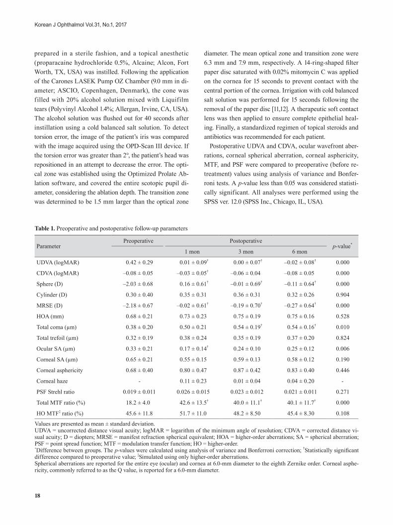
Table 1. Preoperative and postoperative follow-up parameters
ParameterPreoperative Postoperative
p-value*
1 mon 3 mon 6 mon
UDVA (logMAR) 0.42 ± 0.29 0.01 ± 0.09† 0.00 ± 0.07† –0.02 ± 0.08† 0.000
CDVA (logMAR) –0.08 ± 0.05 –0.03 ± 0.05† –0.06 ± 0.04 –0.08 ± 0.05 0.000
Sphere (D) –2.03 ± 0.68 0.16 ± 0.61† –0.01 ± 0.69† –0.11 ± 0.64† 0.000
Cylinder (D) 0.30 ± 0.40 0.35 ± 0.31 0.36 ± 0.31 0.32 ± 0.26 0.904
MRSE (D) –2.18 ± 0.67 –0.02 ± 0.61† –0.19 ± 0.70† –0.27 ± 0.64† 0.000
HOA (mm) 0.68 ± 0.21 0.73 ± 0.23 0.75 ± 0.19 0.75 ± 0.16 0.528
Total coma (µm) 0.38 ± 0.20 0.50 ± 0.21 0.54 ± 0.19† 0.54 ± 0.16† 0.010
Total trefoil (µm) 0.32 ± 0.19 0.38 ± 0.24 0.35 ± 0.19 0.37 ± 0.20 0.824
Ocular SA (µm) 0.33 ± 0.21 0.17 ± 0.14† 0.24 ± 0.10 0.25 ± 0.12 0.006
Corneal SA (µm) 0.65 ± 0.21 0.55 ± 0.15 0.59 ± 0.13 0.58 ± 0.12 0.190
Corneal asphericity 0.68 ± 0.40 0.80 ± 0.47 0.87 ± 0.42 0.83 ± 0.40 0.446
Corneal haze - 0.11 ± 0.23 0.01 ± 0.04 0.04 ± 0.20 -
PSF Strehl ratio 0.019 ± 0.011 0.026 ± 0.015 0.023 ± 0.012 0.021 ± 0.011 0.271
Total MTF ratio (%) 18.2 ± 4.0 42.6 ± 13.5† 40.0 ± 11.1† 40.1 ± 11.7† 0.000
HO MTF‡ ratio (%) 45.6 ± 11.8 51.7 ± 11.0 48.2 ± 8.50 45.4 ± 8.30 0.108
Values are presented as mean ± standard deviation.UDVA = uncorrected distance visual acuity; logMAR = logarithm of the minimum angle of resolution; CDVA = corrected distance vi-sual acuity; D = diopters; MRSE = manifest refraction spherical equivalent; HOA = higher-order aberrations; SA = spherical aberration; PSF = point spread function; MTF = modulation transfer function; HO = higher-order.*Difference between groups. The p-values were calculated using analysis of variance and Bonferroni correction; †Statistically significant difference compared to preoperative value; ‡Simulated using only higher-order aberrations.Spherical aberrations are reported for the entire eye (ocular) and cornea at 6.0-mm diameter to the eighth Zernike order. Corneal asphe-ricity, commonly referred to as the Q value, is reported for a 6.0-mm diameter.
19
B Chung, et al. OPA for Residual Refractive Errors
Results
Patients
The study included 24 eyes from 15 enrolled patients, including eight males and seven females. The mean patient age was 33 years (range, 25 to 45 years). Twelve eyes (eight patients) had undergone a previous LASIK procedure, nine eyes (five patients) a previous LASEK procedure, and three eyes (two patients) had undergone PRK. The mean interval between the first refractive procedure and retreatment was 108 ± 60 months (range, 12 months to 20.9 years). No in-traoperative or postoperative complications were detected.
Refractive and visual acuity outcomes
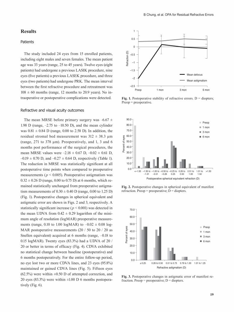
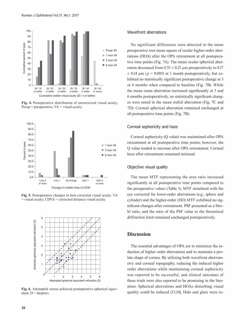
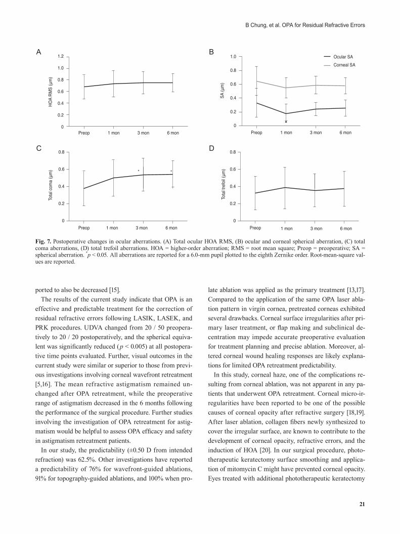
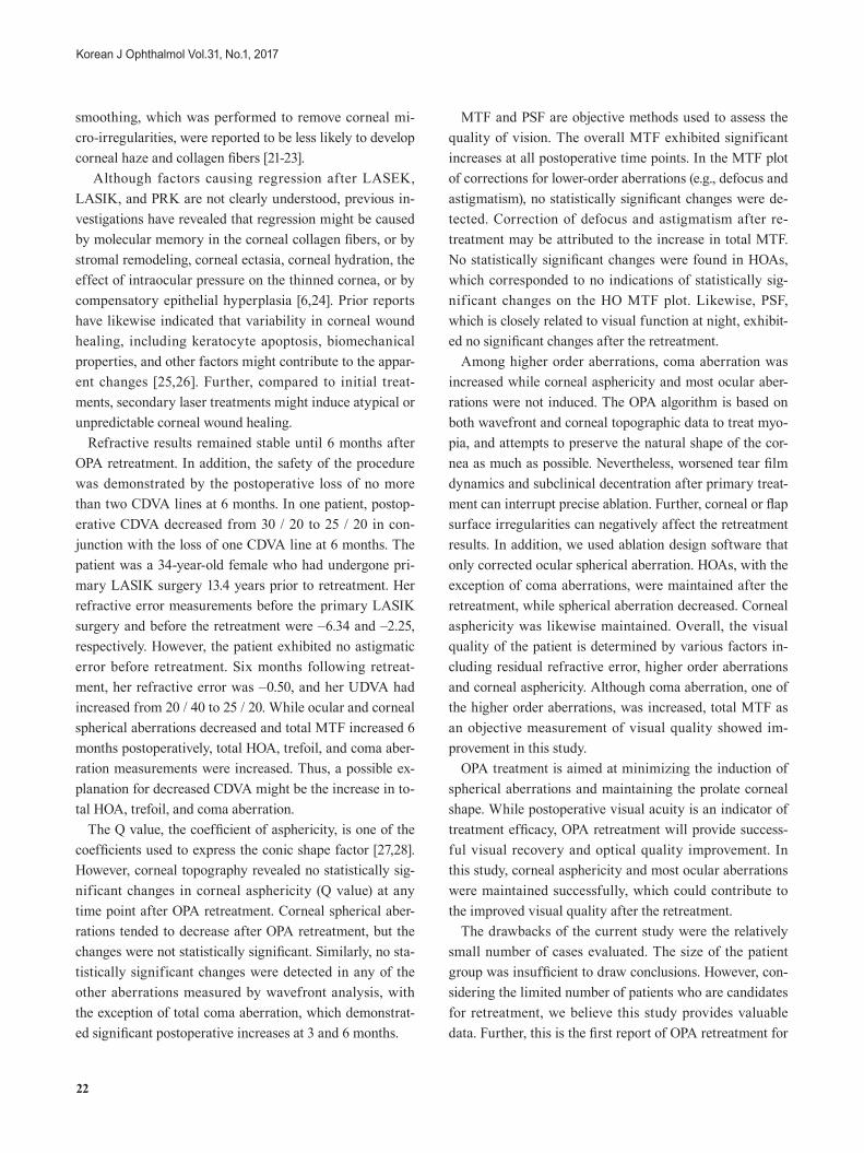
The mean MRSE before primary surgery was –6.67 ± 1.98 D (range, –2.75 to –10.50 D), and the mean cylinder was 0.81 ± 0.84 D (range, 0.00 to 2.58 D). In addition, the residual stromal bed measurement was 312 ± 38.3 µm (range, 271 to 378 µm). Preoperatively, and 1, 3 and 6 months post performance of the surgical procedures, the mean MRSE values were –2.18 ± 0.67 D, –0.02 ± 0.61 D, –0.19 ± 0.70 D, and –0.27 ± 0.64 D, respectively (Table 1). The reduction in MRSE was statistically significant at all postoperative time points when compared to preoperative measurements (p < 0.005). Postoperative astigmatism was 0.32 ± 0.26 D (range, 0.00 to 0.75 D) at 6 months, which re-mained statistically unchanged from preoperative astigma-tism measurements of 0.30 ± 0.40 D (range, 0.00 to 1.25 D) (Fig. 1). Postoperative changes in spherical equivalent and astigmatic error are shown in Figs. 2 and 3, respectively. A statistically significant increase (p < 0.001) was detected in the mean UDVA from 0.42 ± 0.29 logarithm of the mini-mum angle of resolution (logMAR) preoperative measure-ments (range, 0.10 to 1.00 logMAR) to –0.02 ± 0.08 log-MAR postoperative measurements (20 / 50 to 20 / 20 as Snellen equivalent) acquired at 6 months (range, –0.18 to 0.15 logMAR). Twenty eyes (83.3%) had a UDVA of 20 / 20 or better in terms of efficacy (Fig. 4). CDVA exhibited no statistical change between baseline (postoperative) and 6 months postoperatively. For the entire follow-up period, no eye lost two or more CDVA lines, and 23 eyes (95.8%) maintained or gained CDVA lines (Fig. 5). Fifteen eyes (62.5%) were within ±0.50 D of attempted correction, and 20 eyes (83.3%) were within ±1.00 D 6 months postopera-tively (Fig. 6).
1
0.5
0
–0.5
–1
–1.5
–2
–2.51 monPreop
Refra
ction
(D)
3 mon 6 mon
Mean defocus
Mean astigmatism
90.0
80.0
70.0
60.0
50.0
40.0
30.0
20.0
10.0
0.0
Perc
ent o
f eye
s
Postoperative spherical equivalent refraction (D)
<–1.50 –1.50 to–1.01
–1.00 to–0.51
–0.50 to–0.26
–0.25 to0.25
0.26 to0.50
0.51 to1.00
1.01 to1.50
>1.50
Preop1 mon3 mon6 mon
70.0
60.0
50.0
40.0
30.0
20.0
10.0
0.0
Perc
ent o
f eye
s
Refractive astigmatism (D)≤ 0.25 0.26 to 0.50 0.51 to 0.75 0.76 to 1.00 1.01 to 1.25
Preop1 mon3 mon6 mon
Fig. 1. Postoperative stability of refractive errors. D = diopters; Preop = preoperative.
Fig. 2. Postoperative changes in spherical equivalent of manifest refraction. Preop = preoperative; D = diopters.
Fig. 3. Postoperative changes in astigmatic error of manifest re-fraction. Preop = preoperative; D = diopters.
20
Korean J Ophthalmol Vol.31, No.1, 2017
Wavefront aberrations
No significant differences were detected in the mean preoperative root mean square of ocular higher-order aber-rations (HOA) after the OPA retreatment at all postopera-tive time points (Fig. 7A). The mean ocular spherical aber-ration decreased from 0.33 ± 0.21 µm preoperatively to 0.17 ± 0.14 µm (p < 0.005) at 1 month postoperatively, but ex-hibited no statistically significant postoperative change at 3 or 6 months when compared to baseline (Fig. 7B). While the mean coma aberration increased significantly at 3 and 6 months postoperatively, no statistically significant chang-es were noted in the mean trefoil aberration (Fig. 7C and 7D). Corneal spherical aberration remained unchanged at all postoperative time points (Fig. 7B).
Corneal asphericity and haze
Corneal asphericity (Q value) was maintained after OPA retreatment at all postoperative time points; however, the Q value tended to increase after OPA retreatment. Corneal haze after retreatment remained minimal.
Objective visual quality
The mean MTF representing the area ratio increased significantly at all postoperative time points compared to the preoperative values (Table 1). MTF simulated with the eye corrected for lower-order aberrations (e.g., sphere and cylinder) and the higher-order (HO) MTF exhibited no sig-nificant changes after retreatment. PSF presented as a Stre-hl ratio, and the ratio of the PSF value to the theoretical diffraction limit remained unchanged postoperatively.
Discussion
The essential advantages of OPA are to minimize the in-duction of higher order aberrations and to maintain a pro-late shape of cornea. By utilizing both wavefront aberrom-etry and corneal topography, reducing the induced higher order aberrations while maintaining corneal asphericity was reported to be successful, and clinical outcomes of these trials were also reported to be promising in the liter-ature. Spherical aberrations and HOAs disturbing visual quality could be reduced [13,14]. Halo and glare were re-
100
90
80
70
60
50
40
30
20
10
0
Cum
ulativ
e pe
rcen
t of e
yes
Cumulative snellen visual acuity (20 / x or better)
20 / 16or better
20 / 20or better
20 / 25or better
20 / 32or better
20 / 40or better
20 / 40or worse
Preop VA1 mon VA3 mon VA6 mon VA
100.0
90.0
80.0
70.0
60.0
50.0
40.0
30.0
20.0
10.0
0.0
Perc
ent o
f eye
s
Changes in snellen lines of CDVA
Loss 2or more
Gain 2or more
Loss 1 No change Gain 1
1 mon VA3 mon VA6 mon VA
0
6
5
4
3
2
1
1 2 3 4 5 6Attempted spherical equivalent refraction (D)
Achie
ved
sphe
rical
equiv
alent
refra
ction
(D)
Fig. 4. Postoperative distribution of uncorrected visual acuity. Preop = preoperative; VA = visual acuity.
Fig. 5. Postoperative changes in best corrected visual acuity. VA = visual acuity; CDVA = corrected distance visual acuity.
Fig. 6. Attempted versus achieved postoperative spherical equiv-alent. D = diopters.
21
B Chung, et al. OPA for Residual Refractive Errors
ported to also be decreased [15]. The results of the current study indicate that OPA is an
effective and predictable treatment for the correction of residual refractive errors following LASIK, LASEK, and PRK procedures. UDVA changed from 20 / 50 preopera-tively to 20 / 20 postoperatively, and the spherical equiva-lent was significantly reduced (p < 0.005) at all postopera-tive time points evaluated. Further, visual outcomes in the current study were similar or superior to those from previ-ous investigations involving corneal wavefront retreatment [5,16]. The mean refractive astigmatism remained un-changed after OPA retreatment, while the preoperative range of astigmatism decreased in the 6 months following the performance of the surgical procedure. Further studies involving the investigation of OPA retreatment for astig-matism would be helpful to assess OPA efficacy and safety in astigmatism retreatment patients.
In our study, the predictability (±0.50 D from intended refraction) was 62.5%. Other investigations have reported a predictability of 76% for wavefront-guided ablations, 91% for topography-guided ablations, and 100% when pro-
late ablation was applied as the primary treatment [13,17]. Compared to the application of the same OPA laser abla-tion pattern in virgin cornea, pretreated corneas exhibited several drawbacks. Corneal surface irregularities after pri-mary laser treatment, or flap making and subclinical de-centration may impede accurate preoperative evaluation for treatment planning and precise ablation. Moreover, al-tered corneal wound healing responses are likely explana-tions for limited OPA retreatment predictability.
In this study, corneal haze, one of the complications re-sulting from corneal ablation, was not apparent in any pa-tients that underwent OPA retreatment. Corneal micro-ir-regularities have been reported to be one of the possible causes of corneal opacity after refractive surgery [18,19]. After laser ablation, collagen fibers newly synthesized to cover the irregular surface, are known to contribute to the development of corneal opacity, refractive errors, and the induction of HOA [20]. In our surgical procedure, photo-therapeutic keratectomy surface smoothing and applica-tion of mitomycin C might have prevented corneal opacity. Eyes treated with additional phototherapeutic keratectomy
1.2
1.0
0.8
0.6
0.4
0.2
01 monPreop 3 mon 6 mon
HOA
RMS
(μm
)
0.8
0.6
0.4
0.2
0Preop
* *
Tota
l com
a (μ
m)
1 mon 3 mon 6 mon
1.0
0.8
0.6
0.4
0.2
01 monPreop 3 mon 6 mon
SA (μ
m)
Ocular SACorneal SA
Tota
l tref
oil (μ
m)
0.8
0.6
0.4
0.2
0Preop 1 mon 3 mon 6 mon
Fig. 7. Postoperative changes in ocular aberrations. (A) Total ocular HOA RMS, (B) ocular and corneal spherical aberration, (C) total coma aberrations, (D) total trefoil aberrations. HOA = higher-order aberration; RMS = root mean square; Preop = preoperative; SA = spherical aberration. *p < 0.05. All aberrations are reported for a 6.0-mm pupil plotted to the eighth Zernike order. Root-mean-square val-ues are reported.
A
C
B
D
22
Korean J Ophthalmol Vol.31, No.1, 2017
smoothing, which was performed to remove corneal mi-cro-irregularities, were reported to be less likely to develop corneal haze and collagen fibers [21-23].
Although factors causing regression after LASEK, LASIK, and PRK are not clearly understood, previous in-vestigations have revealed that regression might be caused by molecular memory in the corneal collagen fibers, or by stromal remodeling, corneal ectasia, corneal hydration, the effect of intraocular pressure on the thinned cornea, or by compensatory epithelial hyperplasia [6,24]. Prior reports have likewise indicated that variability in corneal wound healing, including keratocyte apoptosis, biomechanical properties, and other factors might contribute to the appar-ent changes [25,26]. Further, compared to initial treat-ments, secondary laser treatments might induce atypical or unpredictable corneal wound healing.
Refractive results remained stable until 6 months after OPA retreatment. In addition, the safety of the procedure was demonstrated by the postoperative loss of no more than two CDVA lines at 6 months. In one patient, postop-erative CDVA decreased from 30 / 20 to 25 / 20 in con-junction with the loss of one CDVA line at 6 months. The patient was a 34-year-old female who had undergone pri-mary LASIK surgery 13.4 years prior to retreatment. Her refractive error measurements before the primary LASIK surgery and before the retreatment were –6.34 and –2.25, respectively. However, the patient exhibited no astigmatic error before retreatment. Six months following retreat-ment, her refractive error was –0.50, and her UDVA had increased from 20 / 40 to 25 / 20. While ocular and corneal spherical aberrations decreased and total MTF increased 6 months postoperatively, total HOA, trefoil, and coma aber-ration measurements were increased. Thus, a possible ex-planation for decreased CDVA might be the increase in to-tal HOA, trefoil, and coma aberration.
The Q value, the coefficient of asphericity, is one of the coefficients used to express the conic shape factor [27,28]. However, corneal topography revealed no statistically sig-nificant changes in corneal asphericity (Q value) at any time point after OPA retreatment. Corneal spherical aber-rations tended to decrease after OPA retreatment, but the changes were not statistically significant. Similarly, no sta-tistically significant changes were detected in any of the other aberrations measured by wavefront analysis, with the exception of total coma aberration, which demonstrat-ed significant postoperative increases at 3 and 6 months.
MTF and PSF are objective methods used to assess the quality of vision. The overall MTF exhibited significant increases at all postoperative time points. In the MTF plot of corrections for lower-order aberrations (e.g., defocus and astigmatism), no statistically significant changes were de-tected. Correction of defocus and astigmatism after re-treatment may be attributed to the increase in total MTF. No statistically significant changes were found in HOAs, which corresponded to no indications of statistically sig-nificant changes on the HO MTF plot. Likewise, PSF, which is closely related to visual function at night, exhibit-ed no significant changes after the retreatment.
Among higher order aberrations, coma aberration was increased while corneal asphericity and most ocular aber-rations were not induced. The OPA algorithm is based on both wavefront and corneal topographic data to treat myo-pia, and attempts to preserve the natural shape of the cor-nea as much as possible. Nevertheless, worsened tear film dynamics and subclinical decentration after primary treat-ment can interrupt precise ablation. Further, corneal or flap surface irregularities can negatively affect the retreatment results. In addition, we used ablation design software that only corrected ocular spherical aberration. HOAs, with the exception of coma aberrations, were maintained after the retreatment, while spherical aberration decreased. Corneal asphericity was likewise maintained. Overall, the visual quality of the patient is determined by various factors in-cluding residual refractive error, higher order aberrations and corneal asphericity. Although coma aberration, one of the higher order aberrations, was increased, total MTF as an objective measurement of visual quality showed im-provement in this study.
OPA treatment is aimed at minimizing the induction of spherical aberrations and maintaining the prolate corneal shape. While postoperative visual acuity is an indicator of treatment efficacy, OPA retreatment will provide success-ful visual recovery and optical quality improvement. In this study, corneal asphericity and most ocular aberrations were maintained successfully, which could contribute to the improved visual quality after the retreatment.
The drawbacks of the current study were the relatively small number of cases evaluated. The size of the patient group was insufficient to draw conclusions. However, con-sidering the limited number of patients who are candidates for retreatment, we believe this study provides valuable data. Further, this is the first report of OPA retreatment for
23
B Chung, et al. OPA for Residual Refractive Errors
residual refractive errors following laser surgery. Finally, the results indicate that OPA retreatment provided effec-tive and reliable surgical outcomes, and objective visual performance revealed significant improvement after re-treatment.
Conflict of Interest
No potential conflict of interest relevant to this article was reported.
Acknowledgements
This study was partially supported by a grant of the Ko-rean Health Technology R&D Project, Ministry of Health & Welfare, Republic of Korea (HI14C2044).
References
1. Rashad KM. Laser in situ keratomileusis retreatment for residual myopia and astigmatism. J Re f ract Surg 2000;16:170-6.
2. Hersh PS, Fry KL, Bishop DS. Incidence and associations of retreatment after LASIK. Ophthalmology 2003;110:748-54.
3. Alio JL, Muftuoglu O, Ortiz D, et al. Ten-year follow-up of laser in situ keratomileusis for high myopia. Am J Ophthal-mol 2008;145:55-64.
4. Kanellopoulos AJ, Asimellis G. Refractive and keratometric stability in high myopic LASIK with high-frequency femto-second and excimer lasers. J Refract Surg 2013;29:832-7.
5. Aslanides IM, Kolli S, Padroni S, Arba Mosquera S. Stabil-ity of therapeutic retreatment of corneal wavefront custom-ized ablation with the SCHWIND CAM: 4-year data. J Re-fract Surg 2012;28:347-52.
6. Bragheeth MA, Fares U, Dua HS. Re-treatment after laser in situ keratomileusis for correction of myopia and myopic astigmatism. Br J Ophthalmol 2008;92:1506-10.
7. Toda I, Yamamoto T, Ito M, et al. Topography-guided abla-tion for treatment of patients with irregular astigmatism. J Refract Surg 2007;23:118-25.
8. El-Danasoury A, Bains HS. Optimized prolate corneal ab-lation: case report of the first treated eye. J Refract Surg
2005;21(5 Suppl):S598-602. 9. El Danasoury AM, Holladay J, Waring GO 3rd, et al. A
contralateral, randomized comparison of optimized prolate ablation and conventional LASIK for myopia with the NIDEK excimer laser platform. J Refract Surg 2012;28:453-61.
10. Fantes FE, Hanna KD, Waring GO 3rd, et al. Wound heal-ing after excimer laser keratomileusis (photorefractive ker-atectomy) in monkeys. Arch Ophthalmol 1990;108:665-75.
11. Maldonado MJ. Intraoperative MMC after excimer laser surgery for myopia. Ophthalmology 2002;109:826.
12. Jain S, McCally RL, Connolly PJ, Azar DT. Mitomycin C reduces corneal light scattering after excimer keratectomy. Cornea 2001;20:45-9.
13. Kang EC, Choi BJ, Kim EK, Kim TI. Clinical outcomes of optimized prolate ablation and custom aspheric treatment in laser-assisted subepithelial keratectomy. J Cataract Re-fract Surg 2012;38:445-52.
14. Choi BJ, Park YM, Lee JS. Clinical outcomes between op-tical path difference custom aspheric treatment and opti-mized prolate ablation photorefractive keratectomy in my-opia exceeding 8 diopters. Eye (Lond) 2015;29:356-62.
15. Chayet A, Bains HS. Prospective, randomized, dou-ble-blind, contralateral eye comparison of myopic LASIK with optimized aspheric or prolate ablations. J Refract Surg 2012;28:112-9.
16. Alio JL, Pinero DP, Plaza Puche AB. Corneal wave-front-guided enhancement for high levels of corneal coma aberration after laser in situ keratomileusis. J Cataract Re-fract Surg 2008;34:222-31.
17. Kermani O, Schmiedt K, Oberheide U, Gerten G. Topo-graphic- and wavefront-guided customized ablations with the NIDEK-EC5000CXII in LASIK for myopia. J Refract Surg 2006;22:754-63.
18. Vinciguerra P, Azzolini M, Radice P, et al. A method for examining surface and interface irregularities after photo-refractive keratectomy and laser in situ keratomileusis: predictor of optical and functional outcomes. J Refract Surg 1998;14(2 Suppl):S204-6.
19. Balestrazzi E, De Molfetta V, Spadea L, et al. Histological, immunohistochemical, and ultrastructural findings in hu-man corneas after photorefractive keratectomy. J Refract Surg 1995;11:181-7.
20. Oshika T, Klyce SD, Applegate RA, et al. Comparison of corneal wavefront aberrations after photorefractive kera-tectomy and laser in situ keratomileusis. Am J Ophthalmol
24
Korean J Ophthalmol Vol.31, No.1, 2017
1999;127:1-7. 21. Vinciguerra P, Azzolini M, Airaghi P, et al. Effect of de-
creasing surface and interface irregularities after photore-fractive keratectomy and laser in situ keratomileusis on op-tical and functional outcomes. J Refract Surg 1998;14(2 Suppl):S199-203.
22. Vinciguerra P, Camesasca FI, Randazzo A. One-year re-sults of butterfly laser epithelial keratomileusis. J Refract Surg 2003;19(2 Suppl):S223-6.
23. Serrao S, Lombardo M. One-year results of photorefractive keratectomy with and without surface smoothing using the technolas 217C laser. J Refract Surg 2004;20:444-9.
24. Bas AM, Onnis R. Excimer laser in situ keratomileusis for
myopia. J Refract Surg 1995;11(3 Suppl):S229-33. 25. Mohan RR, Hutcheon AE, Choi R, et al. Apoptosis, necro-
sis, proliferation, and myofibroblast generation in the stro-ma following LASIK and PRK. Exp Eye Res 2003;76:71-87.
26. Rober ts C. Biomechanics of the cornea and wave-front-guided laser refractive surgery. J Refract Surg 2002;18:S589-92.
27. Lindsay R, Smith G, Atchison D. Descriptors of corneal shape. Optom Vis Sci 1998;75:156-8.
28. Gatinel D, Haouat M, Hoang-Xuan T. A review of mathe-matical descriptors of corneal asphericity. J Fr Ophtalmol 2002;25:81-90.