Embed Size (px)
Citation preview
Organization of Monoterpene Biosynthesis in Mentha.Immunocytochemical Localizations of GeranylDiphosphate Synthase, Limonene-6-Hydroxylase,Isopiperitenol Dehydrogenase, and Pulegone Reductase1
Glenn W. Turner and Rodney Croteau*
Institute of Biological Chemistry, Washington State University, Pullman, Washington 99164
We present immunocytochemical localizations of four enzymes involved in p-menthane monoterpene biosynthesis in mint: thelarge and small subunits of peppermint (Mentha x piperita) geranyl diphosphate synthase, spearmint (Mentha spicata) (2)-(4S)-limonene-6-hydroxylase, peppermint (2)-trans-isopiperitenol dehydrogenase, and peppermint (1)-pulegone reductase. Allwere localized to the secretory cells of peltate glandular trichomes with abundant labeling corresponding to the secretory phaseof gland development. Immunogold labeling of geranyl diphosphate synthase occurred within secretory cell leucoplasts, (2)-4S-limonene-6-hydroxylase labeling was associated with gland cell endoplasmic reticulum, (2)-trans-isopiperitenol dehydro-genase labeling was restricted to secretory cell mitochondria, while (1)-pulegone reductase labeling occurred only in secretorycell cytoplasm. We discuss this pathway compartmentalization in relation to possible mechanisms for the intracellularmovement of monoterpene metabolites, and for monoterpene secretion into the extracellular essential oil storage cavity.
Monoterpenes are a large and diverse class ofvolatile C10 isoprenoids that are the major constituentsof many plant essential oils and resins. These naturalproducts play important chemoecological roles in theinteractions of plants with their environments. Somemonoterpenes have been implicated as allelopathicagents, and they often directly, or indirectly, protectplants from herbivores and pathogens (Pickett, 1991;Harborne, 1991; Langenheim, 1994; Wise and Croteau,1999; Hallahan, 2000). As important constituents offloral scents, monoterpenes also function to attractpollinators (Dudareva and Pichersky, 2000). Someplants release volatile monoterpenes and sesquiter-penes in response to herbivore damage that function toattract predatory insects that in turn feed on, orparasitize, the herbivorous insects (Langenheim,1994; Degenhardt et al., 2003). In conifer species,mechanical wounding, insect attack, or applicationsof methyl jasmonate can induce resin secretion anddifferentiation of traumatic resin ducts withinwounded tissues, producing a protective barrier ofresin at the site of wounding (Steele et al., 1995; Trappand Croteau, 2001; Franceschi et al., 2002; Martin et al.,2002). Many plant species constitutively produce largequantities of terpenoid-rich resins and essential oilswithin specialized glandular tissues, such as glandulartrichomes, secretory cavities, and secretory ducts
(Fahn, 1979, 2000). These natural stores of plantterpenoids probably serve as deterrents to herbivorousinsects, but they also provide commercially importantsources of flavorings, fragrances, resins, and pharma-ceuticals (Langenheim, 1994; Wise and Croteau, 1999).The glandular cells of these secretory tissues are ofinterest for their remarkable ability to rapidly generatesubstantial amounts of specific terpenoid products.
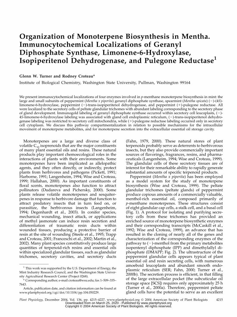
Peppermint (Mentha x piperita) has been employedas a model system for the study of monoterpenebiosynthesis (Wise and Croteau, 1999). The peltateglandular trichomes (peltate glands) of peppermintproduce copious amounts of a commercially valuable,menthol-rich essential oil, composed primarily ofp-menthane monoterpenes. These structures consistof eight glandular cap cells, a stalk cell, and a basal cell(Fig. 1). A protocol for isolating and purifying secre-tory cells from these trichomes has provided anenriched source of monoterpene biosynthetic enzymesand their corresponding transcripts (McCaskill et al.,1992; Wise and Croteau, 1999), an advance that hasresulted in the cloning of nearly all of the genes andcharacterization of the corresponding enzymes of thepathway to (2)-menthol from the primary metabolitesisopentenyl diphosphate (IPP) and dimethylallyl di-phosphate (DMAPP; Fig. 2). The ultrastructure of thepeppermint glandular cells appears typical of plantessential oil and resin secreting cells, with numerousamoeboid leucoplasts and abundant smooth endo-plasmic reticulum (SER; Fahn, 2000; Turner et al.,2000b). The secretion process is efficient, in that fillingof the large extracellular pocket (the subcuticular oilstorage space [SCS]) requires only approximately 25 h(Turner et al., 2000a). Therefore, peppermint peltategland cells have the potential to serve as an excellent
1 This work was supported by the U.S. Department of Energy, theMint Industry Research Council, and the Washington State Univer-sity Agricultural Research Center (Project 0268).
* Corresponding author; e-mail [email protected]; fax 1–509–335–7643.
Article, publication date, and citation information can be found atwww.plantphysiol.org/cgi/doi/10.1104/pp.104.050229.
Plant Physiology, December 2004, Vol. 136, pp. 4215–4227, www.plantphysiol.org � 2004 American Society of Plant Biologists 4215 www.plantphysiol.orgon March 25, 2020 - Published by Downloaded from
Copyright © 2004 American Society of Plant Biologists. All rights reserved.
model for the cell biology of plant oil glands, suppor-ted by the established molecular genetics and enzy-mology of (2)-menthol biosynthesis. Determining thesubcellular organization of monoterpene biosynthesisis an important step in understanding how thesehighly specialized secretory cells function.
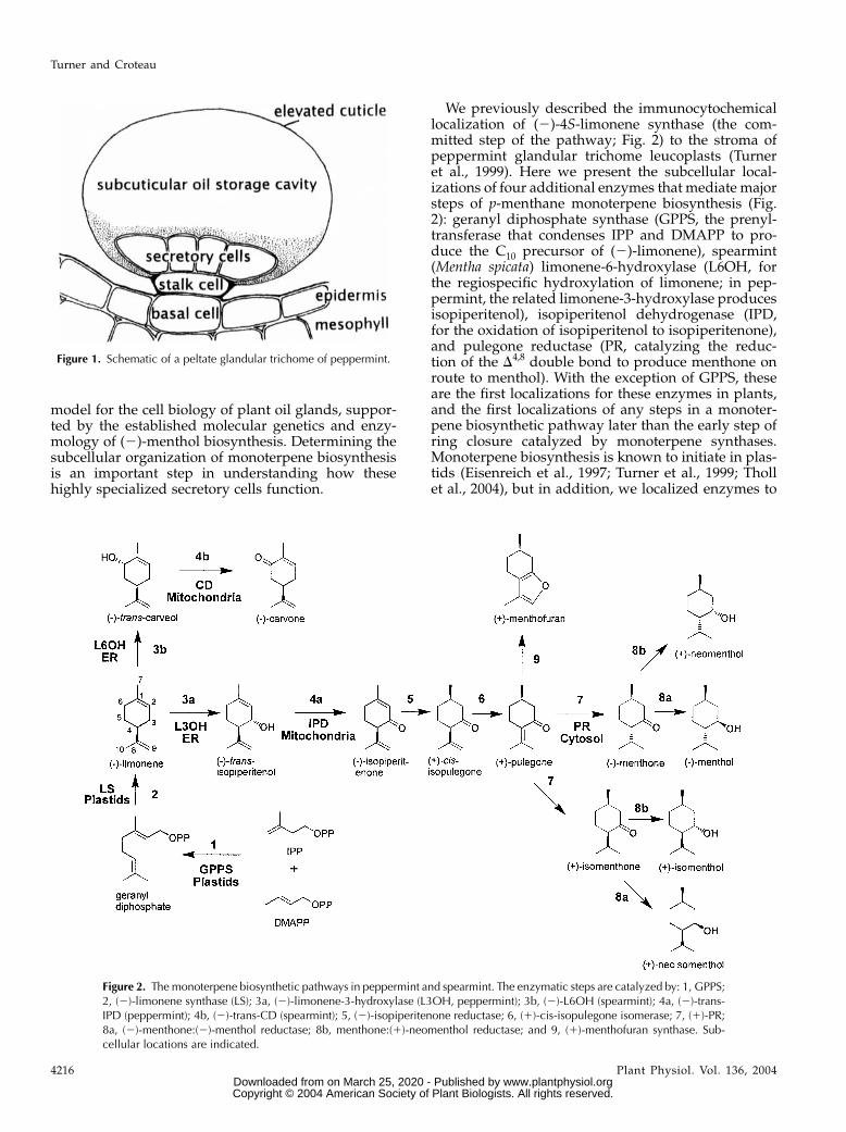
We previously described the immunocytochemicallocalization of (2)-4S-limonene synthase (the com-mitted step of the pathway; Fig. 2) to the stroma ofpeppermint glandular trichome leucoplasts (Turneret al., 1999). Here we present the subcellular local-izations of four additional enzymes that mediate majorsteps of p-menthane monoterpene biosynthesis (Fig.2): geranyl diphosphate synthase (GPPS, the prenyl-transferase that condenses IPP and DMAPP to pro-duce the C10 precursor of (2)-limonene), spearmint(Mentha spicata) limonene-6-hydroxylase (L6OH, forthe regiospecific hydroxylation of limonene; in pep-permint, the related limonene-3-hydroxylase producesisopiperitenol), isopiperitenol dehydrogenase (IPD,for the oxidation of isopiperitenol to isopiperitenone),and pulegone reductase (PR, catalyzing the reduc-tion of the D4,8 double bond to produce menthone onroute to menthol). With the exception of GPPS, theseare the first localizations for these enzymes in plants,and the first localizations of any steps in a monoter-pene biosynthetic pathway later than the early step ofring closure catalyzed by monoterpene synthases.Monoterpene biosynthesis is known to initiate in plas-tids (Eisenreich et al., 1997; Turner et al., 1999; Thollet al., 2004), but in addition, we localized enzymes to
Figure 1. Schematic of a peltate glandular trichome of peppermint.
Figure 2. The monoterpene biosynthetic pathways in peppermint and spearmint. The enzymatic steps are catalyzed by: 1, GPPS;2, (2)-limonene synthase (LS); 3a, (2)-limonene-3-hydroxylase (L3OH, peppermint); 3b, (2)-L6OH (spearmint); 4a, (2)-trans-IPD (peppermint); 4b, (2)-trans-CD (spearmint); 5, (2)-isopiperitenone reductase; 6, (1)-cis-isopulegone isomerase; 7, (1)-PR;8a, (2)-menthone:(2)-menthol reductase; 8b, menthone:(1)-neomenthol reductase; and 9, (1)-menthofuran synthase. Sub-cellular locations are indicated.
Turner and Croteau
4216 Plant Physiol. Vol. 136, 2004 www.plantphysiol.orgon March 25, 2020 - Published by Downloaded from
Copyright © 2004 American Society of Plant Biologists. All rights reserved.
endoplasmic reticulum, mitochondria, and the cytosol.We combine these observationswithdata relating to themonoterpene content of peppermint gland cells, thesecretion rate, and monoterpene solubility to presenta new model of monoterpene intracellular transloca-tion and secretion.
RESULTS
Specificity of Affinity-Purified Antibodies
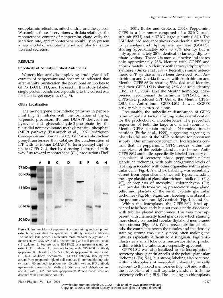
Western-blot analysis employing crude gland cellextracts of peppermint and spearmint indicated thatafter affinity purification the polyclonal antibodies toGPPS, L6OH, IPD, and PR used in this study labeledsingle protein bands corresponding to the correct Mrsfor their target enzymes (Fig. 3).
GPPS Localization
The monoterpene biosynthetic pathway in pepper-mint (Fig. 2) initiates with the formation of the C5terpenoid precursors IPP and DMAPP derived frompyruvate and glyceraldehyde-3-phosphate by theplastidial nonmevalonate, methylerythritol phosphate(MEP) pathway (Eisenreich et al., 1997; Rodrıguez-Concepcion and Boronat, 2002). GPPSs are short-chainprenyltransferases that catalyze the condensation ofIPP with its isomer DMAPP to form geranyl diphos-phate (GPP; C10), thereby directing isoprenoid path-way flux toward monoterpene (C10) production (Tholl
et al., 2001; Burke and Croteau, 2002). PeppermintGPPS is a heteromer composed of a 28-kD smallsubunit (SSU) and a 37-kD large subunit (LSU). TheLSU deduced sequence shows considerable similarityto geranylgeranyl diphosphate synthase (GGPPS),sharing approximately 65% to 75% identity but isonly approximately 25% identical to farnesyl diphos-phate synthase. The SSU is more distinctive and sharesonly approximately 25% identity with GGPPS andapproximately 17% identity with farnesyl diphosphatesynthase. (Burke et al., 1999). Recently, similar hetero-meric GPP synthases have been described from An-tirrhinum and Clarkia flowers, with Antirrhinum andMentha GPPS-SSUs sharing 53% deduced identity,and their GPPS-LSUs sharing 75% deduced identity(Tholl et al., 2004). Like the Mentha homologs, coex-pressed recombinant Antirrhinum GPPS-SSU andGPPS-LSU produced GPP. Unlike the Mentha GPPS-LSU, the Antirrhinum GPPS-LSU showed GGPPSactivity when expressed alone.
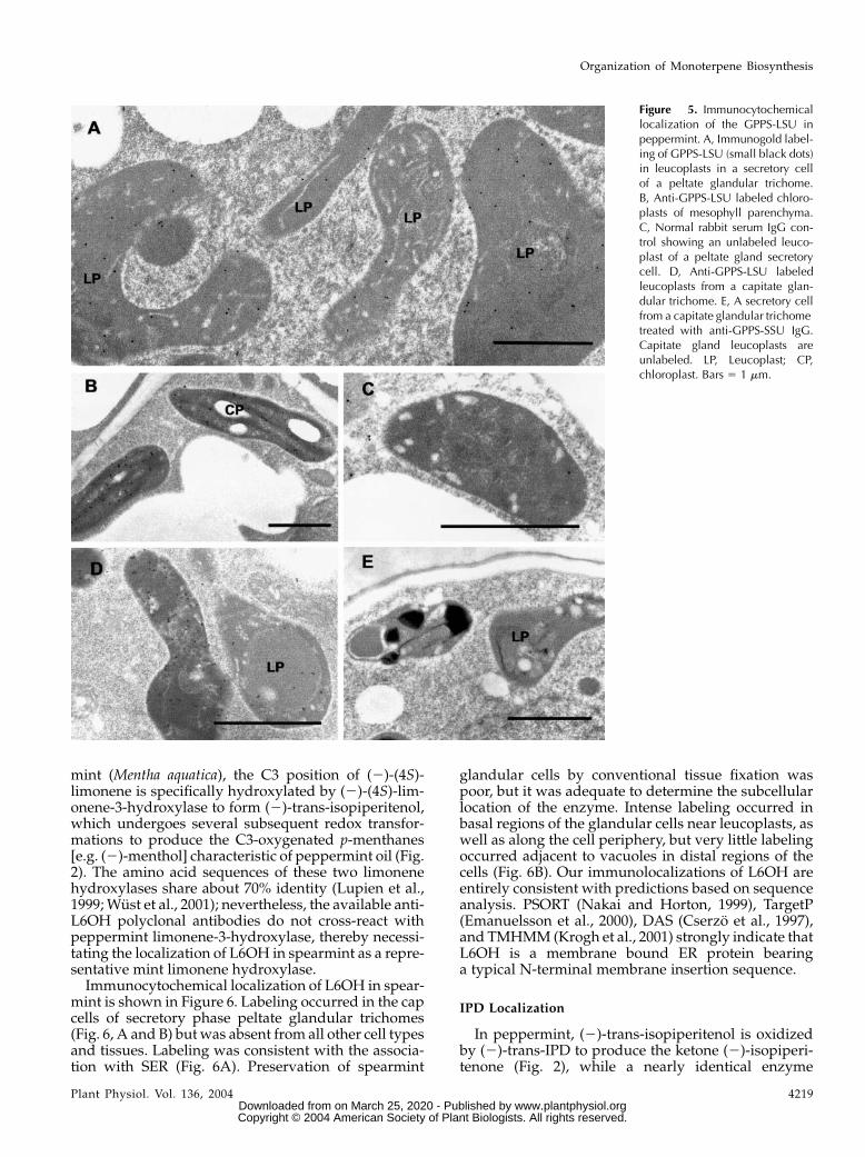
Presumably, the subcellular distribution of GPPSis an important factor affecting substrate allocationfor the production of monoterpenes. The preproteinsequences of both the large and small subunits ofMentha GPPS contain probable N-terminal transitpeptides (Burke et al., 1999), suggesting targeting toplastids (the site of the MEP pathway for precursorsupply). Our immunocytochemical localizations con-firm that, in peppermint, GPPS resides within theleucoplasts of the peltate glandular trichomes. Anti-GPPS-SSU antibodies specifically labeled secretory cellleucoplasts of secretory phase peppermint peltateglandular trichomes, with only background levels oflabeling associated with other organelles within glan-dular cells (Fig. 4, A and B). Labeling was essentiallyabsent from organelles of other cell types, includingthe large plastids of glandular trichome stalk cells (Fig.4C), chloroplasts of mesophyll chlorenchyma (Fig.4D), proplastids from young presecretory stage glandcells, and plastids of the small capitate glandulartrichomes (Fig. 5E). Significant labeling was absent inthe preimmune serum IgG controls (Fig. 4, E and F).
Within the leucoplasts, the GPPS-SSU label ap-peared to be frequently, but not consistently, associatedwith tubular plastid membranes. This was most ap-parent with chemically fixed glands for which stainingmore clearly contrasted tubular thylakoid membranesfrom stroma (Fig. 4A). With freeze-substituted plas-tids, the contrast between the tubules and the denselystaining stroma was usually poor, often making thetubules especially difficult to distinguish. Figure 4Billustrates a small lobe of a freeze-substituted plastidwithin which the tubules are especially apparent.
GPPS-LSU was also localized to the leucoplasts ofsecretory phase glandular cells of the peltate glandulartrichomes (Fig. 5A), but strong labeling also occurredwithin chloroplasts of mesophyll chlorenchyma cellsadjacent to thylakoid membranes (Fig. 5B) and withinthe leucoplasts of small capitate glandular trichomesecretory cells (Fig. 5D). The labeling in chloroplasts
Figure 3. Immunoblots of peppermint or spearmint gland cell proteinextracts demonstrating the specificity of affinity-purified antibodies.The far left lane presents molecular mass markers (5 mg/band). A,Representative SDS-PAGE of a peppermint gland cell protein extract(18 mg/lane). B, Representative SDS-PAGE of a spearmint gland cellextract (11 mg/lane). C, Immunoblotting with GPPS-SSU antibody(peppermint), (D) with GPPS-LSU antibody (peppermint), and (E) with(2)-L6OH antibody (spearmint). (2)-L6OH antibody labeling wasabsent from peppermint gland cell extracts. F, Immunoblotting with(2)-trans-IPD antibody (peppermint), (G) with (2)-trans-IPD antibody(spearmint), presumably labeling (2)-trans-carveol dehydrogenase,and (H) with (1)-PR antibody (peppermint). Protein bands were notdetected with preimmune controls.
Organization of Monoterpene Biosynthesis
Plant Physiol. Vol. 136, 2004 4217 www.plantphysiol.orgon March 25, 2020 - Published by Downloaded from
Copyright © 2004 American Society of Plant Biologists. All rights reserved.
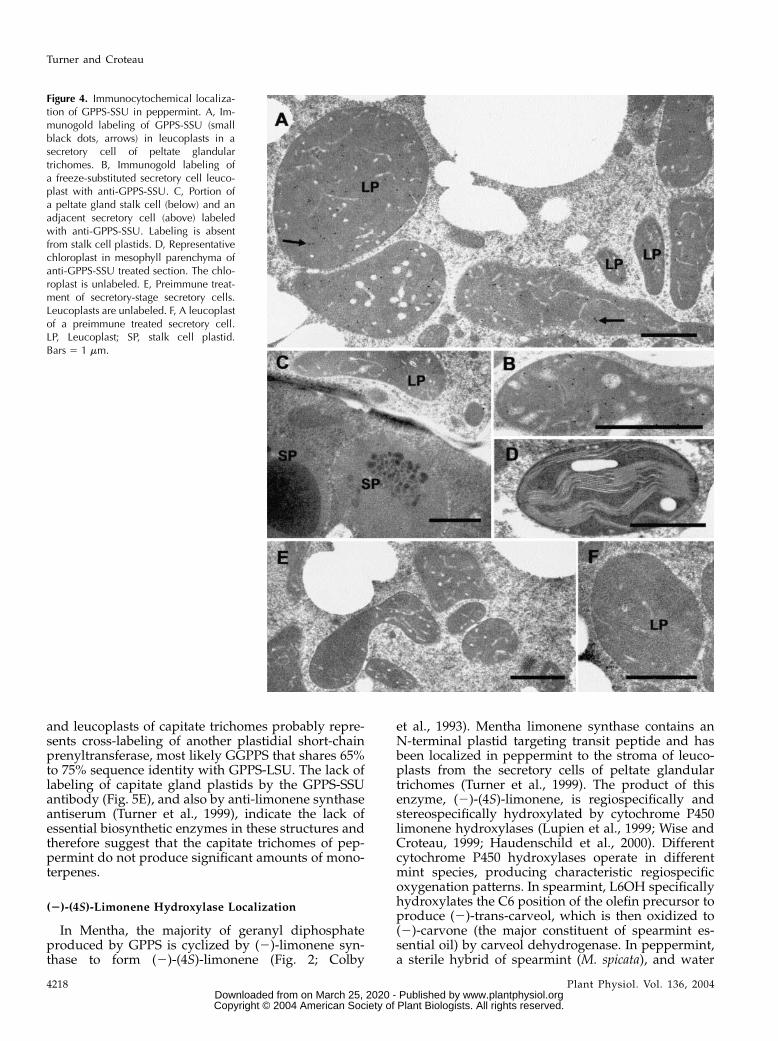
and leucoplasts of capitate trichomes probably repre-sents cross-labeling of another plastidial short-chainprenyltransferase, most likely GGPPS that shares 65%to 75% sequence identity with GPPS-LSU. The lack oflabeling of capitate gland plastids by the GPPS-SSUantibody (Fig. 5E), and also by anti-limonene synthaseantiserum (Turner et al., 1999), indicate the lack ofessential biosynthetic enzymes in these structures andtherefore suggest that the capitate trichomes of pep-permint do not produce significant amounts of mono-terpenes.
(2)-(4S)-Limonene Hydroxylase Localization
In Mentha, the majority of geranyl diphosphateproduced by GPPS is cyclized by (2)-limonene syn-thase to form (2)-(4S)-limonene (Fig. 2; Colby
et al., 1993). Mentha limonene synthase contains anN-terminal plastid targeting transit peptide and hasbeen localized in peppermint to the stroma of leuco-plasts from the secretory cells of peltate glandulartrichomes (Turner et al., 1999). The product of thisenzyme, (2)-(4S)-limonene, is regiospecifically andstereospecifically hydroxylated by cytochrome P450limonene hydroxylases (Lupien et al., 1999; Wise andCroteau, 1999; Haudenschild et al., 2000). Differentcytochrome P450 hydroxylases operate in differentmint species, producing characteristic regiospecificoxygenation patterns. In spearmint, L6OH specificallyhydroxylates the C6 position of the olefin precursor toproduce (2)-trans-carveol, which is then oxidized to(2)-carvone (the major constituent of spearmint es-sential oil) by carveol dehydrogenase. In peppermint,a sterile hybrid of spearmint (M. spicata), and water
Figure 4. Immunocytochemical localiza-tion of GPPS-SSU in peppermint. A, Im-munogold labeling of GPPS-SSU (smallblack dots, arrows) in leucoplasts in asecretory cell of peltate glandulartrichomes. B, Immunogold labeling ofa freeze-substituted secretory cell leuco-plast with anti-GPPS-SSU. C, Portion ofa peltate gland stalk cell (below) and anadjacent secretory cell (above) labeledwith anti-GPPS-SSU. Labeling is absentfrom stalk cell plastids. D, Representativechloroplast in mesophyll parenchyma ofanti-GPPS-SSU treated section. The chlo-roplast is unlabeled. E, Preimmune treat-ment of secretory-stage secretory cells.Leucoplasts are unlabeled. F, A leucoplastof a preimmune treated secretory cell.LP, Leucoplast; SP, stalk cell plastid.Bars 5 1 mm.
Turner and Croteau
4218 Plant Physiol. Vol. 136, 2004 www.plantphysiol.orgon March 25, 2020 - Published by Downloaded from
Copyright © 2004 American Society of Plant Biologists. All rights reserved.
mint (Mentha aquatica), the C3 position of (2)-(4S)-limonene is specifically hydroxylated by (2)-(4S)-lim-onene-3-hydroxylase to form (2)-trans-isopiperitenol,which undergoes several subsequent redox transfor-mations to produce the C3-oxygenated p-menthanes[e.g. (2)-menthol] characteristic of peppermint oil (Fig.2). The amino acid sequences of these two limonenehydroxylases share about 70% identity (Lupien et al.,1999;Wust et al., 2001); nevertheless, the available anti-L6OH polyclonal antibodies do not cross-react withpeppermint limonene-3-hydroxylase, thereby necessi-tating the localization of L6OH in spearmint as a repre-sentative mint limonene hydroxylase.Immunocytochemical localization of L6OH in spear-
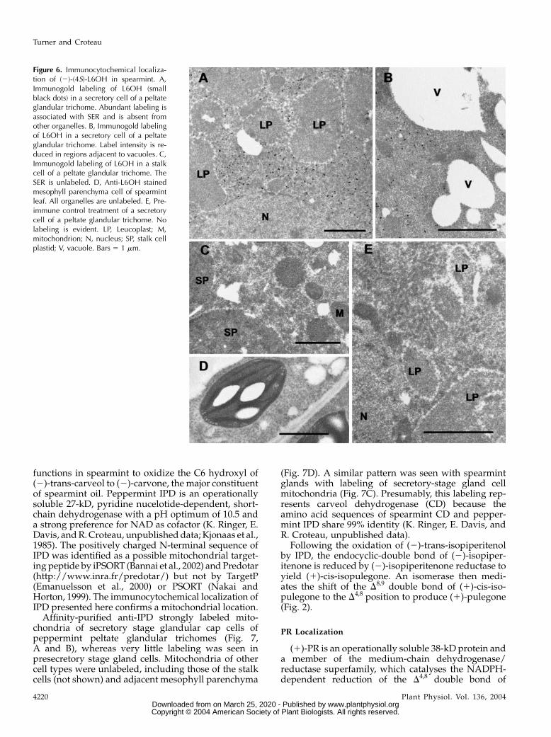
mint is shown in Figure 6. Labeling occurred in the capcells of secretory phase peltate glandular trichomes(Fig. 6, A and B) butwas absent from all other cell typesand tissues. Labeling was consistent with the associa-tion with SER (Fig. 6A). Preservation of spearmint
glandular cells by conventional tissue fixation waspoor, but it was adequate to determine the subcellularlocation of the enzyme. Intense labeling occurred inbasal regions of the glandular cells near leucoplasts, aswell as along the cell periphery, but very little labelingoccurred adjacent to vacuoles in distal regions of thecells (Fig. 6B). Our immunolocalizations of L6OH areentirely consistent with predictions based on sequenceanalysis. PSORT (Nakai and Horton, 1999), TargetP(Emanuelsson et al., 2000), DAS (Cserzo et al., 1997),and TMHMM (Krogh et al., 2001) strongly indicate thatL6OH is a membrane bound ER protein bearinga typical N-terminal membrane insertion sequence.
IPD Localization
In peppermint, (2)-trans-isopiperitenol is oxidizedby (2)-trans-IPD to produce the ketone (2)-isopiperi-tenone (Fig. 2), while a nearly identical enzyme
Figure 5. Immunocytochemicallocalization of the GPPS-LSU inpeppermint. A, Immunogold label-ing of GPPS-LSU (small black dots)in leucoplasts in a secretory cellof a peltate glandular trichome.B, Anti-GPPS-LSU labeled chloro-plasts of mesophyll parenchyma.C, Normal rabbit serum IgG con-trol showing an unlabeled leuco-plast of a peltate gland secretorycell. D, Anti-GPPS-LSU labeledleucoplasts from a capitate glan-dular trichome. E, A secretory cellfrom a capitate glandular trichometreated with anti-GPPS-SSU IgG.Capitate gland leucoplasts areunlabeled. LP, Leucoplast; CP,chloroplast. Bars 5 1 mm.
Organization of Monoterpene Biosynthesis
Plant Physiol. Vol. 136, 2004 4219 www.plantphysiol.orgon March 25, 2020 - Published by Downloaded from
Copyright © 2004 American Society of Plant Biologists. All rights reserved.
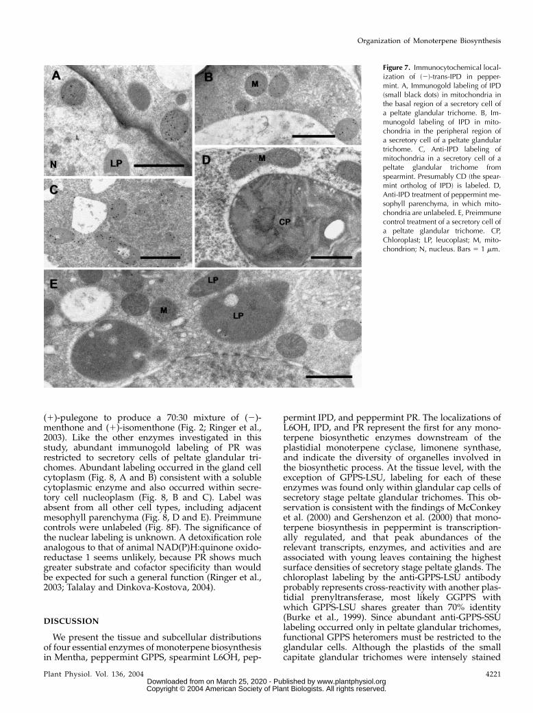
functions in spearmint to oxidize the C6 hydroxyl of(2)-trans-carveol to (2)-carvone, the major constituentof spearmint oil. Peppermint IPD is an operationallysoluble 27-kD, pyridine nucelotide-dependent, short-chain dehydrogenase with a pH optimum of 10.5 anda strong preference for NAD as cofactor (K. Ringer, E.Davis, andR.Croteau, unpublisheddata; Kjonaas et al.,1985). The positively charged N-terminal sequence ofIPD was identified as a possible mitochondrial target-ing peptide by iPSORT (Bannai et al., 2002) andPredotar(http://www.inra.fr/predotar/) but not by TargetP(Emanuelsson et al., 2000) or PSORT (Nakai andHorton, 1999). The immunocytochemical localization ofIPD presented here confirms a mitochondrial location.
Affinity-purified anti-IPD strongly labeled mito-chondria of secretory stage glandular cap cells ofpeppermint peltate glandular trichomes (Fig. 7,A and B), whereas very little labeling was seen inpresecretory stage gland cells. Mitochondria of othercell types were unlabeled, including those of the stalkcells (not shown) and adjacent mesophyll parenchyma
(Fig. 7D). A similar pattern was seen with spearmintglands with labeling of secretory-stage gland cellmitochondria (Fig. 7C). Presumably, this labeling rep-resents carveol dehydrogenase (CD) because theamino acid sequences of spearmint CD and pepper-mint IPD share 99% identity (K. Ringer, E. Davis, andR. Croteau, unpublished data).
Following the oxidation of (2)-trans-isopiperitenolby IPD, the endocyclic-double bond of (2)-isopiper-itenone is reduced by (2)-isopiperitenone reductase toyield (1)-cis-isopulegone. An isomerase then medi-ates the shift of the D8,9 double bond of (1)-cis-iso-pulegone to the D4,8 position to produce (1)-pulegone(Fig. 2).
PR Localization
(1)-PR is an operationally soluble 38-kD protein anda member of the medium-chain dehydrogenase/reductase superfamily, which catalyses the NADPH-dependent reduction of the D4,8 double bond of
Figure 6. Immunocytochemical localiza-tion of (2)-(4S)-L6OH in spearmint. A,Immunogold labeling of L6OH (smallblack dots) in a secretory cell of a peltateglandular trichome. Abundant labeling isassociated with SER and is absent fromother organelles. B, Immunogold labelingof L6OH in a secretory cell of a peltateglandular trichome. Label intensity is re-duced in regions adjacent to vacuoles. C,Immunogold labeling of L6OH in a stalkcell of a peltate glandular trichome. TheSER is unlabeled. D, Anti-L6OH stainedmesophyll parenchyma cell of spearmintleaf. All organelles are unlabeled. E, Pre-immune control treatment of a secretorycell of a peltate glandular trichome. Nolabeling is evident. LP, Leucoplast; M,mitochondrion; N, nucleus; SP, stalk cellplastid; V, vacuole. Bars 5 1 mm.
Turner and Croteau
4220 Plant Physiol. Vol. 136, 2004 www.plantphysiol.orgon March 25, 2020 - Published by Downloaded from
Copyright © 2004 American Society of Plant Biologists. All rights reserved.
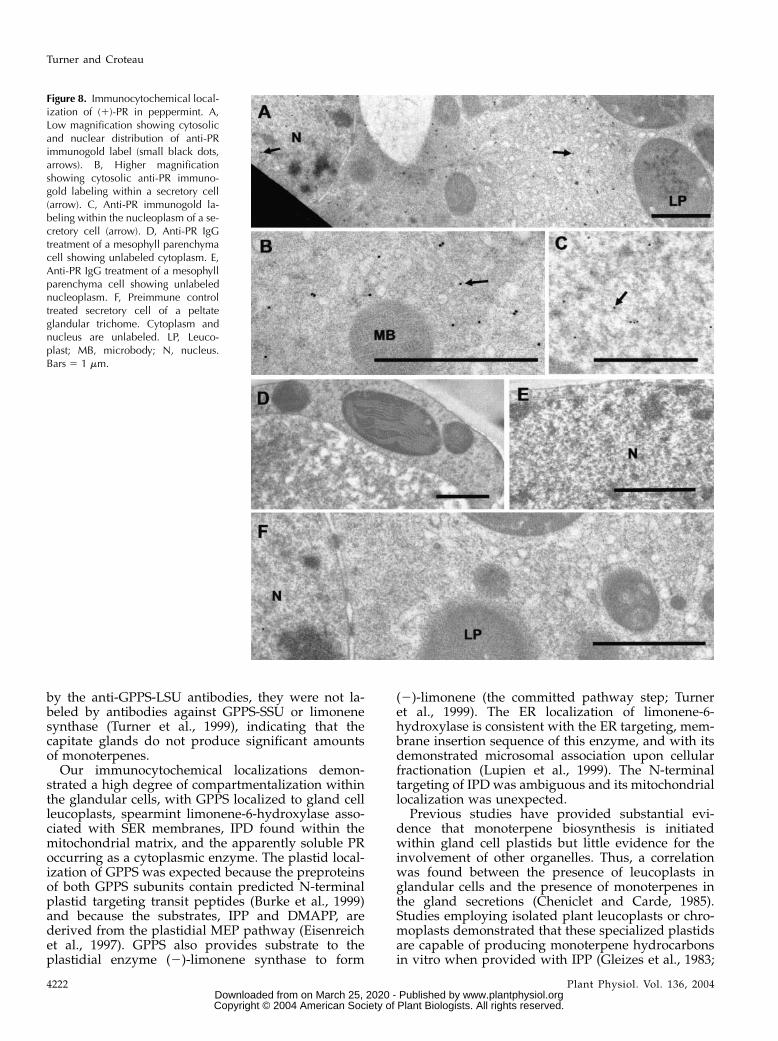
(1)-pulegone to produce a 70:30 mixture of (2)-menthone and (1)-isomenthone (Fig. 2; Ringer et al.,2003). Like the other enzymes investigated in thisstudy, abundant immunogold labeling of PR wasrestricted to secretory cells of peltate glandular tri-chomes. Abundant labeling occurred in the gland cellcytoplasm (Fig. 8, A and B) consistent with a solublecytoplasmic enzyme and also occurred within secre-tory cell nucleoplasm (Fig. 8, B and C). Label wasabsent from all other cell types, including adjacentmesophyll parenchyma (Fig. 8, D and E). Preimmunecontrols were unlabeled (Fig. 8F). The significance ofthe nuclear labeling is unknown. A detoxification roleanalogous to that of animal NAD(P)H:quinone oxido-reductase 1 seems unlikely, because PR shows muchgreater substrate and cofactor specificity than wouldbe expected for such a general function (Ringer et al.,2003; Talalay and Dinkova-Kostova, 2004).
DISCUSSION
We present the tissue and subcellular distributionsof four essential enzymes of monoterpene biosynthesisin Mentha, peppermint GPPS, spearmint L6OH, pep-
permint IPD, and peppermint PR. The localizations ofL6OH, IPD, and PR represent the first for any mono-terpene biosynthetic enzymes downstream of theplastidial monoterpene cyclase, limonene synthase,and indicate the diversity of organelles involved inthe biosynthetic process. At the tissue level, with theexception of GPPS-LSU, labeling for each of theseenzymes was found only within glandular cap cells ofsecretory stage peltate glandular trichomes. This ob-servation is consistent with the findings of McConkeyet al. (2000) and Gershenzon et al. (2000) that mono-terpene biosynthesis in peppermint is transcription-ally regulated, and that peak abundances of therelevant transcripts, enzymes, and activities and areassociated with young leaves containing the highestsurface densities of secretory stage peltate glands. Thechloroplast labeling by the anti-GPPS-LSU antibodyprobably represents cross-reactivity with another plas-tidial prenyltransferase, most likely GGPPS withwhich GPPS-LSU shares greater than 70% identity(Burke et al., 1999). Since abundant anti-GPPS-SSUlabeling occurred only in peltate glandular trichomes,functional GPPS heteromers must be restricted to theglandular cells. Although the plastids of the smallcapitate glandular trichomes were intensely stained
Figure 7. Immunocytochemical local-ization of (2)-trans-IPD in pepper-mint. A, Immunogold labeling of IPD(small black dots) in mitochondria inthe basal region of a secretory cell ofa peltate glandular trichome. B, Im-munogold labeling of IPD in mito-chondria in the peripheral region ofa secretory cell of a peltate glandulartrichome. C, Anti-IPD labeling ofmitochondria in a secretory cell of apeltate glandular trichome fromspearmint. Presumably CD (the spear-mint ortholog of IPD) is labeled. D,Anti-IPD treatment of peppermint me-sophyll parenchyma, in which mito-chondria are unlabeled. E, Preimmunecontrol treatment of a secretory cell ofa peltate glandular trichome. CP,Chloroplast; LP, leucoplast; M, mito-chondrion; N, nucleus. Bars 5 1 mm.
Organization of Monoterpene Biosynthesis
Plant Physiol. Vol. 136, 2004 4221 www.plantphysiol.orgon March 25, 2020 - Published by Downloaded from
Copyright © 2004 American Society of Plant Biologists. All rights reserved.
by the anti-GPPS-LSU antibodies, they were not la-beled by antibodies against GPPS-SSU or limonenesynthase (Turner et al., 1999), indicating that thecapitate glands do not produce significant amountsof monoterpenes.
Our immunocytochemical localizations demon-strated a high degree of compartmentalization withinthe glandular cells, with GPPS localized to gland cellleucoplasts, spearmint limonene-6-hydroxylase asso-ciated with SER membranes, IPD found within themitochondrial matrix, and the apparently soluble PRoccurring as a cytoplasmic enzyme. The plastid local-ization of GPPS was expected because the preproteinsof both GPPS subunits contain predicted N-terminalplastid targeting transit peptides (Burke et al., 1999)and because the substrates, IPP and DMAPP, arederived from the plastidial MEP pathway (Eisenreichet al., 1997). GPPS also provides substrate to theplastidial enzyme (2)-limonene synthase to form
(2)-limonene (the committed pathway step; Turneret al., 1999). The ER localization of limonene-6-hydroxylase is consistent with the ER targeting, mem-brane insertion sequence of this enzyme, and with itsdemonstrated microsomal association upon cellularfractionation (Lupien et al., 1999). The N-terminaltargeting of IPD was ambiguous and its mitochondriallocalization was unexpected.
Previous studies have provided substantial evi-dence that monoterpene biosynthesis is initiatedwithin gland cell plastids but little evidence for theinvolvement of other organelles. Thus, a correlationwas found between the presence of leucoplasts inglandular cells and the presence of monoterpenes inthe gland secretions (Cheniclet and Carde, 1985).Studies employing isolated plant leucoplasts or chro-moplasts demonstrated that these specialized plastidsare capable of producing monoterpene hydrocarbonsin vitro when provided with IPP (Gleizes et al., 1983;
Figure 8. Immunocytochemical local-ization of (1)-PR in peppermint. A,Low magnification showing cytosolicand nuclear distribution of anti-PRimmunogold label (small black dots,arrows). B, Higher magnificationshowing cytosolic anti-PR immuno-gold labeling within a secretory cell(arrow). C, Anti-PR immunogold la-beling within the nucleoplasm of a se-cretory cell (arrow). D, Anti-PR IgGtreatment of a mesophyll parenchymacell showing unlabeled cytoplasm. E,Anti-PR IgG treatment of a mesophyllparenchyma cell showing unlabelednucleoplasm. F, Preimmune controltreated secretory cell of a peltateglandular trichome. Cytoplasm andnucleus are unlabeled. LP, Leuco-plast; MB, microbody; N, nucleus.Bars 5 1 mm.
Turner and Croteau
4222 Plant Physiol. Vol. 136, 2004 www.plantphysiol.orgon March 25, 2020 - Published by Downloaded from
Copyright © 2004 American Society of Plant Biologists. All rights reserved.
Mettal et al., 1988; Perez et al., 1990; Soler et al., 1992).In addition, 13C-labeling experiments with basic pre-cursors have indicated that plant monoterpenes arederived from the plastid localized MEP pathway(Eisenreich et al., 1997; Lichtenthaler, 1999; Rodrıguez-Concepcion andBoronat, 2002). Previous immunocyto-chemical localization of peppermint (2)-limonenesynthase established that this key enzyme is presentonly within secretory cell leucoplasts of the peltateglandular trichomes (Turner et al., 1999). Recently,Tholl et al. (2004) employed transmission electronmicroscopy immunocytochemistry to localize the An-tirrhinum GPPS-SSU to plastids of Antirrhinum petalcells.Bouvier et al. (2000) reported the immunofluores-
cence localizations for a putative homodimeric GPPsynthase (AtGPPS, GenBank no. CAC16849.1), DXPsynthase, a ‘‘monoterpene synthase,’’ and GGPP syn-thase in several plant species. Although organelletargeting-sequence analysis programs consistentlypredict that the putative AtGPPS is targeted to mito-chondria, Bouvier et al. (2000) localized all theseenzymes to plastids, with strong labeling of mesophyllparenchyma chloroplasts in Arabidopsis, Pinus, andCitrofortunella, as well as strong labeling of secre-tory cell leucoplasts in Pinus and Citrofortunella.This seemingly abundant, constitutive expression ofAtGPPS in chlorenchyma of diverse species contrastswith our findings for Mentha. It is possible that thereliance of Bouvier et al. (2000) on crude polyclonalantiserum could have compromised the specificity oftheir probes; false positive labeling artifacts associatedwith the use of crude antiserum have been describedby (Tavares et al., 2002).The subcellular compartmentalization of monoter-
pene biosynthesis in diverse localeswithin peppermintglandular cells presents questions concerning the co-ordinated intracellular movement of monoterpene me-tabolites between organelles during production andsecretion. In speculating about this process, enzymelocalization in the context of intracellular transport,possible oil secretion mechanisms, and the knownmonoterpene content of peppermint gland cells mustbe considered. The rate of oil secretion is moderatelyrapid, in that the glandular cells of a peltate glandulartrichome secrete approximately twice their cellularvolume when filling the subcuticular oil storage spacein roughly 20 to 30 h (Turner et al., 2000a). This event
translates to a SCS filling rate of approximately 1.7 31022 (60.33 1022) nmol h21 gland21, and secretion fluxacross the plasma membrane of approximately 1.8 31026 (60.4 3 1026) nmol mm22 h21.
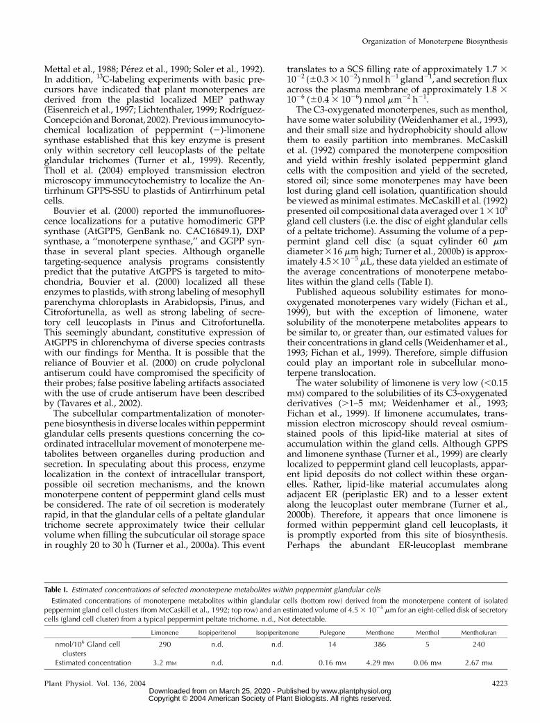
The C3-oxygenated monoterpenes, such as menthol,have some water solubility (Weidenhamer et al., 1993),and their small size and hydrophobicity should allowthem to easily partition into membranes. McCaskillet al. (1992) compared the monoterpene compositionand yield within freshly isolated peppermint glandcells with the composition and yield of the secreted,stored oil; since some monoterpenes may have beenlost during gland cell isolation, quantification shouldbe viewed as minimal estimates. McCaskill et al. (1992)presented oil compositional data averaged over 13106
gland cell clusters (i.e. the disc of eight glandular cellsof a peltate trichome). Assuming the volume of a pep-permint gland cell disc (a squat cylinder 60 mmdiameter316 mm high; Turner et al., 2000b) is approx-imately 4.531025 mL, these data yielded an estimate ofthe average concentrations of monoterpene metabo-lites within the gland cells (Table I).
Published aqueous solubility estimates for mono-oxygenated monoterpenes vary widely (Fichan et al.,1999), but with the exception of limonene, watersolubility of the monoterpene metabolites appears tobe similar to, or greater than, our estimated values fortheir concentrations in gland cells (Weidenhamer et al.,1993; Fichan et al., 1999). Therefore, simple diffusioncould play an important role in subcellular mono-terpene translocation.
The water solubility of limonene is very low (,0.15mM) compared to the solubilities of its C3-oxygenatedderivatives (.1–5 mM; Weidenhamer et al., 1993;Fichan et al., 1999). If limonene accumulates, trans-mission electron microscopy should reveal osmium-stained pools of this lipid-like material at sites ofaccumulation within the gland cells. Although GPPSand limonene synthase (Turner et al., 1999) are clearlylocalized to peppermint gland cell leucoplasts, appar-ent lipid deposits do not collect within these organ-elles. Rather, lipid-like material accumulates alongadjacent ER (periplastic ER) and to a lesser extentalong the leucoplast outer membrane (Turner et al.,2000b). Therefore, it appears that once limonene isformed within peppermint gland cell leucoplasts, itis promptly exported from this site of biosynthesis.Perhaps the abundant ER-leucoplast membrane
Table I. Estimated concentrations of selected monoterpene metabolites within peppermint glandular cells
Estimated concentrations of monoterpene metabolites within glandular cells (bottom row) derived from the monoterpene content of isolatedpeppermint gland cell clusters (from McCaskill et al., 1992; top row) and an estimated volume of 4.53 1025 mm for an eight-celled disk of secretorycells (gland cell cluster) from a typical peppermint peltate trichome. n.d., Not detectable.
Limonene Isopiperitenol Isopiperitenone Pulegone Menthone Menthol Menthofuran
nmol/106 Gland cellclusters
290 n.d. n.d. 14 386 5 240
Estimated concentration 3.2 mM n.d. n.d. 0.16 mM 4.29 mM 0.06 mM 2.67 mM
Organization of Monoterpene Biosynthesis
Plant Physiol. Vol. 136, 2004 4223 www.plantphysiol.orgon March 25, 2020 - Published by Downloaded from
Copyright © 2004 American Society of Plant Biologists. All rights reserved.
contacts (Turner et al., 2000b) facilitate transfer oflimonene to the outer leaflet of ER membranes. At thislocale, limonene-3-hydroxylase mediates hydroxyl-ation of the olefin to produce (2)-trans-isopiperitenol.
Hydroxylation of monoterpene olefins greatly in-creases their water solubility (Weidenhamer et al.,1993; Li et al., 1998; Fichan et al., 1999), and the activityof mitochondrial IPD might produce a concentrationgradient sufficient to drive rapid diffusion of (2)-trans-isopiperitenol into mitochondria. Alternatively,some type of terpenoid carrier protein, amitochondrialmembrane pump, or transient contacts between SERand mitochondrial membranes might facilitate isopi-peritenol movement. Following oxidation, the nextpathway step is mediated by isopiperitenone reduc-tase, an operationally soluble, NADPH-dependent,short-chain reductase (Ringer et al., 2003) that lacksapparent targeting sequence information and is pre-sumed to be cytosolic. Thus, it appears that (2)-isopiperitenone produced by mitochondrial IPD isimmediately exported to the cytosol. Seemingly, thetransfer of (2)-trans-isopiperitenol to the mitochon-drial matrix, its conversion to (2)-isopiperitenone, andthe subsequent export of (2)-isopiperitenone to thecytosol are very efficient, because neither (2)-trans-isopiperitenol nor (2)-isopiperitenone can be found inany but trace quantities in either the secreted oil orwithin the glandular cells (McCaskill et al., 1992). Thisrapid conversion occurs in spite of the widespreadsubcellular distribution of ER localized limonene-3-hydroxylase (which produces (2)-trans-isopiperite-nol) and the relatively small volume occupied by thegland cell mitochondria (approximately 6% of cellvolume; G. Turner and R. Croteau, unpublished data).
PR was localized to the cytosol, and preliminaryimmunocytochemical results indicate that menthonereductase is also cytosolic (G. Turner and R. Croteau,unpublished data); thus, the final steps of monoter-pene biosynthesis in peppermint occur in the cytosol.Within peppermint glandular cells, most lipid-likedeposits are associated with ER membranes. Thesedeposits are especially abundant at ER of the periph-eral cytoplasm, suggesting a directional movement oflipids (terpenoids) toward the secretory plasma mem-brane (Turner et al., 2000a, 2000b). If these lipid-likedeposits represent the terminal monoterpene prod-ucts, menthone and menthol, partitioned into ERmembranes, then the abundant, direct ER-plasmamembrane contacts (described in Turner et al., 2000b)could facilitate transfer of these monoterpenes fromthe ER to the plasma membrane. The mechanism ofsecretion is unknown, but an active process seemslikely, primarily because the secretion is directionaland because peppermint gland cell membranes showselectivity towardmonoterpene types (McCaskill et al.,1992). For example, McCaskill et al. (1992) noted that60% of the menthofuran produced in peppermintglands is selectively retained within the glandularcells. An ATP binding cassette (ABC) transporterwould be a good candidate for such an essential oil
pump. Putative ABC transporters are well representedin the peppermint gland expressed sequence taglibrary (Lange et al., 2000), and an inducible, plasmamembrane, ABC transporter of the pleiotropic drugresistance subfamily has been implicated in diterpenesecretion from tobacco epidermal cells (Jasinski et al.,2001). It is interesting to note that menthone stored inthe SCS apparently remains in equilibrium with dis-solved cytosolic menthone, because this ketone isconverted to menthol by menthone reductase duringthe late postsecretion phase of gland development(McConkey et al., 2000). A more detailed investigationof menthone reductase and its role in monoterpenemetabolism is in progress.
MATERIALS AND METHODS
Plant Materials, Secretory Cell Extracts, andAntibody Production
Peppermint (Mentha x piperita L. cv Black Mitcham) plants were grown
with a 16-h photoperiod in a controlled environment chamber as previously
described (Gershenzon et al., 2000). Spearmint (Mentha spicata) plants were
grown with a 16-h photoperiod in a greenhouse with supplemental lighting
from sodium vapor lights. Peltate glandular trichome secretory cells were
isolated by a surface abrasion technique previously described (Gershenzon
et al., 1992). The isolated glandular cells were sonicated three times for 30 s at
1/4 power with a VirTis (Gardiner, NY) Virsonic sonicator in a chilled 100 mM
Na2HPO4-NaH2PO4, pH 7.4, buffer, containing 250 mM Suc, 5% (w/v)
Amberlite XAD-4 resin, 1% PVPP-40, 1 mM dithiothreitol, 1 mM EDTA, and
1 mM benzamidine. The resulting lysates were filtered, homogenized with
a glass homogenizer, and centrifuged for 30 min at 18,000g, followed by 90
min at 195,000g at 4�C. The resulting pellets and supernatants were employed
for immunoblot analysis. Polyclonal antibodies, generated in rabbits against
purified recombinant proteins (see below), were prepared by the contractor,
Alpha Diagnostic International (San Antonio, TX).
Protein Purification and Production of Antibodies
Production of antibodies to recombinant L6OH (Lupien et al., 1999) and to
both subunits of recombinant GPP synthase (Burke et al., 1999) was described
previously. cDNAs encoding PR and IPD from a peppermint oil gland cell
library (Lange et al., 2000; Ringer et al., 2003) were subcloned into pSBET
(Schenk et al., 1995) and expressed in Escherichia coli BL21-CodonPlus cells
(Stratagene, La Jolla, CA) by procedures previously described (Ringer et al.,
2003). One-liter cultures were grown to A600 5 0.5 at 37�C in Luria-Bertani
medium containing 50 mg kanamycin/mL, then induced with 1mM isopropyl-
b-D-thiogalactopyranoside and grown overnight at 15�C. The cells were then
pelleted by centrifugation at 5,000 rpm, resuspended in chilled 50 mMMOPSO
buffer, pH 7.5, that contained 1mM benzamidine, and sonicated three times for
15 s at 1/4 power with a Virsonic sonicator (VirTis). The resulting lysate was
centrifuged at 18,000g for 30min, then at 195,000g for 90min at 4�C, to afford the
soluble enzyme fraction that was separated by chromatography on Source 30Q
anion-exchange resin (Amersham Biosciences, Piscataway, NJ) with a salt
gradient (0–1 M NaCl) in 50 mM MOPSO buffer, pH 7.5. The target enzymes,
located by activity assay (Ringer et al., 2003),were then subjected to SDS-PAGE,
and the corresponding proteins of the correctMrwere excised from the gels and
used to generate antibodies in rabbits (Alpha Diagnostic International).
Partially purified recombinant PR obtained with Source 30Q anion-
exchange chromatography, as described above, was further purified for use
in affinity purification of polyclonal anti-PR antibodies. Source 30Q fractions
demonstrating ample PR activity were combined and loaded onto a phenyl
sepharose FPLC column (Amersham Biosciences) in a loading buffer contain-
ing final concentrations of 50 mM sodium phosphate buffer, 1 mM dithio-
threitol, 10% (v/v) glycerol, and 1.5 M (NH4)2SO4. After washing with 10 mL
of the loading buffer, proteins were eluted with a decreasing ammonium
sulfate gradient [1.5–0 M (NH4)2SO4], and fractions were assayed for PR
Turner and Croteau
4224 Plant Physiol. Vol. 136, 2004 www.plantphysiol.orgon March 25, 2020 - Published by Downloaded from
Copyright © 2004 American Society of Plant Biologists. All rights reserved.
activity (Ringer et al., 2003). Recombinant PR was then gel purified by SDS-
PAGE to provide 99% pure PR for use in affinity purification of anti-PR
antibodies.
The cDNA encoding IPD was also subcloned into the pBAD-TOPO vector
(Invitrogen, Carlsbad, CA) and expressed in E. coli One Shot TOP10 cells
(Invitrogen) to generate a fusion protein bearing a C-terminal His6 tag. One-
liter cultures were grown to an optical density of A6005 0.5, induced with 0.1%
Ara, and then grown overnight at 17�C. Soluble extracts were prepared as
before, and the recombinant proteins were purified using a Ni-NTA agarose
column (Qiagen USA, Valencia, CA) as per the manufacturer’s instructions.
The His6-tagged recombinant protein (purity.95%) was then used for affinity
purification of anti-IPD antibodies.
Antibody Affinity Purification
All crude antisera were affinity purified before use for immunocytochem-
istry. The total IgG fractions from antisera and preimmune sera were first
isolated by FPLC using HiTrap Protein A affinity columns (Amersham
Biosciences). Approximately 3 mL of serum was diluted with 7 mL of 20 mM
sodium phosphate buffer, pH 7.5, then filtered and loaded onto a 1-mL
protein A column. After rinsing with 10 mL of 20 mM phosphate buffer, the
bound IgG was eluted with 2 mL of citric acid-sodium phosphate buffer, pH
3.5, and immediately neutralized with 120 mL of 1 M Tris-HCl, pH 9. The re-
sulting antisera IgG fraction typically contained approximately 1 mg protein/
mL. The IgG fraction was then affinity purified against the corresponding
target protein. For this purpose, approximately 300 mg of the purified re-
combinant target enzyme was covalently linked to 0.5 mL (109 beads) of
tosyl-activated M-280 magnetic dynabeads (Dynal A.S., Oslo) as per the
manufacturer’s instructions. Magnetic fields from small neodynium magnets
were used to isolate the coated beads during antibody purification. Coated
dynabeads were first suspended in a blocking solution of 3% bovine serum
albumin (BSA) in Tris-buffered saline containing Tween 20 (TBST; 10 mM Tris-
HCl, pH 7.5, 250 mM NaCl, and 0.3% Tween 20) for 2 to 4 h at 4�C, and then
incubated overnight at 4�C in a solution containing 300 mL of protein A
purified antisera IgG and 1.2 mL of the BSA-TBST blocking solution. The
beads were then rinsed three times for 10 min each with TBSTand finally with
H2O before eluting the bound IgG with 300 mL of Gly-HCl buffer, pH 2.5, for
45 s. The IgG solution was immediately neutralized with 30 mL of 1 M Tris-
HCl, pH 8.5. The affinity-purified IgG solution was then centrifuged at 10,000g
for 5 min to remove any remaining protein-coated dynabeads.
Immunoblots
Immunoblots were performed to test the specificity of antibodies toward
the target enzymes (Fig. 3). Secretory cell protein extracts were separated
using one-dimensional SDS-PAGE on 10% to 12% gels and then electroblotted
to nitrocellulose according to instructions for the Bio-Rad (Hercules, CA)
Mini-Protean Gel Electrophoresis and Mini-Transblot systems. The nitrocel-
lulose blots were immersed for 2 to 4 h (room temp) or overnight (4�C) ina blocking solution containing either 5% nonfat dry milk in TBST, or 3% BSA
in TBST. Blots were rinsed with TBST and then transferred to half strength
blocking solution (2.5% dry milk or 1.5% BSA) containing a dilute solution of
the purified primary antibody. Blots were incubated in primary antibody
solutions for 4 h (room temp) or overnight (4�C). The blots were then rinsed
three times (10 min each) in TBST and then transferred to a half strength
blocking solution containing a 1:5,000 dilution of goat anti-rabbit antibodies
conjugated to alkaline phosphatase. Blots were incubated with the secondary
antibodies for 1 to 2 h (room temperature), then rinsed well with TBST and
water prior to two 10-min rinses in alkaline phosphatase reaction buffer (0.1 M
Tris-HCl, pH 9.5, with 0.1 M NaCl, and 5 mM MgCl2). Antibody-labeled
proteins were stained for several minutes in alkaline phosphatase reaction
buffer containing 0.5 mg nitro-blue tetrazolium/mL and 0.17 mg 5-bromo-4-
chloro-3-indolyl phosphate/mL to form dark deposits on the immunolabeled
proteins. The reaction was stopped by washing blots with 20 mM Tris-HCl, pH
7.5, containing 0.5 mM EDTA.
Tissue Fixation and Sectioning
Rapid freezing, freeze-substitution, and embedment in LRWhite resin (Ted
Pella, Redding, CA) of high-pressure frozen, freeze-substituted peppermint
leaf tissues were performed in the Electron Microscopy Laboratory at the
University of California, Berkeley. Small leaf discs, approximately 1 mm in
diameter, from young peppermint leaves were rapidly frozen in a Balzers
(BAL TEC AG, Balzers, Liechtenstein) model HPM 010 high pressure freezer
and then freeze-substituted at 290�C for 72 h in a Leica (Wetzler, Germany)
AFS automatic freeze substitution device. The freeze-substitution fluid con-
sisted of anhydrous acetone containing 0.25% uranyl acetate, 0.1% glutaral-
dehyde, and 0.01% OsO4. After warming slowly to room temperature, the
acetone mixture was exchanged for ethanol, and the ethanol was then
exchanged for LR White resin in a short, graded series of steps. Infiltration
with LR White resin was allowed to proceed overnight at room temp. The
resin was then polymerized in a Pelco model 3440 research microwave oven
(Ted Pella) at full power for 45 min.
Spearmint and some peppermint specimens were fixed by immersing
1-mm leaf discs overnight in a chilled (4�C) fixative solution containing 0.5%
(v/v) glutaraldehyde, 2% (v/v) paraformaldehyde, and 50 mM PIPES buffer,
pH 7.3. These specimens were then dehydrated in a graded ethanol series
and infiltrated with LR White resin. After infiltration, the resin was allowed
to polymerize overnight at 50�C in a conventional oven. Sectioning was
accomplished using a Diatome (Diatome U.S., Hatfield, PA) diamond knife
and a Leica Ultracut R ultramicrotome at the Washington State University
Electron Microscopy Center. Silver sections were collected on uncoated 300-
mesh nickel grids.
Immunocytochemistry
Thin sections on nickel grids were incubated for 1 to 2 h at room temp in
a TBST blocking solution containing 1% (w/v) IgG-free BSA (Jackson Immu-
noResearch Laboratories, West Grove, PA) and 1% (v/v) normal donkey
serum. After blocking, specimens were transferred to a primary antibody
solution containing TBST-BSA (0.5% IgG-free BSA) and either affinity-purified
anti-serum IgG, or the equivalent concentration of the appropriate protein
A purified preimmune IgG. The concentrations of the IgG solutions were
determined by Bradford protein assays (Bio-Rad) using an IgG standard curve.
Typically, each grid was incubated for 4 h room temp in 30 mL of solution
containing 5 to 15 mg IgG/mL. After incubationwith the primary antibody, the
sections were rinsed a minimum of three times for 10 min with TBST, then
transferred to a TBST blocking solution containing 2.5% to 5% colloidal gold
conjugated donkey anti-rabbit secondary antibodies (Jackson ImmunoRe-
search). Sectionswere incubated for 1 to 2 h, rinsedwith TBST, and followed by
rinses with distilled water. After immuno-labeling, the immunogold-labeled
sectionswere counter-stained for 12minwith a uranyl acetate-KMnO4 solution
consisting of 3 parts 2% aqueous uranyl acetate, and 1 part 1% KMnO4, mixed
and filtered immediately prior to staining (Franceschi et al., 1994). After
thorough rinsing, the grids were coated with a thin film of Formvar (Electron
Microscopy Sciences, Fort Washington, PA) to provide additional support to
the sections. Stained sections were observed with a JEOL JEM 1200EX electron
microscope and photographed with Kodak (Rochester, NY) electron micros-
copy film.
Protein Localization Prediction Algorithms
Protein sequences were examined for possible organelle targeting motifs
with the following prediction programs: TargetP (Emanuelsson et al., 2000),
ChloroP (Emanuelsson et al., 1999), and PSORT (Nakai and Horton, 1999), all
available through the ExPASy (Expert Protein Analysis System) proteomics
server (http://us.expasy.org/) sponsored by the Swiss Institute of Bioinfor-
matics.
Estimate of Monoterpene Flux
Since both the dimensions of the glandular cells and the volume of the SCS
are known (Turner et al., 2000a, 2000b), the flux of monoterpenes across the
gland cell plasma membrane can be estimated. The SCS is a raised hemi-
spherical pocket with a diameter of approximately 65 mm and a volume of
approximately 7.2 3 1025 mL. Taking 25 h (65 h) for the filling time (Turner
et al., 2000a) and assuming that the bulk of the secretion is composed of
p-menthane monoterpenes with densities of approximately 0.9 g/mL, the
filling rate is approximately 1.7 3 1022 (63.4 3 1023) nmol h21 gland21. The
disc of glandular cells can be approximated by a short cylinder 60 mm in
diameter and 16 mm high, divided into eight cells by four lateral partitions.
Assuming that most of the secretion exits the glandular cells along their outer
and lateral walls, the secretory surface area (excluding the basal wall con-
tacting the stalk cell) is approximately 9,700 mm2. The flux of monoterpenes
Organization of Monoterpene Biosynthesis
Plant Physiol. Vol. 136, 2004 4225 www.plantphysiol.orgon March 25, 2020 - Published by Downloaded from
Copyright © 2004 American Society of Plant Biologists. All rights reserved.
across the secretory plasma membrane is then approximately 1.8 3 1026
(60.4 3 1026) nmol mm22 h21.
Upon request, all novel materials described in this publicationwill bemade
available in a timely manner for noncommercial research purposes, subject to
the requisite permission from any third-party owners of all or parts of the
material. Obtaining any permissions will be the responsibility of the requestor.
ACKNOWLEDGMENTS
We thank Kent McDonald and the staff of the Electron Microscope
Laboratory at the University of California, Berkeley, for assistance with
freeze-substituted samples. We thank the staff of the Electron Microscopy
Center at Washington State University for technical support, John Rogers for
helpful discussions, Yujia Wu, Stefan Jennewein, Ed Davis, Kerry Ringer, and
Charles Burke for helpful discussions and technical assistance, and Julianna
Gothard for growing the plants.
Received August 2, 2004; returned for revision September 28, 2004; accepted
September 29, 2004.
LITERATURE CITED
Bannai H, Tamada Y, Maruyama O, Nakai K, Miyano S (2002) Extensive
feature detection of N-terminal protein sorting signals. Bioinformatics
18: 298–305
Bouvier F, Suire C, d’Harlingue A, Backhaus RA, Camara B (2000)
Molecular cloning of geranyl diphosphate synthase and compartmen-
tation of monoterpene synthesis in plant cells. Plant J 24: 241–252
Burke C, Croteau R (2002) Interaction with the small subunit of geranyl
diphosphate synthase modifies the chain length specificity of geranyl-
geranyl diphosphate synthase to produce geranyl diphosphate. J Biol
Chem 277: 3141–3149
Burke CC, Wildung MR, Croteau R (1999) Geranyl diphosphate synthase:
cloning expression and characterization of this prenyltransferase as
a heterodimer. Proc Natl Acad Sci USA 96: 13062–13067
Cheniclet C, Carde J-P (1985) Presence of leucoplasts in secretory cells and of
monoterpenes in the essential oil: a correlative study. Isr J Bot 34: 219–238
Colby SM, Alonso WR, Katahira EJ, McGarvey DJ, Croteau R (1993) 4S-
Limonene synthase from the oil glands of spearmint (Mentha spicata):
cDNA isolation characterization and bacterial expression of the cata-
lytically active monoterpene cyclase. J Biol Chem 268: 23016–23024
Cserzo M, Wallin E, Simon I, von Heijne G, Elofsson A (1997) Prediction
of transmembrane a-helices in prokaryotic membrane proteins: the
dense alignment surface method. Protein Eng 10: 673–676
Degenhardt J, Gershenzon J, Baldwin IT, Kessler A (2003) Attracting
friends to feast on foes: engineering terpene emission to make crop
plants more attractive to herbivore enemies. Curr Opin Biotechnol 14:
169–176
Dudareva N, Pichersky E (2000) Biochemical and molecular genetic
aspects of floral scents. Plant Physiol 122: 627–633
Eisenreich W, Sagner S, Zenk MH, Bacher A (1997) Monoterpene essential
oils are not of mevalonoid origin. Tetrahedron Lett 38: 3889–3892
Emanuelsson O, Nielsen H, Brunak S, von Heijne G (2000) Predicting
subcellular localization of proteins based on their N-terminal amino
acid sequence. J Mol Biol 300: 1005–1016
Emanuelsson O, Nielsen H, von Heijne G (1999) ChloroP a neural
network-based method for predicting chloroplast transit peptides and
their cleavage sites. Protein Sci 8: 978–984
Fahn A (1979) Secretory Tissues in Plants. Academic Press, London, pp
158–222
Fahn A (2000) Structure and function of secretory cells. Adv Bot Res 31:
38–75
Fichan I, Larroche C, Gros JB (1999) Water solubility vapor pressure and
activity coefficients of terpenes and terpenoids. J Chem Eng Data 44: 56–62
Franceschi VR, Ding B, Lucas WJ (1994) Mechanism of plasmodesmata
formation in characean algae in relation to evolution of intercellular
communication in higher plants. Planta 192: 347–358
Franceschi VR, Krekling T, Christiansen E (2002) Application of methyl
jasmonate on Picea abies (Pinaceae) stems induces defense-related
responses in phloem and xylem. Am J Bot 89: 578–586
Gershenzon J, McCaskill D, Rajaonarivony JIM, Mihaliak C, Karp F,
Croteau R (1992) Isolation of secretory cells from plant glandular
trichomes and their use in biosynthetic studies of monoterpenes and
other gland products. Anal Biochem 200: 130–138
Gershenzon J, McConkey ME, Croteau RB (2000) Regulation of mono-
terpene accumulation in leaves of peppermint. Plant Physiol 122: 205–213
Gleizes M, Pauly G, Carde J-P, Marpeau A, Bernard-Dagan C (1983)
Monoterpene hydrocarbon biosynthesis by isolated leucoplasts of
Citrofortunella mitis. Planta 159: 373–381
Hallahan DL (2000) Monoterpenoid biosynthesis in glandular trichomes of
labiate plants. Adv Bot Res 31: 77–120
Harborne JB (1991) Recent advances in the ecological chemistry of plant
terpenoids. In JB Harborne, FA Tomas-Barberan, eds, Ecology Chemistry
and Biochemistry of Plant Terpenoids. Clarendon Press, Oxford, pp 399–426
Haudenschild C, Schalk M, Karp F, Croteau R (2000) Functional expres-
sion of regiospecific cytochrome P450 limonene hydroxylases fromMint
(Mentha spp) in Escherichia coli and Saccharomyces cervisiae. Arch Bio-
chem Biophys 379: 127–136
Jasinski M, Stukkens Y, Degand H, Purnelle B, Marchand-Brynaert J,
Boutry M (2001) A plant plasma membrane ATP binding cassette-type
transporter is involved in antifungal terpenoid secretion. Plant Cell 13:
1095–1107
Kjonaas RB, Venkatachalam KV, Croteau R (1985) Metabolism of mono-
terpenes: oxidation of isopiperitenol to isopiperitenone and subse-
quent isomerization to piperitenone by soluble enzyme preparations
from peppermint (Mentha piperita) leaves. Arch Biochem Biophys 238:
49–60
Krogh A, Larsson B, von Heine G, Sonnhammer ELL (2001) Predicting
transmembrane protein topology with a hidden Markov model: appli-
cation to complete genomes. J Mol Biol 305: 567–580
Lange BM, Wildung MR, Stauber EJ, Sanchez C, Pouchnik D, Croteau R
(2000) Probing essential oil biosynthesis and secretion by functional
evaluation of expressed sequence tags from mint glandular trichomes.
Proc Natl Acad Sci USA 97: 2934–2939
Langenheim JH (1994) Higher-plant terpenoids-a phytocentric overview of
their ecological roles. J Chem Ecol 20: 1223–1280
Li J, Perdue EM, Pavlostathis SG, Araujo R (1998) Physicochemical
properties of selected monoterpenes. Environ Int 58: 353–358
Lichtenthaler HK (1999) The 1-deoxy-D-xylulose-5-phosphate pathway of
isoprenoid biosynthesis in plants. Annu Rev Plant Physiol Plant Mol
Biol 50: 47–65
Lupien S, Karp F, Wildung M, Croteau R (1999) Regiospecific cytochrome
P450 limonene hydroxylases from Mint (Mentha) species: cDNA iso-
lation characterization and functional expression of (-)-4S-limonene-3-
hydroxylase and (-)-4S-limonene-6-hydroxylase. Arch Biochem Biophys
368: 181–192
Martin D, Tholl D, Gershenzon J, Bohlmann J (2002) Methyl jasmonate
induces traumatic resin ducts, terpenoid resin biosynthesis and terpe-
noid accumulation in developing xylem of Norway spruce stems. Plant
Physiol 129: 1003–1018
McCaskill D, Gershenzon J, Croteau R (1992) Morphology and mono-
terpene biosynthetic capabilities of secretory-cell clusters isolated from
glandular trichomes of peppermint (Mentha piperita). Planta 187: 445–454
McConkey ME, Gershenzon J, Croteau RB (2000) Developmental regula-
tion of monoterpene biosynthesis in the glandular trichomes of pep-
permint. Plant Physiol 122: 215–223
Mettal U, Boland W, Beyer P, Kleinig H (1988) Biosynthesis of mono-
terpene hydrocarbons by isolated chromoplasts from daffodil flowers.
Eur J Biochem 170: 613–616
Nakai K, Horton P (1999) PSORT: a program for detecting the sorting
signals of proteins and predicting their subcellular localization. Trends
Biochem Sci 24: 34–35
Perez LM, Pauly G, Carde J-P, Belingheri L, Gleizes M (1990) Biosynthesis
of limonene by the isolated chromoplasts from Citrus sinensis fruits.
Plant Physiol Biochem 28: 221–229
Pickett JA (1991) Lower terpenoids as natural insect control agents. In JB
Harborne, FA Tomas-Barberan, eds, Ecology Chemistry and Biochem-
istry of Plant Terpenoids. Clarendon Press, Oxford, pp 297–313
Ringer KL, McConkey ME, Davis EM, Rushing GW, Croteau R (2003)
Monoterpene double-bond reductases of the (-)-menthol biosynthetic
pathway: isolation and characterization of cDNAs encoding (-)-isopi-
peritenone reductase and (1)-pulegone reductase of peppermint. Arch
Biochem Biophys 418: 80–92
Turner and Croteau
4226 Plant Physiol. Vol. 136, 2004 www.plantphysiol.orgon March 25, 2020 - Published by Downloaded from
Copyright © 2004 American Society of Plant Biologists. All rights reserved.
Rodrıguez-Concepcion M, Boronat A (2002) Elucidation of the meth-
ylerythritol phosphate pathway for isoprenoid biosynthesis in bacteria
and plastids. A metabolic milestone achieved through genomics.
Plant Physiol 130: 1079–1089
Schenk PM, Baumann S, Mattes R, Steinbiss H-H (1995) Improved high-
level expression system for eukaryotic genes in Escherichia coli using T7
RNA polymerase and rare ArgtRNAs. Biotechniques 19: 196–200
Soler E, Feron G, Clastre M, Dargent R, Gleizes M, Ambid C (1992)
Evidence for a geranyl-diphosphate synthase located within the plastids
of Vitis vinifera L cultivated in vitro. Planta 187: 171–175
Steele CL, Lewinsohn E, Croteau R (1995) Induced oleoresin biosynthesis
in grand fir as a defense against bark beetles. Proc Natl Acad Sci USA 92:
4164–4168
Talalay P, Dinkova-Kostova AT (2004) Role of nicotinamide quinone
oxidoreductase 1 (NQO1) in protection against toxicity of electrophiles
and reactive oxygen intermediates. Methods Enzymol 382: 355–364
Tavares R, Vidal J, van Lammeren A, Kreis M (2002) Non-purified anti-
peptide sera generate tissue specific artifacts in immunohistochemical
staining of Arabidopsis thaliana. Plant Sci 162: 309–314
Tholl D, Croteau R, Gershenzon J (2001) Partial purification and charac-
terization of the short-chain prenyltransferases geranyl diphosphate
synthase and farnesyl diphosphate synthase from Abies grandis (grand
fir). Arch Biochem Biophys 386: 233–242
Tholl D, Kish CM, Oriova I, Sherman D, Gershenzon J, Pichersky E,
Dudareva N (2004) Formation of monoterpenes in Antirrhinum majus
and Clarkia breweri flowers involves heterodimeric geranyl diphosphate
synthases. Plant Cell 16: 977–992
Trapp S, Croteau R (2001) Defensive resin biosynthesis in conifers. Annu
Rev Plant Physiol 52: 689–724
Turner GW, Gershenzon J, Croteau RB (2000a) Distribution of peltate
glandular trichomes on developing leaves of peppermint. Plant Physiol
124: 655–663
Turner GW, Gershenzon J, Croteau RB (2000b) Development of peltate
glandular trichomes of peppermint. Plant Physiol 124: 665–679
Turner G, Gershenzon J, Nielson EE, Froehlich JE, Croteau R (1999)
Limonene synthase, the enzyme responsible for monoterpene biosyn-
thesis in peppermint, is localized to leucoplasts of oil gland secretory
cells. Plant Physiol 120: 879–886
Weidenhamer JD, Macias FA, Fischer NH, Williamson GB (1993) Just how
insoluble are monoterpenes? J Chem Ecol 19: 1799–1807
Wise ML, Croteau R (1999) Biosythesis of monoterpenes. In DE Cane, ed,
Comprehensive Natural Products Chemistry, Vol 2, Isoprenoids In-
cluding Carotenoids and Steroids. Elsevier, Oxford, pp 97–153
Wust M, Little DB, Schalk M, Croteau R (2001) Hydroxylation of limonene
enantiomers and analogs by recombinant (-)-limonene 3- and 6-hydroxy-
lases from mint (Mentha) species: evidence for catalysis within steri-
cally constrained active sites. Arch Biochem Biophys 387: 125–136
Organization of Monoterpene Biosynthesis
Plant Physiol. Vol. 136, 2004 4227 www.plantphysiol.orgon March 25, 2020 - Published by Downloaded from
Copyright © 2004 American Society of Plant Biologists. All rights reserved.


![GPPS Merchant Presentation Gopro Aug2011[1]](https://img.pdfslide.us/doc/110x75/55a7593c1a28ab79458b4608/gpps-merchant-presentation-gopro-aug20111.jpg)