Embed Size (px)
Citation preview
Zhang et al
Trop J Pharm Res, November 2015; 14(11): 2099
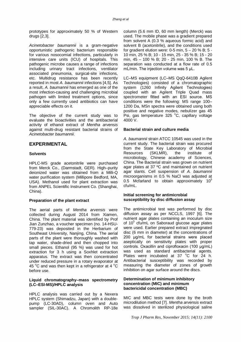
Tropical Journal of Pharmaceutical Research November 2015; 14 (11): 2099-2106 ISSN: 1596-5996 (print); 1596-9827 (electronic)
© Pharmacotherapy Group, Faculty of Pharmacy, University of Benin, Benin City, 300001 Nigeria. All rights reserved.
Available online at http://www.tjpr.org
http://dx.doi.org/10.4314/tjpr.v14i11.21 Original Research Article
Antibacterial Activity and Mode of Action of Mentha arvensis Ethanol Extract against Multidrug-Resistant Acinetobacter baumannii
Ling Zhang1, Shu-gen Xu1, Wei Liang2, Jun Mei1, Yue-ying Di1, Hui-hua Lan1, Yan Yang1, Wei-wei Wang1, Yuan-yuan Luo1 and Hou-zhao Wang1* 1Central Laboratory, The 174th Hospital of the Chinese People's Liberation Army, The Affiliated Chenggong Hospital of Xiamen University, 2Hospital Infection Management Department, The First Affiliated Hospital of Xiamen University, Xiamen 361003 China *For correspondence: Email: [email protected]; Tel/Fax: 0086-592-6335560 Received: 19 May 2015 Revised accepted: 6 October 2015
Abstract
Purpose: To evaluate the antibacterial effect of ethanol extract of Mentha arvensis against multi-drug resistant Acinetobacter baumannii using liquid chromatography–mass spectrometry (LC-ESI-MS). Methods: Disc diffusion and microdilution assays were used to evaluate the antibacterial effect of the extract by measuring the zone of inhibition, minimum inhibitory concentration (MIC) and and minimum bacteriocidal concentration (MBC) of the extract against the test bacteria. Scanning electron microscopy (SEM) was employed to evaluate the morphological changes induced by the extract in cellular membrane of the bacteria. Reactive oxygen species (ROS) generation and protein leakage from the bacterial cells induced by the extract were also evaluated. Results: The extract showed dose-dependent growth inhibitory effects against A. baumannii with MIC and MBC of 23.5 and 72.1 µg/mL, respectively. The extract also induced potent ROS generation and protein leakage in A. baumannii bacterial cells. SEM findings revealed that the extract induced potential cellular damage which increased with increasing extract concentration. Conclusion: The ethanol extract of Mentha arvensis is a potent antibacterial agent against A. baumannii and acts by inducing lethal cellular damage to the bacterium. Keywords: Mentha arvensis, Acinetobacter baumannii, Reactive oxygen species, Antibacterial activity, Cellular membrane damage
Tropical Journal of Pharmaceutical Research is indexed by Science Citation Index (SciSearch), Scopus, International Pharmaceutical Abstract, Chemical Abstracts, Embase, Index Copernicus, EBSCO, African Index Medicus, JournalSeek, Journal Citation Reports/Science Edition, Directory of Open Access Journals (DOAJ), African Journal Online, Bioline International, Open-J-Gate and Pharmacy Abstracts
INTRODUCTION Infectious diseases caused by bacterial and fungal infections are the key causes of mortality in tropical and subtropical countries. It is now well established that due to the indiscriminate uses of various antibiotic, predominantly synthetic drugs, the pathogenic microbes have acquired multidrug resistance which have created severe health issues mainly in developing countries. Most of the time, this multi-
drug resistance could lead to serious epidemic since, no drug can have any visible effect on the pathogenic microbes [1]. The less accessibility and high price of new generation antibiotics demands looking for the substances from alternative medicines with proved antimicrobial potential. A huge number of medicinal plants have been reported to show antimicrobial activity especially against drug resistant microbes. It is estimated that plants have provided the
Zhang et al
Trop J Pharm Res, November 2015; 14(11): 2100
prototypes for approximately 50 % of Western drugs [2,3]. Acinetobacter baumannii is a gram-negative opportunistic pathogenic bacterium responsible for various nosocomial infections, particularly in intensive care units (ICU) of hospitals. This pathogenic microbe causes a range of infections including urinary tract infections, ventilator associated pneumonia, surgical-site infections, etc. Multidrug resistance has been recently reported in most A. baumannii infections [4,5]. As a result, A. baumannii has emerged as one of the most infection-causing and challenging microbial pathogen with limited treatment options, since only a few currently used antibiotics can have appreciable effects on it. The objective of the current study was to evaluate the bioactivities and the antibacterial activity of ethanol extract of Mentha arvensis against multi-drug resistant bacterial strains of Acinetobacter baumannii. EXPERIMENTAL Solvents HPLC-MS grade acetonitrile were purchased from Merck Co., (Darmstadt, GER). High–purity deionized water was obtained from a Milli-Q water purification system (Millipore Bedford, MA, USA). Methanol used for plant extraction was from ANPEL Scientific Instrument Co. (Shanghai, China). Preparation of the plant extract The aerial parts of Mentha arvensis were collected during August 2014 from Xiamen, China. The plant material was identified by Prof Jian Zunzhao, a voucher specimen (no. 14-HSU-779-23) was deposited in the Herbarium of Southeast University, Nanjing, China. The aerial parts of the plant were thoroughly washed with tap water, shade-dried and then chopped into small pieces. Ethanol (95 %) was used for hot extraction for 3 h using a Soxhlet extraction apparatus. The extract was then concentrated under reduced pressure in a rotary evaporator at 45 oC and was then kept in a refrigerator at 4 oC before use. Liquid chromatography–mass spectrometry (LC–ESI-MS)/HPLC analysis HPLC analysis was carried out by a Nexera HPLC system (Shimadzu, Japan) with a double-pump (LC-30AD), column oven and Auto sampler (SIL-30AC). A Chromolith RP-18e
column (5.6 mm ID, 60 mm length) (Merck) was used. The mobile phase was a gradient prepared from solvent A (0.3 % aqueous formic acid) and solvent B (acetonitrile), and the conditions used for gradient elution were: 0-5 min, 5 – 20 % B; 5 - 10 min, 25 % B; 10 - 15 min, 25 - 35 % B; 15 - 20 min, 45 – 100 % B; 20 - 25 min, 100 % B. The separation was conducted at a flow rate of 0.5 mL/min. The injection volume was 5 μL. LC–MS equipment (LC–MS QqQ-6410B Agilent Technologies) consisted of a chromatographic system (1260 Infinity Agilent Technologies) coupled with an Agilent Triple Quad mass spectrometer fitted with an ESI source. MS conditions were the following: MS range 100–1200 Da, MSn spectra were obtained using both positive and negative modes, nebulizer gas 45 Psi, gas temperature 325 oC, capillary voltage 4000 V. Bacterial strain and culture media A. baumannii strain ATCC 10545 was used in the current study. The bacterial strain was procured from the State Key Laboratory of Microbial Resources (SKLMR), the institute of microbiology, Chinese academy of Sciences, China. The Bacterial strain was grown on nutrient agar plates at 37 ºC and maintained on nutrient agar slants. Cell suspension of A. baumannii microorganisms in 0.5 % NaCl was adjusted at 0.5 Mcfarland to obtain approximately 105 cfu/mL. Initial screening for antimicrobial susceptibility by disc diffusion assay The antimicrobial test was performed by disc diffusion assay as per NCCLS, 1997 [6]. The nutrient agar plates containing an inoculum size of 105 cfu/mL on Saboraud glucose agar plates were used. Earlier prepared extract impregnated disc (6 mm in diameter) at the concentrations of 200 μg/mL for bacterial strains were placed aseptically on sensitivity plates with proper controls. Oxacillin and ciprofloxacin (100 μg/mL) was used as standard antibacterial agents. Plates were incubated at 37 oC for 24 h. Antibacterial susceptibility was recorded by measuring the diameter of zones of growth inhibition on agar surface around the discs. Determination of minimum inhibitory concentration (MIC) and minimum bactericidal concentration (MBC) MIC and MBC tests were done by the broth microdilution method [7]. Mentha arvensis extract was dissolved in sterilized physiological saline
Zhang et al
Trop J Pharm Res, November 2015; 14(11): 2101
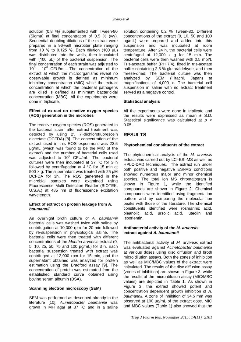
solution (0.8 %) supplemented with Tween-80 (Sigma) at final concentration of 0.5 % (v/v). Sequential doubling dilutions of the extract were prepared in a 96-well microtiter plate ranging from 10 % to 0.125 %. Each dilution (100 μL) was distributed into the wells, then inoculated with (100 μL) of the bacterial suspension. The final concentration of each strain was adjusted to 105 - 106 CFU/mL. The concentration of the extract at which the microorganisms reveal no observable growth is defined as minimum inhibitory concentration (MIC) while the extract concentration at which the bacterial pathogens are killed is defined as minimum bactericidal concentration (MBC). All the experiments were done in triplicate. Effect of extract on reactive oxygen species (ROS) generation in the microbes The reactive oxygen species (ROS) generated in the bacterial strain after extract treatment was detected by using 2’, 7’-dichlorofluorescein diacetate (DCFDA) [8]. The concentration of the extract used in this ROS experiment was 23.5 μg/mL (which was found to be the MIC of the extract) and the number of bacterial cells used was adjusted to 105 CFU/mL. The bacterial cultures were then incubated at 37 °C for 3 h followed by centrifugation at 4 °C for 15 min at 500 × g. The supernatant was treated with 25 μM DCFDA for 3h. The ROS generated in the microbial samples were examined using Fluorescence Multi Detection Reader (BIOTEK, U.S.A.) at 485 nm of fluorescence excitation wavelength. Effect of extract on protein leakage from A. baumannii An overnight broth culture of A. baumannii bacterial cells was washed twice with saline by centrifugation at 10,000 rpm for 20 min followed by re-suspension in physiological saline. The bacterial cells were then treated with different concentrations of the Mentha arvensis extract (0, 5, 10, 25, 50, 75 and 100 µg/mL) for 3 h. Each bacterial suspension treated with extract was centrifuged at 12,000 rpm for 15 min, and the supernatant obtained was analyzed for protein estimation using the Bradford assay [9]. The concentration of protein was estimated from the established standard curve obtained using bovine serum albumin (BSA). Scanning electron microscopy (SEM) SEM was performed as described already in the literature [10]. Acinetobacter baumannii was grown in MH agar at 37 ºC and in a saline
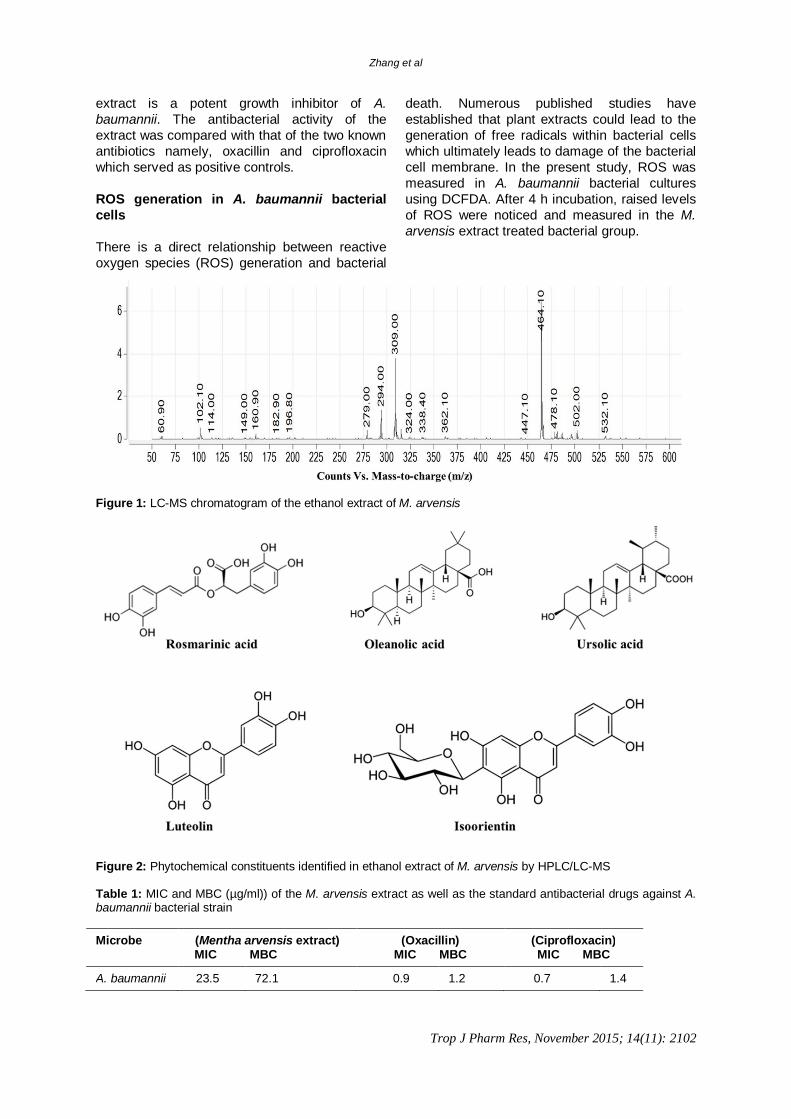
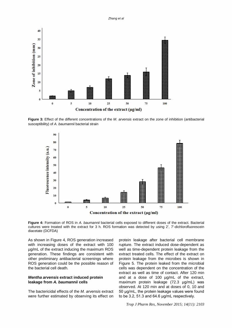
solution containing 0.2 % Tween-80. Different concentrations of the extract (0, 10, 50 and 100 µg/mL) were prepared and added into this suspension and was incubated at room temperature. After 24 h, the bacterial cells were centrifuged at 12,000 x g for 15 min. The bacterial cells were then washed with 0.5 mol/L Tris-acetate buffer (PH 7.4), fixed in tris-acetate buffer containing 2.5 % glutaraldehyde, and then freeze-dried. The bacterial culture was then analyzed by SEM (Hitachi, Japan) at magnifications of 4,000 x. The bacterial cell suspension in saline with no extract treatment served as a negative control. Statistical analysis All the experiments were done in triplicate and the results were expressed as mean ± S.D. Statistical significance was calculated at p < 0.05. RESULTS Phytochemical constituents of the extract The phytochemical analysis of the M. arvensis extract was carried out by LC–ESI-MS as well as HPLC-DAD techniques. The extract run under both positive and negative ESI-MS conditions showed numerous major and minor chemical species. The total ion MS chromatogram is shown in Figure 1, while the identified compounds are shown in Figure 2. Chemical compounds were identified using fragmentation pattern and by comparing the molecular ion peaks with those of the literature. The chemical constituents identified were rosmarinic acid, oleanolic acid, ursolic acid, luteolin and Isoorientin. Antibacterial activity of the M. arvensis extract against A. baumannii The antibacterial activity of M. arvensis extract was evaluated against Acinetobacter baumannii at various doses using disc diffusion and broth micro dilution assays. Both the zones of inhibition as well as MIC/MBC values of the extract were calculated. The results of the disc diffusion assay (zones of inhibition) are shown in Figure 3, while the results of the micro dilution assay (MIC/MBC values) are depicted in Table 1. As shown in Figure 3, the extract showed potent and concentration dependent growth inhibition of A. baumannii. A zone of inhibition of 34.5 mm was observed at 100 µg/mL of the extract dose. MIC and MBC values (Table 1) also showed that the
Zhang et al
Trop J Pharm Res, November 2015; 14(11): 2102
extract is a potent growth inhibitor of A. baumannii. The antibacterial activity of the extract was compared with that of the two known antibiotics namely, oxacillin and ciprofloxacin which served as positive controls. ROS generation in A. baumannii bacterial cells There is a direct relationship between reactive oxygen species (ROS) generation and bacterial
death. Numerous published studies have established that plant extracts could lead to the generation of free radicals within bacterial cells which ultimately leads to damage of the bacterial cell membrane. In the present study, ROS was measured in A. baumannii bacterial cultures using DCFDA. After 4 h incubation, raised levels of ROS were noticed and measured in the M. arvensis extract treated bacterial group.
Figure 1: LC-MS chromatogram of the ethanol extract of M. arvensis
Figure 2: Phytochemical constituents identified in ethanol extract of M. arvensis by HPLC/LC-MS Table 1: MIC and MBC (µg/ml)) of the M. arvensis extract as well as the standard antibacterial drugs against A. baumannii bacterial strain Microbe (Mentha arvensis extract)
MIC MBC (Oxacillin)
MIC MBC (Ciprofloxacin) MIC MBC
A. baumannii 23.5 72.1 0.9 1.2 0.7 1.4
Zhang et al
Trop J Pharm Res, November 2015; 14(11): 2103
Figure 3: Effect of the different concentrations of the M. arvensis extract on the zone of inhibition (antibacterial susceptibility) of A. baumannii bacterial strain
Figure 4: Formation of ROS in A. baumannii bacterial cells exposed to different doses of the extract. Bacterial cultures were treated with the extract for 3 h. ROS formation was detected by using 2’, 7’-dichlorofluorescein diacetate (DCFDA) As shown in Figure 4, ROS generation increased with increasing doses of the extract with 100 µg/mL of the extract inducing the maximum ROS generation. These findings are consistent with other preliminary antibacterial screenings where ROS generation could be the possible reason of the bacterial cell death. Mentha arvensis extract induced protein leakage from A. baumannii cells The bactericidal effects of the M. arvensis extract were further estimated by observing its effect on
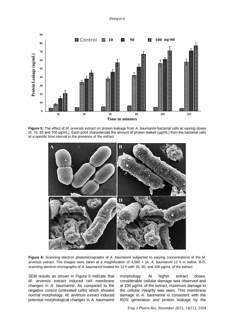
protein leakage after bacterial cell membrane rupture. The extract induced dose-dependent as well as time-dependent protein leakage from the extract treated cells. The effect of the extract on protein leakage from the microbes is shown in Figure 5. The protein leaked from the microbial cells was dependent on the concentration of the extract as well as time of contact. After 120 min and at a dose of 100 µg/mL of the extract, maximum protein leakage (72.3 μg/mL) was observed. At 120 min and at doses of 0, 10 and 50 μg/mL, the protein leakage values were found to be 3.2, 51.3 and 64.6 μg/mL respectively.
Zhang et al
Trop J Pharm Res, November 2015; 14(11): 2104
Figure 5: The effect of M. arvensis extract on protein leakage from A. baumannii bacterial cells at varying doses (0, 10, 50 and 100 µg/mL). Each point characterizes the amount of protein leaked (μg/mL) from the bacterial cells at a specific time interval in the presence of the extract
Figure 6: Scanning electron photomicrographs of A. baumannii subjected to varying concentrations of the M. arvensis extract. The images were taken at a magnification of 4,000 × (A, A. baumannii 12 h in saline; B-D. scanning electron micrographs of A. baumannii treated for 12 h with 10, 50, and 100 µg/mL of the extract SEM results as shown in Figure 6 indicate that M. arvensis extract induced cell membrane changes in A. baumannii. As compared to the negative control (untreated cells) which showed normal morphology, M. arvensis extract induced potential morphological changes in A. baumannii
morphology. At higher extract doses, considerable cellular damage was observed and at 100 µg/mL of the extract, maximum damage to the cellular integrity was seen. This membrane damage in A. baumannii is consistent with the ROS generation and protein leakage by the
Zhang et al
Trop J Pharm Res, November 2015; 14(11): 2105
extract. ROS generation can eventually lead to membrane damage which was demonstrated by SEM in this study. DISCUSSION There has been an enormous increase in the multidrug resistant strains of clinically relevant bacterial pathogens which is posing a great challenge to the scientists. This increase in multidrug resistance is believed to arise due to the indiscriminate use of antibiotics. Further, the non-availability coupled with rising cost of new generation antibiotics have led to an exponential increase in the number of deaths occurring due to infectious diseases. As such, there is a pressing need for novel, cheap and effective anti-infective drugs. This has led to the search for effective antimicrobial agents from plants, with the purpose of discovering potentially useful antimicrobial compounds that can serve as source and template for the synthesis of new antimicrobial drugs [11,12]. Plants have been known to synthesize active secondary metabolites such as phenolic compound that is found in essential oils with well-known potent anti-microbial activities, with applications in some pharmaceuticals, alternative medicines and natural therapies [13-15]. M. arvensis is one of the members of Lamiaceae which is commonly called Corn mint and Japanese mint, an essential oil bearing crop cultivated for natural menthol, extensively used in pharmaceutical, cosmetic and flavoring industries. Mints have been used in all continents of the world. The leaves of M. arvensis L, the common edible aromatic herb has been known to possess various pharmacological properties including antimicrobial properties [16]. In the current study, we observed potent antibacterial properties of the ethanolic extract of M. arvensis against A. baumannii bacterial strains. The antibacterial effect of the extract from our data could be due to induction of reactive oxygen species (ROS) generation, protein leakage and membrane damage caused by the extract. Phytochemical analysis revealed that the extract constituents were various phenolic and terpenoid compounds including rosmarinic acid, oleanolic acid, ursolic acid, luteolin and Isoorientin. The antibacterial activity of the extract could possibly be due to these phenolic and terpenoid compounds. CONCLUSION Mentha arvensis extract exhibits significant antibacterial activity against drug resistant A. baumannii bacterial strains by causing protein
leakage, inducing membrane damage and promoting ROS generation. ACKNOWLEDGEMENT This work was supported by grants from the Science and Technology Innovation Fund of Xiamen City (no. 3502Z20134024) and the Medical Science and Technique Fund for Youth Development of the PLA (no. 13QNP047) and the Medical Science and Technology Innovation Fund of Nanjing Military Region of PLA (no. 2013ZD27) and Natural Science Foundation of Fujian Province of China (no. 2013D006) REFERENCES 1. Weisser R, Asscher AW, Winpenny J. In vitro reversal of
antibiotic resistance by DTA. Nature 1966; 219(5161): 1365-1366.
2. Balandrin MF, Klocke JA, Wutule ES, Bollinger WH. Natural plant chemicals: Sources of industrial and medicinal materials. Science 1985; 228(4704): 1154–1160.
3. Jones FA. Herbs-useful plants. Their role in history and today. Eur J Gastroenterology Hepatol 1996; 8(12): 1227–1231.
4. Clock SA, Cohen B, Behta M, Ross B, Larson EL. Contact precautions for multidrug-resistant organisms: Current recommendations and actual practice. Am J Infect Control 2010; 38(2): 105–111.
5. Maragakis LL, Perl TM. Acinetobacter baumannii: epidemiology, antimicrobial resistance, and treatment options. Clin Infect Dis 2008; 46(8): 1254–1263.
6. National Committee for Clinical Laboratory Standards. Performance standards for antimicrobial disk susceptibility tests. Approved standard M2-A6. National Committee for Clinical Laboratory Standards, Wayne, Pa. 1997.
7. Yu J, Lei J, Yu H, Cai X, Zou G. Chemical composition and antimicrobial activity of the essential oil of Scutellaria barbata. Phytochemistry 2004; 65(7): 881-884.
8. Kye IS, Jeon YS, No JK, Kim YJ, Lee KH, Shin KH, Kim J, Yokozawa T, Chung HY. Reactive oxygen scavenging activity of green tea polyphenols. J Korea Gerontol 1999; 9: 10–17.
9. Bradford MM. A rapid and sensitive method for quantitation of protein-dye binding. Ann Biochem 1976; 72: 248–254.
10. Agizzio AP, Da Cunha M, Carvalho AO, Oliveira MA, Ribeiro SFF, Gomes VM. The antifungal properties of a 2S albumin-homologous protein from passion fruit seeds involve plasma membrane permeabilization and ultrastructural alterations in yeast cells. Plant Sci 2006; 171(4): 515-522.
11. Pretorius JC, Magama S, Zietsman PC. Growth inhibition of plant pathogenic bacteria and fungi by extracts
Zhang et al
Trop J Pharm Res, November 2015; 14(11): 2106
from selected South African plant species. S Afri J Bot 2003; 20: 188-192.
12. Moreillion P, Que YA, Glauser MP. Staphylococcus aureus (Including Staphyloccal Toxic shock). In ‘Principles and Practice of Infectious diseases.’ (Ed.) Mandell GL, Bennett JE, Dolin R.6th ed. Published by Churchill livingstone Pennyslyvania 2005; 2: 2333- 2339.
13. Nascimento Grislene GF, Juliana Locatevi Paulo C. Freitas Giuliana L. Silva. Antibacterial activity of plant extracts and phytochemicals on antibiotics resistant bacteria. Brazilian J Microbiol 2000; 31(4): 247-256.
14. Wang M, Li J, Rangarajan M, Shao Y, Lavoie EJ, Huang Tc, Ho Ci. Anti-oxidative phenolic compounds from sage (Salvia officinalis). J Agric Food Chem 1998; 46: 4869-4873.
15. Rios JL, Recio Mc, Villar A. Screening methods for natural products with antimicrobial activity: A review of the Literature. J Ethaopharonacol 1998; 23(2-3): 177-49.
16. Srinivas P, Arun T. Antibacterial Activity and Phytochemical Screening of Mentha arvensis Linn. against Proteus mirabilis from Urinary Tract Infected Patients. Intern J Pharm Tech Res 2012; 4(4): 1735-1744.