Embed Size (px)
Citation preview
RSC Advances
PAPER
Ope
n A
cces
s A
rtic
le. P
ublis
hed
on 1
2 Fe
brua
ry 2
021.
Dow
nloa
ded
on 1
0/12
/202
1 9:
04:1
1 A
M.
Thi
s ar
ticle
is li
cens
ed u
nder
a C
reat
ive
Com
mon
s A
ttrib
utio
n-N
onC
omm
erci
al 3
.0 U
npor
ted
Lic
ence
.
View Article OnlineView Journal | View Issue
The metabolomi
aDepartment of Pharmacognosy, Faculty of P
Sciences and Arts (MSA), Giza, EgyptbMolecular Discovery Group, Computation
Rothamsted Research, AL5 2JQ, Harpenden,cDepartment of Pharmacognosy, Faculty of
EgyptdDepartment of Pharmacognosy, Faculty o
11795, EgypteDepartment of Pharmacognosy, Faculty of P
Minia, EgyptfDepartment of Pharmacognosy, Faculty of P
Egypt. E-mail: [email protected] of Microbiology and Immun
University, Giza, EgypthDepartment of Pharmaceutics and Industria
6 University, Giza, EgyptiDepartment of Pharmacognosy, Faculty of P
EgyptjDepartment of Pharmacognosy, Faculty of P
32897, Egypt
Cite this: RSC Adv., 2021, 11, 7318
Received 3rd November 2020Accepted 13th January 2021
DOI: 10.1039/d0ra09334c
rsc.li/rsc-advances
7318 | RSC Adv., 2021, 11, 7318–7330
c analysis of five Mentha species:cytotoxicity, anti-Helicobacter assessment, and thedevelopment of polymeric micelles for enhancingthe anti-Helicobacter activity
Riham O. Bakr, a Ahmed Tawfike, b Heba A. El-Gizawy,c Nashwa Tawfik,d
Usama Ramadan Abdelmohsen, *ef Miada F. Abdelwahab,f Walaa A. Alshareef, g
Sahar M. Fayez,h Shereen M. S. El-Mancy, h Ahlam M. El-Fishawy,i
Mostafa A. Abdelkawyi and Marwa A. A. Fayed j
Mentha species are medicinally used worldwide and remain attractive for research due to the diversity of
their phytoconstituents and large therapeutic indices for various ailments. This study used the
metabolomics examination of five Mentha species (M. suaveolens, M. sylvestris, M. piperita, M. longifolia,
and M. viridis) to justify their cytotoxicity and their anti-Helicobacter effects. The activities of species
were correlated with their phytochemical profiles by orthogonal partial least square discriminant analysis
(OPLS-DA). Tentatively characterized phytoconstituents using liquid chromatography high-resolution
electrospray ionization mass spectrometry (LC-HR-ESI-MS) included 49 compounds: 14 flavonoids, 10
caffeic acid esters, 7 phenolic acids, and other constituents. M. piperita showed the highest cytotoxicity
to HepG2 (human hepatoma), MCF-7 (human breast adenocarcinoma), and CACO2 (human colon
adenocarcinoma) cells using 3-(4,5-dimethylthiazol-2-yl)-2,5-diphenyltetrazolium bromide (MTT) assays.
OPLS-DA and dereplication studies predicted that the cytotoxic activity was related to benzyl
glucopyranoside-sulfate, a lignin glycoside. Furthermore, M. viridis was effective in suppressing the
growth of Helicobacter pylori at a concentration of 50 mg mL�1. OPLS-DA predicted that this activity
was related to a dihydroxytrimethoxyflavone. M. viridis extract was formulated with Pluronic® F127 to
develop polymeric micelles as a nanocarrier that enhanced the anti-Helicobacter activity of the extract
and provided minimum inhibitory concentrations and minimum bactericidal concentrations of 6.5 and
50 mg mL�1, respectively. This activity was also correlated to tentatively identified constituents, including
rosmarinic acid, catechins, carvone, and piperitone oxide.
harmacy, October University for Modern
al and Analytical Science Department,
UK
Pharmacy, October 6 University, Giza,
f Pharmacy, Helwan University, Cairo,
harmacy, Deraya University, 61111 New
harmacy, Minia University, 61519 Minia,
; Tel: +2-86-2347759
ology, Faculty of Pharmacy, October 6
l Pharmacy, Faculty of Pharmacy, October
harmacy, Cairo University, 11562 Cairo,
harmacy, University of Sadat City, Sadat
1. Introduction
The genus Mentha in the family Lamiaceae (Labiatae) is highlycomplex, including 61 species and hundreds of subspecies,varieties, and cultivars.1,2 Mint has been used worldwide sinceancient eras in its fresh or dried forms as it displays numerousbenecial properties.3,4 This wide and signicant bioactivity istypically correlated with the diversity of the secondary metab-olites of this genus.3,5 Chemical proling of Mentha speciesconrms the presence of volatile constituents and poly-phenolics, including avones, avonols, avanones, phenolicacids, and tannins.6–9 Hydroxycinnamic acid derivatives, chieyrosmarinic acid, are widely spread in the family Lamiaceae andpredominate other phenolics. These phytoconstituents showimportant biological and pharmacological activities, includingantioxidant, antiviral, antiobesity, and antibacterial effects.10–13
Many investigations of Mentha species are available; however,the interest in studying mint and its application in
© 2021 The Author(s). Published by the Royal Society of Chemistry
Paper RSC Advances
Ope
n A
cces
s A
rtic
le. P
ublis
hed
on 1
2 Fe
brua
ry 2
021.
Dow
nloa
ded
on 1
0/12
/202
1 9:
04:1
1 A
M.
Thi
s ar
ticle
is li
cens
ed u
nder
a C
reat
ive
Com
mon
s A
ttrib
utio
n-N
onC
omm
erci
al 3
.0 U
npor
ted
Lic
ence
.View Article Online
pharmaceutical formulations is continuous. All species in thisgenus display multiple bioactive constituents that show no sideeffects, even in doses ten times higher than their therapeuticdoses. Therefore, they are highly safe in therapy, and investi-gation of their possible activities is of high importance.
Plantmetabolomics is crucial for improving our understandingof metabolite levels, facilitating differentiation, quantitation, andcomparing the large numbers of secondary metabolites.14 Further,metabolomics contributes signicantly to the study of differencesbetween plant phenotypic and genotypic physiology and biologywhen combined with functional genomics.15
Cancer is considered to be a signicant global healthproblem, accounting for many thousands of deaths; therefore,exploring new drugs and chemotherapeutics to improve treatmentare of high priority with a main target to overcome their fatal sideeffects. Therefore, a current focus on the potential of naturalproducts as anticancer candidates and investigation of theiractivity is favored to minimize the side effects of chemicallyderived drugs and benet from their high therapeutic value.16,17
Moreover, peptic ulcers are considered a major global healthproblem, and one of their main causes is infection with Heli-cobacter pylori bacteria. This bacterium is a agellated, Gram-negative coccobacillus that affects 50% of the world's pop-ulation. Its prevalence is higher in developing countries than indeveloped countries.18 H. pylori cases are associated with severepathologies, including gastric cancer and peptic ulcer.19
Different treatment regimens are proposed to eradicate H.pylori, including the use of two or more antibiotics coupled witha proton pump inhibitor.20 Antimicrobial resistance is a leadingcause of standard triple therapy failure inmost countries, whichhighlights the need for alternative compounds with provenantimicrobial activity. Natural products are excellent sourcesthat may allow the discovery of synthesized active constituentsor derivatized secondary metabolites to provide new antimi-crobials that successfully overcome bacterial resistance.
However, the conventional administration of natural drugsis hindered due to their low solubility, permeability, orbioavailability. Finding suitable pharmaceutical dosage formsto increase their absorption and their potency is in greatdemand. Nanonization may be an appropriate strategy toaddress these problems and improve the physicochemical prop-erties of natural drug preparations.21 Polymeric micelles, as core/shell nanoscale dispersion systems formed by self-assembly ofvarious amphiphilic block copolymers, may contribute as a dosageform to help solve this problem. These micelles are characterizedby small sizes (�10–200 nm), biodegradability for easy elimina-tion, ability to encapsulate poorly water-soluble drugs, high drugloading capacity and reproducibility, and low cost.22–24 Nano-particles enhance formulation parameters and improve the ther-apeutic potential of hydrophobic drugs.25–27
Pluronic® F127 is a biodegradable and biocompatible poly-mer that is used successfully to develop nanoscale polymericmicelles. The polymer promotes the drug encapsulation,permeation and therapeutic potential of various drugs28–31
The conventional use of Mentha species for the treatment ofgastrointestinal symptoms and its inclusion in GermanCommission E lists for treatment of gastrointestinal
© 2021 The Author(s). Published by the Royal Society of Chemistry
disorders,32 in most cases without identication of phenotypicplasticity and genetic variability because of extensive hybrid-ization, triggered our interest in comparing the chemicalproles of veMentha species cultivated in Egypt:M. suaveolens,M. sylvestris, Mentha � piperita, M. longifolia, and M. viridis.Chemical components were assessed using liquid chromatog-raphy with high-resolution electrospray ionization mass spec-trometry (LC-HR-ESI-MS), followed by principal componentanalysis (PCA). The objectives were to assess the cytotoxic andanti-Helicobacter activities of different extracts and comparethem by orthogonal partial least squares discriminant analysis(OPLS-DA) as well as to develop Pluronic® F127 polymericmicelles as a nanocarrier for the most effective Mentha extract.Further, in the study, we characterized the prepared systems toinvestigate their improved antimicrobial activity against H.pylori as an adjuvant treatment for peptic ulcer.
2. Material and experimentalmethods2.1. Plant material and extraction procedure
The medicinal plants studied were Mentha suaveolens, M. syl-vestris and M. longifolia (L.) Huds sub-spp. schimperi Briq. Thevoucher samples were kindly authenticated by Dr Gemma L.C.Bramley, Curator of the Lamiaceae collection HerbariumDepartment, Library, Art & Archives Directorate, Royal BotanicGardens, Kew, Richmond Surrey, U.K., Voucher No. M-20/313,20-01-2020 II, and 25-6-2015, respectively. M. piperita and M.viridis were kindly authenticated by Dr Mohamed El-Gebaly,Senior Botanist at El-Orman Botanic Garden, Egypt (VoucherNo. 20-01-2020-I, 20-01-2020 V respectively). The leaves of eachplant under study were collected from the Experimental Station ofMedicinal and Aromatic Plants, Department of Pharmacognosy,Faculty of Pharmacy, Cairo University, Cairo, Egypt, inMarch 2019.Voucher specimens were deposited in the Herbarium of theDepartment of Pharmacognosy, Faculty of Pharmacy, CairoUniversity, Cairo, Egypt. The fresh leaves were air-dried at roomtemperature, then ground into a ne powder. 100 g of each samplewas extracted with 70% ethanol (3� 200mL) for 30min in a waterbath at 50 �C.33 The samples were then cooled and ltered, and thesolvent was evaporated at 45 �C using a rotary evaporator (Buchi R-300, USA) and stored at 4 �C until further use.
2.2. Anti-proliferative assay
2.2.1. Cytotoxic activity. The cytotoxicity of the 70% etha-nolic extracts was evaluated in cell lines using the 3-(4,5-dimethylthiazol-2-yl)-2,5-diphenyltetrazolium bromide (MTT)assay.34 HepG2 (human hepatoma), MCF-7 (human breastadenocarcinoma), and CACO2 (human colon adenocarcinoma)cells were obtained from the American Type Culture Collection(ATCC, Rockville, MD, USA; HPACC, Salisbury, UK) and weremaintained in RPMI medium (Merck, Darmstadt, Germany) sup-plemented with 10% fetal bovine serum (FBS). Cancer cells werecultured at 37 �C, 5% (v/v) CO2 in RPMI1640 medium supple-mented with 5% (v/v) fetal bovine serum (FBS), 1% (w/v) L-gluta-mine, 1% sodium pyruvate and 0.4% (w/v) antibiotics (50 U mL�1
RSC Adv., 2021, 11, 7318–7330 | 7319
RSC Advances Paper
Ope
n A
cces
s A
rtic
le. P
ublis
hed
on 1
2 Fe
brua
ry 2
021.
Dow
nloa
ded
on 1
0/12
/202
1 9:
04:1
1 A
M.
Thi
s ar
ticle
is li
cens
ed u
nder
a C
reat
ive
Com
mon
s A
ttrib
utio
n-N
onC
omm
erci
al 3
.0 U
npor
ted
Lic
ence
.View Article Online
penicillin, 50 mg mL�1 streptomycin). Cells were routinely sub-cultured twice per week. All chemicals and reagents werepurchased from Sigma Aldrich (Darmstadt, Germany). Tonormalize the cell viability values, each plate included a triplicateof cells treated with the compound carrier DMSO to dene 100%viable cells as well as a triplicate of cells incubated with a cytotoxicmixture (200 ng mL�1 Tumor Necrosis Factor (TNF), 200 ng mL�1
CD95L (Fas ligand), 200 ng mL�1 TNF-related apoptosis-inducingligand (TRAIL), 25 g mL�1 cycloheximide (CHX), 1% (w/v) sodiumazide) to dene maximal cell death and, thus, 0% viability. Allother viability values were normalized according to the averages ofthese triplicates and analyzed by Graph Pad Prism 5 soware (LaJolla, CA, USA).
2.3. Screening of the anti-Helicobacter activity of Menthaspecies
2.3.1. Strain of Helicobacter pylori and culturing. Heli-cobacter pylori ATCC® 43504™ was kindly obtained from theMicrobiology Department (Faculty of Medicine) of October 6University Hospital, Giza, Egypt, on chocolate agar plates. Purecolonies were sub-cultured directly onto supplementedColumbia blood agar with DENT aer a positive urease test;then, Gram staining of a smear prepared from the pure colonieswas performed. The cultures were incubated for a minimum of3 days and a maximum of 7 days at 37 �C under microaerophilicconditions (CO2 5%). The H. pylori strain was identied bycolony morphology, observing the characteristic spiral shape inthe Gram staining (Gram-negative), and positive oxidase andcatalase tests. The isolates were stored at �80 �C for 5 to 10months in Columbia broth medium containing 15% glycerol.
2.3.2. Disc diffusion test. Kirby-Bauer disc diffusion wasused to analyze the susceptibility of H. pylori ATCC® 43504™ todifferent Mentha species extracts.35,36 Each extract residue (50mg) was dissolved in one mL of dimethyl sulfoxide (DMSO),sterilized through a Millipore lter (0.22 mm) and then loadedon a sterile lter paper disc (6 mm in diameter). Ten mL of sup-plemented Columbia blood agar (CBA) medium was poured intosterile Petri dishes (as a basal layer), followed by surface inocula-tion ofH. pylori suspensions (100mL of brothmedium/3mL of 108
CFU) to attain a 0.5 MacFarland turbidity standard (0.5� 108 CFUmL�1). Sterile lter paper discs loadedwithMentha species extracts(50 mg mL�1) were placed on top of the supplemented Columbiablood agar plates. Discs (6 mm diameter, Hi-Media, India) with 10mg per disc of metronidazole and DMSO were used as a positiveand a negative control against H. pylori, respectively. The inhibi-tion zones were measured by a Vernier caliper, recorded andconsidered as an indication of antibacterial activity.37 The anti-microbial activity was determined in triplicate.
2.4. LC-MS analysis
An Agilent 1100 HPLC Series system (Agilent, USA) equipped witha degasser, binary gradient pump, column thermostat, autosam-pler, and U.V. detector was used. The HPLC system was coupledwith an Agilent 1100 mass spectrometer (LC-MS analysis IonTrap V.L.). For the separation, a reverse-phase analytical columnwas employed (Zorbax SB-C18, 100� 3.0 mm i.d., 3.5 mmparticle);
7320 | RSC Adv., 2021, 11, 7318–7330
the work temperature was 48 �C. The detection of the compoundswas performed in both U.V. and M.S. mode. The U.V. detector wasset at 330 nm until 17.5 min; then, it was set at 370 nm. The MSsystem was operated using an electrospray ion source in negativeand positive modes. The chromatographic data were processedusingChemStation andDataAnalysis soware fromAgilent, USA. Themobile phase was a binary gradient: methanol and acetic acid 0.1%(v/v). The elution started with a linear gradient, beginning with 5%methanol and ending at 42% methanol, for 35 minutes; then, 42%methanol was used for the next 3 minutes. The ow rate was 1mL min�1 and the injection volume was 5 mL. Compounds werequalitatively identied based on spectral matching of the signals andspectra of each polyphenol with the available library and literature.
2.5. Preparation of the most effective Mentha extractpolymeric micelles
Preliminary screening using different concentrations of themost effective Mentha extract and polymer ratios were per-formed. Extract polymeric micelles with a 100 mg mL�1
concentration and an extract-to-polymer ratio of 1 : 3 werenally developed. The formulae were prepared using the thin-lm hydration method. Weights of 500 mg extract and 1.5 gPluronic® F127 were completely dissolved separately in 5 mLmethanol. Then, the two solutions were combined, and themixture was stirred for a further 15 min. The solvent was evapo-rated to form a dried thin lm of the extract and polymer matrixusing either of the following methods. In the rst method, themixture was transferred to a porcelain dish and then was placed ina domestic microwave (Microwave, LG, 2450 MHz, China) oper-ating at a power of 1000W for a short pulse of 1min for 10 cycles toachieve complete drying. For the second method, the mixture wastransferred to a round ask connected to a rotary evaporator(Rotavapor, Heidolph VV 2000, Burladingen, Germany) rotated ata rotation speed of 120 rpm and 50 �C for one h under vacuum.The obtained dry lm for each case was hydrated with 5mL doubledistilled water and stirred for 30 min to obtain a homogeneousmicellar dispersion (100 mg mL�1). The prepared formulae werestored in tightly closed containers. They were codedMvP-M for theformula prepared using a microwave and MvP-R for the formuladesigned using the rotary evaporator.
2.6. Characterization of the prepared Mentha polymericmicelles
2.6.1. Visual inspection and determination of particle size(PS), polydispersity index (PDI) and zeta potential (ZP). Theprepared formulae were examined visually for appearance andhomogeneity.
The PS and PDI of 100-fold diluted formulae were deter-mined at 25 �C by dynamic light scattering (DLS) using a Zeta-sizer (Nano Z.S., Malvern, U.K.). Also, the ZP was measured toinvestigate the physical stability. Measurements were per-formed in triplicate at 25 �C.
2.6.2. Screening of anti-Helicobacter activity. The anti-Helicobacter activity of the prepared formula was determinedusing the disc diffusion test described in Section 2.4.2 using
© 2021 The Author(s). Published by the Royal Society of Chemistry
Table 1 Cytotoxic activities of the crude extracts (IC50 mg mL�1)a
Tested sample (70%ethanolic extracts) HepG-2 (liver) MCF-7 (breast) CACO2 (colon)
M. suaveolens*** 52.19 � 5.6 55.07 � 4.9 61.06 � 4.8M. sylvestris*** 51.30 � 3.9 52.81 � 5.5 58.11 � 6.3M. piperita* 11.52 � 4.2 7.97 � 5.6 13.72 � 3.3M. longifolia*** 54.23 � 2.3 56.54 � 7.2 61.25 � 5.9M. viridis*** 58.61 � 4.3 51.74 � 6.4 62.36 � 4.3Doxorubicin 5.72 � 0.7 5.17 � 0.6 5.81 � 0.5
a The results were obtained from three independent replicateexperiments and expressed as means � standard deviation. Thestatistical signicance of the results was tested using one-way Analysisof Variance (ANOVA) and Tukey–Kramer multiple Comparisons Testbetween Doxorunicin and the different plant extracts. The p valuesignicance was represented as an asterisk (*) for p < 0.05 and threeasterisks (***) for p < 0.001.
Paper RSC Advances
Ope
n A
cces
s A
rtic
le. P
ublis
hed
on 1
2 Fe
brua
ry 2
021.
Dow
nloa
ded
on 1
0/12
/202
1 9:
04:1
1 A
M.
Thi
s ar
ticle
is li
cens
ed u
nder
a C
reat
ive
Com
mon
s A
ttrib
utio
n-N
onC
omm
erci
al 3
.0 U
npor
ted
Lic
ence
.View Article Online
a concentration of 100 mg mL�1 for the most potent Menthaextract and its formulae.
2.6.3. Determination of minimum inhibitory concentra-tions (MICs) and minimum bactericidal concentrations (MBCs)of the most effective Mentha extract and polymeric micelles.The most effectiveMentha extracts (dissolved in DMSO), MvP-Mand MvP-R, were manipulated to determine their MICs andMBCs. Broth microdilution was performed with 96-well U-shaped bottom microtiter plates following Clinical and Labo-ratory Standards Institute (CLSI) guidelines (2017).38,39
The tested samples were used in an initial concentration of100 mg mL�1 and then serially diluted two-fold to obtainconcentrations in the range of 0.098–50 mg mL�1. The inoc-ulum of H. pylori was prepared in Columbia broth, with theOD600 nm adjusted to 0.5. Equal volumes (50 mL) of broth anddifferent concentrations of each sample were incubated at 37 �Cfor 72 h under microaerophilic conditions. Bacteria-free brothand 50 mL DMSO were used as a negative control. The bacterialdensity was read at 620 nm using an ELISA reader. The lowestconcentration at which no bacterial density was determined wasthe MIC value. The experiment was performed in triplicate.40
For MBC determination, 10 mL of mixtures containingMenthaextract and H. pylori as well as MvP-M and MvP-R (differentconcentrations) were inoculated on respective agar plates (asdetailed above), and the bacterial counts were determined andexpressed as CFU mL�1 aer incubation at 37 �C for 72 h. TheMBC was determined as the lowest concentration at which theincubated H. pylori strain showed no detectable colonies on therespective agar plates. The experiment was performed threeindependent times.
2.6.4. Surface morphology studies by transmission elec-tronic microscopy (TEM). Based on the previous characteriza-tion results, the formula with the best criteria was selected. Themorphology of the selected formula was investigated by TEM(JEM-2100, Jeol® Tokyo, Japan). TEM offers valuable informa-tion on the inner structure of a sample, such as its crystalstructure, morphology, and stress state information.41,42
A drop of a diluted aqueous dispersion of the formula wasdropped onto copper-coated carbon grids, dried, and stainedwith 2% w/v phosphotungstic acid solution; then, the excesswas absorbed by lter paper.
2.7. Statistical analysis
Statistical analysis of the data was performed using one-wayANOVA followed by Turkey multiple range tests. All the dataare presented as themean� SE of 3 determinations. The level ofsignicance was set at p < 0.05. Statistical calculations wereperformed using SPSS version 15.0 soware. The p valuesignicance is represented as an asterisk (*) for p < 0.05, twoasterisks (**) for p < 0.01 and three asterisks (***) for p < 0.001.
3. Results and discussion3.1. Cytotoxic activity
The ve extracts were tested for their cytotoxic activity againstthree human cancer cell lines (Table 1). Only M. piperita was
© 2021 The Author(s). Published by the Royal Society of Chemistry
able to induce cytotoxicity towards the tested cell lines, with IC50
values ranging from 7–13 mg mL�1. Previous studies showed thepowerful anticancer effect against breast cell carcinoma of M.piperita leaf extract ethyl acetate, with cellular apoptosis, inhi-bition of proliferation, perturbation of oxidative balance, andupregulation of Bax gene.43 Additionally, the reported phenolicswere demonstrated to scavenge cOH and superoxide free radi-cals and to inhibit lipid peroxidation.44,45
3.2. Anti-Helicobacter activity of Mentha species
Different Mentha species were investigated for their effectsagainst the H. pylori strain using the disc diffusion method. Theresults are recorded in Table 2. M. viridis, M. piperita, and M.suaveolens caused suppression of bacterial growth at a concen-tration of 50 mg mL�1, with inhibition zones of 14 mm, 12 mm,and 10mm, respectively.M. viridis was the most effective extractin retarding microbial growth of all the tested isolates. Incontrast, extracts of M. sylvestris and M. longifolia were noteffective (no zone of inhibition). Essential oils of M. viridis andM. piperita were reported for their anti-Helicobacter activities.46
3.3. Tentative identication of the major phytoconstituentsin Mentha species
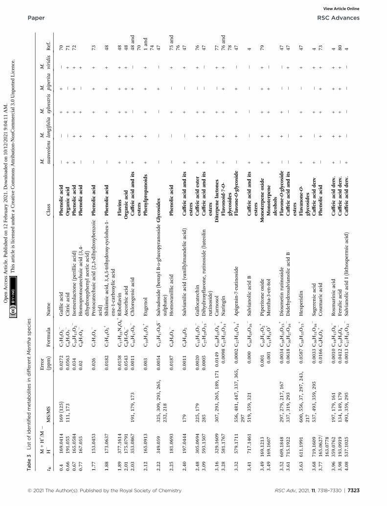
Metabolites were identied in the veMentha species using LC/MS in both positive and negative modes, where the peaks wereidentied using an in-house library. Forty-seven compoundsbelonging to different classes have been tentatively identied,including avonoids (13), hydroxycinnamic esters (12), organicacids, phenolic acids, ascorbic acid, and riboavin (22). Theidentities, retention times, and observed molecular and frag-ment ions of the tentatively identied components are pre-sented in Table 3. The caffeic acid esters identied weresalvianolic acid, chlorogenic acid, salvianolic acid B, sagerenicacid, rosmarinic acid, salvianolic acid A, I, and didehy-drosalvianolic acid, which are widespread in all the studiedplants. Other metabolites which were common in all speciesinclude isomenlactone (perillic acid), naringin, ladanin (dihy-droxy, dimethoxyavone), vanillyl alcohol, 11-cisretinoic acid,
RSC Adv., 2021, 11, 7318–7330 | 7321
Table 2 Antibacterial activity tests of Mentha species (50 mg mL�1) against Helicobacter pyloria,b
Plant extracts
Helicobacterpylori ATCC® 43504™
Zone of inhibition (mm)
M. suaveolens (50 mg mL�1)** 10 � 2.0M. sylvestris (50 mg mL�1) —M. piperita (50 mg mL�1)** 12 � 1.5M. longifolia (50 mg mL�1) —M. viridis (50 mg mL�1)** 14 � 1.5Positive control (10 mg per disc of metronidazole) 17 � 0
a n ¼ 3, mean � SD. b The results were obtained from three independent replicate experiments and expressed as means � standard deviation. Thestatistical signicance of the results was tested using one-way Analysis of Variance (ANOVA) and Tukey–Kramer multiple Comparisons Test. The pvalue signicance was represented two asterisks (**) for p < 0.01.
RSC Advances Paper
Ope
n A
cces
s A
rtic
le. P
ublis
hed
on 1
2 Fe
brua
ry 2
021.
Dow
nloa
ded
on 1
0/12
/202
1 9:
04:1
1 A
M.
Thi
s ar
ticle
is li
cens
ed u
nder
a C
reat
ive
Com
mon
s A
ttrib
utio
n-N
onC
omm
erci
al 3
.0 U
npor
ted
Lic
ence
.View Article Online
carvone, eugenol, riboavin, antheraxanthin, 9-cis retinolacetate, and piperitone oxide. Those results were in agreementwith those reported for other Mentha species.47–49 M. viridis,which showed the best anti-Helicobacter activity, was tentativelyidentied to contain a variety of active constituents, includingprotocatechuic, shikimic, homovanillic, ferulic, salvianolic,sagerinic, and rosmarinic acids, in addition to the presence ofavonoids, such as gallocatechin, hesperidin, epicatechin, andnaringin, and monoterpenes, such as carvone and piperitoneoxide. Catechins have been reported to eradicate H. pylori when incombination with sialic acid.50 Rosmarinic acid was also proved tohave an excellent drug-like prole in an in silico screening of shi-kimate kinase, showing a possible synergistic effect with manyantibiotics.51 Additionally, monoterpenes have been proved to havegastroprotective and possible anti-Helicobacter effects.52 Mean-while, M. piperita, which showed the best cytotoxic activity, wasdistinguished by its phenolic acid as well as its avonoid contentsin addition to the presence of salvadoside, a lignan glycosidepreviously identied in Salvadora persica;53 these epoxy carotenoidsrepresent lipophilic-active compounds of xanthophylls in foodsand play a great role in cancer prevention,54,55 including viola-xanthin, previously identied in Mentha species,48 and the mainconstituent in Dunaliella tertiolecta, which demonstrated a power-ful antiproliferative effect against breast cell carcinoma56 in addi-tion to antheraxanthin.
3.4. Metabolomics analysis
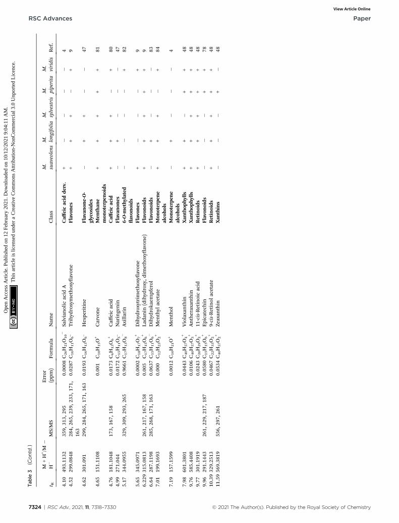
To widen the coverage of the metabolome of Mentha, the posi-tive and negative mass spectral data were combined into onedata matrix. The data were processed viaMZmine2 according toa method developed previously in our lab.57 The processed datawere then moved into an in-house database Excel le witha built-in Dictionary of Natural Products Database (DNP) fordereplication purposes.58 Principle component analysis (PCA)was performed to test the similarity and/or the variation in thechemical proles among the tested species. PCA is an unsu-pervised multivariate data analysis that aims to reduce thedimensionality of the data to reveal clusters, groups, and/oroutliers among the observations.59 The PCA score plot(Fig. 1A) showed respective total variances of 81% and 10% for
7322 | RSC Adv., 2021, 11, 7318–7330
PC1 and PC2 and demonstrated the clustering of M. suaveolensand M. sylvestris extracts (UA-62 and UA-64, respectively). Thisresult indicated the similar chemical ngerprints of thoseextracts. Moreover, PCA showed a dispersal ofM. viridis (UA-66),M. piperita (UA-65) and M. longifolia (UA-63) extracts, whichdemonstrated their unique chemical proles. The PCA loadingplot (Fig. 1B) highlighted the metabolites that contributed tothis variation, as M. viridis (UA-66) extract was characterized bymolecules at m/z (retention time in minutes) 345.097 [M + H]+
(tR 5.65) and 305.069 [M�H]� (tR 2.48). The former correspondsto a dihydroxytrimethoxyavone, equivalent to a molecularformula of C18H17O7
+, while the latter is equivalent to themolecular formula of C15H13O7
�, which corresponds to gallo-catechin in the DNP database. Moreover,M. piperita extract (UA-65) was distinguished by the presence of discriminatorymetabolites at m/z [M � H]� 349.059 (tR 2.22) and 408.042 (tR1.75). They are respectively equivalent to C13H18O9S, whichcorresponds to benzyl glucopyranoside-sulfate or salvadoside.Sulphated glycosides have been previously reported in Mentha� piperita.47 To investigate which compounds are mediating thebiological activity of M. piperita extract, a supervised multivar-iate data analysis was carried out. Orthogonal partial leastsquares discriminant analysis (OPLS-DA) was applied to high-light the molecules that are highly correlated with the biologicalactivity. The OPLS-DA score plot (Fig. 2A) demonstratedcomplete discrimination between the active extract M. piperita(UA-65) and the other inactive Mentha species. The model'smeasures for the goodness of t R2 ¼ 0.91 and prediction Q2 ¼0.67 indicate a strong t model with a high predictive power.The coefficient of variation plot (Fig. 2B) is a very useful tool tocompare the variable magnitude against its reliability, whereregression coefficients related to center-scaled X-variables aredisplayed. This scaling of the data enables comparison of thecoefficients. Thus, these coefficients express how strongly Y(biological activity) is correlated to the systematic part of each ofthe X-variables. Themolecules highly correlated withM. piperitaactivity were checked, and only those with high coefficients ofvariation, with high VIP (variable importance), and whose 95%condence level limits did not cross zero were chosen. Thedescriptive statistics of the model, including the coefficient of
© 2021 The Author(s). Published by the Royal Society of Chemistry
Tab
le3
List
ofidentifiedmetabolitesin
differentMenthasp
ecies
t RM
+H
+/M
�H
�MS/MS
Error
(ppm
)Fo
rmula
Nam
eClass
M.
suaveolens
M.
longifolia
M.
sylvestris
M.
piperita
M.
viridis
Ref.
0.4
169.04
1416
9(125
)0.02
72C7H
5O5�
Gallicacid
Phen
olic
acid
��
++
�70
0.66
191.05
511
1,17
30.05
63C6H
7O7�
Citricacid
Organ
icacid
+�
+�
+71
0.67
165.05
840.03
4C10H
13O2�
Isom
enlacton
e(perillicacid)
Phen
olic
acid
++
++
+72
0.77
167.05
50.02
C8H
7O4�
Hom
oprotocatech
uicacid
(3,4-
dihyd
roxyph
enyl
acetic
acid)
Phen
olic
acid
�+
+�
+
1.77
153.04
530.02
6C7H
5O4�
Protocatechuicacid
(2,3-dihyd
roxybe
nzoic
acid)
Phen
olic
acid
�+
++
+73
1.88
173.06
370.01
82C7H
11O
5+
Shikim
icacid,3
,4,5-trihyd
roxy-cyclohex-1-
ene-1-carboxylic
acid
Phen
olic
acid
�+
++
+48
1.89
377.16
140.01
58C17H
21N4O6+Ribo
avin
Flavins
++
++
+48
2.01
175.07
910.05
43C6H
7O6�
Ascorbicacid
Organ
icacid
�+
++
+48
2.03
353.08
6719
1,17
9,17
30.00
11C16H
17O9�
Chlorogenic
acid
Caff
eicacid
andits
esters
++
�+
�48
and
702.12
165.09
130.00
3C10H
13O2+
Eug
enol
Phen
ylprop
anoids
++
++
+1an
d74
2.22
349.05
932
5,30
9,29
3,26
5,23
2,21
80.00
54C13H
17O9S�
Salvad
oside(ben
zylB
-D-glucopy
ranoside
sulphate)
Glycosides
��
�+
�47
2.25
181.06
930.01
87C9H
9O4�
Hom
ovan
illicacid
Phen
olic
acid
��
+�
+75
and
762.40
197.04
4417
90.00
11C9H
10O
5Sa
lvianilic
acid
(van
illylm
ande
licacid)
Caff
eicacid
andits
esters
+�
+�
+47
2.48
305.06
9422
5,17
90.00
20C15H
13O7�
Gallocatech
inCaff
eicacid
ester
++
+�
+76
3.09
593.15
0728
50.00
05C27H
29O15�
Dihyd
roxyavon
e,rutinoside(luteo
lin
rutinoside)
Caff
eicacid
andits
esters
++
+�
�47
3.16
329.16
0930
7,29
3,26
5,18
9,17
10.01
49C20H
25O4�
Carnosol
Diterpe
nelacton
es�
++
�+
773.28
581.17
670.00
98C27H
33O14+
Naringin
Flavon
oid-7-O-
glycosides
++
++
+76
and
783.32
579.17
1155
6,48
1,44
7,33
7,36
5,29
70.00
02C27H
31O14+
Apigenin-7-rutinoside
Flavon
e-O-glycoside
�+
++
�47
3.41
717.14
6151
9,35
9,32
10.00
0C36H
29O16�
Salvianolic
acid
BCaff
eicacid
andits
esters
+�
��
�4
3.49
169.12
130.00
1C10H
17O2+
Pipe
ritoneoxide
Mon
oterpe
neoxide
++
++
+79
3.49
169.16
070.00
1C11H
21O+
Men
tha-3-en
-8ol
Mon
oterpe
ne
alcohols
++
++
�
3.52
609.18
4829
7,27
9,21
7,16
70.00
34C28H
33O15+
Diosm
etin
rutinoside
Flavon
e-O-glycoside
�+
+�
�47
3.61
715.19
2233
7,31
9,29
30.06
18C36H
27O16�
Dideh
ydrosalvianolic
acid
BCaff
eicacid
andits
esters
��
��
+47
3.63
611.19
9160
0,55
6,37
,297
,243
,21
70.05
87C28H
35O15+
Hespe
ridin
Flavan
one-O-
glycosides
++
+�
+47
3.68
719.16
0953
7,49
3,35
9,29
50.00
33C36H
31O16�
Sagerenic
acid
Caff
eicacid
derv
+�
+�
+4
3.77
165.06
27/
163.07
780.01
66C9H
9O3+
Cou
maric
acid
Phen
olic
acid
�+
++
+73
3.96
359.07
6219
7,17
9,16
10.00
10C18H
15O8�
Rosmarinic
acid
Caff
eicacid
derv.
++
++
+4
3.98
193.09
1913
4,14
9,17
90.04
12C10H
9O4�
Ferulicacid
Caff
eicacid
derv.
�+
++
+80
4.08
537.10
2549
3,35
9,29
50.00
13C27H
21O12�
Salvianolic
acid
I(lithospe
rmic
acid)
Caff
eicacid
derv.
+�
��
�4
© 2021 The Author(s). Published by the Royal Society of Chemistry RSC Adv., 2021, 11, 7318–7330 | 7323
Paper RSC Advances
Ope
n A
cces
s A
rtic
le. P
ublis
hed
on 1
2 Fe
brua
ry 2
021.
Dow
nloa
ded
on 1
0/12
/202
1 9:
04:1
1 A
M.
Thi
s ar
ticle
is li
cens
ed u
nder
a C
reat
ive
Com
mon
s A
ttrib
utio
n-N
onC
omm
erci
al 3
.0 U
npor
ted
Lic
ence
.View Article Online
Tab
le3
(Contd.)
t RM
+H
+/M
�H
�MS/MS
Error
(ppm
)Fo
rmula
Nam
eClass
M.
suaveolens
M.
longifolia
M.
sylvestris
M.
piperita
M.
viridis
Ref.
4.10
493.11
3235
9,31
3,29
50.00
08C26H
21O10�
Salvianolic
acid
ACaff
eicacid
derv.
+�
��
�4
4.52
299.08
4828
4,26
5,23
9,23
3,17
1,16
30.02
87C16H
11O6_
Trihyd
roxymethoxy
avon
eFlavon
es+
++
�+
9
4.62
301.09
129
9,28
4,26
5,17
1,16
30.01
93C16H
13O6_
Hespe
ritine
Flavan
one-O-
glycosides
�+
+�
�47
4.65
151.11
080.00
1C10H
15O+
Carvone
Men
than
emon
oterpe
noids
++
++
+81
4.76
181.10
4817
3,16
7,15
80.01
73C6H
13O
6+
Caff
eicacid
Caff
eicacid
++
+�
+80
4.99
271.04
40.01
72C15H
11O5_
Naringenin
Flavan
ones
�+
��
�47
5.17
344.09
5532
9,30
9,29
3,26
50.96
61C17H
13O8�
Axilla
rin
6-O-m
ethylated
avon
oids
��
��
+82
5.65
345.09
710.00
02C18H
17O7+
Dihyd
roxytrim
ethoxy
avon
eFlavon
es+
��
�+
96.22
931
5.08
1326
1,21
7,16
7,15
80.00
5C17H
15O6+
Lada
nin
(dihyd
roxy,d
imethoxy
avon
e)Flavon
oids
++
++
+9
6.64
287.11
9828
5,26
8,17
1,16
30.06
37C15H
11O6�
Dihyd
roka
empferol
Flavon
oids
�+
��
�83
7.01
199.16
930.00
0C12H
23O2+
Men
thyl
acetate
Mon
oterpe
ne
alcohols
++
+�
+84
7.19
157.15
990.00
12C10H
21O+
Men
thol
Mon
oterpe
ne
alcohols
�+
��
�4
7.98
601.38
010.04
43C40H
57O4+
Violaxanthin
Xan
thop
hylls
++
�+
+48
9.76
585.44
080.01
06C40H
57O3+
Antheraxan
thin
Xan
thop
hylls
++
++
+48
9.77
301.19
190.02
43C20H
29O2+
11-cis-Retinoicacid
Retinoids
++
++
+48
9.96
291.14
4326
1,22
9,21
7,18
70.05
80C15H
15O6+
Epicatech
inFlavon
oids
��
�+
+78
10.3932
9.25
130.04
67C22H
33O2+
9-cis-Retinol
acetate
Retinoids
++
++
+48
11.5956
9.38
1955
6,29
7,26
10.05
34C40H
57O2+
Zeaxan
thin
Xan
thins
��
�+
�48
7324 | RSC Adv., 2021, 11, 7318–7330 © 2021 The Author(s). Published by the Royal Society of Chemistry
RSC Advances Paper
Ope
n A
cces
s A
rtic
le. P
ublis
hed
on 1
2 Fe
brua
ry 2
021.
Dow
nloa
ded
on 1
0/12
/202
1 9:
04:1
1 A
M.
Thi
s ar
ticle
is li
cens
ed u
nder
a C
reat
ive
Com
mon
s A
ttrib
utio
n-N
onC
omm
erci
al 3
.0 U
npor
ted
Lic
ence
.View Article Online
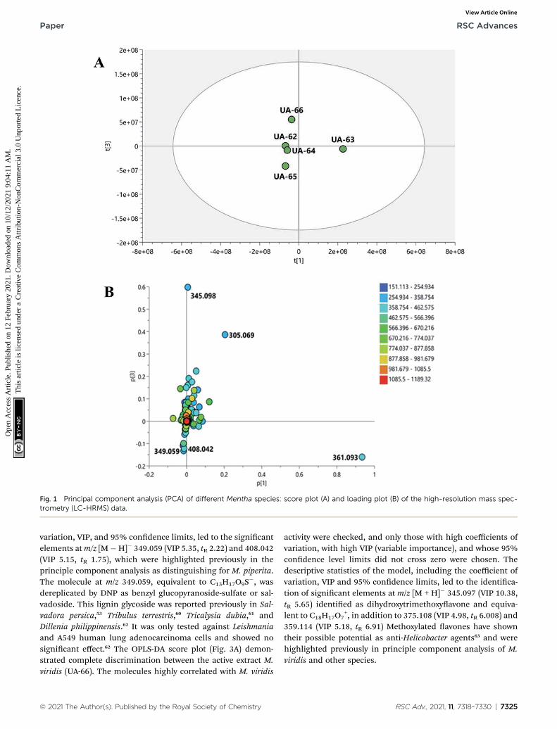
Fig. 1 Principal component analysis (PCA) of different Mentha species: score plot (A) and loading plot (B) of the high-resolution mass spec-trometry (LC-HRMS) data.
Paper RSC Advances
Ope
n A
cces
s A
rtic
le. P
ublis
hed
on 1
2 Fe
brua
ry 2
021.
Dow
nloa
ded
on 1
0/12
/202
1 9:
04:1
1 A
M.
Thi
s ar
ticle
is li
cens
ed u
nder
a C
reat
ive
Com
mon
s A
ttrib
utio
n-N
onC
omm
erci
al 3
.0 U
npor
ted
Lic
ence
.View Article Online
variation, VIP, and 95% condence limits, led to the signicantelements atm/z [M�H]� 349.059 (VIP 5.35, tR 2.22) and 408.042(VIP 5.15, tR 1.75), which were highlighted previously in theprinciple component analysis as distinguishing for M. piperita.The molecule at m/z 349.059, equivalent to C13H17O9S
�, wasdereplicated by DNP as benzyl glucopyranoside-sulfate or sal-vadoside. This lignin glycoside was reported previously in Sal-vadora persica,53 Tribulus terrestris,60 Tricalysia dubia,61 andDillenia philippinensis.62 It was only tested against Leishmaniaand A549 human lung adenocarcinoma cells and showed nosignicant effect.62 The OPLS-DA score plot (Fig. 3A) demon-strated complete discrimination between the active extract M.viridis (UA-66). The molecules highly correlated with M. viridis
© 2021 The Author(s). Published by the Royal Society of Chemistry
activity were checked, and only those with high coefficients ofvariation, with high VIP (variable importance), and whose 95%condence level limits did not cross zero were chosen. Thedescriptive statistics of the model, including the coefficient ofvariation, VIP and 95% condence limits, led to the identica-tion of signicant elements at m/z [M + H]� 345.097 (VIP 10.38,tR 5.65) identied as dihydroxytrimethoxyavone and equiva-lent to C18H17O7
+, in addition to 375.108 (VIP 4.98, tR 6.008) and359.114 (VIP 5.18, tR 6.91) Methoxylated avones have showntheir possible potential as anti-Helicobacter agents63 and werehighlighted previously in principle component analysis of M.viridis and other species.
RSC Adv., 2021, 11, 7318–7330 | 7325
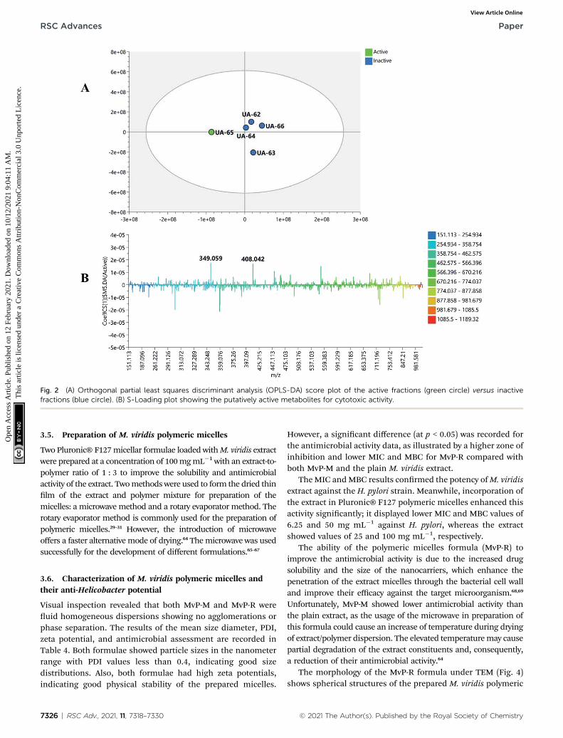
Fig. 2 (A) Orthogonal partial least squares discriminant analysis (OPLS-DA) score plot of the active fractions (green circle) versus inactivefractions (blue circle). (B) S-Loading plot showing the putatively active metabolites for cytotoxic activity.
RSC Advances Paper
Ope
n A
cces
s A
rtic
le. P
ublis
hed
on 1
2 Fe
brua
ry 2
021.
Dow
nloa
ded
on 1
0/12
/202
1 9:
04:1
1 A
M.
Thi
s ar
ticle
is li
cens
ed u
nder
a C
reat
ive
Com
mon
s A
ttrib
utio
n-N
onC
omm
erci
al 3
.0 U
npor
ted
Lic
ence
.View Article Online
3.5. Preparation of M. viridis polymeric micelles
Two Pluronic® F127micellar formulae loaded withM. viridis extractwere prepared at a concentration of 100mgmL�1 with an extract-to-polymer ratio of 1 : 3 to improve the solubility and antimicrobialactivity of the extract. Twomethods were used to form the dried thinlm of the extract and polymer mixture for preparation of themicelles: a microwave method and a rotary evaporator method. Therotary evaporator method is commonly used for the preparation ofpolymeric micelles.29–31 However, the introduction of microwaveoffers a faster alternative mode of drying.64 Themicrowave was usedsuccessfully for the development of different formulations.65–67
3.6. Characterization of M. viridis polymeric micelles andtheir anti-Helicobacter potential
Visual inspection revealed that both MvP-M and MvP-R wereuid homogeneous dispersions showing no agglomerations orphase separation. The results of the mean size diameter, PDI,zeta potential, and antimicrobial assessment are recorded inTable 4. Both formulae showed particle sizes in the nanometerrange with PDI values less than 0.4, indicating good sizedistributions. Also, both formulae had high zeta potentials,indicating good physical stability of the prepared micelles.
7326 | RSC Adv., 2021, 11, 7318–7330
However, a signicant difference (at p < 0.05) was recorded forthe antimicrobial activity data, as illustrated by a higher zone ofinhibition and lower MIC and MBC for MvP-R compared withboth MvP-M and the plain M. viridis extract.
TheMIC andMBC results conrmed the potency ofM. viridisextract against the H. pylori strain. Meanwhile, incorporation ofthe extract in Pluronic® F127 polymeric micelles enhanced thisactivity signicantly; it displayed lower MIC and MBC values of6.25 and 50 mg mL�1 against H. pylori, whereas the extractshowed values of 25 and 100 mg mL�1, respectively.
The ability of the polymeric micelles formula (MvP-R) toimprove the antimicrobial activity is due to the increased drugsolubility and the size of the nanocarriers, which enhance thepenetration of the extract micelles through the bacterial cell walland improve their efficacy against the target microorganism.68,69
Unfortunately, MvP-M showed lower antimicrobial activity thanthe plain extract, as the usage of the microwave in preparation ofthis formula could cause an increase of temperature during dryingof extract/polymer dispersion. The elevated temperaturemay causepartial degradation of the extract constituents and, consequently,a reduction of their antimicrobial activity.64
The morphology of the MvP-R formula under TEM (Fig. 4)shows spherical structures of the prepared M. viridis polymeric
© 2021 The Author(s). Published by the Royal Society of Chemistry
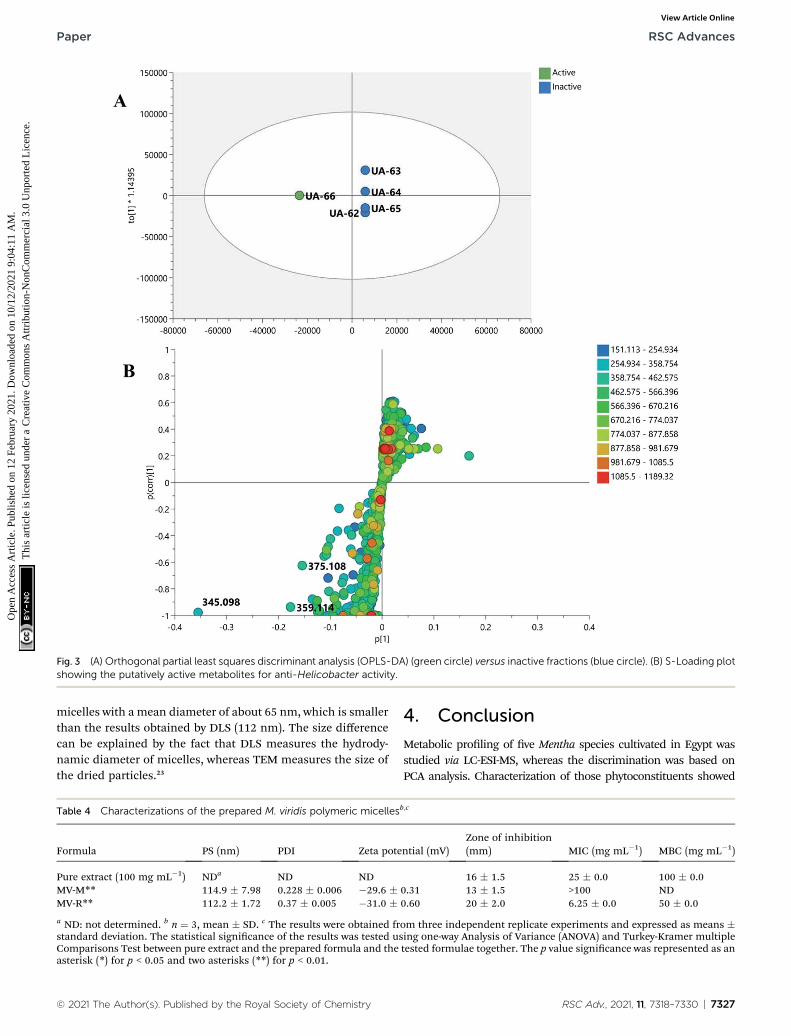
Fig. 3 (A) Orthogonal partial least squares discriminant analysis (OPLS-DA) (green circle) versus inactive fractions (blue circle). (B) S-Loading plotshowing the putatively active metabolites for anti-Helicobacter activity.
Paper RSC Advances
Ope
n A
cces
s A
rtic
le. P
ublis
hed
on 1
2 Fe
brua
ry 2
021.
Dow
nloa
ded
on 1
0/12
/202
1 9:
04:1
1 A
M.
Thi
s ar
ticle
is li
cens
ed u
nder
a C
reat
ive
Com
mon
s A
ttrib
utio
n-N
onC
omm
erci
al 3
.0 U
npor
ted
Lic
ence
.View Article Online
micelles with a mean diameter of about 65 nm, which is smallerthan the results obtained by DLS (112 nm). The size differencecan be explained by the fact that DLS measures the hydrody-namic diameter of micelles, whereas TEM measures the size ofthe dried particles.23
Table 4 Characterizations of the prepared M. viridis polymeric micelles
Formula PS (nm) PDI Zeta pote
Pure extract (100 mg mL�1) NDa ND NDMV-M** 114.9 � 7.98 0.228 � 0.006 �29.6 �MV-R** 112.2 � 1.72 0.37 � 0.005 �31.0 �a ND: not determined. b n ¼ 3, mean � SD. c The results were obtained frstandard deviation. The statistical signicance of the results was tested usComparisons Test between pure extract and the prepared formula and theasterisk (*) for p < 0.05 and two asterisks (**) for p < 0.01.
© 2021 The Author(s). Published by the Royal Society of Chemistry
4. Conclusion
Metabolic proling of ve Mentha species cultivated in Egypt wasstudied via LC-ESI-MS, whereas the discrimination was based onPCA analysis. Characterization of those phytoconstituents showed
b,c
ntial (mV)Zone of inhibition(mm) MIC (mg mL�1) MBC (mg mL�1)
16 � 1.5 25 � 0.0 100 � 0.00.31 13 � 1.5 >100 ND0.60 20 � 2.0 6.25 � 0.0 50 � 0.0
om three independent replicate experiments and expressed as means �ing one-way Analysis of Variance (ANOVA) and Turkey-Kramer multipletested formulae together. The p value signicance was represented as an
RSC Adv., 2021, 11, 7318–7330 | 7327
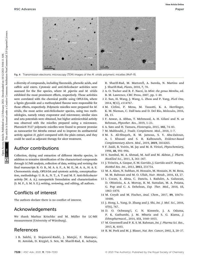
Fig. 4 Transmission electronic microscopy (TEM) images of the M. viridis polymeric micelles (MvP-R).
RSC Advances Paper
Ope
n A
cces
s A
rtic
le. P
ublis
hed
on 1
2 Fe
brua
ry 2
021.
Dow
nloa
ded
on 1
0/12
/202
1 9:
04:1
1 A
M.
Thi
s ar
ticle
is li
cens
ed u
nder
a C
reat
ive
Com
mon
s A
ttrib
utio
n-N
onC
omm
erci
al 3
.0 U
npor
ted
Lic
ence
.View Article Online
a diversity of compounds, including avonoids, phenolic acids, andcaffeic acid esters. Cytotoxic and anti-Helicobacter activities wereassessed for the ve species, where M. piperita and M. viridisexhibited the most prominent effects, respectively. Those activitieswere correlated with the chemical prole using OPLS-DA, wherea lignin glycoside and a methoxylated avone were responsible forthose effects, respectively. Polymeric micelles were prepared for M.viridis, the most active anti-Helicobacter species, using two meth-odologies, namely rotary evaporator and microwave; similar sizesand zeta potentials were obtained, but higher antimicrobial activitywas observed with the micelles prepared using a microwave.Pluronic® F127 polymeric micelles were found to present promiseas nanocarrier for Mentha extract and to improve its antibacterialactivity against H. pylori compared with the plain extract, and theycould be used as adjuvant therapy for ulcer treatment.
Author contributions
Collection, drying and extraction of different Mentha species, inaddition to tentative identication of the characterized compoundsthrough LC/MS analysis, collection of data, writing and revising thenal manuscript: R. O. B., M. A. A. F., A. M. E., M. A. A., H. A. E.Chemometric study, OPLS-DA and cytotoxic activity, conceptualiza-tion, methodology: U. R. A., N. T., A. T and M. F. Anti-Helicobacteractivity (W. A. A.); nanoparticle formulation and characterization(S. M. F., S. M. S. E.); writing, reviewing, and editing, all authors.
Conflicts of interest
The authors declare there is no conict of interest.
Acknowledgements
We thank Markus Krischke and M. Muller for LC-MSmeasurement (University of Wurzburg).
References
1 B. Salehi, Z. Stojanovic-Radic, J. Matejic, F. Sharopov,H. Antolak, D. Kregiel, S. Sen, M. Shari-Rad, K. Acharya,
7328 | RSC Adv., 2021, 11, 7318–7330
R. Shari-Rad, M. Martorell, A. Sureda, N. Martins andJ. Shari-Rad, Plants, 2018, 7, 70.
2 A. O. Tucker and R. F. Naczi, in Mint: the genus Mentha, ed.B. M. Lawrence, CRC Press, 2007, pp. 1–40.
3 Z. Sun, H. Wang, J. Wang, L. Zhou and P. Yang, PLoS One,2014, 9(12), e114767.
4 M. Cirlini, P. Mena, M. Tassotti, K. A. Herrlinger,K. M. Nieman, C. Dall'Asta and D. Del Rio, Molecules, 2016,21, 15.
5 F. Anwar, A. Abbas, T. Mehmood, A. H. Gilani and N. urRehman, Phytother. Res., 2019, 1–23.
6 A. Sato and H. Tamura, Fitoterapia, 2015, 102, 74–83.7 M. Mahboubi, J. Tradit. Complement. Med., 2018, 1–7.8 M. S. Ali-Shtayeh, R. M. Jamous, S. Y. Abu-Zaitoun,A. I. Khasati and S. R. Kalbouneh, Evidence-BasedComplementary Altern. Med., 2019, 2019, 3834265.
9 F. Zaidi, B. Voirin, M. Jay and M. R. Viricel, Phytochemistry,1998, 48, 991–994.
10 S. Sumbul, M. A. Ahmad, M. Asif and M. Akhtar, J. Pharm.BioAllied Sci., 2011, 3, 361–367.
11 J. Teixeira, A. Gaspar, E. M. Garrido, J. Garrido and F. Borges,BioMed Res. Int., 2013, 2013, 251754.
12 M. A. Alam, N. Subhan, H. Hossain, M. Hossain, H. M. Reza,M. M. Rahman and M. O. Ullah, Nutr. Metab., 2016, 13, 27.
13 I. Cocan, E. Alexa, C. Danciu, I. Radulov, A. Galuscan,D. Obistioiu, A. A. Morvay, R. M. Sumalan, M. A. Poiana,G. Pop and C. A. Dehelean, Exp. Ther. Med., 2018, 15,1863–1870.
14 M. Creydt and M. Fischer, Anal. Chem., 2017, 89, 10474–10486.
15 J. Hong, L. Yang, D. Zhang and J. Shi, Int. J. Mol. Sci., 2016,17(6), 767.
16 D. O. Ochwang'i, C. N. Kimwele, J. A. Oduma,P. K. Gathumbi, J. M. Mbaria and S. G. Kiama, J.Ethnopharmacol., 2014, 151, 1040–1055.
17 M. Greenwell and P. K. S. M. Rahman, Int. J. Pharma Sci. Res.,2015, 6, 4103.
18 R. M. Peek and M. J. Blaser, Nat. Rev. Cancer, 2002, 2, 28–37.
© 2021 The Author(s). Published by the Royal Society of Chemistry
Paper RSC Advances
Ope
n A
cces
s A
rtic
le. P
ublis
hed
on 1
2 Fe
brua
ry 2
021.
Dow
nloa
ded
on 1
0/12
/202
1 9:
04:1
1 A
M.
Thi
s ar
ticle
is li
cens
ed u
nder
a C
reat
ive
Com
mon
s A
ttrib
utio
n-N
onC
omm
erci
al 3
.0 U
npor
ted
Lic
ence
.View Article Online
19 S. Martini, C. D'Addario, A. Colacevich, S. Focardi,F. Borghini, A. Santucci, N. Figura and C. Rossi, Int. J.Antimicrob. Agents, 2009, 34, 50–59.
20 A. T. B. Abadi and J. G. Kusters, BMC Gastroenterol., 2016, 16,94–98.
21 Y. D. Taghipour, R. Bahramsoltani, A. M.Marques, R. Naseri,R. Rahimi, P. Haratipour, A. I. Panah, M. H. Farzaei andM. Abdollahi, Daru, J. Pharm. Sci., 2018, 26, 229–239.
22 A. R. Fares, A. N. Elmeshad and M. A. A. Kassem, DrugDelivery, 2018, 25, 132–142.
23 A. S. Deshmukh, P. N. Chauhan, M. N. Noolvi, K. Chaturvedi,K. Ganguly, S. S. Shukla, M. N. Nadagouda andT. M. Aminabhavi, Int. J. Pharm., 2017, 532, 249–268.
24 C. Sun, W. Li, P. Ma, Y. Li, Y. Zhu, H. Zhang, M. Adu-Frimpong, W. Deng, J. Yu and X. Xu, Food Chem. Toxicol.,2020, 137, 111126.
25 M. U. Akbar, K. M. Zia, A. Nazir, J. Iqbal, S. A. Ejaz andM. S. H. Akash, AAPS PharmSciTech, 2018, 19, 2719–2739.
26 M. Abedanzadeh, M. Salmanpour, F. Farjadian,S. Mohammadi and A. M. Tamaddon, J. Drug Delivery Sci.Technol., 2020, 58, 101793.
27 F. Molavi, M. Barzegar-jalali, H. Hamishehkar andH. Hamishehkar, J. Control. Release., 2020, 320, 265–282.
28 Y. Chen, W. Zhang, J. Gu, Q. Ren, Z. Fan, W. Zhong, X. Fangand X. Sha, Int. J. Pharm., 2013, 452, 421–433.
29 J. K. Valenzuela-Oses, M. C. Garcıa, V. A. Feitosa,J. A. Pachioni-Vasconcelos, S. M. Gomes-Filho,F. R. Lourenço, N. N. P. Cerize, D. S. Basseres andC. O. Rangel-Yagui, Mater. Sci. Eng., C, 2017, 81, 327–333.
30 M. Jaiswal, M. Kumar and K. Pathak, Colloids Surf., B, 2015,130, 23–30.
31 J. Oyama, D. S. S. L. Lera-Nonose, A. C. F. H. Ramos-Milare,F. B. Padilha Ferreira, C. F. de Freitas, W. Caetano, N. Hioka,T. G. V. Silveira andM. V. C. Lonardoni, Acta Trop., 2019, 192,11–21.
32 M. Blumenthal, W. Busse, A. Goldberg, J. Gruenwald, T. Halland C. Riggins, The Complete German Commission EMonographs: Therapeutic Guide to Herbal Medicines | Annalsof Internal Medicine | American College of Physicians,American Botanical Council, 1999.
33 A. C. Martin, A. D. Pawlus, E. M. Jewett, D. L. Wyse,C. K. Angerhofer and A. D. Hegeman, RSC Adv., 2014, 4,26325–26334.
34 C. Cheng, E. M. Othman, H. Stopper, R. A. Edrada-Ebel,U. Hentschel and U. R. Abdelmohsen, Mar. Drugs, 2017,15, 383–391.
35 A. W. Bauer, W. M. Kirby, J. C. Sherris and M. Turck, Am. J.Clin. Pathol., 1966, 45, 493–496.
36 J. A. Nazeam, W. A. AL-Shareef, M. W. Helmy and A. E. El-Haddad, Food Chem., 2020, 326, 126993.
37 T. A. Nagy, L. E. Wroblewski, D. Wang, M. B. Piazuelo,A. Delgado, J. Romero-Gallo, J. Noto, D. A. Israel,S. R. Ogden, P. Correa, T. L. Cover and R. M. Peek,Gastroenterology, 2011, 141, 553–564.
38 A. El Haddad, W. AL-Shareef and S. Eid, J. Adv. Pharm. Res.,2019, 3, 143–149.
© 2021 The Author(s). Published by the Royal Society of Chemistry
39 J. Patel, M. Weinstein, G. Eliopolos, S. Jenkins, B. Limbago,A. Mathers, T. Mazzulli, R. Patel, S. Richter, M. Satlin,J. Swenson, M. Traczewsli, J. Turnidge and B. Zimmer,M100: Performance Standards for Antimicrobial SusceptibilityTesting An informational supplement for global applicationdeveloped through the Clinical and Laboratory StandardsInstitute consensus process, 2017.
40 M. H. Taleb, N. F. Abdeltawab, R. N. Shamma,S. S. Abdelgayed, S. S. Mohamed, M. A. Farag andM. A. Ramadan, Molecules, 2018, 23(9), 2164.
41 Q. Yao, Y. Liu, L. Kou, Y. Tu, X. Tang and L. Zhu,Nanomedicine, 2019, 19, 71–80.
42 W. Zhang, Y. Shi, Y. Chen, J. Ye, X. Sha and X. Fang,Biomaterials, 2011, 32, 2894–2906.
43 D. Jain, N. Pathak, S. Khan, G. V. Raghuram, A. Bhargava,R. Samarth and P. K. Mishra, Int. J. Toxicol., 2011, 30, 225–236.
44 Y. X. Li, Y. B. Liu, A. Q. Ma, Y. Bao, M. Wang and Z. L. Sun,Food Sci. Biotechnol., 2017, 26, 1675–1683.
45 M. O. Arruda, S. J. F. Mendes, S. A. Teixeira, L. S. S. DeMesquita, M. N. De Sousa Ribeiro, S. D. S. L. Galvao,M. N. Muscara, E. S. Fernandes and V. Monteiro-Neto, J.Immunol. Res., 2017, 2017, 2078794.
46 H. Imai, K. Osawa, H. Yasuda, H. Hamashima, T. Arai andM. Sasatsu, Microbios, 2001, 106, 31–39.
47 A. Bodalska, A. Kowalczyk, M. Włodarczyk and I. Fecka,Molecules, 2020, 25, 69–88.
48 Y. J. Park, S. A. Baek, Y. Choi, J. K. Kim and S. U. Park,Molecules, 2019, 24, 258.
49 H. J. D. Dorman, M. Kosar, K. H. C. Baser and R. Hiltunen,Nat. Prod. Commun., 2009, 4, 535–542.
50 J. C. Yang, H. C. Yang, C. T. Shun, T. H. Wang, C. T. Chienand J. Y. Kao, Evidence-Based Complementary Altern. Med.,2013, 2013, 248585.
51 P. Fong, C. H. Hao, C. C. Io, P. I. Sin and L. R. Meng,Molecules, 2019, 24(19), 3608.
52 L. L. Perico, M. T. Emılio-Silva, R. Ohara, V. P. Rodrigues,G. Bueno, J. M. Barbosa-Filho, L. R. M. da Rocha,L. M. Batista and C. A. Hiruma-Lima, Biomolecules, 2020,10, 265.
53 M. S. Kamel, K. Ohtani, M. H. Assaf, R. Kasai, M. A. El-Shanawani, K. Yamasaki, A. A. Ali and O. Tanaka,Phytochemistry, 1992, 31, 2469–2471.
54 D. B. Rodriguez-Amaya, Food Carotenoids, JohnWiley & Sons,Ltd, Chichester, UK, 2015.
55 A. R. Khuda-Bukhsh, S. K. Saha and S. Das, in Cancer:Oxidative Stress and Dietary Antioxidants, Elsevier Inc.,2014, pp. 77–89.
56 V. Pasquet, P. Morisset, S. Ihammouine, A. Chepied,L. Aumailley, J. B. Berard, B. Serive, R. Kaas, I. Lanneluc,V. Thiery, M. Lafferriere, J. M. Piot, T. Patrice, J. P. Cadoretand L. Picot, Mar. Drugs, 2011, 9, 819–831.
57 A. Tawke, E. Z. Attia, S. Y. Desoukey, D. Hajjar, A. A. Makki,P. J. Schupp, R. A. Edrada-Ebel and U. R. Abdelmohsen, AMBExpress, 2019, 9, 12.
58 A. Tawke, M. Romli, C. Clements, G. Abbott, L. Young,M. Schumacher, M. Diederich, M. Farag and R. A. Edrada-
RSC Adv., 2021, 11, 7318–7330 | 7329
RSC Advances Paper
Ope
n A
cces
s A
rtic
le. P
ublis
hed
on 1
2 Fe
brua
ry 2
021.
Dow
nloa
ded
on 1
0/12
/202
1 9:
04:1
1 A
M.
Thi
s ar
ticle
is li
cens
ed u
nder
a C
reat
ive
Com
mon
s A
ttrib
utio
n-N
onC
omm
erci
al 3
.0 U
npor
ted
Lic
ence
.View Article Online
Ebel, J. Chromatogr. B: Anal. Technol. Biomed. Life Sci., 2019,1106, 71–73.
59 A. Tawke, R. Tate, G. Abbott, L. Young, C. Viegelmann,M. Schumacher, M. Diederich and R. Edrada-Ebel, Chem.Biodiversity, 2017, 14, e1700040.
60 V. G. Nebieridze, A. V. Skhirtladze, E. P. Kemertelidze andM. Ganzera, Chem. Nat. Compd., 2018, 54, 63–65.
61 J. Shitamoto, S. Sugimoto, K. Matsunami, H. Otsuka,T. Shinzato and Y. Takeda, Chem. Pharm. Bull., 2011, 59,72–77.
62 R. A. S. Macahig, K. Matsunami and H. Otsuka, Chem.Pharm. Bull., 2011, 59, 397–401.
63 T. Isobe, M. Doe, Y. Morimoto, K. Nagata and A. Ohsaki, Biol.Pharm. Bull., 2006, 29, 1039–1041.
64 T. Wong, Curr. Drug Delivery, 2008, 5, 77–84.65 M. Alle, B. reddy G, T. H. Kim, S. H. Park, S. H. Lee and
J. C. Kim, Carbohydr. Polym., 2020, 229, 115511.66 H. Ashraf, T. Anjum, S. Riaz and S. Naseem, Front. Microbiol.,
2020, 11, 238.67 J. Isaac, S. Kaity, S. Ganguly and A. Ghosh, J. Pharm.
Pharmacol., 2013, 65, 219–229.68 V. D. Wagh and O. J. Deshmukh, ISRN Pharm., 2012, 2012, 1–
7.69 E. Haba, S. Bouhdid, N. Torrego-Solana, A. M. Marques,
M. J. Espuny, M. J. Garcıa-Celma and A. Manresa, Int. J.Pharm., 2014, 476, 134–141.
70 J. Sun, F. Liang, Y. Bin, P. Li and C. Duan, Molecules, 2007,12, 679–693.
71 K. Kapp, E. Hakala, A. Orav, L. Pohjala, P. Vuorela, T. Pussa,H. Vuorela and A. Raal, Food Res. Int., 2013, 53, 758–766.
7330 | RSC Adv., 2021, 11, 7318–7330
72 R. Kodama, T. Yano, K. Furukawa, K. Noda and H. Ide,Xenobiotica, 1976, 6, 377–389.
73 M. B. Bahadori, G. Zengin, S. Bahadori, L. Dinparast andN. Movahhedin, Int. J. Food Prop., 2018, 21, 183–193.
74 D. Lu, X. Yuan, S.-J. Kim, J. V. Marques, P. P. Chakravarthy,S. G. A. Moinuddin, R. Luchterhand, B. Herman, L. B. Davinand N. G. Lewis, Plant Biotechnol. J., 2017, 15, 970–981.
75 M. J. Magera, A. L. Stoor, J. K. Helgeson, D. Matern andP. Rinaldo, Clin. Chim. Acta, 2001, 306, 35–41.
76 B. Fatih, K. Madani, M. Chibane and P. Duez, in Aromaticand Medicinal Plants - Back to Nature, InTech, 2017.
77 P. Mena, M. Cirlini, M. Tassotti, K. A. Herrlinger, C. Dall’Astaand D. Del Rio, Molecules, 2016, 21(11), 1576.
78 A. Taamalli, D. Arraez-Roman, L. Abaza, I. Iswaldi,A. Fernandez-Gutierrez, M. Zarrouk and A. Segura-Carretero, Phytochem. Anal., 2015, 26, 320–330.
79 M. Bozovic, A. Pirolli and R. Ragno, Molecules, 2015, 20,8605–8633.
80 Z. Ozer, J. Turk. Chem. Soc., Sect. A, 2018, 5, 445–456.81 D. P. De Sousa, E. Raphael and T. J. Brocksom, Z.
Naturforsch., B: J. Chem. Sci., 2010, 65, 1381–1383.82 F. A. Tomas-Barberan, S. Z. Husain and M. I. Gil, Biochem.
Syst. Ecol., 1988, 16, 43–46.83 G. H. Jang, H. W. Kim, M. K. Lee, S. Y. Jeong, A. R. Bak,
D. J. Lee and J. B. Kim, Saudi J. Biol. Sci., 2018, 25, 1622–1631.84 N. R. Desam, A. J. Al-Rajab, M. Sharma, M. M. Mylabathula,
R. R. Gowkanapalli and M. Albratty, J. King Saud Univ., Sci.,2019, 31, 528–533.
© 2021 The Author(s). Published by the Royal Society of Chemistry