Embed Size (px)
Citation preview
Oral SessionsImproving tissue perfusion: 0001–00050001EFFECTS OF A FLUID CHALLENGE ON REGIONAL TISSUE PERFUSION IN ICUPATIENTS
E. Klijn1, M. van Velzen2, C. Ince1, J. Bakker1, J. Van Bommel1
1Erasmus Medical Center, Intensive Care, Rotterdam, Netherlands, 2Erasmus Medical Center,Experimental Anesthesiology, Rotterdam, Netherlands
INTRODUCTION. Fluid therapy is one of the cornerstones of hemodynamic management incritically ill patients. Although the aim of fluid therapy is to improve regional tissue perfusion,its adequacy is judged by the effect on cardiac stroke volume (SV). It is as yet unknownwhether an increase in SV through a fluid challenge (FC) can also improve commonparameters of regional perfusion. And if so, whether it is possible to predict the effect of a FCon regional perfusion parameters by performing a passive leg raising.
METHODS. Critically ill patients, receiving invasive hemodynamic monitoring andrequiring fluid therapy, were eligible for enrollment. They underwent a 30� passive leg raising(PLR) followed by a fluid challenge of 250 ml colloids. During the PLR and FC, strokevolume and cardiac output (CO) were measured as well as sublingual microcirculatory per-fusion (sidestream dark field imaging), peripheral flow index (photoplethysmography of thefinger) and tissue blood flow of the skin (laser Doppler flowmetry).
RESULTS. 35 patients were included in our study, of which 15 (43%) were fluid responsive (FR;more than 10% increase in SV). Baseline SV and CO were significantly lower in responders than innonresponders, as well as thesublingualperfusion. In the responsivepatients sublingual vesseldensityand skin tissue blood flow increased following FC (Fig. 1), whereas PFI on the finger remained low.During the PLR no changes in any parameter were observed. Fluid responsiveness was best predictedby SV response to PLR (AUC 0.88 ± 0.08, p\0.01) and baseline SV (AUC 0.79 ± 0.08,p\0.01), followed by baseline sublingual vessel density (AUC 0.76 ± 0.09, p\0.05). It was notpossible to predict the response effect of FC on regional perfusion by performing a PLR.
CONCLUSIONS. In septic patients, parameters of regional perfusion are not related to sys-temic hemodynamic parameters and do not respond uniformly to fluid administration. In fact, itis questionable whether regional perfusion does benefit from fluid therapy at all following initialresuscitation. As such, the PLR has no value in optimizing regional perfusion.
Figure 1
0002DOPAMINE AND NOREPINEPHRINE FOR THE TREATMENT OF SHOCK: A PERPROTOCOL ANALYSIS
D. De Backer1, P. Biston2, J. Devriendt3, C. Madl4, D. Chochrad5, C. Aldecoa6,
J.-L. Vincent1, Soap II Investigators
1Erasme University Hospital, Intensive Care, Brussels, Belgium, 2Centre Hospitalier Uni-versitaire de Charleroi, Intensive Care, Charleroi, Belgium, 3Brugmann University Hospital,Intensive Care, Brussels, Belgium, 4Medical University of Vienna, Intensive Care Unit 13H1,Vienna, Austria, 5Centre Hospitalier Etterbeek Ixelles, Intensive Care, Brussels, Belgium,6Rio Hortega University Hospital, Anesthesia and Critical Care, Valladolid, Spain
INTRODUCTION. We recently demonstrated that, compared to norepinephrine, dopaminewas associated with a non significant increase in 28 day mortality (OR 1.17 [0.97–1.42]) (1).However, arrhythmia resulted in discontinuation of the study drug in some patients, whichwas converted to open label norepinephrine in all cases. We conducted a per-protocol analysislimited to patients in whom the study drug was not discontinued.
OBJECTIVES. To evaluate in a per protocol analysis the impact on outcome of dopamineand norepinephrine for the treatment of shock.
METHODS. In a prospective, randomized study (NCT00314704), 858 patients received dopa-mine and 821 norepinephrine, according to a protocol described in detail elsewhere (1). Briefly,patients were randomized within 4 h max of the onset of shock to receive either dopamine (max dose20 mcg/kg min) or norepinephrine (max dose 0.19 mcg/kg min). When the desired mean arterialpressure (MAP) could not be reached with these max doses, an open label norepinephrine wasadded. In the presence of problematic arrhythmias, physicians were allowed to discontinue studydrug and to replace it by any vasopressor agent while blinding of initial allocation was maintained.According to our predefined analytical plan, we analyzed patients in whom study drug was notdiscontinued. Kaplan–Meier curves for estimated survival were compared using a log-rank test.
RESULTS. The study drug was discontinued in 65 of the 1,679 patients enrolled, leaving1,614 patients in the per-protocol analysis. Discontinuation occurred more frequently indopamine than in norepinephrine-treated patients (52 vs. 13 patients, p \ 0.05), so that 806patients in the dopamine group and 808 patients in the norepinephrine group were exposed tostudy drug during the entire shock episode. Kaplan–Meier curves for estimated survivalshowed a increased mortality rate in dopamine treated patients (p = 0.031, Fig. 1).
Kaplan–Meier curves for 28 day survival
CONCLUSIONS. In patients effectively exposed to dopamine or norepinephrine during theentire shock period, dopamine was associated with an increased mortality rate.
REFERENCE(S). (1) De Backer et al. Comparison of dopamine and norepinephrine in thetreatment of shock NEJM 362:779–789;2010
0003MICROCIRCULATION DENSITY AND PERFUSION IN A LOWER BODY NEGA-TIVE PRESSURE (LBNP) MODEL OF CENTRAL HYPOVOLEMIA
D.M.J. Milstein1, S.A. Bartels1, R. Bezemer1, F.J. Wallis de Vries1, C. Ince1
1Academic Medical Center at the University of Amsterdam, Translational Physiology,Amsterdam, Netherlands
AIMS. Hypovolemia and hemorrhagic shock are common life threatening complications andearly reliable detection would be a great advantage for on-time corrective therapeutic inter-ventions. The aim of this study was to investigate the effects of imminent hypovolemia onsublingual microcirculation perfusion and vessel density during a lower body negativepressure (LBNP) model of progressive central hypovolemia.
METHODS. For the present investigation, 8 healthy male volunteers with a mean age of29 ± 3.6 years were recruited for a cardiovascular screening exercise stress test prior toinclusion for the study. During the LBNP protocol, the subjects were exposed to sequentialincreasing negative pressures of -20, -40, and -60 mmHg while resting in a supine positionwith their legs sealed in the LBNP chamber at the level of the iliac crest. In addition to continuousregistration of cardiac output (CO) and mean arterial pressure (MAP), sublingual perfused vesseldensity (PVD) (1) and microvascular flow indices (MFI) (2) were measured using sidestreamdark-field (SDF) imaging before (T0), during (T1; -60 mmHg), and after (T2) LBNP.
RESULTS. There were no significant differences in mean CO and MAP in our subjects.Figures 1 and 2 illustrate microcirculation PVD and MFI for all time points respectively.
Figures 1 and 2
CONCLUSIONS. LBNP significantly attenuates microcirculation perfusion and smallmicrovessels in the sublingual microcirculation may to be a good indicator of the early onsetof central hypovolemia.
REFERENCE(S). (1) De Backer D, Hollenberg S, Boerma C, Goedhart P, Buchele G,Ospina-Tascon G, Dobbe I, Ince C. How to evaluate the microcirculation: report of a roundtable conference. Crit Care 2007;11(5):R101 (2) Boerma EC, Mathura KR, van der Voort PH,Spronk PE, Ince C. Quantifying bedside-derived imaging of microcirculatory abnormalities inseptic patients: a prospective validation study. Crit Care 2005;9(5):R601–6.
0004THE EFFECTS OF NON-LEUKOREDUCED RED BLOOD CELL TRANSFUSIONSON MICROCIRCULATION IN MIXED SURGICAL PATIENTS
B. Ayhan1,2, K. Yuruk3, S. Koene3, A. Sahin2, C. Ince1,3, U. Aypar2
1Erasmus Medical Center, Intensive Care Medicine, Rotterdam, Netherlands, 2HacettepeUniversity Hospital, Intensive Care Medicine, Ankara, Turkey, 3Academic Medical Center atthe University of Amsterdam, Translational Physiology, Amsterdam, Netherlands
AIMS. The impact of the storage process on oxygen-carrying properties of red blood cells andthe efficacy of red blood cell transfusions (BTX) concerning tissue oxygenation remain anissue of debate in transfusion medicine. Storage time and leukocyte content probably interactsince longer storage duration would be expected to cause greater accumulation of leukocyte-derived cytokines and red blood cell injury. The aim of this study was to investigate theeffects of storage and efficacy of fresh (stored for less than 1 week) versus aged (stored formore than 3 weeks) non-leukoreduced BTX on sublingual microvascular density andmicrovascular flow in mixed surgical patients.
METHODS. 18 surgical patients were included in this study. Patients were randomlyassigned into two groups as patients receiving fresh BTX (Group A) and patients receivingaged BTX (Group B). Sublingual microcirculatory functional capillary density (FCD) andmicrovascular flow index (MFI) were assessed using orthogonal polarization spectral (OPS)imaging. Measurements and collection of blood samples were performed after induction ofgeneral anesthesia, before BTX and 30 min after the BTX ended.
RESULTS. In both groups BTX caused an increase in systemic hemoglobin concentration(Fig. 1). BTX increased FCD in group A, while FCD remained unaffected in group B (Fig. 2).Changes in MFI following BTX in both groups are depicted in Fig. 3.
Figures
CONCLUSIONS. Fresh non-leukoreduced BTX in surgical patients were effective inimproving microcirculatory perfusion by elevating the number of perfused microvessels.However, stored BTX did not have the capacity to maintain effective microcirculatoryfunction.
S86 23rd ESICM ANNUAL CONGRESS – BARCELONA, SPAIN – 9–13 OCTOBER 2010

0005NITROGLYCERIN FAILS TO RECRUIT THE MICROCIRCULATION OF THEINTESTINAL VILLI IN SHEEP ENDOTOXEMIA
V.S. Kanoore Edul1, G. Ferrara1, M.O. Pozo1, G. Murias1, E. Martins1, C. Canullan1,
H.S. Canales1, E. Estenssoro1, C. Ince2, A. Dubin1
1Facultad de Ciencias Medicas, Universidad Nacional de La Plata, Catedra de FarmacologıaAplicada, La Plata, Argentina, 2Academic Medical Center, University of Amsterdam,Translational Physiology, Amsterdam, Netherlands
INTRODUCTION. Hypoperfusion of intestinal villi still persists after fluid resuscitation ofendotoxemic shock (1).
OBJECTIVES. To show that nitroglycerin improves the microcirculation of the villi.
METHODS. Twenty sheep were anesthetized and mechanically ventilated. Endotoxemicshock was produced by the infusion of 5 lg/kg of endotoxin followed by 4 lg/kg/h for 1500 .After 600 of shock, hydroxyethyl starch resuscitation was given to normalize oxygen trans-port, and sheep were randomized to control (n = 10) or nitroglycerin (n = 10) groups.Nitroglycerin was infused at a rate of 0.2 lg/kg/min during 900. Mean arterial blood pressure(MAP), cardiac output (CO), intestinal blood flow (Qgut), intramucosal-arterial PCO2
(DPCO2), and lactate were measured at baseline (00), during shock and after resuscitation. Themicrocirculation was evaluated by sidestream darkfield (SDF) imaging. Villi perfused cap-illary density (PCD) and microvascular flow index (MFI) were calculated.
RESULTS. Hemodynamic and perfusion variables were similarly compromised during shockin both groups. Hyperlactatemia, intramucosal acidosis and villi hypoperfusion that developedduring shock were not affected by the resuscitation with nitroglycerin.
TABLE 1
MAP
(mmHg)
CO
(l/min)
Qgut
(ml/min)
DPCO2
(mmHg)
Lactate
(mmol/l)
Villi
MFI
Villi PCD (mm/
mm2)
Baseline Control 97 ± 13 2.8 ± 0.8 404 ± 210 8 ± 8 2.1 ± 0.8 3.0 ± 0.0 18.2 ± 3.1
Nitroglycerin 92 ± 15 2.4 ± 0.9 401 ± 207 6 ± 6 1.7 ± 0.6 3.0 ± 0.0 17.3 ± 3.0
Shock Control 56 ± 24* 2.6 ± 0.9 329 ± 167* 15 ± 9* 4.3 ± 1.6* 2.5 ± 0.2* 14.9 ± 2.5*
Nitroglycerin 51 ± 17* 2.4 ± 1.1 312 ± 183* 13 ± 11* 3.6 ± 1.2* 2.3 ± 0.3 15.1 ± 3.7*
Resuscitation Control 91 ± 25 3.3 ± 0.8* 427 ± 163 15 ± 9* 5.6 ± 1.7* 2.7 ± 0.2* 15.9 ± 3.1
Nitroglycerin 75 ± 22* 3.1 ± 1.1* 363 ± 195 13 ± 11* 5.1 ± 2.3* 2.6 ± 0.3* 16.8 ± 4.5
p \ 0.05 versus baseline
CONCLUSIONS. Nitroglycerin was unable to improve the microcirculation of the intestinal villi.
REFERENCE. (1) Dubin A, Kanoore Edul VS, Pozo M, Murias G, Canullan C, Martins E,Ferrara G, Canales H, Laporte M, Estenssoro E, Ince C: Persistent villi hypoperfusionexplains intramucosal acidosis in sheep endotoxemia. Crit Care Medicine 2008; 36:535–542
GRANT ACKNOWLEDGMENT. Supported by the grant PICT-2007-00912, AgenciaNacional de Promocion Cientıfica y Tecnologica, Argentina.
Acute renal failure in the critically ill: 0006–00100006THE ROLE OF INITIAL RENAL HYPOPERFUSION IN RENAL ENDOXEMIC-INDUCED MICROCIRCULATORY FAILURE IN RATS
M. Legrand1,2, R. Bezemer2, E.G. Mik3, D. Payen1, C. Ince2
1Lariboisiere University Hospital, Anesthesiology and Critical Care, Paris, France,2Academic Medical Center at the University of Amsterdam, Translational Physiology,Amsterdam, Netherlands, 3Erasmus Medical Center, University Medical Center, Anesthesi-ology, Rotterdam, Netherlands
INTRODUCTION. Microcirculatory failure may be involved in the development of sepsis-related acute kidney injury.
OBJECTIVES. We conducted this study to determine the role of initial renal hypoperfusionin the endotoxemia-induced renal microcirculatory failure.
METHODS. 24 Wistar rats were randomized in 4 groups: sham group; LPS group; LPSgroup with early fluid resuscitation preventing drop of renal blood flow (EARLY group); LPSgroup with a delayed (i.e. 2 h) fluid resuscitation (LATE group).
Cortical microvascular blood flow was measured using speckle laser imaging techniques withassessment of microvascular blood flow distribution. Renal function was assessed with cre-atinine clearance. Serum levels of IL-6, IL-10 and TNF-a were measured.
RESULTS. LPS infusion resulted in 80% drop of renal blood flow with marked fall ofcortical microcirculatory blood flow. Fluid resuscitation could only partially restored mi-crocirculatory blood flow in the LATE and EARLY group despite preservation of renal bloodflow and increase aortic blood flow in the later group. Microvascular blood flow heterogeneitywith a left shift on histograms persisted in both resuscitated groups (Fig. 1). All rats in thecontrol group were anuric. Fluid resuscitation in both the LATE and EARLY groups partiallyprevented decrease of creatinine clearance which still remained lower than the sham-operatedgroup. Serum cytokines levels decreased in the resuscitated groups with no differencebetween the EARLY and LATE group.
Cortical microcirculatory blood flow distributionCONCLUSIONS. Our study shows that endotoxaemia induces intrarenal alterations of thecortical microcirculation and oxygenation which appear to be poorly dependant on themacrohemodynamics. This may explain why renal failure yet progresses after correction ofshock due to intrarenal microvascular oxygen supply heterogeneity.
GRANT ACKNOWLEDGMENT. M. Legrand was partially supported by a grant from theFrench minister of foreign affairs (EGIDE-LAVOISIER).
0007ONLY EARLY SEPSIS-RELATED SEVERE ACUTE KIDNEY INJURY ISASSOCIATED WITH LOWER SURVIVAL INDEPENDENTLY OF SHOCK:A PROSPECTIVE MULTICENTRE STUDY
M. Legrand1, A.-C. Lukaszewicz1, E. Gayat1, B. Megarbane2, E. Azoulay3, F. Fieux4, D. Payen1
1Lariboisiere University Hospital, Anesthesiology and Critical Care, Paris, France, 2Lari-boisiere University Hospital, Medical ICU, Paris, France, 3Saint-Louis University Hospital,Medical ICU, Paris, France, 4Saint-Louis University Hospital, Anesthesiology and CriticalCare, Paris, France
INTRODUCTION. Sepsis is the leading cause of acute kidney injury (AKI) in intensive careunits but debate remains concerning its prognosis.
OBJECTIVES. To determine whether AKI is an independent factor of prognosis related ornot to the severity of shock.
METHODS. This multi-centre study was approved by the local Committee (# CCPPRB 2061)and involved patients from four medical-surgical Intensive Care Units in Paris, France. Patientswith criteria of severe sepsis or septic shock (ACCP/SCCM consensus conference) (1) having atleast two organ failures defined by the SOFA score were included between 01/2004 and 1/2006.After excluding patients with chronic renal failure (n = 23) 3 groups were identified based onAKIN and SOFArenal scores: (1) patients with no early (within 48 h) AKI (serum creati-nine B 110 lmol/L; i.e. SOFArenal 0), (2) patients with mild AKI (serum creatinine [110 lmol/L and B 300 lmol/L; i.e. SOFArenal 1, 2) and (3) patients with severe AKI (serum creati-nine[ 300 lmol/L or urine output\ 500 ml/day or requiring renal replacement therapy; i.e.SOFArenal 3, 4). To assess the role of shock in the development of early AKI patients were stratifiedon the vasopressor dependency index (2) to treat cardiovascular failure. This index was calculatedas the ratio of catecholamine index to the MAP. The catecholamine index was calculated as:(dopamine dose 9 1) + (dobutamine dose 9 1) + (adrenaline dose 9 100) + (noradrenalinedose 9 100) + (phenylephrine dose 9 100). A multivariate analysis with a competitive riskmodel was used for mortality risk. p \ 0.05 was considered as statistically significant.
RESULTS. 43 had no AKI, and 132 had AKI, 73 with mild AKI and 59 with severe AKI.During the first 48 h, 29 patients had severe sepsis and 146 were in shock. Severe early AKIpatients had a significantly higher rate of mortality at day 28 (42%) compared to patients withmild AKI (23%) or no AKI (11%, p \ 0.05). In multivariate analysis using a competitive riskmodel, severe AKI was found to be an independent factor of mortality after adjusting for age,gender, presence of diabetes or cancer, and the SOFA score with exclusion of the renal item(HR 2.22 [1.26–3.92], p = 0.005). The severity of cardiovascular failure (based on vaso-pressor dependency index) did not differ between groups of AKI.
CONCLUSIONS. Only early severe sepsis-related AKI appears to be an independent factorof mortality as part of multiple organ failure syndrome. Furthermore, occurrence of AKI doesnot seem to be driven by the severity of cardiovascular failure.
REFERENCE(S). 1. Bone et al., RC, Chest 1992;101:1481–1483. 2. Cruz et al., JAMA2009;301:2445–2452.
GRANT ACKNOWLEDGMENT. This work was supported in part by ‘‘Programme Hos-pitalier Recherche Clinique (PHRC) AORO2006’’ and by ‘‘Quadrienal plan for research fromthe French Ministry of Research’’ EA 3509.
0008IS A TARGETED KT/V OF 1.2 PER DIALYSIS SESSION ACHIEVABLE IN SEPTICSHOCK OR SEVERE SEPSIS PATIENTS WITH ACUTE KIDNEY INJURY?
F.G. Brivet1, F.M. Jacobs1, D. Prat1, M. Rovani1, M. Morgand1, B. Sztrymf1
1Hopital Antoine Beclere, Medical Intensive Care Unit, Clamart, France
INTRODUCTION. The optimal dialysis dose in patients with acute kidney injury (AKI) isunknown. Whereas, Schiffl et al. previously suggested that daily dialysis should be provided,the recent VA/NIH Acute Renal Failure Trial Network (ATN) concluded that intensive renalsupport in ICU patients did not improve the outcome (1, 2). Despite differences in the meandelivered Kt/V urea per session, the delivered dialysis dose per week in the trice weekly groupof ATN study were comparable with those of Schiffl daily dialysis group. These resultsprovide evidence of the importance of the weekly dose of dialysis.
OBJECTIVES. To test the efficiency of on line adjustments of dialysis parameters in order todecrease the number of dialysis sessions in patients with septic shock or severe sepsis andAKI by targeting a Kt/V C 1.2 per session.
METHODS. All patients (pts) with AKI requiring renal support and hemodynamically stable wereprospectively enrolled during a 18 months period. Each session was performed thru a triple lumencatheter (12 Fr 20 cm length, medial and proximal blood flow rate respectively 285 and 305 ml/min, Arrow USA) with an AK 200 hemodialysis monitor (Gambro Sweden) equipped with aDiascan module for automatic measurement of ionic dialysance. Dialysate flow was set at 500 ml/min, blood pump speed and duration of the session being adjusted on line to reach the targeted Kt/V.Forty pts of 69 year-old (median, IQR: 58.5–81) with a SAPS II of 65 (50–70) were included; 32 ofthem requiring vasopressor and 28 mechanical ventilation. ICU length of stay was 15 days(5–27.5), hospital mortality: 57.5%. Among the 206 dialysis delivered, 178 sessions were analyzed.
RESULTS.Management of 178 renal support sessions
Session/pt Duration
(min)
Blood flow
(ml/min)
Pre-dial. Urea Post-dial Urea Urea reduction ratio Kt/V
Median: 3 250 243 25.3 10.2 0.59 1.13
IQR: 2–6 240–300 224–265 19.6–33.1 7–14 0.5–0.68 0.84–1.27
The targeted Kt/V was obtained in 43% of the dialysis sessions, in which median durationwere increased at 390 min (277–303) and blood flow were increased at 260 ml/min(249–270). An urea reduction ratio of more than 0.65 occurred in 36% of the cases. Duringthese sessions, catecholamine infusion rate needed to be increased 42 times, volumeexpansion was performed 35 times and catheter dysfunction was reported 37 times Despiteonline management of dialysis sessions and increment of duration, the main factor (AUC:0.718–95% CI: 0.633–0.797), and blood flow, the targeted Kt/V was obtained in less than50% of the sessions. A procedure improvement such as dialysate flow rate increment, shouldbe implemented prior to the reduction of weekly sessions.
CONCLUSIONS. A Kt/V of at least 1.2 per session is difficult to obtain. To avoid inadequatedialysis, before improvement of our dialysis management, daily dialysis remains indicated.
REFERENCE(S). (1) Schiffl et al. N Engl J Med 2002;346:305–10 (2) The VA/NIH acuteRenal Failure Trial Network. N Engl J Med 2008;359:7–20.
23rd ESICM ANNUAL CONGRESS – BARCELONA, SPAIN – 9–13 OCTOBER 2010 S87
0009PERI-OPERATIVE STATIN THERAPY DOES NOT ALTER THE INCIDENCE OFACUTE KIDNEY INJURY AFTER CARDIOPULMONARY BYPASS: CREAT, THECARDIOPULMONARY BYPASS, RENAL INJURY AND ATORVASTATIN TRIAL.CLINICALTRIALS.GOV NCT00910221
J.R. Prowle1, E. Licari1, P. Calzavacca1, E.V. Ligabo1, J.E. Echeverri1, M. Hasse2,
S.M. Bagshaw3, R. Bellomo1
1Austin Health, Intensive Care Unit, Melbourne, Australia, 2Charite University Medicine,Department of Nephrology and Intensive Care Medicine, Berlin, Germany, 3University ofAlberta Hospital, Division of Critical Care Medicine, Edmonton, Canada
INTRODUCTION. After cardiopulmonary bypass, acute kidney injury (AKI), is estimatedto occur in 15% of patients with normal pre-operative renal function. Even mild peri-oper-ative AKI has been independently associated with increased mortality (1). Cardiopulmonarybypass has been shown to induce oxidative stress, leucocyte and endothelial activation andrelease of inflammatory mediators. Such inflammatory states have been associated with AKIin animal models. HMG-CoA reductase inhibitors (statins) have been shown to have anti-oxidant and anti-inflammatory activity and their use has been associated with improvedoutcomes after cardiac surgery in small studies (2).
OBJECTIVES. To assess the effect of peri-operative initiation or continuation of statintherapy (Atorvastatin 40 mg once daily for 4 days) on post-operative AKI in the first 5 daysafter surgery.
METHODS. We conducted prospective, double blinded, randomized, placebo-controlledtrial of 100 patients undergoing cardiopulmonary bypass with a higher risk of peri-operativeAKI. Randomization was stratified by presence or absence of pre-operative statin therapy anddata was analyzed on an intention to treat basis. Primary outcome was defined by the RIFLEconsensus classification of AKI (RIFLE R or greater). Additionally, serum and urine neu-trophil gelatinase-associated lipocalin (NGAL), a biomarker of renal tubular injury, wasassessed as a secondary outcome (3).
RESULTS. Study groups were well matched. Median maximal rise in creatinine after surgerywas 28 lmol/L with Atorvastatin and 29.5 lmol/L with Placebo (p = 0.62), with RIFLE R orgreater occurring in 26 and 32% of patients respectively (p = 0.65). Post-operatively urineand serum NGAL rose significantly, but these changes were similar in treatment group andplacebo. Treatment was well tolerated and reports of adverse event were similar betweengroups.
CONCLUSIONS. In our study peri-operative statin use was not associated with a reducedincidence of subsequent AKI or smaller post-operative elevations in NGAL. This study wastoo small examine any wider potential benefits of statin therapy.
REFERENCE(S). (1) Zanardo G, Michielon P, Paccagnella A, et al. Acute renal failure inthe patient undergoing cardiac operation. Prevalence, mortality rate, and main risk factors.J Thorac Cardiovasc Surg. 1994;107:1489–95. (2) Kulik A, Ruel M. Statins and coronaryartery bypass graft surgery: preoperative and postoperative efficacy and safety. Expert OpinDrug Saf. 2009;8:559–71. (3) Haase M, Bellomo R, Devarajan P, Schlattmann P, Haase-Fielitz A; NGAL Meta-analysis Investigator Group. Accuracy of neutrophil gelatinase-associated lipocalin (NGAL) in diagnosis and prognosis in acute kidney injury: a systematicreview and meta-analysis. Am J Kidney Dis. 2009;54:1012–24.
GRANT ACKNOWLEDGMENT. Austin ICU Research Fund.
0010ALKALINE PHOSPHATASE IMPROVES KIDNEY FUNCTION IN ICU PATIENTSWITH SEPSIS-INDUCED ACUTE KIDNEY INJURY
P. Pickkers1, J. Schouten2, P.-F. Laterre3, J.-L. Vincent4, J. Groeneveld 5, P. Jorens6,H. Spapen7, S. Heemskerk1, H. van der Hoeven1
1Radboud University Nijmegen Medical Centre, Intensive Care Medicine, Nijmegen, Neth-erlands, 2CWZ Nijmegen, Nijmegen, Netherlands, 3UCL Brussels, Brussels, Belgium, 4ULBBrussels, Brussels, Belgium, 5VUmc Amsterdam, Amsterdam, Netherlands, 6UniversityHospital Antwerp, Antwerp, Belgium, 7University Hospital Brussels, Brussels, Belgium
INTRODUCTION. Alkaline phosphatase (AP) is an endogenous detoxifying enzyme that isdepleted in the kidney during an ischemic or inflammatory insult. Administration of APimproves outcomes in animal models and decreases urinary excretion of markers of tubulardamage in a previous sepsis trial.
OBJECTIVES. To evaluate whether AP treatment improves renal function in sepsis patientswith acute kidney injury (AKI).
METHODS. Thirty-six sepsis patients (27 m/9f, age 66 ± 13 years) with evidence forkidney injury (AKIN stage 1) were included in a double-blind, randomized, placebo-con-trolled study on the safety and efficacy of AP. Patients on dialysis were excluded and duringthe study renal replacement therapy (RRT) was started according to the Acute DialysisQuality Initiative (ADQI) criteria. AP was administered intravenously as a bolus injection of67.5 U/kg followed by continuous infusion of 132.5 U/kg for 48 h and followed up for28 days.
RESULTS. Progress in renal parameters was the primary endpoint, namely combined cre-atinine clearance, requirement and duration of RRT, and serum creatinine. The resultsconfirmed that AP treatment is well-tolerated by patients with sepsis and AKI. Creatinineclearance was restored to normal in the AP group within 7 days and remained impaired in theplacebo group (p = 0.02) and this improvement was sustained during the follow-up period(p \ 0.05). Fewer patients in the AP group required RRT (19 vs. 36%, p = 0.29) and therelative duration of RRT was shorter (12 vs. 34% total time in study, p = 0.04). Overallbenefit of AP treatment on renal endpoints was clinically important (p = 0.02). In addition,AP treatment reduced mean length of ICU stay (10.9 vs. 24.5 days, p = 0.02).
CONCLUSIONS. Alkaline phosphatase treatment improves renal function in sepsis patientswith AKI.
Advances in sepsis therapies: 0011–00150011IMPACT OF PROTECTIVE MECHANICAL VENTILATION IN THE MORTALITYOF ACUTE LUNG INJURY IN SEPTIC PATIENTS
R. Ferrer1, M.L. Martinez1, C. de Haro1, A. Navas1, G. Goma1, M.M. Levy2, A. Artigas1
1Sabadell Hospital, CIBER Enfermedaded Respiratorias, Critical Care Centre, Sabadell,Spain, 2Brown University School of Medicine, Rhode Island Hospital, Division of Pulmonary,Sleep and Critical Care Medicine, Providence, United States
INTRODUCTION. Severe sepsis is one of the most common causes of acute lung injury(ALI) and is associated to high mortality. The Surviving Sepsis Campaign (SSC) developedguidelines for management of severe sepsis and septic shock. A performance improvementinitiative via bundles based on key SSC guideline recommendations was able to reducemortality1.
OBJECTIVES. The aim of the study is to analyze in the SSC international database if theprotective ventilation (Pplateau \ 30 cmH2O) is associated to lower mortality in septic patientswith ALI in mechanical ventilation.
METHODS. International prospective multicentric study of a cohort of 3,122 patients withALI (PaO2/FiO2 \ 300 mmHg + mechanical ventilation [ 24 h) of a total of 15,022 septicpatients from the 165 ICUs that participated in the SSC during 2 years. Clinical character-istics, compliance with the quality indicators, use of protective mechanical ventilation(Pplat \ 30 cmH2O) and hospital mortality were analyzed in patients with and without ALI.Pearson’s Chi-squared was used to test differences in categorical variables and to test dif-ferences between the groups for the 13 key quality indicators. The Wilcoxon rank-sum wasused to test differences for continuous variables. Kaplan–Meier methods were used to esti-mate differences in survival. The log-rank test was used to compare hospital survival curvesbetween patients that maintained a mechanically ventilated plateau pressure \ 30 cmH2O andthose who did not. All analyses were run using Stata 10.1, Stata Corporation, College Station,Texas.
RESULTS. Septic patients with ALI (n = 3,122) had more frequently pneumonia as cause ofinfection (68 vs. 38%; p \ 0.001), more organ dysfunction and higher mortality (47 vs. 31%;p \ 0.001) than septic patients without ALI (n = 11,900). In patients with ALI, compliancewith the SSC bundles was associated with lower mortality. Protective mechanical ventilationwas associated to lower mortality by Chi-square (45 vs. 55%; p \ 0.001) and by Kaplan–Meirand log-rank test (p \ 0.001).
CONCLUSIONS. ALI in sepsis is associated to higher mortality, especially when protectivemechanical ventilation is not implemented.
REFERENCE(S). 1. Levy M et al. Crit Care Med 2010;38:367–374.
0012IN-LINE FILTRATION REDUCES SIRS IN CRITICALLY ILL CHILDREN
T. Jack1, M. Boehne1, B.E. Brent1, A. Wessel1, M. Sasse1
1Medical School Hanover, Paed. Cardiology and Intensive Care Medicine, Hannover,Germany
INTRODUCTION. Sepsis, systemic inflammatory response syndrome (SIRS) or organfailure often complicate the clinical course on an intensive care unit. Particulate contami-nation of infusion solution may contribute to the clinical deterioration of these patients.Particles have been shown to induce thrombogenesis, deterioration of microcirculation andmodulation of immunoresponse. The use of in-line filtration with micro-filters almost com-pletely prevents particulate infusion.
OBJECTIVES. We assessed the effect of in-line filtration on the reduction of major com-plications in critically ill children (Clinical Trials.gov ID NCT 00209768).
METHODS. In a randomised, prospective trial 807 paediatric patients admitted to theinterdisciplinary PICU of a tertiary university hospital were assigned to either control orinterventional group, the latter receiving in-line filtration (infusion filter Pall ELD96LLCE/NOE96E, Braun Intrapur Lipid/Intrapur Neonat Lipid) throughout whole infusion therapy.Prior to this study, infusion regiment was optimised to prevent precipitation and incompati-bilities of solutions and drugs. Primary objectives included a reduction in the incidence ofsepsis, thrombosis, systemic inflammatory response syndrome (SIRS), organ failure (liver,lung, kidney, circulation) and mortality.
RESULTS. 807 children (343 female, 464 male) with a heterogeneous background ofunderlying diagnoses and a Gaussian distribution to either control (406 patients) or in-linefiltration group (401 patients) were included. According to the study criteria a significantreduction in the incidence of SIRS for the interventional group (95% CI, 145 vs. 200 patients,P \ 0.001) was evident. No differences were demonstrated for the occurrence of sepsis,thrombosis, organ failure (liver, lung, kidney, circulation) or mortality between the controland interventional group.
CONCLUSIONS. The occurrence of SIRS often complicates the treatment in intensive caremedicine. Inline-filtration is most effective reducing the incidence of SIRS and offers a noveltherapeutic option.
S88 23rd ESICM ANNUAL CONGRESS – BARCELONA, SPAIN – 9–13 OCTOBER 2010
0013CITRULLINE SUPPLEMENTATION IMPROVES THE MICROCIRCULATIONTHROUGH THE ARGININE-NO METABOLISM IN SEPSIS
K.A. Wijnands1, H. Vink2, W.A. Buurman1, W.H. Lamers3, M. Poeze4
1Maastricht University Medical Centre, General Surgery and Nutrim School for Nutrition andToxicology, Maastricht, Netherlands, 2Maastricht University Medical Centre, Physiology,Maastricht, Netherlands, 3Maastricht University Medical Centre, Anatomy and Embryology,Maastricht, Netherlands, 4Maastricht University Medical Centre, General Surgery, Divisionof Traumatology and Nutrim School for Nutrition and Toxicology, Maastricht, Netherlands
INTRODUCTION. Sepsis is characterized by a starved Arginine (ARG)-Nitric Oxide (NO)metabolism, circulating reduced ARG levels and a disturbed the microcirculation. ARGsupplementation did not improve the microcirculation in previous studies, as the upregulatedinflammatory NO-synthase enzyme (iNOS) used the extracellular available ARG, resulting indecreased ARG availability for endothelial NOS (eNOS) induced NO synthesis. Furthermore,sepsis is characterized by increased ARG breakdown, by circulating arginase, leading to anadditional decrease in ARG availability for eNOS. However, the byproduct of NO synthesis,citrulline (CIT), may be a potential therapeutic target for intervention in sepsis, as CIT can berecycled intracellular into ARG.
OBJECTIVE. To asses the effects of CIT supplementation on the microcirculation in sepsis.
METHODS. Mice received a continuous intravenous endotoxin (LPS, 200 lg total) infusionfor 18 h with or without CIT during the last 6 h of endotoxin infusion. Amino acid con-centrations in plasma were measured by HPLC. Side stream dark field (SDF)—imaging wasused to evaluate the microcirculation in jejunal villi. Images were analysed by 2 independentinvestigators, and analysed for the microcirculation, the glycocalyx thickness and the mi-crocirculatory flow index (MFI) with AVA 3.0. Inflammatory cell infiltration was quantifiedby immunohistochemistry for myeloperoxidase (MPO).
RESULTS. The microcirculation was significantly improved in the LPS-CIT group compared tothe LPS group (p \ 0.05). CIT infusion resulted in a significantly increased (p\ 0.05) number ofvisible microvessels (small vessel diameter B 10 lm) compared to the LPS group. The red bloodcell diameter in the LPS group was significantly larger (average difference of 0.4 lm, p \ 0.01)compared to the LPS-CIT group, indicating less glycocalyx degradation in the LPS-CIT groupduring endotoxemia. The MFI improved significantly in the citrulline supplemented groupcompared to the LPS group in the small vessels (p \ 0.05) and in all microcirculatory vessels(p\ 0.05). The heterogeneity index was 0.33 in the LPS treated group compared to 0.06 in theLPS-CIT group. Both plasma ARG and CIT concentrations were significantly increased in LPS-CIT compared to LPS mice without CIT (p\ 0.05). CIT supplementation resulted in decreasedinfiltration of MPO expressing cells in the intestine compared to LPS without CIT mice.
CONCLUSIONS. Citrulline supplementation improves the microcirculation and preventsendotoxin induced glycocalyx degradation during sepsis and is a potential therapeuticintervention to prevent perfusion disturbances with associated organ failure during sepsis.
GRANT ACKNOWLEDGMENT. Eli Lilly ESICM Sepsis Elite Award 2008.
0014EXTERNAL COOLING REDUCES VASOPRESSOR USE IN SEPTIC SHOCK:PRELIMINARY RESULTS FROM THE SEPSISCOOL STUDY
F. Schortgen1, K. Clabault2, N. Hawajri3, J. Devaquet4, A. Mercat5, N. Deye6,
J. Dellamonica7, L. Bouadma8, F. Cook9, O. Beji1, F. Lemaire1, C. Brun-Buisson1,
L. Brochard1, for the SepsisCool Study Group1Hopital Henri Mondor-APHP, Reanimation medicale, Creteil, France, 2CHU de Rouen,Reanimation medicale, Rouen, France, 3Hopital Henri Mondor-APHP, Unite de rechercheclinique, Creteil, France, 4Hopital Foch, Reanimation polyvalente, Suresnes, France, 5CHUd’Angers, Reanimation medicale, Angers, France, 6Hopital Lariboisiere-APHP, Reanimationmedicale et toxicologique, Paris, France, 7CHU de Nice, Reanimation medicale, Nice, France,8Hopital Bichat-Claude Bernard-APHP, Reanimation medicale et des maladies infectieuses,Paris, France, 9Hopital Henri Mondor-APHP, Reanimation chirurgicale, Creteil, France
INTRODUCTION. Fever management remains controversial in sepsis. Control of thermalbalance might improve vascular tone but fever could play a role in host defence.
OBJECTIVES. The aim of this multicentre randomised controlled trial was to determineprimarily whether external cooling might accelerate the weaning of vasopressors in patientswith septic shock.
METHODS. Patients with septic shock treated with epi/norepinephrine infusion and fever over38.3�C were enrolled in 7 centres when also requiring mechanical ventilation and sedation.Patients received external cooling to reach normothermia (36.5–37�C) during 48 h (n = 101) orhad fever respected (n = 99). A goal of 65 mmHg for mean arterial pressure was used in the twogroups. A similar algorithm was used for weaning of vasopressors. The main end point was thenumber of patients achieving a 50% decrease in the initial dose of vasopressor in the two groups.Shock reversal was defined by vasopressor withdrawal for at least 24 h.
RESULTS. At inclusion the two groups (cooling/respect of fever) were similar for age61 ± 14 versus 59 ± 15 years, SAPS III (68 ± 12 vs. 70 ± 13), SOFA score (11 ± 3 vs.11 ± 3), and body core temperature (39.0 ± 0.6 vs. 39.0 ± 0.6�C). A similar number ofpatients received steroids and a PC before enrolment. Body temperature became significantlylower in the cooling group within the 48 h of treatment: 36.8 ± 0.7 vs. 38.4 ± 1.1 at H12 and36.8 ± 0.5 vs. 37.6 ± 1.17�C at H48 (p \ 0.001). The decrease in vasopressor was morerapid in the cooling group (Fig. 1). Shock reversal was 84 vs. 73%, p = 0.07 and in-Hospitalmortality was 45 vs. 48% in the cooling and the respect of fever groups respectively.
Figure
CONCLUSIONS. These preliminary results show that treating fever using external coolingin septic shock patients allows a more rapid decrease in the dose of vasopressor withoutapparent adverse effect.
GRANT ACKNOWLEDGMENT. APHP-SCR 06012.
0015INITIAL ANTIMICROBIAL REGIMENS IN SEVERE SEPSIS: IMPACT ON MOR-TALITY OF COMBINATION THERAPY
A. Diaz-Martin1, R. Ferrer-Roca2, J. Garnacho-Montero1, C. Ortiz-Leyba1, A. Artigas2,
Edusepsis Working Group1H. Virgen del Rocio, Intensive Care Unit, Seville, Spain, 2H. de Sabadell, Critical Care Area,CIBER Enfermedades Respiratorias, Barcelona, Spain
INTRODUCTION. The Surviving Sepsis Campaign (SSC) recommends early administrationof broad spectrum antibiotics in patients with severe sepsis.
We set up to describe the antibiotic treatment regimens prescribed for patients with severesepsis in Spanish ICUs and to analyze the potential therapeutic benefit of combinationtherapy.
METHODS. Edusepsis subanalysis, including all patients with severe sepsis admitted to the77 participating ICUs during 8 months, in three periods between November 2005 and June2007. There was analyzed the time between the presentation of sepsis and the initiation ofantibiotic treatment and empirical antibiotic used in terms of focus and origin of sepsis(community/nosocomial). We also studied the combination therapy compared to mono-therapy, assessing the impact on outcomes of combination therapy in particular. The resultsare presented as frequencies (percentage) or mean ± standard deviation.
RESULTS. There were included 2,782 patients with severe sepsis (age 62.1 ± 16.2 years,61.4% male, APACHE II 21.2 ± 7.6), of whom 1,796 (64.5%) presented communityacquired sepsis and 986 were (35.5%) nosocomial sepsis. The main sources of infection were:pneumonia 1,020 (36.5%), 833 abdominal (29.8%) and 299 urological (10.7%). The mostcommonly used antibiotics were beta-lactam antibiotics 902 (65.7%), carbapenems 345(25.1%), quinolones 286 (20.6%) and aminoglycosides 183 (13.3%).
Patients received antibiotics within 3 h from admission in 39.0%, with no significant dif-ferences between the foci of infection [pneumonia 402 (40%), abdominal 296 (35.7%),urologic 147 (49.3%)] or if it was community (281, 28.5%) or nosocomial sepsis (401,44.8%). Monotherapy was given in 45.1% patients and combination antibiotic therapy in54.9% patients. Mortality was similar in both groups (monotherapy 38.8% vs. combinationtherapy 40.0%, p = 0.63). The multivariate analysis, including those factors with regard tomortality, did not show benefit of combination antibiotic therapy.
CONCLUSIONS. Combination therapy is not associated with a better outcome in this largecohort of patients with severe sepsis. Nevertheless, there is room for improvement since 61%of patients did not receive antibiotic therapy within the first 3 h from admission, as recom-mended by the SSC.
Safety and quality in the ICU: 0016–00200016A MULTIFACETED PROGRAM FOR IMPROVING QUALITY OF CAREIN CRITICALLY ILL PATIENTS: THE IATROREF STUDY (PART III)
M. Garrouste-Orgeas1, A. Tabah2, C. Schwebel2, A. Vesin3, L. Soufir1, C. Adrie4,
M. Thuong4, J.-F. Timsit2,3, Outcomerea study group
1Saint Joseph Hospital Network, ICU, Paris, France, 2Albert Michallon Hospital, ICU, Gre-noble, France, 3Inserm U-578, Clinical Epidemiology of Critically Ill Patients and AirwayCancer, La Tronche, France, 4Delafontaine Hospital, ICU, Saint Denis, France
INTRODUCTION. Intensive care unit patients are at high risk for medical errors (MEs) andadverse events (AEs). The impact of adverse events on mortality indicates an urgent need forprevention programs in ICUs (1).
OBJECTIVES. To test a multi-faceted safety program (MFSP) on the incidence rate of MEs/1,000 patients days. The selected medical errors were: error in administering insulin, inprescribing or administering anticoagulants and error in accidental removal of endotrachealtube (ET) and central venous catheter (CVC). The MFSP was: an on-site slide show edu-cational session for all caregivers, a distribution of educational materials with a pocket cardand quality improvement sessions twice a month.
METHODS. Multicenter cluster randomized study from January 2007 to January 2008recruiting patients in 3 ICUs belonging to the Outcomerea research group in 1 university and2 general hospitals in France, recruiting all patients [ 18 years old. The MFSP directedagainst insulin errors, anticoagulant errors and CVC and ET, versus standard care, wasapplied in random order over the course of 2.5 months during 1 year after an 1-monthobservational period. Data collection was done by an external clinical research assistant.Audits were performed during each period.
RESULTS. 2,117 patients with 15,014 patient-days were enrolled. 8,520 MEs were reportedin 2,117 patients. The incidence rate of MEs was 567.5/1,000 patients days. Among them,1,551 (18.2%) were classified as adverse event (103.3 for 1,000 patients-days). For erroradministering insulin, the implementation of the MFSP was associated with a significantdecrease of errors during (risk ratio (RR, 0.65 [95% CI, 0.52–0.82, p = 0.0003]) and per-sisting after the implementation period (RR, 0.51 [95% CI, 0.35–0.73, p = 0.0004]). Asignificant Hawthorne effect was also unmasked. For accidental removal of central venouscatheter and endotracheal tube, the MFSP decreased the MEs rate, odds ratio [OR], 0.34,[95% CI, 0.15–0.81, p = 0.01]) with a non significant increase in the post intervention phase(OR, 1.65 [95% CI, 0.78–3.48]. The MFSP directed against anticoagulation errors was noteffective (OR = 0.64 [0.26–1.59] but we also observed a significant Hawthorne effect.
CONCLUSIONS. A multifaceted prevention program was effective in reducing insulinerrors and accidental removal of CVC and ET. We observed significant Hawthorne effectsthat emphasize the need of appropriate methods before definitely implementing strategies.The implementation of one of the 3 MFSP was not associated with the increase of othertargeted errors.
TRIAL REGISTRATION. Clinicaltrails.gov Identifier: NCT00461461.
REFERENCE(S). (1) Garrouste-Orgeas M, Timsit JF, Vesin A et al. Selected medical errorsin the intensive care unit: Results of the IATROREF study (Part I and II). Am J Respir CritCare Med 2010, 181: 134–142.
GRANTS. The Iatroref study was supported by the French National Health Agency (PHRCAOMO4-18), and the National Health Authority (4-049).
23rd ESICM ANNUAL CONGRESS – BARCELONA, SPAIN – 9–13 OCTOBER 2010 S89
0017MEASURING QUALITY IN INTENSIVE CARE UNITS: A PROSPECTIVE MULTI-CENTER STUDY IN 27 PORTUGUESE ICUS
E. Gomes1, T. Cardoso1, E. Neutel1, I. Aragao1, O. Ribeiro2, A. Costa-Pereira2, QICU-Quality in ICU
Study Group
1Centro Hospitalar do Porto, Hospital Santo Antonio, Polivalent Intensive Care Unit, Porto, Portugal,2Faculty of Medicine, University of Porto, Servico de Bioestatıstica e informatica medica, Porto,Portugal
INTRODUCTION. Recent years have seen great changes in the way critical care is delivered, withmore resources, new ways of working and service improvements. However, there is always room forimprovement and critical care services should be rigorously and routinely assessed to see that thehighest possible quality of care is being provided.
OBJECTIVES. To evaluate the feasibility of the registration of quality indicators; to measurecompliance to international benchmarks of practice and to identify areas for improvement.
METHODS. All participating units answered a questionnaire about the specific unit characteristics(structure) and a questionnaire about measurement of specific quality indicators (process and out-come). Data was collected on consecutively admitted patients over 60 days. Patients with a ICU LOSless than 6 h and aged less than 18 years were excluded. An on-line database was created and madeavailable for data collection. All units had ethics committee permission to enter the study.
RESULTS. Twenty-seven (out of 41) Portuguese ICU participated in the study corresponding to1,053 patients and 7,735 opportunities to collect quality indicators. Structure ICU quality Indicators:All except 2 were polyvalent ICU and the average number of beds was 8 (IQR: 6–10); the averageSAPSII was 42 (8) and had an occupation rate of 80% (9). In 52% of the hospitals, a cardiac arrestteam or emergency team was implemented and the ICU participated in the team except for two cases.Sedation, weaning, Ventilator associated pneumonia (VAP) and glucose control written protocolsexisted in 10, 19, 50 and 83% respectively in the involved ICUs. In 44% of the ICUs a clinic follow-up was implemented and in 44% of the units there was a measurement of patients/family satisfaction.Most of the ICUs had a microbiological daily consultation (81%). Process and outcome qualityindicators: The 1,053 patients corresponded to 7,735 days in the ICU. The average age was 61 (17),male in 64% of cases, average SAPSII of 43 (18) and a median UCI LOS of 5 days (IQR: 3–10).Comparisons were made with the Spanish Society of Intensive Care Quality Standards. The delayeddischarge rate was 15% (std: 9%), readmission rate was 2% (std: 4%), stress ulcer prophylaxis ratewas 97% (std: 95%), Deep Venous Thrombosis prophylaxis rate was 92% (std: 90%), Elevation ofbed rate was 96% (std: 97%), Unplanned extubation rate was 12/1,000 entubation days (std: 15/1,000), VAP rate was 21.7/1,000 ventilation days (std: 18/1,000) and pressure ulcer was present in 7%of patients. The data collection time was 2 min per patient per day.
CONCLUSIONS. The registration of quality indicators was feasible. Compared to internationalbenchmarks of practice, discharge delays, VAP and pressure ulcer prevention were identified as areasfor improvement in the Portuguese ICUs.
REFERENCE(S). Indicadores de calidad en el enfermo crıtico. Martin, Cabre,Ruiz et al. MedIntensiva. 2008;32(1):23–32
0018CENTRAL VENOUS CATHETER BLOOD-STREAM INFECTIONS (CVC-BSIS) INICUS IN ENGLAND: PHASE 1 PILOT STUDY
T.A. Abrusci1, J.F. Bion1, A. Richardson2, the ‘Matching Michigan’ Collaboration1University of Birmingham, Birmingham, UK, 2Newcastle upon Tyne Hospitals NHS Trust,Newcastle, UK
INTRODUCTION. The National Patient Safety Agency is coordinating a 2 year project to minimiseCVC-BSIs in all adult and paediatric intensive care units (ICUs) in England, using technical and non-technical interventions from research in Michigan demonstrating sustained reductions in CVC-BSIs.(1) The pilot phase included establishing a national reporting system to demonstrate to cliniciansthat there was headroom for improvement. We report here the methodological development and first10 months data.
METHODS. We reviewed and clarified international definitions for CVC-associated, and CVC-related, BSIs, and for denominator measures to calculate infection rates. Before national roll-out, 19ICUs (15 adult, 4 paediatric) in the North-East of England were invited to form a pilot cohort to testdata definitions and collection methods, establish baseline infection rates, and evaluate the inter-ventions. Chief Executives in each hospital were asked to appoint project leads (physician, nurse,infection control, and executive board member) who received training in data collection at baselinefollowed by interventions training at 6 months. Paper-based data submission was replaced by a web-based tool after 9 months. Teleconference calls were used to support project leads.
RESULTS. Participant surveys demonstrated satisfaction with the clarity of infection definitions(77%), and denominator definitions (92%) (CVC-days and CVC-patient days), but piloting revealederrors in denominator measurement necessitating additional training. Monthly data collection wasestimated to take \ 2 h by 44% of participating units, 3–5 h (28%), and [ 5 h (28%). Irrespective ofthe duration, 96% of units considered this an acceptable workload. The half-day training sessionswere insufficient for adequate engagement and participation and were modified for national roll-out.Most infections were reported using the CVC-associated definition (single positive culture + clinicaljudgment of attributability). Median (range) CVC-BSI rates per 1,000 CVC-patient days pre-inter-vention (5 months) were 2.7 (0.0, 11.5), and post-intervention (5 months) were 2.4 (0.0, 7.1).Infection rates by ICU ranged from 0 to 11.5 over the 10 month period; three ICUs (2 adult-1paediatric) reported no infections. Greater variability amongst paediatric ICUs was accentuated by thesmaller denominator.
CONCLUSIONS. Piloting permitted essential modifications to be made before national roll out, andprovided generalisable evidence of need. Most infections were reported using the ‘catheter-associ-ated’ definition. Individual ICU infection rates vary widely.
REFERENCE(S). Pronovost P, Goeschel C, Colantuoni A et al. Sustaining reductions in blood-stream infection in Michigan intensive care units: observational study. BMJ 2010; 340; c309
GRANT ACKNOWLEDGMENT. Department of Health and National Patient Safety Agency.
0019PATIENT SAFETY INCIDENTS ASSOCIATED WITH TRACHEOSTOMIESOCCURRING IN HOSPITAL WARDS: A REVIEW OF REPORTS TO THE UKNATIONAL PATIENT SAFETY AGENCY
B.A. McGrath1, A.N. Thomas2
1University Hospital of South Manchester, ITU, Manchester, UK, 2Salford Royal HospitalNHS Trust, Manchester, UKINTRODUCTION. Tracheostomies are increasingly common in hospital wards and can lead tosignificant patient harm. This is partly due to bed pressures in UK critical care units and the increasinguse of percutaneous and surgical tracheostomies for critical care patients. Commonly, hospital wardslack the infrastructure to care for tracheostomies safely.
OBJECTIVES. Analyse tracheostomy-related critical incidents reported in the UK over a 2 year period.We wished to identify themes and make recommendations to improve patient safety.
METHODS. The search was conducted from 1st October 2005 to 30th September 2007 and was conducted inFebruary 2008 to allow time for incidents to be submitted. The selected incidents were then incorporated into anAccess database (Microsoft Office 2007) and the description of each incident was read and reviewed. Weanalysed 968 tracheostomy-related critical incidents reported to the UK National Patient Safety Agency over a2 yearperiod, identified by key letter searches. We categorized the records to identify recurring themes and thenperformed root cause analysis where possible.
RESULTS. We identified 1,541 incidents from the NPSA incident database originating from hospitalwards during the study period having the defined letter sequences. Of these incidents, 968 were associatedwith tracheostomies; 453 directly affecting patients with the remaining 515 not directly affecting indi-vidual patients. In the 453 incidents where patients were directly affected 338 (75%) were associated withsome identifiable patient harm of which 83 (18%) were associated with more than temporary harm. In 29incidents (6%) some intervention was required to maintain life and in 15 cases the incident may havecontributed to the patient’sdeath. There were 15 cardiac arrests and 26 respiratoryarrests described in theseincidents. Of the 453 incidents, 126 involved equipment and there were 276 blocked or displaced tra-cheostomy tubes described.
TABLE 1 FREQUENCY WITH WHICH PROBLEMS WITH EQUIPMENT WEREDESCRIBED IN THE INCIDENT REPORTSLevel of harm
n No harm Some harm More than
temporary harm
All equipment related incidents 176 84 47.27% 92 52.3% 26 14.8%
Suction equipment 33 15 45.5% 18 54.5% 6 18.2%
No spare tracheostomy tubes 67 50 74.6% 17 25.4% 6 9.0%
Oxygen Equipment 25 6 24.0% 19 76.0% 3 12.0%
All unavailable equipment 100 57 57.0% 43 43.0% 16 16.0%
All incompetent equipment use 46 15 32.6% 31 67.4% 8 17.4%
Equipment problem
n Unavailable Incompetent use Failure Faulty Other
All equipment related incidents 176 100 56.8% 46 26.1% 23 13.1% 8 4.5% 16 9.1%
Note: An individual incident could be classified in multiple fields
CONCLUSIONS. We were able to identify themes in incident reports associated with tracheosto-mies and identify areas where care could be improved to reduce risks to patients. There were anumber of recurrent problems that contributed to incident evolution or severity that would bepotentially avoidable. These include:1. Lack of ward beds or trained staff.2. Equipment not available when needed.3. Incorrect use of equipment and other aspects of poor staff knowledge and skills.4. Inadequate communication.5. Use of tracheostomy tubes that are not appropriate for use on general wards.
We were able to make recommendations include improvements in infrastructure, competency andtraining, equipment provision and use and improvements in communication.
REFERENCE(S). Thomas AN, McGrath BA. Patient safety incidents associated with airway devicesin critical care: a review of reports to the UK National Patient Safety Agency. Anaesthesia.2009;64(4):354–7.
0020VOLUME–OUTCOME RELATIONSHIP FOR RENAL REPLACEMENT THERAPYIN THE ICU
Y.-L. Nguyen1, E.B. Milbrandt1, L. Weissfeld1, J.M. Kahn2, J.-D. Chiche3, B. Guidet4,
P. Aegerter5, J.A. Kellum1, D.C. Angus1
1Critical Care Medicine Department, University of Pittsburgh Medical Center, Pittsburgh,United States, 2Division of Pulmonary, Allergy and Critical Care, University of Pennsylvania,Philadelphia, United States, 3Service de Reanimation Medicale, CHU Cochin, Universite ParisV, Paris, France, 4Service de Reanimation Medicale, CHU Saint-Antoine, Universite Paris VI,Paris, France, 5Departement de Biostatistiques, Hopital Ambroise Pare, Boulogne, FranceINTRODUCTION. Evidence suggests that patients requiring high risk procedures benefit from careat institutions that provide a large volume of these procedures. A volume–outcome relationship isknown for mechanical ventilation in the ICU, yet no one has examined this relationship for renalreplacement therapy (RRT).
OBJECTIVES. We examined the volume-outcome relationship in two countries with differenthealth care systems (France and the United States), hypothesizing that mortality would decrease asannual RRT volume increased.METHODS. We conducted an observational retrospective cohort study using two multicenter ICUdatabases, CUB-Rea (France) and Project IMPACT (United States). We selected all non-surgical adultpatients requiring RRT admitted to either a medical or combined medical-surgical ICU. We assessed theassociation of annual RRT volume with ICU and hospital mortality using multivariable modelingaccounting for clustering and adjusting for age, co-morbidities, admitting diagnosis, SAPS-II, pre-ICUlength of stay (LOS), location before ICU admission, and hospital and ICU characteristics.
RESULTS. Our final cohort included 9,449 patients treated in 32 ICUs in CUB-Rea and 3,498 patientstreated in 76 ICUs in Project IMPACT. Patient demographics did not differ between the two cohorts. Therewas a large variation in RRT delivery across ICUs (3 to 129 patients peryear in CUB-Rea, 1 to 66 in ProjectIMPACT). Overall unadjusted ICU and hospital mortality rates were 45 and 49% in CUB-Rea and 34 and47% in Project IMPACT. After adjustment for patient, ICU, and hospital characteristics, there was noassociation between annual RRT volume and either ICU or hospital mortality whether we treated volumeas a continuous measure or as quartiles. However, being hospitalized in a high volume ICU was associatedwith a shorter length of stay.
CONCLUSIONS. In two large cohorts of patients receiving RRT in France and the United States, wecould not find an association between high ICU RRT volume and decreased mortality.
REFERENCE(S). Kahn JM et al. Hospital volume and the outcomes of mechanical ventilation. NEngl J Med 2006; 355(1):41–50.
GRANT ACKNOWLEDGMENT. Supported by the 2008 ECCRN Levi-Montalcini Bio-medical Science Award of the ESICM.
S90 23rd ESICM ANNUAL CONGRESS – BARCELONA, SPAIN – 9–13 OCTOBER 2010

Acquired infections in the critically ill: 0021–00250021CLINICAL RELEVANCE OF ASPERGILLUS POSITIVE ENDOTRACHEALASPIRATE CULTURES IN CRITICALLY ILL PATIENTS: VALIDATIONOF AN ALGORITHM TO DISCRIMINATE ASPERGILLUS COLONIZATIONFROM INVASIVE PULMONARY ASPERGILLOSISS. Blot1, N. Brusselaers1, F.S. Taccone2, P. Bulpa3, A.-M. Van den Abeele4, G. Dimopoulos5,
J.A. Paiva6, B. Misset7, J. Rello8, K. Vandewoude1, D. Vogelaers1
1Ghent University Hospital, General Internal Medicine & Infectious Diseases, Ghent, Belgium,2Erasmus Hospital Brussels, Brussels, Belgium, 3Mont-Godinne University Hospital, Yvoir, Belgium,4General Hospital Sint Lucas, Ghent, Belgium, 5Athens University Hospital, Athens, Greece,6Hospital de Sao Joao, Porto, Portugal, 7Hopital Saint-Joseph, Paris, France, 8Vall d’Hebron Hospital& IISPV, Barcelona, Spain
INTRODUCTION. Diagnosis of invasive pulmonary aspergillosis (IPA) in ICU patients is problematic asin most cases clinical features are nonspecific and lung biopsies are not always feasible.
OBJECTIVES. To validate a clinical algorithm which aims at discriminating Aspergillus coloni-zation in the respiratory tract from probable IPA [1].
METHODS. We report an interim analysis of the AspICU project (www.aspicu.org), a multicenter (n = 24)observational survey (Nov 2006–Nov 2009) of all ICU patients with a positive Aspergillus culture (n = 398). Aselection was made of all patients with tracheal aspirates positive for Aspergillus spp. and in which histology datawere available (either lung biopsy or autopsy). These cases were reanalyzed using a clinical algorithm [1]developed to differentiate colonization from probable IPA. This algorithm considers the presence of Aspergillusspp. in tracheal aspirates as IPA if there are (1) compatible clinical signs, (2) abnormal medical imaging, and (3)either host risk factors (neutropenia, hemato-oncological malignancy + cytotoxic agents, corticosteroid use,congenital or acquired immunodeficiency) or a broncho-alveolar lavage (BAL) positive for Aspergillus onmicroscopy and culture. The predictive value of this clinical algorithm was subsequently validated using thehistology data.
RESULTS. Within a total of 370 patients with Aspergillus positive tracheal aspirates, histological datawere available in 61 patients. Histology was positive for IPA in 44 cases. Compatible signs and abnormalmedical imaging were present in all 44, host factors or positive BAL in 40/44. Hence, all criteria of thealgorithm necessary for a diagnosis of probable IPA were present in 90.9% (40/44). Histology wasnegative for IPA in 17 cases. In 5 of these cases all criteria necessary for a clinical diagnosis of probableIPA were present (29.4%; 5/17). Hence, the clinical algorithm for probable IPA had a specificity of70.6%, a sensitivity of 76.9%, a positive predictive value of 88.9% and a negative predictive value of75.0%. Receiver operating characteristic curve analysis showed an area under the curve of 80.7% (95%CI: 66.8–94.7%).
CONCLUSIONS. The proposed clinical algorithm demonstrated favorable operating characteristics,suggesting its usefulness as a clinical tool to discriminate between Aspergillus colonization and IPAin critically ill patients.
REFERENCE. (1) Vandewoude K, Blot S, Depuydt P, Benoit D, Temmerman W, Colardyn F,Vogelaers D. Clinical relevance of Aspergillus isolation from respiratory tract samples in critically illpatients. Crit Care 2006; 10; R31.
GRANT ACKNOWLEDGMENT. (1) Unrestricted educational grant from Pfizer Belgium, (2)Research grant from Ghent University, (3) S. Blot is supported by a grant from ESICM and the iMDSoftPatient Safety Award 2008, (4) The AspICU project is endorsed by the ECCRN of the ESICM.
0022RELATIONSHIPS BETWEEN NOSOCOMIAL INFECTIONS AND ACUTE KIDNEYINJURY REQUIRING RENAL REPLACEMENT THERAPY IN ICU. A PROSPEC-TIVE MULTICENTER EPIDEMIOLOGICAL SURVEYC. Guerin1, L. Ayzac2, F. Nguyen3, J. Durand-Gasselin4, P. Chardon5, M.-O. Robert6, P. Beuret7, F.
Petitjean8, F. Thiolliere9, R. Chausset10, C. Combe11, B. Garrigues12, N. Ounis13, T. Illinger14, P.G.Durand15, P. Gery16
1Hopital de la Croix Rousse, Reanimation Medicale, Lyon, France, 2Hospices Civils de Lyon, C-Clin Sud-Est, Pierre Benite, France, 3Hospices Civils de Lyon, C-Clin Sud Est, Pierre Benite, France, 4Hopital deToulon, Toulon, France, 5Hopital de Montpellier, Montepelleir, France, 6Hopital de la Croix Rousse,Reanimation Chirurgicale, Lyon, France, 7Hopital de Roanne, Roanne, France, 8HIA Desgenettes, Lyon,France, 9Centre Hospitalier Lyon Sud, Pierre Benite, France, 10Hopital de Montlucon, Montlucon, France,11Hopital de Villefranche, Villefranche, France, 12Hopital d’Aix en Provence, Aix-en-Provence, France,13Clinique du Vert coteau, Marseille, France, 14Hopital Edouard Herriot, Reanimation Medicale, Lyon,France, 15Hopital Saint Joseph, Lyon, France, 16Hopital Nord, Saint-Etienne, France
INTRODUCTION. There is minimal data on the relationships between the incidence of nosocomialinfections (NI) and renal replacement therapy (RRT) for acute kidney injury (AKI) in ICU.
OBJECTIVES. We aimed at separating the respective role of AKI and RRT in the incidence of NI.
METHODS. We carried out a prospective multicenter epidemiological survey across the NI surveillancenetwork in ICUs in South-East of France. ICUs were recruited from willingness to participate in. Exclusioncriteria for patients were: prior chronic dialysis, RRT without AKI, liver extracorporeal assistance, plasmaexchange, RRT for AKI started before admission to the center. The incidence of first episode of pneumonia(PNE) or bacteraemia (BAC) or urinary tract infection (UTI) was recorded. The risk for NI along with RRTwas adjusted for age, gender, context and origin of admission, immuno-suppression, SAPSII, trachealintubation and antibiotics on admission. We used multivariable-real time segmented Cox’s model withRRT as the segmented variable, taking no RRT as the reference class, and NI as dependent variable.
RESULTS. From January 1, 2007 to June 30, 2009, 8,188 patients were admitted over 14 ICUs ofwhom 647 were included in the RRT group.
For each NI, the incidence was higher in RRT than in no RRT group either before or after RRT onset,except for BAC (Table 1). Between intermittent (IRRT) and continuous (CRRT) RRT, the incidenceof PNE was 43.8 vs. 13.8% before RRT onset (P \ .0001) and 56.3 vs. 86.2% after RRT onset(P \ .0001), UTI 12 vs. 18.4% and 88 vs. 81.6%, respectively (P [ .05) and BAC 15.4 vs. 9.4 and84.5 vs. 90.6%, respectively (P [ .05).
The multivariate analysis showed that the risk for PNE was significantly higher after (RR = 1.22,95%CI [1.03–1.45], P = .024), but not before RRT onset. It increased after RRT in CRRT (RR = 1.22,95%CI [1.02–1.46], P = .027) and before RRT in IRRT (RR = 3.05, 95%CI [1.36–6.84], P = .007).The risk for UTI was not dependent on RRT as a whole. However, it increased after RRT in IRRT(RR = 2.16, 95%CI [1.41–3.32], P \ .001). The risk for BAC significantly increased both before(RR = 1.94, 95%CI [1.20–3.13], P = .006) and after (RR = 1.44, 95%CI [1.12–1.86], P = .005)RRT. It increased both before (RR = 1.91, 95%CI [1.13–3.21], P = .0015) and after (RR = 1.41,95%CI [1.08–1.84], P = .013) CRRT, but was independent on IRRT.
TABLE 1RRT (n = 647) No RRT (n = 7,541)
*P \ .0001 vs. No RRT NI in RRT NI before RRT NI after RRT NI
PNE, n (%) 168 (26.0)* 28 (16.7)* 140 (83.3)* 770 (10.2)
UTI, n (%) 123 (19.0)* 21 (17.1)* 102 (82.9)* 619 (8.2)
BAC, n (%) 119 (18.4)* 12 (1.9)* 107 (16.5)* 330 (4.4)
CONCLUSIONS. These results suggest that RRT per se increased risk of PNE, and RRT togetherwith AKI increased risk of BAC.
REFERENCE(S). None.
0023INFLUENCE ON OUTCOME OF MICROBIOGICAL DOCUMENTATION OFSEVERE INFECTIONS IN CRITICALLY ILL PATIENTS WITH HAEMATOLOG-ICAL MALIGNANCES. FINAL REPORT
R. Zaragoza1, M. Borges2, S. Sancho1, J. Bonastre3, G. Muniz4, R. Granada5, P. Marcos6, EMEHU-
GTEI-SEMICYUC
1Hospital Universitario Dr. Peset, ICU, Valencia, Spain, 2Hospital Son Llatzer, Palma de Mallorca,Spain, 3Hospital Universitario La Fe, Valencia, Spain, 4Hospital Central Asturias, Oviedo, Spain,5Hospital Bellvitge, Barcelona, Spain, 6Hospital Germans Trias i Pujol, Badalona, Spain
INTRODUCTION. Nowadays, haematological malignances have become an important cause ofadmission in ICU specially due to severe infections.
OBJECTIVES. The aim of this study were to asses the principal ‘‘microbiological’’ features of thepatients with a hematological malignance (HMP) admitted in an ICU due to a severe infection, todescribe their updated prognosis and specially to analyze the influence on ICU mortality of micro-biological documentation (MD) of severe infections in HMP.
METHODS. This prospective observational multicenter study was conducted at 70 sites in Spainfrom June 2006 through December 2008. Any patient with any hematological malignance admitted inICU was potentially eligible. Clinical and microbiological features were recorded. A multivariateanalysis was performed to describe independently factors associated with ICU mortality.
RESULTS. Among 450 HMP admitted in an ICU, 293 of them (65.1%) presented a severe infectionas a cause of admission. The age of patients was 53.67 ± 17.01 years. APACHE II score was22.89 ± 8.24. Septic shock was the most frequent clinical presentation (62.8%). The principal originsof infections were: respiratory (57.6%), abdominal (17%) and unknown (9.3%) The majority ofepisodes were nosocomial adquired (58.4%). Associated bacteremia was present in 46.4% of infectedHMP. MD was achieved in 65.1%. The most frequently microorganism isolated were: Escherichiacoli (21.4%), Pseudomonas aeruginosa (11.5%) and Streptococcus pneumoniae (7.3%). ICU mor-tality were 54.6%. In univariate analysis there were no differences in mortality rates between MD orno-MD patients (54.4 vs.54.9%; p = 0.94). Multivariate analysis confirmed APACHE II [ 20 (OR2.4; 95% CI 1.18–4.94), need of mechanical ventilation (OR 8.2; 95%CI 3.92–17.36) and ARDS (OR2.7; 95%CI 1.26–6) as independently factors associated with ICU mortality but no MD.
CONCLUSIONS. Severe infection was the main cause of admission in ICU of HMP and its mostcommon clinical presentation still is septic shock secondary to pulmonary foci. MD was achieved in 2out 3 HMP with severe infection, but had no any influence on ICU mortality
REFERENCE(S). None.
GRANT ACKNOWLEDGMENT. Astra.Zeneca Grant.
0024INCIDENCE OF VENTILATOR-ASSOCIATED PNEUMONIA IN BURN PATIENTSWITH INHALATION INJURY AND THE VALUE OF ROUTINE ENDOTRACHEALASPIRATE SURVEILLANCE CULTURES TO PREDICT INVOLVEMENT OFMULTIDRUG RESISTANT MICROBIAL ETIOLOGY
D. Logie1, N. Brusselaers1,2, S. Monstrey3, D. Vogelaers1,2, S. Blot1,2
1Ghent University, Ghent, Belgium, 2Ghent University Hospital, General Internal Medicine, Ghent,Belgium, 3Ghent University Hospital, Plastic Surgery and Burn Unit, Ghent, Belgium
INTRODUCTION. Burn patients with inhalation injury requiring mechanical ventilation (MV) areat particular risk for ventilator-associated pneumonia (VAP). Routine endotracheal surveillancecultures (SC) may provide information about the causative pathogen in subsequent VAP, and hencecontribute to high rates of early appropriate antibiotic therapy [1].
OBJECTIVES. To assess the value of routine endotracheal SC to predict multidrug resistant (MDR)etiology of VAP in burn patients with inhalation injury.
METHODS. We performed a historical cohort study including all burn patients with inhalation injuryrequiring MV admitted to the burn unit at Ghent University Hospital (2002–2009). Routine endo-tracheal SC are sampled thrice weekly. Only results of the last SC before VAP onset were considered.VAP was diagnosed on clinical, radiological, and microbiological grounds, and supported by aClinical Pulmonary Infection Score (CPIS) [ 6.
RESULTS. 53 patients were included. Median age and total burned surface area were 44y (IQR39–55 years) and 35% (IQR 19–50%) respectively. The median Belgian Outcome in Burn Injuryscore was 5 (IQR, 4–6), reflecting a predicted mortality of 30% (IQR 20–50%) [2]. Overall, 70episodes of VAP occurred in 46 patients (86.8%). Median duration of MV prior to onset VAP was7 days (IQR 4–9 days). The incidence of VAP was 55 episodes/1,000 MV days and112 episodes/1,000 MV days ‘‘at risk’’. The median CPIS was 8 (IQR 6–9). Most prevalent pathogens werePseudomonas aeruginosa (39.8%), Staphylococcus aureus (12.9%), and Enterobacter spp. (10.8%).In 23 episodes (32.9%) at least one MDR causative pathogen was involved, mostly MDR P. aeru-ginosa and ESBL-producing Enterobacteriaceae. The sensitivity and specificity of SC to predictMDR pathogens in subsequent VAP was respectively 0.83 and 0.96. This corresponds with a positivepredictive value (PPV) of 0.87 and a negative predictive value (NPV) of 0.95. A subgroup analysiswas performed on P. aeruginosa (37 episodes). The sensitivity and specificity of SC to predict MDRP. aeruginosa was 0.90 and 0.99, respectively. The PPV and NPV was 0.90 and 0.99 respectively.
CONCLUSIONS. The incidence of VAP in burn patients with inhalation injury is high. In this cohortroutine SC appear to have excellent operating characteristics to predict MDR involvement in VAP.
REFERENCE. 1. Depuydt P, Benoit D, Vogelaers D, et al. Systematic surveillance cultures as a toolto predict involvement of multidrug antibiotic resistant bacteria in ventilator-associated pneumonia.Intensive Care Med 2008; 34: 675–82. 2. The Belgian Outcome in Burn Injury Study Group.Development and validation of a model for prediction of mortality in patients with acute burn injury.Br J Surg 96: 111–7.
GRANT ACKNOWLEDGMENT. S Blot is supported by a grant from ESICM and theiMDSoft Patient Safety Award 2008.
23rd ESICM ANNUAL CONGRESS – BARCELONA, SPAIN – 9–13 OCTOBER 2010 S91

0025EFFECTS OF DURATION OF MECHANICAL VENTILATION ON BACTERIALBIOFILM TRANSLOCATION AND VENTILATOR-ASSOCIATED PNEUMONIA INPIGS
L. Saucedo1, G. Li Bassi1, L. Fernandez1, M. Rigol1, D. Marti1, I. Roca2, A. Mons2,
N. Luque1, M. Esperatti1, R. Peredo3, A. San Jose1, M. Ferrer1, J. Vila2, A. Torres1
1Hospital Clinic, Pneumology and Critical Care, Barcelona, Spain, 2Hospital Clinic, Micro-biology, Barcelona, Spain, 3Hospital de Sabadell, Critical Care, Barcelona, Spain
INTRODUCTION. Clinical studies have failed to clearly demonstrate whether biofilmformation within the tracheal tube (ETT) can cause ventilator-associated pneumonia (VAP)and were unable to differentiate between bacterial translocation from within the ETT and bycolonized secretions within the airways.
OBJECTIVES. This study assesses effects of length of stay under mechanical ventilation(MV) on bacterial biofilm translocation and role of ETT biofilm in the pathogenesis of VAP.
METHODS. ETTs were obtained from an associated ongoing study, in which pigs underwentbacterial challenge by 10 mL of 108 colony-forming units (cfu)/mL of pathogenic P. aeruginosaATCC 27853 (PA) ceftriaxone-resistant into the buccal pouch, and 72 h of MV. 6 healthyLandrace/Large White pigs (30 ± 3 kg) were intubated, MV (volume-controlled, VT 8 ml/kg,PEEP 0, RR adjusted to maintain pH between 7.35–7.45, TI/TTOT 0.25), and randomized into 4groups based on the length of stay on MV (6, 12, 24 and 48 h). Ceftriaxone was administeredduring the study to prevent endogenous colonization. Upon autopsy, 11 tissue samples from thebronchial tree were excised for PA quantification. Additionally, in pigs that showed clinicalsigns of lung infection, macroscopically confirmed at autopsy, lung tissue samples were biop-sied. Sections of the distal ETT were obtained for microbiology and confocal scanning lasermicroscopy (CSLM) studies. Tissue samples data are reported as mean log concentrations per gof tissue. ETT data are reported as mean log concentrations per cm of ETT.
RESULTS. The observed mean inspiratory flow was 16.9 ± 2.9 L/min. The inner lumen of 5(83%) ETTs were highly colonized, mean PA concentrations was 5.06 ± 2.94 cfu/cm and didnot differ between groups (p = 0.83). Mean bronchial PA colonizations of the 5 pigs on MVthrough colonized ETTs are reported in Fig. 1. Mean bronchial colonization was linearlycorrelated with the hours of MV (p = 0.004, Adj r2 0.93). None of the pigs developed VAP.SCLM analysis of 2 out of 3 tubes confirmed presence of patchy biofilm.
Figure 1
CONCLUSIONS. Following PA biofilm formation on the internal lumen of the ETT, lengthof stay on MV increases risks for bronchial bacterial colonization. However, our data supportthe hypothesis that ETT biofilm bacterial translocation, during MV up to 48 h, does notovercome host’s defenses and does not cause VAP.
*LS and GLB have equally contributed to this work*.
GRANT ACKNOWLEDGMENT. CIBERES, IDIBAPS, University Barcelona.
Poster SessionsMonitoring patients with acute respiratory failure:0026–00390026THE EFFECT OF POSTURE ON VENTILATION AND CARDIAC RELATEDIMPEDANCE CHANGES IN HEALTHY ADULTS
C.A. Grant1,2, T. Pham1, J. Hough1, K. Foster1, T. Riedel1,3, C. Stocker1, A. Schibler1
1Mater Children’s Hospital, Paediatric Critical Care Research Group, Paediatric IntensiveCare Unit, South Brisbane, Australia, 2Queensland University of Technology, Institute ofHealth and Biomedical Innovation, Kelvin Grove, Australia, 3University Children’s Hospitaland University of Bern, Paediatric and Neonatal Intensive Care, Bern, Switzerland
INTRODUCTION. Electrical impedance tomography (EIT) detects changes in regionalelectrical impedance provoked by air and blood volume variations within the chest. Theseparation of the ventilation and cardiac related impedance signal imposes analytical diffi-culties. The ventilation signal is several magnitudes larger than the cardiac signal and theirfrequency characteristics differ. A novel approach to differentiate the ventilation and cardiacrelated impedance signals is presented.
OBJECTIVES. This approach was used to examine the gravity dependence of ventilation,cardiac and ventilation/cardiac related impedance changes.
METHODS. Ten healthy adults were measured during a breath hold (BH) and while spon-taneously breathing (SB) in supine, prone, left and right lateral positions. EIT data wereanalysed with and without filtering for the respiratory and heart rates. Regions of pulmonaryperfusion were identified and ventilation, cardiac and ventilation/cardiac related impedanceprofiles were generated.
RESULTS. During BH, lung regions with pulsatile impedance change could be identified andcorrelated to simultaneously recorded ECG signals. Post hoc filtering of the EIT data at theheart and respiratory rates did not impact on the quality of the impedance signals obtained.Ventilation and cardiac related impedance distribution were independent of gravity in supineand prone position but were gravity dependent in lateral positions.
CONCLUSIONS. EIT detects perfusion related impedance changes and can assess positionaland regional differences in ventilation and cardiac related impedance relationships.
GRANT ACKNOWLEDGMENT. This work has been supported in parts by a GoldenCasket Grant (Queensland, AUS) and the Preston James Fund (Brisbane QLD, AUS).
0027DIGITALIZED CHEST-X RAY TO ASSESS THE LUNG RECRUITMENT WITHPEEP AT THE BEDSIDE
F. Wallet1, B. Delannoy1, F. Bayle1, V. Leray1, G. Bourdin1, J.-C. Richard1, C. Guerin1
1Reanimation Medicale Hopital Croix Rousse, Lyon, France
OBJECTIVES. We aimed at assessing the potential of digitalized Chest-X ray to set PEEP inARDS patients at the bedside.
METHODS. In patients with ARDS (consensus criteria), the following measurements wereperformed at PEEP 5 then at PEEP 10 cmH2O: arterial blood gas, digitalized Chest-X ray instandardized conditions, end-expiratory occlusion (total PEEP) then end-inspiratory occlusion(Pplat,rs) and low flow (7 L/min) inflation of the respiratory system up to 35 cmH2O from theventilator. The density on regions of interest drawn in each lung was measured by usingPhotoshop CS3 Extended� and ImageJ softwares. Recruted volume (Vrec) was measuredfrom the pressure–volume curves as the change in lung volume at 20 cmH2O between PEEP 5and 10. Static compliance of the respiratory system was computed from the occlusiontechnique.
RESULTS. Nineteen ARDS patients were included. Between PEEP 5 and 10, PaO2 signif-icantly increased from 93 ± 26 to 107 ± 33 mmHg (p = 0.044), and Vrec averaged162 ± 183 ml (P \ 0.05 vs. 0.). There was a strong correlation in density measured with eachsoftware (R2 = 0.962, p \ 0.0001). There was no correlation between the change in densityand Vrec across the 2 levels of PEEP (Fig. 1). There was also no correlation between changein density and change in PaO2, Pplat,rs and static compliance.
Figure 1
CONCLUSIONS. Changing PEEP from 5 to 10 cmH2O increased PaO2 and induced Vrec,without any correlation to the change in lung density. It seems that the numerical analysis ofthe Chest-X ray lacks of sensitivity to detect changes of lung volume in ARDS patients atthose levels of PEEP.
0028INFLUENCE OF LUNG COLLAPSE ON PULMONARY HEMODYNAMICS
A. Santos1, G. Tusman2, S.H. Bohm3, M. Perez1, J.L. Gracia4, F. Suarez-Sipmann1
1Fundacion Jimenez Diaz-Capio, Intensive Care Unit, Madrid, Spain, 2Hospital Privado deComunidad, Anesthesiology, Mar del Plata, Argentina, 3CSEM Centre Suisse d’Electroniqueet de Microtechnique SA, Research Centre for Nanomedicine, Landquart, Switzerland,4Fundacion Jimenez Diaz-Capio, Anesthesiology, Madrid, Spain
INTRODUCTION. In acute lung injury the hemodynamic effects of PEEP are difficult topredict since they are also influenced by the lung’s condition. High PEEP levels are associatedwith minimum collapse but may increase pulmonary vascular resistance (PVR) and decreasecardiac output, whereas low levels result in increased cardiac output and significant lungcollapse that may also increase PVR.
OBJECTIVES. Describe and compare pulmonary hemodynamic at two distinct lung con-ditions: One with minimum (min-Col) and another with maximum (max-Col) collapse.
METHODS. We studied eight pigs (mean weight 26 kg) subjected to repeated lung lavagesplus 3 h of injurious ventilation. We performed a lung recruitment manoeuvre followed by adecremental PEEP trial using Vt 6 mL/kg; RR 30 bpm and FiO2 1. PEEP was reduced from20 down to 6 cmH2O in steps of 2 cmH2O and maintained at each level for 5 min. Hemo-dynamic and gas exchange data were collected just prior to changing PEEP. In the post-hocanalysis the point at which shunt increased by more than 15% from the preceding step‘‘inflection point’’ was used to separate min-Col from max-Col. Data from both these con-ditions were pooled separately and then analyzed, compared and correlated. In addition, wemeasured effective pulmonary perfusion (EPP = total minus the shunted cardiac output).
RESULTS. The mean PEEP level dividing min-Col from max-Col was 14 ± 2 cmH2O. Shunt,PEEP and hemodynamic parameters are shown in Table 1. At min-Col changes in shunt were notcorrelated with changes in cardiac output (R2 = 0.13) whereas at max-Col they were highlycorrelated (R2 = 0.82) (p \ 0.05). Effective pulmonary remained unchanged.
CONCLUSIONS. Pulmonary hemodynamics were highly influenced by the presence orabsence of lung collapse. The minimum collapse condition showed better pulmonaryhemodynamics despite the use of high PEEP levels. In the collapsed condition at lower PEEPspulmonary vascular resistance, pulmonary arterial pressure, shunt and cardiac outputincreased at constant effective pulmonary perfusion.
TABLE 1
min-Col max-Col p
PEEP (cmH2O) 16.8 ± 2.7 8.7 ± 3.1 \0.001CO (L/min) 2.4 ± 0.9 3.5 ± 0.9 \0.001Shunt (%) 11 ± 5 29 ± 16 \0.001EPP (L/min) 2.1 ± 0.6 2.3 ± 0.4 NSPAPm (mmHg) 29 ± 5 38 ± 7 \0.001PCWP (mmHg) 13.2 ± 3.1 9.6 ± 2.8 \0.01PAPm-PCWP (mmHg) 17.8 ± 5.8 29 ± 7.8 \0.001PVR (dyn s/cm5) 626 ± 177 690 ± 177 \0.01CVP (mmHg) 10 ± 5 8 ± 6 \0.05
S92 23rd ESICM ANNUAL CONGRESS – BARCELONA, SPAIN – 9–13 OCTOBER 2010
0029INFLUENCE OF LEVEL OF PEEP IN THE LUNG SOUND SIGNAL MEASURED BYVIBRATION RESPONSE IMAGING
G.J. Gonzalez Dıaz1, N. Llamas Fernandez1, J.A. Garcıa1, L. Capilla1, A. Lopez Martınez1,
A. Carrillo Alcaraz1
1Hospital Morales Meseguer, ICU, Murcia, Spain
INTRODUCTION. The vibration response imaging (VRI) was developed in order tosimultaneously process multi-sensor signals from lung sound recordings and to generate adynamic representation of breath sound distribution using gray-scale coding. The dynamicnature of the image, which provides detailed information about breath sound timing betweenthe different lung locations and quantification of regional distribution of breath sounds.
OBJECTIVES. To analyze changes in the quantitative assessment of breaths sound distri-bution in relation to changes in PEEP in mechanically ventilated patients.
METHODS. We studied 13 critically ill patients on mechanical ventilation, 10 with uni-lateral lung disease (pneumonia, atelectasis or contusion) and the rest with lungs withoutpathological findings in the radiograph, so we analyzed 16 lungs without radiographic find-ings (61.5%) and 10 with consolidation/atelectasis. VRI analysis was performed with 3different levels of PEEP (0, 5 and 10 cmH2O) at 15 min to change the level. Comparisonswere performed using the Friedman test and Mann–Whitney.
RESULTS. The quantitative assessment of breaths sound distribution shown in the right lung51.7, 53.4 and 50.8% with 3 levels of PEEP and the left 48.3, 46.6 and 49.2% respectively(p = 0.980). None of the analyzed lung regions (upper, middle and bottom), both right andleft lung, shows significant changes in vibration energy to different levels of PEEP. Healthylungs show a better quantitative assessment of breaths sound distribution with respect to thosewho show radiographic findings with PEEP 0 (54.6 vs. 42.6%, p = 0.019), which is no longersignificant with PEEP 5, to decrease the difference between both types of lung (51.6 vs.47.4%, p: 0.223) and PEEP 10 difference between the two increases as compared to theprevious level of PEEP (52.8 vs. 45.5%; p: 0.082).
CONCLUSIONS. The quantitative distribution of sound breaths are not modified by varyingthe levels of PEEP or the global lung nor in different lung regions, analyzed by VRI. Healthylungs are more quantitative pathological breaths that sound when the patient is ventilated withPEEP 0, but the difference disappears by increasing the level of PEEP.
REFERENCE(S). Dellinger RP, Smith J. Regional distribution of acoustic-based lungvibration as function of mechanical ventilation mode. Critical Care 2007 11:R26
0030WHAT DO THORACIC CT SCANS REVEAL IN THE ADULT ICU CLINICALPRACTICE?
M. Perroni de Oliveira1, C.S.V. Barbas1,2, T.R.D. Lara1, P.F. Scherer1, P.R. Sanches1,
F. Colombari1, G.O. Leite1, O.F.P. Santos1, E. Knobel1
1Hospital Israelita Albert Einstein, Sao Paulo, Brazil, 2Universidade de Sao Paulo, Pneu-mology, Sao Paulo, Brazil
INTRODUCTION. The study of computerized thoracic tomography patterns can be of greathelp in the diagnosis of the causes of acute respiratory failure in the ICU patients. Wehypothesized that the consecutive analysis of a series of thoracic CTs will contribute to themanagement of these critically ill patients.
OBJECTIVES. To study, over a three-month period, the thoracic CTs performed in the AdultICU in the Albert Einstein Hospital in Sao Paulo, Brazil.
METHODS. From May 1st to August 31st, 2009, all the thoracic CTs were analyzed by tworadiologists from the Albert Einstein Hospital staff according to a pre-established protocol:
(1) presence of parenchymal consolidations;
(2) ground-glass opacities;
(3) septal thickening;
(4) atelectasis
(5) pleural effusions;
(6) pneumothorax
(7) pneumomediastinum;
(8) subcutaneous emphysema;
(9) presence of nodules;
(10) presence of masses;(11) presence of cysts;(12) emphysema;(13) bronchial thickening.
RESULTS. Hundred and sixteen thoracic CTs were performed and analyzed over the studyperiod, from 64 (55.1%) males and 52 (44.9%) females. The mean age of the patients was65.30 ± 19.03 years. Thoracic CT analysis revealed: (1) parenchyma consolidations: 59(50.8%); (2) ground-glass opacities: 72 (62%); (3) septal thickening: 41 (35.2%); (4) ate-lectasis: 78 (67.2%); (5) pleural effusions: 94 (81%) (6) presence of pneumothorax: 10(8.6%); (7) pneumomediastinum: 3 (2.5%); (8) subcutaneous emphysema: 11 (9.4%); (9)nodules: 30 (25.8%); (10) presence of masses: 2 (1.7%); (11) presence of cysts: 2 (1.7%); (12)emphysema: 21 (18.1%); (13) bronchial thickening: 51 (43.9%).
CONCLUSIONS. Thoracic CT is a useful tool for a detailed analysis of the lung paren-chyma, specially in the detection of ground-glass opacities, consolidations and atelectasis,improving the diagnostic possibilities and management of acute respiratory failure.
REFERENCE(S). ICU imaging. Hill JR, Horner PE, Primack SL. Clin Chest Med. 2008Mar;29(1):59–76, vi. Review.
GRANT ACKNOWLEDGMENT. Albert Einstein Hospital.
0031EXTRAVASCULAR LUNG WATER INDEX IN PATIENTS DURING AND AFTERELECTIVE BRAIN TUMOR SURGERY: A PROPOSAL FOR NORMAL VALUES
S. Wolf1,2, A. Rieß3, J.F. Landscheidt2, C.B. Lumenta2, L. Schurer2, P. Friederich3
1Charite Campus Virchow, Department of Neurosurgery, Berlin, Germany, 2Klinikum Bo-genhausen, Neurosurgery, Muenchen, Germany, 3Klinikum Bogenhausen, Anesthesiology,Muenchen, Germany
INTRODUCTION. Extravascular lung water index (EVLWI) may present a valuable markerfor the severity and treatment of acute lung injury and acute respiratory distress syndrome.Measured by single indicator transpulmonary thermodilution and indexed to predicted bodyweight, a threshold of 10 ml/kg is currently regarded as the upper limit of normality. How-ever, so far only critically ill patients were studied and data from subjects with normalcardiovascular function is lacking.
OBJECTIVES. To prospectively investigate EVLWI in patients without cardiopulmonarycompromise.
METHODS. Patients requiring elective brain tumor surgery were equipped with a trans-pulmonary thermodilution device (PiCCO 7.0, Pulsion Medical Systems AG, Munich,Germany). Triplicate EVLWI measurements were performed after induction of anesthesia(time point 1), before (time point 2), during (time points 3 and 4) and after surgery (time point5) as well as after extubation (time point 6) and before discharge from the neurosurgical ICU(time point 7). Data were recorded electronically and investigated with a random effect modelto cope for multiple measurements per individual.
RESULTS. 641 valid measurements were performed in 101 patients (60 female/41 male,Fig. 1). No patient showed clinical signs of over-hydration or cardiopulmonary failure and allwere discharged regularly from the ICU on postoperative day one. Indexed to predicted bodyweight, females had a mean EVLWI of 9.1 (SD 3.0, range 5–23) ml/kg and males had a meanEVLWI of 8.0 (SD 1.9, range 4–19) ml/kg (p \ 0.001). 26% of the measurements in femalesand 11% in males exceeded the threshold of 10 ml/kg. No significant differences werebetween the different time points of measurement (p = 0.28) or during anesthesia and afterextubation (p = 0.86).
CONCLUSIONS. Measured with single indicator transpulmonary thermodilution andindexed to predicted body weight, EVLWI frequently shows values above the previouslyestablished normality threshold of 10 ml/kg in patients without cardiopulmonary compro-mise. Females present significantly higher values than male patients. As we are not aware ofany abnormal hemodynamic profile for brain tumor patients, we propose our findings as aclose approximation to normal values for EVLWI.
GRANT ACKNOWLEDGMENT. Supported by Pulsion Medical Systems AG, Munich,Germany.
Figure 1
0032DUAL EFFECTS IN PULMONARY VASCULAR REACTIVITY ASSOCIATED TOVENTILATOR INDUCED LUNG INJURY IN RATS
L. Martinez-Caro1, C. Menendez2, J. Moral-Sanz2, N. Nin1, J.A. Lorente1,
F. Perez-Vizcaıno2, A. Esteban1
1Hospital Universitario de Getafe, CIBERES, Madrid, Spain, 2Departamento de Farm-acologıa. Universidad Complutense de Madrid, Madrid, Spain
INTRODUCTION. Ventilator-induced lung injury is associated with pulmonary inflamma-tion and edema, hypoxemia and systemic vascular dysfunction.
OBJECTIVES. To analyze the effects of high VT mechanical ventilation in pulmonaryvascular reactivity ex vivo.
METHODS. Male SD rats (weight 325–375 g) were ventilated during 2 h with one of twoprotocols: low (VT = 9 ml/kg, PEEP = 5 cmH2O) and high VT (VT = 25 mlk/kg,PEEP = 0 cmH2O). At the end of the experiment the lung was extracted and resistancepulmonary arteries (PA) were isolated and mounted in a myograph to analyze ex vivo vas-cular reactivity to vasoconstrictor agents. Potassium currents were analyzed in pulmonarymyocytes.
RESULTS. Hypoxia-induced contractions increased significantly in rats undergoing high VTmechanical ventilation (58 ± 15%). Sphyngomyelinase, an enzyme involved in hypoxicpulmonary vasoconstriction, induced comparable responses in the two groups. Voltage-dependent potassium current density and their hypoxia-induced inhibition were similar inboth groups. However, the iNOS inhibitor 14,000 W, markedly increased contractions toserotonin (5-HT) in rings from rats ventilated with high but not with low VT. iNOSexpression was up-regulated in vessels from rats undergoing high VT ventilation (RT-PCR).
CONCLUSIONS. VILI is associated with a duel effect in vasoconstrictor responses ofpulmonary arteries. The inhibitory effect is associated with iNOS up-regulation. Theincreased response obeys to not yet identified mechanisms. These results are in line with theknown effects of sepsis and endotoxinemia on pulmonary vascular reactivity.
GRANT ACKNOWLEDGMENT. Instituto de Salud Carlos III, FIS 08/1726, FundacionMutua Madrilena.
23rd ESICM ANNUAL CONGRESS – BARCELONA, SPAIN – 9–13 OCTOBER 2010 S93
0033TIME-DEPENDENCE INFLUENCE ON THE HEMODYNAMIC REPERCUSSIONSAND POSITIVE END-EXPIRATORY PRESSURE TITRATION
N.S. Carvalho1, A.C. Jardim-Neto1, V.G.L. Mancin2, L.F. Paula2, G. Casulari1, W.A. Zin2,F.C. Jandre1, A. Giannella-Neto1, A.R. Carvalho2
1Federal University of Rio de Janeiro, Biomedical Engineering Program, Rio de Janeiro,Brazil, 2Federal University of Rio de Janeiro, Carlos Chagas Filho Institute of Biophysics,Rio de Janeiro, Brazil
INTRODUCTION. The titration of positive end-expiratory pressure (PEEP) frequentlyrequires the use of high airway pressures (Paw) during long periods for stabilization ofmechanical properties of the lungs, which may results in hemodynamic repercussions.
AIMS. To evaluate the time-dependence influence on the estimation of the PEEP that min-imizes the respiratory system elastance (Ers) as well as the hemodynamic outcomes related toa recruitment maneuver (RM) followed by a decremental PEEP trial.
METHODS. Six males Wistar rats (250–320 g) were instilled with lipopolysaccharide and, after24 h, paralyzed and mechanically ventilated (VT of 6 ml/kg, ZEEP, RR of 90 bpm, I:E ratio of 1:2in room air; Baseline). The PEEP was reduced from 8 to 0 cmH2O in each animal: (a) in a rampshape; total time of 1 min (Ramp Group); (b) in steps of 1 cmH2O, 30 s per step; total time of4 min (Fast-Step Group); (c) in steps of 1 cmH2O, 5 min per step; total time of 40 min (Slow-StepGroup). Before each titration, a RM was performed by increasing the PEEP to 3, 6, 7 and 8 cmH2O,30 s per step, after 5 min of stabilization at Baseline settings. The volume (V) was calculated bythe numerical integration of the flow. The Ers and resistance (Rrs) were estimated in a breath-by-breath basis by the least squares method (LSM), as well as by the recursive least square method(RLS) with three different forgetting time constants (5, 10 and 20 s), using the linear single-compartment model. The PEEP of minimal Ers (PmE), at each strategy, was determined and meanarterial pressure (MAP) at Baseline was compared to the MAP during the PEEP titration. Datawere presented as median and range values. Statistical analysis was performed with the Kruskal–Wallis considering a P \ 0.05. Multiple comparisons were corrected by the Bonferroni method.
RESULTS. The PmE ranged between 4 and 5 cmH2O in all animals and its determination was notaffected by the LSM or by the RLS with different forgetting time constants. In the Ramp-Group,the PmE obtained with the LSM was significantly lower than in the Fast- and Slow-Step groups(4.0[3.6–4.0 cmH2O s/l] vs. 5.0[5.0–5.0 cmH2O s/l] and 5.0[5.0–5.0 cmH2O s/l], respectively;P \ 0.001). No significant differences in MAP were observed among groups at Baseline. Areduction in MAP was observed immediately after the RM in all groups and was significantlydifferent in the Slow-Step Group compared to the Baseline (140 [126–151 mmHg] vs. 70[64–93 mmHg], respectively; P = 0.00052). During the PEEP titration, the MAP progressivelyincreased with a recovered time proportional to the time of the maneuver.
CONCLUSIONS. The PEEP titration with a fast stepwise or ramp PEEP reduction seems tobe equivalent to identify the PmE as compared to the conventional slow stepwise reduction ofPEEP. Despite similar hemodynamics effects, the mean arterial pressure time-recovery seemsproportional to the total time of PEEP titration.
GRANT ACKNOWLEDGMENT. FAPERJ, CNPq and CAPES.
0034INCIDENCE AND PROGNOSTIC VALUE OF ACUTE COR PULMONALEAND PATENT FORAMEN OVALE IN EARLY ARDS: PRELIMINARY RESULTS
G. Lheritier1,2, A. Legras3, J.-P. Frat4, A. Courte5, J.-P. Gouello6, F. Bontemps7,
A. Grumann2,8, J.-B. Amiel2,8, P. Vignon2,8,9
1Dupuytren Teachning Hospital, Medical-Surgical ICU, Limoges, France, 2CIC-P 0801Limoges, Limoges, France, 3Teaching hospital of Tours, Medical ICU, Tours, France,4Teaching hospital of Poitiers, Medical ICU, Poitiers, France, 5St Brieuc Hospital, ICU, Saint-Brieuc, France, 6St Malo Hospital, Saint-Malo, France, 7La Roche Sur Yon Hospital, LaRoche-Sur-Yon, France, 8Dupuytren Teachning Hospital, medical-surgical ICU, Limoges,France, 9Faculty of Medicine, University of Limoges, Limoges, France
INTRODUCTION. Acute increase in right ventricular (RV) afterload secondary to acuterespiratory distress syndrome (ARDS) may result in acute cor pulmonale (ACP). The inci-dence of ACP in ARDS patients submitted to protective ventilation has been estimated around25%. The incidence of PFO during the early phase of ARDS is unknown and the prognosticvalue of ACP and PFO remains to prospectively establish.
OBJECTIVES. To prospectively determine the incidence of ACP and PFO in ventilated patientsadmitted to the ICU for ARDS and to evaluate the prognostic value of ACP and PFO in ARDS patients.
METHODS. This observational study started in November 2009 in 9 ICUs to prospectivelyenroll 150 patients over a 2-year period. Patients were eligible if [ 18 year-old and pre-senting with ARDS \ 48 h. Transeophageal echocardiography (TEE) was performed bytrained intensivists and digitally recorded. TEE studies were reviewed independently by 3experts in echocardiography. RV was deemed dilated on a visually estimated end-diastolicventricular area ratio [ 0.6 in the four-chamber view, ACP was diagnosed based on theassociation of a dilated RV and paradoxical septal motion in the transgastric short-axis view,PFO was detected by a positive contrast study in the bicaval view. These qualitative diagnoseswere compared between readers using the Kappa test. An univariate analysis was only per-formed due to small sample size between survivors and non survivors.
RESULTS. During a 6-month period, 34 patients were studied (median [25th–75th percen-tiles] age: 55 years [25–74]; SAPSII: 42 [17–75]; PaO2/FiO2: 100 [42–185]; VT: 6.0 ml/kg[5.3–6.2]; PEEP: 10 cmH2O [5–14]; plateau pressure: 24 cmH2O [19–32]; PaCO2: 46[44–57]). 13 patients (34%) received a vasopressor support. Etiology of ARDS was: H1N1(n = 11), bacterial pneumonia (n = 8), septic shock (n = 7), aspiration (n = 4), and others(n = 4). Day 28 mortality was 12% (n = 4). RV dilatation was observed in 11 patients(32%), ACP in 4 patients (12%), and PFO in 10 patients (29%). Right atrial opacification bythe contrast study was subtotal (precluding the exclusion of a PFO) due to the presence of alarge Eustachian valve in 9 patients (26%). RV dilatation (P = 0.05) and ACP (P = 0.012)were more frequently encountered in patients who died, whereas the presence of PFO failed toreach statistical significance (P = 0.07). Diagnostic agreement between the 3 readers was fairfor the visual diagnosis of RV dilatation (Kappa: 0.59), good for the identification of ACP(Kappa: 0.87), and excellent for the identification of PFO (Kappa: 1).
CONCLUSIONS. In this population of ARDS patients submitted to protective ventilation,the incidence of ACP was lower than that previously reported, but PFO was observed innearly one-third of the cases. Qualitative diagnosis of ACP and PFO using TEE is repro-ducible. The prognostic value of ACP and PFO remains to be confirmed in the entire cohort ofpatients.
GRANT ACKNOWLEDGMENT. SRLF 2009.
0035DIFFUSION TEST ESTIMATES THE INTRA PULMONARY SHUNTBY NON INVASIVE WAY
S. Larraza R1, G.F. Vazquez de Anda1,2, J. Talavera2, L. de la Cruz3, D. Rodrıguez2,
L. Florez C4
1Hospital Materno Perinatal ‘Monica Pretelini’ ISEM, Toluca, Mexico, 2Centro de Investi-gacion en Ciencias Medicas de la Universidad Autonoma del Estado de Mexico, Toluca,Mexico, 3Centro Medico ISSEMyM, Toluca, Mexico, 4TEC de Monterrey, BiomedicalEngineering, Monterrey, Mexico
INTRODUCTION. Intra Pulmonary shunt may be estimated by experimental data from aDiffusion Test (DT), i.e. the Pulse Oxymetry Saturation (SpO2) versus the Inspired PartialPressure of Oxygen (PiO2) [1]. We hypothesized that the downward shift displayed by the DTcurve with respect to the Severinghaus equation curve will estimate the Shunt.
METHODS. This is a retrospective study using data from 448 DTs performed to 146mechanically ventilated patients. Using the experimental data—i.e. SpO2, PiO2, ArterialPressure of Oxygen (PaO2) and Arterial Pressure of Carbon Dioxide (PaCO2)—an equation wasdeveloped by polynomial regression for each performed DT. Taking the curve of the Sever-inghaus equation as a base and assuming that PaO2 is equal to the Alveolar Partial Pressure ofOxygen (PaO2). At 100 mmHg of PaO2 we established a Reference Point (RP), where it wasdetermined the Ideal SpO2 (ISat) as 97.7% and the Ideal Rate of change between the SpO2 andPAO2 (IDS) as 0.000654 per 1 mmHg. The downward shift, i.e. Calculated Shunt (CS), was setas the difference between the ISat and the Calculated SpO2 (CSat) from the DT equation at theCalculated Alveolar Pressure of Oxygen (CPAO2). The CPAO2 is the pressure at which the DTequation presents a rate of change between SpO2 and PaO2 equal to IDS. The Venous Admixtureestimated Shunt (VAES) was determined by the standard equations. The Kruskal–Wallis ranksum test was employed to compare the CS with the VAES.
RESULTS. The results are shown in Table 1. An average bias of 0.0686 ± 0.00423 isregarded between the median of the CS and the VAES, when data is grouped according to theAECC ALI/ARDS definition. We considered as Group 2 (G2) all the patients that were notclassified either as Normal (G1), ALI (G3) or ARDS (G4). Furthermore, the Kruskal–Wallisrank sum test points that the VAES G1, G2 and G3 are not distinguishable. However,considering the CS, G1 and G4 are different from G2 and G3 that are overlapped.
TABLE 1 VENOUS ADMIXTURE AND CALCULATED SHUNT
AECCALI/
ARDS
definition
G1 (n = 58) G2 (n = 220) G3 (n = 89) G4 (n = 81)
Median ± SD 0.106 ±
0.0832*§
0.0368 ±
0.0213**
0.136 ±
0.132*§
0.0625 ±
0.0571**}0.131 ±
0.087*§
0.0682 ±
0.0464**
0.224 ±
0.137*
0.154
± 0.101**
95% Confidence
interval
0.0825–
0.130*
0.0303–
0.0434**
0.121–
0.150*
0.0594–
0.0656**
0.111–
0.151*
0.0640–
0.0724**
0.193–
0.254*
0.129–
0.180**
*Venous Admixture estimated Shunt, **Calculated Shunt, Kruskal–Wallis rank sumtest §}p value [ 0.05, for the rest of the paired Groups p-value \ 0.01
CONCLUSIONS. CS allows the estimation of the intra pulmonary Shunt compared withVAES. CS seems to be more precise than VAES since CS differentiates G1 and G4 from G2and G3 whereas just G4 is different by VAES, so the Normal group is different form ALI (andsome kind of lung lesion G2) and ARDS. The study showed that it is possible to determineshunt according to a close dependence between SpO2 and FiO2.
REFERENCE. (1) Vazquez de Anda GF et al. Desaturation Index: A new bedside nonin-vasive index to identify lung dysfunction. Crit. Care Med; 2003; 31 (Supplement):A392
0036CHARACTERIZATION OF THE HYPOTENSION INDUCED BY HIGH TIDALVOLUME MECHANICAL VENTILATION
M.C. Marin1, L. Martinez-Caro1, E. Gonzalez1, A. Ferruelo1, N. Nin1, J.A. Lorente1,
P. Fernandez-Segoviano1, A. Esteban1
1Hospital Universitario de Getafe, CIBERES, Madrid, Spain
INTRODUCTION. Mechanical ventilation-induced lung injury (VILI) is associated withorgan dysfunction and hypotension.
OBJECTIVES. To prove that mechanical ventilation-induced hypotension can be correctedby the administration of iv fluids or vasoconstrictor drugs.
METHODS. Male Sprague-Dawley rats were subjected to two ventilatory strategies usinglow and high tidal volume (VT): VT = 9 mL/kg, PEEP = 5 cmH2O; and VT = 25 mL/kgand PEEP = 0 cmH2O, for 2.5 h. Two additional groups were ventilated with high VT and,when mean arterial pressure (MAP) fell [ 10% below baseline they received either normalsaline (NaCl 0.9%, 30 mL/kg/h)) or noradrenaline (starting at 0.1 lg/kg/min), to keep(MAP) ± 10% of baseline. MAP (carotid artery catheter) and aortic blood flow (QAO,ultrasonic blood flow probe) were monitored. Arterial blood gases and lactate concentration aswell as dry/wet weight lung ratio were measured. Means were compared by ANOVA.
RESULTS. VILI was associated with hypoxemia, pulmonary edema and a fall in MAP andQAo, and hyperlactatemic acidosis. Changes in MAP and QAo occurred 60 min after havingstarted high VT mechanical ventilation. Treatment with fluids or noradrenaline was able tokeep MAP within the target range, prevented the fall in QAo, and attenuated the hypoxemiaand the hyperlactatemia. Treatment with noradrenaline decreased whereas treatment withfluids worsened pulmonary edema.
CONCLUSIONS.
(1) VILI is associated with hypotension;(2) mechanical ventilation-induced hypotension can be corrected by iv fluids or pressor
agents;(3) the time course of MAP suggests that the hypotension is not due to changes in preload
but possibly to an inflammatory response.
GRANT ACKNOWLEDGMENT. Instituto de Salud Carlos III, FIS 08/1726, FISFundacion Mutua Madrilena.
S94 23rd ESICM ANNUAL CONGRESS – BARCELONA, SPAIN – 9–13 OCTOBER 2010
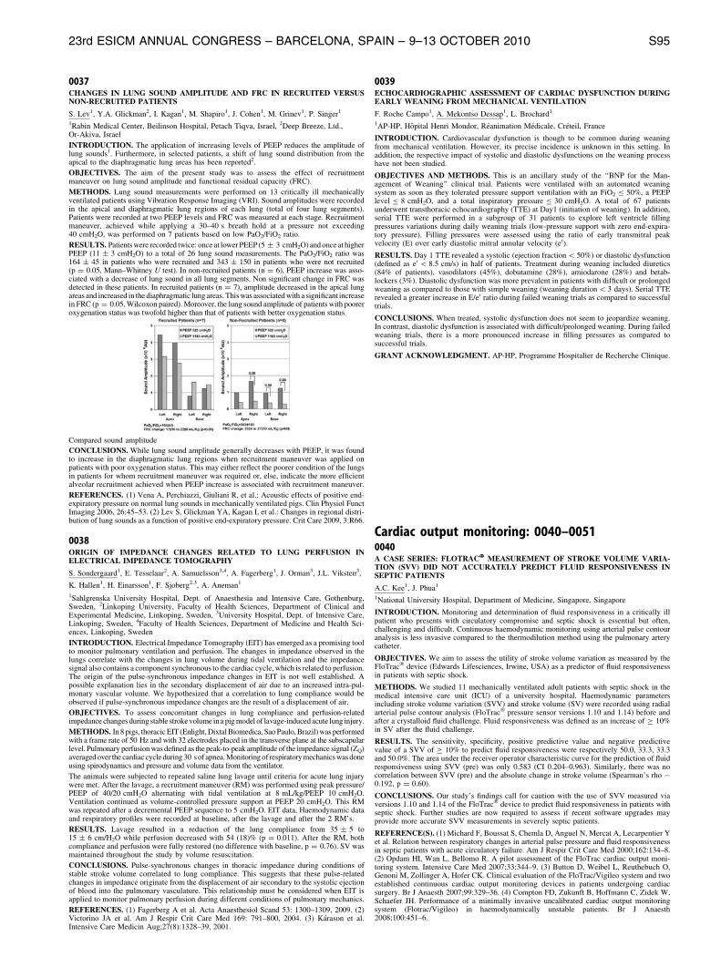
0037CHANGES IN LUNG SOUND AMPLITUDE AND FRC IN RECRUITED VERSUSNON-RECRUITED PATIENTS
S. Lev1, Y.A. Glickman2, I. Kagan1, M. Shapiro1, J. Cohen1, M. Grinev1, P. Singer1
1Rabin Medical Center, Beilinson Hospital, Petach Tiqva, Israel, 2Deep Breeze, Ltd.,Or-Akiva, Israel
INTRODUCTION. The application of increasing levels of PEEP reduces the amplitude oflung sounds1. Furthermore, in selected patients, a shift of lung sound distribution from theapical to the diaphragmatic lung areas has been reported2.
OBJECTIVES. The aim of the present study was to assess the effect of recruitmentmaneuver on lung sound amplitude and functional residual capacity (FRC).
METHODS. Lung sound measurements were performed on 13 critically ill mechanicallyventilated patients using Vibration Response Imaging (VRI). Sound amplitudes were recordedin the apical and diaphragmatic lung regions of each lung (total of four lung segments).Patients were recorded at two PEEP levels and FRC was measured at each stage. Recruitmentmaneuver, achieved while applying a 30–40 s breath hold at a pressure not exceeding40 cmH2O, was performed on 7 patients based on low PaO2/FiO2 ratio.
RESULTS. Patients were recorded twice: once at lower PEEP (5 ± 3 cmH2O) and once at higherPEEP (11 ± 3 cmH2O) to a total of 26 lung sound measurements. The PaO2/FiO2 ratio was164 ± 45 in patients who were recruited and 343 ± 150 in patients who were not recruited(p = 0.05, Mann–Whitney U test). In non-recruited patients (n = 6), PEEP increase was asso-ciated with a decrease of lung sound in all lung segments. Non significant change in FRC wasdetected in these patients. In recruited patients (n = 7), amplitude decreased in the apical lungareas and increased in the diaphragmatic lung areas. This was associated with a significant increasein FRC (p = 0.05, Wilcoxon paired). Moreover, the lung sound amplitude of patients with pooreroxygenation status was twofold higher than that of patients with better oxygenation status.
Compared sound amplitude
CONCLUSIONS. While lung sound amplitude generally decreases with PEEP, it was foundto increase in the diaphragmatic lung regions when recruitment maneuver was applied onpatients with poor oxygenation status. This may either reflect the poorer condition of the lungsin patients for whom recruitment maneuver was required or, else, indicate the more efficientalveolar recruitment achieved when PEEP increase is associated with recruitment maneuver.
REFERENCES. (1) Vena A, Perchiazzi, Giuliani R, et al.: Acoustic effects of positive end-expiratory pressure on normal lung sounds in mechanically ventilated pigs. Clin Physiol FunctImaging 2006, 26:45–53. (2) Lev S, Glickman YA, Kagan I, et al.: Changes in regional distri-bution of lung sounds as a function of positive end-expiratory pressure. Crit Care 2009, 3:R66.
0038ORIGIN OF IMPEDANCE CHANGES RELATED TO LUNG PERFUSION INELECTRICAL IMPEDANCE TOMOGRAPHY
S. Sondergaard1, E. Tesselaar2, A. Samuelsson3,4, A. Fagerberg1, J. Orman3, J.L. Viksten3,
K. Hallen1, H. Einarsson1, F. Sjoberg2,3, A. Aneman1
1Sahlgrenska University Hospital, Dept. of Anaesthesia and Intensive Care, Gothenburg,Sweden, 2Linkoping University, Faculty of Health Sciences, Department of Clinical andExperimental Medicine, Linkoping, Sweden, 3University Hospital, Dept. of Intensive Care,Linkoping, Sweden, 4Faculty of Health Sciences, Department of Medicine and Health Sci-ences, Linkoping, Sweden
INTRODUCTION. Electrical Impedance Tomography (EIT) has emerged as a promising toolto monitor pulmonary ventilation and perfusion. The changes in impedance observed in thelungs correlate with the changes in lung volume during tidal ventilation and the impedancesignal also contains a component synchronous to the cardiac cycle, which is related to perfusion.The origin of the pulse-synchronous impedance changes in EIT is not well established. Apossible explanation lies in the secondary displacement of air due to an increased intra-pul-monary vascular volume. We hypothesized that a correlation to lung compliance would beobserved if pulse-synchronous impedance changes are the result of a displacement of air.
OBJECTIVES. To assess concomitant changes in lung compliance and perfusion-relatedimpedance changes during stable stroke volume in a pig model of lavage-induced acute lung injury.
METHODS. In 8 pigs, thoracic EIT (Enlight, Dixtal Biomedica, Sao Paulo, Brazil) was performedwith a frame rate of 50 Hz and with 32 electrodes placed in the transverse plane at the subscapularlevel. Pulmonary perfusion was defined as the peak-to-peak amplitude of the impedance signal (ZQ)averaged over the cardiac cycle during 30 s ofapnea. Monitoring of respiratory mechanics was doneusing spirodynamics and pressure and volume data from the ventilator.
The animals were subjected to repeated saline lung lavage until criteria for acute lung injurywere met. After the lavage, a recruitment maneuver (RM) was performed using peak pressure/PEEP of 40/20 cmH2O alternating with tidal ventilation at 8 mL/kg/PEEP 10 cmH2O.Ventilation continued as volume-controlled pressure support at PEEP 20 cmH2O. This RMwas repeated after a decremental PEEP sequence to 5 cmH2O. EIT data, Haemodynamic dataand respiratory profiles were recorded at baseline, after the lavage and after the 2 RM’s.
RESULTS. Lavage resulted in a reduction of the lung compliance from 35 ± 5 to15 ± 6 cm/H2O while perfusion decreased with 54 (18)% (p = 0.011). After the RM, bothcompliance and perfusion were fully restored (no difference with baseline, p = 0.76). SV wasmaintained throughout the study by volume resuscitation.
CONCLUSIONS. Pulse-synchronous changes in thoracic impedance during conditions ofstable stroke volume correlated to lung compliance. This suggests that these pulse-relatedchanges in impedance originate from the displacement of air secondary to the systolic ejectionof blood into the pulmonary vasculature. This relationship must be considered when EIT isapplied to monitor pulmonary perfusion during different conditions of pulmonary mechanics.
REFERENCES. (1) Fagerberg A et al. Acta Anaesthesiol Scand 53: 1300–1309, 2009. (2)Victorino JA et al. Am J Respir Crit Care Med 169: 791–800, 2004. (3) Karason et al.Intensive Care Medicin Aug;27(8):1328–39, 2001.
0039ECHOCARDIOGRAPHIC ASSESSMENT OF CARDIAC DYSFUNCTION DURINGEARLY WEANING FROM MECHANICAL VENTILATION
F. Roche Campo1, A. Mekontso Dessap1, L. Brochard1
1AP-HP, Hopital Henri Mondor, Reanimation Medicale, Creteil, France
INTRODUCTION. Cardiovascular dysfunction is though to be common during weaningfrom mechanical ventilation. However, its precise incidence is unknown in this setting. Inaddition, the respective impact of systolic and diastolic dysfunctions on the weaning processhave not been studied.
OBJECTIVES AND METHODS. This is an ancillary study of the ‘‘BNP for the Man-agement of Weaning’’ clinical trial. Patients were ventilated with an automated weaningsystem as soon as they tolerated pressure support ventilation with an FiO2 B 50%, a PEEPlevel B 8 cmH2O, and a total inspiratory pressure B 30 cmH2O. A total of 67 patientsunderwent transthoracic echocardiography (TTE) at Day1 (initiation of weaning). In addition,serial TTE were performed in a subgroup of 31 patients to explore left ventricle fillingpressures variations during daily weaning trials (low-pressure support with zero end-expira-tory pressure). Filling pressures were assessed using the ratio of early transmitral peakvelocity (E) over early diastolic mitral annular velocity (e0).
RESULTS. Day 1 TTE revealed a systolic (ejection fraction \ 50%) or diastolic dysfunction(defined as e0\ 8.5 cm/s) in half of patients. Treatment during weaning included diuretics(84% of patients), vasodilators (45%), dobutamine (28%), amiodarone (28%) and betab-lockers (3%). Diastolic dysfunction was more prevalent in patients with difficult or prolongedweaning as compared to those with simple weaning (weaning duration \ 3 days). Serial TTErevealed a greater increase in E/e0 ratio during failed weaning trials as compared to successfultrials.
CONCLUSIONS. When treated, systolic dysfunction does not seem to jeopardize weaning.In contrast, diastolic dysfunction is associated with difficult/prolonged weaning. During failedweaning trials, there is a more pronounced increase in filling pressures as compared tosuccessful trials.
GRANT ACKNOWLEDGMENT. AP-HP, Programme Hospitalier de Recherche Clinique.
Cardiac output monitoring: 0040–00510040A CASE SERIES: FLOTRAC� MEASUREMENT OF STROKE VOLUME VARIA-TION (SVV) DID NOT ACCURATELY PREDICT FLUID RESPONSIVENESS INSEPTIC PATIENTS
A.C. Kee1, J. Phua1
1National University Hospital, Department of Medicine, Singapore, Singapore
INTRODUCTION. Monitoring and determination of fluid responsiveness in a critically illpatient who presents with circulatory compromise and septic shock is essential but often,challenging and difficult. Continuous haemodynamic monitoring using arterial pulse contouranalysis is less invasive compared to the thermodilution method using the pulmonary arterycatheter.
OBJECTIVES. We aim to assess the utility of stroke volume variation as measured by theFloTrac� device (Edwards Lifesciences, Irwine, USA) as a predictor of fluid responsivenessin patients with septic shock.
METHODS. We studied 11 mechanically ventilated adult patients with septic shock in themedical intensive care unit (ICU) of a university hospital. Haemodynamic parametersincluding stroke volume variation (SVV) and stroke volume (SV) were recorded using radialarterial pulse contour analysis (FloTrac� pressure sensor versions 1.10 and 1.14) before andafter a crystalloid fluid challenge. Fluid responsiveness was defined as an increase of C 10%in SV after the fluid challenge.
RESULTS. The sensitivity, specificity, positive predictive value and negative predictivevalue of a SVV of C 10% to predict fluid responsiveness were respectively 50.0, 33.3, 33.3and 50.0%. The area under the receiver operator characteristic curve for the prediction of fluidresponsiveness using SVV (pre) was only 0.583 (CI 0.204–0.963). Similarly, there was nocorrelation between SVV (pre) and the absolute change in stroke volume (Spearman’s rho -0.192, p = 0.60).
CONCLUSIONS. Our study’s findings call for caution with the use of SVV measured viaversions 1.10 and 1.14 of the FloTrac� device to predict fluid responsiveness in patients withseptic shock. Further studies are now required to assess if recent software upgrades mayprovide more accurate SVV measurements in severely septic patients.
REFERENCE(S). (1) Michard F, Boussat S, Chemla D, Anguel N, Mercat A, Lecarpentier Yet al. Relation between respiratory changes in arterial pulse pressure and fluid responsivenessin septic patients with acute circulatory failure. Am J Respir Crit Care Med 2000;162:134–8.(2) Opdam HI, Wan L, Bellomo R. A pilot assessment of the FloTrac cardiac output moni-toring system. Intensive Care Med 2007;33:344–9. (3) Button D, Weibel L, Reuthebuch O,Genoni M, Zollinger A, Hofer CK. Clinical evaluation of the FloTrac/Vigileo system and twoestablished continuous cardiac output monitoring devices in patients undergoing cardiacsurgery. Br J Anaesth 2007;99:329–36. (4) Compton FD, Zukunft B, Hoffmann C, Zidek W,Schaefer JH. Performance of a minimally invasive uncalibrated cardiac output monitoringsystem (Flotrac/Vigileo) in haemodynamically unstable patients. Br J Anaesth2008;100:451–6.
23rd ESICM ANNUAL CONGRESS – BARCELONA, SPAIN – 9–13 OCTOBER 2010 S95
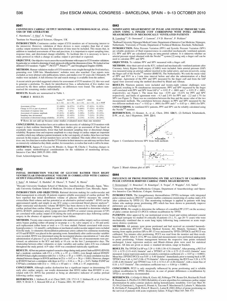
0041CONTINUOUS CARDIAC OUTPUT MONITORS: A METHODOLOGICAL ANAL-YSIS OF THE LITERATURE
C. Hawthorne1, J. Glen1, S. Young1
1Institute for Neurological Sciences, Glasgow, UK
INTRODUCTION. Continuous cardiac output (CCO) monitors are of increasing interest tothe intensivist. However, validation of these devices is more complex than that of staticcardiac output monitors because the dimension of time must be included. This means that, inaddition to standard assessments of bias and precision, it is important to report sampling time,response time, and directional change reliability. To facilitate this it is necessary to have astudy protocol in which a change in cardiac output is provoked or expected.
OBJECTIVE. Ourobjectivewas toassess the recent literature with respect to CCOmonitor validation.Inparticularwe wished todetermine if study protocols reflected the dimension of time. Welooked at fourdifferent CCO monitors: VigileoTM, PiCCOTM, PulseCoTM, and Oesophageal Doppler (ODM).
METHODS. Human validation studies of CCO monitors were sought through the Ovid interface,generating over 3,000 hits. Manufacturers’ websites were also searched. Case reports wereexcluded, as were abstract-only publications, letters, and studies over 10 years old. Ultimately, 99studies were included. A full reference list and search strategy is available from the authors.
A recent article provided suggested criteria for assessment of CCO monitors [1]: this was usedto generate a proforma. To check for interobserver bias, a subselection of five studies wasassessed by the three authors independently; no differences were found. The authors sum-marised the remaining studies individually.
RESULTS. Results are summarised in Table 1.
TABLE 1 RESULTS
VigileoTM
(n = 32)
PiCCOTM
(n = 35)
LiDCOTM
(n = 15)
ODM
(n = 33)
Total
(n = 99)W
Assessment of bias 32 (100%) 34 (97%) 13 (87%) 27 (82%) 91 (92%)
Change in CO as part of protocol 22 (69%) 27 (77%) 9 (60%) 17 (52%) 62 (63%)
Assessment of response time 1 (3%) 2 (6%) 1 (7%) 0 (0%) 4 (4%)
Assessment of response amplitude 13 (41%) 21 (60%) 3 (20%) 5 (15%) 32 (32%)
Sampling time documented 18 (56%) 15 (43%) 5 (33%) 7 (21%) 35 (35%)
Assessment of directional change reliability 15 (47%) 15 (43%) 2 (13%) 4 (12%) 28 (28%)
W Rows do not add up because some studies evaluated more than one monitor
CONCLUSIONS. Researchers have yet to address the necessity of validating CCO monitors withrespect to their realtime functionality. While most studies give an assessment of bias based onessentially static measurements, fewer than half document sampling time or directional changereliability. Response time and response amplitude to a step change in cardiac output are importantvariables which may influence patient treatment; in the vast majority of studies, these have not beenassessed. In this respect, all four monitors have yet to be validated. This study offers two perspec-tives: one, for clinicians to realise that the CCO monitor in their intensive care unit may not have beenas extensively validated as they think; another, for researchers, to realise that work is still to be done.
REFERENCE. Squara P, Cecconi M, Rhodes A, Singer M, Chiche J. Tracking changes incardiac output: methodological considerations for the validation of monitoring devices,Intensive Care Med 2009;35:1801–1808.
Grant Acknowledgement: None.
0042INITIAL DISTRIBUTION VOLUME OF GLUCOSE RATHER THAN RIGHTVENTRICULAR END-DIASTOLIC VOLUME IS CORRELATED WITH CARDIACOUTPUT FOLLOWING CARDIAC SURGERY
J. Saito1, H. Ishihara1, E. Hashiba2, H. Okawa1, T. Tsubo2, K. Hirota1
1Hirosaki University Graduate School of Medicine, Anesthesiology, Hirosaki, Japan, 2Hiro-saki University Graduate School of Medicine, Division of Intensive Care, Hirosaki, Japan
INTRODUCTION AND OBJECTIVES. Rational decision making for cardiovascular andfluid management in critically ill patients requires reliable assessment of cardiac preload. Wehave reported that initial distribution volume of glucose (IDVG) measures the centralextracellular fluid volume and has potential as an alternative preload variable1). IDVG can beapproximated rapidly and simply in any ICU using a conventional blood glucose analyzer2).Right ventricular end-diastolic volume (RVEDV) has been shown to be a better indicator ofcardiac preload than cardiac filling pressure3). This study was intended to determine whetherIDVG, RVEDV, pulmonary artery wedge pressure (PAWP) or central venous pressure (CVP)are correlated with cardiac output (CO) during the early postoperative days following cardiacsurgery in the absence of apparent congestive heart failure.
METHODS. Twenty-nine consecutive patients who underwent cardiac surgery such as coronaryartery bypass grafting (either off-pump or on-pump: n = 6), valve surgery (n = 10) and aorticarch replacement (either hemi or total: n = 13) were studied. Patients associated with excesshyperglycemia ([ 14 mmol/L), arrhythmias or mechanical cardiovascular support were excludedfrom the study. A volumetric thermodilution pulmonary artery catheter for continuous monitoringof CO and RVEDV was placed in the operating room. Immediately after cardiovascular variableswere recorded, IDVG was determined using the incremental plasma concentration at 3 min afteradministration of glucose (5 g) as described previously2). Three sets of measurements were per-formed; on admission to the ICU and daily at 10 a.m. on the first 2 postoperative days. Therelationship between either volumetric or static variables and cardiac index (CI) was evaluatedthroughout the study period. A P value\0.05 was considered statistically significant.
RESULTS. Allbutonepatients requiredvasoactivedrugsduringstudyperiod. IndexedIDVG(IDVGI)had a moderate correlation with CI (r = 0.56, n = 87, p\0.001), even though indexed RVEDV(RVEDVI)hadaslightcorrelationwithCI (r = 0.29, n = 87, p = 0.007).ALinearcorrelationwasalsoobtained between changes in IDVGI and those in CI (r = 0.47, n = 58, p\0.001). However, changesin RVEDVI had not a correlation with those in CI (r = 0.10, n = 58, p = 0.47). Neither PAWP norCVP had a correlation with CI (r = -0.02, n = 65 and r = -0.06, n = 87, respectively).
CONCLUSION. Although cardiac dysfunction has a significant impact on determining COearly after cardiac surgery, our results demonstrate that IDVG rather than RVEDV is cor-related with CO. IDVG has potential as being an alternative indicator of cardiac preloadfollowing cardiac surgery.
REFERENCES. 1. Ishihara H, et al. Chest 2005; 128: 1713–9. 2. Ishihara H, et al. Crit Care2005; 9: R144–9. 3. Kincaid EH et al. J Trauma 2001; 50: 659–65.
0043NONINVASIVE MEASUREMENT OF PULSE AND SYSTOLIC PRESSURE VARI-ATION USING A FINGER CUFF CORRESPOND WITH INTRA ARTERIALMEASUREMENTS IN MECHANICALLY VENTILATED PATIENTS
B. Lansdorp1,2, D. Ouweneel2, J. Lemson1, J.V.D. Hoeven1, P. Pickkers1
1Radboud University Nijmegen Medical Centre, Department of Intensive Care Medicine, Nijmegen,Netherlands, 2University of Twente, Department of Technical Medicine, Enschede, Netherlands
INTRODUCTION. Pulse Pressure Variation (PPV) and Systolic Pressure Variation (SPV)are reliable predictors of fluid responsiveness in controlled mechanically ventilated patients[1]. PPV and SPV are calculated using an intra-arterial catheter. It is unknown whether anarterial pressure signal obtained with the NexfinTM system [2] using only a finger cuff can beused to calculate PPV and SPV.
OBJECTIVES. To validate PPV and SPV measured with a finger cuff.
METHODS. After their arrival on the ICU, sedated and mechanically ventilated patients afterCoronary Artery Bypass Graft surgery (CABG) were included. Intra arterial pressure (IAP)was measured using an arterial catheter inserted in the radial artery, and non-invasively, usingthe finger cuff of the NexfinTM monitor (BMEYE, The Netherlands). We took the mean valueof PPV and SVV in a 1-min time interval before and after the administration of a fluidchallenge. Agreement of the PPV and SPV measured by the finger cuff and from the IAPsignal were assessed using the method described by Bland and Altman.
RESULTS. Nineteen patients were included and twenty-eight volume challenges wereanalyzed, resulting in 56 simultaneous measurements. PPV and SPV measured by the fingercuff correlated with PPV and SPV from IAP (r2 = 0.92, P \ .0001 and r2 = 0.93, P \ .0001,respectively), see Figure 1. The mean bias was -0.95 and -0.22% for PPV and SPVrespectively, and limits of agreement were -4.3 and 2.4% for PPV and -2.2 and 1.7% forSPV (see Figure 2). There was no correlation between the bias and the mean value of the twomeasurement methods. The correlation between changes in PPV and SPV measured by thetwo different methods was r2 = 0.82 (p \ .0001) for PPV and r2 = 0.83 (p \ .0001) for SPV.
CONCLUSIONS. In ventilated ICU patients, PPV and SPV can be reliably calculated usingthe NexfinTM monitor.
REFERENCE(S). (1) Kramer, A., et al., Chest, 2004. 126(5). (2) Eeftinck Schattenkerk,D.W., et al., Am J Hypertens, 2009. 22(4).
Figure 1: Correlation between SPV from both methods
Figure 2: Bland–Altman plot of SPV
0044INFLUENCE OF PRONE POSITIONING ON THE ACCURACY OF CALIBRATEDPULSE CONTOUR DERIVED CARDIAC INDEX MEASUREMENTS
J. Grensemann1, U. Bruecken1, D. Knuettgen1, S. Trojan1, F. Wappler1, S.G. Sakka1
1University Hospital Witten/Herdecke Cologne, Department of Anaesthesiology and Opera-tive Intensive Care Medicine, Cologne, Germany
INTRODUCTION. The transpulmonary thermodilution (TPTD) technique with integratedpulse contour analysis (PiCCO�-system) enables continuous monitoring of cardiac index (CI)after calibration by TPTD [1]. This monitoring technique is applied in patients with lungfailure who undergo prone positioning (PP) which has been shown to potentially improvepulmonary gas exchange [2].
OBJECTIVE. We sought to determine the influence of a modified PP (135�) on the accuracyof pulse contour derived CI (PCCI) without recalibration by TPTD.
PATIENTS: After approval by our institutional review board and written informed consentby a legal surrogate we studied 16 critically ill patients (11#, 5$, age 20–71 years) who weremechanically ventilated due to acute lung injury following lung contusions or acute respi-ratory distress syndrome.
METHODS. All patients were prone positioned and had received an extended haemody-namic monitoring (PiCCO�, Pulsion Medical Systems AG, Munich, Germany). Beforeturning from supine position (SP) to PP, CI was measured by TPTD (TPTDCI) and PCCI wascalibrated. Ten minutes after positioning, PCCI was read from the monitor and then recali-brated by TPTD. After 8–10 h, PP was ended and measurements were performed analogouslyto prone positioning. Volume management between the respective time points remainedunchanged. Linear regression analysis and Bland–Altman plots were used for statisticalanalysis. All data are given as mean ± standard deviation, range in brackets.
RESULTS. The TPTDCI in SP was 3.29 ± 0.88 (1.50–4.51) l/min/m2. After proning, a PCCI of3.51 ± 0.79 (2.15–4.63) l/min/m2 and a TPTDCI of 3.56 ± 0.75 (2.33–4.63) l/min/m2 weremeasured. Linear regression analysis revealed a correlation coefficient of r = 0.87 (p \ 0.0001).Mean bias (TPTDCI-PCCI) was 0.05 ± 0.40 l/min/m2. Immediately prior to turning back to SP,TPTDCI was 3.46 ± 0.67 (2.06–4.75) l/min/m2. After re-positioning, the PCCI was 3.28 ± 0.70(2.00–4.48) l/min/m2 and TPTDCI was 3.10 ± 0.74 (1.47–4.28) l/min/m2, with a mean bias of0.18 ± 0.37 l/min/m2. The correlation coefficient was r = 0.84 (p \ 0.0001).
CONCLUSION. PCCI is only marginally influenced by prone positioning and is reliablewithout recalibration by TPTD. However, in case of greater differences a recalibration byTPTD is nevertheless recommended.
REFERENCES. (1) Godje O, Hoke K, Goetz AE, Felbinger TW, Reuter DA, Reichart B, FriedlR, Hannekum A, Pfeiffer UJ (2002) Reliability of a new algorithm for continuous cardiac outputdetermination by pulse-contour analysis during hemodynamic instability. Crit Care Med 30:52–58 (2) Gattinoni L, Tognoni G, Pesenti A, Taccone P, Mascheroni D, Labarta V, MalacridaR, Di Giulio P, Fumagalli R, Pelosi P, Brazzi L, Latini R (2001) Effect of prone positioning onthe survival of patients with acute respiratory failure. N Engl J Med 345: 568–573
S96 23rd ESICM ANNUAL CONGRESS – BARCELONA, SPAIN – 9–13 OCTOBER 2010
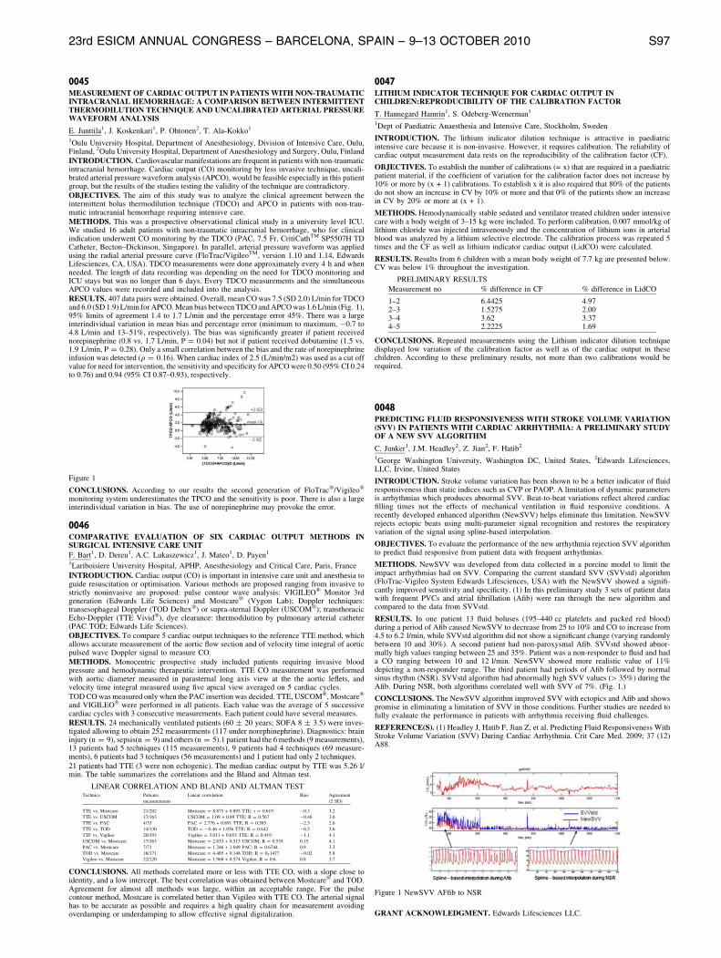
0045MEASUREMENT OF CARDIAC OUTPUT IN PATIENTS WITH NON-TRAUMATICINTRACRANIAL HEMORRHAGE: A COMPARISON BETWEEN INTERMITTENTTHERMODILUTION TECHNIQUE AND UNCALIBRATED ARTERIAL PRESSUREWAVEFORM ANALYSIS
E. Junttila1, J. Koskenkari1, P. Ohtonen2, T. Ala-Kokko1
1Oulu University Hospital, Department of Anesthesiology, Division of Intensive Care, Oulu,Finland, 2Oulu University Hospital, Department of Anesthesiology and Surgery, Oulu, Finland
INTRODUCTION. Cardiovascular manifestations are frequent in patients with non-traumaticintracranial hemorrhage. Cardiac output (CO) monitoring by less invasive technique, uncali-brated arterial pressure waveform analysis (APCO), would be feasible especially in this patientgroup, but the results of the studies testing the validity of the technique are contradictory.
OBJECTIVES. The aim of this study was to analyze the clinical agreement between theintermittent bolus thermodilution technique (TDCO) and APCO in patients with non-trau-matic intracranial hemorrhage requiring intensive care.
METHODS. This was a prospective observational clinical study in a university level ICU.We studied 16 adult patients with non-traumatic intracranial hemorrhage, who for clinicalindication underwent CO monitoring by the TDCO (PAC, 7.5 Fr, CritiCathTM SP5507H TDCatheter, Becton–Dickinson, Singapore). In parallel, arterial pressure waveform was appliedusing the radial arterial pressure curve (FloTrac/VigileoTM, version 1.10 and 1.14, EdwardsLifesciences, CA, USA). TDCO measurements were done approximately every 4 h and whenneeded. The length of data recording was depending on the need for TDCO monitoring andICU stays but was no longer than 6 days. Every TDCO measurements and the simultaneousAPCO values were recorded and included into the analysis.
RESULTS. 407 data pairs were obtained. Overall, mean CO was 7.5 (SD 2.0) L/min for TDCOand 6.0 (SD 1.9) L/min for APCO. Mean bias between TDCO and APCO was 1.6 L/min (Fig. 1),95% limits of agreement 1.4 to 1.7 L/min and the percentage error 45%. There was a largeinterindividual variation in mean bias and percentage error (minimum to maximum, -0.7 to4.8 L/min and 13–51%, respectively). The bias was significantly greater if patient receivednorepinephrine (0.8 vs. 1.7 L/min, P = 0.04) but not if patient received dobutamine (1.5 vs.1.9 L/min, P = 0.28). Only a small correlation between the bias and the rate of norepinephrineinfusion was detected (q = 0.16). When cardiac index of 2.5 (L/min/m2) was used as a cut offvalue for need for intervention, the sensitivity and specificity for APCO were 0.50 (95% CI 0.24to 0.76) and 0.94 (95% CI 0.87–0.93), respectively.
Figure 1
CONCLUSIONS. According to our results the second generation of FloTrac�/Vigileo�
monitoring system underestimates the TPCO and the sensitivity is poor. There is also a largeinterindividual variation in bias. The use of norepinephrine may provoke the error.
0046COMPARATIVE EVALUATION OF SIX CARDIAC OUTPUT METHODS INSURGICAL INTENSIVE CARE UNITF. Bart1, D. Dereu1, A.C. Lukaszewicz1, J. Mateo1, D. Payen1
1Lariboisiere University Hospital, APHP, Anesthesiology and Critical Care, Paris, France
INTRODUCTION. Cardiac output (CO) is important in intensive care unit and anesthesia toguide resuscitation or optimisation. Various methods are proposed ranging from invasive tostrictly noninvasive are proposed: pulse contour wave analysis: VIGILEO� Monitor 3rdgeneration (Edwards Life Sciences) and Mostcare� (Vygon Lab); Doppler techniques:transesophageal Doppler (TOD Deltex�) or supra-sternal Doppler (USCOM�); transthoracicEcho-Doppler (TTE Vivid�), dye clearance: thermodilution by pulmonary arterial catheter(PAC TOD; Edwards Life Sciences).
OBJECTIVES. To compare 5 cardiac output techniques to the reference TTE method, whichallows accurate measurement of the aortic flow section and of velocity time integral of aorticpulsed wave Doppler signal to measure CO.
METHODS. Monocentric prospective study included patients requiring invasive bloodpressure and hemodynamic therapeutic intervention. TTE CO measurement was performedwith aortic diameter measured in parasternal long axis view at the the aortic leflets, andvelocity time integral measured using five apical view averaged on 5 cardiac cycles.
TOD CO was measured only when the PAC insertion was decided. TTE, USCOM�, Mostcare�
and VIGILEO� were performed in all patients. Each value was the average of 5 successivecardiac cycles with 3 consecutive measurements. Each patient could have several measures.
RESULTS. 24 mechanically ventilated patients (60 ± 20 years; SOFA 8 ± 3.5) were inves-tigated allowing to obtain 252 measurements (117 under norephinephrine). Diagnostics: braininjury (n = 9), sepsis(n = 9) and others (n = 5).1 patient had the 6 methods (9 measurements),13 patients had 5 techniques (115 measurements), 9 patients had 4 techniques (69 measure-ments), 6 patients had 3 techniques (56 measurements) and 1 patient had only 2 techniques.
21 patients had TTE (3 were non echogenic). The median cardiac output by TTE was 5.26 l/min. The table summarizes the correlations and the Bland and Altman test.
LINEAR CORRELATION AND BLAND AND ALTMAN TESTTechnics Patients
measurements
Linear correlation Bias Agreement
(2 SD)
TTE vs. Mostcare 21/202 Mostcare = 8.875 + 0.895 TTE; r = 0.619 -0.3 3.2
TTE vs. USCOM 17/163 USCOM = 1.09 + 0.89 TTE; R = 0.567 -0.46 3.6
TTE vs. PAC 4/33 PAC = 2.776 + 0.891 TTE; R = 0.585 -2.3 2.6
TTE vs. TOD 14/130 TOD = -0.46 + 1.056 TTE; R = 0.642 -0.3 3.6
TTF vs. Vigileo 20/193 Vigileo = 3.011 + 0.653 TTE; R = 0.419 -1.1 4.1
USCOM vs. Mostcare 17/163 Mostcare = 2.833 + 0.513 USCOM; R = 0.535 0.15 4.1
PAC VS. Mostcare 7/71 Mostcare = 1.246 + 1.049 PAC; R = 0.6744 0.9 3.3
TOD VS. Mostcare 18/171 Mostcare = 4.485 + 0.148 TOD; R = 0T1477 -0.02 5.8
Vigileo vs. Mostcare 22/220 Mostcare = 1.968 + 0.574 Vigileo; R = 0.6 0.8 3.7
CONCLUSIONS. All methods correlated more or less with TTE CO, with a slope close toidentity, and a low intercept. The best correlation was obtained between Mostcare� and TOD.Agreement for almost all methods was large, within an acceptable range. For the pulsecontour method, Mostcare is correlated better than Vigileo with TTE CO. The arterial signalhas to be accurate as possible and requires a high quality chain for measurement avoidingoverdamping or underdamping to allow effective signal digitalization.
0047LITHIUM INDICATOR TECHNIQUE FOR CARDIAC OUTPUT INCHILDREN:REPRODUCIBILITY OF THE CALIBRATION FACTOR
T. Hannegard Hamrin1, S. Odeberg-Wernerman1
1Dept of Paediatric Anaesthesia and Intensive Care, Stockholm, Sweden
INTRODUCTION. The lithium indicator dilution technique is attractive in paediatricintensive care because it is non-invasive. However, it requires calibration. The reliability ofcardiac output measurement data rests on the reproducibility of the calibration factor (CF).
OBJECTIVES. To establish the number of calibrations (= x) that are required in a paediatricpatient material, if the coefficient of variation for the calibration factor does not increase by10% or more by (x + 1) calibrations. To establish x it is also required that 80% of the patientsdo not show an increase in CV by 10% or more and that 0% of the patients show an increasein CV by 20% or more at (x + 1).
METHODS. Hemodynamically stable sedated and ventilator treated children under intensivecare with a body weight of 3–15 kg were included. To perform calibration, 0.007 mmol/kg oflithium chloride was injected intravenously and the concentration of lithium ions in arterialblood was analyzed by a lithium selective electrode. The calibration process was repeated 5times and the CF as well as lithium indicator cardiac output (LidCO) were calculated.
RESULTS. Results from 6 children with a mean body weight of 7.7 kg are presented below.CV was below 1% throughout the investigation.
PRELIMINARY RESULTS
Measurement no % difference in CF % difference in LidCO
1–2 6.4425 4.972–3 1.5275 2.003–4 3.62 3.374–5 2.2225 1.69
CONCLUSIONS. Repeated measurements using the Lithium indicator dilution techniquedisplayed low variation of the calibration factor as well as of the cardiac output in thesechildren. According to these preliminary results, not more than two calibrations would berequired.
0048PREDICTING FLUID RESPONSIVENESS WITH STROKE VOLUME VARIATION(SVV) IN PATIENTS WITH CARDIAC ARRHYTHMIA: A PRELIMINARY STUDYOF A NEW SVV ALGORITHM
C. Junker1, J.M. Headley2, Z. Jian2, F. Hatib2
1George Washington University, Washington DC, United States, 2Edwards Lifesciences,LLC, Irvine, United States
INTRODUCTION. Stroke volume variation has been shown to be a better indicator of fluidresponsiveness than static indices such as CVP or PAOP. A limitation of dynamic parametersis arrhythmias which produces abnormal SVV. Beat-to-beat variations reflect altered cardiacfilling times not the effects of mechanical ventilation in fluid responsive conditions. Arecently developed enhanced algorithm (NewSVV) helps eliminate this limitation. NewSVVrejects ectopic beats using multi-parameter signal recognition and restores the respiratoryvariation of the signal using spline-based interpolation.
OBJECTIVES. To evaluate the performance of the new arrhythmia rejection SVV algorithmto predict fluid responsive from patient data with frequent arrhythmias.
METHODS. NewSVV was developed from data collected in a porcine model to limit theimpact arrhythmias had on SVV. Comparing the current standard SVV (SVVstd) algorithm(FloTrac-Vigileo System Edwards Lifesciences, USA) with the NewSVV showed a signifi-cantly improved sensitivity and specificity. (1) In this preliminary study 3 sets of patient datawith frequent PVCs and atrial fibrillation (Afib) were ran through the new algorithm andcompared to the data from SVVstd.
RESULTS. In one patient 13 fluid boluses (195–440 cc platelets and packed red blood)during a period of Afib caused NewSVV to decrease from 25 to 10% and CO to increase from4.5 to 6.2 l/min, while SVVstd algorithm did not show a significant change (varying randomlybetween 10 and 30%). A second patient had non-paroxysmal Afib. SVVstd showed abnor-mally high values ranging between 25 and 35%. Patient was a non-responder to fluid and hada CO ranging between 10 and 12 l/min. NewSVV showed more realistic value of 11%depicting a non-responder range. The third patient had periods of Afib followed by normalsinus rhythm (NSR). SVVstd algorithm had abnormally high SVV values ([ 35%) during theAfib. During NSR, both algorithms correlated well with SVV of 7%. (Fig. 1.)
CONCLUSIONS. The NewSVV algorithm improved SVV with ectopics and Afib and showspromise in eliminating a limitation of SVV in those conditions. Further studies are needed tofully evaluate the performance in patients with arrhythmia receiving fluid challenges.
REFERENCE(S). (1) Headley J, Hatib F, Jian Z, et al. Predicting Fluid Responsiveness WithStroke Volume Variation (SVV) During Cardiac Arrhythmia. Crit Care Med. 2009; 37 (12)A88.
Figure 1 NewSVV AFfib to NSR
GRANT ACKNOWLEDGMENT. Edwards Lifesciences LLC.
23rd ESICM ANNUAL CONGRESS – BARCELONA, SPAIN – 9–13 OCTOBER 2010 S97
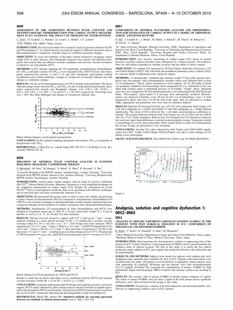
0049ASSESSMENT OF THE AGREEMENT BETWEEN PULSE CONTOUR ANDTRANSPULMONARY THERMODILUTION FOR CARDIAC OUTPUT MEASURE-MENT IN ICU PATIENTS: THE IMPACT OF THERAPEUTIC INTERVENTIONS
L. Zoric1, D. Candela1, L. Mattatia1, G. Louart1, L. Muller1, J.Y. Lefrant1
1CHU NIMES, Pole ARDU, Nimes, France
INTRODUCTION. Several recent studies have reported a lack of agreement between the PCand TP techniques [1, 2], without directly assessing the impact of different treatments such asfluid administration, vasopressor, inotropic drugs or ventilatory parameter changes.
OBJECTIVES. To assess the reliability of the pulse contour method for measuring cardiacoutput (CO) in daily practice, after therapeutic maneuver-free periods and different thera-peutic interventions that can influence vascular compliance and systemic vascular resistancesin intensive care unit patients (ICU).
METHODS. In ICU patients requiring the CO monitoring, transpulmonary thermodilutionCO (TPCO) and pulse contour CO (PCCO) were measured with a PiCCO device after thera-peutic maneuver-free periods (B and [ 1 h) and after therapeutic interventions withoutrecalibration such as fluid challenges, changes in vasopressor or inotropic infusion rates andchanges in ventilatory settings.
RESULTS. 352 sets of CO measurement pairs in 63 ICU patients were performed. The biases(and percentage errors) between PCCO and TPCO for the overall paired measurement, thera-peutic maneuver-free periods and therapeutic changes were 0.20 ± 1.09 (32.8%), -0.01 ± 0.93 (18% \ 1 h, 39% [ 1 h) and 0.37 ± 1.18 (34%) respectively. Percentage errorwas [ 30% after fluid challenges and changes in vasopressor infusion rates.
Bland–Altman diagram overall paired measurements
CONCLUSIONS. In ICU patients requiring therapeutic interventions, PCCO is frequently indisagreement with TPCO.
REFERENCE(S). (1) Bein B et al., Anesth Analg 2007;105:107–13 (2) Rodig G et al., Br JAnaesth 1999;82:525–30.
0050PRECISION OF ARTERIAL PULSE CONTOUR ANALYSIS IN PATIENTSREQUIRING HIGH-DOSE VASOPRESSOR THERAPY
S. Metzelder1, M. Fries2, M. Reinges3, S. Reich1, G. Marx2, R. Rossaint1, S. Rex1
1University Hospital of the RWTH Aachen, Anesthesiology, Aachen, Germany, 2UniversityHospital of the RWTH Aachen, Intensive Care, Aachen, Germany, 3University Hospital of theRWTH Aachen, Neurosurgery, Aachen, Germany
INTRODUCTION. Arterial pulse contour analysis with the need for invasive calibration(PiCCOTM, Pulsion Medical Systems, Munchen, Germany) is a well established technique forthe continuous measurement of cardiac output (CO). Despite the widespread use of thePiCCOTM-device in perioperative medicine, data on its agreement with reference techniquesand its precision have not been systematically reported so far.
OBJECTIVES. We performed the present study in order to assess the validity and precisionof pulse contour CO-measurements (PCCO) compared to transpulmonary thermodilution CO(TPCO) as the reference technique in hemodynamically instable patients requiring high-dosevasopressor-therapy for the treatment of cerebral vasospasms after subarachnoid hemorrhage.
METHODS. Simultaneous CO measurements by bolus thermodilution and PCCO wereobtained in 20 patients (mean age (± SD) 45 ± 8 years, mean body weight 74 ± 12 kg) atbaseline as well as 2, 6, 12, 24, 48 and 72 h after inclusion.
RESULTS. Patients received vasoactive support with 0.57 ± 0.49 lg kg-1 min-1 norepi-nephrine resulting in a mean arterial pressure of 103 ± 13 mmHg and a systemic vascularresistance index of 1.751 ± 442 dyn s cm-5 m-2. 136 CO-data pairs were analyzed. TPCOranged from 5.2 to 14.3 l min-1 (mean ± SD 8.6 ± 2.0 l min-1) and PCCO ranged from 5.0 to16.4 l min-1 (mean ± SD 8.6 ± 2.1 l min-1). Bias and limits of agreement (1.96 SD of thebias) were -0.1 and 1.7 l min-1, resulting in an overall percentage error of 19.7%. The precisionof TPCO measurements was 2.6%, while PCCO-measurements yielded a precision of 19.1%.
Bland–Altman for CO-measurements by TPCO and PCCO
Besides it could also be shown that there was no correlation between PCCO and systemicvascular resistance (SVR) (R = 0.110; R2 = 0.0121; p = 0.202).
CONCLUSIONS. In patients undergoing triple-H-therapy and requiring extensive vasoactivesupport, PCCO-values obtained by pulse contour analysis showed clinically acceptable agree-ment with intermittent TPCO-measurements. The precision of PCCO-measurements was belowthe cut-off of 20%, statistically indicating the interchangeability of the two methods [1].
REFERENCE(S). Bland JM, Altman DG: Statistical methods for assessing agreementbetween two methods of clinical measurement. Lancet 1986, 1:307–310
0051COMPARISON OF ARTERIAL WAVEFORM ANALYSIS AND THERMODILU-TION FOR ESTIMATION OF CARDIAC OUTPUT IN A MODEL OF ABDOMINALAORTIC ANEURYSM RUPTURE
P. Suk1, I. Cundrle Jr.1, J. Hruda1, M. Helan1, J. Krbusik1, M. Vlasin2, M. Matejovic3,
M. Pavlik1, V. Sramek1
1St. Anna University Hospital, Masaryk University, ICRC, Department of Anesthesia andIntesive Care, Brno, Czech Republic, 2University of Veterinary and Pharmaceutical Sciences,ICRC, Brno, Czech Republic, 3University Hospital and Charles University Pilsen, 1stDepartment of Internal Medicine, Pilsen, Czech Republic
INTRODUCTION. Less invasive monitoring of cardiac output (CO) based on arterialpressure waveform analysis becomes more widespread in a clinical practice. Nevertheless,there are still debates regarding its absolute accuracy and the ability to detect changes.
OBJECTIVES. To compare CO measured by FloTrac/Vigileo (Edwards Lifesciences, US)and LiDCO Rapid (LiDCO, UK) with bolus thermodilution pulmonary artery catheter (PAC)in a porcine model of abdominal aortic aneurysm rupture.
METHODS. 14 mechanically ventilated pigs (median weight 37 kg) under general anes-thesia were investigated. After instrumentation, baseline values were obtained after at least2 h of stabilization. ‘‘Shock’’ phase (simulation of AAA rupture): 26 (24–28) ml/kg of bloodwas gradually withdrawn and hemorrhagic shock maintained for 4 h. Abdominal cavity wasfilled with warmed saline to abdominal pressure of 20 mmHg. ‘‘Clamp’’ phase: infrarenalaorta was cross clamped for 90 min and hemodynamics was resuscitated with shed blood andfluids. ‘‘Post-surgery’’ phase lasted 11 h and pigs were subsequently sacrificed. Hemody-namics was obtained at baseline, every 30 min for first 2 h of hemorrhage, every 1 h untilpostoperative phase and every 2 h till the end of the study. Data are presented as median(IQR), appropriate non-parametric tests were used for statistical analysis.
RESULTS. Baseline CO measured by PAC was 145 (122–161) ml/kg/min. Both Vigileo 214(185–263) ml/kg/min (p = 0.029) and LiDCO 77 (60–100) ml/kg/min (p = 0.004) differedsignificantly. The course of CO is shown in Fig. 1, all values are presented as a difference tobaseline. The median difference between PAC and Vigileo was 34 (12–60)% and for LiDCO106 (36–171)%. Study limitations: Both devices were designed for CO estimation in humansbut we do not expect huge differences in arterial system properties in pigs. Young pigs reactedto hemorrhage by severe sinus tachycardia which caused failure in some CO measurementsbut at least 10 pigs are presented at every timepoint.
CONCLUSIONS. Absolute CO values obtained by both Vigileo and LiDCO differ signifi-cantly from PAC. Unlike LiDCO Rapid, FloTrac/Vigileo was able to track changes in COduring severe hemorrhage.
GRANT ACKNOWLEDGMENT. IGA MZCR NS 10109-4 and VZ MSM 0021620819.
Figure 1
Analgesia, sedation and cognitive dysfunction 1:0052–00650052CHANGES IN URINARY 8-HYDROXY-2-DEOXYGUANOSINE (8-OHDG) IN THEPATIENTS WITH POST SURGICAL SEDATION IN ICU: COMPARISON OFMIDAZOLAM AND DEXMEDETOMIDINE
K. Ikeda1, T. Ikeda2, H. Taniuchi2, S. Suda2, M. Hiramatsu2
1Tokyo-Medical University, Department of Acuter and Critical Care Medicine, Tokyo, Japan,2Hachiouji Medical Center of Tokyo Medical University, Tokyo, Japan
INTRODUCTION. Most important role of postoperative sedation is suppressing stress of thepatients in ICU. Urinary 8-hydroxy-2-deoxyguanosine (8-OHdG) can be a good biomarker foroxidative stress in clinical research. The aim of this study is to assess the free radicalproduction under sedation in ICU and compare the production between with midazolam anddexmedetomidine.
SUBJECTS AND METHODS. Subjects were twenty-five patients with sedation after neckmalignant tumor operation and ventilated for 48 h in ICU. Patients with renal failure wereexcluded from this study. All patients received fentanyl (2 lg/kg/day), fifteen patients werewith midazolam (0.1 mg/kg/h: M-Group) and ten patients were with dexmedetomidine(0.2–0.7 lg/kg/h: D-Group) We examined the concentration of urinary 8-OHdG by highperformance liquid chromatography (HPLC) method with coolaray system every morning inICU.
RESULTS. The average value of urinary 8-OHdG of healthy human volunteer is 4 ng/ml.The values of urinary 8-OHdG were less than 2 ng/ml in the both groups and no significantdifferences were observed between the groups in this study.
CONCLUSIONS. Postoperative sedation with both midazolam and dexmedetomidine wereeffective in suppressing oxidative stress in ICU patients.
S98 23rd ESICM ANNUAL CONGRESS – BARCELONA, SPAIN – 9–13 OCTOBER 2010

0053EXPERIENCE IN USING REMIFENTANIL FOR LONG-TERM SEDATION([72 HOURS) IN CRITICALLY ILL PATIENTS UNDER MECHANICALVENTILATION
J.L. Martinez Melgar1, E. Moreno Lopez2, A. Pais Almozara1, T. Sanchez de Dios1,
S. Freita Ramos1, E. Saborido Paz1, J.I. Chico Caballas1
1Hospital Montecelo, Intensive Care Unit, Pontevedra, Spain, 2Hospital Montecelo, Anes-thesiology, Pontevedra, Spain
AIM. To evaluate the use of remifentanil (R) in continuous infusion for long-term (more than72 h) sedoanalgesia of mechanically ventilated critically ill patients admitted to our IntensiveCare Unit (ICU).
METHOD. A prospective descriptive study was carried out during a period of 3 years (fromOctober 2006 to October 2009) in mechanically ventilated patients receiving sedoanalgesiawith remifentanil in continuous infusion. The following parameters were analyzed: age,gender, APACHE II score, reason for sedation, mean dosage of R in the first 72 h and fromthen on, maximum dosage of R (maintained for at least 6 h), reason for withdrawal or need forassociation of other sedatives to the sedoanalgesia protocol, type of sedatives associated,reason for that association (tolerance, RASS out of range, dose [ 15 lg/kg/h), comorbidities(renal failure, liver failure, need for vasopressor amines), level of sedation according to theRASS scale, days under mechanical ventilation, days under sedoanalgesic dosage (\6 lg/kg/h), length of stay in the ICU.
RESULTS. Patients receiving sedoanalgesia with R (at least 12 h): 242 (159 of them formore than 72 h). Gender: 90 male (69 female). Mean age: 60.7 ± 19 years, APACHE IIscore: 18.8 ± 6.1. Reason for sedoanalgesia: neurological 38, respiratory 59, traumatological31, surgical 21 and poisoning 10 patients. Mean R dosage in the first first 72 h of sedoan-algesia: 9.86 ± 1.75 lg/kg/h and after 72 h: 12.90 ± 2.16 lg/kg/h. Maximum dosage of R:18.24 ± 3.25 lg/kg/h. Reason for withdrawal or need for association: extubation 106patients, death 7 and association of other sedatives 46 patients (25 due to acquired tolerance, 9due to dosage [ 15 lg/kg/h and 12 patients due to non-optimal level of sedation or out ofrange according to the RASS scale). The associated sedatives were: propofol in 27 patients,neuroleptics in 9 and benzodiacepins in 12. Of these 46 patients, 28 need a change in thesedation protocol (20 due to medical reasons and 8 due to bad control of sedation).Comorbidities: renal failure 10, liver failure 7 and need for vasopressor amines in 22. Daysspent under mechanical ventilation: 11 ± 3.8, days under sedoanalgesic dosage: 3.8 ± 1.7and length of stay in the ICU: 14 ± 7 days.
CONCLUSIONS. The use of R for long-term sedation (for more than 72 h) at a dosage of11.32 lg/kg/h provides good quality of sedation (RASS score between +1 and -3) in 66% ofthe patients of our series. 29% of the patients need the association of other sedatives, usingpropofol in 59% of these patients. The main reason for association was acquired tolerance(15%), followed by non-optimal RASS levels (7.5%).
0054INTRAVENOUS TRAMADOL PLUS METAMIZOL VERSUS EPIDURAL MOR-PHINE FOR PAIN RELIEF AFTER MAJOR ABDOMINAL SURGERY IN THEELDERY
E. Grizelj Stojcic1, V. Nesek Adam1, V. Mrsic1, A. Smiljanic1, A.M. Pavicic1, K. Sakic1
1Sveti Duh Clinical Hospital, University Department of Anesthesiology, Resuscitation andIntensive Care, Zagreb, Croatia
INTRODUCTION. Poorly controlled pain in the postoperative period can lead to slowrecovery and life threatening complications, especially in elderly patients. It has also beensuggested that the quality of postoperative analgesia could decrease delirium incidence andreduce duration of hospital stay in the elderly patients. However, the ideal postoperativeanalgesia management of elderly surgical patients in intensive care units remains to bedetermined. Since, continuous epidural analgesia provides the required level of analgesia tosupport early mobilization and significant reduction in pulmonary and cardiovascular mor-bidity in the early postoperative period, we postulated that the use of low dose of continuousepidural morphine might improve postoperative analgesia and reduce undesirable side effectsin elderly patientsTherefore, the present study was designed to evaluate the effects of mor-phine administered via epidural patients controlled analgesia and intravenoustramadol + metamizol on postoperative pain control and side effects in elderly patients aftermajor abdominal surgery.
OBJECTIVES. The purpose of this study was to compare the analgesic efficacy of morphineadministered via epidural patients controlled analgesia (EPCA) with our standard analgesicfor postoperative pain treatment, intravenous tramadol + metamizol in eldery patientsundergoing major abdominal surgery.
METHODS. Forty patients older than 70 years undergoing major abdominal surgery wererandomly assigned to two groups. Group I received epidural morphine 1 mg at the end ofsurgery and used a patients controlled analgesia device programmed to deliver morphine0.2 mg/h, 0.2 mg per bolus. Group II received intravenous infusion of 400 mg tramadol plus5 mg metamizol in 500 ml electrolyte infusion. The patients in group II received 125 ml ofthe infusion solution as a loading dose over 20 min (corresponding to 100 mg tramadol plus1.25 mg metamizol) Postoperative analgesia was tested at rest on a visual analogue pain scale(0 = no pain, 10 = worst possible pain) at 1, 2, 6, 12 and 24 h after surgery. Patients’satisfaction, arterial oxygen saturation, respiratory rate, episodes of nausea, vomiting, pruritusand dizziness were also noted.
RESULTS. Both groups obtained adequate pain relief, and there were no between-groupdifferences in pain scores. There were no significant respiratory differences but the patients inthe epidural group were more sedated. In the tramadol metamizol group 10 patients weretreated for PONV while 5 of the patients in the morphine group showed PONV.
CONCLUSIONS. We conclude that combination of tramadol and metamizole providedpostoperative analgesia equivalent to that provided by epidural morphine in early post-operative period. The both analgesic regimens were safe and suitable for the management ofpostoperative pain in elderly patients.
0055COMPLIANCE TO SEDATION PROTOCOLS IN A UK INTENSIVE CARE UNIT
H. Poon1, J. Hulme2
1Sandwell and West Birmingham Hospitals NHS Trust, Birmingham, UK, 2Sandwell andWest Birmingham Hospitals NHS Trust, Intensive Care Medicine and Anaesthesia, Bir-mingham, UK
INTRODUCTION. A substantial amount of patients in intensive care units (ITU) receive aninappropriate level of sedation with a tendency for over-sedation. Although the ideal ITUsedation practice is not known, many units use a protocol-based approach incorporating bestpractice consensus. The use of daily interruption of sedation infusions can reduce over-sedation and is included in our current guidelines.
OBJECTIVES. The audit assesses compliance to our current protocols for sedation scoringand adjustment of sedation infusions. Provision of sedation breaks in patients sedated morethan 1-day is evaluated.
METHODS. Retrospective review of ITU inpatients’ daily record charts during June and July2009 at Sandwell and West Birmingham Hospitals NHS Trust in two 16-bedded ITU. 184charts were reviewed.
RESULTS. Our guidelines recommend hourly sedation scoring from 0600 h to midnight and3 hourly scoring from midnight till 0600 h; at 0000, 0300 and 0600 h. 86% of the reviewedcharts did not have a recorded hourly score. 81% did not have 3 hourly scores overnight.
75% of the charts contain Ramsay score -2 or -3; 29% of the charts contain Ramsay score 2or 3. Per protocol, sedation infusion is stopped at Ramsay score -2 or -3 but in 71% of casesthis did not occur. A sedation bolus should be given at a score of 2 or 3. 56% did not receive arecorded bolus.
Our guidelines advocate restarting a sedation infusion at a lower rate after it has stopped andthe patient is less sedated and increasing the rate after a bolus is given. However, correctinfusion rate adjustments were only performed in 46% of cases. Often, rates were changed butwithout first stopping, or administering a drug bolus.
Out of 184 sedation days, there were only 62 true daily breaks.
CONCLUSIONS. There is a tendency to over-sedation in our ITU; less so under-sedation.Common practice deviates from protocol and there is poor documentation. Scores arerecorded less often than protocol: there may continuous assessment by nursing staff that is notrecorded. Boluses are given more often than documented on patient charts. Review of thecurrent guidance is required to address appropriate frequency of formal scoring, nurse-ledcompulsory sedation breaks and pharmacokinetic education about the necessity to stop orbolus before infusion rate alterations.
REFERENCE(S). (1) Jackson et al. The incidence of sub-optimal sedation in the ICU: asystematic review. Critical Care Vol 13 No 6 (2) Reschreiter et al. Sedation practice in theintensive care unit: a UK national survey. Critical Care Vol 12 No 6 (3) Kress JP, PohlmanAS, O0Connor MF, Hall JB: Daily interruption of sedative infusions in critically ill patientsundergoing mechanical ventilation. N Engl J Med 2000, 342:1471–1477. (4) Adam C, RosserD, Manji M: Impact of introducing a sedation management guideline in intensive care.Anaesthesia 2006, 61:260–263.
0056THE IMPACT OF DAILY INTERRUPTION OF SEDATION (DIS) ON SEDATIONLEVELS IN A GENERAL SCOTTISH INTENSIVE CARE UNIT (ICU)
S. McAree1, J. Erskine1
1Victoria Infirmary, Anaesthesia and Intensive Care, Glasgow, UK
INTRODUCTION. Daily interruption of sedation has been shown to decrease length of stay inICU1. Recent national guidance in Scotland2 promotes DIS as part of a care bundle to decreaseventilator associated pneumonia. A 6 week retrospective first round audit was completed prior tothe introduction of DIS and demonstrated 30% of patient hours where sedation could beimproved. A second round audit of current practice was therefore undertaken to assess theimpact of DIS on sedation levels. Our aim was to identify the frequency of periods wheresedation levels were undesirable and investigate any clinical reason behind these.
OBJECTIVES. To assess whether DIS would improve sedations levels of our patients alreadymanaged with a simple sedation algorithm (SA) aiming at a Ramsay sedation score level 3–43.
METHODS. Six months following the introduction of DIS, a second period of 6 weeks wasstudied retrospectively where the ICU daily charts were examined to look at: demographicdata, sedation score, and whether DIS had been successfully carried out.
RESULTS. 21 cases were identified: 13 males and 8 females with an average age 61 years andsimilar illness severity scores to the first group. DIS was carried out on 44 patient days, omittedon 3 days and contraindicated on a further 36. 1,395 patient hours were examined. 419 (30%)were excluded due to contraindications such as refractory hypoxia, drug overdose, and end of lifecare. The remaining patient hours were compared to the first round group [1,540 patient hourswith 308 contraindications (20%)] using the Mann–Whitney test as shown in Fig. 1.
Impact of DIS on sedation levels in ICU
CONCLUSIONS. We could not demonstrate any difference in sedation levels following intro-duction of DIS on our unit. We found a large proportion of patients where DIS was unsuitable.
REFERENCE(S). (1) Kress JP, Pohlman AS, O0Connor MF, Hall JB: Daily interruption ofsedative infusions in critically ill patients undergoing mechanical ventilation. N Engl J Med2000, 342:1471–1477. (2) Scottish Intensive Care Society Audit Group Ventilator AssociatedPneumonia Prevention Bundle.http://www.sicsag.scot.nhs.uk/SubGroup/VAP_Prevention_Bundle_Bedside_Aide_Memoire.pdf. Last accessed 20th April 2010.
GRANT ACKNOWLEDGMENT. Audit completed without grant.
23rd ESICM ANNUAL CONGRESS – BARCELONA, SPAIN – 9–13 OCTOBER 2010 S99
0057DELIRIUM ASSESSMENT ON THE ICU
N. Fitton1, S. Jagannathan1, Z. Khan1
1City Hospital Birmingham, Birmingham, UK
INTRODUCTION. Delirium, an acute and fluctuating disturbance of consciousness andcognition, is a common manifestation of acute brain dysfunction in critically ill patients,occurring in up to 69% of ventilated patients in the United Kingdom1. Delirium is inde-pendently associated with more deaths, longer hospital stay, and higher overall cost2.Hypoactive delirium is much more common, is easily missed and is associated with a worseprognosis than hyperactive delirium3.
OBJECTIVES. The aim of our project was to quantify the presence of delirium on ourcritical care unit and to survey the practice of delirium assessment within our region.
METHODS. We conducted a prospective audit at the Critical Care Unit, Birmingham CityHospital, UK. We used the Confusion Assessment Method for the Intensive Care Unit (CAM-ICU) to assess for delirium daily on all our patients for a period of 2 weeks. Simultaneously,we conducted a telephone survey to elicit the delirium assessment practice of the other unitswithin the West Midlands region of the Unite Kingdom.
RESULTS. We carried out our assessment on 24 patients over a 2-week period, whichresulted in 68 assessments (55 were on non ventilated patients and 13 on ventilated patients).12 assessments could not be completed because of deep sedation or language barrier. In totalwe had 17 assessments positive for delirium giving an incidence of 30% for those patients wecould fully assess. All of the patients with a positive assessment had hypoactive delirium. Thetelephone survey revealed only three of the twenty units in our region routinely assessed fordelirium. None of the units had a formal management protocol/guideline for delirium.
CONCLUSIONS. Delirium is a significant problem on our critical care unit. The incidencewas lower than that reported in the literature this might be because firstly we were unable toassess a number of patients due to the patients being deeply sedated, and secondly we notedmany of the patients during the assessment period were high dependency patients withminimal organ dysfunction. Despite a large number of recent publications on delirium andincreasing awareness amongst critical care professionals most of the units in our region do notroutinely assess for delirium. Without regular assessment most delirium will go unrecognisedand thus opportunities to instigate preventative measures and early management will bemissed.
REFERENCE(S). (1) Page V. Sedation and delirium assessment in the ICU. Care of theCritically ill 2008; 24:153–58 (2) Thomason J, Shintani A, Peterson J et al. Intensive care unitdelirium is an independent predictor of longer hospital stay: a prospective analysis of 261non-ventilated patients. Critical Care 2005; 9:R375–381. (3) Page V. Optimum sedation anddelirium assessment in ICU. JICS 2010; 11(S1):8–11.
0058PROPOFOL RELATED INFUSION SYNDROME TREATED EFFECTIVELY WITHCONTINUOUS HEMOFILTRATION IN BRAIN INJURED ADULTS
M. Fragou1, A. Zacharaki1, P. Zotos1, K. Tsikritsaki1, A. Damelou1, C. Tsakalakis1,
G. Koukoulitsios1, C. Mandila1, M. Tsirantonaki1, M. Paidonomos1, K. Papakonstantinou1,D. Karakitsos1, A. Karabinis1
1G. Gennimatas General Hospital of Athens, Department of Intensive Care Unit, Athens,Greece
INTRODUCTION. The potentially lethal propofol related infusion syndrome (PRIS) may beobserved in trauma patients. We present 6 cases of PRIS successfully treated with continuoushemofiltration in the intensive care unit (ICU).
METHODS. Six patients aged 20–45 years old (4 females) who all sustained brain injurywith cerebral oedema alongside other traumatic damage (i.e., fractures, abdominal trauma) arehereby presented. Upon admission to the ICU all patients received large propofol doses(25–30 ml/h of propofol infusion 2%), in an attempt to lower cerebral metabolic demandalong with vasopressors to maintain a normal mean arterial pressure. Three to 5 days fol-lowing admission all patients developed metabolic acidosis (base deficit ranged from -6 to-12 mmol/L), hyperkalemia (potassium concentration ranged from 5.8 to 6.9 mmol/L),evidence of muscle cell degradation (creatine kinase and myoglobin concentrations rangedfrom 30,000 to 49,000 U/L and from 1,900 to 2,700 lg/L, respectively) and lipaemia (tri-glyceride concentrations ranged from 4 to 4.4 mmol/L). At that time all patients wereclinically stable and usual laboratory tests as well as cultures were inconclusive, hence adiagnosis of PRIS was suggested.
RESULTS. Three out of six patients developed global left ventricular dysfunction, which wasdocumented by echocardiographic evaluation, with normal cardiac enzymes, while allpatients developed acute renal failure. Cardiac and renal failure were observed within24–48 h following the manifestation of the above abnormal laboratory findings. Continuoushemofiltration was initiated promptly on a daily basis in all cases, while the administration ofpropofol was discontinued. Abnormal laboratory findings normalized within 2–4 days, whilecardiac and renal function gradually ameliorated within a week, following therapy in allcases. No deaths were recorded.
CONCLUSIONS. The initiation of continuous hemofiltration therapy is a crucial therapeutictool for the elimination of propofol and its potentially toxic metabolites in cases of PRIS andmay have a beneficial effect upon survival in these cases.
0059DOUBLE-BLIND COMPARISON OF INTRAVENOUS BUTORPHANOLAND FENTANYL FOR ANALGESIA DURING CHEST TUBE REMOVAL
L. Zurong1, W. Yichun1
1Intensive Care Unit of Hunan Province Tumor Hospital, Changsha, China
OBJECTIVES. Our purpose was to compare the analgesic properties, effect, and side effectsof intravenous butorphanol and fentanyl during chest tube removal in cardiac surgery patients.
METHODS. Seventy-four patients with cardiac surgery were enrolled before chest tuberemoval. Each patient received standard doses of either fentanyl (50 lg) or butorphanol(1 mg) before chest tube removal in a double-blind manner. Pain intensity and pain distresswere measured before analgesic administration, immediately after chest tube removal, and20 min later Pain quality was measured immediately after chest tube removal. Level ofsedation was measured before and 20 min after chest tube removal.
RESULTS. The fentanyl (n = 37) and butorphanol (n = 37) groups were identical withrespect to age, race, sex, and weight. Pain intensity, pain distress, and sedation levels did notdiffer significantly between groups. However, procedural pain intensity (mean 3.26, SD 3.00)and pain distress (mean 2.98, SD 3.18) scores for all were low. Patients remained alert,regardless of which analgesic was administered.
CONCLUSIONS. If used correctly, either fentanyl or butorphanol can substantially reducepain during chest tube removal without causing adverse sedative effects. Thus, clinicians maychoose either safe and effective analgesic interventions during chest tube removal.
0060DELIRIUM MANAGEMENT IN CRITICAL CARE: A SURVEY OF CURRENTOPINIONS OF HEALTHCARE PROFESSIONALS
R. Bird1, J. Holmes2, D. Monkhouse3
1The James Cook University Hospital, Department of Anaesthesia, Middlesbrough, UK,2Universiy of Leeds, Faculty of Medicine and Health, Leeds, UK, 3The James CookUniversity Hospital, Critical Care Directorate, Middlesbrough, UK
INTRODUCTION. Delirium is defined as an acute alteration of mental status, with either adisturbance of consciousness or a change in cognition which develops over a short period oftime and fluctuates during the course of the day. Reported prevalence of delirium in criticalcare varies widely from 20 to 80%. Despite this, delirium remains grossly under-recognisedand is often thought to be temporary and of little consequence in critical care. It is howeverone of the most frequent complications and, after adjusting for age, gender and severity ofillness is an independent risk factor for prolonged length of stay and mortality (1). Currentrecommendations are for the assessment and diagnosis of delirium using simple validatedtools (2). Pharmacological intervention should be considered when reversible precipitatingfactors have been corrected. Haloperidol is considered the drug of choice.
OBJECTIVES. The purpose of this study was to detect knowledge and awareness ofdelirium, attitudes and behaviours towards its assessment and pharmacological managementin critical care units of two large UK teaching hospitals
METHODS. A 15-point survey was distributed to all senior medical and nursing staffemployed in Critical Care at the Leeds General Infirmary and The James Cook UniversityHospital, Middlesbrough. A total of 114 questionnaires were collected after four follow uprounds via web-based survey tool ‘‘Survey Monkey’’.
RESULTS. The survey detected a significant awareness of the problem delirium poses inICU. The vast majority (84%) of practitioners did not screen for delirium routinely. Ofthe16% who screened only 1 in 10 used screening tools that were appropriate, the majoritylacked the knowledge of suitable methods to do so. 82% of respondents felt that they requiredtools to aid diagnosis of delirium in their unit. The pharmacological management of deliriumvaried significantly, with a wide range of drugs used, suggesting the need for guidance. Themajority of respondents (95%) felt that they needed guidelines for treatment of delirium, 90%felt that guidelines would change their practice.
CONCLUSIONS. Despite an awareness of the problem delirium poses in ICU, data from thissurvey shows a lack of knowledge of assessment and treatment. Given that most respondentsneeded guidelines, we have developed a delirium treatment protocol and implementedthe Confusion Assessment Method for ICU (CAM-ICU) training package for all staff in theJames Cook ICU. Following its implementation we plan to re-evaluate in the hope that theawareness of delirium can be met with the appropriate knowledge to implement a sustainedchange in practice.
REFERENCE(S). (1) Pun BT and Ely EW. The impotance of diagnosing and managingdelirium. Chest 2007; 132:624–636. (2) Ely EW, Margolin R, Francis J, et al. Evaluation ofdelirium in critically ill patients: validation of the Confusion Assessment Method for theIntensive Care Unit (CAM-ICU). Crit Care Med 2001; 29:1370–1379
S100 23rd ESICM ANNUAL CONGRESS – BARCELONA, SPAIN – 9–13 OCTOBER 2010
0061EFFICACY AND SAFETY OF SUFENTANYL IN PATIENTS WITH COMPLI-CATED SEDATION
A. Hernandez-Luna1, H.R. Lopez1, M. Poblano-Morales1, C.I. Olvera1, J.E. Etulain1,
G. Camarena1, J. Franco-Granillo1
1The American British Cowdray Medical Center, Intensive Care Unit, Mexico City, Mexico
INTRODUCTION. Complicated sedation refers to the need of excessive number of sedativesthat do not produce the desired pharmacologic effect, but the adverse effects mainly hemo-dynamic consequences. Suboptimal sedation implies agitation, ventilator asynchrony, etc.
OBJECTIVE. To determine effectiveness and safety of Sufentanyl in ICU patients withcomplicated sedation.
METHODS. Prospective study of all patients with complicated sedation admitted to the ICUfrom September 2008–January 2010. Complicated sedation was defined as the necessity ofmore than 0.22 mg/kg/h of Midazolam or Propofol [ 3.0 mg/kg/h (or both). Sufentanyl wasstarted at a rate of 1 mcg/kg/h and doses were adjusted according to Ramsay, RASS and BISSscale every 3 h.
RESULTS. 50 patients were included, 35 male (70%), mean APACHE II score 21 ± 5.
Midazolam was suspended in 77% after 6 h. Maximum dose of Sufentanyl was 3 mcg/kg/h.Bilirrubin and creatinin did not change from initial values and there was no effect in enteralnutrition tolerance. BIS values improved 22% from 50 ± 10 to 39 ± 9 (p = NS) and therewere less hemodynamic effects as well as less necessity of amines and sedatives. Results areshown in Table 1.
TABLE 1Hour 0 3 h 6 h 12 h 24 h P
Nore pinephirne
mcg/min
16.5 ± 9 15.4 ± 8.5 14.4 ± 8.6 11.9 ± 8.2 10 ± 5.12 0.0002
Vasopression
UI/HR
0.04 ± 0.09
n = 19
0.03 ± 0.1
n = 18
0.03 ± 0.01
n = 16
0.03 ± 0.01
n = 13
0.03 ± 0.01
n = 10
0.005
Propofol mg/kg/h 2.01 ± 1.3
n = 46
2.4 ± 1.04
n = 43
1.9 ± 0.8
n = 41
1.6 ± 0.93
n = 38
1.4 ± 0.9
n = 33
\0.0001
Midazolam
mcg/kg/h
0.26 ± 11
n = 27
0.22 ± 0.11
n = 20
0.18 ± 0.10
n = 17
0.15 ± 0.10
n = 13
0.15 ± 0.10
n = 9
0.01
RASS -3
and -4
42% 86% 0.001
RAMSAY
4&5
28% 90% 0.001
CONCLUSIONS. Sufentanyl is an efficient and safe sedative that reduces necessity of moresedatives, amines and generates adequate sedation without renal or hepatic effects.
0062SURVEY OF GLASGOW ANAESTHETISTS USE OF AND ATTITUDE TOWARDSETOMIDATE
A.S. Puxty1, J. Kinsella1, K. Anderson1
1Glasgow Royal Infirmary, Department of Anaesthetics, Glasgow, UK
INTRODUCTION. Etomidate is a sedative agent often used for the induction of critically illpatients. It is, however, a controversial drug with effects on the steroid axis that have beensuggested may lead to a poor outcome. So far this has only been proven in infusions of thedrug. Despite this some have called for its withdrawal altogether
OBJECTIVES. To determine the attitude of anaesthetists and ICU consultants in five hos-pitals in a major UK city towards the use of etomidate.
METHODS. An online questionnaire was constructed using SurveyMonkey (Portland,Oregon, USA). This was then sent out to all anaesthetists in Glasgow via an E-mail link. Areminder was sent after 2 weeks. Trainees from one of the hospitals were unable to becontacted via E-mail and so hard copies of the questionnaire were sent to them.
RESULTS. Of a total of 243 anaesthetists in Glasgow (trainees and consultants), 175(72%)completed the questionnaire successfully.
Of those answering the questionnaire, 18.2% were SHO level, 23.9% SpR, 57.4% consultantsand 0.6% SAS grade. These respondents sub-specialities/specialist interest were: general(46%), pain (8%), ICU (22.2%), obstetrics (14.8%) and cardiac (9.1%).
Overall 64(95% CI 57–71)% of respondents were concerned about etomidate’s effect onsteroid synthesis, although when asked about induction of an emergency laparotomy 53(46–60)% would still use it [with 34 (27–41)% avoiding it and 12(7–17)% responding thatthey were never involved in emergency laparotomies]. Of those using etomidate, 27(18–36)% would avoid it in a septic patient, and a further 33(23–43)% would give steroidcover.
The most common reason for using etomidate was cardiovascular stability [98 (96–100)%].Other reasons given were simple dosing (6 [1–11])% and habit (13[6–20])%. More than onereason was allowed.
Looking for differences between groups the following was found: while ICU consultantsreported more concern over etomidate (75 vs. 50%, p = 0.044), there was no difference inetomidate usage in emergency laparotomy (65% of general vs. 50% of ICU consultants,p = 0.31). Trainees were more likely to have their habit changed by the recent literature onetomidate (56 vs. 41%, p = 0.035).
CONCLUSIONS. There is concern among anaesthetists and ICU consultants regarding theuse of etomidate but usage has not changed markedly by most. Further evidence of harmwould likely need to be demonstrated before abandonment of the drug.
REFERENCES. (1) Watt I, Ledingham IM. Mortality amongst multiple trauma patientsadmitted to an intensive care unit. Anaesthesia 1984; 39:973–81 (2) Wagner RL, White PF,Kan PB, Rosenthal MH, Feldman D. Inhibition of adrenal steroidogenesis by the anestheticetomidate. New England Journal of Medicine 1984; 310: 1536–42. (3) Morris C, McAllisterC. Etomidate for emergency anaesthesia; mad, bad and dangerous to know? Anaesthesia2005; 60: 737–740 (4) Annane D. ICU physicians should abandon the use of etomidate!Intensive Care Med (LETTER) 2005;31:325–6.
0063PUPILLOMETRY: A PREDICTIVE PARAMETER TO ADJUST ANALGESIADURING NOXIOUS PROCEDURES IN ICU SEDATED PATIENTS
D. Dereu1, A.-C. Lukaszewicz1, F. Bart1, D. Payen1
1Lariboisiere University Hospital, AP-HP, University Paris 7, Anesthesiology and CriticalCare Department, Paris, France
INTRODUCTION. Nociception and requirement of additional analgesia before painfulprocedures in ICU are difficult to anticipate. Under anaesthesia, the direct pupil light reflexreflects the sympatho-vagal balance and remains altered by pain and sedative drugs. Thepupillometry is a reproductive, non invasive method based on automated flash light deviceassessing pupillary variations [1].
OBJECTIVES. To study pupillometric parameters to anticipate the tolerance to painfulstimuli (surgical debridement).
METHODS. Eligibility criteria: sedated patients (morphinomimetics + benzodiazepines,BPS = 3) in days after invasive surgery for cervical necrotizing fasciitis (CNF), complicatedor not with mediastinitis, requiring a surgical debridement three times a day.
Clinical evaluation performed before and during debridement: heart rate (HR), mean arterialpressure (MAP) and Behavioral Pain Score (BPS) [2]. Pupillometric test before debridement(calibrated bright flash of one-second at 150 Lux, (Neurolight, ID MED) with recording ofpupillary parameters: minimum and maximum diameter, variation rate, latency, velocity.
Noxious procedure was defined as an increase of at least one point of the BPS (change infacial expression, upper limb movement or ventilator synchrony). The additional analgesiawas decided blindly from pupillometric values.
Analysis compared two groups defined on BPS variation induced by the procedure: Group 1with DBPS C 1 and Group 2 with DBPS = 0. Comparison of pupillometric parametersbefore procedure between the 2 groups. Results expressed as median (interquartile range,IQR), Mann–Whitney test, significance at p \ 0.05.
RESULTS. 24 patients with cervical cellulitis of which 5 complicated by mediastinitis, H/Fratio = 15/9, age 49 years (40), IGS 33 (17). In this population, during the procedure, HRand MAP were unchanged, the BPS increased significantly [from 3(0) to 5(2), p = 0.0004]but remained unchanged in 8 patients (33%) (group 2 = high tolerance).
Pupillometric parameters before procedure
Before procedure Group 1 (n = 16) Group 2 (n = 8) p
Min Diameter (mm) 2.8 (0.4) 1.8 (0.2) 0.0001Maxi Diameter (mm) 4.3 (0.6) 2.3 (0.3) \0.0001Variation rate (%) 38 (6) 14 (10) 0.0001Latency (s) 214 (48) 198 (42) nsVelocity (mm/s) 3.9 (0.5) 1.1 (0.5) 0.0001HR (beat/min) 86 (4) 103 (14) 0.0466MAP (mmHg) 105 (9) 81 (14) 0.0022
CONCLUSIONS. All pupillometric parameters, except latency, were discriminant for sub-sequent debridement tolerance with significantly lower values in the group without painexperience. The pupillometric test seemed adapted to this clinical practice for evaluation ofnociception status and may help for rationalizing analgesia for short noxious procedures.
REFERENCE(S). [1] Leslie K, Anesthesiology 1996; 84: 52–63 [2] Payen JF, Crit. CareMed 2001; 29:2258–2263.
0064EFFECTS OF KETAMINE, PROPOFOL AND SEVOFLURANE IN MECHANI-CALLY VENTILATED HEALTHY RATS
P. Spieth1,2, L. Seaone1,3, M. Parotto1, D.C. Mazer1, A. Slutsky1, H. Zhang1
1University of Toronto and St. Michael’s Hospital, Departments of Anesthesia, Medicine,Physiology and Interdepartmental Division of Critical Care Medicine; Keenan ResearchCenter in the Li Ka Shing Knowledge Institute of St. Michael0s Hospital, Toronto, Canada,2University Hospital Dresden, Department of Anesthesia and Intensive Care Therapy,Dresden, Germany, 3Complejo Hospitalario Universitario A Coruna, Department of CriticalCare Medicine, La Coruna, Spain
INTRODUCTION. Ketamine and Propofol are commonly used for intravenous sedation incritical ill patients. The volatile anesthetic Sevoflurane may be more frequently used inintensive care after introduction of the anesthetic conserving device (AnaConDa).
OBJECTIVES. This study was performed to compare the effects of Ketamine, Propofol andSevoflurane on pulmonary function and inflammatory response in mechanically ventilatedhealthy rats.
METHODS. 21 Sprague-Dawley rats (250–350 g) were randomized to receive 4 h ofmechanical ventilation (FiO2 0.35, VT 6 mL/kg, PEEP 3 cmH2O, respiratory rate 80/min)with ketamine + xylazine (KET, induction: 100 mg/kg + 10 mg/kg i.p.; maintenance: 30 mg/kg/h + 3 mg/kg/h i.v.), propofol (PROP, induction: 100 mg/kg i.p.; maintenance: 30 mg/kg/hi.v.) or sevoflurane (SEVO, induction and maintenance 2.0 Vol.%). Muscle paralysis wasachieved by administration of pancuronium (0.5 mg/kg/h i.v.). Gas exchange, hemodynamics,lung mechanics, spatial distribution of pulmonary blood flow (PBF) and inflammatoryresponse were measured.
RESULTS. Arterial blood pressure did not differ significantly among groups, but heart ratewas significantly decreased with KET compared to PROP and SEVO. SEVO significantlyimproved oxygenation compared to KET. Peak and mean airway pressures as well as Elas-tance and Resistance were significantly increased with KET as compared to PROP and SEVO.Only SEVO was associated with a redistribution of PBF towards ventral lung regions. Neitherpulmonary wet to dry ratio nor Myeloperoxidase or Macrophage Inflammatory Protein 2levels differed significantly among groups.
CONCLUSIONS. In mechanically ventilated healthy rats, sedation with SEVO and PROPwere associated with improved lung mechanics compared to KET. The redistribution of PBFtowards better ventilated lung regions and the improved oxygenation compared to KET favorsthe role of SEVO as alternative sedation strategy in intensive care.
GRANT ACKNOWLEDGMENT. This study was supported by the Canadian Institute ofHealth Research (Drs. Mazer, Slutsky and Zhang) and the German Research Foundation (Dr.Spieth).
23rd ESICM ANNUAL CONGRESS – BARCELONA, SPAIN – 9–13 OCTOBER 2010 S101
0065PROPOFOL VS. MIDAZOLAM: TREATING DELIRIUM AFTER MAJORORTHOPEDIC SURGERIES IN HIGH-RISK PATIENTS WITH CHRONICRENAL FAILURE
S. Akin1, P. Ergenoglu1, P. Poyraz1, A. Aribogan1
1Baskent University School of Medicine, Anesthesiology and Reanimation Department,Adana, Turkey
INTRODUCTION. Delirium is an acute and fluctuating change in mental status, withinattention and altered levels of consciousness. The incidence of delirium in orthopedicpatients was ranges from 5.1 to 61%. Delirium may present before or after the patientundergoing surgical procedure and has demonstrate increasing risk, including mortality.
OBJECTIVES. The purpose of this study was to compare the effectiveness and tolerabilityof intravenous propofol versus midazolam infusions in postoperative agitation/deliriumtherapy with using an epidural infusion for postoperative analgesia after major orthopedicsurgeries in high-risk patients with chronic renal failure.
METHODS. With the institutional ethic committee approval; eighty-two high-risk chronicrenal failure patients (ASA III, BUN [ 40 mg/dl and serum creatinin value [ 2) after a majorhip or knee operations with a diagnosis of agitation/delirium were eligible for the study in thepostoperative period. Richmond agitation sedation scale (RASS) or Confusion AssessmentMethod for the ICU (CAM-ICU) were used for sedation/orientation levels. All agitatedpatients had an lumbar epidural catheter was inserted preoperatively in combination withspinal anesthesia intraoperatively. After the surgery the catheter was loaded with 0.25%25 mg bupivacaine at the T12 to L1 sensory levels and a continuous infusion of 0.125%bupivacaine was commenced at 2–4 mL/h in combination with patient controlled analgesia ofmeperidine (2 mg/bolus). For agitation/delirium therapy in group P (n = 40) propofol wasused with intravenous loading dose of 1–2 mg/kg in a bolus and followed by continuousinfusion at 1–3 mg/kg/h and group M (n = 42) midazolam was used with intravenous loadingdose of 0.05–0.2 mg/kg in a bolus and followed by continuous infusion at 0.05–0.3 mg/kg/hto ensure a target of RASS in +1 and -1 values. Hemodynamic parameters (heart rates,systolic and diastolic blood pressures), oxygen saturation with sedation-agitation scales weremonitored periodically for 24 h. Adverse events were recorded. p \ 0.05 showed statisticallysignificant.
RESULTS. The groups were demographical comparable (p [ 0.05). Group P patientsreached the target RASS scores earlier in group p (p \ 0.05). Hemodynamic values weresignificantly higher in group M (p \ 0.05). The epidural bupivacaine consumption was sig-nificantly lower in group P with a limited analgesic requirement (p \ 0.05) and n treatmentrequired adverse events was seen in group P (p [ 0.05).
CONCLUSIONS. Intravenous propofol infusion may be an effective and safe approachadjunct to epidural analgesia for possible postoperative agitation/delirium treatment aftermajor orthopedic surgeries in high-risk patients with chronic renal failure.
REFERENCE(S). Crit Care Med. 2010 Feb;38(2):409–18.
Int Psychogeriatr. 2007 Apr;19(2):197–214.
Ventilator-associated pneumonia: 0066–00790066PREVENTION AND MANAGEMENT OF VENTILATOR ASSOCIATED PNEUMO-NIA: A SURVEY ON CURRENT PRACTICES BY INTENSIVISTS PRACTICING INTHE INDIAN SUBCONTINENT
Y. Javeri1, D. Juneja1, O. Singh1, P. Nasa1, V. Arora1, A. Chaturvedi1, A. Borah1, R. Dang1
1Max Super Speciality Hospital, Critical Care Medicine, New Delhi, India
INTRODUCTION. Ventilator-associated pneumonia (VAP) is the most common nosoco-mial infection among the critically ill patients admitted in the intensive care units (ICUs).Implementation of the available international evidence-based guidelines and recommenda-tions to prevent and manage VAP into the clinical setting may not be adequate leading to sub-optimal patient care and increased VAP rates.
OBJECTIVES. To assess the implementation of selected VAP prevention strategies, and tolearn how VAP is managed by the intensivists practicing in the Indian Subcontinent.
METHODS. Three hundred 10-point questionnaires were distributed during an internationalcritical care conference. 131 (43.7%) were returned and 126 (42%) questionnaires of dele-gates from India, Nepal and Sri Lanka were analyzed.
RESULTS. Most of the intensivists (96.8%), reported using VAP bundles in their ICUs witha high proportion including head elevation (98.4%), chlorhexidine mouthcare (83.3%), stressulcer prophylaxis (96.8%), heat and moisture exchangers (HME, 92.9%), early weaning(94.4%), and hand washing (97.6%) as part of their VAP bundle. Use of subglottic secretiondrainage (SSD, 45.2%) and closed suction systems (CSS, 74.6%) was also reported by manyintensivists, whereas, use of selective gut decontamination was reported by only 22.2% ofrespondents. Most common method for sampling used for diagnosis of VAP was endotrachealsuction by 86 (68.3%) intensivists, and only 0.8% intensivists reported using protected-sample brush. Gram negative organisms (pseudomonas, acinetobacter) were reported to be themost commonly isolated organisms. Majority of respondents (39.7%) reported using protonpump inhibitors for stress ulcer prophylaxis. Majority (84.1%) believed that VAP contributedto increased mortality in their ICUs with 47.6% treating VAP with an antibiotic course lastingfor 7–14 days. De-escalating therapy was considered, in patients responding to treatment, by57.9 and 65.9% considered adding empirical MRSA coverage and 63.5% considered addingnebulised antibiotics in certain high risk patients.
CONCLUSIONS. Overall there was good concordance regarding VAP prophylaxis amongthe intensivists with a majority adhering to evidence based recommendations and guidelines.Even though the gap between recommended guidelines and the actual clinical practice isclosing, we could identify certain issues like the choice of agent for stress ulcer prophylaxis,use of HME, SSD and CSS, where there still exists some practice variability and opportunitiesfor improvement to provide better patient care.
REFERENCES. 1. Richards MJ, Edwards JR, Culver DH, et al. Nosocomial infections incombined medical-surgical ICUs in the United States. Infect Control Hosp Epidemiol 2000;21:510–5.2. Sinuff T, Muscedere J, Cook D, et al. Ventilator-associated pneumonia: Improving
outcomes through guideline implementation. J Crit Care 2008;23:118–25.
0067PROGNOSTIC VALUE OF PROCALCITONIN LEVELS IN PREDICTING DEATHFOR PATIENTS WITH VENTILATOR-ASSOCIATED PNEUMONIA
X. Liao1, Y. Kang1
1West China Hospital of Sichuan University, Department of Critical Care Medicine, Chen-gdu, China
OBJECTIVES. To reassess the value of individual levels and dynamic alteration of pro-calcitonin (PCT) in predicting the outcome of ventilator-associated pneumonia(VAP) patient.
METHODS. Forty adult patients with VAP were studied and divided into two groupsaccording to their outcome at day 28 after diagnosis of VAP: death and survival. Serum PCTlevels were measured on Days 1, 4 and 7 (D1, D4, and D7, respectively) and their alterationbetween different days (kinetics, 4PCT) were calculated. To control the study, AcutePhysiology and Chronic Health Evaluation II (APACHE II), Sequential Organ FailureAssessment (SOFA), and Clinical Pulmonary Infection Score (CPIS) and C-reactive protein(CRP) were also recorded and analyzed. All parameters have been investigated as indepen-dent variables in relation to 28-day death as dependent variable.
RESULTS. The increase of PCT levels on day 1, 4 and 7 were significantly predictive ofdeath in univariate analysis with area under curve (AUC) and 95% CI of 0.732 (0.568, 0.896),0.741 (0.546, 0.937) and 0.797 (0.606, 0.989), respectively. However, kinetics of PCT levelsamong day 1, 4 and 7 did not show a significant difference between favorable and unfavorableoutcomes (p [ 0.05). Multivariate analysis revealed that only SOFA (7) [OR = 6.381,(95%CI: 2.690, 15.141)] and APACHE II (4) [OR = 1.468 (95%CI: 1.210, 1.781)] predictdeath.
CONCLUSIONS. Neither PCT individual levels nor their kinetics during VAP course canpredict patient outcome. Comprehensive evaluation of patients with multiple methods, incombination with PCT levels, may increase predictive accuracy in the future.
GRANT ACKNOWLEDGMENT. We have no competing interests.
0068WHEN IS A VENTILATOR ASSOCIATED PNEUMONIA (VAP) NOT A VAP:THE IMPACT OF DIFFERENCES IN DATA PRESENTATION
N. Singatullina1, D. Turnbull1
1Sheffield Teaching Hospitals, Department of Anaesthesia, Sheffield, UK
INTRODUCTION. The presence of new or progressive radiological infiltrates, pyrexia,leukopenia or leukocytosis, and the presence of purulent tracheal aspirates are key featuresdefining the presence or absence of VAP. The incidence of VAP is dependent upon thenumber of diagnostic criteria applied, and the demographics of the critical care population towhich it refers. A UK government directed ‘‘patient safety first’’ campaign in the UK, hashighlighted VAP as a complication related to mechanical ventilation that can be reducedthrough the introduction of a ‘‘care bundle’’. However, benchmarking between different unitshas practical limitations.
OBJECTIVES. Selective data presentation can distort the perceived occurrence of criticalevents in order to deliver performance targets. We compared the incidence of VAP between aneurosurgical and general intensive care unit in the same hospital. A variety of denominatorswere applied to explore the potential for data manipulation to realise performance targets
METHODS. A local database of neurosurgical and general intensive care admissions, cov-ering a 6 month period, was analysed retrospectively
RESULTS. 322 and 96 subjects were admitted to GITU and NITU respectively in the6 month period. 165 (51%) and 59 (61%) subjects were ventilated for more than 2 days.Table 1 summarises the demographics and measures of performance between the two units.A P value \0.05 was considered significant. Parametric and Non-Parametric tests wereapplied as appropriate.
TABLE 1 RESULTS
Value Measured GITU NITU pValue
Number of subjects 332 96Age (Mean ± SD) 58(18) 53(17) 0.006Male/female 196/135 47/49 0.001ICU stay duration (Mean ± SD) 6.2(9.7) 7.0(7.7) 0.045Number of Subjects at risk of VAP (ventilation
duration [2 days)165(51%) 59(61%) 0.052
Incidence of VAP total 21.9% 34.4% 0.01Incidence of VAP at risk 42.7% 44.1% 0.83Incidence of VAP per 1,000 ventilator days 36.5(12.4) 46.4(19.4) 0.38Incidence of VAP per 1,000 ventilator days at risk 53.0(25.0) 43.8(28.4) 0.49
CONCLUSIONS. Measures of performance between disparate units may wrongly identify apoorly performing unit. The incidence of VAP alone is therefore unlikely to allow for a faircomparison between units. This analysis demonstrates the impact of changes in denominatorupon the perceived difference in VAP rates.
REFERENCE(S). 1) P Eggimann, S Hugonnet, H Sax, S Touveneau, JC Chevrolet, D PittetVentilator-associated pneumonia: caveats for benchmarking. Intensive Care Med 2003; 29:2086–9.2) C Fisher, B Downes Performance measurement and metric manipulation in the public
sector. Business Ethics 2008; 17: 245–58.3) TR Lied Small hospitals and performance measurement: implications and strategies. Int J
Health Care Qual Assur 2001; 14: 168–73.
S102 23rd ESICM ANNUAL CONGRESS – BARCELONA, SPAIN – 9–13 OCTOBER 2010
0069VENTILATOR-ASSOCIATED PNEUMONIA IN PATIENTS AT THE QUEENELIZABETH NATIONAL SPINAL INJURIES UNIT (QENSIU) FOR SCOTLAND
S.J. Wilson1, S.P. Young1, P. Edgar1, A. Mclean2
1Institute of Neurological Sciences, Department of Neuroanaesthetics, Glasgow, UK, 2QueenElizabeth National Spinal Injuries Unit for Scotland, Glasgow, UK
INTRODUCTION. Respiratory failure in the patient following acute spinal cord injury (SCI) iscommon. Weaning such a patient from mechanical ventilation (MV) can prove difficult andsometimes impossible. With a prolonged duration of MV the patient is at risk of developingventilator-associated pneumonia (VAP)1. VAP is associated with not only mortality but alsoconsiderable morbidity, prolonged duration of ventilation and increased cost of hospitalisation.
OBJECTIVES. To determine the incidence of VAP in patients ventilated at the QENSIU, tocalculate MV resource utilisation, and to ascertain whether VAP relates to outcome in acute SCI.
METHODS. We undertook a retrospective case note review of all patients ventilated at theQENSIU during a 5 year period. Patients were identified using a local computerised database.The QENSIU receives referral of patients with SCI from the whole country (pop * 5.1 m).VAP was defined pragmatically as deteriorating gas exchange more than 48 h following MV,coupled with a raised CRP or WCC, together with either a positive sputum sample or newinfiltrates on CXR. Statistics: descriptive, median (range); analytical, Mann–Whitney U andFisher’s exact test as appropriate.
RESULTS.Demographics
Age Gender Length of stay Duration of MV
51 (15–78) y 46M: 12F 182 (7–567) d 20 (1–490) d
Of the 58 ventilated patients, 32 patients (56%) developed VAP. 52 patients were successfullyweaned from ventilation. Total number of ventilator days was 2,640 in 5 years.
No VAP vs. VAP
No VAP VAP p value
Age 50 (15–78) y 53 (20–78) y 0.43Duration of MV 10 (1–85) d 32 (4–490) d 0.0006Length of stay 187 (7–309) d 182 (16–567) d 0.75
Death occurred in 22% of patients, and was significantly greater in the group who wereretrospectively assigned to the VAP cohort:
VAP vs Survival
Of the 13 deaths, 8 occurred following discharge from the unit (6 of whom developed asubsequent VAP).CONCLUSIONS. VAP incidence was apparently high in this patient population (56%), par-ticularly in comparison to the reported general ICU population incidence (8–20%)2. This maypartially reflect our pragmatic diagnostic criteria, applied retrospectively. VAP was also asso-ciated with an increase in mortality following discharge in patients with SCI. A modified VAPbundle adapted to SCI patients is being developed locally, based on recent recommendations3.
REFERENCES. 1. J Crit Care 2006; 21: 56 2. Crit Care Med 1999; 27: 887 3. IntensiveCare Med 2010; 36: 773.
0070NURSES FEED-BACK ASSESSMENT IN A VENTILATOR-ASSOCIATED PNEU-MONIA (VAP) PREVENTION
M. Llaurado1,2, E. Diaz3,4, T. Lisboa3, M. Ricart5, N. Feijoo6, E. Sole7, E. Figueras8, M.
Aragones3, J. Rello1,9
1CIBER Enfermedades Respiratorias, Barcelona, Spain, 2Vall d’Hebron University Hospital,Research Institute, Barcelona, Spain, 3University Hospital Joan XXIII, Tarragona, Spain,4Institut d’Investigacio Sanitaria Pere Virgili, Tarragona, Spain, 5Sant Pau University Hos-pital, Barcelona, Spain, 6l’Hospitalet General Hospital, L’Hospitalet, Spain, 7UniversityHospital Arnau de Vilanova, Lleida, Spain, 8Delfos Medical Center, Barcelona, Spain,9University Hospital Vall d’Hebron, Barcelona, Spain
INTRODUCTION. The implementation of a prevention programme with feedback to nurses andhealthcare workers (HCW) was associated with better outcomes and increased compliance [1].
OBJECTIVES. To assess the effects of feed-back in a study based on implementing a 5-variables care bundle to prevent VAP.
METHODS. A multicenter and prospective study was carried out in 5 ICUs. The 5 elementcare bundle consisted in hands hygiene, oral hygiene, monitoring cuff pressure, sedationvacation/adjustment and avoid ventilator circuits changing. Beweekly feed-back displayingposters with information was carried out to update HCW in the compliance of the preventionmeasures and VAP incidence. A 6 questions with open answer questionnaire was performed6 months after the implementation of the feed-back in all ICUs to assess the knowledge of thecompliance measures, VAP incidence, opinion of the study and improvement suggestions.
RESULTS. 82 questionnaires were obtained. The median experience in intensive care was10.7 years (SD 7.9–15.2). 100% of the staff was aware of the study: 95.8% agree that was useful,3.1% disagree and 1.0% didn’t know. Regarding if they thought that in their units they had a highVAP incidence, 22.2% answered that they did, 71.6% answered negatively and 1.7% didn’tknow. Asking about the knowledge of the prevention measures’ compliance 41.4% answeredright, 4.2% said they knew but didn’t specify the percentage, 51.1% didn’t know and 3.3% didn’tanswer the question. Finally, about the VAP incidence, 37.0% answered right, 6.7% said theyknew but didn’t specify it, 50.8% didn’t know and 5.5% didn’t answer the question. 14.1% addedcomments, the most relevant was: 73.3% of the respondents wanted more verbal information.
CONCLUSIONS. Poster Feed-back failed to improve compliance on VAP care bundle due tolack of information. Our survey suggests to implement other communication strategies basedon direct verbal interaction to improve implementation.
REFERENCE(S). (1) Hawe CS, et al. Intensive Care Med. 2009;35:1180–6.
GRANT ACKNOWLEDGMENT. Supported by FIS PI O7/9060 (FADO-2) and AI 07/90031.
0071RISK FACTORS AND PROGNOSIS OF POLYMICROBIAL VENTILATOR-ASSO-CIATED PNEUMONIA IN CRITICALLY ILL PATIENTS
M. Katsiari1, C. Nikolaou1, A. Sakkalis1, E. Papadakis1, A. Voulgaridis1, A. Xydaki1,
E. Mainas1, A. Maguina1
1Konstantopoulion General Hospital, Intensive Care Unit, Athens, Greece
OBJECTIVE. Aim of our study is to evaluate possible risk factors for developing polymi-crobial ventilator-associated pneumonia (VAP), as well as its impact on Intensive Care Unit(ICU) length of stay (LOS) and ICU mortality.
METHODS. Prospective observational study in a multidisciplinary 8-bed ICU. During a 3-year period, 39 consecutive patients who developed VAP during ICU stay were enrolled in thestudy. Data collected included: Age, gender, APACHE II score on admission, days onmechanical ventilation and CPIS score on day of VAP diagnosis, presence of chronicobstructive pulmonary disease (COPD), diabetes mellitus (DM), chronic renal failure (CRF),ICU LOS and mortality. Patients were stratified into two groups according to the number ofpathogens isolated and quantitatively cultured in tracheal aspirates: Group A included patientswho developed monomicrobial VAP (n = 28) and Group B included patients who developedpolymicrobial VAP (n = 11). Data were analyzed using student’s t test, Mann–Whitney ranksum test, Chi-square and Fisher Exact test.
RESULTS. Age (66 ± 12 vs. 73 ± 9 years, p = 0.108) and APACHE II score (21.4 ± 9.1vs. 21.3 ± 6.4, p = 0.978) were similar in both groups. All isolates cultured have been gramnegative pathogens. Frequency of COPD (46.4 vs. 45.4%, p = 0.763), CRF (10.7 vs. 0%,p = 0.545) and DM (25 vs. 27.3%, p = 1) was found comparable between the two groups.Polymicrobial VAP developed significantly later after mechanical ventilation onset (medianday: 7 vs. 18, p \ 0.001). Severity of VAP, evaluated by CPIS score (6.7 ± 2.6 vs. 6.2 ± 2.7,p = 0.574) and arterial oxygenation (PaO2/FiO2: 151 ± 47 vs. 128 ± 45, p = 0.171), werefound comparable between the two groups. ICU LOS was significantly longer in Group B(35 ± 22 vs. 58 ± 44, p = 0.03). Despite higher ICU mortality in patients who developedpolymicrobial VAP (63.6 vs. 39.3%), this difference was not found statistically significant(p = 0.31).
CONCLUSIONS. Polymicrobial VAP developed significantly later after mechanical venti-lation onset, whereas COPD, CRF and DM were not identified as risk factors. Althoughpolymicrobial VAP contribute to significantly longer ICU LOS, no impact on ICU mortalityhas been clearly documented in our study.
0072ORAL DENTAL HYGIENE AND VENTILATOR-ASSOCIATED PNEUMONIAPREVENTION IN AN ICU SETTING: COMPARISON BETWEEN TWO METHODS(PRELIMINARY DATA OF A RANDOMISED PROSPECTIVE STUDY)
M. Zouka1, I. Soultati1, H. Hari2, C. Pourzitaki1, G. Paroutsidou3, E. Thomaidou1, P. Partala1,M. Giannakou3
1AHEPA University Hospital, Anesthesiology and Intensive Care, Thessaloniki, Greece,2AHEPA University Hospital, 1st Department of Internal Medicine, Thessaloniki, Greece,3AHEPA University Hospital, Thessaloniki, Greece
INTRODUCTION. Oral dental hygiene is the one intervention not included in the IHI(Institute of Healthcare Improvement) VAP bundle.
OBJECTIVES. To compare the effectiveness of two different methods of oral dental hygieneon ventilator-associated pneumonia (VAP) prevention using the CPIS (Clinical PulmonaryInfection Score).
METHODS. Twenty-seven critically ill patients, aged 21–79 years were studied. Patientswere randomly separated in 2 groups: Group 1 (n = 14) received oral care with toothbrushing using the Bass technique followed by lavage with chlorhexidine 0.12% (CHX)solution in NaCl 0.9% (CHX:NaCl 0.9% = 3:1). Hexetidine 0.1% (HEX) was used for orallavage alone in group 2 (n = 13). Demographics, SOFA, APACHE II, GCS and CPIS scoreswere recorded upon patient admission. On days 2, 5, 8 and 15, BAL and tongue surfacecultures were obtained. The endpoints taken into account were the CPIS on days 2, 5, 8, and15 as well as the number and species of bacteria cultivated. A CPIS C 6 was consideredpositive for VAP. Two-way ANOVA, t test for independent samples, Mann–Whitney U,Pearson correlation and Kruskal–Wallis tests were used for statistical analysis. P \ 0.05 wasconsidered statistically significant.
RESULTS. Patient data analysis showed that both groups were homogeneous. On days 5, 8and 15 there were more negative tongue surface cultures in the CHX group than in the HEXgroup (p = 0.03). Even though the CPIS did not differ significantly between the 2 groups, astrong tendency of faster and greater reduction in the CHX group was observed. Moreover, acontinuous gradual improvement of the CPIS score was recorded in both groups on days 5, 8and 15. The overall incidence of VAP was similar in both groups: group 1, 14.4% (n = 2) andgroup 2, 15.4% (n = 2). The same bacteria (p \ 0.001) developed in both BAL and tonguecultures on days 8 (p \ 0.001) and 15 (p \ 0.001). Nevertheless, the CPIS was positive forVAP only in the above 4 patients on day 8, while day 15 was totally free of VAP.
CONCLUSIONS. Even though the CPIS improvement was more remarkable in group 1, bothgroup scores showed parallel improvement overtime, the CHX group being in a slightly morepropitious position. Diligence in patient oral care seems to be an additional effective practicefor VAP prevention, independently of the method used.
REFERENCE(S). Binkley, C. et al. Survey of oral care practices in US intensive care units.American Journal of Infection Control 2004; 32(3): 161–169.
23rd ESICM ANNUAL CONGRESS – BARCELONA, SPAIN – 9–13 OCTOBER 2010 S103
0073HOSPITAL ACQUIRED PNEUMONIA AFTER LUNG RESECTION SURGERYDISPLAYS CHARACTERISTIC CYTOKINE PROFILES
M. White1,2, I. Martin-Loeches3, D.G. Doherty4, V. Young5, D. Kelleher2, R. McManus2,T. Ryan1
1St James Hospital, Anaesthesia and Intensive Care Medicine, Dublin, Ireland, 2TrinityCollege, Clinical Medicine, Dublin, Ireland, 3Joan XXIII University Hospital, Critical CareDepartment, Tarragona, Spain, 4Trinity College, Clinical Immunology, Dublin, Ireland, 5StJames Hospital, Cardiothoracic Surgery, Dublin, Ireland
INTRODUCTION. Specific patterns of cytokine gene expression are reported to be asso-ciated with the occurrence of infection in humans [1, 2]. It is unclear whether thesecharacteristic profiles are a consequence of an established disease process or precede theinfective process.
OBJECTIVES. Our primary endpoint was to determine whether hospital acquired pneu-monia was associated with differential gene expression of IFN-c, TNF a and IL-12 family ofcytokines. Secondary endpoint was to identify whether alteration in gene expression precededthe clinical onset of infection.
METHODS. 60 consecutive patients undergoing elective thoracic surgery were recruited.Hospital acquired pneumonia was diagnosed as per NNIS guidelines, independent of studyinvestigators. mRNA and protein levels were analysed pre operatively, 24 h and 5 days postoperatively.
RESULTS. 41 patients had an uncomplicated recovery. 19 patients developed hospitalacquired pneumonia. IL-6, IL-10, IL12-p35, IL-23-p19, IL-27-p28, TNF-a, and IFN-c mRNAand protein levels of IL-6, IL-23, and IFN-c in peripheral blood were analysed before surgery,24 h and day 5 post surgery. IL-23p19 mRNA levels were reduced in the pneumonia group(15,465; 8,071–51,614) compared to non pneumonia group (31,420; 7,101–206,943) day 1post surgery (P = 0.02). IFN-c mRNA levels were reduced in the pneumonia group (305;102–1,522) compared to non pneumonia group (640; 144–1,820) (P = 0.03) day 5 postsurgery. Absolute copy numbers of mRNA per 10 million copy numbers of b-Actin arequoted. All values are quotes as median and 10th to 90th centile range.
CONCLUSIONS. Patients with postoperative hospital acquired pneumonia exhibit distinctpatterns of cytokine gene expression. These distinctive patterns manifest before the clinicalonset of pneumonia.
REFERENCE(S).
[1] O0Dwyer MJ, Mankan AK, White M et al. The human host response to infection isassociated with distinct patterns of interleukin 23 and interleukin 27 expression.Intensive Care Medicine 2008;34(4): 8
[2] O0Dwyer MJ, Mankan AK, Stordeur P et al. The occurrence of severe sepsis and septicshock are related to distinct patterns of cytokine gene expression. Shock 2006;26(6): 6
GRANT ACKNOWLEDGMENT. AAGBI & Royal City of Dublin Hospital Trust.
0074IMPACT OF IMPLEMENTATION OF A VENTILATOR CARE BUNDLE ON THEINCIDENCE OF VENTILATION-ACQUIRED PNEUMONIA
M.C.I. Valdovinos Mahave1, J. Monton Dito1, C. Sanchez Polo1, J. Luz Gomez de
Travecedo1
1’Obispo Polanco’ Hospital, ICU, Teruel, Spain
INTRODUCTION. Ventilation-acquired pneumonia (VAP) may cause significant morbidity,mortality and increases in health care costs.
OBJECTIVES. The aim of this study was to evaluate the impact of the implementation of aventilator bundle on the incidence of VAP in our Intensive Care Unit (ICU).
METHODS. Prospective, observational study. During a 1 year period (January 09–10) aventilator care bundle based on the CDC and Canadian Guidelines1,2 was applied to eachpatient requiring more than 48 h of mechanical ventilation (MV). The ventilator care bundleconsisted in:Hand hygiene. Use of barrier precautions. Preference of noninvasive ventilation(NIV). Orotracheal intubation. Semirecumbent position. Continuous aspiration of subglotticsecretions. Manteinance of the endotracheal cuff pressure. Oral care with chlorhexidine. Dailysedation vacation and assessment of readiness for weaning. No scheduled ventilator circuitchanges. Ventilation circuit maintenance. Stress bleeding profilaxis. We followed generalmeasures for infection control:Single patient room. Microbiologic surveillance. Monitoringand early removal of invasive devices. Program to reduce antimicrobial prescriptions. Foreach ventilated patient the following data was registered:Age, APACHE II, the reason ofadmission, risk factors, use NIV, MV duration, timing of tracheostomy, time of diagnosis ofVAP, microbiological data, length of stay and mortality in ICU. The compliance with thebundle was registered in a check list. The application of the ventilator care bundle impact wascompared with historical data.
RESULTS. 67 ventilated patients were included. The median age was 73 years. The meanAPACHE II was 22.5 ± 8.4. The reason of admission was in a 87% medical and 13%surgical. The most frequent risk factors were: age[65 years, emergency surgery, pulmonarydisease and immunosuppression. NIV was used in 23%. The median duration of MV was8 days. Tracheostomy was done in 19.5% patients with a delay of 15 ± 7 days. There were 3late-onset VAP episodes and the rate of VAP per 1,000 ventilator days was 4.57. The VAPwere caused by Candida albicans, Enterobacter aerogenes and Pseudomona aeruginosa. Thelength of ICU stay was 17.6 days. Mortality was 26% (only one patient with VAP). The rateof compliance with ventilator care bundle was 100%. The rate of VAP per 1,000 ventilatordays after implementation of the bundle decreased significantly (Table 1).
TABLE 1 RATE OF VAP PER 1,000 VENTILATOR-DAYS
YEAR ENVIN study TERUEL–ICU
2007 14.52% 22.34%2008 14.95% 12.54%2009 11.44% 4.57%
CONCLUSIONS. The incidence of VAP was reduced after implementation of a ventilatorcare bundle.
REFERENCE(S). 1. CDC 2004. Guidelines for preventing health-care associated pneumo-nia. MMWR;53(RR-3):1–36.2. The Canadian Critical Care Trials Group 2008. Comprehensive evidence-based clinical
practice guidelines for ventilator-associated pneumonia: prevention. J Crit Care23:126–137.
0075A MULTIDISCIPLINARY APPROACH TO MEET THE CHALLENGE OF DIAG-NOSING AND MONITORING VENTILATOR ASSOCIATED PNEUMONIA ON THEINTENSIVE CARE UNIT
C. Allan1, J. Coakes1, S. Mathieu1, M. Lee2, S. Wylie2, M. Williams1
1Queen Alexandra Hospital, Department of Critical Care, Portsmouth, UK, 2Queen AlexandraHospital, Department of Microbiology, Portsmouth, UK
IINTRODUCTION: There is currently no consensus on the diagnostic criteria for ventilatorassociated pneumonia (VAP) which has led to significant variation in the reported incidence(9–28%)1. ICUs need a reliable and accurate diagnostic system so that the efficacy of mea-sures to reduce the incidence of VAP can be assessed, and if VAP is to be considered as aquality indicator of performance.
OBJECTIVE. To establish a process to diagnose VAP that can be used for continuoussurveillance and to assess the effect of new therapeutic interventions.
METHOD. Potential diagnoses of VAP were identified prospectively by a senior intensive caredoctor and clinical data was collected to calculate a modified clinical pulmonary infection score(CPIS)2. Each case was subsequently reviewed at a multidisciplinary team (MDT) review forumwith intensive care and microbiology clinicians. The case details, modified CPIS and semi-quantitative microbiological culture results were used to reach a consensus view on the diagnosis.A pilot audit performed over a 6 week period in 2009, gave a VAP incidence of 5% (6 VAPs; 118ventilated patients; 483 ventilator days). A change to our ventilator care bundle was subsequentlyinstigated, with the addition of chlorhexidine paste to daily mouth care before repeat surveillance.
RESULT: 366 patients were admitted to the critical care unit over 112 days. 199 patientswere ventilated during a total 759 ventilator days. 12 patient episodes were identified aspotential VAP and discussed at the MDT forum. 3 cases were diagnosed as VAP (incidence1.5%) and 1 patient diagnosed as tube associated pneumonia. The other cases were excludedeither due to clinical circumstances (n = 5) or absence of positive microbiology (n = 3).
CONCLUSION. We have developed a process for identifying and diagnosing VAP within ourcritical care unit that continues to be used for ongoing surveillance. Collaboration between theintensive care team and the microbiology department, along with a MDT forum, has been funda-mental to this process. Our data highlights the difficulty in diagnosing VAP and the need for multi-disciplinary expertise. Of the 12 patient episodes prospectively identified as potential VAP, 9 weredeemed not to be VAP when clinical data and results were available for review at the MDT forum.The reduction of VAP incidence between the pilot study and subsequent screening suggests benefitfrom the introduction of oral chlorhexidine, however we recognise other contributing factors.
Controversially, ICUs in the UK are increasingly being asked to provide data on VAP inci-dence as a quality indicator. We believe that we have a process that is robust and allows us toaccurately monitor the incidence of VAP.
REFERENCE. 1. B Chandler et al. Ventilator-associated pneumonia: a concise review. JICS(2009) Vol 102. Masterton R et al. Guidelines for the management of hospital-acquired pneumonia in the
UK. J Antimicrobial Chemotherapy (2008) 62, 5–34.
0076C-REACTIVE PROTEIN: A TOOL IN THE FOLLOW-UP OF NOSOCOMIALPNEUMONIA
M.S. Moreno1, H. Nietmann1, C.M. Matias1, S.M. Lobo2
1Sao Jose do Rio Preto Medical School-Hospital de Base, Sao Jose do Rio Preto, Brazil, 2SaoJose do Rio Preto Medical School-Hospital de Base, Servico de Terapia Intensiva, Sao Josedo Rio Preto, Brazil
INTRODUCTION. Hospital-acquired pneumonias and ventilator-associated pneumonias arecommon complications and important causes of increased morbidity, mortality rate and costs.
OBJECTIVES. The aim of this study was to examine the clinical usefulness of serial C-reactive protein (CRP) dosages in patients with nosocomial pneumonia (NP).
METHODS. Prospective and observational study performed in a 24-beds Intensive CareUnit. Sixty-four patients with NP, including non-ventilated patients and those with ventilatorassociated-pneumonia were included. Daily measurements of CRP were performed and CRPratios were calculated from the day of antibiotic prescription (D0) until day 10. Patients werethan classified according to the CRP ratios in 2 groups: ‘good’ response (CRP ratios lowerthan 0.67 at day 10) and ‘poor’ response, that included patients whose CRP ratios remainedmore than 0.67 (no response) and those in whom the CRP ratio decreased to less than 0.67 butthen increased persistently to C0.67 (bi-phasic response).
RESULTS. The poor response group (n = 34) had a mortality rate of 53% in comparison to20% in the good response group (n = 30) (RR = 2.65; 95% CI, 1.21–5.79, p = 0.01). Sig-nificant differences between the two groups were found on CRP ratios at Day 4 (p = 0.01).The adequacy of antibiotic therapy was much lower in the group poor response in comparisonto the group good response, 14 vs. 67%, (p = 0.008), respectively. Multiresistant pathogenswere found in 95% of the patients with inappropriate therapy antibiotic, mostly Acinetobacterbaumannii, Pseudomonas aeruginosa, Klebsiella pneumoniae and Staphylococcus aureus.
CONCLUSIONS. Daily CRP measurements in patients with nosocomial pneumonia may beuseful in the identification of patients with poor outcome, as early as day 4, and detect patientswith inappropriate antimicrobial therapy.
CRP ratios in patients with good and poor response
CRP ratios in good and poor response groups
REFERENCE. Povoa P, Coelho L, Almeida E, Fernandes A, Mealha R, Moreira P, SabinoH. C-reactive protein as a marker of ventilator-associated pneumonia resolution: a pilot study.Eur Respir J 2005; 25(5):804–812
S104 23rd ESICM ANNUAL CONGRESS – BARCELONA, SPAIN – 9–13 OCTOBER 2010

0077PRESENCE OF MIMIVIRUS IN BRONCHOALVEOLAR LAVAGE FLUID OFCRITICALLY ILL PATIENTS SUSPECTED OF VENTILATOR-ASSOCIATEDPNEUMONIA
C.F.M. Linssen1, M.J. Vanspauwen1, C.A. Bruggeman1, R. Schnabel2, M. Drent3,W.N.K.A. van Mook2,4, D.C.J.J. Bergmans2
1Maastricht University Medical Centre, Medical Microbiology, Maastricht, Netherlands,2Maastricht University Medical Centre, Intensive Care Medicine, Maastricht, Netherlands,3Maastricht University Medical Centre, Respiratory Medicine, Maastricht, Netherlands,4Maastricht University Medical Centre, Internal Medicine, Maastricht, Netherlands
INTRODUCTION. Acanthamoeba polypaga mimivirus, a recently discovered giant virus,belongs to the amoebae-associated micro-organisms (AAMO). Some other AAMO, such asLegionella pneumophila, are known to cause pneumonia in various patient-groups. Antibodiesto mimivirus have been found in patients presenting with pneumonia suggesting a potentialrole for this virus as a respiratory pathogen. In addition, positive serology for the mimiviruswas associated with an increased duration of mechanical ventilation and intensive care unit(ICU) stay in patients with ventilator-associated pneumonia (VAP).
OBJECTIVES. The aim of the present study was to evaluate the occurrence of mimivirus inbronchoalveolar lavage fluid (BALF) samples of critically ill patients suspected of VAP bymeans of real time polymerase chain reaction (RT-PCR).
METHODS. This study was conducted at the intensive care unit (ICU) of the MaastrichtUniversity Medical Centre, a 750-bed hospital. All consecutive BALF samples obtainedbetween January 2005 and October 2009 from patients suspected of VAP were eligible forinclusion. Data on BALF total cell count, differential cell count, quantitative bacterial cultureand detection of viruses, mycobacteriae and fungi were noted. BALF samples were retrievedfrom -80�C storage. All samples were analyzed by an RT-PCR targeting the mimivirus.
RESULTS. A total of 260 BALF samples from 214 patients (139 male, 75 female) wereincluded in the study. The mean age in the study group was 63 (19–84) years. A total of 105out of 260 (40%) suspected episodes of VAP (86 patients), were microbiologically confirmedbacterial VAP. Of the patients without a microbiologically confirmed VAP, seven werediagnosed with Pneumocystis pneumonia, three with a herpes simplex virus type 1 pneu-monia, one with human metapneumovirus infection and one with tuberculosis. Unfortunately,neither in the bacterial VAP positive nor in the bacterial VAP negative BALF samples thepresence of mimivirus DNA could be demonstrated.
CONCLUSIONS. Although mimivirus was suggested as a potential respiratory pathogen inpatients with pneumonia, its presence could not be confirmed in this study-population ofcritically ill patients suspected of VAP.
0078BIOFILM PRODUCTION OF MRSA INCREASES DUE TO PHENOTYPICCHANGES INSIDE THE TRACHEAL TUBE DURING EXPERIMENTAL VENTI-LATOR-ASSOCIATED PNEUMONIA
L. Fernandez-Barat1, M. Ferrer1, J.M. Sierra2, M.M. Sole3, G. Li Bassi1, P. Martinez1, M.
Rigol1, M. Esperatti1, N. Luque1, D. Soy4, F. Marco3, J. Vila3, A. Torres1
1Hospital Clinic, IDIBAPS, CibeRes, Pneumology, Barcelona, Spain, 2Hospital Clinic, ID-IBAPS, Infectious Diseases, Barcelona, Spain, 3Hospital Clinic, IDIBAPS, Microbiology,Barcelona, Spain, 4Hospital Clinic, IDIBAPS, CibeRes, Pharmacy, Barcelona, Spain
INTRODUCTION. Bacterial biofilm within the internal surface of the endotracheal tube(ETT) may contribute to ventilator-associated pneumonia. The gene expression pattern ofthose bacteria, embedded within biofilm matrix, greatly differs from their planktonic coun-terpart and allows survival benefits and increased virulence.
OBJECTIVES. To compare biofilm production capability of planktonic versus sessilemethicillin-resistant Staphylococcus aureus (MRSA) retrieved from within ETTs, and toassess whether antimicrobials can hinder those processes. Ultimately, to study any change inplanktonic versus sessile bacterial DNA.
METHODS. We previously developed a model of pneumonia in pigs mechanically ventilatedup to 84 h and challenged into both lungs with MRSA. From those studies, 35 ETTs of pigs,treated with placebo (n = 10), Linezolid (n = 9) or Vancomycin (n = 16) were retrieved.Distal and medial parts of each ETT were processed (70 samples). The planktonic MRSAstrain, inoculated to the pigs, was compared with sessile MRSA strains, retrieved from theinternal surface of the ETTs, in capability to form biofilm, assessed through the adhesion-to-a-plaque method for gram-positive bacteria. The optical density of biofilm was measured usinga Microplate Reader at wave length k = 450 nm and results are proportional to the planktoniccontrol strain (value = 1). Pulsed-field Gel Electrophoresis (PFGE) was used to determinepotential bacterial DNA recombination.
RESULTS. We isolated MRSA strains in 52 (74%) samples (17 treated with placebo, 11 withLinezolid and 24 with Vancomycin). MRSA isolated from within the ETT produced morebiofilm in vitro (mean value 2.19, 95% confidence interval 1.87–2.51, p \ 0.0001 comparedwith the planktonic inoculated strain) without differences between the 3 groups (2.11 ± 0.96,2.07 ± 1.09 and 2.30 ± 1.13 for placebo, Linezolid and Vancomycin, respectively,p = 0.82). We did not find any DNA recombination of MRSA retrieved from the ETT incomparison to planktonic MRSA.
CONCLUSIONS. The capability of MRSA to produce biofilm increases after adherence tothe internal surface of ETT. This increased capability is likely due to phenotypic changes.Antimicrobial treatment did not influence MRSA biofilm production capability.
GRANT ACKNOWLEDGMENT. CibeRes (CB06/06/0028)-ISCiii, 2009 SGR 911, ERSFellowship, IDIBAPS
0079FREQUENT RECORDING OF RISK FACTORS AS A TOOL TO PREVENT VAP
M. Parisi1, I. Papakonstantinou1, C. Goga1, E. Kampisiouli1, D. Tsimpouki1, S. Fika1,
T. Pitsolis1, E. Tripodaki1, A. Papastylianou1, I. Mparampoutis1, C. Routsi1, S. Nanas1
1First Critical Care Department, Medical School, University of Athens, Evangelismos Hos-pital, Athens, Greece
INTRODUCTION. Ventilator-associated pneumonia (VAP) is a common complication inmechanically ventilated patients and is associated with increased morbidity, mortality andcosts. Treatment-related risk factors associated with VAP, include prolonged duration ofmechanical ventilation, lack of antiseptic techniques, improper patient’s position, inappro-priate cuff pressure, peptic ulcer prophylaxis, and deep sedation.
OBJECTIVES. The aim of this study is to evaluate the presence of these risk factors andtheir association with VAP development in a multidisciplinary ICU.
METHODS. One hundred and fifty-seven critically ill patients consecutively admitted to amultidisciplinary ICU, were prospectively studied. Forty-one of them, developed VAP (32M/9Fage: 51 ± 22 years, APACHE II score on admission 14 ± 6, SOFA score on admission 8 ± 3).Inclusion criteria were intubation \12 h prior to ICU admission or after 48 h of admission.Exclusion criteria included: prior hospitalization in another ICU, ICU stay\48 h, brain death, age\18 years old and pregnancy. Bronchial secretions, samples from the pharynx and gastricsecretions were collected from each patient 3 times weekly (on days 1, 3 and 7). We also docu-mented the known risk factors for VAP; sedation depth (using the RAMSAY scale), cuff pressure,head-bed elevation, reintubation, ventilator circuit changes, tracheostomy, the presence of levin,deep venous thrombosis and stress ulcer prophylaxis, three times weekly per patient
RESULTS. In this selected sample of patients, the incidence of VAP was 26 per 1,000ventilation days. Out of this sample, 15% underwent at least one endotracheal tube change,24% had at least once their ventilator circuit changed, 19% underwent a tracheostomy, 93%had levin, 14% did not have head-bed elevation above 30�, 87% had Ramsay scale 5 or 6,19% had cuff pressure B15 cmH2O, 58% did not received deep venous thrombosis pro-phylaxis and all of them received H2-receptors antagonists, as gastric ulcer prophylaxis.
CONCLUSIONS. These results indicate that several risk factors for VAP, do indeed applyfor the cases under study and that the incidence of VAP is relatively high. Therefore, theimplementation of preventive strategies and the reinforcement of VAP bundles can result inbeneficial effects on the appearance of VAP, in the monitored ICU.
REFERENCE(S). Blamoun J, et al. Efficacy of an expanded ventilator bundle for thereduction of ventilator associated pneumonia in the medical intensive care unit. AmericanJournal of Infection Control. 2008;1–4.
Muscedere JG et al. Comprehensive evidence-based clinical practice guidelines for ventilator-associated pneumonia: Prevention. Journal of Critical Care. 2008;23:126–137.
American Thoracic Society (ATS). Guidelines for the Management of Adults with Hospital-acquired, Ventilator-associated, and Healthcare-associated Pneumonia.
Am J Respir Crit Care Med 2005;171: 388–416
Advances in mechanical ventilation: 0080–00930080INTERNATIONAL STUDY USE OF HIGH FREQUENCY CHEST WALL OSCIL-LATION (HFCWO) IN SECRETION MANAGEMENT IN THE MECHANICALLYVENTILATED PATIENT
A. Esquinas1, E. Mantellini2, F. Leon Benitez3, J. Cortes3, G. Gonzalez Dıaz4, L. Perrero5,
A. Gatta5, N. Decorato2, S. Scarpa2, B. Becker6, on behalf HFCWO working group.1Hospital Morales Meseguer, Intensive Care Unit, Murcia, Spain, 2Respiratory and CardiacRehabilitation Department of Rehabilitation, Alessandria, Italy, 3Intensive Care Unit, Hos-pital Comarcal, Melilla, Spain, 4Hospital Morales Meseguer, Murcia, Spain, 5NeurologicRehabilitation Department of Rehabilitation, Alessandria, Italy, 6Hill-Rom Inc, St. Paul, MN.,United States
INTRODUCTION: In the mechanically ventilated patient, and clinical conditions associatedwith bronchial secretions, adequate airway clearance is important to facilitate gas exchange.This study was performed to determine the effects of airway clearance with high-frequencychest wall oscillation (HFCWO) using The Vest� Airway Clearance System in themechanically ventilated patients.
OBJECTIVES: Analyze effects of early and aggressive airway clearance via The Vest�
Airway Clearance System during mechanical ventilation.
METHODS. International prospective study, three European hospitals. Over a 2 year period127 ICU patients with clinical conditions associated with acute respiratory failure (ARF),requiring mechanically ventilation were enrolled in the study. All patients were treated withhigh-frequency chest wall oscillation (HFCWO) to control bronchial secretions. HFCWO wasperformed twice per day for 15 min per session. Patients served as their own control.
RESULTS. Patient characteristics were as follows: Age = 66.70 years; female = 32(25.19%); APACHE II score = 26.05; SAPS II = 46.82; Tracheostomy = 24 (18.89%);noninvasive mechanical ventilation (NIV) = 14 (11.02%); endotracheal intubation = 89(70.07%); Average days mechanical ventilation = 24.72 days. Type ARF in the mechani-cally ventilated patients associated with bronchial secretions were: Group A = ARF-medicallung diseases, n = 31 (24%); Infections, n = 20 (15.7%); Non-infections n = 11 (8.6%);Skin, n = 1 (0.78%); Urologic n = 1 (0.78%); Urinary, n = 3 (2.36%); Neuromusculardiseases, n = 13 (10.2%); Group B = Postoperative abdominal surgery infections: Abdom-inal n = 16 (12.5%); Post operatory abdominal surgery, n = 38 (29.9%); GroupC = Difficult weaning n = 24 (18.8%) and Group D = Atelectasis, n = 20.
Effective airway clearance: based on amount of secretions cleared and improvement inoxygenation index, was achieved in 126 patients (99.2%); incidence of ventilator-associatedpneumonia was 7% (9 of 127 patients) improved from 15% to 20% usually seen in thesepatients. Resolution of atelectasis: 16 of 20 subjects (80%). Gas exchange (pre- vs. post-HFCWO): pH: 7.36 ± .05 vs. 7.37 ± .05 (p = .029); PaO2: 57.41 ± 16 vs. 73.75 ± 28(p = .004); PCO2: 54.58 ± .17 vs. 52.25 ± .23 (p = .101); SaO2: 97.13 ± .3.1 vs.96.56 ± 2.82. Mean FiO2 was .49 ± .17 at baseline vs. 46 ± .23 post-HFCWO. 107mechanical ventilated patients (84.25%) were successfully weaned in 24 ± 12 days; Mor-tality = 33 (25.9%).
CONCLUSIONS. These data suggest efficacy and tolerance in airways clearance secretionsin mechanically ventilated patients in intensive care units.
GRANT ACKNOWLEDGMENT. Partial funding support in the form of devices wasprovided by Hill-Rom Inc.
23rd ESICM ANNUAL CONGRESS – BARCELONA, SPAIN – 9–13 OCTOBER 2010 S105
0081WHAT VENTILATION AND BLOOD GASES ARE ‘‘DESIRABLE’’ IN ADULTMECHANICALLY VENTILATED PATIENTS
M. Wysocki1, J.M. Arnal2, M. Feissel3, E. L’Her4, J. Roeseler5, E. Bialais6, F. Lellouche7
1HAMILTON MEDICAL AG, Medical Research, Bonaduz, Switzerland, 2CHI Toulon/LaSeyne sur Mer, Service de reanimation, Toulon, France, 3Centre Hospitalier Belfort, Servicede reanimation, Belfort, France, 4CHU de la Cavale Blanche, Reanimation medicale, Brest,France, 5Cliniques Universitaires Saint-Luc, Brussels, Belgium, 6Cliniques UniversitairesSaint Luc, Brussels, Belgium, 7Institut Universitaire de Cardiologie et de Pneumologie,Quebec, Canada
INTRODUCTION. Transfer of clinical/physiological knowledge in medical devices such asmechanical ventilators is getting popular in order to shorten the time spent on the ventilator andto improve patient’s safety. Several closed loop systems (SmartCare�, IntelliVent�) alreadyavailable on the market need further investigations to assess whether ventilation and arterialblood gases obtained with such closed loop systems fit the user’s expectations as compared toconventional ventilation i.e. manual setting of the ventilator. However little is known on whatventilation and arterial blood gases are ‘‘desirable’’.
OBJECTIVES. Theaimof thepresent investigation is toestimate fromapanelofclinicians the rangesof ‘‘desirable’’ tidal volume (TV), plateau pressure (Pplat), SpO2, EtCO2 and respiratory rate (RR).
METHODS. The Delphi method [1] was used to achieve an agreement regarding ‘‘desirable’’TV, Pplat, SpO2, EtCO2 and RR. Between October 2009 and April 2010, four intensivists andone respiratory therapist from 5 hospitals/3 countries (Canada, France, Belgium) and havingdifferent adult ICU populations were entering rounds to find an agreement regarding the‘‘desirable’’ values for 4 different underlying conditions (ALI/ARDS, Chronic hypercapnia,Brain injury, none of the previous) and for 2 respiratory muscles states (active or passive).
RESULTS. Three rounds were needed to find an agreement. ‘‘Desirable’’ values are sum-marized in the table.
‘‘Desirable’’ values
Underlying condition Resp muscles ‘‘Desired’’ values
None Passive 92 B SpO2 \ 96%ALI/ARDS Passive 88 B SpO2 \ 96%None or ALI/ARDS Active 92 B SpO2 \ 96%Brain injury Passive or active 95 B SpO2 \ 99%Chronic hyperCO2 Passive or active 88 B SpO2 \ 94%None Passive 30 B EtCO2 \ 41 mmHgALI/ARDS Passive 30 B EtCO2 \ 45 mmHgNone or ALI/ARDS Active 25 B EtCO2 \ 45 mmHgBrain injury Passive or active 30 B EtCO2 \ 33 mmHgChronic hyperCO2 Passive 40 B EtCO2 \ 45 mmHgChronic hyperCO2 Active 40 B EtCO2 \ 55 mmHgNone Passive 6 B TV \ 10 mL/Kg PBWALI/ARDS Passive 5 B TV \ 8 mL/kg PBWNone or ALI/ARDS Active 6 B TV \ 10 mL/Kg PBWBrain injury Passive or active 6 B TV \ 10 mL/Kg PBWChronic hvperCO2 Passive or active 7 B TV \ 10 mL/kg PBWNone Passive 6 B Pplat \ 25 cmH2OALI/ARDS Passive 6 B Pplat \ 30 cmH2ONone or ALI/ARDS Active 6 \ Pplat \ 25 cmH2OBrain injury Passive or active 6 \ Pplat \ 25 cmH2OChronic hyperCO2 Passive or active 6 \ Pplat \ 25 cmH2ONone or ALI/ARDS Active 12 \ RR \ 30 b/minBrain injury Active 15 \ RR \ 35 b/minChronic hyperCO2 Active 15 \ RR \ 35 b/min
They were more discussions on ‘‘desirable’’ ranges in chronic hypercapanic patients ascompared to ALI/ARDS patients.
CONCLUSIONS. The present ‘‘desirable’’ values will be further estimate on a larger panelof clinicians and should help evaluating how much closed loop systems keep the patient insuch ‘‘desirable’’ ranges.
REFERENCE(S). [1] Harold A. Linstone and Murray Turoff, Editors Linstone & Turoff (1975).
0082EFFECTIVENESS AND ADVERSE EVENTS OF HIGH FREQUENCY OSCILLA-TORY VENTILATION FOR THE PATIENTS WITH ACUTE RESPIRATORYDISTRESS SYNDROME
T. Niwa1, R. Hasegawa1, M. Kawase1, Y. Kondoh2, H. Taniguchi2
1Tosei General Hospital, Emergency and Intensive Care Medicine, Seto, Japan, 2ToseiGeneral Hospital, Respiratory Medicine and Allergy, Seto, Japan
INTRODUCTION. High frequency oscillatory ventilation (HFOV) is characterized by highmean airway pressure (MAP) to prevent atelectasis and extremely small tidal volumes. HFOVis an ideal ventilatory mode for improvement in oxygenation and lung protection. However,there are no data showing that HFOV improves mortality or prognosis of the patients withacute respiratory distress syndrome (ARDS).
OBJECTIVES. This study examined the effectiveness and adverse events associated withHFOV for ARDS patients in whom target oxygenation and ventilation could not be main-tained by conventional mechanical ventilation (CMV).
METHODS. A prospective observational study was conducted in ARDS patients whounderwent HFOV. Trends in oxygenation and ventilatory parameters were recorded beforeand during HFOV (baseline and 2, 24, 48, 72 h later). Mortality at 30 days and adverse eventswere investigated during mechanical ventilation, especially barotraumas and risk factors forthem in the patient background and ventilator settings.
RESULTS. Thirty-six patients, including 28 with pulmonary ARDS, were enrolled. Oxy-genation index (OI) improved significantly after 24 h of HFOV (17.7 ± 11.4 vs. 21.9 ± 8.5;p = 0.037). Twenty-three patients (64%) including 11 with exacerbation of interstitialpneumonia had died by 30 days. There was no significant difference in OI trend during thefirst 72 h between survivors and non-survivors at 30 days (p = 0.31). Of the 35 patients(excluding one who had developed pneumothorax before HFOV treatment), 11 (31%)developed barotraumas. There was not a significant difference in ventilatory settings betweenthe barotrauma group and non-barotrauma group. In multivariate analysis only exacerbationof interstitial pneumonia was seen as a significant risk factor for barotraumas (odds ratio 7.77,95% confidence interval (CI) 1.02–59.34).
CONCLUSIONS. HFOV improved oxygenation in patients with life-threatening respiratoryfailure, but the effect in decreasing mortality was unclear. The background disease, such asexacerbation of interstitial pneumonia, seems to be a risk factor for barotraumas during HFOV.
REFERENCES. 1)Derdak S, Mehta S, Stewart TE, Smith T, Rogers M, Buchman TG, Carlin B,Lowson S, Granton J; Multicenter Oscillatory Ventilation For Acute Respiratory DistressSyndrome Trial (MOAT) Study Investigators (2002) High-Frequency Oscillatory Ventilationfor Acute Respiratory Distress Syndrome in Adults. Am J Respir Crit Care Med 166:801–808
2) Bollen CW, van Well GT, Sherry T, Beale RJ, Shah S, Findlay G, Monchi M, Chiche JD,Weiler N, Uiterwaal CS, van Vught AJ (2005) High frequency oscillatory ventilationcompared with conventional mechanical ventilation in adult respiratory distresssyndrome: a randomized controlled trial. Critical Care 9:R430–R439.
0083CONTINUOUS LATERAL ROTATIONAL THERAPY (CLRT) AND INFLAMMA-TORY RESPONSE IN POSTTRAUMATIC ACUTE LUNG INJURY (ALI):A PROSPECTIVE RANDOMIZED STUDY
T. Bein1, F. Schiewe-Langgartner1, R. Strobel1, M. Gruber1, M. Nerlich2, B.M. Graf1
1University Hospital, Anesthesia and Intensive Care, Regensburg, Germany, 2UniversityHospital, Trauma Surgery, Regensburg, Germany
INTRODUCTION. Pulmonary inflammatory response and progressive ARDS may com-plicate posttraumatic ALI. CLRT by a motor-driven bed is used in trauma patients to improvegas exchange and to prevent from ventilation-associated complications. We investigated theeffect of CLRT on the inflammatory response in the posttraumatic course.
OBJECTIVES. To assess systemic and pulmonary cytokines (Interleukin (IL-6, IL-8),Intercellular Adhesion Molecule-1 (ICAM-1) before and 5 days after begin of CLRT. Con-ventionally positioned patients served as a control group.
METHODS. After approval by our Institutional Review Board 27 trauma patients presentingwith ALI (PaO2/FiO2 \ 300) were prospectively randomized in CLRT (n = 13) and controlgroup (n = 14). Cytokines were assessed from serum (S) and broncho-alveolar lavage (BAL)after admission at the ICU and on day 5, respectively.
RESULTS. The mean age was 44 ± 11 years and mean Injury Severity Score was 48 ± 7.Pulmonary gas exchange improved significantly in CLRT in comparison with conventionalpositioning on day 5. Changes in cytokine levels are presented in Table 1 (median and 25/75-percentile values, * = p \ 0.05 within groups and $ = p \ 0.05 between groups, Wilcoxonand Mann–Whitney-U test).
TABLE 1 IL-6, IL-8, ICAM-1 LEVELS IN SERUM AND BAL
S1-
CLRT
S5-
CLRT
S1-
control
S5-
control
BAL1-
CLRT
BAL5-
CLRT
BAL1-
control
BAL5-
control
IL-6 (pg/ml) 348
(125–559)
35 (23–110)* 323
(126–
1,302)
155
(50–460)*$
4,350 (1,565–
6,236)
15,775
(1,565–
32,970)
3,995 (2,050–
5,752)
10,657
(856–21,329)
IL-8 (pg/ml) 34
(18–81)
16 (5–20)* 32
(19–
142)
16 (7–251) 4,137
(965–
5,220)
16,490 (1,820–
34,710)
3,980
(2,990–
7,937)
7,306 (856–
14,415)
ICAM-1
(ng/
ml)
771
(700–1,337)
1,678
(1,434–
2,002)*
1,089 (691–
1,508)
1,961 (1,167–
2,730)*
2,426 (927–3,396) 11,152
(2,099–32,463)* 1,520
(691–
13,511)
7,323
(1,133–
15,420)
Serum IL-6 and IL-8 levels were reduced statistically significant on day 5 in both groups, butthis effect was significantly more pronounced in CLRT-group. In BAL cytokines tended to beincreased in CLRT and control group on day 5. ICAM-1 levels were increased in S and BALafter 5 days treatment.
CONCLUSIONS. The early use of CLRT reduces the systemic inflammatory response (IL-6,IL-8), but has no influence on regional pulmonary cytokine expression. CLRT might be ahelpful tool for stabilization in trauma patients with ALI.
REFERENCE(S). Christie RJ: Therapeutic positioning of the multiple-injured trauma patientin ICU. Br J Nurs 2008; 17:638
GRANT ACKNOWLEDGMENT. The study was supported by Kinetic Concepts Inc. (KCI)Europe, Amsterdam.
0084ADAPTIVE SUPPORT VENTILATION WITH END-TIDAL CO2 CLOSED LOOPCONTROL VS. CONVENTIONAL VENTILATION
D.S. Sulemanji1, A. Marchese2, M. Wysocki3, R. Kacmarek1
1Harvard Medical School and Massachusetts General Hospital, Departments of Anesthesia,Critical Care and Pain Medicine and Respiratory Care, Boston, United States, 2MassachusettsGeneral Hospital, Department of Respiratory Care, Boston, United States, 3Hamilton, Bon-aduz, Sweden
OBJECTIVES. The aim of this study was to compare the performance of Adaptive SupportVentilation (ASV) with and without closed loop control by end tidal CO2 (ETCO2) (ASVCO2) tothat of pressure control ventilation (PCV) and volume control ventilation (VCV) during simulatedARDS and to compare the ability of all modes to manage ETCO2, tidal volume (VT), and plateaupressure (PP) as respiratory mechanics, PEEP and minute volume changed.
METHODS. ASV and ASVCO2 were compared to a VT of 6 ml/kg in VCV and PCV using theMichigan Instruments test lung set up with CO2 titrated into one of the test lung chambers. Thecompliance of the system was set at 35 and 15 ml/cmH2O and resistance at 7.7 cmH2O/L/min. Atbaseline ventilation CO2 was titrated to establish a CO2 production of 3 ml/kg PBW/min (70 kg)or 210 ml/min. After stabilization and data collection CO2 production was increased to 280 ml/min and then to 350 ml/min then decreased to 140 ml/min. After each CO2 adjustment andstabilization period data was collected. Statistical analyses were performed by ANOVA orKruskal–Wallis tests where appropriate, a p value\0.05 was considered significant.
RESULTS. Overall ETCO2 level in ASVCO2 (45.3 ± 6.8 mmHg) was higher than in othermodes (ASV 35.6 ± 13.5, VCV 29.4 ± 9.5, PCV 31.7 ± 9.7 mmHg) (p \ 0.001). At thelowest compliance ETCO2 in ASVCO2 (48.6 ± 6.8 mmHg) was higher than in VCV(29.8 ± 9.8 mmHg) and PCV (33.4 ± 10.1 mmHg).
Overall VT was similar in all modes except that VT in ASVCO2 (5.3 ± 1.7 ml/kg) was lowerthan in VCV (6.3 ± 0.2 ml/kg) (p = 0.033). At the lowest compliance VT in ASV(3.9 ± 0.5 ml/kg) and ASVCO2 (3.7 ± 0.5 ml/kg) were lower than in VCV (6.4 ± 0.1 ml/kg) and PCV (5.9 ± 0.1 ml/kg) (p \ 0.001).
Overall, PP in ASV (29 ± 1.9 cmH2O) and ASVCO2 (28.6 ± 2 cmH2O) were lower than inVCV (33.8 ± 8.2 cmH2O) (p = 0.001). At the lowest compliance PP in ASV(30.4 ± 0.5 cmH2O) and ASVCO2 (30 ± 0 cmH2O) were lower than in both VCV(41.6 ± 2.1 cmH2O) and PCV (39.0 ± 2.1 cmH2O (p \ 0.001). In these scenarios, PP inPCV was significantly lower than in VCV (p = 0.003).
When PP was C28 cmH2O, PP in ASV [30 (29–31) cmH2O] and ASVCO2 [30 (28–30)cmH2O] were lower than in VCV [39.5 (28–44) cmH2O] and PCV [37 (28–41) cmH2O](p = 0.024). At the lowest compliance PP in ASVCO2 [30 (30–30) cmH2O] was the lowest,PP in VCV [42 (39–44) cmH2O] was the highest and PP in ASV [30 (30–31) cmH2O] waslower than the PP in PCV [39 (37–41) cmH2O] (p \ 0.001).
CONCLUSION. ASVCO2 allows ETCO2 to gradually increase, resulting in greater hyper-capnia than ASV, VCV or PCV and lower VT than VCV or PCV. ASVCO2 and ASVmaintained lower PP than VCV or PCV.
S106 23rd ESICM ANNUAL CONGRESS – BARCELONA, SPAIN – 9–13 OCTOBER 2010
0085HIGH FREQUENCY OSCILLATORY VENTILATION; A REVIEW OF ITS USEWITHIN OUR CRITICAL CARE NETWORK
N. Coulson1, J. Walker1
1Royal Liverpool University Hospital, Department of Intensive Care Medicine, Liverpool, UK
INTRODUCTION. High frequency oscillation ventilation (HFOV) is a lung protectiveventilation mode for which mortality benefit in adult acute respiratory distress syndrome(ARDS) is as yet unproven. International guidelines based on expert opinion recommendoxygenation targets of PaO2 of 7.3–10.6 kPa and permissive hypercapnia with a target pH7.25–7.35.1 HFOV is available in 6 intensive care units (ICU) in our local Critical CareNetwork. There is no local guideline as to when and how to use HFOV.
OBJECTIVES. To assess when and how HFOV is used in our Network, adherence topublished guidelines, complications and outcomes.
METHODS. The following data were collected prospectively by the clinical team on a data-sheet of patients undergoing HFOV, then analysed centrally: patient age, primary diagnosis,respiratory diagnosis, initial APACHE, number of days conventional ventilation (IPPV) prior toHFOV, number of days HFOV, prone position or nitric oxide prior to HFOV, ICU survival,complications, and arterial blood gases and ventilator settings at intervals after initiating HFOV.
RESULTS. 3 hospitals in the Network contributed data on 30 patients between July 2007 andJanuary 2010. The mean age of the patients was 48 (range 22–76), mean Apache on ICUadmission was 17 (SD 5.72) (from 20 patients), and median duration of IPPV prior to HFOVwas 5.5 days (range 0.08–41). 7 patients were proned and 8 received nitric oxide prior to thecommencement of HFOV. Median duration of HFOV was 3 days (range 0.5–17).
The greatest increase in PaO2:FiO2 (P/F) ratio from the time prior to commencing HFOVoccurred at 72 h, with median values increasing from 10.6 to 22.4 kPa. After this point therewas no significant improvement in P/F ratios. Appropriate oxygenation targets (PaO2
7.3–10.6 kPa) at this point were achieved in 55% of patients; 45% had a PaO2 above thetarget range. pH target of 7.25–7.35 was achieved in 30% of patients at 72 h after com-mencing HFOV; 62% had a pH above this range, and 8% below this range.
The diagnosis of the respiratory failure in the majority of patients was ARDS (21 of 30), othercauses included inhalation injury and pneumonia. However, all patients met ARDS criteriabased on their P/F ratio of B26.7 kPa.
ICU mortality was 59%. 6 patients had one complication each attributed to HFOV: 2pneumothorax, 1 bronchopleural fistula, 1 uncontrollable CO2 retention, 2 hypotension.
CONCLUSIONS. Our high mortality in this group of patients despite improved oxygenationconcurs with published data. However, potential improvements with adherence to pH andoxygenation targets could be made to ensure optimal use of HFOV as a lung protective strategy.
REFERENCE(S). 1) Fessler H et al. Crit Care Med 2007; 35:1649–1654
GRANT ACKNOWLEDGMENT. n/a.
0086PROSPECTIVE RANDOMIZED CROSS-OVER CONTROLLED STUDY COMPAR-ING ADAPTIVE SUPPORT VENTILATION (ASV) AND A FULLY CLOSE LOOPCONTROL SOLUTION (INTELLIVENT�) IN ADULT ICU PATIENTS WITHACUTE RESPIRATORY FAILURE
J.-M. Arnal1, M. Wysocki2, D. Demory1, R. Lopez2, D. Novotnik2, G. Durisch2,
T. Laubscher2, F. Bruehschwein2, J. Durand-Gasselin1
1Hopital Font Pre, Service de Reanimation, Toulon, France, 2Hamilton Medical, MedicalResearch, Bonaduz, Switzerland
INTRODUCTION. Intellivent� is a fully close loop solution available for passive and activebreathing patients on invasive mechanical ventilation. Minute ventilation (MV) is automat-ically adjusted to keep end-tidal CO2 (EtCO2) within expert-based acceptable ranges. PEEPand FiO2 are automatically adjusted based on the ARDSnetwork PEEP-FiO2 Table (1) to keepSpO2 within expert-based acceptable ranges.
OBJECTIVES. This randomized cross-over controlled study was designed to assess safety,gas exchanges, and ventilator outputs obtained with Intellivent� as compared to AdaptiveSupport Ventilation (ASV)(2) in ICU ventilated patients with acute respiratory failure.
METHODS. The study was approved by ethic committee and inform consents were obtained bynext-of-kin. 50 intubated, sedated and ventilated patients were included (age = 67 ± 15 years,SAPS II = 50 ± 18, 31 and 19 ALI/ARDS and normal lungs patients, respectively). Patients wereventilated using a G5 (Hamilton Medical, Switzerland) with a dedicated software. ASV and Intel-livent� weredelivered in random order for twoperiodsof2 h with half an hourof washout in between.Tidal volume (VT), peak pressure (Ppeak), SpO2, and EtCO2 were continuously recorded. Blood gasanalysis and plateau pressure (Pplat) were measured at the end of each period. A paired t test was usedto compare ventilation and oxygenation parameters between ASV and Intellivent� period.
RESULTS. No patient was removed from Intellivent� for major safety issue. Intellivent� delivereda lower MV (7.1 ± 1.8 vs. 7.9 ± 1.7 L/min, p = 0.003), PEEP (9 ± 5 vs. 10 ± 4 cmH2O,p = 0.01), FiO2 (35 ± 13 vs. 45 ± 18%, p\0.001), Vt/PBW (8.1 ± 0.8 vs. 8.4 ± 0.8 mL/KgPBW, p = 0.003), and Pplat (22 ± 6 vs. 24 ± 6 cmH2O, p = 0.01) than ASV. Static compliance,PaO2/FiO2 ratio and PaCO2 were not different between ASV and Intellivent�. With both ASV andIntellivent�, VT was between 6 and 10 mL/Kg PBW in all the patients. Figure 1 is showing thedistribution of mean of individual breath by breath peak pressure during the two recording periods.
Figure 1
CONCLUSIONS. Intellivent� delivered lower volumes and pressures than ASV for equiv-alent results on gas exchange suggesting more efficient ventilation.
REFERENCE(S). (1) ARDS Network. N Engl J Med 2000. (2) Arnal. Intensive Care Med 2008.
GRANT ACKNOWLEDGMENT. n/a.
0087HIGH FREQUENCY OSCILLATORY (HFOV) USED AS RESCUE THERAPYIN THE ACUTE RESPIRATORY DISTRESS SYNDROME IN ADULTS (ARDS)
R. Leon Lopez1, A. Mula Gomez1, J. Vargas Vasserort1, J.M. Duenas Jurado1,
F. Moran Moliz1, J. Varo Leon1, R. Guerrero Pabon1, J.M. Serrano Simon1
1H.U. Reina Sofıa, Intensive Care Unit, Cordoba, Spain
OBJETIVES: To evaluate the efficacy and safety of HFOV in ARDS on failure of conventionalmechanical ventilation (CMV), in terms of gas exchange, survival at 30 days and complications.
METHODS. Prospective collection data from all ICU patients with ARDS (ConsensusConference1994), with failure of CMV, from August 2003 to April 2010. Inclusion criteria:FiO2 [ 0.7, PaO \ 60 mmHg, total Peep [ 10 cmH2O, or plateau pressure C 35 cmH2O.No response criteria to HFOV: DPaO2/FiO2 \ 50 after 24 h of treatment, despite increasedairway pressure (Paw) 40 cmH2O and FiO2 1; PaCO2 C 80 mmHg, pH \ 7.20.
INTERVENTIONS. The Oscillator Sensormedics 3100B� was used. The initial Paw was2–3 cmH2O higher than Paw in CMV, defined to maintain SaO2 88% or PaO2 55 mmHg. IfSaO2 was C88–90%, then FiO2 was reduced to a FiO2 target B0.60. If SaO2 was B88%, thenPaw was increased by 1–2 cmH2O to a maximum Paw of 40 cmH2O. The PaCO2 target was35–80 mmHg, with pH C 7.25. PaCO2 was adjusted increasing pressure amplitude (DP). Ifhypercapnoea persisted despite a DP [ 100 cmH2O, frequency was reduced in steps of0.5 Hz to a lower limit of 3 Hz. Frequency was the highest possible value at which the pHgoal could be maintained. At the start of HFOV, after of aspiration or disconnection, we madea recruitment maneuver: maintaining Paw 40–45 cmH2O for 40–45 s. Patients requiring aFiO2 B 0.40 and Paw B 22 cmH2O were weaned to CMV.
STATISTIC. Base-line and outcome variables are expressed in mean ± SD; medians IQR(25th–75th percentiles); and percentages. Comparisons of measured variables over time wasmade by a paired t test. Comparisons between survivors and nonsurvivors were determined bytwo-tailed Mann–Whitney U rank sum test. Logistic regression was used to identify signifi-cant baseline characteristics associated with mortality.
MEASUREMENTS AND RESULTS. Demographic variables, severity, blood gases andhemodynamic parameters during transition from CMV to HFOV, 30 min, 4, 12, 24, and 48 h afterapplication of HFOV were measured. Patients N:81 (57 men, 24 women). APACHE II: 24 (20–30).The main diagnosis was pneumonia N: 41(50.6%). After 12 h of HFOV significant improvementwas observed in gas exchange, showing an increased PaO2/FiO2 ratio and decreased PCO2 com-pared to baseline; as well as increase in the systemic blood pressure. Responders N:50(66.7%) inprimary pulmonary N:31(38.3%). The most frequent complications were: pneumothoraxN:7(8.6%). Other treatments applied: Prone N:15(18.5%), NO:12(15.4%), CRRT N:40(49.4%).Mortality at 30 days N:48(59.3%), 88.6% of cases by multisystem failure. Age and number of daysreceiving CMV prior to HFOV were identified as the significant predictors of mortality.
CONCLUSIONS. Our results suggest that HFOV has benefits in improving efficiency in gasexchange and security in the mechanical ventilation rescue treatment in patients with severeARDS. Significant higher mortality was observed in prolonged CMV prior to HFOV. Mor-tality may not be related to respiratory disease per se.
0088USING THE MURRAY LUNG INJURY SCORE TO OPTIMISE HIGH FREQUENCYOSCILLATING VENTILATION
P. Byrne1, M. O’Connell2, F. Hegarty1, D. Collins3
1Saint James Hospital, Medical Physics and Clinical Engineering, Dublin, Ireland, 2SaintJames Hospital, Intensive Care Unit, Dublin, Ireland, 3Saint James Hospital, Dept. Anaes-thesia, Dublin, Ireland
INTRODUCTION. Experimental evidence shows that High Frequency Oscillating Venti-lation (HFOV) leads to reduced ventilator induced lung injury.
OBJECTIVES. The aim of this work was to assess the change in the Murray Lung InjuryScore after ventilation with HFOV.
METHODS. The study assessed how HFOV settings such as Continuous Distending Pressure(CDP) and Amplitude (DP) impact on differences in the Murray Lung Injury Score (MLIS)and its individual components.
RESULTS. Between June 2006 and August 2009 36 patients underwent 49 trials on HFOV.Results show that there was a mean reduction in the MLIS of 0.34 after ventilation withHFOV, p value \0.001.
Patient ventilation characteristics
Mean days with ARDS prior to HFOV 6.93 (SD = 7.37)
Mean days on CV prior to HFOV 7.8 (SD = 12.6)
Prior to HFOV After HFOV
Gas exchange pH 7.31 (SD = 0.1) 7.41 (SD = 0.3)
pCO2 7.31 (SD = 1.52) 6.98 (SD = 1.52)
pO2 11.05 (SD = 4.3) 15.65 (SD = 7.27)
Oxygenation Index 21.83 (SD = 11.2) 11.81 (SD = 7.1)
Conventional ventilation airway pressures PEEP 12 (SD = 3) 11 (SD = 3)
MPaw 20 (SD = 5) 18 (SD = 5)
Murray lung injury score change
A mean improvement in the oxygenation ratio, PaO2/FiO2, of 56 was found after ventilation withHFOV, p value = 0.004. Those patients treated with a mean CDP C 30 cmH2O experienced a meanimprovement in PaO2/FiO2 of 14.6 while patients treated with a mean CDP\30 cmH2O experienceda mean improvement in PaO2/FiO2 of 115, p value = 0.044. A mean improvement in compliance of2.4 cm/H2O was found, p value = 0.264. Patients treated with meanDP C 70 cmH2O experienced amean disimprovement in compliance of 2.29 ml/H2O while those treated with meanDP\70 cmH2O experienced a mean improvement incomplianceof 5.55 ml/H2O, pvalue = 0.043.
CONCLUSIONS. It has been demonstrated that HFOV brings about a reduction in theMurray lung injury Score. Improvements in oxygenation are greatest at a mean ContinuousDistending Pressures below 30 cmH2O and that improvements in compliance are greatestwhen mean Amplitude is below 70 cmH2O. This demonstrates that HFOV may contribute toimprovements in lung condition when applied in these conditions.
REFERENCE(S). Rotta AT, Gunnarsson B, Fuhrman BP, et al.: Comparison of lung pro-tective ventilation strategies in a rabbit model of acute lung injury. Crit Care Med 2001;29:2176–2184.
Murray JF, Matthay MA, Luce JM, et al. An expanded definition of the Adult RespiratorySyndrome. Am Rev Respir Dis 1989; 138: 720–723.
23rd ESICM ANNUAL CONGRESS – BARCELONA, SPAIN – 9–13 OCTOBER 2010 S107
0089INFLUENCE OF ELECTRICAL ACTIVITY OF DIAPHRAGM ON THE EFFECT OFDIFFERENT LEVEL OF ASSISTANCE ON RESPIRATORY PATTERN DURINGNEURALLY ADJUSTED VENTILATORY ASSIST
M. Milan1, E. Saccavino2, S. Isgro1, A. Zanella1, B. Vergnano2, G. Foti2, A. Pesenti1,
N. Patroniti1
1Milano Bicocca University-San Gerardo Hospital, Monza, Italy, 2San Gerardo Hospital,Monza, Italy
INTRODUCTION. During neurally adjusted ventilatory assist (NAVA) pressure applied to theairways by the ventilator is moment-by-moment proportional (according to a proportionality factorcalled ‘‘NAVA gain’’, unit: cmH2O/lV) to the electrical activity of the diaphragm (EAdi, unit:lV), asmeasured through a modified nasogastric tube with a multiple array of esophageal electrodes.Independently from its physiological correlation with real diaphragm activity, EAdi and its variationsare the only variables through which different NAVA gains may affect patient respiratory pattern.
OBJECTIVES. We explored how EAdi influence the effect of varying the NAVA gain ontidal volume and peak airway pressure.
METHODS. We included twelve patients recovering from acute respiratory failure. Allpatients, connected to a Servo-i ventilator (Maquet Critical Care, Solna, Sweden) able ofdelivering both PSV and NAVA, underwent to a random application of 7 increasing gains(0.5-1-1.5-2-2.5-3-4 cmH2O/lV) during NAVA and 4 increasing pressure support (4-8-12-16 cmH2O) during PSV. Each level was maintained at least 10 min. All changes in EAdi,tidal volume (Vt), and peak airway pressure (Pawpeak) were referenced respect to theirrespective values at NAVA gain 0.5.
RESULTS. Increased NAVA gain lead to decrease of EAdi and increase of Pawpeak. Vt increasedsignificantly only at NAVA level 1 and 1.5. Both DVt and DPawpeak were significantly correlatedwith EAdi0.5 (EAdi at gain 0.5) (p\ 0.01), DEAdi (p\ 0.001), and DGain (p\ 0.02). Patientswith higher EAdi0.5 showed bigger decrease in EAdi with changing NAVA gains (p\ 0.001).When inserted in a multiple linear regression model EAdi, DEAdi, and DGain accounted for 44%of DVt variations (DVt = -81.846 + EAdi 9 10.789 - DEADi 9 25.7 + DGain 9 8.272,p \ 0.001), and for 66% of DPawpeak (DPawpeak = -6.16 + EAdi 9 3.46 - DEAD-i 9 1 + DGain 9 1.1, p \ 0.001). The range of DPawpeak and DVt was positively correlated withEAdi0.5 (p \ 0.001). In patient with lower EAdi0.5 there were levels of PS for which it was notpossible to find a NAVA gain producing an equivalent level of Pawpeak.
CONCLUSIONS. During NAVA the effect of varying NAVA gain on Vt and Pawpeak isdependent on the level of EAdi at low level of assistance. Patients with lower EAdi show lessinfluence of increasing NAVA gain on Vt and Pawpeak. In patients with low EAdi it may notbe possible to match equivalent Pawpeak in NAVA and PSV. Thus Pawpeak, as commonly usedin many studies comparing NAVA with PSV, may not be an adequate variable to matchequivalent level of assistance.
0090EARLY HIGH FREQUENCY OSCILLATION IN PATIENTS WITH ACUTE RESPI-RATORY DISTRESS SYNDROME REDUCES REQUIREMENT OF ADJUNCTIVETHERAPIES
S.M. Ahmed1,2, S.C. Elliot3, Z.J. Beardow3, A. Mallick1
1Leeds General Infirmary, Leeds Teaching Hospital, Anaesthetics & Intensive Care, Leeds,UK, 2J N Medical College, Aligarh Muslim University, Anaesthesiology, Aligarh, India,3Leeds Teaching Hospital, Critical Care, Leeds, UK
INTRODUCTION. Lung protective ventilation strategies is considered ‘gold standard’ forpatients with acute respiratory distress syndrome (ARDS). High-frequency oscillation (HFO)has been used as a rescue measure and claimed to protect the lungs from ventilator inducedlung injury (1). Theoretically HFO should be considered early in the course of ARDS.
OBJECTIVES. We were interested to determine the time course to achieve optimal gasexchange with early HFO and its impact on the use of other adjunctive therapies; inhalednitric oxide, prone positioning and extracorporeal oxygenation or carbon dioxide removal.
METHODS. After Ethical approval, a total of 79 adult patients with ARDS were enrolled in thisprospective observational study. The oscillation (Metran Co. Ltd, 12-18, 2-Chome, Kawaguchi,Saitama, Japan) was started when FiO2 [ 0.7 or level of PEEP [ 15 cmH2O were required on aconventional ventilator. HFO frequencies, cycle volume and bias flow were altered until optimalgas exchange achieved. The pattern of gas exchange was observed for the first 24 h and otheradjunctive therapies were used when required. The following parameters were recorded:duration of HFO, arterial blood gases during the first 24 h, frequency of need for other therapies,incidence of barotraumas, and re-requirement of HFO once weaned from.
RESULTS. A total of 79 patients (age between 18–74 years, male:female ratio = 43:26)were ventilated 1–3 days with conventional ventilator before start of HFO. Duration of HFOvaried from 2 to 13 days (4 median). There was a significant improvement in PaO2 at 8–12 h(p = 0.001) following the onset of HFO (Fig. 1). The number of patients requiring othertherapies are very low; Inhaled nitric oxide n = 4, prone positioning n = 2, both pronepositioning and nitric oxide n = 1, ECMO/Novalung n = 0. Seven patients required re-HFOonce weaned and another two developed pneumothoraces requiring chest drainage.
CONCLUSIONS. The data of this study demonstrates that significant improvement in gasexchange occurs in patients with ARDS following 8–12 h of HFO. Early oscillation also appears toreduce the need for other adjunctive therapies. Further studies of early HFO strategy are warranted.
REFERENCE. 1. Derdak S, Mehta S, Stewart TE, et al. High-frequency oscillatory venti-lation for acute respiratory distress syndrome in adults: a randomized, controlled trial. Am JRespir Crit Care Med 2002, 166:801–808.
Figure 1
0091THE INTRODUCTION OF NEURALLY ADJUSTED VENTILATORY ASSIST(NAVA) INTO A CENTRAL LONDON TEACHING HOSPITAL AND A COMPAR-ISON WITH CONVENTIONAL PRESSURE SUPPORT
D. Hadfield1, L. Colorado1, A. Vercueil1, P. Hopkins1
1Kings Health Partners AHSC, Critical Care, London, UK
INTRODUCTION. Neurally adjusted ventilatory assist (NAVA) is a novel method forpatient triggering of pressure support. Instead of relying on conventional pressure or flowtriggering, the peak diaphragmatic EMG signal (Edi) is used to precisely synchronise patientdemand with ventilatory assist. Although, the physiology and science underpinning NAVAhas been described, less is known about its clinical application and efficacy (1). In particular,clinical superiority over conventional pressure support has not been established.
OBJECTIVES. Here, we describe the clinical, nursing and operational lessons learnt fromthe introduction of 10 NAVA platforms (Servo-i, Maquet) into a 32-bedded central Londonintensive care unit over 2 years. In parallel we present some novel data from an observationalcohort study examining the effect of NAVA on sedation use and ventilator days.
METHODS. A protocol for the safe measurement of Edi in critically ill ventilated patientswas developed through multidisciplinary review and discussion. A 2 year registry of clinicallessons and adverse events from the introduction of NAVA was kept. In a parallel observa-tional cohort study we examined three populations of ventilated patients (n = 27) matched forAPACHE II, oxygenation index and compliance. Group 1 had no NAVA catheter; group 2had Edi signal measured during conventional pressure support; and group 3 utilised the Edisignal to act as a neural trigger to drive ventilatory support.
RESULTS. The measurement of Edi was not associated with a difference in hospital mor-tality. There was a significant reduction in sedation use, muscle relaxation, ventilator days andICU days in patients with a NAVA catheter (Fig. 1). However, there was no significantdifference in any of these parameters with Edi measurement guiding conventional pressuresupport versus NAVA.
Ventilator days in NAVA versus pressure support
CONCLUSIONS. In this study we show that neurally adjusted ventilatory assist can besafely and effectively be introduced into a ‘non-expert’ intensive care unit. We further showthat reductions in sedation use and ventilator days appear to be related to the earlier intro-duction of sedation holds and reductions in ventilatory assist (with consequent earlierspontaneous breathing trials) in order to acquire an Edi signal rather than due to an intrinsicadvantage gained by improved patient-ventilator synchronisation with NAVA.
REFERENCE. Sinderby C et al. 1999. Nat Med 5: 1433–6.
GRANT ACKNOWLEDGMENT. Maquet
0092HIGH FREQUENCY OSCILLATORY VENTILATION IN A PEDIATRIC INTEN-SIVE CARE UNIT
I. Tassiou1, K. Papazoglou1, P. Bonos1, A. Prezerakou1, D. Tzira1
1PICU P&A Kyriakou Children’s Hospital, Athens, Greece
INTRODUCTION. High Frequency Oscillatory Ventilation (HFOV) is, from a theoreticalpoint of view, an ideal method of ventillation in pediatric patients. Nowdays HFOV hasgained more indications in children after adequate published data.
OBJECTIVES. The aim of this study was to investigate the effectiveness of HFOV inchildren who remained on respiratory failure after Controlled Mechanical Ventilation (CMV).
METHODS. We studied 17 children supported by HFOV, aged 1 month to 16 years. Theduration of the study was 4 years (January 2006–December 2009). The total number ofadmissions to ICU supported by CMV during this period was 363 (4.68% supported byHFOV). In these patients HFOV was used because CMV failed to control ventilation aftersevere and persistent hypoxemia (PO2 B 60–50 mmHg, FiO2 [ 85–90%).
The causes of the respiratory failure were: infections n = 8; 47.05% (immunodeficiencyn = 3, tuberculosis n = 1, aplastic anemia n = 1, congenital cardiomyopathy n = 2, sepsisn = 1), RSV infection-bronchiolitis n = 3; 17.64%, bronchopulmonary dysplasias n = 3;17.64%, leukemias n = 2; 11.76%, burn-ARDS n = 1; 5.88%.
The mean duration of HFOV was 3.2 days (12 h to 10 days). The parameters of ventillationranged: FiO2: 100–50%, mean pressure: 22–29 cmH2O, DP:45–75 cmH2O. The I time andHertzs ranged within the acceptable for age limits. Closed suction circuit was used. We didn’tnotice any side effects from cardiovascular system or distension of the thorax.
RESULTS. Our results showed that 9/17 patients (52.94%) recovered with regular weaningvia CMV. Six patients (35.29%) died because of their underlying disease and not from theirrespiratory failure. Three of our patients (17.64%) discontinued the HFOV treatment earlybecause of hemorrhage from the trachea.
CONCLUSIONS. HFOV could be a very useful tool in the pediatric intensivists’ hands as itis a safe mode of ventilation when used with specific indications. It is well tolerated with noneed for extra sedation. It improves ventilation using low pressures, in severe hypoxemicpatients. Further studies are needed in order to establish its effectiveness.
REFERENCE(S). 1. High-frequency oscillatory ventilation in children: a single-centerexperience of 53 cases: Fieke YAM Slee-Wijffels, Klara RM van der Vaart, Jos WR Twisk,Dick G Markhorst, and Frans B Plotz. Critical Care. 2005; 9(3): R274–R279.2. The role of high-frequency oscillatory ventilation in paediatric intensive care: Stephen
D Playfor. Critical Care 2005, 9:249–250
S108 23rd ESICM ANNUAL CONGRESS – BARCELONA, SPAIN – 9–13 OCTOBER 2010

0093TITRATION OF ASSISTANCE IN NEURALLY ADJUSTED VENTILATORYASSIST (NAVA) COMPARED TO PRESSURE SUPPORT VENTILATION
G. Carteaux1, A. Lyazidi1, A.W. Thille1, L. Brochard1
1Henri Mondor Hospital, Creteil, France
INTRODUCTION. Neurally Adjusted Ventilatory Assist (NAVA) is a new ventilatory modewhich delivers assistance in proportion to the electrical activity of the diaphragm (EAdi).EAdi and respiratory effort indices decrease when assistance increases in NAVA. NAVAshould also theoretically allow perfect synchronization between the patient and the ventilator,and no asynchrony has been described with this mode. NAVA level adjustment at bedside,however, is still a matter of research. We assessed breathing pattern, respiratory effort indices,and patient-ventilator synchronization throughout a titration of NAVA and of pressure supportventilation (PSV).
OBJECTIVES. We assessed breathing pattern, respiratory effort indices, and patient-ven-tilator synchronization throughout a titration of NAVA and of pressure support ventilation(PSV).
METHODS. Physiological study conducted in seven patients (preliminary results) during theweaning. Airway pressure (Paw) and flow, esophageal and gastric pressures and the EAdiwere continuously recorded. For each patient, the following levels of assistance were con-secutively applied: in PSV: 7-10-15-20-25 cmH2O; in NAVA: 0.5-1-1.5-2-2.5-3-4-5-7 cmH2O/mvolt. Results are given as median and [25th–75th percentiles], and tidal volume(Vt) in ml/kg of predicted body weight.
RESULTS. The increase from the lower to the higher level of assistance was associated withan increase in Vt from 6.7 [5.8–7.0] to 9.4 [8.4–11.3] ml/kg in PSV and from 6.7 [6.2–8.7] to8.7 [7.7–11.8] ml/kg in NAVA, an increase of Paw from 14 [13–16] to 31 [30–32] cmH2O inPSV and from 17 [14–21] to 46 [42–48] cmH2O in NAVA (p = 0.028 for the highest levels),and a reduction in esophageal pressure–time product by 49 [39–52]% in PSV and 72[63–82]% in NAVA (significantly lower with NAVA for the highest levels). In 4 of 7 patients,the Paw reached a plateau when increasing NAVA level. Wasted efforts were detected inPSV, mainly in case of over-assistance, and double-triggerings were observed in NAVAirrespective of the assistance level. Several patients exhibited a large respiratory variabilitywith periodic oscillations with high NAVA levels. Coefficient of variation for EAdi wassignificantly higher than for the Vt in PSV, but this also occurred at higher values of assis-tance in NAVA.
CONCLUSIONS. Looking for a Paw phase plateau during a stepwise increase in NAVAlevel was not possible for each patient. The settings interval from 0.5 to 7 cmH2O/mvolt ofNAVA seems to be larger than 7–25 cmH2O of PSV in term of Paw value and unloading ofrespiratory muscles. We report patient-ventilator asynchrony with NAVA. Comparison of therespective variability of Vt and EAdi could be useful to identify the ideal range of NAVAlevel for each patient.
Progress in ventilatory support: 0094–01070094EFFECTS OF OSCILLATORY DEVICES DURING NONINVASIVE MECHANICALVENTILATION IN ACUTE HYPERCAPNIC RESPIRATORY FAILURE (AHRF).A CLINICAL PILOT STUDY
A. Esquinas1, F.M. Santiago Martın2, on behalf International Working group of New Tech-nologies in NIV.1Hospital Morales Meseguer, Intensive Care Unit, Murcia, Spain, 2Unidad de Anestesiologiay Reanimacion, Granada, Spain
INTRODUCTION. High frequency oscillatory airflow (HFOA) may improve gas exchangein conventional invasive mechanical ventilation.
OBJECTIVES. We describe technical possibilities, tolerance and gas exchange with com-bination of oscillation device with NIMV.
METHODS. A specific Oscillator device (Sensor Medics, Vyasis) was selected to changeconventional airflow to oscillatory airflow. Connected by ‘‘T’’ piece with oscillatory device-respiratory circuit-face mask. We selected BiPAP ventilator (Vision Resp Inc), facial mask.BiPAP mode (IAP/EPAP cmH2O) to achieve stable mechanical ventilation (T1). A pro-gressive increment in rate of oscillation (HZ) and power oscillation to achieve airflow change(T2).
RESULTS. Twenty-one patients, 66 (33–87) years, IPAP: 15 ± 3.7 cmH2O; EPAP:7 ± 0.8 cmH2O; Volume minute (L/min), 13 ± 5.6; volume tidal (385 ± 120) ml; RR:29 ± 7.7. Oscillatory device setting: HZ = 23.5 ± 1.97. We recording change in four con-ditions: T0 = pre-NMIV; T1 = NIMV conventional BiPAP mode ventilation; T2 = BiPAPmode with Oscillatory device and T3 = without oscillatory NIMV. Measurements: compli-ance, comfort, monitoring, gas exchange and respiratory rate (RR).
Ventilatory effects. NIV-HFOD-prototype
Pre(T0) T1 T2 T3
RR 29 29 ± 7.7 23.5 ± 1.97 24 ± 3.4PaO2 (mmHg) 118.3 ± 47 112.12 ± 67 104.4 ± 45 93.5 ± 22FiO2 0.43 ± 0.15 0.57 ± 0.22 0.50 ± 0.19 0.44 ± 0.9pCO2 (mmHg) 71.37 ± 38 55.9 ± 19 52. 4 ± 18 63.7 ± 21SatO2 93.8 ± 5.4 96.14 ± 3.0 97.71 ± 3.5 98.42 ± 1.39pH 7.33 ± 0.06 7.30 ± 0.08 7.33 ± 0.10 7.35 ± 0.10CO3H mEq/L 31.38 ± 6.8 33.38 ± 8.8 33.20 ± 8.7 34.18 ± 9.6Vol tidal (ml) 385.4 ± 120 385 ± 120 578.50 ± 110
CONCLUSIONS. NIMV with new external oscillatory devices may technically possible withgood tolerance and have influence in gas exchange. This may implicate new clinical andtechnical approach in NIV.
GRANT ACKNOWLEDGMENT. Cardinal Health/VIASYS Respiratory Care.
0095VENTILATOR PERFORMANCE CHARACTERISTICS DURING AIRWAY PRES-SURE RELEASE VENTILATION: A LUNG MODEL STUDY
T. Yoshida1, A. Uchiyama1, T. Mashimo1, Y. Fujino1
1Osaka University Graduate School of Medicine, The Department of Anesthesiology andIntensive Care Medicine, Suita, Japan
OBJECTIVES. To evaluate the differences of PEEPi during the release phase, referred tomechanical pressure release from Phigh to Plow, and the inspiratory work of breathing (WOB)during Phigh among currently available APRV ventilators, using a lung model (TTl1600, MI).
METHODS. The six APRV ventilators were evaluated in this study: 1) Nellcor Puritan-Bennett840 (Puritan-Bennett), 2) Evita XL (Drager), 3) Servo I (MAQUET), 4) Avea (VIASYS), 5) G5(Hamilton) and 6) Engstrom (GE healthcare). Ventilator settings in APRV ventilators were set asfollows: Phigh/Plow 25 cmH2O/0 cmH2O, Thigh/Tlow 4.5 s/0.5 s. When evaluated PEEPi duringthe release phase, Tlow was diminished by steps of 0.1 s from 1.0 to 0.2 s. The inspiratorypressure–time product (PTP), as an index of WOB, was directly measured under with tidalvolume/respiratory rate of 300 ml and 30/min. Measurement for each setting was performed in arandom order. Three breaths were analyzed and averaged for each measurement.
RESULTS. The results are shown in Fig. 1. The progressive diminution of Tlow caused a significantincrease of PEEPi in all ventilators (p\ .001). Depending on ventilators used, PEEPi varied sig-nificantly between 0 and 4.7 ± 0.02 cmH2O at Tlow of 0.5 s and between 9.4 ± 0.07 and15.7 ± 0.04 cmH2O at Tlow of 0.2 s. PEEPi was always lowest using Servo i and/or Evita XL andhighest with Avea (p\ .001). With regard to the inspiratory PTP, Servo i had significantly lower PTPthan any APRV ventilator (p\ .001) and PTP was highest with Avea and Hamilton G5 (p\ .001).PTP varied significantly between 0.67 ± 0.02 cmH2O•s/b and 1.81 ± 0.03 cmH2O•s/b.
CONCLUSIONS. A large range of variability in PEEPi and PTP among APRV ventilatorswas observed. The understanding of this observation can lead to more appropriate ventilatorysettings of APRV in each ARDS patient.
Figure 1
0096A BENCH EVALUATION OF THE PERFORMANCE OF COUGH ASSIST DEVICEFACING ENDOTRACHEAL TUBE AND TRACHEOTOMY CANULA
C. Guerin1, G. Bourdin1, B. Delannoy1, V. Leray1, F. Bayle1, M. Germain2, J.-C. Richard1
1Reanimation Medicale Hopital Croix Rousse, Lyon, France, 2Laboratoire d’ExplorationsFonctionnelles Respiratoires, Hopital de la Croix Rousse, Lyon, France
INTRODUCTION. Cough assist can be performed by using a device, Coflator�, which ismostly dedicated to patients breathing spontaneously without endotracheal tube (ETT) ortracheotomy canula (TRACH). However, Coflator� can be used in patients with trachealprotheses, as in patients with critical illness myopathy. ETT or TRACH can challenge theperformance of the device by offering increased resistance.
OBJECTIVES. To perform bench study to test the hypothesis that ETT or TRACH canreduce the insufflated (VTI) and/or exsufflated (VTE) volume as provided by Coflator�.
METHODS. Coflator� is connected to a pneumatic model (TTL, Michigan Instruments) viaETT Mallinckrodt 6.5, 7.0, 7.5, 8.0, 8.5) or TRACH (Mallinckrodt 6.0, 7.0, 8.0). The set up istested in 4 conditions of compliance (C ml/cmH2O) and resistance (R cmH2O/L/s): C30R0,C30R5, C60R0, C60R5. The device is set in manual mode, at the highest inspiratory flow,inspiratory/expiratory time 3/1, 1 s pause after expiration. Three pressures are used, 30, 40 and50 cmH2O in both inflation and deflation. A baseline condition without any tracheal prothesisnor R served as reference. Airflow and pressure upstream ETT or TRACH are measured (BioPacMP150). Five insufflations followed each by an exsufflation are performed in each condition.The data are analyzed by using a linear mixed effects model where ETT or TRACH, and pressurehave fixed effects and C or R a random effect. The dependent variable is VTI and VTE. The mainend-point is the slope of the relationships between VTI or VTE and pressure.
RESULTS. The VTI-pressure slope was significantly lower with any ETT than baseline atboth C (Fig. 1A and B). The same was true for TRACH. Even though these differences werestatistically significant the reduction in VTI from the baseline averaged 160 ml with ETTs asa whole. The VTE-pressure relationships were similar for ETT and TRACH and showed nodifference from baseline at C 30 (Figure 1D). However, at C60, the relationships are scattered(Figure 1C). Moreover, expiratory pressure of -40 and -50 cmH2O were not reached forETT 6.0, 8.0 and 8.5 and TRACH 7.0 and 8.0.
Figure 1
CONCLUSIONS. ETT and TRACH slightly but significantly change the performance of thedevice. However, small sized ETT or TRACH in C60 condition substantially modify theefficacy of the exsufflation. These findings desserves assessment in patients for relevance.
REFERENCE(S). none.
23rd ESICM ANNUAL CONGRESS – BARCELONA, SPAIN – 9–13 OCTOBER 2010 S109
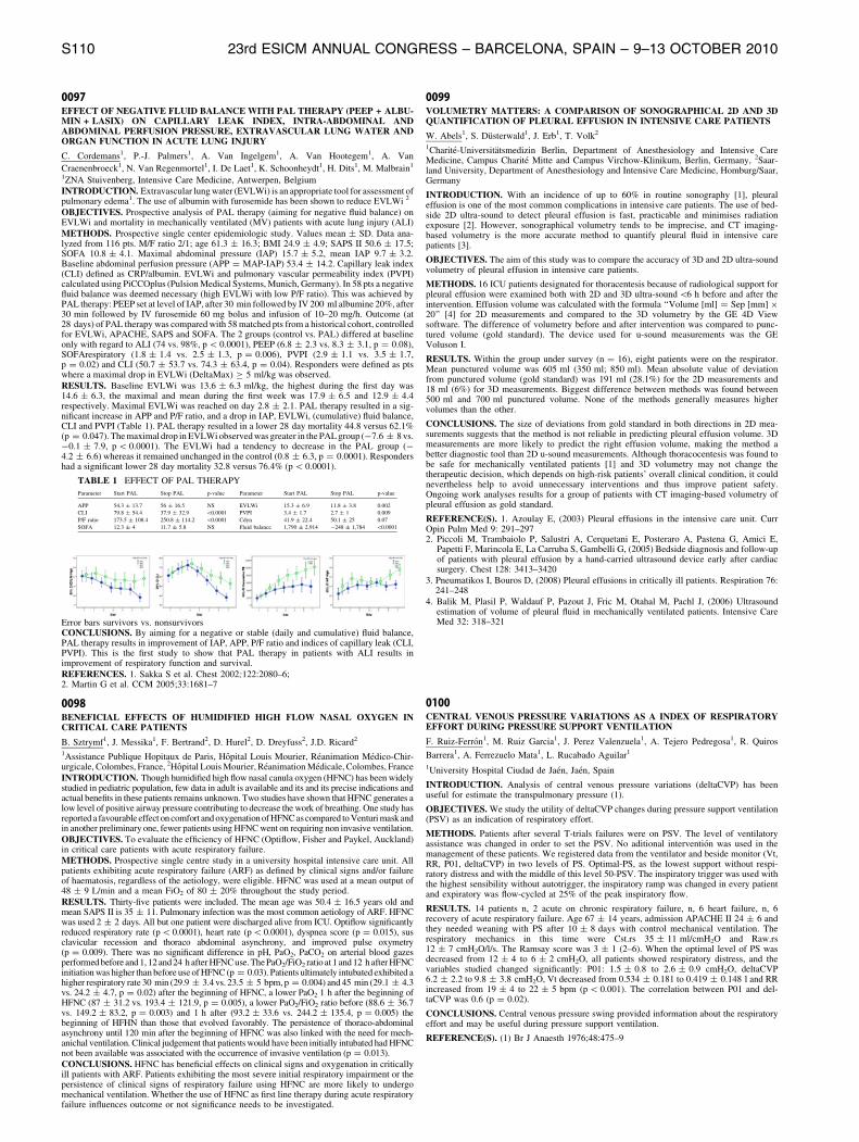
0097EFFECT OF NEGATIVE FLUID BALANCE WITH PAL THERAPY (PEEP + ALBU-MIN + LASIX) ON CAPILLARY LEAK INDEX, INTRA-ABDOMINAL ANDABDOMINAL PERFUSION PRESSURE, EXTRAVASCULAR LUNG WATER ANDORGAN FUNCTION IN ACUTE LUNG INJURY
C. Cordemans1, P.-J. Palmers1, A. Van Ingelgem1, A. Van Hootegem1, A. Van
Craenenbroeck1, N. Van Regenmortel1, I. De Laet1, K. Schoonheydt1, H. Dits1, M. Malbrain1
1ZNA Stuivenberg, Intensive Care Medicine, Antwerpen, Belgium
INTRODUCTION. Extravascular lung water (EVLWi) is an appropriate tool for assessment ofpulmonary edema1. The use of albumin with furosemide has been shown to reduce EVLWi 2
OBJECTIVES. Prospective analysis of PAL therapy (aiming for negative fluid balance) onEVLWi and mortality in mechanically ventilated (MV) patients with acute lung injury (ALI)
METHODS. Prospective single center epidemiologic study. Values mean ± SD. Data ana-lyzed from 116 pts. M/F ratio 2/1; age 61.3 ± 16.3; BMI 24.9 ± 4.9; SAPS II 50.6 ± 17.5;SOFA 10.8 ± 4.1. Maximal abdominal pressure (IAP) 15.7 ± 5.2, mean IAP 9.7 ± 3.2.Baseline abdominal perfusion pressure (APP = MAP-IAP) 53.4 ± 14.2. Capillary leak index(CLI) defined as CRP/albumin. EVLWi and pulmonary vascular permeability index (PVPI)calculated using PiCCOplus (Pulsion Medical Systems, Munich, Germany). In 58 pts a negativefluid balance was deemed necessary (high EVLWi with low P/F ratio). This was achieved byPAL therapy: PEEP set at level of IAP, after 30 min followed by IV 200 ml albumine 20%, after30 min followed by IV furosemide 60 mg bolus and infusion of 10–20 mg/h. Outcome (at28 days) of PAL therapy was compared with 58 matched pts from a historical cohort, controlledfor EVLWi, APACHE, SAPS and SOFA. The 2 groups (control vs. PAL) differed at baselineonly with regard to ALI (74 vs. 98%, p \ 0.0001), PEEP (6.8 ± 2.3 vs. 8.3 ± 3.1, p = 0.08),SOFArespiratory (1.8 ± 1.4 vs. 2.5 ± 1.3, p = 0.006), PVPI (2.9 ± 1.1 vs. 3.5 ± 1.7,p = 0.02) and CLI (50.7 ± 53.7 vs. 74.3 ± 63.4, p = 0.04). Responders were defined as ptswhere a maximal drop in EVLWi (DeltaMax) C 5 ml/kg was observed.
RESULTS. Baseline EVLWi was 13.6 ± 6.3 ml/kg, the highest during the first day was14.6 ± 6.3, the maximal and mean during the first week was 17.9 ± 6.5 and 12.9 ± 4.4respectively. Maximal EVLWi was reached on day 2.8 ± 2.1. PAL therapy resulted in a sig-nificant increase in APP and P/F ratio, and a drop in IAP, EVLWi, (cumulative) fluid balance,CLI and PVPI (Table 1). PAL therapy resulted in a lower 28 day mortality 44.8 versus 62.1%(p = 0.047). The maximal drop in EVLWi observed was greater in the PAL group (-7.6 ± 8 vs.-0.1 ± 7.9, p \ 0.0001). The EVLWi had a tendency to decrease in the PAL group (-4.2 ± 6.6) whereas it remained unchanged in the control (0.8 ± 6.3, p = 0.0001). Respondershad a significant lower 28 day mortality 32.8 versus 76.4% (p \ 0.0001).
TABLE 1 EFFECT OF PAL THERAPY
Parameter Start PAL Stop PAL p-value Parameter Start PAL Stop PAL p-value
APP 54.3 ± 13.7 56 ± 16.5 NS EVLWi 15.3 ± 6.9 11.8 ± 3.8 0.002
CLI 79.8 ± 54.4 37.9 ± 32.9 \0.0001 PVPI 3.4 ± 1.7 2.7 ± 1 0.009
P/F ratio 173.5 ± 108.4 250.8 ± 114.2 \0.0001 Cdyn 41.9 ± 22.4 50.1 ± 25 0.07
SOFA 12.3 ± 4 11.7 ± 5.8 NS Fluid balance 1,790 ± 2,914 -248 ± 1,784 \0.0001
Error bars survivors vs. nonsurvivorsCONCLUSIONS. By aiming for a negative or stable (daily and cumulative) fluid balance,PAL therapy results in improvement of IAP, APP, P/F ratio and indices of capillary leak (CLI,PVPI). This is the first study to show that PAL therapy in patients with ALI results inimprovement of respiratory function and survival.
REFERENCES. 1. Sakka S et al. Chest 2002;122:2080–6;2. Martin G et al. CCM 2005;33:1681–7
0098BENEFICIAL EFFECTS OF HUMIDIFIED HIGH FLOW NASAL OXYGEN INCRITICAL CARE PATIENTS
B. Sztrymf1, J. Messika1, F. Bertrand2, D. Hurel2, D. Dreyfuss2, J.D. Ricard2
1Assistance Publique Hopitaux de Paris, Hopital Louis Mourier, Reanimation Medico-Chir-urgicale, Colombes, France, 2Hopital Louis Mourier, Reanimation Medicale, Colombes, France
INTRODUCTION. Though humidified high flow nasal canula oxygen (HFNC) has been widelystudied in pediatric population, few data in adult is available and its and its precise indications andactual benefits in these patients remains unknown. Two studies have shown that HFNC generates alow level of positive airway pressure contributing to decrease the work of breathing. One study hasreported a favourable effect on comfort and oxygenation of HFNC ascompared to Venturi mask andin another preliminary one, fewer patients using HFNC went on requiring non invasive ventilation.
OBJECTIVES. To evaluate the efficiency of HFNC (Optiflow, Fisher and Paykel, Auckland)in critical care patients with acute respiratory failure.
METHODS. Prospective single centre study in a university hospital intensive care unit. Allpatients exhibiting acute respiratory failure (ARF) as defined by clinical signs and/or failureof haematosis, regardless of the aetiology, were eligible. HFNC was used at a mean output of48 ± 9 L/min and a mean FiO2 of 80 ± 20% throughout the study period.
RESULTS. Thirty-five patients were included. The mean age was 50.4 ± 16.5 years old andmean SAPS II is 35 ± 11. Pulmonary infection was the most common aetiology of ARF. HFNCwas used 2 ± 2 days. All but one patient were discharged alive from ICU. Optiflow significantlyreduced respiratory rate (p\ 0.0001), heart rate (p\ 0.0001), dyspnea score (p = 0.015), susclavicular recession and thoraco abdominal asynchrony, and improved pulse oxymetry(p = 0.009). There was no significant difference in pH, PaO2, PaCO2 on arterial blood gazesperformed before and 1, 12 and 24 h after HFNCuse. The PaO2/FiO2 ratio at 1 and 12 h after HFNCinitiation was higher than before use of HFNC (p = 0.03). Patients ultimately intubated exhibited ahigher respiratory rate 30 min (29.9 ± 3.4 vs. 23.5 ± 5 bpm, p = 0.004) and 45 min (29.1 ± 4.3vs. 24.2 ± 4.7, p = 0.02) after the beginning of HFNC, a lower PaO2 1 h after the beginning ofHFNC (87 ± 31.2 vs. 193.4 ± 121.9, p = 0.005), a lower PaO2/FiO2 ratio before (88.6 ± 36.7vs. 149.2 ± 83.2, p = 0.003) and 1 h after (93.2 ± 33.6 vs. 244.2 ± 135.4, p = 0.005) thebeginning of HFHN than those that evolved favorably. The persistence of thoraco-abdominalasynchrony until 120 min after the beginning of HFNC was also linked with the need for mech-anichal ventilation. Clinical judgement that patients would have been initially intubated had HFNCnot been available was associated with the occurrence of invasive ventilation (p = 0.013).
CONCLUSIONS. HFNC has beneficial effects on clinical signs and oxygenation in criticallyill patients with ARF. Patients exhibiting the most severe initial respiratory impairment or thepersistence of clinical signs of respiratory failure using HFNC are more likely to undergomechanical ventilation. Whether the use of HFNC as first line therapy during acute respiratoryfailure influences outcome or not significance needs to be investigated.
0099VOLUMETRY MATTERS: A COMPARISON OF SONOGRAPHICAL 2D AND 3DQUANTIFICATION OF PLEURAL EFFUSION IN INTENSIVE CARE PATIENTS
W. Abels1, S. Dusterwald1, J. Erb1, T. Volk2
1Charite-Universitatsmedizin Berlin, Department of Anesthesiology and Intensive CareMedicine, Campus Charite Mitte and Campus Virchow-Klinikum, Berlin, Germany, 2Saar-land University, Department of Anesthesiology and Intensive Care Medicine, Homburg/Saar,Germany
INTRODUCTION. With an incidence of up to 60% in routine sonography [1], pleuraleffusion is one of the most common complications in intensive care patients. The use of bed-side 2D ultra-sound to detect pleural effusion is fast, practicable and minimises radiationexposure [2]. However, sonographical volumetry tends to be imprecise, and CT imaging-based volumetry is the more accurate method to quantify pleural fluid in intensive carepatients [3].
OBJECTIVES. The aim of this study was to compare the accuracy of 3D and 2D ultra-soundvolumetry of pleural effusion in intensive care patients.
METHODS. 16 ICU patients designated for thoracentesis because of radiological support forpleural effusion were examined both with 2D and 3D ultra-sound \6 h before and after theintervention. Effusion volume was calculated with the formula ‘‘Volume [ml] = Sep [mm] 920’’ [4] for 2D measurements and compared to the 3D volumetry by the GE 4D Viewsoftware. The difference of volumetry before and after intervention was compared to punc-tured volume (gold standard). The device used for u-sound measurements was the GEVoluson I.
RESULTS. Within the group under survey (n = 16), eight patients were on the respirator.Mean punctured volume was 605 ml (350 ml; 850 ml). Mean absolute value of deviationfrom punctured volume (gold standard) was 191 ml (28.1%) for the 2D measurements and18 ml (6%) for 3D measurements. Biggest difference between methods was found between500 ml and 700 ml punctured volume. None of the methods generally measures highervolumes than the other.
CONCLUSIONS. The size of deviations from gold standard in both directions in 2D mea-surements suggests that the method is not reliable in predicting pleural effusion volume. 3Dmeasurements are more likely to predict the right effusion volume, making the method abetter diagnostic tool than 2D u-sound measurements. Although thoracocentesis was found tobe safe for mechanically ventilated patients [1] and 3D volumetry may not change thetherapeutic decision, which depends on high-risk patients’ overall clinical condition, it couldnevertheless help to avoid unnecessary interventions and thus improve patient safety.Ongoing work analyses results for a group of patients with CT imaging-based volumetry ofpleural effusion as gold standard.
REFERENCE(S). 1. Azoulay E, (2003) Pleural effusions in the intensive care unit. CurrOpin Pulm Med 9: 291–2972. Piccoli M, Trambaiolo P, Salustri A, Cerquetani E, Posteraro A, Pastena G, Amici E,
Papetti F, Marincola E, La Carruba S, Gambelli G, (2005) Bedside diagnosis and follow-upof patients with pleural effusion by a hand-carried ultrasound device early after cardiacsurgery. Chest 128: 3413–3420
3. Pneumatikos I, Bouros D, (2008) Pleural effusions in critically ill patients. Respiration 76:241–248
4. Balik M, Plasil P, Waldauf P, Pazout J, Fric M, Otahal M, Pachl J, (2006) Ultrasoundestimation of volume of pleural fluid in mechanically ventilated patients. Intensive CareMed 32: 318–321
0100CENTRAL VENOUS PRESSURE VARIATIONS AS A INDEX OF RESPIRATORYEFFORT DURING PRESSURE SUPPORT VENTILATION
F. Ruiz-Ferron1, M. Ruiz Garcia1, J. Perez Valenzuela1, A. Tejero Pedregosa1, R. Quiros
Barrera1, A. Ferrezuelo Mata1, L. Rucabado Aguilar1
1University Hospital Ciudad de Jaen, Jaen, Spain
INTRODUCTION. Analysis of central venous pressure variations (deltaCVP) has beenuseful for estimate the transpulmonary pressure (1).
OBJECTIVES. We study the utility of deltaCVP changes during pressure support ventilation(PSV) as an indication of respiratory effort.
METHODS. Patients after several T-trials failures were on PSV. The level of ventilatoryassistance was changed in order to set the PSV. No aditional intervention was used in themanagement of these patients. We registered data from the ventilator and beside monitor (Vt,RR, P01, deltaCVP) in two levels of PS. Optimal-PS, as the lowest support without respi-ratory distress and with the middle of this level 50-PSV. The inspiratory trigger was used withthe highest sensibility without autotrigger, the inspiratory ramp was changed in every patientand expiratory was flow-cycled at 25% of the peak inspiratory flow.
RESULTS. 14 patients n, 2 acute on chronic respiratory failure, n, 6 heart failure, n, 6recovery of acute respiratory failure. Age 67 ± 14 years, admission APACHE II 24 ± 6 andthey needed weaning with PS after 10 ± 8 days with control mechanical ventilation. Therespiratory mechanics in this time were Cst.rs 35 ± 11 ml/cmH2O and Raw.rs12 ± 7 cmH2O/l/s. The Ramsay score was 3 ± 1 (2–6). When the optimal level of PS wasdecreased from 12 ± 4 to 6 ± 2 cmH2O, all patients showed respiratory distress, and thevariables studied changed significantly: P01: 1.5 ± 0.8 to 2.6 ± 0.9 cmH2O, deltaCVP6.2 ± 2.2 to 9.8 ± 3.8 cmH2O, Vt decreased from 0.534 ± 0.181 to 0.419 ± 0.148 l and RRincreased from 19 ± 4 to 22 ± 5 bpm (p \ 0.001). The correlation between P01 and del-taCVP was 0.6 (p = 0.02).
CONCLUSIONS. Central venous pressure swing provided information about the respiratoryeffort and may be useful during pressure support ventilation.
REFERENCE(S). (1) Br J Anaesth 1976;48:475–9
S110 23rd ESICM ANNUAL CONGRESS – BARCELONA, SPAIN – 9–13 OCTOBER 2010
0101VISUAL ESTIMATION OF PATIENT HEIGHT AND ITS CONSEQUENCESON TIDAL VOLUME SETTINGS
R. Miguel Montanes1, E. Kipnis1, F. Remi1, B. Leroy1, B. Tavernier2, B. Vallet2, G. Lebuffe2
1Centre Hospitalier Universitaire de Lille, Reanimation Chirurgicale-Pole d’Anesthesie-Reanimation, Lille, France, 2Centre Hospitalier Universitaire de Lille, Pole d’Anesthesie-Reanimation, Lille, France
INTRODUCTION. The use of a tidal volume of 6 ml/kg of predicted body weight is part ofthe management of patients presenting with ARDS (1) and prevents ventilator induced lunginjury in ICU patients undergoing mechanical ventilation (2). The setting of tidal volume isbased on patient height. Height seems to be more often estimated by the ICU staff thanactually measured (3).
OBJECTIVES. Our study aimed to assess the accuracy of the visual estimation of patientheight and its impact on ventilator settings.
METHODS. Thirty-two patients admitted to a surgical ICU were prospectively included inthis observational study. Patients had their height visually estimated by 18 ICU staff members(6 doctors, 6 nurses and 6 nurse-assistants) and then measured. We also measured the kneeheight from which height can be extrapolated. We then compared the mean estimated heightfor each patient to the actual measured height with the use of Bland and Altman plots and theconsequences of the measurement error on the tidal volume setting.
RESULTS. In most patients visual estimation overestimates the actual height. The mea-surement errors result in an increase in the tidal volume up to +2 ml/kg. This remains truewhatever the subgroup studied: all patients (bias: 4.5 ± 5.52, 95% CI: -6.32;15.32), malepatients (bias: 4.02 ±6.29, 95% CI -8.3;16.35), female patients (bias: 5.5 ± 2.29, 95% CI1.01–9.99) and whatever the type of assessor. Extrapolated height from knee height resultsinvariably in an underestimation of actual height (bias -1 ± 4.5, 95% CI -11.68; 9.68).
CONCLUSIONS. Our results prove that neither visual estimation nor knee height mea-surement are reliable surrogates for measured height. Therefore, measuring patient heightshould be mandatory in critically ill patients in order to minimise ventilator-induced lunginjury.
REFERENCE(S). (1) The acute respiratory distress syndrome network. Ventilation withlower tidal volumes as compared with traditional tidal volumes for acute lung injury and acuterespiratory distress syndrome. N Engl J Med 2000; 18: 1301–1308
(2) Determann RM, Royakkers A, Wolthuis EK, Vlaar AP, Choi G, Paulus F, HofstraJJ, deGraaf MJ, Korevaar JC, Schultz MJ. Ventilation with lower tidal volumes as compared withconventionnal tidal volumesfor patients without acute lung injury: a preventive randomizedcontrolled trial. Crit Care 2010; 1: R1
(3) Leary TS, Milner QJ, Niblett DJ. The accuracy of the estimation of body weight andheight in the intensive care unit. European Journal of Anaesthesiology 2000; 11: 698–703.
0102VENTILATORY PATTERNS IN A MEDICAL INTENSIVE CARE UNIT. A CROSSSECTIONAL STUDY
J. Cebrian1, B. Balerdi1, A. Navarro1, E. Parreno1, A.V. Bommel1
1Hospital Universitario La Fe, Valencia, Spain
OBJECTIVES. To describe our in-ICU ventilatory patterns with special attention to patientswith acute lung injury (ALI) or acute respiratory distress syndrome (ARDS)
METHODS. During 2 months (March and April 2010) we have performed a 4 days crosssectional study measuring the main characteristics of our patients, ventilatory modes andrespirator settings. To overcome the small size we have bootstrapped our main quantitativevariables in order to achieve the best estimates. We have performed 1,000 random samples ofour data by using Bias Corrected Accelerated percentiles method. Categorical variables areshowed as frequencies and quantitative data as mean and 95th confidence interval. Epidataand S-Plus statistical packets were used.
RESULTS. 20 over 44 patients were ventilated during the four study days (Prevalence 45%).Mean age was 60.51 years (52.05–67.9) and 65.0% were men. SAPS 3 was 63.77(54.33–71.37). The main reason for mechanical ventilation was pneumonia (35.0%) and themost prevalent ventilation mode was controlled volume ventilation (55.0%) followed bypressure support (20.0%). Mean PaO2/FiO2 was 211.3 (163.62–279.35), mean respiratory rate(RR) was 19 (16–22) and mean tidal volume (TV) was 6.65 ml/kg (5.85–7.65). Regarding 13patients with ALI/ARDS the following values were obtained: TV 5.9 ml/kg (5.01–6.62), RR20 (17–24), Peep 10 cmH2O (8 to11), Peak pressure 30.82 cmH2O (25.07–36.38) and Plateaupressure 23.6 cmH2O (16.86–30.62).
Tidal volume
CONCLUSIONS. Our data agree with the lower TV strategy recommended in the currentpublications and guidelines. In spite of small size of this preliminary study it is possible toreach good estimates by using intensive computer techniques.
0103AUTOMATIC DETECTION OF PATIENT-VENTILATOR ASYNCHRONIESDURING PRESSURE SUPPORT VENTILATION
F. Mojoli1,2, A. Orlando2, I. Curro2, M. Pozzi2, A. Venti2, G. Matrone3, G. Magenes3,
A. Braschi1,2
1Universita degli Studi di Pavia, Dipartimento di Scienze Chirurgiche, Rianimatorie, Ria-bilitative e dei Trapianti d’Organo, Pavia, Italy, 2Fondazione IRCCS Policlinico S. Matteo,Servizio di Anestesia e Rianimazione I, Pavia, Italy, 3Universita degli Studi di Pavia, Di-partimento di Informatica e Sistemistica, Pavia, Italy
OBJECTIVES. To evaluate the effectiveness of a new algorythm in automatic detection ofpatient-ventilator asynchronies during Pressure Support Ventilation (PSV).
METHODS. 3,016 respiratory acts were recorded in 4 ICU patients selected because ofsevere asynchrony with the ventilator (G5, Hamilton Medical) during PSV. By visual analysis(VA) of airway pressure and flow trajectory, ineffective efforts and inspiratory/expiratorydelays were detected. The results of VA were compared with those provided by a newalgorithm based on the automatic analysis (AA) of flow trajectory.
RESULTS. VA identified 773 ineffective efforts (26% of patients acts). Among 2,243assisted acts, average inspiratory and expiratory delay was 380 ± 214 ms and 134 ± 215 ms,respectively. Significant inspiratory and expiratory delay ([300 ms) occurred in 1,255 (56%)and in 422 acts (19%), respectively. Automatic analysis was able to identify 2,994 of 3,016patients acts (99%). In 2,656 cases (88%), both inspiratory muscles contraction and relaxationwere detected. In 338 acts (11%) only relaxation was identified. Compared with VA, inspi-ratory and expiratory delay of AA was 138 ± 134 and 29 ± 208 ms, respectively. AArecognized the start and the end of patient’s effort before the ventilator in 2,142 (95%) and1,477 (66%) of assisted acts. Sensitivity and Specificity of AA in detecting ineffective efforts,inspiratory delays [300 ms, expiratory delays [300 ms were 99 and 98%, 78 and 85%, 81and 87% respectively.
CONCLUSIONS. The new algorithm proved to be efficient as a real-time, continuousmonitoring system of patient-ventilator interaction. The advantage over traditional flow andpressure based triggers has to be tested.
0104CLINICAL APPLICATION OF A COMPUTERIZED ALARM SYSTEM FORMECHANICALLY VENTILATED PATIENTS. A PILOT STUDY
L. Blanch1,2,3, B. Sales2, R. Fernandez3,4, O. Garcıa-Esquirol1, A. Estruga1, E. Chacon1,J. Montanya2, A. Villagra1,3, M. Borelli5, U. Lucangelo5, A. Hernandez6, G.M. Albaiceta7,E. Fernandez-Mondejar8, G. Murias9
1Corporacio Parc Taulı, Intensive Care Department, Sabadell, Spain, 2Fundacio Parc Taulı,Sabadell, Spain, 3Ciber de Enfermedades Respiratorias, Madrid, Spain, 4Fundacio Althaia,Manresa, Spain, 5Ospedale di Cattinara, Trieste, Italy, 6Hospital Central de la Defensa,Madrid, Spain, 7Hospital Central de Asturias, Oviedo, Spain, 8Hospital Virgen de las Nieves,Critical Care, Granada, Spain, 9Clinica Bazterrica, Critical Care, Buenos Aires, Argentina
INTRODUCTION. Mechanical ventilation remains as one of the most difficult safety issuesin the ICU, but parameters commonly monitored by ventilators only depict the most extremerisks for patients. New computerized approaches may manage exhaustive data and mayinclude ‘‘intelligent’’ software that mirrors expert decision making.
OBJECTIVES. To test the clinical usefulness of a new computerized system as a herald forclinically significant alarms and its possible impact in outcome.
METHODS. Twenty-five mechanically ventilated patients were continuously monitored in asingle mixed ICU in a university-affiliated hospital. We recorded age, diagnosis, PaO2/FiO2,APACHE II and SOFA on admission. The computerized system grabs and process data fromdifferent devices, usually a monitor and a respirator, and evaluates the most relevant events ina ventilated patient. All the algorithms were designed and validated with the clinical staff.Data of ventilated patients were recorded at the ICU during 8 months and a total of 4,615,589breaths from 25 different patients, each of them with a register of at least 50% of totalventilation time, were collected. Data include biomedical signals (waves and trends) as wellas all the clinical events detected by the system, including trapped gas at end-expiration,presence of secretions, double-cycling, asynchronies during expiration, pulse pressure vari-ation in patients not triggering the ventilator and stress index ([1.2 or \0.8) in thoseventilated with square airflow. Outcome variables were ICU length of stay, hospital stay andmortality. Statistical analysis included multiple regression models for length of stay andmortality.
RESULTS. Median and interquartile range were: age 61 [45.50–72], PaO2/FiO2 200 mmHg[132–260], SOFA 7 [4–10], ICU length of stay 11.5 days [7.5–18], hospital length of stay 21days [6–37]. The frequency of alarms were: trapped gas at end-expiration: 20.7%, presenceof secretions 1.3%, double-cycling 1.1%, asynchronies during expiration 5.2%, stress index[1.2 1.1%, stress index\0.8 2.5%, and pulse pressure variation 5.1%. Multiple regressionanalysis found PaO2/FiO2 associated with length of MV and close to significance with hos-pital stay and mortality, but any of the computerized alarms reached yet the level ofsignificance.
CONCLUSIONS. The computerized system is able to detect and review more clinicallysignificant problems than clinical routine. However, its impact to define patient outcomeswarrants further investigation.
GRANT ACKNOWLEDGMENT. Supported in part by Fundacio Parc Taulı, Obra SocialCaixa Sabadell, ISCiii PI09/91074, Plan AVANZA TSI-020302-2008-38.
23rd ESICM ANNUAL CONGRESS – BARCELONA, SPAIN – 9–13 OCTOBER 2010 S111

0105BACTERAEMIA FOLLOWING PERCTANEOUS DILATATIONAL TRACHEOS-TOMY: THE RELATIONSHIP TO RESPIRATORY SECRETION COLONISATIONAND THE QUESTION OF ANTIBIOTIC PROPHYLAXIS
L. Anderson1, R. Cooke2, T. Cope1, G. Dempsey1
1University Hospital Aintree, Critical Care Unit, Liverpool, UK, 2University Hospital Aintree,Department of Medical Microbiology, Liverpool, UK
INTRODUCTION. The role of antibiotic prophylaxis (AP) prior to percutaneous dilatationaltracheostomy (PDT) is unclear. A recent survey showed that the majority of UK Critical CareUnits (CCU) do not routinely prescribe AP1 despite bacteraemia and ventilator associatedpneumonia (VAP) rates following PDT of 102 and 18%3 respectively.
OBJECTIVES. Our aim was to determine the bacteraemia rate in the 48 h following PDTwith particular reference to colonisation of respiratory secretions.
METHODS. We have performed a retrospective analysis of all patients undergoing PDTbetween November 2003 and December 2008 in the CCU of University Hospital Aintree,Liverpool, UK. The medical microbiology database (MMD) was searched to determine thosepatients with colonised respiratory secretions in the 48 h prior to PDT and those with positiveblood cultures (BC) in the 48 h following PDT. Respiratory cultures were subdividedaccording to whether the sputum culture resulted in a pure growth (PG)(C++ of a singleorganism), a mixed growth (MG) or no growth (NG).
RESULTS. During the study period 489 patients underwent PDT. Of these, 488 wereidentified on the MMD. 292 patients had respiratory samples cultured in the 48 h prior toPDT.
Bacteraemia incidence with secretion colonisation
Respiratory culture growth
Blood culture Pure (n = 61) Mixed (n = 49) None (n = 182)
Positive n (%) 4 (6.6) [n = 3 withsame pathogen]
2 (4.1) 9 (4.9)
Negative n (%) 33 (54.1) 18 (36.7) 89 (48.9)
There was no significant difference in the bacteraemia rates post PDT. p = 0.69 Chi squared.
CONCLUSIONS. In this retrospective series there is no demonstrated association betweenpositive respiratory cultures and bacteraemia following PDT. There does not seem to be a rolefor specific AP when performing PDT.
REFERENCE(S). 1. Hanna-Jumma S, Higgins D, Kinnear J, Wright M, JICS2010.11:31–33.
2. Teoh N, Parr M, Finfer SR, Anaesthesia Intensive Care 1997.25:354–357.
3. Rello J, Diaz E, Chest 2003.124:2239–2243.
0106ACUTE RESPIRATORY DISTRESS SYNDROME: PERFORMANCE OFVENTILATOR AT SIMULATED ALTITUDE
J.-P. Tourtier1, E. Forsans1, E. Fontaine1, L. Franck1, N. Libert1, L. Grasser1, M. Borne1
1Military Hospital Val-de-Grace, Paris, France
INTRODUCTION. Ventilation of Acute Respiratory Distress Syndrome (ARDS) is achallenge. High tidal volume (Vt) is harmful and there is definitely a need for lack ofvariations between delivered and set Vt on the ventilators that is being used.
OBJECTIVES. We have assessed the ability of the ventilator T-bird VS02 and LTV-1000 todeliver to a lung model with ARDS a set tidal volume (Vt) at different simulated altitudes.
METHODS. We used a decompression chamber to mimic the hypobaric environment at arange of simulated cabin altitudes of 1,500, 2,500 and 3,000 m (4,000, 6,670, 8,000 feet).Ventilators were tested with realistic parameters. Vt was set at 400 and 250 ml in an ARDSlung model. The positive end expiratory pressures (PEEP) were set at 10 and 15 cmH2O.Pressure drop across the pneumotachograph was measured by a differential pressure trans-ducer (EnertecTM). The spirometer was checked at each altitude using a calibration syringe.The inspired oxygen content (FiO2) was 100%. Respiratory rate was 20 breaths/min. The ratioinspiratory time/expiratory time was 1/1. The protocol included three measurements for eachsimulated altitude. Comparisons of preset to actual measured values were accomplished usinga t test for each altitude. A significant difference was defined by p \ 0.05.
RESULTS. The standard deviation for the three measurements obtained at each altitude wasconsistently less than 10 ml. Respiratory rate delivered was 20 breaths/min in all cases.Variation of PEEP did not change the volume delivered. The T-bird VS02 showed a decreasein volume delivered. Comparisons of actual delivered Vt and set Vt demonstrated a significantdifference starting at 1,500 m for a Vt set of 400 ml, at 2,500 m for Vt set of 250 ml. At thesealtitudes, the variations between Vt set and delivered were more than 10%. With decreasingbarometric pressure, the LTV-1000 showed mostly an increase in volume delivered. Com-parisons of actual delivered Vt and set Vt demonstrated a significant difference at 2,500 m fora Vt set of 400 ml, at 3,000 m for Vt set of 250 ml. The delivered tidal volume remainedwithin 10% of the set Vt. Assuming that the patient is ventilated at sea level and gas exchangeis normal, the movement to altitude would result in an increase in tidal volume which might infact represent a clinical event.
CONCLUSIONS. The LTV-1000 met the trial targets in all settings, whereas the T-bird-VSO2 did not compensate well for altitude and progressively delivered lower volumes asbarometric pressure decreased. Such variations between delivered and set Vt suggest lack ofefficacy of altimetric correction in hypobaric conditions in some devices. The LTV1000showed a moderate increase in volume delivered for ARDS lung model with increasingaltitude, but maintained the delivered volume within 10% of the set Vt up to 3,000 m. Theaccuracy of the Vt delivery was superior with the LTV-1000 than with the T-birdVSO2.
0107NASAL HIGH-FLOW OXYGEN THERAPY VS. STANDARD OXYGEN THERAPYVIA VENTURI MASK AFTER EXTUBATION: PRELIMINARY RESULTS OF ARANDOMIZED, CONTROLLED TRIAL
F.A. Idone1, R. Festa1, F. Antonicelli1, F. Di Muzio1, D. Maniglia2, A. Cataldo1, M. Anto-
nelli1, S.M. Maggiore1
1Department of Anesthesiology and Intensive Care, Policlinico A. Gemelli, UniversitaCattolica del Sacro Cuore, Roma, Italy, 2Department of Anesthesiology and Intensive Care,Policlinico P. Giaccone, Universita degli Studi di Palermo, Palermo, Italy
INTRODUCTION. Oxygen therapy is commonly used to correct residual oxygenationimpairment in the post-extubation period. This is usually done through a Venturi mask whichallows to deliver predetermined fractions of oxygen humidified with bubble humidifiers. Thelow humidity delivered by such devices and the use of an oro-nasal mask may, however,reduce patient’s comfort, possibly resulting in mask displacement or removal and consequentoxygen desaturations. Nasal high-flow (NHF) oxygen therapy allows to deliver high-flowoxygen, humidified with heated humidifiers and delivered through nasal cannulae, with thepotential to improve comfort and efficacy.
OBJECTIVES. In patients requiring oxygen therapy after extubation, we compared NHF vs.Venturi mask in terms of oxygenation and comfort.
METHODS. Patients who were mechanically ventilated for more than 24 h, passed aspontaneous breathing trial, and had PaO2/FiO2 \ 300 at the end of the trial were randomizedto receive oxygen with NHF or Venturi mask after extubation. Exclusion criteria were:tracheostomy, age \18, pregnancy, or anticipated need for noninvasive ventilation afterextubation. In both groups, FiO2 was set to obtain SpO2 between 92 and 98% (88–95% inCOPD patients). With NHF, flow rate was set at 50 L/min. Arterial blood gases, respiratoryrate, and discomfort were assessed at 1, 3, 6, 12, 24, 36, and 48 h. Discomfort was assessed byasking patients to rate their discomfort with the used device by using a numerical scale from 0(no discomfort) to 10 (maximum imaginable discomfort). Discomfort symptoms were alsoassessed for the dryness of the delivered oxygen (dryness of the mouth, throat, nose, difficultyto swallow and throat pain). Incidence of desaturations and interface displacement was alsoassessed.
RESULTS. Thirty-five patients were included (18 NHF, 17 Venturi mask). No difference wasobserved in the baseline characteristics at inclusion. Arterial blood gases were similarbetween groups, except at 36 h when PaO2/FiO2 was greater with NHF (358 vs. 273,p = 0.05). Respiratory rate was lower with NHF at all time points (22 ± 4 vs. 26 ± 4 at 24 h,p = 0.01). At 24 and 36 h, general discomfort was lower with NHF than with Venturi mask(4 ± 2 vs. 6 ± 3 at 24 h, p = 0.01). Improvement in discomfort with NHF concerned par-ticularly dryness of mouth (3 ± 3 vs. 7 ± 3 at 24 h, p \ 0.01) and throat (3 ± 3 vs. 6 ± 3 at24 h, p = 0.01). Oxygen desaturations (94 vs. 61% patients, p = 0.02; 76 vs. 25 episodes,p \ 0.01) and interface displacement requiring an intervention (76 vs. 33% patients,p = 0.01) were more frequent with the Venturi mask.
CONCLUSIONS. Our preliminary results suggest that NHF is an effective method fordelivering oxygen therapy after extubation, with greater comfort, less interface displacementand less oxygen desaturations than Venturi mask.
Noninvasive ventilation: 0108–01210108HUMIDIFICATION AND TEMPERATURE WHEN A USING HELMETFOR NONINVASIVE POSITIVE PRESSURE VENTILATION
K. Ueta1, N. Iguchi1, Y. Goto1, N. Ohta1, A. Uchiyama1, Y. Fujino1
1Osaka University Hospital, Intensive Care Unit, Suita, Japan
INTRODUCTION. Recently, ventilation helmets have been developed to improve NPPVtolerance and problems with other methods of delivery. While humidification during helmetdelivery of ventilation has been indicated1, optimal humidification settings have not beenclearly elucidated.
OBJECTIVES. To evaluate the optimal humidifier water temperature when using a helmetfor noninvasive positive pressure ventilation.
METHODS. Twelve healthy individuals underwent 8 cmH2O CPAP ventilation with 21%oxygen. Each was sequentially tested in the following order: using the helmet withouthumidification at ambient temperature, with humidification with unheated chamber water, andwith humidification with the chamber water at 31, 34, and 37�C. At each setting, after a20 min stabilization period, measurements were taken. Comfort level at each setting wasevaluated using a visual analog scale (VAS) rated zero (most comfortable) to ten (leastcomfortable).
RESULTS. Temperature and relative and absolute humidity inside the helmet, and VASscores statistically significantly increased as the humidification chamber water temperatureincreased. The lowest VAS, 1.6 ± 0.8, was obtained when water in the humidifier chamberwas at ambient temperature.
CONCLUSIONS. For patient comfort during CPAP using a helmet, the most desirableconditions are likely to obtained by humidifying without heating, that is by leaving the waterin the humidifier chamber at room temperature.
REFERENCE: 1 Chiumello D, Chierichetti M, Tallarini F, Cozzi P, Cressoni M, Polli F,Colombo R, Castelli A, Gattinoni L (2008). Effect of a heated humidifier during continuouspositive airway pressure delivered by a helmet. Crit Care 12(2):R55.
S112 23rd ESICM ANNUAL CONGRESS – BARCELONA, SPAIN – 9–13 OCTOBER 2010

0109FACTORS PREDICTING FAILURE IN THE USE OF NON-INVASIVE MECHAN-ICAL VENTILATION WITH A HELMET, IN PATIENTS WITH ACUTE POST-OPERATIVE RESPIRATORY FAILURE
F.J. Redondo Calvo1, M. Madrazo Delgado1, R. Villazala Gonzalez1, A.S. Yuste Pena1,R. Una Orejon2, E. Alday Munoz3, G. Bernal Garcıa1
1Hospital Universitario de Ciudad Real, Ciudad Real, Spain, 2Hospital Universitario La Paz,Madrid, Spain, 3Hospital Universitario La Princesa, Madrid, Spain
INTRODUCTION. The physiological and clinical effects of non-invasive ventilation (NIV)on acute post-operative respiratory failure are relatively unknown. The aim of this study wasto determine the prediction factors for failure in the use of NIV with a helmet in this context.
METHODS. The use of NIV was assessed for a period of 2 years, in a post-operativeintensive care unit (ICU). Demographic data was collected, as well as ARF and arterial gasreadings. Haemodynamic changes were assessed using PiCCOTM technology and the clinicaldevelopment of patients was recorded. All patients who developed acute respiratory failure(ARF) were treated using NIV as their primary care, and the two groups for the study weredetermined in this way, depending on whether the technique was successful, or the patientsrequired intubation. The risk factors that determined failure in the application of NIV weresubsequently determined.
RESULTS. Of the 56 patients presenting with post-operative ARF treated with NIV using ahelmet, 41 did not require intubation (73.2%). Following a multivariate analysis using logisticregression, we determined that there are four independent risk factors for the failure of NIV.The primary causes of respiratory failure are as follows: acute respiratory distress syndrome(ARDS) and PNEUMONIA, and in second place, the high initial EVLWI (extravascular lungwater index) value, as a protective factor, is the increase in the pO2/FiO2 ratio after the firsthour of NIV application.
CONCLUSIONS. NIV using a helmet could provide an effective alternative to conventionalventilation in selected patients with post-operative ARF.
REFERENCE(S).
– Jaber S, Delay JM, Chanques G, et al. Outcomes of patients with acute respirtatoru failureafter abdominal surgery treated with noninvasive positive pressure ventilation. Chest2005; 128:2688–2695
– Squadrone V, Coha M, Cerrutti E, Schellino MM, Biolino P, Occella P, Belloni G,Vilianis G, Fiore G, Cavallo F, Ranieri VM. Continous positive airway pressure fortreatment of postoperative hypoxemia. JAMA 2005; 293:589–595
– Antonelli M, Conti G, Moro ML, Esquinas A, Gonzalez-Diaz G, Confalonieri M, Pelaia P,Principi T, Gregoretti C, Beltrame F, Pennisi MA, Arcangeli A, Proietti R, Passariello M,Meduri GU. Predictors of failure of noninvasive positive pressure ventilation in patientswith acute hypoxemic respiratory failure: a multi-center study. Intensive Care Med 2001;27:1718–28
– Sakka SG, Klein M, Reinhart K, et al.: Prognostic value of extravascular lung water incritically ill patients. Chest 2002; 122(6):2080–6
GRANT ACKNOWLEDGMENT. We express our gratitude to the Mutua Madrilena Fun-dation (Madrid, Spain) and to the nursing staff on the unit for their kind and generous help.Departament of Anesthesiology. Hospital General Ciudad Real. Spain.
0110NONINVASIVE MECHANICAL VENTILATION ORGANIZATION. AN INTERNA-TIONAL SURVEY IN PREHOSPITAL AND EMERGENCY MEDICINE.COPERNICUS INTERNATIONAL SURVEY. PHASE-I
A. Esquinas1, A. Bellone2, R. Scala3, S.K. Sharma4, G. Meyanci Koksal5, B. John6, G.Sempere Montes7, S. Budweiser8, J.M. Carratala Perales9, M. Koldehoff10, C. MaquilonOrtiz11, R.M. Iban Ochoa12, M. Folgado13, P. Torok14, J. Gomez Company15, A. Almela16,T.K. Lim17, C. Suero Mendez18, U. Anttalainen19, on behalf International Association ofNoninvasive Mechanical Ventilation.1Hospital Morales Meseguer, Intensive Care Unit, Murcia, Spain, 2Ospedale Valduce, Como,Italy, 3Hospital S-Donato, Arezzo, Italy, 4All India Institute Of Medical Sciences, New Delhi,India, 5Hospital Istanbul University, Cerrahpasa Medical School, Istanbul, Turkey, 6Uni-versity Hospital, Newark, United States, 7Hospital Dr. Peset, Valencia, Spain, 8DonaustaufHospital, Center For Pneumology, Donaustauf, Germany, 9Hospital General Universitario,Alicante, Spain, 10Hospital University Hospital Of Duisburg-Essen, Bmt Center, Essen,Germany, 11Clinica Davila, Santiago de Chile, Chile, 12Hospital Universitario Rio Hortega,Valladolid, Spain, 13Hospital Zamora, Zamora, Spain, 14Hospital Vranovska Nemnocnica,N.O., Slovakia, 15Servicio de Urgencias, Hospital Virgen de la Arrixaca, Murcia, Spain,16Servicio De Urgencias Del. Hospital Arnau De Vilanova, Valencia, Spain, 17NationalUniversity Hospital, Singapore, Singapore, 18Hospital H.R.U. Carlos Haya, Malaga, Spain,19Turku University, Central Hospital, Pulmonary Department, Turku, Finland
INTRODUCTION. Noninvasive mechanical ventilation (NIV) is a first line intervention inemergency department and hospitals.
OBJECTIVES. Analyze NIV practice in emergency departments.
METHODS. We have development an international epidemiology survey (March 2009) byelectronic questionnaire to know NIV organization, equipment and training in emergencydepartments (Phase I).
DESIGN. International multicenter prospective.
RESULTS. We have enrolled information from 57 hospitals: Spain (42); Italy (2); India (1);USA (3); Slovakia (1); Turkey (1); Germany (2); Australia(2); Chile (1); Singapore (1);Finland (1) during to analyze noninvasive practice in prehospital and emergency medicine,equipment, interfase, ventilatory modes and common clinical applications. Major resultsduring 1 month period analysis were: noninvasive mechanical ventilation were applied in pre-hospital: 3 (57) (5.2%). NIMV commonly was applied follow a objective pprotocol of NIMV:(54) (57) (94.7%). Global rate indication of NIV were COPD exacerbation (90%) and cardiacpulmonary edema (10–60%). All NIV applications were successful applications in emergencydepartments (avoid ETI 80%) with minor complications (12.2%) (skin nose lesion). Equip-ment more relavant were (CPAP devices (60%) and facial mask (90%) was more frequent,follow total face(5%); nasal mask (3%); helmet (2%); type ventilary mode: cpap ventilatorymode was frequent used as first line (60%). All hospitals have educational NIV-programs 54(56) (96.42%) for emergency staff physicians.
CONCLUSIONS. Preliminary results to know NIV organization Copernicus InternationalSurvey (Phase I), in hospital showed that in Emergency departments follow common indi-cations and hospital organization.
GRANT ACKNOWLEDGMENT. International Association NIV.
0111DOOR TO NON-INVASIVE VENTILATION (NIV) TIME IN ACUTE TYPE 2RESPIRATORY FAILURE: IMPROVING DELAYS TO APPROPRIATE NIV IN ADISTRICT GENERAL HOSPITAL IN UK: COMPLETING AN AUDIT LOOP
S. Kumar1, A. Chakraborty1, D. Pandit2
1The Dudley Group of Hospitals NHS FoundationTrust, Acute Medicine, Dudley, UK, 2TheDudley Group of Hospitals NHS FoundationTrust, Respiratory and Critical Care Medicine,Dudley, UK
INTRODUCTION. Current British Thoracic Guidelines (BTS) (1) suggest NIV should bestarted within 1 h of not responding to the maximal medical therapy for acute type 2 respi-ratory failure patients. Following an earlier audit presented as an abstract at ESICM 2008 (2),which looked at possible delays in starting NIV, protocols were implemented to start NIVearlier at Russells Hall hospital acute admissions unit.
OBJECTIVE. The aim was to assess the delays in starting the appropriate patients on NIV atadmission looking at impact on adverse outcomes.
METHOD. Data was collected retrospectively using BTS NIV audit tool. 54 cases admittedwith type 2 respiratory failure in our hospital between January and March 2010 who neededNIV were included in the audit. Delay was subdivided into a) door to first arterial blood gas(ABG) sampling b) ABG to decision for NIV and c) from decision to actual starting of NIV.
RESULTS. The mean time to get an ABG from admission was 180 min. 6 cases with delaymore than 10 h skewed the data. The median time, which was more representative of the usualdelay between admission and first ABG, was 51 min. Our audit showed that 28 out of 54(52%) patients had ABG within an hour compared to 11 out of 19 (58%) in the last audit.
The mean time from admission to NIV was 285 min, showing an improvement of 40 min.The median value for door to NIV time was 186 min which accounted for the cases withextreme delays. 31 out of 54 (57%) patients had NIV within 4 h of admission compared to 12out of 19 (63%) from last audit.
Only 2 out of 54 (3.7%) patients had delay of more than 30 min in starting NIV once decisionwas made compared to 15 out of 18 (83%) showing a significant improvement from previousaudit.
Sub-analysis of the data showed out of these patients who were discharged, 17 (71%) had anadmission pH C 7.25 and 7 (29%) had an admission pH \ 7.25. Out of the patients who died,13 (62%) had NIV started after 4 h and 8 (38%) had NIV started within 4 h of admission. Thissuggests that earlier and appropriate NIV can have a positive effect on patient outcome.
CONCLUSIONS. We have shown considerable improvement in door to NIV time using asimplified hospital protocol and educating the junior doctors at front door in reducing delaysin a life saving intervention preventing escalation to invasive ventilation. Writing upguidelines and hospital protocol and educating NIV nurses has significantly reduced the delaybetween decision making and implementing NIV. Reinforcement and further education at thefront door should be continued to reduce delay and avoid extreme wait in assessment andimplementation of NIV.
REFERENCE. 1. National guidelines in management of respiratory failure: Concise Guid-ance to Good Practice series, No 11. London: RCP, 2008. 2. Delays to appriopriate noninvasive ventilation: Ibrahim et al. http://www.esicm.org/data/ModuleSoumission/resume/html/2008/0355.html
0112COMPARISON OF USE OF NONINVASIVE VENTILATION IN THE TREATMENTOF EXACERBATIONS OF CHRONIC RESPIRATORY FAILURE IN PATIENTSWITH CHRONIC OBSTRUCTIVE PULMONARY DISEASE AND MORBIDOBESITY
N. Llamas Fernandez1, A. Lopez Martınez1, A. Carrillo Alcaraz1, M.D.l.N. Alcazar1,
S.M. Botıas Arnau1, C.R. Hernandez Romero1, G.J. Gonzalez Dıaz1
1Hospital Morales Meseguer, ICU, Murcia, Spain
INTRODUCTION. Although noninvasive ventilation (NIV) has been widely used in patientswith acute on chronic respiratory failure (ACRF) due to chronic obstructive pulmonary dis-ease (COPD), series studying patients with pulmonary restriction due to morbid obesity (MO)are rare (1), despite the disease is highly prevalent in our environment.
OBJECTIVES. The aim of our study is to analyze and compare the effectiveness of NIV inpatients with COPD and OM.
METHODS. We analyzed all patients admitted to ICU for a period of 14 years with diag-nosis of ACRF due to COPD or MO and treated with noninvasive ventilation. NIV successwas defined as the avoidance of endotracheal intubation, survival in ICU and at least 24 h on amedical ward with no signs or symptoms of respiratory failure. Variables are expressed asmeans ± standard deviation and percentages. Comparison between variables by Pearson’s v2
test and Student t. We analyzed survival and hospital readmission per year (log Rank test).
RESULTS. During the study period, 475 patients were admitted with exacerbation of COPDand 160 with MO. All patients were treated with two levels of pressure. Age differs betweenCOPD and OM, 70 ± 10 and 73 ± 11 years, respectively (p = 0.003), as well as the per-centage of men, 87.6 and 21.9% (p \ 0.001). Neither the presence of do-not-intubate order(29.3 and 27.5%, p = 0.670) or the need for home oxygen therapy (45.9 and 41.9%,p = 0.307) differed in the two groups. Respiratory parameters in COPD and MO were pH:7.22 ± 0.09 and 7.22 ± 0.08 (p = 0.788), PaCO2: 85 ± 23 and 84 ± 18 (p = 0.625), PaO2/FiO2: 174 ± 41 and 171 ± 40 (p = 0.379) and respiratory rate 31 ± 8 and 30 ± 7(p = 0.118). Failure of NIV was in COPD: 54 patients (11.4%) and MO: 9 (5.6%)(p = 0.036), late respiratory failure was13 and 7% (p = 0.041), hospital mortality: 17.5 and6.3% (p = 0.001), hospital readmission per year: 46.4 and 51.1% (p = 0.875) and mortalityat 1 year: 29.3 and 18.1% (p = 0.005).
CONCLUSIONS. Although the characteristics of patients with OM and respiratory failureare very similar to COPD patients, both the hospital course as alter a year are better in MOtreated with NIV.
REFERENCE(S). (1) Nava S, Hill NS. Non-invasive ventilation in acute respiratory failure.Lancet 2009; 374: 250–59.
23rd ESICM ANNUAL CONGRESS – BARCELONA, SPAIN – 9–13 OCTOBER 2010 S113
0113EFFICACY AND OUTCOME OF NON INVASIVE VENTILATION USING ADAP-TIVE SUPPORT VENTILATION IN ACUTE RESPIRATORY FAILURE: APROSPECTIVE COHORT STUDYG. Raja Amarnath1, A.S. Arunkumar1, V. Mahesh Vakamudi1, S. Sanjayprabhu1, S. Ramesh1
1Sri Ramachandra University, Critical Care, Chennai, India
INTRODUCTION. Adaptive support Ventilation (ASV)� (Hamilton Galileo) has been shown to result inbetter patient synchrony, reduced weaning times and reduced work load for the ICU staff (1, 2). But, data islacking on its efficacy, especially as Non invasive ventilation (NIV) due to the concern of being closed loopventilation. Also, little is known about the risk factors of late NIV failure in patients who improve initially (3)
OBJECTIVES. To analyze the efficacy of NIV using ASV in Acute Respiratory Failure (RF) andidentify risk factors of Late NIV failure
METHODS. On consecutive Patients with acute RF from May2008 to February2010, NIV was insti-tuted and maintained using ASV, and patients were withdrawn or Intubated based on pre defined criteria.APACHI II on admission, ABG, Vitals and SOFA scores at various points in time were noted. Primaryoutcome was failure of NIV. Secondary outcomes were hospital mortality and ICUdays.
RESULTS. 205 patients were included. Admission APACHE II and SOFA were 20.02 ± 7.46 and5.27 ± 2.58, respectively (Mean ± SD).
Different types of RF in the study populationType of RF Number of patients (%)
HYPOXIC RF-PULMONARY (PaO2/FiO2 \ 300 and PaCO2 \ 45 mmHg) Eg:Pneumonia, ARDS, Pleural Effusion, Contusion etc. 81 (39.5%)
HYPERCAPNIC RF (PaCO2 [ 45 mmHg and PaO2/FiO2 [ 300) 31 (15.1%)
HYPOXIC RF-NON PULMONARY (PaO2/FiO2 \ 300 and PaCO2 \ 45 mmHg) Eg:Cardiac failure,Fluid overload etc. 28 (13.7%)
COMBINED RF (PaO2/FiO2 \ 300 & PaCO2 [ 45 mmHg) 65 (31.7%)
Over all success of NIV was 66.3%.
NIV outcome in RF with different causes (p = 0.00)Cause of RF NIV SUCCESS number (%) NIV FAILURE number (%)
PRIMARY ARDS 6 (23.1%) 20 (76.9%)
SECONDARY ARDS 8(36.4%) 14(63.6%)
COPD 48 (86%) 2 (4%)
CHEST TRAUMA 10 (83.3%) 2 (16.7%)
CARDIAC FAILURE 18 (100%) 0 (0%)
Higher Respiratory rate, lower GCS, and lower PaO2/FiO2 before initiation of NIV and 2 h afterinitiation of NIV independently predicted NIV failure. Hypercapnic RF was significantly associatedwith NIV success and reduced mortality. Worsening SOFA from ICU admission to initiation of NIVpredicted late NIV failure (p = 0.002). Overall mortality was 27.8%. NIV failure was an independentpredictor of increased mortality (73.9%) vs. (4.4% in patients with NIV success) (P = 0.00).Worsening of SOFA score from the day of NIV initiation to the day of intubation independentlypredicted Mortality in patients with NIV failure (p = 0.00). Higher APACHE II and DELTA SOFAscores were associated with increased ICU and hospital stay (p = 0.05).
CONCLUSIONS. ASV is a feasible mode in NIV with its advantages of better patient synchrony anduser friendliness. Hypoxic RF (especially ARDS and Pneumonia) has poorest outcome in NIV.Worsening of SOFA from the day of ICU admission to the day of NIV initiation independentlypredicted NIV failure. Using these trends in SOFA scores, it is possible to predict Late NIV failure.
REFERENCE(S). (1) Tassaux D,Patient-ventilator interactions during partial ventilatory sup-port,Crit Care Med 2002;30 (2) Petter,Automatic weaning with ASV,Anes Analg 2003;97 (3) MorettiM,NIV failure after initial success. Thorax 2000;5
0114VENTILATOR PERFORMANCE WITH DIFFERENT VENTILATORS DURINGHELMET NIVR. Costa1, G. Spinazzola1, O. Festa1, G. Ferrone1, F. Cipriani1, D. Micci1, R. Cioccolini1, G. Conti1
1UCSC, Anaesthesiology and Intensive Care, Rome, Italy
INTRODUCTION. During NIV, patient–ventilator interaction may be markedly affected by theinterface used and the ventilator performance, the latter determined by ventilator characteristics: triggertype and sensibility, slope of pressurization process, cycling criteria, presence of a ‘‘NIV-mode’’. TheHelmet has been demonstrated to be a complex interface that may markedly alter ventilator functioning.
OBJECTIVES. The aim of this bench study was to evaluate the performances of 5 different ven-tilators during Helmet NIV in terms of patient-ventilator interaction.
METHODS. A mannequin connected to an active test lung breathing at two respiratory rates (20 and 30breaths/min), was ventilated with an helmet with 5 different ventilators (Puritan Bennet 840, PB; OmegaDatex, GE; Maquet Servo I, SI; Draeger Evita XL; Zephyros Medisize, ZP) in PSV mode set at two different
pressurization times (Timepress) and expiratory trigger thresholds (Trexp): Default setting (DS, Timepress/Trexp
50%/25%) and Fast setting (FS, Timepress/Trexp 80%/60%) (Table 1). The flow trigger was set at the lowestvalue not determining auto-cycling. We evaluated ventilators performance determining: Trigger pressure drop(DPtrigger), Inspiratory Pressure–Time Product (PTPt), Pressure–Time Product at 300 and 500 ms (PTP 300and PTP 500). Patient-ventilator interaction was evaluated determining: Inspiratory trigger delay (Delaytrinsp),Expiratory trigger delay (Delaytrexp), Pressurization Time (Timepress), Time of synchrony (Timesyn).
RESULTS. See Table 2, Figs. 1 and 2.
TABLE 1 SETTING FOR EACH VENTILATORTimepress Trexp
PB DS FS 50% 80% 25% 60%
GE DS FS 250 ms 100 ms 25 ms 50 ms
SI DS FS 0.2 s 0.3 s 25% 60%
ZP DS FS 0.8 s 0.4 s 35% 70%
Evita XL DS FS 1 s 0.4 s 22% 52%
RR 20 RR 20 RR 20 RR 20 RR 20 RR 30 RR 30
ZP PB GE SI Evita XL ZP PB
Timec (sec) DS 1.05±0.01** Timec(sec) FS 0.74±0.02**
Timec (sec) DS 0.58±0.01†† Timec(sec) FS 0.53±0.01††
Timec (sec) DS 0.77±0.01§§ Timec(sec) FS 0.66±0.03
Timec (sec) DS 0.90±0.01## Timec(sec) FS 0.67±0.01
Timec (sec) DS 1.31±0.01θθ Timec(sec) FS 0.80±0.01θθ
Timec (sec) DS 1.27±0.04** Timec(sec) FS 0.80±0.02
Timec (sec) DS 0.57±0.01†† Timec (sec) FS 0.49±0.01††
Ti/Ttotmec DS 0.44** Ti/Ttotmec FS 0.30**
Ti/Ttotmec DS 0.26†† Ti/Ttotmec FS 0.22††
Ti/Ttotmec DS 0.29§§ Ti/Ttotmec FS 0.25
Ti/Ttotmec DS 0.35## Ti/Ttotmec FS 0.27
Ti/Ttotmec DS 0.53 θθ Ti/Ttotmec FS 0.32 θθ
Ti/Ttotmec DS 0.63** Ti/Ttotmec FS 0.51
Ti/Ttotmec DS 0.39 Ti/Ttotmec FS 0.32††
TV(ml) DS 482±6.12** TV(ml) FS 616.4±9.21**
TV(ml) DS 914.6±2.19†† TV(ml) FS 854.8±1.79
TV(ml) DS 940.4±3.13§§ TV(ml) FS 856.6±22.85
TV(ml) DS 840.4±3.29## TV(ml) FS 711±3.54##
TV(ml) DS 707±2.74θθ TV(ml) FS 808±2.24 θθ
TV(ml) DS 472±2.74** TV(ml) FS 439.6±8.05**
TV(ml) DS 740.6±14.42†† TV(ml) FS 738±2.24††
ΔPtrigger (cmH2O) DS 0.52±0.03 ΔPtrigger(cmH2O) FS 0.25±0.05
ΔPtrigger (cmH2O) DS 0.37±0.02 ΔPtrigger(cmH2O) FS 0.28±0.02
ΔPtrigger (cmH2O) DS 0.42±0.05 ΔPtrigger(cmH2O) FS 0.42±0.02
ΔPtrigger (cmH2O) DS 0.92±0.06 ΔPtrigger (cmH2O) FS 0.81±0.06
ΔPtrigger (cmH2O) DS 1.01±0.18 ΔPtrigger (cmH2O) FS 0.66±0.17
ΔPtrigger (cmH2O) DS 0.63±0.05 ΔPtrigger (cmH2O) FS 0.63±0.05
ΔPtrigger (cmH2O) DS 0.92±0.04 ΔPtrigger (cmH2O) FS 0.85±0.1
PTPt(cmH2O/sec) DS 0.09±0.01 PTPt(cmH2O/sec) FS 0.04±0.02
PTPt(cmH2O/sec) DS 0.05±0.0 PTPt(cmH2O/sec) FS 0.03±0.01
PTPt(cmH2O/sec) DS 0.06±0.01 PTPt(cmH2O/sec) FS 0.04±0.01
PTPt(cmH2O/sec) DS 0.15±0.02 PTPt(cmH2O/sec) FS 0.16±0.02
PTPt(cmH2O/sec) DS 0.3±0.08θθ PTPt(cmH2O/sec) FS 0.14±0.02
PTPt(cmH2O/sec) DS 0.12±0.02 PTPt(cmH2O/sec) FS 0.13±0.01
PTPt(cmH2O/sec) DS 0.15±0.03 PTPt(cmH2O/sec) FS 0.13±0.02
PTP500(%of ideal PTP500) DS 7% PTP500(%of ideal PTP500) FS 19%
PTP500(%of ideal PTP500) DS 31%† PTP500(%of ideal PTP500) FS 42%†
PTP500(%of ideal PTP500) DS 24%§ PTP500(%of ideal PTP500) FS 32%§
PTP500(%of ideal PTP500) DS 22%# PTP500(%of ideal PTP500) FS 15%#
PTP500(%of ideal PTP500) DS 6%θ PTP500(%of ideal PTP500) FS 11% θ
PTP500(%of ideal PTP500) DS 8% PTP500(%of ideal PTP500) FS 12%
PTP500(%of ideal PTP500) DS 24% PTP500(%of ideal PTP500) FS 25%
Figure 1
Figure 2
CONCLUSIONS. Technical characteristics of ventilators may strongly affect patient–ventilatorinteraction during mechanical ventilation and mostly during helmet NIV. High ventilator performancecan minimize the drawbacks related to the helmet.
0115CAN END TIDAL CARBON DIOXIDE (PETCO2) MONITORING REPLACEARTERIAL BLOOD GAS (ABG) ANALYSIS IN THE ASSESSMENT OF COPDPATIENTS RECEIVING NON INVASIVE VENTILATION (NIV)?
L.P. Mansfield1, S.V. Baudouin1
1Newcastle upon Tyne Hopsitals NHS Trust, Critical Care, Newcastle, UK
INTRODUCTION. NIV is a recommended treatment for hypercapnic exacerbations of chronicobstructive pulmonary disease (COPD). The British Thoracic Society recommend ABG tensions(PaCO2) should be measured in patients receiving NIV. ABG sampling is invasive and may beassociated with complications. NIV services are often run by nurses or physiotherapists needing ABGresults to facilitate decision making, many of whom cannot obtain ABG’s. End tidal carbon dioxidemonitoring may be an alternative.
OBJECTIVES. To assess end tidal CO2 monitoring in patients with hypercapnic exacerbations ofCOPD requiring NIV
METHODS. Simultaneous measurement of PaCO2 and PETCO2 was performed in 3 groups ofpatients. PaCO2 was measured using arterial blood gas analysis and PETCO2 was measured usingnon-invasive capnography. The groups were; Phase 1a: 20 mechanically ventilated patients postcoronary artery bypass graft, used to establish the reliability of the end tidal carbon dioxide monitor ina homogenous group of previously well patients. Phase 1b: 5 patients with COPD who did not havesymptoms associated with an exacerbation, used to assess the use of a non invasive sampling deviceand assess the sampling method in stable COPD patients. Phase 2: 28 patients with a hypercapnicexacerbation of COPD requiring NIV. Capnography was monitored continuously in this group andPETCO2 values were calculated based on a mean value 1 min before and after the arterial blood gassample. This was to avoid any sampling error as it was impossible to isolate the exact moment ofarterial puncture. Agreement between the sampling methods was assessed using the Bland–Altmanmethod.
RESULTS.
Phase 1a: Mean difference between PaCO2 and PETCO2: 0.69 kPa. Limits of agreement: -0.12 to1.50 kPa
Phase1b: Mean difference between PaCO2 and PETCO2: 2.16 kPa. Limits of agreement:-1.56 to5.87 kPa
Phase 2
CONCLUSIONS. ETCO2 monitoring cannot replace ABG sampling in the assessment of COPDpatients receiving NIV.
REFERENCE(S). British Thoracic Society Standards of Care Committee (2002). Non-invasiveventilation in acute respiratory failure, Thorax, 57, p.192–211
Plant PK, Owen JL & Elliott MW (2000). A Multicentre Randomised Controlled Trial of the EarlyUse of Non-invasive Ventilation for Acute Exacerbations of Chronic Obstructive Pulmonary Diseaseon General Respiratory Wards, Lancet, vol 355: p.1931–1935
GRANT ACKNOWLEDGMENT. ‘Microcap Plus’, a microstream capnometer supplied byOridion.
0116NONINVASIVE VENTILATION DOES NOT ADD MORE HARM WHEN IT FAILS:SUBANALYSIS OF DATA FROM A MULTICENTER STUDY
M. Delgado1, A. Tizon1, J. Gonzalez1, P. Monedero1, M. Garcıa-Sanchez1, M. De la Torre1,
P. Ibanez1, F. Frutos1, F. Del Nogal1, M. Gomez1, A. Marcos1, V. Gomez-Tello1, R. Fernandez1,
Sabadell Score Group1Sant Joan de Deu Hospital. Xarxa Assistencial de Manresa, Intensive Care Unit, Manresa, Spain
INTRODUCTION. Noninvasive ventilation (NIV) is a first-line treatment for exacerbation ofobstructive pulmonary disease and cardiogenic pulmonary edema. Several studies suggest that NIVfailure could increase the risk of mortality, mainly due to the delay in tracheal intubation.
OBJECTIVES. We aimed to evaluate the possible harm of NIV failure in routine practice amongSpanish ICUs.
METHODS. We extracted patients with acute respiratory failure requiring either invasive or non-invasive mechanical ventilation in 32 Spanish ICUs during the 3-month period of the validation of theSabadell Score (1). We recorded demographic parameters and treatments received during the ICUstay. Patients were followed until hospital discharge or death.
RESULTS. We analyzed 4,132 patients, of whom 1,602 (39%) received only invasive mechanicalventilation (IMV) and 529 (13%) received NIV. NIV succeeded in 50% of patients, but the other 50%required intubation. NIV failure was more common in neurologic (70%) and post operatory (13%)and less frequent in coronary patients (11%). Mortality was lower than predicted in NIV patients (22vs. 33%) and similar to predicted in IMV patients (27 vs. 29%). Mortality was lower than predicted inpatients in whom NIV was successful (12 vs. 28%) and (similar or slightly lower than to predicted) inthose in whom NIV failed (32 vs. 38%).
CONCLUSIONS. Routine use of NIV seems to confer a benefit, even when it fails and intubation isneeded.
REFERENCE(S). (1) Fernandez R et al. Ward mortality after ICU discharge: a multicenter vali-dation of the Sabadell Score. Intensive Care Med 2010 (in press).
S114 23rd ESICM ANNUAL CONGRESS – BARCELONA, SPAIN – 9–13 OCTOBER 2010

0117COMPARISON OF NONINVASIVE VENTILATION IN TWO TURBINE-BASEDVENTILATORS DURING BILEVEL PRESSURE: SINGLE VERSUS (VS) DOUBLECIRCUIT. A CLINICAL STUDY
J.M. Serrano Simon1, N. Munoz Guillen1, M.P. Reyes Parras1, M.D. Bautista Rodriguez1, A.Rodriguez Perea1, V. Paradas Torralvo1, R. Leon-Lopez1, I. Alcalde Mayayo1, R. Guerrero Pabon1
1University Hospital Reina Sofia, Intensive Care Unit, Cordoba, Spain
INTRODUCTION. Technological development has provided specific ways for ventilationNIV with new-generation ICU ventilators, including those turbine-based that reduce the leaks,but these systems remain to be evaluated in clinical settings.
OBJECTIVE. To evaluate the clinical impact of NIV modes of two turbine-based ventilator:NIV ventilator with single limb vs. ICU ventilator with dual limb circuit; in terms of respi-ratory muscles and mechanics, breathing pattern and asynchrony during bilevel pressure.
METHODS. Patients admitted to ICU with acute respiratory failure requiring NIV werestudied. We used two turbine-based ventilators: BiPAP Vision and Esprit Respironics, eachequipped with AutoTrak system, but each with different types of pneumatic circuits andconnection to the patient. Ventilatory setting: Each ventilator is applied on S/T mode, IPAPlevel was set for patient confort, EPAP5cmH2O. Inspiratory trigger: AutoTrack in bothventilators and flow trigger in Esprit. Using Esprit ventilator expiratory trigger 25%. Thestudy was conducted in absence and presence of leaks. All patients are ventilated with bothventilators in all conditions for 60 min. Measurements: Flow, airway pressure(Paw) andesophageal pressure(Pes) were recorded. We evaluate parameters on effort, work of breathing,ventilatory pattern, and asynchrony events. Statistical Analysis: The results are shown asmeans ± SD. Comparative statistics relied on the Wilcoxon test.
RESULTS. Patients:6 male, age 65 ± 9, APACHE II 19. Reasons for NIV: COPD(N2),CABG postoperative(N1), pneumonia(N2), Sepsis(N1). At time of the starting the study:pH7.35, PaCO2 50 ± 2 mmHg, PaO2 59 ± 2 mmHg, FiO2 40 ± 1%. Using flow, Paw and Pestracings was observed more asynchrony during NIV with Esprit vs. Vision. Tables 1 and 2shows the parameters on respiratory muscles.
TABLE 1 RESPIRATORY PARAMETERS. EVALUATION OF TRIGGER
Vision Esprit flow trigger Esprit auto
WOBpatient
(J.L-1)
0.085 ± 0.3 0.73 ± 0.35** 0.64 ± 0.34**
PTP (cmH2O s min-1) 45.58 ± 4.15 201.61 ± 87.66** 172.63 ± 63**
P0.1(cmH2O) 1.61 ± 1.53 7.07 ± 5.11** 5.55 ± 3.52**
Ti/Ttot (%) 51.75 ± 14.88 65.88 ± 25.90** 47.59 ± 18.07*
RR/VT (B.L.min) 20.62 ± 7.58 54.61 ± 23.41** 69.05 ± 12.99**
TABLE 2 RESPIRATORY PARAMETERS. DURING LEAKS 15.4 L/MIN
Vision Esprit flow trigger Esprit auto
WOBpatient (J.L-1) 0.55 ± 0.38 0.73 ± 0.35** 0.42 ± 0.49
PTP (cmH2O s.min-1) 94.42 ± 48.86 169.61 ± 115.71** 131.75 ± 63.75*
P0.1 (cmH2O) 1.93 ± 1.49 7.06 ± 5.11** 3.1 ± 1.08*
Ti/Ttot 65.97 ± 46.80 46.37 ± 10.02** 49.25 ± 32.68*
RR/VT (B.L.min) 15.54 ± 5.32 54.60 ± 23.40** 44.43 ± 8,.4**
** p \ 0.001, * p \ 0.05
CONCLUSIONS. The application of NIV with specific ventilator Vision allows reduction inasynchrony, effort and work of breathing of the patient as compared to NIV Esprit ventilator,both in the presence or absence of leaks. Since both ventilators are turbined-based and havesimilar trigger design, the fundamental difference could be attributed to unique breathingsystem versus dual circuit, which involve significant changes at the level of internal pneu-matic system, primarily on the characteristics and function of the valves.
0118FACTORS OF NON-INVASIVE VENTILATION FAILURE IN CRITICALLY ILLPATIENTS WITH HYPOXEMIA DE NOVO
M. Ortiz1, C. Mora1, J. Valles1, A. Villagra1
1Hospital de Sabadell, CIBER Enfermedades Respiratorias, Institut Universitari, FundacioParc Taulı, UAB, Sabadell, Spain
INTRODUCTION. The effectiveness of non-invasive ventilation (NIV) in the setting of hyp-oxemia de novo remains controversial. It has been detected that patients in whom NIV fails andintubation is required have a high mortality. Otherwise, in patients in whom NIV avoids intubation,survival rate is also high.
OBJECTIVES. To identify the factors involved in success or failure of NIV in critically illpatients with hypoxemia de novo.
METHODS. We retrospectively studied all the patients admitted in our 26-bed intensive care unit(ICU) from January 2008 to December 2009 with the diagnosis of hypoxemia de novo. Do-not-intubate patients were excluded. The indication of NIV was at medical discretion, as well asintubation criteria. We defined the hypoxemia de novo as acute non hypercapnic respiratory failuredue to a different cause from cardiogenic pulmonary edema. We defined two groups of patients: 1)NIV failure, patients who required intubation, and 2) NIV success, patients who did not requireintubation. We collected demographical variables (age and gender), etiology of the hypoxemia,severity scores on admission (SAPS II and SOFA), Glasgow Coma Scale (GCS), respiratory rate(RR), pulsioximetry (SpO2), pH and FiO2, before and 2 h after starting NIV, episodes of noso-comial respiratory infection, length of stay (LOS) in ICU and ICU mortality. We compared bothgroups using the Mann–Whitney non-parametric test. P \ 0.05 was statistically significant.
RESULTS. We studied 35 patients (12 women and 23 men) with a mean age of 65 ± 18 years. Theetiology of respiratory failure was: ARDS (n = 12), pneumonia (n = 13) and others (n = 10).There were 20 patients in the NIV failure group (57%), and 15 in the NIV success group (43%). NIVfailure rate was higher when hypoxemia was due to ARDS (P =\ 0.05).
Factors of NIV failure
Success (N = 15) Failure (N = 20) P
SAPS II 30 ± 8 41 ± 9 NS
SOFA 4 ± 3 5 ± 2 0.059
RR before (bpm) 25 ± 6 29 ± 7 NS
SpO2 before (%) 94 ± 4 88 ± 9 0.035
FiO2 before (%) 53 ± 20 67 ± 20 0.029
RR before (bpm) 21 ± 5 28 ± 8 0.008
SpO2 2 h after (%) 95 ± 2 94 ± 3 NS
FiO2 2 h after (%) 45 ± 12 61 ± 18 0.005
Success (N = 15) Failure (N = 20) P
Respiratory infections (%) 0 30 0.020
LOS ICU (days) 6 ± 3 20 ± 19 0.001
Mortality (%) 13 45 0.049
CONCLUSIONS. NIV failed in most of the patients with hypoxemia de novo. When NIV didnot avoid intubation, respiratory infection rate, LOS in ICU and mortality were higher. Ourdata suggest that NIV may be tried in patients with a less severe hypoxemia de novo from adifferent cause than ARDS. But if after 2 h of NIV the patient keeps tachypneic and/or stillrequires FiO2 higher than 60% intubation should not be delayed.
0119WHEN SHOULD NON-INVASIVE VENTILATION BE USED? A THREE YEARANALYSIS OF CARE IN A UK TEACHING HOSPITAL
G.B.S. McNeill1, M. Naik1
1Norfolk and Norwich University Hospital, Critical Care Centre, Norwich, UK
INTRODUCTION. There is a strong evidence base for using Non-Invasive Ventilation(NIV) to manage acute respiratory failure either due to acute exacerbations of ChronicObstructive Pulmonary Disease (COPD) or Acute Pulmonary Oedema (APO). However thereremains a lack of evidence for the use of NIV in other causes of acute respiratory failure suchas community acquired and hospital acquired pneumonia.
OBJECTIVES. This retrospective analysis aimed to assess outcomes following instigationsof NIV in a variety of clinical conditions. Outcome data for COPD/APO and non-COPD/APOgroups were compared. We assessed whether outcomes differed between these groups. Inaddition we wished to assess how outcomes varied across non-COPD, non-APO conditions.
METHODS. In a busy UK teaching hospital critical care centre (CCC), all patients whoreceived NIV from December 2007 until March 2010 were identified. Diagnoses and outcomedata including need for instigation of IPPV and survival to CCC discharge were recorded.
RESULTS. 255 patients received NIV on the CCC over the 3-year period. 46 (18%) ofpatients had a primary diagnosis of either COPD or APO. Non-COPD/APO cases included 82(39%) patients with community acquired pneumonia (CAP), 53 (27%) patients with hospitalacquired pneumonia (HAP), 28 (13%) patients with acute lung injury (ALI), 46 (22%)patients with other diagnoses.
2 (4%) of patients in the COPD/APO group died during their CCC admission and 4 (9%)required escalation to IPPV. Comparatively, 62 (30%) patients in the non-COPD/APO groupdied during their CCC admission (p \ 0.05) and 117 (56%) required escalation to IPPV(p \ 0.001). Subgroup analysis revealed 27 (33%) patients with CAP died during their CCCadmission and 43 (52%) required escalation to IPPV. Comparatively 14 (26%) patients withHAP died and 36 (68%) required escalation to IPPV. 9 (32%) patients with ALI died and 14(50%) required escalation to IPPV from NIV.
CONCLUSIONS. The risk of death or NIV treatment failure was significantly higher in thenon COPD/APO group. Within this group, risk of treatment failure was highest in patientswith hospital acquired pneumonia, although the majority of patients with community acquiredpneumonia would also experience treatment failure and require escalation to IPPV. Carefulevaluation is required when considering ventilatory strategies for non-COPD/APO patients.Appreciation that certain conditions may have a higher NIV failure rate than others mayinfluence timing of initiation of IPPV therapy.
REFERENCE(S). Peter JV, Moran JL, Philps-Hiughes J et al. Effect of non-invasive positivepressure ventilation (NIPPV) on mortality in patients with acute cardiogenic pulmonaryoedema: a meta-analysis. Lancet 2006; 367:1155–63. Brochard L, Mancebo J, Wysocki Met al., Non-invasive ventilation for acute exacerbations of chronic obstructive pulmonarydisease. N Engl J Med 1995; 333: 817–22.
0120COMPARISON OF PATIENTS RESPIRATORY EFFORT WITH THREEDIFFERENT NON INVASIVE VENTILATORS
A. Cordoba1, A. Lyazidi1, H. Prigent2, A.W. Thille1, J.C. Richard3, F. Lofaso2, L. Brochard1
1ICU, Unite Inserm U955, CHU Henri Mondor, Creteil, France, 2Physiology Department,Raymond Poincre Hospital, Garches, France, 3ICU, CHU Charles Nicolle, Rouen, France
INTRODUCTION. During pressure support noninvasive ventilation (PS-NIV) ventilatorsperformance may have a major impact on patient’s work of breathing (WOB). This could bepredicted from bench test.
OBJECTIVES. The aim of this study was to compare patient’s respiratory effort with threedifferent noninvasive ventilators currently used on critical care patients and selected from abench study.
METHODS. Six patients treated by NIV to prevent respiratory failure after extubation wereincluded. Each subject was successively submitted to a randomly assigned 15 min-period ofPS-NIV with three different ventilators: BiPAP Vision (Respironics), Elisee 250 (Resmed)and Oxylog 3000 (Drager Medical). These ventilators have different performances in a benchcomparison. Ventilatory settings were adjusted for the first ventilator and maintained for thefollowings. PS level was increased in order to obtain a tidal volume of 6–8 ml/kg of bodyweight (PS 9 ± 2 cmH2O). Flow, airway and oesophageal pressures were recorded. Theoesophageal pressure time product (PTPoes) and tidal oesophageal swing (DPoes) weremeasured to evaluate patient’s respiratory effort.
RESULTS. No significant differences in tidal volume, respiratory rate and autoPEEP werefound between ventilators. The DPoes and PTPoes, however, were significantly higher withOxylog 3000 as compared to BiPAP Vision and Elisee 250, as expected from the benchcomparison.
Results table
Patients(N = 6) Oxylog 3000 Bisee 250 Bi PAP Vision P-value
Vte(ml) 404 ± 187 390 ± 206 440 ± 196 0.867RR(/min) 26.5 ± 6.5 27 ± 8.4 24.5 ± 3.9 0.788Ti/Tct(%) 44 ± 5 43 ± 3 42 ± 3 0.923Auto PEEP (cmH2O) 0.5 ± 0 0.3 ± 0.5 0.4 ± 0 0.181DPoes (cmH2O) 6.6 ± 4.5 3.9 ± 3.1* 4.4 ± 2.4* 0.001PTPoes
(cmH2O s min-1)408 ± 318 204 ± 162± 285±156* 0.001
Data are shown as mean ± SD*p \ 0.05 vs. Oxylog 3000
CONCLUSIONS. Different performances of ventilators observed in a bench comparisonhave a real impact on patient’s respiratory effort during NIV
GRANT ACKNOWLEDGMENT. PHRC (AP-HP).
23rd ESICM ANNUAL CONGRESS – BARCELONA, SPAIN – 9–13 OCTOBER 2010 S115

0121A REVIEW OF NON INVASIVE VENTILATION AS FIRST LINE RESPIRATORYASSISTANCE OVER 2 YEARS IN A UNIVERSITY TEACHING HOSPITAL
A. Biswas1, A. Mulvey2, P.J. Neligan3
1University College Hospital Galway, Anaesthesia, Galway, Ireland, 2University CollegeHospital Galway, Dept of Intensive Care Medicine, Galway, Ireland, 3Galway UniversityHospitals, Dept Anaesthesia and Intensive Care, Galway, Ireland
INTRODUCTION. Non invasive positive pressure ventilation (NIV) is widely used to treatobstructive sleep apnea, atelectasis, hypercarbic respiratory failure and pulmonary edema.There are limited data on NIV0s efficacy in hypoxic respiratory failure.
OBJECTIVE. To investigate the epidemiology and outcomes of patients administered NIVas first line respiratory support in a mixed medical-surgical ICU over a 2 year period (Jan2008–Dec 2009), in an academic medical center.
METHODOLOGY. Data abstraction from ICU database, clinical care manager and chartreview.
RESULTS. 64 surgical patients (SP) and 260 medical patients (MP) were administered NIV.The SP were 58% male, and had a median age of 73 years. The MP were 56% male, and had amedian age of 69 years. 42% of SP were admitted with type 1 respiratory failure (T1RFPaO2 \ 9 kPa), 22% were admitted with type 2 respiratory failure (T2RF PaCO2 [ 6 kPa)and the remainder were admitted with respiratory distress (RD). 34% of MP were admittedwith T1RF (PaO2 \ 9 kPa), 48% were admitted with T2RF (PaCO2 [ 6 kPa) and theremainder were admitted with RD. The median length of stay (MLOS) was 3 days for SP(range 0–50); the MLOS for MP was 3 days (range 0–46).
SP were commenced on NIV on average 1.52 h after admission (range 0–6 h), and remainedon NIV for a median of 19.5 (range 1–304) h. 22% of surgical patients required intubation,and the mortality rate was 9.4%.
MP were commenced on NIV on average 1 h after admission (range 0–6 h), and remained onNIV for a median of 20 (range 1–356) h. 30% of medical patients required intubation, and themortality rate was 22%.
Logistic regression was applied to all datasets. Among medical and surgical patients there wasno correlation between the type of respiratory failure, initial blood gas or pH and the need forsubsequent intubation, or risk of death. Hematology patients had a mortality rate of 65% andaccounted for 16% of overall deaths. Oncology patients also had a 65% mortality rate, andaccounted for 45% of overall deaths. Amongst the MP that presented with hypoxemia, theintubation rate was 29% and the mortality rate was 29% (although not all patients that diedwere intubated). Amongst the MP that presented with hypercarbia, the intubation rate was24% and the mortality rate 13%.
SUMMARY. NIV successfully prevented intubation in more than 70% of patients. Patientspresenting with hypoxic respiratory failure were no more likely to be intubated than thosepresenting with hypercarbia. Two-thirds of hematology and oncology patients treated initiallywith NIV subsequently died.
Microcirculation: Monitoring and treatment:0122–01330122DYNAMICS OF INTERSTITIAL CALCIUM IN RAT MYOCARDIAL ISCHEMIAREPERFUSION INJURY IN VIVO
S.H. Huang1, J. Rong2, Z.K. Wu3, Y. Li1, J. Zhang1
1The 3rd Affiliated Hospital of Sun Yat-sen University, Cardiothoracic Surgery Department,Guangzhou, China, 2The 3rd Affiliated Hospital of Sun Yat-sen University, Cardiac SurgeryDepartment, Guangzhou, China, 3The First Affiliated Hospital of Sun Yat-sen University,Cardiac Surgery Department, Guangzhou, China
INTRODUCTION. Intracellular calcium([Ca++]ic) overload is the key factor for myocardialischemia reperfusion injury(IR), however there was no report for interstitial Ca++ concen-tration ([Ca++]ex) dynamics.
OBJECTIVES. To plot the [Ca++]ex dynamics in rat myocardial IR in vivo.
METHODS. A microdialysis system was composed and the time delay of the system,recovery time, was introduced and tested with a fluids switching method. Twelve SD ratswere divided into IR or control group. Myocardial IR was induced by ligating (20 min) orreleasing (60 min) the suture underlying LAD. Mycrodialyisis probe was implanted into theleft ventricular myocardium perfusion area to be occluded. Dialysate samples were collectedevery 10 min. Blood samples were drawn at the beginning and at the end of the procedures.Dialysate calcium concentration ([Ca++]i) was detected with an atomic absorption spectro-photometer. Serum calcium and cTnT were detected.
RESULTS. Recovery time for the microdialysis system was 20 min, recovery rate was 16%.[Ca++]i showed no changes during ischemia and descended immediately after reperfu-sion,reached the lowest level at 20 min after reperfusion, then escalated slowly while keepinglower than control with significant difference. There was no difference in serum calcium atthe beginning (control vs. IR, mmol/L: 2.35 ± 0.31 vs. 2.63 ± 0.2, p = 0.093) and endingpoint (control vs. IR, mmol/L: 2.38 ± 0.34 vs. 2.66 ± 0.15, p = 0.095). Serum cTnT of bothgroups showed no difference at the begining (control vs. IR, ng/mL: 0.47 ± 0.38 vs.0.74 ± 0.46, p = 0.287) and rose respectively at the 60 min after reperfusion (control vs. IR,ng/mL: 1.62 ± 0.6 vs. 4.29 ± 2.22, p = 0.031). Given the recovery time and recovery rate,baseline of rat myocardial [Ca++]ex was estimated as 1.26±0.22 mmol/L and the [Ca++]exwas speculated steady in ischemia, desending 33% at the start of reperfusion, then escalatingslowly.
CONCLUSIONS. Recovery time was an important parameter for mycrodialysis technique,which should not be neglected and need to be tested. Our data suggest that [Ca++]ex in ratmyocardial IR in vivo kept steady in ischemia, descended rapidly at the initial reperfusionthen rebounded slowly.
REFERENCE(S). 1. Giorgi C, Romagnoli A, Pinton P, Rizzuto R. Ca2+ signaling, mito-chondria and cell death. Curr Mol Med. 2008; 8:119–130.2. Javadov S, Karmazyn M. Mitochondrial permeability transition pore opening as an end-
point to initiate cell death and as a putative target for cardioprotection. Cell PhysiolBiochem. 2007; 20:1–22.
3. Murphy E, Steenbergen C. Ion transport and energetics during cell death and protection.Physiology (Bethesda). 2008; 23:115–123.
4. Lee MS, Wu YS, Yang DY, Lee JB, Cheng FC. Significantly decreased extracellularmagnesium in brains of gerbils subjected to cerebral ischemia. Clin Chim Acta. 2002;318:121–125.
0123ADMISSION HYPERLACTATEMIA: CAUSES, INCIDENCE AND IMPACT ONOUTCOME OF PATIENTS ADMITTED IN A GENERAL MEDICAL INTENSIVECARE UNIT
D. Juneja1, Y. Javeri1, O. Singh1, P. Nasa1, N. Garg1, M. Gupta1, R. Suvarna1
1Max Super Speciality Hospital, Critical Care Medicine, New Delhi, India
INTRODUCTION. Serum lactate is one of the most commonly used biomarker in sepsispatients. But hyperlactatemia may also be present secondary to other causes in patientsadmitted to a medical intensive care unit (MICU). There is dearth of data regarding othercauses of hyperlactatemia and its impact on outcome.
OBJECTIVES. To evaluate the causes, incidence and impact on outcome of admissionhyperlactatemia in patients admitted to a general MICU.
METHODS. Data were retrospectively collected from the patient records for all adult patientsadmitted in the MICU during the 15-months period. Data regarding patient demographics,probable cause of hyperlactatemia, presence of shock on admission, need for organ support andICU outcome were recorded. Patients were divided into two groups based on admission lactatelevels: high lactate, with levels of 2 mmol/l or more and normal lactate, with levels less than2 mmol/l. Patients in these two groups were compared in terms of need for organ support andICU mortality. The efficacy to discriminate between survivors and non-survivors was assessedby Area Under the Receiver Operating Characteristic Curve (AUROC).
RESULTS. Admission hyperlactatemia was present in 199/653 (30.47%) patients. Shock wasthe commonest cause for hyperlactatemia, present in 106 (53.3%) patients, followed byrespiratory and renal failure in 26 (13.1%) and 16 (8%) patients, respectively. The meanlactate levels among survivors were 1.64 ± 1.56 mmol/l as compared to 4.77 ± 4.72 mmol/lamong non-survivors (p = 0.000). The AUROC for predicting ICU mortality was 0.803 (95%CI; 0.753–0.853). The sensitivity and specificity of serum lactate (cut-off [ 2 mmol/l) topredict ICU mortality was 74.8 and 77.8%, respectively, with a positive and negative pre-dictive value of 38.69 and 94.27, respectively. The odds ratio for death in patients withadmission hyperlactatemia was 10.39 (95% CI; 6.378–16.925) with a relative risk of 1.538(95% CI; 1.374–1.721). On sub-group analysis, in patients without shock too, ICU mortalitywas significantly increased in patients with hyperlactatemia (1.3 vs. 6.45%, p = 0.009).
Comparison in patients with/without hyperlactatmia
Parameter ofinterest
Normal lactate(n = 454)
Admission hyperlactatemia(n = 199)
pvalue
Mean age ± SD 57.99 ± 18.74 59.6 ± 18.13 0.306
Sex, males (%) 264 (58.15) 114 (57.29) 0.863
Mean APACHE II score 12.39 ± 7.17 19.42 ± 9.97 0.000*
Need for inotropes (%) 64 (14.09) 101 (50.75) 0.000*
Need for RRT (%) 47 (10.35) 45 (22.61) 0.000*
Need for MV (%) 76 (16.74) 97 (48.74) 0.000*
Mean days in ICU 4.96 ± 5.63 4.99 ± 4.75 0.936
Mean days in hospital 9.31 ± 7.46 8.38 ± 8.56 0.163
ICU mortality (%) 26 (5.73) 77 (38.69) 0.000*
CONCLUSIONS. Admission hyperlactatemia is common in a general MICU and is asso-ciated with increased need for organ support and ICU mortality, irrespective of presence ofshock. Shock was the commonest cause for hyperlactatemia followed by respiratory and renalfailure.
0124CINE PHASE-CONTRAST MAGNETIC RESONANCE IMAGING FORTHE MEASUREMENT OF ORGAN BLOOD FLOW IN CRITICAL ILLNESS
J.R. Prowle1, M.P. Molan2, E. Hornsey2, R. Bellomo1
1Austin Health, Intensive Care Unit, Melbourne, Australia, 2Austin Health, Department ofRadiology, Melbourne, Australia
INTRODUCTION. During critical illness alterations in blood flow are thought to predisposeto organ dysfunction and hemodynamic therapy is often targeted at maintaining organ per-fusion. However, abnormal blood flow distribution during critical illness may cause regionalblood flows to correlate poorly with systemic haemodynamics (1). Currently, our under-standing of blood flow distribution during critical illness in humans has been limited by theinvasiveness of established techniques for its measurement.
OBJECTIVES. Phase-contrast MRI (PC MRI) represents an entirely non-invasive, contrast-free, method of measuring blood flow in major blood vessels (2, 3). We sought to apply thistechnique to technique to the measurement of organ blood flow in the critically ill.
METHODS. In a pilot proof of concept study, we measured renal and portal blood flow byPC MRI critically ill humans with sepsis, multi-organ dysfunction and acute kidney injury(AKI). In 5 individuals cardiac output was measured by thermo-dilution in the ICU, in theremaining patients we measured cardiac output (ascending aortic flow) and also descendingthoracic aortic blood flow using PC MRI techniques.
RESULTS. We studied 10 critically ill individuals with severe sepsis and AKI. Whenstudied, 8 were mechanically ventilated, 9 were on continuous haemofiltration and 5 requiredvasopressors. Transport and MRI examinations were carried out without complication. Inthese patients, median cardiac index was 3.5 L/min/m2 (range 1.6–8.7), median renal bloodflow 482 ml/min (335–1,137) and median renal fraction of cardiac output 7.1% (4.4–10.8).Median portal blood flow was 881 ml/min (550–1,800). Descending aortic blood flow(measured in 5 patients) ranged between 65 and 85% of cardiac output (median 75%).
CONCLUSIONS. Phase-contrast MRI can efficiently and safely assess organ perfusionduring critical illness in man. Near simultaneous measurement of cardiac output enablesorgan blood flow to be assessed in the context of the global circulation. Preliminary obser-vations suggest renal blood flow is consistently reduced as a fraction of cardiac output inestablished AKI. PC MRI may be valuable to future investigation of organ dysfunction andvasoactive therapies in sepsis and critical illness.
REFERENCE(S). 1) Ruokonen E, Takala J, Kari A, et al. Regional blood flow and oxygentransport in septic shock. Crit Care Med 1993;21:1296–303.
2) Bax L, Bakker CJ, Klein WM, et al. Renal blood flow measurements with use of phase-contrast magnetic resonance imaging: normal values and reproducibility. J Vasc IntervRadiol. 2005;16:807–14.
3) Sommer G, Corrigan G, Fredrickson J, et al. Renal blood flow: measurement in vivo withrapid spiral MR imaging. Radiology. 1998;208:729–34.
GRANT ACKNOWLEDGMENT. Australia and New Zealand Intensive Care SocietyIntensive Care Foundation. Austin ICU Research Fund.
S116 23rd ESICM ANNUAL CONGRESS – BARCELONA, SPAIN – 9–13 OCTOBER 2010
0125PERMISSIVE HYPERCAPNIA RESULTS IN A DECREASED FUNCTIONAL VES-SEL DENSITY IN THE SKIN OF EXTREMELY LOW BIRTH WEIGHT INFANTS
O. Genzel-Boroviczeny1, K. Schottmayer1, S. Henneberger1, U. Thome2
1LMU, Neonatology, Munich, Germany, 2University Hospital Leipzig, Leipzig, Germany
INTRODUCTION. Due to the increasing number of extremely low birth weight infants longterm ventilation and its aftermath has become an important issue. One of the newer conceptsfor gentle ventilation is permissive hypercapnia.
OBJECTIVES. We were interested in the effects of the higher pCO2-levels on the micro-circulation of infants with birh weights \1,000 g.
METHODS. Data were collected from 12 infants, who were randomized either to treatmentwith permissive hypercapnia or normocapnia. Inclusion criteria were a birth weight between400 and 1,000 g, a gestational age from 23rd to 28th+6 weeks, intubation during the first 24 hof life and no malformations. The pCO2 target range was increased stepwise and was15 mmHg higher in the intervention group. Skin microvascular parameters were assessednoninvasively with SDF on the right arm every 24 h during the first week of life and on the14th day.
RESULTS. pCO2 (AUC: 639 ± 62 vs. 744 ± 62) differed significantly between the twogroups (p = 0.004). Functional vessel density (FVD) was significantly lower in the inter-vention group on the 14th day of life (395 ± 43 vs. 281 ± 67 cm/cm2; p = 0.04). Theproportion of small vessels increased in the control group whereas they decreased slightly inthe intervention group, but did not reach stat. sig. Increasing target pCO2 lead to a temporaryhyperdynamic flow in both groups.
CONCLUSIONS. pCO2-levels influence significantly the microcirculation in preterminfants. Elevation of pCO2-levels leads to a decrease in FVD, presumably due to shunting andvasoconstriction and might cause temporarily hyperdynamic flow.
0126EFFECTS OF HEMODILUTION WITH DIFFERENT SOLUTIONS ON WHOLEBLOOD VISCOSITY
M. Piagnerelli1, J.-L. Vincent1, D. De Backer1
1Universite Libre de Bruxelles, Dpt of Intensive Care, Brussels, Belgium
INTRODUCTION. Increased whole blood viscosity (WBV) with fluid resuscitation canimprove the microcirculation in an animal model of severe hemorrhagic shock (1). However,measurements of WBV in vitro may fail to reflect in vivo WBV. We studied the effects ofhydroxylethylstarch 6% 130/0.4-HES-, albumin 4%, Ringer’s lactate (RL) and autologousplasma on WBV at various shear rates to reflect all circulation compartments (microcircu-lation, arterial, venous).
METHODS. Blood from 6 healthy volunteers were diluted with HES, albumin 4%, RL orautologous plasma to obtain a final hematocrit of 30%. In vitro WBV measurements weremade by the Rheolog TM device (Rheologics, Exton, PA), a new viscometer with a U-formedcapillary. The flow rate (determined by the rate of change in height of the columns of blood)is directly related to the pressure drop across the capillary tube. The shear rate (from 1,000 to1 s-1) and viscosity of the sample can be mathematically derived. Results were expressed asmedian values (with 25–75% intervals) and compared by ANOVA with Bonferroni correc-tion. A p value \0.05 was considered as statistically significant.
RESULTS. Hemodilution with RL and albumin decreased significantly the WBV for allshear rate compared with autologous plasma and HES (Fig. 1).
CONCLUSIONS. In contrast to albumin and Ringer’s lactate, HES and autologous plasmaincreased the whole blood viscosity, suggesting that these solutions may be preferred insevere hemorrhagic shock to better preserve plasma viscosity and microcirculation.
REFERENCE(S). 1. Cabrales et al. Shock 2006; 23: 549–555
Figure 1
0127THE BENEFITS OF CONTINUOUS BLOOD GAS MONITORING ON PATIENTOUTCOME
A. Olah1, G. Varga1, A. Muller1, N. Gal1, K. Nemeth1, J. Roznar2, I. Kriszbacher3, I. Boncz4,A. Sebestyen5, J. Marton-Simora6, J. Betlehem6
1University of Pecs Faculty of Health Sciences, Department of Nursing Science, Pecs,Hungary, 2University of Pecs Faculty of Health Sciences, Department of Foreign Languages,Szombathely, Hungary, 3University of Pecs Faculty of Health Sciences, Institute of HealthScience, Pecs, Hungary, 4University of Pecs Faculty of Health Sciences, Department ofHealth Policy and Economics, Pecs, Hungary, 5Baranya County Health Insurance Fund, Pecs,Hungary, 6University of Pecs, Faculty of Health Sciences, Department of Emergency Care,Pecs, Hungary
OBJECTIVE. Comparative study of the continuous and intermittent blood gas monitoringtechnology.
METHODS. We divided into two groups the randomly selected sample from the scope ofpatients come through open-heart operation assisted with extracorporeal support at the Uni-versity of Pecs: therapeutic (continuous blood gas monitoring/CDI-500) and control(intermittent sampling) group. After the retrospective data collection we carry out the analysiswith (prevalence) frequency and confidence interval calculation and Khi square test.
RESULTS. The following accompanying diseases occurred significantly higher rate in thetherapeutic group: AMI (p = 0.007), kidney disease (p = 0.009), chronic pulmonary disease(p = 0.001), and the aggregation of the accompanying diseases showed also significantly highdegree (p = 0.001). The long interval operations occurred significantly higher rate(p = 0.036) in the therapeutic group, and the times of the aorta clinch (p = 0.025) and theperfusion (p = 0.001) was also significantly longer. Despite of that during the perfusion in asignificantly more cases remained the rates in the normal range concerning to the therapeuticgroup (pH: p \ 0.001; BE: p \ 0.001; pCO2: p \ 0.001), and the prevalence of the restart ofthe heart showed also significantly higher rate (p = 0.005).
CONCLUSIONS. The continuous blood gas analyses assure reliable and the postoperativerecovery assisted ECC circulation support. This assists considerably for keeping the param-eters in the physiological limits even in the higher rate of the incidences of complexoperations and accompanying diseases. This could contribute to lower incidence of sideeffects, preventing the causeless elevation of the postoperative hospital charges.
REFERENCE. 1. Irizarry R, Reiss A. Beyond blood gases: making use of additional oxy-genation parameters and plasma electrolytes in the emergency room. Compend Contin EducVet. 2009;31(10):E1–5.
0128ERYTHROCYTAPHERESIS AND SUBLINGUAL MICROVASCULAR FLOWIN SEVERE PLASMODIUM FALCIPARUM MALARIA
N. Siegenthaler1, R. Giraud1, K. Bendjelid1
1Geneva University Hospitals, Geneva, Switzerland
INTRODUCTION. Despite optimal macrocirculatory parameters of perfusion, capillary flowand tissus perfusion can be severely reduced. In case of severe Plasmodium falciparummalaria capillary flow is impaired despite normal macrocirculatory parameters. Erytrocy-tapheresis, decreasing sequestration and rheological disturbance, could improve capillaryperfusion.
OBJECTIVES. Describe the changes in capillary perfusion after erythrocytapheresis duringsevere falciparum malaria.
METHODS. We report two cases of severe falciparum malaria and describe the evolution ofthe sublingual capillary perfusion after erytrocytapheresis. The sublingual microcirculationhas been studied with Sidestream dark-field imaging (MicroScan; MicrovisonMedicalTM,Amsterdam). The device was applied on the lateral side of the tongue and the video images(2–3 captures of 5–12 s.) of capillary perfusion were recorded. The microcirculatory scoreswere analysed offline: small vessels (\20 lm) density (number of vessels/mm), percent ofcontinuously perfused small vessels (PPV%) and Mean Flow Index (MFI).
RESULTS.
Case 1. Severe falciparum malaria with high parasitemia (45%), sepsis and multiple organsfailure (ALI, renal failure, neurologic disability). Before erythrocytapheresis: Mean arterialpressure (MAP): 83 mmHg (norepinephrine: 0.3 lg/kg/min), cardiac index (CI): 4.1 L/min/m2, central venous blood saturation (ScVO2): 83%, lactate 3.2 mmol/l. Microcirculatoryperfusion was low: low capillary density (8.53/mm) with a few perfused vessels (32.6%) and asluggish flow (MFI: 1.3). After erythracytapheresis: Parasitemia decreased (4.1%). MAP:75 mmHg (norepinephrine:0.1 lg/kg/min), CI: 3.75 L/min/m2, ScVO2: 70.6%, lactate:2 mmol/l. The capillary perfusion has improved: capillary density increased (13.6/mm), theproportion of perfused vessel increased (87%) and flow was continuous in most vessels (MFI:3). Clinical evolution was rapidly favourable and the patient was discharged from theintensive care unit.
Case 2. Severe falciparum malaria with high parasitemia (24%) and acute renal failure.Before erythracytapheresis: Macrohemodynamic parameters were normal but microcircula-tion was reduced: vessels density (14.6/mm) with 71% of small vessels perfused and the flowwas slow in most vessels (MFI: 2.16). After erythracytapheresis: Parasitemia decreased(7.9%). Sublingual microcirculation has improved with an increase in small vessels density(18.1/mm) among which 93.6% were perfused with a continuous flow (MFI: 3). The patienthad a good outcome.
CONCLUSIONS. Microcirculation monitoring should be assessed specifically in somecritically ill patients, even if macrocirculatory parameters are in the normal range. Duringsevere Plasmodium falciparum malaria, this monitoring could be specifically important toassess the effect of erytrocytapheresis therapy on tissue perfusion.
23rd ESICM ANNUAL CONGRESS – BARCELONA, SPAIN – 9–13 OCTOBER 2010 S117
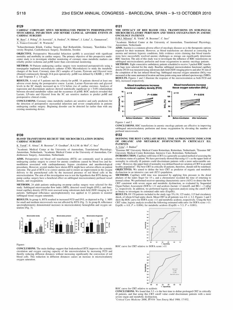
0129CARDIAC CORONARY SINUS MICRODIALYSIS PREDICTS PERIOPERATIVEMYOCARDIAL INFARCTION AND SEVERE CLINICAL ADVERSE EVENTS INCARDIAC SURGERY
W. Rees1, J. Poling1, D. Jozwicki1, A. Ferdosi1, N. Hubner1, J. Liska2, L. Gunnarsson2,
A. Franco-Cereceda2, H. Warnecke1
1Schuechtermann Klinik, Cardiac Surgery, Bad Rothenfelde, Germany, 2Karolinksa Uni-versity Hospital, Cardiothoracic Surgery, Stockholm, Sweden
OBJECTIVE. Perioperative Myocardial Infarction (poMI) is associated with significantmortality and morbidity in cardiac surgery. The primary objective of this prospective multi-center study is to investigate whether monitoring of coronary sinus metabolic markers canreliably predict ischemia and poMI faster than conventional monitoring.
METHOD. 50 Patients undergoing cardiac surgery were monitored perioperatively using atransjugular implanted microdialysis catheter (CMA Microdialysis) to study the metabolicchanges of the heart. Coronary sinus (SC) samples of Lactate, Pyruvate and Glycerol wereobtained continuously through 24-h post-operatively. poMI was defined by CKMB C 100 U/L and Troponin T C 1.0 lg/L.
RESULTS. A total of 5 patients met the criteria for poMI. 14 patients showed at least oneadverse event during the postoperative course. Lactate, Lactate–Pyruvate-ratio and glycerollevels in the SC sharply increased up to 12 h before rise of cardiac enzymes. Analyses ofregression and discriminate analyses showed statistically significant (p \ 0.05) relationshipsbetween elevated metabolite values and the occurence of poMI. ROC analysis revealed thatLactate, LP-ratio and Glycerol from the SC are sensitive markers to predict poMI andpostoperative clinical events.
CONCLUSIONS. Coronary sinus metabolic markers are sensitive and early predictors forthe detection of perioperative myocardial infarction and severe complications in patientsundergoing cardiac surgery. Beginning disorder can be detected far earlier than with anyexisting monitoring device.
0130BLOOD TRANSFUSIONS RECRUIT THE MICROCIRCULATION DURINGCARDIAC SURGERY
K. Yuruk1, E. Almac1, R. Bezemer1, P. Goedhart1, B.A.J.M. de Mol2, C. Ince1
1Academic Medical Center at the University of Amsterdam, Translational Physiology,Amsterdam, Netherlands, 2Academic Medical Center at the University of Amsterdam, Car-diothoracic Surgery, Amsterdam, Netherlands
AIMS. Perioperative red blood cell transfusions (BTX) are commonly used in patientsundergoing cardiac surgery to correct for anemic conditions caused by blood loss and he-modilution associated with cardiopulmonary bypass circulation and anesthesiologicalprocedures. However, several studies have shown BTX might have adverse effects on patientoutcome. The goal of BTX is to correct anemia and to ensure an improvement in the oxygendelivery to the parenchymal cells by the increased presence of red blood cells in themicrocirculation. The aim of this investigation was to test the hypothesis that BTX during on-pump cardiac surgery have a beneficial effect on sublingual microcirculatory perfused vesseldensity, and oxygenation.
METHODS. 24 adult patients undergoing on-pump cardiac surgery were selected for thisstudy. Sublingual microvascular flow index (MFI), detected vessel length (DVL), and func-tional capillary density (FCD) were assessed using sidestream dark-field (SDF) imaging in 12patients. Sublingual reflectance spectrophotometry was applied in 12 patients to monitorsublingual tissue oxygen saturation.
RESULTS. In group A, BTX resulted in increased FCD and DVL as depicted in Fig. 1. MFIfor small and medium microvessels was not affected by BTX (Fig. 2). In group B, reflectancespectrophotometry demonstrated increases in microcirculatory hemoglobin and oxygen sat-uration (Fig. 3).
Figures
CONCLUSIONS. The main findings suggest that leukoreduced BTX improves the systemiccirculation and oxygen carrying capacity of the microcirculation by increasing FCD andthereby reducing diffusion distances without increasing significantly the convection of redblood cells. This reduction in diffusion distances causes an increase in microcirculatoryoxygen saturation.
0131THE EFFICACY OF RED BLOOD CELL TRANSFUSIONS ON SUBLINGUALMICROCIRCULATORY PERFUSION AND TISSUE OXYGENATION IN ANEMICONCOLOGY PATIENTSD.M.J. Milstein1, K. Yuruk1, R. Bezemer1, C. Ince1
1Academic Medical Center at the University of Amsterdam, Translational Physiology,Amsterdam, NetherlandsAIMS. Anemia is a common adverse effect of oncologic diseases as is the therapeutic optionsrequired for their treatment. However, as blood transfusions are directed at correcting foranemia and intrinsic hypoxic conditions, little evidence exists claiming that blood transfu-sions have successfully resolved anemic challenges as storage can significantly deteriorateRBC function. The aim of this study was to investigate the influence of RBC transfusions onsublingual microcirculatory perfusion and tissue oxygenation in anemic oncology patients.METHODS. Eight consecutive ambulatory patients scheduled to receive 3 packed RBC transfu-sion bags were selected for this study. Baseline sublingual microcirculation functional capillarydensity (FCD) was measured using sidestream dark-field (SDF) imaging prior to and after 30 min ofthe completion of the last infused blood bag. Sublingual mucosal oxygen saturation (StO2) wasmeasured at the same anatomical location and time points using near-infrared spectroscopy (NIRS).RESULTS. Figures 1 and 2 illustrate the response of the microcirculation as both FCD andStO2 increased respectively.
Figures 1 and 2
CONCLUSIONS. RBC transfusions in anemic oncology patients are effective in improvingsublingual microcirculatory perfusion and tissue oxygenation by elevating the number ofperfused microvessels.
0132DEFINING THE BEST CAPILLARY REFILL TIME AS PROGNOSTIC INDICATOROF ORGANIC AND METABOLIC DYSFUNCTION IN CRITICALLY ILLPATIENTSA. Lima1, J. Bakker2
1Erasmus MC University Medical Centre Rotterdam, Rotterdam, Netherlands, 2Erasmus MCUniversity Medical Centre Rotterdam, Intensive Care, Rotterdam, NetherlandsINTRODUCTION. Capillary refill time (CRT) is a generally accepted method of assessing thecirculatory status of a patient. We have previously showed that using 4.5 s as the upper limit ofnormality in critically ill patients could discriminate patients with a more unfavourable out-come1. However, this upper limit of normality was defined based on variation of CRT in an adulthealthy population2. The best CRT in critically ill patients, therefore, should still be redefined.OBJECTIVES. We aimed to define the best CRT as predictor of organic and metabolicdysfunction in an intensive care unit (ICU) population.METHODS. Capillary refill time was measured by applying firm pressure to the distalphalanx of the index finger for 15 s, and a chronometer recorded the time of returning tonormal colour. We performed receiver operating characteristic curve (AUC) to detect the bestCRT consistent with severe organ and metabolic dysfunction, as evaluated by SequentialOrgan Failure Assessment (SOFA)[11 and acidosis (lactate[2 mmol/L and BE\-3 mEq/L), respectively. In addition, we performed logistic regression analysis using the cutoff CRTas binary to investigate its estimated odds ratio (Exp(B)).RESULTS. Of 170 patients included in the study (age 55±16; 125 male), 115 had circulatoryshock, of whom 62 had septic shock. Mean CRT in all patients was 4.6 ± 2.2. Figures 1 and 2show the ROC curve for SOFA score[11 and metabolic acidosis, respectively. Using the bestCRT value, logistic analysis revelled the following estimated odds ratio: for SOFA score[11:Exp(B) = 6.8; P = 0.004); for metabolic acidosis (Exp(B) = 5.3; P = 0.001).
ROC curve for CRT relative to SOFA score [11
ROC curve for CRT relative to acidosisCONCLUSIONS. We found that 5.5 s is the best time to define prolonged CRT in criticallyill patients, and that using this CRT cutoff value could discriminate patients with a moresevere organ and metabolic dysfunction.1Critical Care Medicine 2008, 37:934 2Ann Emerg Med 1988; 17:932.
S118 23rd ESICM ANNUAL CONGRESS – BARCELONA, SPAIN – 9–13 OCTOBER 2010
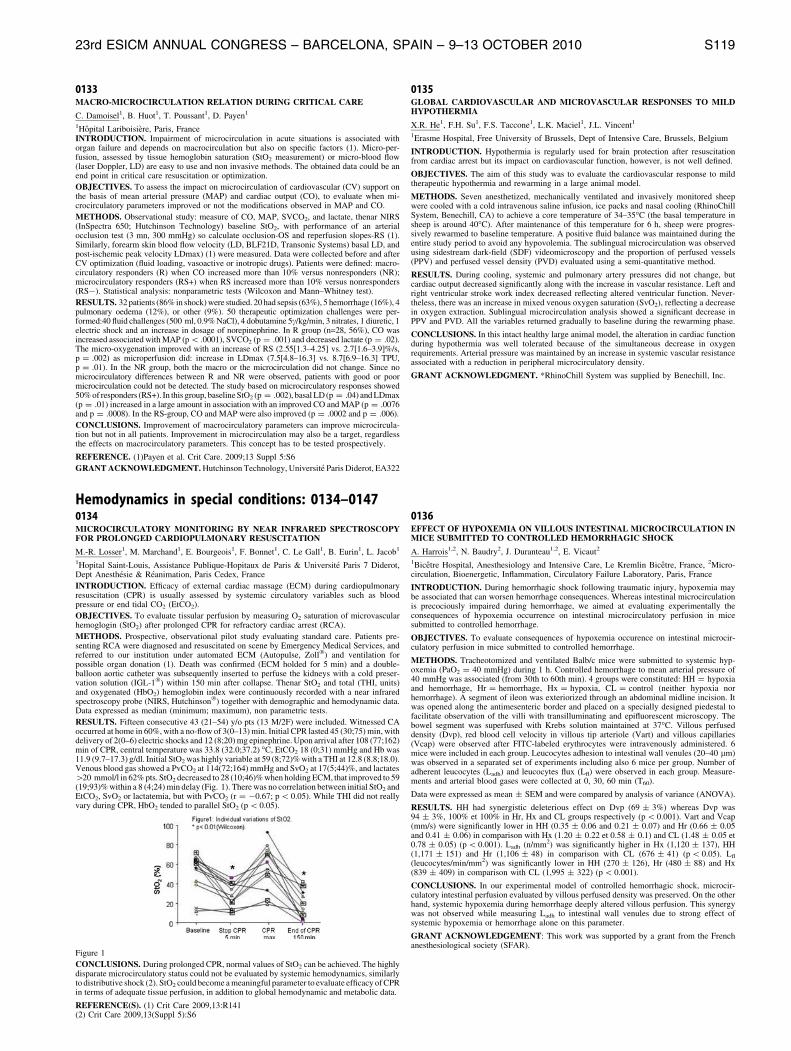
0133MACRO-MICROCIRCULATION RELATION DURING CRITICAL CARE
C. Damoisel1, B. Huot1, T. Poussant1, D. Payen1
1Hopital Lariboisiere, Paris, FranceINTRODUCTION. Impairment of microcirculation in acute situations is associated withorgan failure and depends on macrocirculation but also on specific factors (1). Micro-per-fusion, assessed by tissue hemoglobin saturation (StO2 measurement) or micro-blood flow(laser Doppler, LD) are easy to use and non invasive methods. The obtained data could be anend point in critical care resuscitation or optimization.
OBJECTIVES. To assess the impact on microcirculation of cardiovascular (CV) support onthe basis of mean arterial pressure (MAP) and cardiac output (CO), to evaluate when mi-crocirculatory parameters improved or not the modifications observed in MAP and CO.
METHODS. Observational study: measure of CO, MAP, SVCO2, and lactate, thenar NIRS(InSpectra 650; Hutchinson Technology) baseline StO2, with performance of an arterialocclusion test (3 mn, 300 mmHg) so calculate occlusion-OS and reperfusion slopes-RS (1).Similarly, forearm skin blood flow velocity (LD, BLF21D, Transonic Systems) basal LD, andpost-ischemic peak velocity LDmax) (1) were measured. Data were collected before and afterCV optimization (fluid loading, vasoactive or inotropic drugs). Patients were defined: macro-circulatory responders (R) when CO increased more than 10% versus nonresponders (NR);microcirculatory responders (RS+) when RS increased more than 10% versus nonresponders(RS-). Statistical analysis: nonparametric tests (Wilcoxon and Mann–Whitney test).
RESULTS. 32 patients (86% in shock) were studied. 20 had sepsis (63%), 5 hemorrhage (16%), 4pulmonary oedema (12%), or other (9%). 50 therapeutic optimization challenges were per-formed:40 fluid challenges (500 ml, 0.9% NaCl), 4 dobutamine 5c/kg/min, 3 nitrates, 1 diuretic, 1electric shock and an increase in dosage of norepinephrine. In R group (n=28, 56%), CO wasincreased associated with MAP (p \ .0001), SVCO2 (p = .001) and decreased lactate (p = .02).The micro-oxygenation improved with an increase of RS (2.55[1.3–4.25] vs. 2.7[1.6–3.9]%/s,p = .002) as microperfusion did: increase in LDmax (7.5[4.8–16.3] vs. 8.7[6.9–16.3] TPU,p = .01). In the NR group, both the macro or the microcirculation did not change. Since nomicrocirculatory differences between R and NR were observed, patients with good or poormicrocirculation could not be detected. The study based on microcirculatory responses showed50% of responders (RS+). In this group, baseline StO2 (p = .002), basal LD (p = .04) and LDmax(p = .01) increased in a large amount in association with an improved CO and MAP (p = .0076and p = .0008). In the RS-group, CO and MAP were also improved (p = .0002 and p = .006).
CONCLUSIONS. Improvement of macrocirculatory parameters can improve microcircula-tion but not in all patients. Improvement in microcirculation may also be a target, regardlessthe effects on macrocirculatory parameters. This concept has to be tested prospectively.
REFERENCE. (1)Payen et al. Crit Care. 2009;13 Suppl 5:S6
GRANT ACKNOWLEDGMENT. Hutchinson Technology, Universite Paris Diderot, EA322
Hemodynamics in special conditions: 0134–01470134MICROCIRCULATORY MONITORING BY NEAR INFRARED SPECTROSCOPYFOR PROLONGED CARDIOPULMONARY RESUSCITATION
M.-R. Losser1, M. Marchand1, E. Bourgeois1, F. Bonnet1, C. Le Gall1, B. Eurin1, L. Jacob1
1Hopital Saint-Louis, Assistance Publique-Hopitaux de Paris & Universite Paris 7 Diderot,Dept Anesthesie & Reanimation, Paris Cedex, France
INTRODUCTION. Efficacy of external cardiac massage (ECM) during cardiopulmonaryresuscitation (CPR) is usually assessed by systemic circulatory variables such as bloodpressure or end tidal CO2 (EtCO2).
OBJECTIVES. To evaluate tissular perfusion by measuring O2 saturation of microvascularhemoglogin (StO2) after prolonged CPR for refractory cardiac arrest (RCA).
METHODS. Prospective, observational pilot study evaluating standard care. Patients pre-senting RCA were diagnosed and resuscitated on scene by Emergency Medical Services, andreferred to our institution under automated ECM (Autopulse, Zoll�) and ventilation forpossible organ donation (1). Death was confirmed (ECM holded for 5 min) and a double-balloon aortic catheter was subsequently inserted to perfuse the kidneys with a cold preser-vation solution (IGL-1�) within 150 min after collapse. Thenar StO2 and total (THI, units)and oxygenated (HbO2) hemoglobin index were continuously recorded with a near infraredspectroscopy probe (NIRS, Hutchinson�) together with demographic and hemodynamic data.Data expressed as median (minimum; maximum), non parametric tests.
RESULTS. Fifteen consecutive 43 (21–54) y/o pts (13 M/2F) were included. Witnessed CAoccurred at home in 60%, with a no-flow of 3(0–13) min. Initial CPR lasted 45 (30;75) min, withdelivery of 2(0–6) electric shocks and 12 (8;20) mg epinephrine. Upon arrival after 108 (77;162)min of CPR, central temperature was 33.8 (32.0;37.2) �C, EtCO2 18 (0;31) mmHg and Hb was11.9 (9.7–17.3) g/dl. Initial StO2 was highly variable at 59 (8;72)% with a THI at 12.8 (8.8;18.0).Venous blood gas showed a PvCO2 at 114(72;164) mmHg and SvO2 at 17(5;44)%, and lactates[20 mmol/l in 62% pts. StO2 decreased to 28 (10;46)% when holding ECM, that improved to 59(19;93)% within a 8 (4;24) min delay (Fig. 1). There was no correlation between initial StO2 andEtCO2, SvO2 or lactatemia, but with PvCO2 (r = -0.67; p \ 0.05). While THI did not reallyvary during CPR, HbO2 tended to parallel StO2 (p \ 0.05).
Figure 1
CONCLUSIONS. During prolonged CPR, normal values of StO2 can be achieved. The highlydisparate microcirculatory status could not be evaluated by systemic hemodynamics, similarlyto distributive shock (2). StO2 could become a meaningful parameter to evaluate efficacy of CPRin terms of adequate tissue perfusion, in addition to global hemodynamic and metabolic data.
REFERENCE(S). (1) Crit Care 2009,13:R141(2) Crit Care 2009,13(Suppl 5):S6
0135GLOBAL CARDIOVASCULAR AND MICROVASCULAR RESPONSES TO MILDHYPOTHERMIA
X.R. He1, F.H. Su1, F.S. Taccone1, L.K. Maciel1, J.L. Vincent1
1Erasme Hospital, Free University of Brussels, Dept of Intensive Care, Brussels, Belgium
INTRODUCTION. Hypothermia is regularly used for brain protection after resuscitationfrom cardiac arrest but its impact on cardiovascular function, however, is not well defined.
OBJECTIVES. The aim of this study was to evaluate the cardiovascular response to mildtherapeutic hypothermia and rewarming in a large animal model.
METHODS. Seven anesthetized, mechanically ventilated and invasively monitored sheepwere cooled with a cold intravenous saline infusion, ice packs and nasal cooling (RhinoChillSystem, Benechill, CA) to achieve a core temperature of 34–35�C (the basal temperature insheep is around 40�C). After maintenance of this temperature for 6 h, sheep were progres-sively rewarmed to baseline temperature. A positive fluid balance was maintained during theentire study period to avoid any hypovolemia. The sublingual microcirculation was observedusing sidestream dark-field (SDF) videomicroscopy and the proportion of perfused vessels(PPV) and perfused vessel density (PVD) evaluated using a semi-quantitative method.
RESULTS. During cooling, systemic and pulmonary artery pressures did not change, butcardiac output decreased significantly along with the increase in vascular resistance. Left andright ventricular stroke work index decreased reflecting altered ventricular function. Never-theless, there was an increase in mixed venous oxygen saturation (SvO2), reflecting a decreasein oxygen extraction. Sublingual microcirculation analysis showed a significant decrease inPPV and PVD. All the variables returned gradually to baseline during the rewarming phase.
CONCLUSIONS. In this intact healthy large animal model, the alteration in cardiac functionduring hypothermia was well tolerated because of the simultaneous decrease in oxygenrequirements. Arterial pressure was maintained by an increase in systemic vascular resistanceassociated with a reduction in peripheral microcirculatory density.
GRANT ACKNOWLEDGMENT. *RhinoChill System was supplied by Benechill, Inc.
0136EFFECT OF HYPOXEMIA ON VILLOUS INTESTINAL MICROCIRCULATION INMICE SUBMITTED TO CONTROLLED HEMORRHAGIC SHOCK
A. Harrois1,2, N. Baudry2, J. Duranteau1,2, E. Vicaut2
1Bicetre Hospital, Anesthesiology and Intensive Care, Le Kremlin Bicetre, France, 2Micro-circulation, Bioenergetic, Inflammation, Circulatory Failure Laboratory, Paris, France
INTRODUCTION. During hemorrhagic shock following traumatic injury, hypoxemia maybe associated that can worsen hemorrhage consequences. Whereas intestinal microcirculationis precociously impaired during hemorrhage, we aimed at evaluating experimentally theconsequences of hypoxemia occurrence on intestinal microcirculatory perfusion in micesubmitted to controlled hemorrhage.
OBJECTIVES. To evaluate consequences of hypoxemia occurence on intestinal microcir-culatory perfusion in mice submitted to controlled hemorrhage.
METHODS. Tracheotomized and ventilated Balb/c mice were submitted to systemic hyp-oxemia (PaO2 = 40 mmHg) during 1 h. Controlled hemorrhage to mean arterial pressure of40 mmHg was associated (from 30th to 60th min). 4 groups were constituted: HH = hypoxiaand hemorrhage, Hr = hemorrhage, Hx = hypoxia, CL = control (neither hypoxia norhemorrhage). A segment of ileon was exteriorized through an abdominal midline incision. Itwas opened along the antimesenteric border and placed on a specially designed piedestal tofacilitate observation of the villi with transilluminating and epifluorescent microscopy. Thebowel segment was superfused with Krebs solution maintained at 37�C. Villous perfuseddensity (Dvp), red blood cell velocity in villous tip arteriole (Vart) and villous capillaries(Vcap) were observed after FITC-labeled erythrocytes were intravenously administered. 6mice were included in each group. Leucocytes adhesion to intestinal wall venules (20–40 lm)was observed in a separated set of experiments including also 6 mice per group. Number ofadherent leucocytes (Ladh) and leucocytes flux (Lfl) were observed in each group. Measure-ments and arterial blood gases were collected at 0, 30, 60 min (T60).
Data were expressed as mean ± SEM and were compared by analysis of variance (ANOVA).
RESULTS. HH had synergistic deleterious effect on Dvp (69 ± 3%) whereas Dvp was94 ± 3%, 100% et 100% in Hr, Hx and CL groups respectively (p \ 0.001). Vart and Vcap(mm/s) were significantly lower in HH (0.35 ± 0.06 and 0.21 ± 0.07) and Hr (0.66 ± 0.05and 0.41 ± 0.06) in comparison with Hx (1.20 ± 0.22 et 0.58 ± 0.1) and CL (1.48 ± 0.05 et0.78 ± 0.05) (p \ 0.001). Ladh (n/mm2) was significantly higher in Hx (1,120 ± 137), HH(1,171 ± 151) and Hr (1,106 ± 48) in comparison with CL (676 ± 41) (p \ 0.05). Lfl
(leucocytes/min/mm2) was significantly lower in HH (270 ± 126), Hr (480 ± 88) and Hx(839 ± 409) in comparison with CL (1,995 ± 322) (p \ 0.001).
CONCLUSIONS. In our experimental model of controlled hemorrhagic shock, microcir-culatory intestinal perfusion evaluated by villous perfused density was preserved. On the otherhand, systemic hypoxemia during hemorrhage deeply altered villous perfusion. This synergywas not observed while measuring Ladh to intestinal wall venules due to strong effect ofsystemic hypoxemia or hemorrhage alone on this parameter.
GRANT ACKNOWLEDGEMENT: This work was supported by a grant from the Frenchanesthesiological society (SFAR).
23rd ESICM ANNUAL CONGRESS – BARCELONA, SPAIN – 9–13 OCTOBER 2010 S119
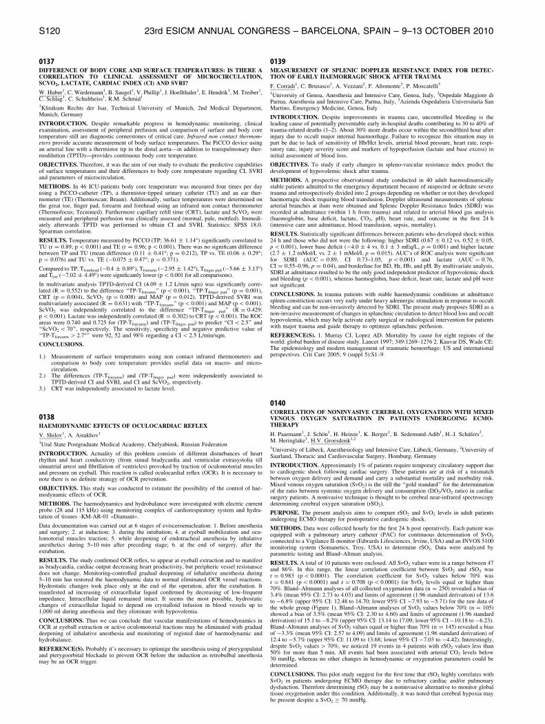
0137DIFFERENCE OF BODY CORE AND SURFACE TEMPERATURES: IS THERE ACORRELATION TO CLINICAL ASSESSMENT OF MICROCIRCULATION,SCVO2, LACTATE, CARDIAC INDEX (CI) AND SVRI?
W. Huber1, C. Wiedemann1, B. Saugel1, V. Phillip1, J. Hoellthaler1, E. Hendrik1, M. Treiber1,C. Schlag1, C. Schultheiss1, R.M. Schmid1
1Klinikum Rechts der Isar, Technical University of Munich, 2nd Medical Department,Munich, Germany
INTRODUCTION. Despite remarkable progress in hemodynamic monitoring, clinicalexamination, assessment of peripheral perfusion and comparison of surface and body coretemperature still are diagnostic cornerstones of critical care. Infrared non contact thermom-eters provide accurate measurement of body surface temperatures. The PiCCO device usingan arterial line with a thermistor tip in the distal aorta—in addition to transpulmonary ther-modilution (TPTD)—provides continuous body core temperature.
OBJECTIVES. Therefore, it was the aim of our study to evaluate the predictive capabilitiesof surface temperatures and their differences to body core temperature regarding CI, SVRIand parameters of microcirculation.
METHODS. In 46 ICU-patients body core temperature was measured four times per dayusing a PiCCO-catheter (TP), a thermistor-tipped urinary catheter (TU) and an ear ther-mometer (TE) (Thermoscan; Braun). Additionally, surface temperatures were determined onthe great toe, finger pad, forearm and forehead using an infrared non contact thermometer(Thermofocus; Tecnimed). Furthermore capillary refill time (CRT), lactate and ScVO2 weremeasured and peripheral perfusion was clinically assessed (normal, pale, mottled). Immedi-ately afterwards TPTD was performed to obtain CI and SVRI. Statistics: SPSS 18.0.Spearman correlation.
RESULTS. Temperature measured by PiCCO (TP; 36.61 ± 1.14�) significantly correlated toTU (r = 0.89; p \ 0.001) and TE (r = 0.96; p \ 0.001). There was no significant differencebetween TP and TU (mean difference (0.11 ± 0.41�; p = 0.212), TP vs. TE (0.06 ± 0.29�;p = 0.076) and TU vs. TE (-0.075 ± 0.47�; p = 0.371).
Compared to TP, Tforehead (-0.4 ± 0.89�), Tforearm (-2.95 ± 1.42�), Tfinger pad (-5.66 ± 3.13�)and Ttoe (-7.02 ± 4.49�) were significantly lower (p\ 0.001 for all comparisons).
In multivariate analysis TPTD-derived CI (4.09 ± 1.2 L/min sqm) was significantly corre-lated (R = 0.552) to the difference ‘‘TP-Tforearm’’ (p \ 0.001), ‘‘TP-Tfinger pad’’ (p = 0.001),CRT (p = 0.004), ScVO2 (p = 0.008) and MAP (p = 0.012). TPTD-derived SVRI wasmultivariately associated (R = 0.631) with ‘‘TP-Tforearm’’ (p \ 0.001) and MAP (p \ 0.001).ScVO2 was independently correlated to the difference ‘‘TP-Tfinger pad’’ (R = 0.429;p \ 0.001). Lactate was independently correlated (R = 0.302) to CRT (p \ 0.001). The ROCareas were 0.740 and 0.725 for (TP-Tforearm) and (TP-Tfinger pad) to predict ‘‘CI \ 2.5’’ and‘‘ScVO2 \ 70’’, respectively. The sensitivity, specificity and negative predictive value of‘‘TP-Tforearm [ 2.7�’’ were 92, 52 and 98% regarding a CI \ 2.5 L/min/sqm.
CONCLUSIONS.
1.) Measurement of surface temperatures using non contact infrared thermometers andcomparison to body core temperature provides useful data on macro- and micro-circulation.
2.) The differences (TP-Tforearm) and (TP-Tfinger pad) were independently associated toTPTD-derived CI and SVRI, and CI and ScVO2, respectively.
3.) CRT was independently associated to lactate level.
0138HAEMODYNAMIC EFFECTS OF OCULOCARDIAC REFLEX
V. Shilov1, A. Astakhov1
1Ural State Postgraduate Medical Academy, Chelyabinsk, Russian Federation
INTRODUCTION. Actuality of this problem consists of different disturbances of heartrhythm and heart conductivity (from sinual bradycardia and ventricular extrasystolia tillsinuatrial arrest and fibrillation of ventricles) provoked by traction of oculomotorial musclesand pressure on eyeball. This reaction is called oculocardial reflex (OCR). It is necessary tonote there is no definite strategy of OCR prevention.
OBJECTIVES. This study was conducted to estimate the possibility of the control of hae-modynamic effects of OCR.
METHODS. The haemodynamics and hydrobalance were investigated with electric currentprobe (28 and 115 kHz) using monitoring complex of cardiorespiratory system and hydra-tation of tissues -KM-AR-01 «Diamant».
Data documentation was carried out at 6 stages of evisceroenucleation: 1. Before anesthesiaand surgery; 2. at induction; 3. during the intubation; 4. at eyeball mobilization and ocu-lomotorial muscles traction; 5. while deepening of endotracheal anesthesia by inhalativeanesthetics during 5–10 min after preceding stage; 6. at the end of surgery, after theextubation.
RESULTS. The study confirmed OCR reflex, to appear at eyeball extraction and to manifestas bradycardia, cardiac output decreasing heart productivity, but peripheric vessel resistanscedoes not change. Monitoring-controlled gradual deepening of inhalative anesthesia during5–10 min has restored the haemodynamic data to normal eliminated OCR vessel reactions.Hydrostatic changes took place only at the end of the operation, after the extubation. Itmanifested ad increasing of extracellular liquid confirmed by decreasing of low-frequentimpedance. Intracellular liquid remained intact. It seems the most possible, hydrostaticchanges of extracellular liquid to depend on crystalloid infusion in blood vessels up to1,000 ml during anesthesia and they eliminate with hypovolemia.
CONCLUSIONS. Thus we can conclude that vascular manifestations of hemodynamics inOCR at eyeball extraction or active oculomotorial tractions may be eliminated with gradualdeepening of inhalative anesthesia and monitoring of registed date of haemodynamic andhydrobalance.
REFERENCE(S). Probably it’s necessary to optimige the anesthesia using of pterygopalataland pterygoorbital blockade to prevent OCR before the induction as retrobulbal anesthesiamay be an OCR trigger.
0139MEASUREMENT OF SPLENIC DOPPLER RESISTANCE INDEX FOR DETEC-TION OF EARLY HAEMORRAGIC SHOCK AFTER TRAUMA
F. Corradi1, C. Brusasco1, A. Vezzani2, F. Altomonte3, P. Moscatelli3
1University of Genoa, Anesthesia and Intensive Care, Genoa, Italy, 2Ospedale Maggiore diParma, Anesthesia and Intensive Care, Parma, Italy, 3Azienda Ospedaliera Universitaria SanMartino, Emergency Medicine, Genoa, Italy
INTRODUCTION. Despite improvements in trauma care, uncontrolled bleeding is theleading cause of potentially preventable early in-hospital deaths contributing to 30 to 40% oftrauma-related deaths (1–2). About 30% more deaths occur within the second/third hour afterinjury due to occult major internal haemorrhage. Failure to recognize this situation may inpart be due to lack of sensitivity of Hb/Hct levels, arterial blood pressure, heart rate, respi-ratory rate, injury severity score and markers of hypoperfusion (lactate and base excess) ininitial assessment of blood loss.
OBJECTIVES. To study if early changes in spleno-vascular resistance index predict thedevelopment of hypovolemic shock after trauma.
METHODS. A prospective observational study conducted in 40 adult haemodinamicallystable patients admitted to the emergency department because of suspected or definite severetrauma and retrospectively divided into 2 groups depending on whether or not they developedhaemorragic shock requiring blood transfusion. Doppler ultrasound measurements of splenicarterial branches at ilum were obtained and Splenic Doppler Resistance Index (SDRI) wasrecorded at admittance (within 1 h from trauma) and related to arterial blood gas analysis(haemoglobin, base deficit, lactate, CO2, pH), heart rate, and outcome in the first 24 h(intensive care unit admittance, blood transfusion, sepsis, mortality).
RESULTS. Statistically significant differences between patients who developed shock within24 h and those who did not were the following: higher SDRI (0.67 ± 0.12 vs. 0.52 ± 0.05,p \ 0.001), lower base deficit (-4.0 ± 4 vs. 0.1 ± 3 mEq/L, p = 0.001) and higher lactate(2.7 ± 1.2 mMol/L vs. 2 ± 1 mMol/L p = 0.015). AUC’s of ROC analysis were significantfor SDRI (AUC = 0.89, CI 0.73–1.05, p \ 0.001) and lactate (AUC = 0.76,CI = 0.55–0.96, p = 0.04), and borderline for BD, Hr, Hb, and pH. By multivariate analysis,SDRI at admittance resulted to be the only good independent predictor of hypovolemic shockand bleeding (p \ 0.001), whereas haemoglobin, base deficit, heart rate, lactate and pH werenot significant.
CONCLUSIONS. In trauma patients with stable haemodynamic conditions at admittancespleen constriction occurs very early under heavy adrenergic stimulation in response to occultbleeding and can be non-invasively detected by SDRI. The present study proposes SDRI as anon-invasive measurement of changes in splanchnic circulation to detect blood loss and occulthypovolemia, which may help activate early surgical or radiological intervention for patientswith major trauma and guide therapy to optimize splanchnic perfusion.
REFERENCE(S). 1. Murray CJ, Lopez AD: Mortality by cause for eight regions of theworld: global burden of disease study. Lancet 1997; 349:1269–1276 2. Kauvar DS, Wade CE:The epidemiology and modern management of traumatic hemorrhage: US and internationalperspectives. Crit Care 2005; 9 (suppl 5):S1–9
0140CORRELATION OF NONINVASIVE CEREBRAL OXYGENATION WITH MIXEDVENOUS OXYGEN SATURATION IN PATIENTS UNDERGOING ECMO-THERAPY
H. Paarmann1, J. Schon1, H. Heinze1, K. Berger1, B. Sedemund-Adib1, H.-J. Schafers2,M. Heringlake1, H.V. Groesdonk1,2
1University of Lubeck, Anesthesiology und Intensive Care, Lubeck, Germany, 2University ofSaarland, Thoracic and Cardiovascular Surgery, Homburg, Germany
INTRODUCTION. Approximately 1% of patients require temporary circulatory support dueto cardiogenic shock following cardiac surgery. These patients are at risk of a mismatchbetween oxygen delivery and demand and carry a substantial mortality and morbidity risk.Mixed venous oxygen saturation (SvO2) is the still the ‘‘gold standard’’ for the determinationof the ratio between systemic oxygen delivery and consumption (DO2/VO2 ratio) in cardiacsurgery patients. A nonivasive technique is thought to be cerebral near-infrared spectroscopydetermining cerebral oxygen saturation (rSO2).
PURPOSE. The present analysis aims to compare rSO2 and SvO2 levels in adult patientsundergoing ECMO therapy for postoperative cardiogenic shock.
METHODS. Data were collected hourly for the first 24 h post operatively. Each patient wasequipped with a pulmonary artery catheter (PAC) for continuous determination of SvO2
connected to a Vigilance II-monitor (Edwards Lifesciences, Irvine, USA) and an INVOS 5100monitoring system (Somanetics, Troy, USA) to determine rSO2. Data were analyzed byparametric testing and Bland–Altman analysis.
RESULTS. A total of 10 patients were enclosed. All SvO2 values were in a range between 47and 86%. In this range, the linear correlation coefficient between SvO2 and rSO2 wasr = 0.983 (p \ 0.0001). The correlation coefficient for SvO2 values below 70% wasr = 0.841 (p \ 0.0001) and r = 0.708 (p \ 0.0001) for SvO2 levels equal or higher than70%. Bland–Altmann analyses of all collected oxygenation data (n = 250) revealed a bias of3.4% (mean 95% CI: 2.73 to 4.03) and limits of agreement (1.96 standard derivation) of 13.6to -6.8% (upper 95% CI: 12.48 to 14.70; lower 95% CI -7.93 to -5.71) for the raw data ofthe whole group (Figure 1). Bland–Altmann analyses of SvO2 values below 70% (n = 105)showed a bias of 3.5% (mean 95% CI: 2.30 to 4.60) and limits of agreement (1.96 standardderivation) of 15.1 to -8.2% (upper 95% CI: 13.14 to 17.09; lower 95% CI -10.18 to -6.23).Bland–Altmann analyses of SvO2 values equal or higher than 70% (n = 145) revealed a biasof -3.3% (mean 95% CI: 2.57 to 4.09) and limits of agreement (1.96 standard derivation) of12.4 to -5.7% (upper 95% CI: 11.09 to 13.68; lower 95% CI -7.03 to -4.42). Interestingly,despite SvO2 values [ 70%, we noticed 19 events in 4 patients with rSO2 values less than50% for more than 5 min. All events had been associated with arterial CO2 levels below30 mmHg, whereas no other changes in hemodynamic or oxygenation parameters could bedetermined.
CONCLUSIONS. This pilot study suggest for the first time that rSO2 highly correlates withSvO2 in patients undergoing ECMO therapy due to refractory cardiac and/or pulmonarydysfunction. Therefore determining rSO2 may be a noninvasive alternative to monitor globaltissue oxygenation under this condition. Additionally, it was noted that cerebral hypoxia maybe present despite a SvO2 C 70 mmHg.
S120 23rd ESICM ANNUAL CONGRESS – BARCELONA, SPAIN – 9–13 OCTOBER 2010

0141CARDIAC FUNCTION DURING SEVERE IMMERSION HYPOTHERMIA ANDREWARMING IN PIGS
O.M. Filseth1,2, O.-J. How3,4, T. Kondratiev5, T.M. Gamst6, T. Tveita2,6
1University Hospital of Northern Norway, Dept of Anaesthesiology and Intensive CareMedicine, Tromsø, Norway, 2University of Tromsø, Department of Anaesthesiology, Instituteof Clinical Medicine, Tromsø, Norway, 3University Hospital of Northern Norway, Depart-ment of Cardiothoracic Surgery, Tromsø, Norway, 4University of Tromsø, Department ofMedical Physiology, Tromsø, Norway, 5University of Tromsø, Departement of Anaesthesi-ology, Institute of Clinical Medicine, Tromsø, Norway, 6University Hospital of NorthernNorway, Department of Anaesthesiology and Intensive Care Medicine, Tromsø, Norway
INTRODUCTION/OBJECTIVES. We developed a minimally invasive, closed chest pigmodel to describe central hemodynamic function during surface cooling, steady state hypo-thermia (1 h at 25�C) and surface rewarming.
METHODS. Eight anesthetized juvenile pigs were acutely catheterized for measurement ofleft ventricular (LV) pressure–volume loops (conductance catheter), cardiac output (Swan-Ganz), and for vena cava inferior occlusion.
RESULTS1: During severe hypothermia (25�C) cardiac index (CI), stroke index, meanarterial pressure and indexes of LV contractility (PRSW2 and dP/dTmax) were reduced. Afterrewarming all variables remained reduced, except for CI that returned to prehypothermicvalues due to increased heart rate. Systemic vascular resistance (SVR), LV isovolumetricrelaxation time (Tau) and oxygen content in arterial and mixed venous blood increased during25�C, while LV end diastolic pressure (LVEDP) was constant. After rewarming SVR andLVEDP were reduced, while Tau and the blood oxygen contents normalized. Troponin-T andTNF-a were constant during 25�C but increased after rewarming. Albumine plasma con-centration was reduced during 25�C and remained so after rewarming.
CONCLUSIONS. Surface cooling to 25�C followed by rewarming caused reduction ofsystolic, but not diastolic LV function. There were no signs of inadequate global oxygenationthroughout experiments. The posthypothermic increase in troponin-T may reflect degradationof myocyte troponins secondary to a hypothermia-induced calcium overload. The increase intumour necrosis factor alpha together with a posthypothermic reduction of plasma albuminconcentration may indicate that the cooling and rewarming initiated an inflammatoryresponse.
REFERENCES. 1 Differences are only described if statistically significant (p \ 0.05).2 PRSW measured at 30�C only.
0142DOPAMINE INFUSIONS IN NORMOTHERMIA, SEVERE HYPOTHERMIA ANDDURING REWARMING: PHARMACOKINETICS AND CARDIOVASCULARFUNCTION IN A PIG MODEL OF SURFACE COOLING AND REWARMING
O.M. Filseth1,2, O.-J. How3, T. Kondratiev4, T.M. Gamst1, G. Sager5, T. Tveita1,4
1University Hospital of Northern Norway, Department of Anaesthesiology and Intensive CareMedicine, Tromsø, Norway, 2University of Tromsø, Department of Anesthesiology, Instituteof Clinical Medicine, Tromsø, Norway, 3University Hospital of Northern Norway, Depart-ment of Cardiothoracic Surgery, Tromsø, Norway, 4University of Tromsø, Department ofAnaesthesiology, Institute of Clinical Medicine, Tromsø, Norway, 5University of Tromsø,Department of Pharmacology, Tromsø, Norway
INTRODUCTION/OBJECTIVES. Catecholamines are administered routinely in hypo-thermic patients to support a presumably inadequate circulation. We developed a minimallyinvasive pig model to describe pharmacokinetics and cardiovascular pharmacodynamiceffects of dopamine (DA) in hypothermia caused by surface cooling.
METHODS. Sixteen anesthetized juvenile pigs were instrumented with left ventricular (LV)pressure volume and pulmonary catheters for assessment of ventricular function and globalhemodynamics. DA at different doses (4, 8 and 16 lg/kg/min) was infused at normothermia,at severe hypothermia (25�C), and subsequently at moderate hypothermia (30–34�C) and38�C during rewarming. One group (n = 8) received placebo (saline) at 25 and 30�C.
RESULTS. DA half-time was 5.4 ± 0.8 min at normothermia, which increased to11.6 ± 0.9 min at 25�C. Comparison of DA infusion of 16 lg/kg/min at normothermia and25�C demonstrated fourfold increase in hypothermic DA plasma concentration. At normo-thermia cardiac index (CI), heart rate (HR), and contractility indexes (PRSW and dP/dTmax)increased 30–90% by DA (16 lg/kg/min) compared to baseline, coincidently with a 24–27%decrease in isovolumetric relaxation time and systemic vascular resistance index (SVRi).During 25�C DA (16 lg/kg/min) infusion elevated SVRi, dP/dTmax and HR by 30–71%,while Tau and stroke index decreased by 31–43%. Interestingly, LV end systolic and diastolicvolumes along with LV end diastolic pressure (EDP) decreased by 48–70% following DA atsevere hypothermia. Cardiovascular effects of DA at 30–34�C during rewarming wereprincipally similar to normothermic results.
CONCLUSIONS. Increased SVRi at high-dose dopamine at 25�C suggests alpha-adrenergicinvolvement not seen in normothermia. LV systolic and diastolic function improved inresponse to DA at 25�C but CO was unaffected, leaving the conclusion that high-dose DA hadno beneficial effect on cardiovascular function at severe hypothermia. Pharmacokineticfindings suggest a fourfold reduction in DA infusion rates at 25�C to obtain similar plasmaDA concentration as in normothermia.
0143EXPERIENCE IN USE OF ECMO IN CARDIOGENIC SHOCK IN HOSPITAL LA FEOF VALENCIA
M.P. Fuset Cabanes1, S. Torregrosa Puerta2, F. Perez Esteban1, R. Gimeno Costa1,J.M. Bonastre Mora1
1H.U. La Fe, Critical Care Service, Valencia, Spain, 2H.U. La Fe, Cardiac Surgery Service,Valencia, Spain
INTRODUCTION. Extracorporeal membrane oxygenator (ECMO) is a device that givecardiac and respiratory support during several days or weeks.
We use the ECMO as cardiac support since December 2006. Our hospital is the centre ofreference in Valencia region for heart transplantation and national reference in use of ECMO.
OBJECTIVES. To describe clinical characteristics, evolution and survival of patients thatreceived ECMO support for cardiogenic shock.
METHODS. Consecutive patients with cardiogenic shock treated with ECMO betweenDecember 2006 and December 2009. Categorical data are expressed as percentage andcontinuous data as the mean ± DS.
RESULTS. We studied 41 patients, mean age 51.6 ± 10.2 years, 68% male. The etiology ofcardiogenic shock was: 44% (n = 18) dilated cardiomyopathy, 37% (n = 15) acute myo-cardial infarction, 12% (n = 5) acute cardiac allograft rejection and 7% (n = 3) acutemyocarditis. The duration of ECMO support was 118.8 ± 89.8 h. Weaning was possible in22% (n = 9) and the ECMO was used as a bridge to transplantation in 51% (n = 21). 30-daysurvival was 60 and 58.5% of our serie were discharged from the hospital. In 7 cases theECMO was withdrown as a result of a limiting treatment decision.
CONCLUSIONS. The ECMO offers the survival in an important percentage of patients incardiogenic shock that would die without this device. It’s useful as bridge to recovery andbridge to heart transplantation. It’s important to consider the ECMO withdrawal if there isn’tclinical viability.
REFERENCE(S). ECMO. Extracorporeal Cardiopulmonary Support in Critical Care 3rd.Edition. Extracorporeal Life Support Organitation, Ann Arbor. Michigan. ISBN 0-9656756-2–9Oxigenacion de membrana extracorporea para soporte cardıaco o respiratorio en adultos.Torregrosa, Fuset et al. Cirugıa Cardiovascular, vol. 16, Num. 2/2009Combes A, Leprince P, Luyt CE, et al. Outcomes and longterm quality of life of patientssupported by extracorporeal membrane oxygenation for refractory cardiogenic shock. CritCare Med. 2008;36:1404–11.Extracorporeal Membrane Oxygenation for Treating Severe Cardiac and Respiratory Failurein Adults: Part 2—Technical Considerations. Sidebotham D, McGeorge A, McGuinness S,Edwards M, Willcox T, Beca J. J Cardiothorac Vasc Anesth. 2010 Feb;24(1):164–72.Fiser SM, Tribble CG, Kaza AK, et al.: When to discontinue extracorporeal membraneoxygenation for postcardiotomy support. Ann Thorac Surg 71:210–214, 2001.
0144MODERATE THERAPEUTIC HYPOTHERMIA IN CARDIAC ARREST (CA).FIRST NINE MONTHS AFTER A PROTOCOL STARTING
A. Hurtado Doce1, M.J. Garcia Monje1, J. Gulias1, F. Pampin1, L. Seoane1, M. Madruga1,
M. Solla1, S. Fojon1, M. Mourelo Farina1, P. Jimenez1
1A Coruna Universitary Hospital, Intensive Care Unit, A Coruna, Spain
OBJECTIVES. To describe the characteristics of patients with CA and its management withmoderate hypothermia using Arctic Sun� device with hydrogel patches.
METHODS. Descriptive, observational and retrospective study of patients who suffered CAand received moderate therapeutic hypothermia (33�C) according to the protocol imple-mented in a coronary intensive care unit of a tertiary hospital. We collected patients from June2009 to April 2010, first months of this therapy in our hospital. Moderate therapeutichypothermia is applied using the Arctic Sun� device consisting of hydrogel patches applied tothe skin covering 40% of the body surface. The device is connected to a temperature controlconsole, measuring core temperature with an urinary catheter. We analyzed demographiccharacteristics, cardiovascular risk factors and other relevant comorbidities. We collected dataabout the CA, its initial treatment and its ICU management with moderate hypothermia,analyzing length of events and systemic and neurological outcome at discharge from ICU. Wealso collected data about the infectious complications during the ICU stay.
RESULTS. During this period, moderate therapeutic hypothermia was applied to 14 patientswith a mean age of 56 ± 14 years. 78.6% were male. The most frequent cardiovascular riskfactor was cigarette smoking, present in 50% of individuals. The CA cause was an AMI by70% of cases; however, myocardial infarction or angina was documented before the eventonly in 14.3% of patients. The CA event was outside the hospital in 78.6% of cases and theinitial heart rate recorded was ventricular fibrillation in 64.3% of cases. The average CAlength was 18.6 ± 8 min. Obtaining a temperature of 33�C took between 3 and 6 h from theCA in most cases; and this temperature was maintained for an average of 31 ± 6 h. Theaverage time of induction of hypothermia was 2.5 h. The re-heating was performed between0.3 to 0.5�C per hour, averaging 9 h to reach temperatures of 36.5�C. Midazolam sedationwas performed in all patients and severe chills required muscle relaxation with cisatracuriumin 71.4% of patients. Infectious complications occurred in 64.3% of patients, the mostcommon site of infection was respiratory. The average stay was 18 days. At the time of ICUdischarge, average GCS was 14 and the average GOS was 4. Mortality was 14.3% (2patients).
CONCLUSIONS.
1- Implementation of a therapeutic hypothermia protocol is feasible.2- Infectious complications are common, being respiratory ones the most observed.3- The Arctic Sun � device is quick and safe for induction of moderate therapeutic
hypothermia.
23rd ESICM ANNUAL CONGRESS – BARCELONA, SPAIN – 9–13 OCTOBER 2010 S121
0145HYPOTHERMIA DECREASES NYTROTYROSINE AND NITRATES/NITRITESPRODUCTION IN CARDIAC ARREST SURVIVORS
P. Ostadal1, A. Kruger1, D. Vondrakova1, J. Herget2
1Cardiovascular Center, Na Homolce Hospital, Department of Cardiology, Prague, CzechRepublic, 22nd Faculty of Medicine, Charles University in Prague, Department of Physiology,Prague, Czech Republic
INTRODUCTION. Protective effect of therapeutic hypothermia in cardiac arrest survivorson mortality and neurological outcomes has been repeatedly shown.
OBJECTIVES. Up to now, it is not clear, however, whether mild hypothermia influencesalso markers of oxidative stress and nitric oxide production.
METHODS. Eleven patients after out-of-hospital cardiac arrest were included into this study,all were treated with mild hypothermia using endovascular system Thermodard XP. Targetcore temperature 33�C was maintained for 12 h, re-warming rate was set at 0.15�C per hour,followed by normothermia of 36.8�C. Blood samples for measurement of nitrotyrosine andnitrates/nitrites were taken at admission and then every 6 h for 2 days.
RESULTS. During hypothermia the levels of nitrotyrosine and nitrates/nitrites were com-parable with baseline values. In re-warming period serum levels of both parameters graduallyincreased and in normothermia the levels were significantly higher as compared with hypo-thermia: nitrotyrosine 14.87 ± 2.01 vs. 8.03 ± 1.68 lM/L, p = 0.027; nitrates/nitrites105.47 ± 30.65 vs. 55.62 ± 10.74 lM/L, p = 0.04.
CONCLUSIONS. Our results revealed that during mild hypothermia in cardiac arrest sur-vivors the levels of nitrotyrosine and nitrates/nitrites are significantly lower. These dataindicate that the reduction of oxidative stress and suppressed nitric oxide production may beinvolved in the protective effect of hypothermia.
GRANT ACKNOWLEDGMENT. This study was supported by the grant of the CzechMinistry of Health, Nr. 00000064203.
0146QUANTITATIVE RESPONSE OF BLOOD VOLUMES IN A JUVENILEHEMORRHAGIC SHOCK MODEL
A. Vigani1, A. Shih1, C. Bandt2, R. Pariaut3, P. Queiroz3, N. Thuramalla4
1University of Florida, Veterinary Medical Center, Large Animal Clinical Science, Gaines-ville, United States, 2University of Florida, Veterinary Medical Center, Small Animal ClinicalScience, Gainesville, United States, 3School of Veterinary Medicine, Louisiana State Uni-versity, Small Animal Clinical Sciences, Baton Rouge, United States, 4Transonic SystemsInc., Ithaca, United States
INTRODUCTION. New volumetric variables of preload, such as total end-diastolic volumeindex (TEDVI) and active circulation volume index (ACVI) and central blood volume index(CBVI), have been shown to be good predictors of fluid responsiveness. During acute changesof intravascular volume, such as hemorrhagic shock, these variables allow a more accurateintervention.
OBJECTIVES. The aim of our study was to investigate the changes in TEDVI, ACVI, CBVIin a juvenile model of hemorrhagic shock.
METHODS. Seven anesthetized ponies (1–4 months of age) were studied at normovolemia(Base), after blood withdrawal to mean arterial pressure (MAP) of 40 mmHg (Hemo), afterinfusion of norepinephrine to a MAP of 75 mmHg (NE), and after retransfusion (Resu).TEDVI, ACVI, CBVI were measured by Ultrasound Dilution (UD) technology with CoStatusdevice. Data were analyzed using Kruskal–Wallis analysis and Dunn’s t test. Comparison offluid load agreement by Blant Altman.
RESULTS. TEDVI and ACVI had significant change during Hemo and Resu status. Per-centage of TEDVI and ACVI changes agreed with percentage of blood volume removed/infused with bias and limits of agreement (LOA) 4% (-21.4, 29.3) and -10.6 (-29.5 8.2%)respectively. NE administration induced MAP and CVP significant changes, whereas TEDVIand ACVI remained unchanged. CBVI showed high variability and seemed to be inconsistenton the identification of the volume status.
CONCLUSIONS. In this animal model, TEDVI and ACVI were superior to CBVI inaccurately reflecting hemorrhage and were also suitable to predict fluid responsiveness. NEadministration did not affect the volumetric variables TEDVI and ACVI.
REFERENCE(S).1Journal of Cardiothoracic and Vascular Anesthesia 2007, 21, 5: 650–6542 Pediatric Anesthesia 2009 19: 688–694.
0147PREDICTORS OF NEUROLOGICAL RECOVERY IN REFRACTORY IN HOSPI-TAL CARDIAC ARREST
A. Papastylianou1, S. Malachias1, S. Zakynthinos1, E. Zakynthinos2, D. Makris2, S. Sourlas3,
S. Aloizos3, S. Mentzelopoulos1
1University of Athens, Intensive Care Medicine, Athens, Greece, 2University of Thessaly,Intensive Care Medicine, Larissa, Greece, 3401 General Army Hospital Athens, IntensiveCare Medicine, Athens, Greece
INTRODUCTION. In refractory in-hospital cardiac arrest, survival to hospital dischargewith neurological recovery is low and may not exceed 10% (1).
OBJECTIVES. We sought to identify independent predictors of post-arrest neurologicalrecovery, and of survival to hospital discharge with neurological recovery.
METHODS. In the course of a pre-planned interim analysis, we analyzed the data from 220 par-ticipants of NCT00729794. This three-center, double blind, placebo-controlled, clinical trial is ongoing(estimated enrollment = 350 patients) and aims to asses the efficacy of combined vasopressin andepinephrine during cardiopulmonary resuscitation (CPR) and of steroid administration during and afterCPR. Post-arrest neurological recovery was defined as Glasgow Coma Scale Score[9 documented atleast once by 2 study-independent physicians in patients not receiving sedation for at least 24 h. Weidentified a total of 65 patients who were subjected to at least one post-arrest assessment of theirneurological status. Subsequently, we used backward stepwise logistic regression, and assessed thefollowing potential predictors: cause of cardiac arrest (cardiac vs. non-cardiac); area of cardiac arrestoccurrence (monitored vs. non-monitored); use of therapeutic hypothermia; number of CPR cycles;mean arterialpressure and serum lactate at 15 min followingresuscitation; andpatient group allocation.
RESULTS. The sole independent predictor of post-arrest neurological recovery was the occurrence ofthe cardiac arrest in an area of monitored patient care (i.e., intensive or coronary care unit, and operatingor emergency room): odds ratio: 3.83, 95% confidence interval = 1.26–11.63; P = 0.018. The soleindependent predictor of survival to hospital discharge with neurological recovery was the serum lactateconcentration at 15 min after resuscitation: odds ratio: 1.22; 95% confidence interval = 1.02–1.46.
CONCLUSIONS. The results of this preliminary analysis suggest that post-arrest neurologicalrecovery seems to depend more on the use of pre-arrest patient monitoring rather than the employedCPR protocol. Also, patients with lower, early post-arrest serum lactate concentration seem to have abetter chance of surviving to hospital discharge without concurrent, severe neurological deficits.
REFERENCE(S). 1. Arch Intern Med. 2009;169(1):15–24.
GRANT ACKNOWLEDGMENT. NCT00729794 is funded by the Thorax Foundation,Athens, Greece.
Caring for coronary artery and valvular diseases:0148–01610148EFFECTS OF INTRAOPERATIVE ELECTRICAL DEFIBRILLATION ON BIOCHEM-ICAL MARKERS OF MYOCARDIAL DAMAGE IN ELECTIVE BYPASS SURGERYB. Heijnen1, L.P. Reusen1, N.M. Tolenaar1, W.B. Gerritsen2, W.J.P. van Boven3, P. Bruins1,H.P.A. van Dongen1
1St Antonius Hospital, Anesthesiology, Pain Medicine and Intensive Care, Nieuwegein,Netherlands, 2St Antonius Hospital, Clinical Chemistry, Nieuwegein, Netherlands, 3St An-tonius Hospital, Thoracic Surgery, Nieuwegein, Netherlands
INTRODUCTION. Because of the risk of postoperative myocardial infarction, CABGpatients are routinely monitored by ECG and biomarkers (CK-MB, TnT). The influence ofsurgery and elective cardioversion on these markers is well studied. However the influence ofepicardial defibrillation is not yet established. Recent literature suggest hFABP as a newmarker. It’s value is still under investigation
OBJECTIVES. To quantify the attribution of intra-operative defibrillation on markers ofmyocardial injury (CK, CK-MB, TnT and hFABP).
METHODS. Single centre prospective study in which 50 elective CABG patients wereincluded in a 2 month period in 2007. Patients with valve, emergency, off-pump surgery orrethoracotomies were excluded. Patients were grouped as having had defibrillation or nodefibrillation during surgery. Serum levels of CK, CK-MB, TnT and hFABP were analyzed inblood samples taken at arrival on the ICU and at 4, 8 and 16 h after admission to the ICU.Levels of these biochemical markers were compared using a paired t test.
RESULTS.
TABLE 1
Demographic characteristic
Characteristic Defbrillation No defbrillation P valve
Patients (number) 14 (39%) 36 (61%)Age (years) 65 ± 7.7 65.9 ± 10.l 0.75Sex (male/female) 28/8 12/2 0.51EURO Score 2.9 ± 1.5 2.8 ± 1.5 0.8Distal anastomosis (number) 4.4 ± 1.0 4.2 ± 0.9 0.4CPB duration (min) 89.1 ± 23.5 91.6 ± 18.7 0.73Aortic cross clamping duration (min) 60.3 ± 16.0 60.6 ± 13.2 0.96Operation temperature (degree celsius) 33.1 ± 1.0 3.28 ± 1.6 0.4Autologe blood returned (cc) 477.1 ± 155.9 509.7 ± 176.3 0.53Postoperative ventilation time (min) 314 ± 84 341 ± 148 0.44Intravascular volume postoperative in 24 h (cc) 2,321 ± 572 2,297 ± 505 0.89Myocardial infarction (%) 0 0 1.0Complications (%) 0 0 1.0
All data presented as mean ± standrad deviation
TABLE 2
Marker levels at T = 0, 4, 8 and 16 h in the not defibrillated and defibrillated group
Variable T = 0 mean p value T = 4 mean p value T = 8 mean p value T = 16 mean p value
CK (U/L)
No defib 114 ± 33.6 0.08 179 ± 65.3 0.07 259 ± 127 0.71 422 ± 331 0.56
Defib 110 ± 19.8 151 ± 39.2 240 ± 167 605 ± 1,128
CK-MB (U/L)
No defib 1.3 ± 5.5 0.75 144 ± 15.3 0.12 20.1 ± 13.6 0.18 24.9 ± 137 0.72
Defib 2.0 ± 7.5 7.0 ± 14.1 14.1 ± 13.6 23.0 ± 17.6
TnT (lg/L)
No defib 0.135 ± 0.93 0.07 0.403 ± 0.230 0.17 0.365 ± 0.224 0.16 0.237 ± 0.147 0.59
Defib 0.100 ± 0.398 0.331 ± 0.127 0.298 ± 0.102 0.237 ± 0.092
hFABP(ng/mL)
No defib 16.4 ± 7.28 0.52 11.23 ± 9.44 0.73 15.58 ± 17.51 0.39
Defib 14.82 ± 7.71 12.26 ± 8.31 10.87 ± 5.93
All data presented as mean ± standrad deviation
TABLE 3
Marker levels at T = 0, 4, 8 and 16 h in the not defibrillated and defibrillated group
Variable T = 0 mean p value T = 4 mean p value T = 8 mean p value T = 16 mean p value
CK (U/L)
\=30) 100 ± 21 0.8 147 ± 41 0.47 0.09 0.01[30 104 ± 2 170 ± 24
CK-MB (U/L)\=30) 2.0 ± 8 0.70 5 ± 13 0.38 0.06 0.17
[30 0 ± 0 16 ± 22TnT (lg/L)
\=30) 0.1 ± 0.03 0.27 0.31 ± 0.09 0.16 0.20 0.81[30 0.13 ± 0.09 0.45 ± 0.28
hFABP (ng/mL)\=30) 14 ± 7 0.06 10 ± 6 0.03 0.85
[30 28 ± 9 25 ± 8
CONCLUSIONS. 1. Intraoperative defibrillation has no effects on the levels of CK, CK-MB,TnT and hFABP. 2. Intraoperative defibrillation with more than 30 Joules may influence theconcentration of CK, CK-MB and hFABP.
S122 23rd ESICM ANNUAL CONGRESS – BARCELONA, SPAIN – 9–13 OCTOBER 2010
0149AUDIT ON MANAGEMENT OF NEW ONSET ATRIAL FIBRILLATION INGENERAL INTENSIVE CARE UNIT AT UNIVERSITY TEACHING HOSPITAL
S. Nallapareddy1, M. Naik1, J. Nortje1
1Norfolk and Norwich University Hospital, Norwich, UK
INTRODUCTION. Atrial fibrillation (AF) is the most common arrhythmia with incidence of12–15% in general intensive care units (ITU)1. Early identification and treatment of AF is pivotal inreducing mortality and morbidity. Although National Institute of Clinical Excellence (NICE) hasgiven general guidance there are currently no specific guidelines for treating AF patients in ITU. Thisis resulting in much variation the way AF patients are treated in ITU. Therefore we performed anaudit to see how compliant we are to NICE guidelines in managing new AF patients.
OBJECTIVES. Whether we are:
1. Confirming AF with 12 lead ECG?2. Adequately investigating causes of AF?3. Anticoagulating high risk AF patients to prevent thromboembolic complications along
with rate control?
METHODS. We conducted a retrospective audit, identified patients considered to be high risk ofdeveloping AF by searching patient database of our unit (Metavision) over a 3 month period in 2009.We excluded patients aged B65 years, stayed in ITU for B48 h and those in AF at admission.
RESULTS. In 3 month period we found 145 at risk patients, 29 of them developed new AF (20%).Below is the results table showing management of new AF in our general ITU:
Results table
New onset AF in ITU (n = 29)
Confirmation of new
AF by 12 lead ECG
Investigations to find common
causes of new AF
Management of
new AF inn ITU
17 (59%) Infextion (checking temperature full blood
counl. U & Es. Chest X-Ray) 29
(100%)
Fluid and electrolyte corrections 29 (100%)
Thyroid function tests 3 (11%) Eletrical card oversion 0%
Enchocardiography: 9 (31%) 1st line drug treatment
1. amiodarone 19(66%)
Ischaemic Heart Disease (IHD) 12 (42%) 2. Digoxin 10(34%)
Post surgical admissions 22 2nd line drug treatment
1. Beta blockers 6
Stroke risk stratification based on NICE
Guidance:
2. Calcium channel biockers 1
High risk patients for stroke 15 (52%) 3. Ic antiarrythmics 1
Medium risk patients for stroke 12 (41%) Stroke risk stratification 3 (11%)
Low risk for stroke 2 (7%) Anticoagulation offered 4 (14%)
CONCLUSIONS. Atrial fibrillation is a common problem associated with morbidity andmortality in critically ill patients; however, evidence-based recommendations are lackingleading to variability in treatment. Our audit confirmed variability and low compliance toNICE in treating new AF. Inconsistency in using appropriate first line drugs for rate controland inadequate thromboprophylaxis reflects lack of familiarity with NICE guidelines.
Educating ITU medical staff and promoting the use of well validated, easy to rememberCHADS2 scoring system2 might improve compliance with NICE guidance. Also,promotingHEMORR2HAGES scoring system3 for assessing risk of bleeding and CARAT tool to guideprescribing antithrombotics may allow ITU physicians to anticoagulate more patients with AFwith less fear of bleeding complications.
REFERENCE(S). 1. NICE:2006 (AF-036)
2. Chronic atrial fibrillation: incidence, prevalence and prediction of stroke using the Congestiveheart failure, Hypertension, Age[75, Diabetes mellitus and prior Stroke or transient ischemic attack(CHADS2) risk stratification scheme. Am Heart J 2008;156:57–64.
3. Clinical classification schemes for predicting hemorrhage: results from the national registryof atrial fibrillation (NRAF). Am Heart J 2006;151:713–19.
0150THE ROLE OF THROMBOCYTOPENIA IN ACUTE CORONARY SYNDROMES
M. Majal1, A. Sinkovic1
1University Medical Centre Maribor, Maribor, Slovenia
INTRODUCTION. In patients with acute coronary syndromes (ACS) combined antiplateletand anticoagulant therapy is recommended in addition to percutaneous coronary revascu-larization. Heparins and glycoprotein IIb/IIIa receptor inhibition can be associated withimmune-mediated thrombocytopenia of clinical significance in less than 2%, resulting inmajor bleedings and increased mortality rate.
OBJECTIVES. To evaluate the incidence of thrombocytopenia and its impact on in-hospitalcomplications—bleedings, reinfarctions, in-hospital heart failure and mortality in patients with ACS.
METHODS. Retrospective evaluation of patients admitted during 3 months, fulfilling the criteria forACS: rest chest pain up to 36 h, changes in standard ECG with or without ST-elevation with or withoutelevated serum troponin I. Serum Troponin I was estimated by immunochemical method (Boehringer,Mannheim, Germany, normal levels \0.15 lg/L). Patients were treated by combined antiplatelettherapy, heparins and percutaneous coronary revascularization. Platelets were estimated by automaticanalyzer Sysmex XE2100, Kobe, Japan (normal levels 140–340 9 109/L). Thrombocytopenia wasdefined as platelet count less than 140 9 109/L or a drop in platelet count of more than 50% during in-hospital stay. We registered demographic, laboratory, clinical data and in-hospital mortality.
RESULTS. We included 78 ACS patients, 82.1% (64/78) with and 17.9 (14/78) without ST-elevation (64.1% men, mean age 63.8 ± 13.3 years). Mean admission troponin I was11.7 ± 24.9 lg/L, platelet count 212.6 ± 68.8 9 109/L. In-hospital thrombocytopenia wasobserved in 24.4% of patients. In thrombocytopenic patients in comparison to non-thrombocyto-penic ones we observed significantly increased mean age (70.2 ± 10.9 vs. 61.8 ± 13.4 years,p = 0.016) and admission serum creatinine (172.7 ± 142.1 vs. 102.3 ± 37.7 lmol/L, p = 0.001),significantly decreased admission systolic blood pressure (123.8 ± 28.9 vs. 139.8 ± 27.6 mmHg,p = 0.034) and HDL-cholesterol (1.1 ± 0.3 mmol/L vs. 1.3 ± 0.4 mmol/L, p = 0.048), signifi-cantly increased bleedings (26.3 vs. 6.8%, p = 0.028), in-hospital heart failure (57.9 vs. 22%,p = 0.003), but nonsignificantly increased reinfarctions (5.3 vs. 3.4%), arrhythmias (36.8 vs. 32.2%)and in in-hospital mortality (21 vs. 6.7%). Thrombocytopenic patients were less likely treated bypercutaneous coronary revascularization (63.2 vs. 88.1%, p = 0.044). Admission thrombocytope-nia in comparison to normal admission platelet count was associated with significant increase in in-hospital mortality (60 vs. 5%, p = 0.0209) and ICU-mortality (42.9 vs. 5.6%, p = 0.0094).
CONCLUSIONS. Thrombocytopenia, observed in more than 20% of ACS patients, was associ-ated with in-hospital complications and mortality, especially thrombocytopenia on admission.
REFERENCE(S). GRACE Investigators. Thrombocytopenia in patients with an acute cor-onary syndrome (from GRACE). Am J Cardiol 2009; 103: 175–80.
0151A CASE OF TWO BROKEN HEARTS ON ONE DAY: DO NOT FORGET THERELATIVES!
F. de Lange1, I.A.C. van der Bilt2, D.A. Dongelmans1
1Academic Medical Center University of Amsterdam, Department of Critical Care Medicine,Amsterdam, Netherlands, 2Academic Medical Center University of Amsterdam, Departmentof Cardiology, Amsterdam, Netherlands
INTRODUCTION. Stress cardiomyopathy, also known Apical Ballooning or Takotsubocardiomyopathy (TTS), has been recognized for several years. This syndrome is characterizedby transient systolic dysfunction of the apex or mid segments of the left ventricle (LV) in theabsence of coronary artery disease. Several forms of mostly physical stress may evoke thissyndrome. In this case we describe a very uncommon cause for TTS in an unusual situation.
CASE DESCRIPTION. A 69-year-old woman without cardiovascular history found herhusband non-responsive in bed. After resuscitation he was admitted to ICU.
Visiting her husband, she complained of chest pains, shortness of breath and hyperventilation.Physical examination revealed no abnormalities but her ECG showed deep negative T-wavesin leads I, II, III, AVF, V3–V6. Her Troponin T level was 0.789 lg/l (ref \ 0.03), NT-pro-BNP was 7,812 ng/l (ref\301). CK was 226 ng/l with CKMB of 27 ng/l. Echocardiographyshowed very poor LV function with the typical apical ballooning of the LV along withhyperkinesis of the basal ring (Fig. 1). There was no coronary artery disease.
She was admitted and treated with beta-blockers. Within days, the enzymatic changes nor-malized and echocardiography showed improved LV function with and normalization of theapical segments. She made full recovery within 2 weeks.
DISCUSSION. ICU admittance has significant impact on family members. In the acute phaseof the illness, most medical attention goes to the admitted patient. Especially when prognosisis poor, stress to the family may be considerable. Mostly spouses and relatives with femalegender are at the highest risk for depression and anxiety disorders1. In contrast, little is knownabout the occurrence in relatives of broader physical symptoms like pain and nausea2 or evenacute onset severe medical conditions requiring treatment.
In our case the wife experienced pain, anxiety and nausea along with hyperventilation.However, the underlying disease was a severe cardiomyopathy requiring admittance andtreatment. The TTS cardiomyopathy is known to ICU physicians in relation to subarachnoidhemorrhage, but most likely not in the context of severe emotional stress.
In summary, we stress the importance for intensive care physicians to be alert to the fact thatdespite many diverse symptoms related to stress and anxiety, relatives can develop acutemedical conditions as well.
REFERENCES. 1) Pochard, Crit Care Med 20012) McAdam, Crit Care Med 2010
TTE four chamber view showing LV aneurysm
0152VALVE SURGERY ON THE BEATING HEART IN PATIENTS WITH IMPAIREDLEFT VENTRICULAR FUNCTION: IMMEDIATE POSTSURGICAL RESULTS INTHE INTENSIVE CARE UNIT
H. Murayama1, H. Kito1, S. Asano1, M. Oba1, K. Sugimoto1
1Chiba Prefectural Cardiovascular Center, Chiba, Japan
INTRODUCTION. In order to avoid myocardial stunning after cardiopelagic arrest, we haveintroduced valve surgery on the beating-heart especially in patients with low left ventricularejection fraction (LVEF) since 2003.
METHODS. A retrospective observation study. Demographic profiles, operative data andshort term outcomes in the ICU were reviewed in the 22 patients who underwent beating-heart(B-H) operation. We also compared B-H operation group (2003–2010) and conventionalcardiac arrest (C-A) operation group (before 2003). Both groups of patients were similar withrespect to preoperative demographics (age, co-morbidities, LV function).
RESULTS. In the B-H operation group, mean age was 70 years (55–78). Preoperative meanNYHA functional class was 3.5. and the mean LVEF was 28.7%. 15 patients underwent singlevalve operation, and the rests needed combined valve operation or CABG. 17 patients wereincluded in the C-A operation group, with mean age of 63 years (23–82), NYHA functionalclass of 3.5 and mean LVEF of 29.3%.
In the B-H operation group, no DC shock was needed, whereas 53% of the patient with C-A operationneeded DC shock after aortic unclamp. In the single aortic valve replacement, B-H operation grouphad a tendency of shorter assist perfusion time after intracardiac procedure (38.5 vs. 45.1 min). In theICU, inotropic support (maximum dose of dopamine) was much less (4.6 vs. 7.3 r) than conventionalC-A operation (P = 0.002) and additional IABP support was not required (0 vs. 53% in C-Aoperation). Low cardiac output syndrome was not encountered in the B-H operation group (0 vs. 6%in C-A operation). No major postoperative complication was encountered except ventriculartachycardia in one patient. There was no 30 day mortality (0 vs. 6% in C-A operation).
CONCLUSIONS. In our series, valve surgery on the beating-heart had a superior postoperativehemodynamics and lower associated morbidity compared to conventional cardiac-arrest oper-ation. This procedure is recommended especially in the patients with impaired LV function.
REFERENCE(S). Borut Gersak (2003) A Technique for Aortic Valve Replacement on theBeating Heart With Continuous Retrograde Coronary Sinus Perfusion With Warm Oxygen-ated Blood Ann Thorac Surg 76:1310–1311
23rd ESICM ANNUAL CONGRESS – BARCELONA, SPAIN – 9–13 OCTOBER 2010 S123
0153HEART RATE TURBULENCE AND RISK STRATIFICATION IN ACUTE CORO-NARY SYNDROME PATIENTS
P.R. Harris1, P.K. Stein2, B.J. Drew1
1University of California San Francisco, Physiological Nursing, San Francisco, United States,2Washington University, School of Medicine, Heart Rate Variability Laboratory, St. Louis,United States
INTRODUCTION. Abnormal heart rate turbulence (HRT) is a significant risk for adverseoutcomes after myocardial infarction (MI). Less is known about HRT measured during acutecoronary syndrome, i.e. ST-elevation MI (STEMI), non-ST-elevation MI (non-STEMI), andunstable angina (UA).
OBJECTIVES. Does HRT measured during daytime or nighttime predict: One-year all-causemortality in ACS?; Hospital readmission within one-year?
METHODS. Secondary analysis of the IMMEDIATE AIM study, prospective clinical trial ofpatients presenting to the emergency department (ED) with symptoms of ACS (N = 1,308):Holter recordings of 303 patients, positive for ACS and admitted to the hospital, started45 min (median time) after arrival in the ED; 1-year follow up after hospital discharge in 70%of the sample; Recordings scanned to exclude artifact and non-sinus rhythm. HRT analysisperformed using research software at the Washington University Heart Rate Variability Lab;HRT parameters measured:
1) turbulence onset (TO), which characterizes the initial rate acceleration after a ventricularpremature contraction (VPCs); and
2) turbulence slope (TS), which characterizes the subsequent oscillation in heart rate.
RESULTS. 187 Holter recordings eligible for HRT analysis; 116 eliminated due to unana-lyzable rhythm, \5 VPCs needed to calculate HRT, or recording time \12-h. 121 patientswere diagnosed with UA, 46 with NSTEMI, and 20 with STEMI. 17 patients died and 84 werere-hospitalized during follow up. HRT measures were dichotomized into low and high-riskgroups based on previously reported cutpoints: TO \ 0% normal, TO C 0% abnormal;TS [ 2.5 ms/beat = normal, TS B 2.5 abnormal. Chi square statistics calculated. Findingsinclude: abnormal 24-h TS significantly associated with 1-year mortality [odds ratio (OR) 3.7(p = 0.008)]; re-hospitalization significantly associated with both abnormal 24-h TO (OR1.83, p = 0.046), and 24-h TS (OR 2.1, p = 0.018); abnormal Night (0000–0600) TO andDay (0800–2000) TS also significantly associated with 1-year mortality (OR 3.8, p \ 0.05 forboth); abnormal daytime TO (OR 2.4, p = 0.007) and TS (OR 2.4, p = 0.012) each signif-icantly associated with re-hospitalization.
CONCLUSIONS. Patients with ACS who have a TS \ 2.5 measured over 24 h or during thedaytime are at higher risk of dying within 1 year after hospitalization. Those who either haveTO C 0% or TS B 2.5 have a greater risk of re-hospitalization. Assessment during thedaytime only might provide sufficient information for risk stratification. HRT measured closeto ACS symptom onset may aid in risk stratification.
REFERENCE(S). Bauer, A, Maiik, M, Schmidt, G, et al. Heart rate turbulence; Standards ofmeasurement, physiological interpretation, and clinical use. J of the American College ofCardiology, 2008;52:1353–65.
GRANT ACKNOWLEDGMENT. IMMEDIATE AIM: NHLBI, NIH (RO1HL69753).
0154THE RELATIONSHIP BETWEEN TIMI RISK SCORE AND CORONARY ARTERYDISEASE
P.F. Cueto1, H. Tajes1, P. Garrido1, C. Rovira1, M. Magret1, I. Vallverdu1
1Hospital Universitari Sant Joan de Reus, Intensive Care Unit, Reus, Spain
INTRODUCTION. Non-ST elevation acute coronary syndrome (NSTEACS) is a predomi-nant disease in coronary units and not all patients underwent percutaneous coronaryintervention (PCI) during their hospitalization. The thrombolysis in myocardial infarction riskscore (TRS) predicts adverse clinical outcomes in patients with NSTEACS and could be doneat hospital admission.
OBJECTIVES. We tried to find a correlation between TRS and the severity of coronaryartery disease (CAD) found in coronary angiography.
METHODS. We analyzed all consecutive patients with NSTEACS admitted to intensive careunit from June 2008 to December 2009. All patients were stratified at admission with TRS.PCI were performed when it were indicated. For the study we grouped patients according toTRS and the severity and extend of CAD. Considering the TRS the patients were classifiedinto three categories: TRS 0–2, TRS 3–4 and TRS 5–7 and considering the results of thecoronary angiography were grouped into three categories: normal angiogram, one or twovessel disease and three vessel or left main disease. We excluded patients without PCI.Qualitative variables are expressed as absolute value and percentage and quantitative vari-ables are expressed as means ± standard deviation or median ± interquartile range whencorrespond.
Comparisons between groups were made with the v2 or Fisher’s exact test for categoricalvariables and Mann–Whitney test for quantitative variables.
RESULTS. A total of 51 patients were admitted with NSTEACS during the period of thestudy and 41 underwent to PCI. Age median were higher in patients with TRS 5–7 than othergroups (71.1 years ± 7.5 p \ .01). Men percentage and in-hospital mortality were similar inall groups (pNS). Between groups there weren’t significant differences in prevalence ofdiabetes, hypertension, dyslipidemia, smoking, mean first troponin I and mean highest tro-ponin I (pNS).
The v2 for all comparisons were 17.1 (p \ .01). Normal angiogram were most likely found inpatients with TRS 0–2 than in those with TRS[2 (p \ .05 OR 10, 95% CI 1.61–62). One ortwo vessel disease were found more often in those with TRS 3–4 than in those with TRS\3 o[4 (p \ .01 OR 5.83, 95% CI 1.51–22.4). Three vessel or left main disease were found moreoften in those with TRS 5–7 (p \ .01 OR 10.4, 95% CI 2.3–47.5).
CONCLUSIONS. The relationship between TRS and clinical outcomes (recurrent angina,acute myocardial infarction and death) is well known but its relation with the extent and theseverity of CAD is not well determined. In our study we found a correlation of TRS with thenumber of vessels affected in coronary angiography, making the TRS as a good predictor ofthe extent and the severity of CAD.
0155THE PRACTICE OF THE USE OF ANTICOAGULATION FOR ACUTE-ONSETATRIAL FIBRILLATION IN THE INTENSIVE CARE UNITS OF THE NORTHEAST REGION OF UK; A REGIONAL AUDIT
A.B. Ratnaparkhi1, J. Walton1
1Freeman Hospital, Anaesthetics, Newcastle upon Tyne, UK
INTRODUCTION. Acute onset atrial fibrillation (AF) is common phenomenon in the intensivecare unit. Atrial fibrillation poses risk for thromboembolism. Practice of commencing anticoag-ulation after acute onset AF varies in different intensive care units. Anticoagulation comes with itsown side effects in the already compromised patients in the intensive care unit. This regional auditwas carried out in intensive care units of the north east region of the UK.
OBJECTIVES. To assess the practice of use of anticoagulation after acute onset of atrialfibrillation in the intensive care units.
METHODS. Postal questionnaire were sent to the 15 intensive care units of the north eastregion of the UK including two cardiac surgical intensive care units. The questions askedwere; is there a protocol in your unit? Are you aware of any guidelines? If yes, whichguidelines? Do you commence anticoagulation for acute onset AF? What do you use foranticoagulation and in what dose? After what duration of onset of AF you consider startinganticoagulation? How long do you continue anticoagulation? Do you commence anti platelettherapy? We also put six clinical scenarios with acute onset atrial fibrillation. The aim was toassess if the units consider stroke risk stratification for commencing the anticoagulation. Oneexample is; how would you manage anticoagulation for a 73 year old patient with hyper-tension and diabetes, presented with sepsis following pneumonia.
a) Full anticoagulationb) Prophylactic anticoagulation onlyc) Antiplatelet therapyd) Other
RESULTS. We received responses from 14 out of 15 intensive care units. The management ofanticoagulation strategy was different in different unit. Two units were aware of the NICEguidelines, one unit was aware of the ACCP guidelines and two units were aware of the otherguidelines. Ten units responded that they commence anticoagulation for acute onset AF. Com-monly used anticoagulation was low molecular weight heparin. Four units use anticoagulationwithin less than 48 h of the onset of AF. There was no fixed duration for the continuation of theanticoagulation. Different units consider various factors before commencing anticoagulation.
CONCLUSIONS. Use of anticoagulation in acute onset AF varies in the different units. Eachunit takes into account different factors for the commencement of anticoagulation. This audithighlights the possible need for the evidence based protocol for the use of anticoagulation inacute onset AF in intensive care units.
REFERENCE(S). National Institute for Health and Clinical Excellence UK guideline No 36,June 2006
0156THE EXTRACORPOREAL MEMBRANE OXYGENATOR (ECMO) AS A BRIDGETO CARDIAC TRANSPLANTATION IN DILATED CARDIOMYOPATHY
R. Gimeno1, M.P. Fuset1, F. Perez1, S. Torregrosa2, J. Bonastre1, J. Botella1
1Hospital Universitario La Fe, Medicina Intensiva, Valencia, Spain, 2Hospital UniversitarioLa Fe, Cirugıa Cardiaca, Valencia, Spain
INTRODUCTION. ECMO provides cardiac and respiratory support during several days orweeks.
OBJECTIVES. To study of the clinical features and analytical features of those patients withdilated cardiomyopathy treated with ECMO as a bridge to cardiac transplantation in order todetermine which parameters are useful to predict the outcome
METHODS. A retrospective study from December 2006 to December 2009. All patients weredivided into two groups: the A group: patients who died before transplantation; the B grouppatients who got transplantation. Several clinical and analytical characteristics are comparedbefore starting ECMO, at 12 and 24 h after the onset and immediately before withdrawing (‘‘endtime’’) ECMO treatment (either for transplantation or for death). Qualitative variables areexpressed as % and quantitative ones a mean and standard deviation (SD). Chi square and T studenttest are used as appropriated. A p \ 0.05 denotes statics significance.
RESULTS. We studied 18 patients, 66.7% were male, age 53.2 ± 8.5 years. Duration ofECMO treatment: 102.63 ± 63.13 h. Four patients (27.8%) were included in A and 14 (72%)in B. No differences in age, gender and cardiovascular risk factors were observed. Patients ofthe A group show higher levels of urea, total serum bilirubin, creatinine LDH, GOT, GPT,proBNP, myocardial enzymes and lactate. On the other hand, they show lower levels ofplatelets and Quick index in all measurements. Nevertheless, only the parameters shown inthe table reached significant differences:
Characteristics
A group (n:4) B group (n:14) p
Before ECMO Urea (mg/dl) 125.2 ± 60.4 65.8 ± 23.6 0.007Before ECMO Creatinine (mg/dl) 2.5 ± 1.2 1.2 ± 0.4 0.002Before ECMO Quick (%) 35 ± 24.1 54.7 ± 14.3 0.05Before ECMO Platelets (9103/mmc) 82.7 ± 48 201.3 ± 56.7 0.005Before ECMO Lactate (mmol/l) 10 ± 9 3.7 ± 2.9 0.04212 h Urea (mg/dl) 138 ± 46.1 66.5 ± 25 0.00112 h Creatinine (mg/dl) 2.9 ± 1.52 1.1 ± 0.42 0.00112 h Procalcitonin (ng/ml) 15.08 ± 15 1.5 ± 1.16 0.015End time Creatinine (mg/dl) 2.1 ± 0.9 0.9 ± 0.5 0.006
CONCLUSIONS. There are statistically significant differences between patients who diedand patients who survived to be transplantated. The presence of multiorgan failure and severetissue oxygen hypoperfusion, and its persistence after initiated treatment, denotes a worseprognose.
The study of this differences could be useful to decide which patients benefit of ECMOtreatment.
S124 23rd ESICM ANNUAL CONGRESS – BARCELONA, SPAIN – 9–13 OCTOBER 2010
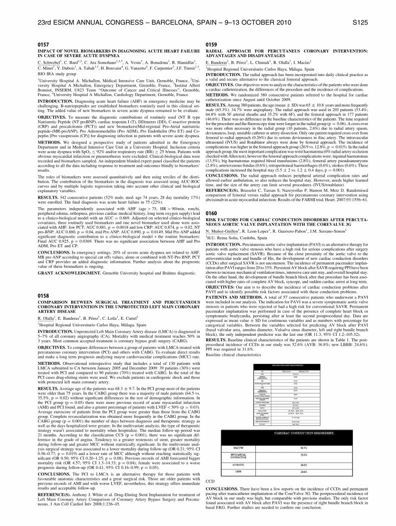
0157IMPACT OF NOVEL BIOMARKERS IN DIAGNOSING ACUTE HEART FAILUREIN CASE OF SEVERE ACUTE DYSPNEA
C. Schwebel1, C. Bard1,2, C. Ara Somohano1,2,3, A. Vesin3, A. Bonadona1, R. Hamidfar1,
C. Minet1, Y. Dubois1, A. Tabah1,3, H. Bouvaist4, G. Vanzetto4, F. Carpentier2, J.F. Timsit1,3,
BIO IRA study group1University Hospital A. Michallon, Medical Intensive Care Unit, Grenoble, France, 2Uni-versity Hospital A Michallon, Emergency Department, Grenoble, France, 3Institut AlbertBonniot, INSERM, U823 Team ‘‘Outcome of Cancer and Critical Illnesses’’, Grenoble,France, 4University Hospital A Michallon, Cardiology Department, Grenoble, France
INTRODUCTION. Diagnosing acute heart failure (AHF) in emergency medicine may bechallenging. B-natriopeptides are established biomarkers routinely used in this clinical set-ting. The added value of new biomarkers in severe acute dyspnea remained to be evaluate.
OBJECTIVES. To measure the diagnostic contributions of routinely used (NT B typeNatriuretic Peptide (NT proBNP), cardiac troponin I (T), DDimeres (DD), C-reactive protein(CRP) and procalcitonin (PCT)) and new biomarkers(Mid-regional Pro-Atrial natriureticpeptide-(MR-proANP), Pro Adrenomedullin (Pro ADM), Pro Endothelin (Pro ET) and Co-peptin [Pro vasopressin (CP)] for diagnosing infection in patients with severe acute dyspnea.
METHODS. We designed a prospective study of patients admitted in the EmergencyDepartment and in Medical Intensive Care Unit in a University Hospital. Inclusion criteriawere acute dyspnea with SpO2 B 92% and/or respiratory rate (RR) C 25 b/min. Patients withobvious myocardial infarction or pneumothorax were excluded. Clinical-biological data wererecorded and biomarkers sampled. An independent blinded expert panel classified the patientsaccording to all the data including response to treatment and outcomes blindly to biomarkers’results.
The roles of biomarkers were assessed quantitatively and then using terciles of the distri-bution. The contribution of the biomarkers in the diagnosis was assessed using AUC-ROCcurves and by multiple logistic regression taking into account other clinical and biologicalexplanatory variables.
RESULTS. 342 consecutive patients (52% male, med. age 74 years, 28 day mortality 17%)were enrolled. The final diagnosis was acute heart failure in 75 (22%).
The parameters independently associated with AHF (age [ 75, HR [ 90/min, ronchi,peripheral edema, orthopnea, previous cardiac medical history, long term oxygen supply) leadto a clinico-biological model with an AUC = 0.869. Adjusted on selected clinico-biologicalcovariates, three routinely used biomarkers and one novel biomarker used alone were asso-ciated with AHF: low PCT: AUC 0.881, p = 0.0018 and low CRP: AUC 0.874, p = 0.02, NTpro-BNP: AUC 0.880, p = 0.04, mid Pro ANP: AUC 0.890, p = 0.0149. Mid Pro ANP addedsignificant diagnostic contribution to a clinico-biological model + the routine biomarkers:Final AUC 0.925, p = 0.0369. There was no significant association between AHF and ProADM, Pro ET and CP.
CONCLUSIONS. In emergency settings, 20% of severe acute dyspnea are related to AHF.MR pro ANP according to special cut offs values, alone or combined with NT-Pro BNP, PCTand CRP provides an added diagnostic information. Further analysis about the prognosticvalue of these biomarkers is ongoing.
GRANT ACKNOWLEDGMENT. Grenoble University hospital and Brahms diagnostic.
0158COMPARISON BETWEEN SURGICAL TREATMENT AND PERCUTANEOUSCORONARY INTERVENTION IN THE UNPROTECTED LEFT MAIN CORONARYARTERY DISEASE
R. Olalla1, E. Banderas1, B. Perez1, C. Leila1, E. Curiel1
1Hospital Regional Universitario Carlos Haya, Malaga, Spain
INTRODUCTION. Unprotected Left Main Coronary Artery disease (LMCA) is diagnosed in5–7% of all coronary angiography (CA). Mortality with medical treatment reaches 50% in3 years. Most common accepted treatment is coronary bypass graft surgery (CABG).
OBJECTIVES. To compare differences between a group of patients with LMCA treated withpercutaneous coronary intervention (PCI) and others with CABG. To evaluate direct resultsand make a long term prognosis analyzing mayor cardiovascular complications (MCC) rate.
METHODS. Observational retrospective study that includes a total of 130 patients withLMCA submitted to CA between January 2005 and December 2009: 39 patients (30%) weretreated with PCI and compared to 90 patients (70%) treated with CABG. In the total of thePCI cases drug-eluting stents were used. We exclude patients in cardiogenic shock and thosewith protected left main coronary artery.
RESULTS. Average age of the patients was 68.3 ± 9.7. In the PCI group most of the patientswere older than 75 years. In the CABG group there was a majority of male patients (64.5 vs.35.5%, p = 0.02) without significant differences in the rest of demographic information. Inthe PCI group (p = 0.05) there were more previous record of acute myocardial infarction(AMI) and PCI found, and also a greater percentage of patients with LVEF\50% (p = 0.03).Average euroscore of patients from the PCI group were greater than those from the CABGgroup. Complete revascularization was obtained more frequently in the CABG group. In theCABG group (p = 0.001) the number of days between diagnosis and therapeutic strategy aswell as the days hospitalized were greater. In the multivariate analysis, the type of therapeuticstrategy wasn0t associated to mortality when hospitalize. The median follow-up period was21 months. According to the classification CCS (p = 0.001), there was no significant dif-ference in the grade of angina. Tendency to a greater restenosis of stent, greater mortalityduring follow-up and greater MCC without statistically significant. In the multivariate anal-ysis surgical strategy was associated to a lower mortality during follow-up (OR 0.21; 95% CI0.56–0.77; p = 0.019) and a lower rate of MCC although without reaching statistically sig-nificant (OR 0.50; 95% CI 0.20–1.25; p = 0.08). Previous records of AMI forecasted biggermortality risk (OR 4.57; 95% CI 1.3–14.33; p = 0.04); female were associated to a worseprognosis during follow-up (OR 0.41; 95% CI 0.16–0.99; p = 0.05).
CONCLUSIONS. The PCI to LMCA is an alternative therapy for those patients withfavourable anatomic characteristics and a great surgical risk. Those are older patients withprevious records of AMI and with worse LVEF, nevertheless, this strategy offers immediateresults and acceptable follow-up.
REFERENCE(S). Anthony J. White et al. Drug-Eluting Stent Implantation for treatment ofLeft Main Coronary Artery Comparison of Coronary Artery Bypass Surgery and Percuta-neous. J Am Coll Cardiol Intv 2008;1:236–45.
0159RADIAL APPROACH FOR PERCUTANEUS CORONARY INTERVENTION:ADVANTAGES AND DISADVANTAGES
E. Banderas1, B. Perez1, L. Chimali1, R. Olalla1, I. Macıas1
1Hospital Regional Universitario Carlos Haya, Malaga, Spain
INTRODUCTION. The radial approach has been incorporated into daily clinical practice asa valid and secure alternative to the classical femoral approach.
OBJECTIVES. Our objectives were to analyze the characteristics of the patients who were donea cardiac catheterization, the differences of the procedure and the incidence of complications.
METHODS. We randomized 380 consecutive patients referred to the hospital for cardiaccatheterization since August until October 2009.
RESULTS. Among 380 patients, the age (mean ± SD) was 65 ± 10.8 years and more frequentlymale (65.3%). 34.7% were angioplasty. The radial approach was used in 203 patients (53.4%;64.8% with 5F arterial sheaths and 35.2% with 6F), and the femoral approach in 177 patients(46.6%). There was no difference in the baseline characteristics of the patients. The time requiredfor the procedure and the fluoroscopy time were longer in the radial group (p = 0.06). A cross overwas more often necessary in the radial group (10 patients, 2.6%) due to radial artery spasm,deviousness, loop, unstable catheter or artery dissection. Only one patient required cross over fromfemoral to radial approach (0.26%) due to serious deviousness in iliac artery. The intravascularultrasound (IVUS) and Rotablator always were done by femoral approach. The incidence ofcomplications was higher in the femoral approach group (20.9 vs. 12.8%, p = 0.015). In the radialapproach group, the most important complication was wrist haematoma (0% radial artery occlusionchecked with Allen test), however the femoral approach complications were: inguinal haematomas(13.5%), big haematomas required blood transfusions (2.8%), femoral artery pseudoaneurysms(2.8%), arteriovenous fistulas (0.6%), retroperitoneal haemorrhages (0.6%), strokes (0.6%). Thesecomplications increased the hospital stay (5.5 ± 2 vs. 1.2 ± 0.4 days, p = 0.001).
CONCLUSIONS. The radial approach reduces peripheral arterial complication rates andallowed earlier ambulation, so also reduces the hospital stay. However, needs higher learnedtime, and the size of the artery can limit several procedures (IVUS/rotablator).
REFERENCE(S). Brasselet C, Tassan S, Nazeyrollas P, Hamon M, Metz D. Randomisedcomparison of femoral versus radial approach for percutaneous coronary intervention usingabciximab in acute myocardial infarction: Results of the FARMI trial. Heart. 2007;93:1556–61.
0160RISK FACTORS FOR CARDIAC CONDUCTION DISORDERS AFTER PERCUTA-NEOUS AORTIC VALVE IMPLANTATION WITH THE COREVALVE 3G
N. Munoz-Guillen1, R. Leon-Lopez1, R. Guerrero-Pabon1, J.M. Serrano-Simon1
1H.U. Reina Sofia, Cordoba, Spain
INTRODUCTION. Percutaneous aortic valve implantation (PAVI) is an alternative therapy forpatients with aortic valve stenosis who have a high risk for serious complications after surgeryaortic valve replacement (SAVR). Because of the close proximity of the aortic valve to theatrioventricular node and bundle of His, the development of new cardiac conduction disorders(CCDs) after surgical SAVR is not uncommon. The incidence of permanent pacemaker implan-tation after PAVI ranges from 20 to 33%. Persistent AV block after SAVR requiring PPI have beenshown to increase mechanical ventilation times, intensive care unit stay, and overall hospital stay.On the other hand, the development of bundle branch block after that procedure has been asso-ciated with higher rates of complete AV block, syncope, and sudden cardiac arrest at long term.
OBJECTIVES: Our aim is to describe the incidence of cardiac conduction problems afterPAVI and to identify possible risk factors associated with these conduction problems.
PATIENTS AND METHODS. A total of 57 consecutive patients who underwent a PAVIwere included in our analysis. The indication for PAVI was a severe symptomatic aortic valvestenosis in patients who were rejected or had a high risk for conventional SAVR. Permanentpacemaker implantation was performed in case of the presence of complete heart block orsymptomatic bradycardia, persisting after at least the second postprocedural day. Data areexpressed as mean value ± SD for continuous variables and as numbers with percentage forcategorical variables. Between the variables selected for predicting AV block after PAVI(basal valvular area, annulus diameter, Valsalva sinus diameter, left and right bundle branchblock), the only independent predictor was the last one (OR 11.3, 95% CI 1.2–105.52).
RESULTS. Baseline clinical characteristics of the patients are shown in Table 1. The post-procedural incidence of CCDs in our study was 52.6% (AVB: 36.8%; new LBBB: 24.6%).PPI was required in 31.6%.
Baseline clinical characteristics
CCD
CONCLUSIONS. There have been a few reports on the incidence of CCDs and permanentpacing after transcatheter implantation of the CoreValve 3G. The postprocedural incidence ofAV block in our study was high, but comparable with previous studies. The only risk factorfound associated with AV block after PAVI was the presence of right bundle branch block inbasal EKG. Further studies are needed to confirm our conclusion.
23rd ESICM ANNUAL CONGRESS – BARCELONA, SPAIN – 9–13 OCTOBER 2010 S125
0161RESULTS AND EVOLUTION OF A LARGE SERIES OF PATIENTS WITH MITRALVALVE REPAIR
L. Chimali1, B. Perez1, R. Olalla1, E. Banderas1
1Hospital Regional Universitario Carlos Haya, Malaga, Spain
INTRODUCTION. Mitral valve replacement is the preferred treatment for the mitralinsufficiency. This allows us to save the subvalvular section and ventricular function, also toavoid long-term complications due to the prosthesis or anticoagulant treatment
OBJECTIVES. To evaluate the results in patients who have undergone mitral valve repair ina third-level hospital during the last 12 years.
METHODS AND RESULTS. Between January 1998 and December 2009, 154 patientsunderwent surgery to reconstruct and preserve the mitral valve. Their mean age was58.7 years and 57.1% were men. 66.3% were in functional class III–IV, and 29.2% in atrialfibrillation. The aetiology was myxoid degeneration in 66.9%. The type of repair most oftenperformed was quadrangular resection. A good intraoperative result was seen 83.7% of cases.The in-hospital mortality rate was 2.6 and 11.7% experienced postoperative complications.The median follow-up was 60 months; 79.2% were in functional class I–II, and 39.6% inatrial fibrillation. During the follow-up, relapse of mitral regurgitation C moderate occurred in30.5% of the patients and the mortality rate was 4.5%. Multivariate analysis showed the bestprognosis with quadrangular resection (P \ 0.001) and the worse prognosis with the Alfieritechnique (P = 0.6), as well as a worse prognosis the larger the preoperative atrial size(P = 0.2). A significant reduction in the ventricular diameters was associated with a goodevolution (5.3 ± 8, P \ 0.001). The Kaplan–Meier curves showed that at 3 years 77.3% ofthe patients were asymptomatic.
CONCLUSIONS. Quadrangular resection for prolapse of the posterior valve and a reductionin the diameter of the left ventricle during follow-up were associated with a good prognosis. Agreater preoperative atrial diameter implied a worse prognosis. Relapse of mitral regurgitationand its symptoms is not unusual; mortality in-hospital and during follow-up was acceptable.
REFERENCE(S). Fedak PWM, Mc Carthy PM, Bonow RO. Evolving concepts and tech-nologies in mitral valve repair. Circulation. 2008;117:963–974
Bloodstream infections: 0162–01750162CATHETER RELATED INFECTIONS. A SINGLE CENTER PROSPECTIVE AUDIT
P. Burtin1, P. Mion2, P. Andre3, D. Bessou1, J. Gippet1
1Clinique du Millenaire, Anesthesie-Reanimation, Montpellier, France, 2Clinique du Mille-naire, Laboratoire Bacteriologie, Montpellier, France, 3Clinique du Millenaire, Celluled’Hygiene, Montpellier, France
INTRODUCTION. Cental venous catheter (CVC) are increasingly used outside the IntensiveCare Unit (ICU) in general ward (GW). Few studies have investigated the catheter relatedblood stream infection (CRBSI) rate and safety of CVC in GW compared to ICU (1, 2, 3).
OBJECTIVES. We designed a prospective clinical audit to establish the incidence, relativerisk and risks factors, morbidity and outcome of CVC in our private institution (236 beds).
METHODS. From 01/01/09 to 12/31/09 all CVC were monitored from insertion until returnof the culture results. The following variables were collected: age, sex, insertion site, type ofCVC, presence of diabetes mellitus, renal failure, immunosuppression, respiratory chronicdisease, cirrhosis, antibiotic use, transfusion, mechanical ventilation, tracheostomy, dialysis,central venous pressure measurements, duration of use in ICU and GW, removal indication,culture results, antibiotic used in case of CRBSI and daily cost. Cultures were performedaccording to the quantitative Brun–Buisson technique. CVC colonisation and CRBSI weredefined according to published criteria (4). Patient stay was defined as ICU only, GW only orICU + GW. CRBSI was calculated per 1,000 catheter days (CD).
RESULTS. 1,245 CVC were inserted in 816 patients (mean age 64.5). Mean duration of usewas 7.8 days; 9,355 CD were generated (ICU: 5,706; GW: 3,649). 19 CRBSI occurred; allpatients survived. Overall colonisation rate was 10.15/1000 CD; CRBSI rate was 2.03/1000CD. CRBSI rate was significantly higher for ICU + GW stays (ICU only: 1.26/1000 CD; GWonly: 0/1000 CD; ICU + GW: 3.11/1000 CD; p \ 0.05). All ICU CRBSI were related toGram negative bacteria; 64% of ICU + GW CRBSI were related to Staphylococci bacteria.CRBSI rate significantly increased in relation to CVC duration of use: 0.92/1000 CD beforethe 4th day; 2.77/1000 CD between 10 an 14 days; 4.11/1000 CD after the 25th day;p \ 0.05. 16 CRBSI were treated with antibiotics and CVC removal; 2 CRBSI were treatedby removal alone and 1 CRBSI was treated by antibiotics only (CVC maintained). Antib-iotherapy was used during 9.7 ± 4 days; total cost was 2142€ rating 2.7% of the totalantibiotics annual cost at our institution. 2 patients died of CVC related cause: 1 pneumo-thorax during an ICU stay and 1 air embolism during a GW stay.
CONCLUSIONS. In our institution:
1 - Overall CRBSI rate is low.
2 - Risk factors are mostly related to duration of use and combined stay (ICU + GW).
3 - CVC use outside the ICU is a risk factor of Staphylococcus related CRBSI.
4 - CVC morbidity is not related to CRBSI.
5 - CRBSI are not a cause of inbalancein antibiotic use and cost.
REFERENCE(S). 1. J Hosp Infect 2009; 73: 41–46
2. Infect Control Hosp Epidemiol 2007; 28: 905–909
3. J Hosp Infect 2009; 71: 117–122
4. Am J Infect Control 1988; 16: 128–140.
0163EPIDEMIOLOGY OF THE PRIMARY AND VASCULAR CATHETER-RELATEDBACTERIEMIA IN CRITICAL PATIENTS ADMITTED TO AN INTENSIVE MED-ICINE DEPARTMENT
F. Alvarez Lerma1, P. Olaechea Astigarraga2, M. Palomar Martinez3, J. Insausti Ordenana4,M.J. Lopez Pueyo5, M.P. Gracia Arnillas1, GTEI-SEMICYUC1Hospital del Mar, Servicio de Medicina Intensiva, Barcelona, Spain, 2Hospital de Galdakao,Servicio de Medicina Intensiva, Galdakao (Vizcaya), Spain, 3Hospital Vall d’Hebron, Ser-vicio de Medicina intensiva, Barcelona, Spain, 4Hospital de Navarra, Servicio de MedicinaIntensiva, Pamplona, Spain, 5Hospital Yague, Servicio Medicina Intensiva, Burgos, Spain
INTRODUCTION. In recent years, different aspects of catheter-related bacteremia (CRB)have changed, including the appearance of multiresistant Gram-positive cocci, the knowledgeof limitations of the antibiotics most commonly used for the treatment of this condition(glycopeptides), and the introduction of new active antibiotics against these pathogens.
OBJECTIVE. This article analyzes trends in incidence rates, etiologies, and multiresistantmarkers of pathogens more frequently involved in CRB (including primary bacteremia) inSpanish Intensive Care Units (ICUs).
MATERIAL AND METHODS. Prospective, observational and multicenter study, withvoluntary participation of 74, 105, 112 and 121 ICUs from 71, 97, 103 and 112 hospitals,respectively, between the years 2005 and 2008 (both inclusive). Data included in the ENVIN-HELICS register was used.
RESULTS. The rates of this complication have decreased and the incidence of CRB wasaround 5 episodes per 1000 central venous catheter-days. One-third of the episodes wereassociated with an important systemic response (severe sepsis or septic shock). MultiresistantGram-positive cocci were the most frequent, although Gram-negative bacilli were isolated in30% of the cases and fungi (different Candida spp.) in 6%. Staphylococcus epidermidis andmethicillin-resistant coagulase-negative Staphylococcus persisted in a proportion greater than80%, whereas methicillin-resistant S. aureus decreased to less than 40%.
CONCLUSIONS. Empirical antibiotic treatment in extremely severe situations shouldconsider coverage of the most frequent pathogens, such as methicillin-resistant Gram-positivecocci and Gram-negative bacilli as well as fungi in special situations.
0164EFFECT OF CARE BUNDLE IMPLEMENTATION ON CATHETER RELATEDBLOODSTREAM INFECTION ON THE INTENSIVE CARE UNIT
P. Beckett1, H. Jerrett1, T. Pain1, A. Hermon1, T. Szakmany1
1Royal Glamorgan Hospital, Anaesthetics, Theatres and Critical Care, Llantrisant, UK
INTRODUCTION. Catheter related bloodstream infection (CRBSI) is still a commonproblem on UK ICUs (1). Implementation of care bundles have been advocated to reduce theinfection rate (2).
OBJECTIVES. The aim of the study was to identify the effect of the introduction of theCentral Venous Catheter (CVC) bundle on CRBSI rate on our Critical Care Unit over a three-year period.
METHODS. Retrospective audit on the rate of CRBSI for a 3 months period before theimplementation of the CVC bundle provided baseline data. Prospective audits for the cor-responding 3 months were carried out after the CVC bundle was firmly embedded in clinicalpractice. The data was collected based on the information recorded in our clinical informationsystem (CIS). The CVC bundle consisted hand hygiene, barrier precautions on insertion, 2%chlorhexidine skin preparation, using femoral site as last resort, daily review of necessity ofcentral access, daily inspection of insertion site, use of TPN on a dedicated port and main-taining asepsis when accessing the line. Robust educational program was rolled out during theimplementation phase for medical and nursing staff. Compulsory elements of the care bundlewere recorded in our CIS. We collected data on overall compliance with the bundle, meandwell time, number of CRBSIs, site of infection and whether the patient left the unit with aCVC line in situ. For statistical analysis Chi-square test and Wilcoxon test were used.
RESULTS. Our main results are summarised in Table 1.
TABLE 1
2006 2008 2009
Lines inserted (n) 114 110 92Overall compliance with bundle (%) 55 99 99Mean dwell time (days) 4.4 3.7 3.4CVC related infection (n) 8 2 2Site of infected lines (n) Jugular
(8)Jugular
(2)Jugular
(2)Infection rates/1,000 catheter days 15.9 4.4 3.6Patients transferred to ward with lines in situ
(n)61 39 10
Lines removed prior to transfer (n) 33 52 76
We have seen a significant increase in the compliance with the bundle and it resulted asignificant and sustained reduction in mean dwell time, CVC related infection rate andnumber of patients transferred to the ward with CVC lines (all p \ 0.05). The bundle resultedin bigger scrutiny for CVCs, hence the reduction in the number of lines inserted.
CONCLUSIONS. Our data shows that implementation of care bundles can significantly andsustainably reduce the rate of CRBSI on the ICU in a real life setting. Our previouslyunacceptable infection rates were reduced and now are comparable with the recently pub-lished data (1). Evidence-based catheter-care procedures, guided by healthcare workers0
perceptions and including bedside teaching, reduce significantly the CRBSI rate and dem-onstrate that improving catheter care has a major impact on its prevention.
REFERENCE(S). 1. Tacconelli E et al. J Hosp Infect 2009;72:97–103 2. Pronovost PJ et al.BMJ 2010 Feb 4;340:c309. doi:10.1136/bmj.c309.
S126 23rd ESICM ANNUAL CONGRESS – BARCELONA, SPAIN – 9–13 OCTOBER 2010
0165RESULTS OF A PROGRAM TO REDUCE CATHETER-RELATED BLOOD-STREAM INFECTION IN THE ICU: TWO YEARS’ FOLLOW-UP
J. Trenado1, M. Riera2, R. Jane1, J. Valls1, N. Freixas2, J.M. Nava1
1Hospital Universitari Mutua Terrassa, Intensive Care Department, Terrassa, Spain, 2HospitalUniversitari Mutua Terrassa, Infection Control Nursing, Terrassa, Spain
INTRODUCTION AND OBJECTIVES. To evaluate the incidence of catheter-relatedbloodstream infection (CR-BSI) and of the use of central venous catheters (CVC) after anintensive improvement program aimed at reducing CR-BSI.
METHODS. Before-and-after study in patients admitted to a 12-bed medical-surgical ICUfrom January 2007 through December 2009. In 2008 we implemented an improvementprogram (analysis of barriers, creation of a working group, review of protocols, and imple-mentation of an educational program and checklist) and a set of measures to reduce CR-BSIduring CVC insertion and maintenance based on Provonost et al.’s model (1). In the post-intervention period, we suspended the use of the checklist and evaluated the degree ofcompletion of the online training module ‘‘Bacteremia Zero program’’ and analyzed the staffturnover rate. We have monitored CR-BSI using the ‘‘Estudio Nacional de Vigilancia deInfeccion Nosocomial en UCI’’ (ENVIN-UCI) criteria since 2002. We calculated the inci-dence rate ratio of CR-BSI and CVC utilization ratio for 2007, 2008, and 2009. We comparedthe incidence rate ratios using the Epitab module from the STATA program and utilizationratios using Chi-square tests.
RESULTS. Nine CR-BSI were diagnosed in 2007, one in 2008, and five in 2009. Theincidence rate ratio of CR-BSI in these periods was 3.11, 0.32, and 1.52%, respectively. Theincidence rate ratio in the postintervention period (0.94%) was significantly lower than inthe preintervention period (3.11%) (3.31: 95% CI 1.05–11.32, p = 0.025.) The increase inincidence rate ratio between 2008 and 2009 was not statistically significant (0.32 vs. 1.52%,p = 0.14). The pre- and post-intervention CVC utilization ratios were 0.83 and 0.86,respectively (no significant differences). During the year 2009, and for existing staff in 2008,6 rotating residents, 19 nurses (turnover rate 52%), and 5 nurse’s aides (turnover rate 50%)joined the ICU. The training module was completed by 32% of the new nurses and none of thephysicians or nurse’s aides.
CONCLUSIONS. The program was effective; its effectiveness may be related to theintensity of the measures. A low preintervention incidence rate ratio does not preclude theusefulness of an improvement program.
REFERENCE(S). [1] Pronovost P, Needham D, Berenholtz S, Sinopoli D, Chu H, CosgroveS et al. An intervention to Decrease Catheter-Related Bloodstream Infections in the ICU.N Engl J Med 2006;26:2725–2732
0166MINOCYCLINE-RIFAMPICIN IMPREGNATED CENTRAL VENOUS CATHETERSDO NOT REDUCE THE INCIDENCE OF BLOODSTREAM INFECTIONS
N. Kusadasi1, J. Epker1, J. Bakker1
1Erasmus Medical Center, Rotterdam, Netherlands
INTRODUCTION. In the intensive care unit (ICU) the bloodstream infections (BSI) relatedto the central venous catheters (CVCs) represent a serious clinical complication and are asubstantial economic burden. Although the data are still somewhat controversial, the use ofantibiotic impregnated CVCs is one of the generally accepted approaches in reducing the riskof BSI [1, 2].
OBJECTIVES. In order to determine the efficacy of antibiotic impregnated CVCs in ourclinic we evaluated retrospectively the data of the cultures of CVCs and blood obtained frompatients during their stay at ICU within the last 2 years (January 2008 till August 2009).
METHODS. The unimpregnated catheters were used until December 2008 and the Mino-cycline-Rifampicine (MR) impregnated catheters were introduced in January 2009. The tipand blood culture data of the unimpregnated CVCs (number of cultures = 400) were com-pared with the data of the MR-impregnated CVCs (number of cultures = 281).
RESULTS. Within the first 7 months, the number of unimpregnated CVCs and MR-impregnated CVCs was 221 and 281, respectively. The corresponding incidence of positiveCVC cultures was not significantly different in both groups. There was also no significantdifference of the incidence of the positive CVC cultures within the first 281 catheter tipscultured (number of positive cultures = 89 in both groups). Overall, the percentage ofpositive catheter tip cultures in unimpregnated (number of cultures = 127) and MR-impregnated CVCs (number of cultures = 89) were 31.8 and 31.7%, respectively. Addi-tionally, the percentage of correlated bloodstream infections was 34.7 and 35.1%, respectivelyin the unimpregnated (number of cultures = 51) and MR-impregnated CVCs (number ofcultures = 40), respectively.
CONCLUSIONS. Surprisingly, there was no difference in the incidence of the CVC andbloodstream infections in both groups. We can conclude that the strategy of using MR-impregnated CVCs did not reduce the incidence of catheter related BSI. Although earlierstudies have indicated that MR-impregnated CVCs are cost saving [3], our data add furtherproof to the suggestion that the cost effectiveness of these catheters is at least uncertain.
REFERENCE(S). 1. Ramritu P et al.: A systematic review comparing the relative effec-tiveness of antimicrobial-coated catheters in intensive care units. Am J Infect Control2008;36:104–17.
2. Leon C et al.: Benefits of minocycline and rifampicin-impregnated central venous cathe-ters. Intensive Care Med 2004; 30: 1891–1899.
3. Marciante KD et al.: Which antimicrobial impregnated central venous catheter should weuse? Modeling the costs and outcomes of antimicrobial catheter use. Am J Infect Control2003;31:1–8.
0167CATHETER-RELATED BLOODSTREAM INFECTIONS: INCIDENCE ANDPOTENTIAL CATHETER RELATED RISK FACTORS
M. Katsiari1, C. Nikolaou1, A. Voulgaridis1, A. Sakkalis1, A. Xydaki1, E. Papadakis1, M.Laskou1, A. Kounougeri1, A. Maguina1
1Konstantopoulion General Hospital, Intensive Care Unit, Athens, Greece
OBJECTIVE. To determine the influence of catheter insertion site, use and type on infectionrates associated with central venous catheterization.
METHODS. Prospective observational study conducted in a multidisciplinary 8-bed Inten-sive Care Unit (ICU) of a 300-bed public hospital. Over a period of 3 years, 341 consecutivepatients with a length of stay (LOS) C 72 h were enrolled in the study. Data collected foreach patient included age, gender, APACHE II score on admission, number of central venouscatheters (CVCs) inserted, catheter insertion site (subclavian, jugular or femoral vein),number of catheter lumens (single, double, triple), catheter use (drug and TPN administration,monitoring, haemodialysis), catheter material (antimicrobial coated or not). CRBSI diagnosiswas confirmed based on currently adopted criteria and CVCs were stratified into two groups:Group A included CRBSI cases (n = 65) and Group B included CVCs without CRBSI(n = 993). Data were analyzed using Chi-square.
RESULTS. The mean age and APACHE II score on admission of patients included were64 ± 17 years and 18.7 ± 7.4 respectively. A total of 1,058 catheters were recorded andanalyzed during 7,884 patient-days. During the study period, 65 episodes of CRBSI werediagnosed. Calculated CRBSI incidence was 8.24/1,000 catheter-days. Mean time fromcatheter insertion to bacteremia diagnosis was 10 days. Evaluation of catheter related factorsare presented on Tables 1 and 2.
TABLE 1 SITE OF CATHETER INSERTION AND MATERIAL
Site of catheter insertion (%) Antimicrobial coatedcatheter material (%)
Femoral vein Jugular vein Subclavian vein YES NO
Group A (n = 65) 34.33 40.3 25.37 77.61 22.39Group B (n = 993) 32.29 28.25 39.46 77.03 22.97p-value 0.0399 0.9121
TABLE 2 CATHETER USE AND NUMBER OF LUMENS
Catheter use (%) Number of
catheter lumens (%)
Drug/TPN administration Hemodialysis Swan-Ganz Double Triple
Group A (n = 65) 83.08 15.38 1.54 18.18 81.82Group B (n = 993) 82.35 15.82 1.83 18.51 81.08p-value 0.9807 0.2764
CONCLUSIONS. CRBSI incidence rate in our patients’ cohort was 8.24 per 1,000 catheter-days. Variables related to catheter use, number of lumens and material were not predictive ofCRBSI development. Jugular vein catheterization was the major determinant for CRBSI, withsubclavian vein catheterization being the least contributing.
0168INCIDENCE SHIFT OF INTRAVASCULAR CATHETER RELATED INFECTION INICU PATIENTS
A. Vakalos1, P. Tassioudis1, D. Papadopoulos1
1Xanthi General Hospital, ICU, Xanthi, Greece
INTRODUCTION. Short term central venous catheters (CVC) are widely used in ICUpatients, to administer intravenous fluids, medications, parenteral nutrition and to monitorhemodynamic status. The major complication remains the catheter related bloodstreaminfections (CRBSI). The risk of CRBSI varies according to the intravascular device, theduration of catheter placement and the use of proven preventative strategies.
OBJECTIVES. The aim of our study was to record the incidence of CRBSI in relation withthe total number of day’s central venous catheter placement during the year 2009 and tocompare the data with the year 2008 and to evaluate the difference.
METHODS. During the year 2009, 64 patients admitted to our ICU and included retrospectivelyto our study. Mean age: 58.2 years, mean APACHE II score at admission: 16.8,mean length of stay(LOS): 12.68 days, recorded mortality: 21.47% and predicted mortality: 26.8%. Total number ofICU stay: 812 days. During this period, our ICU purchased untunneled CVC to 60 patients(93.75% of the total) while the total number of CVC placement stay was 735 days.
RESULTS. From all patients, 27 (42.1%) developed infection from any reason during the ICUstay. 8 patients developed CRBSI, 12.5% of the total patient number and 29.62% of the patientswho developed any infection. We recorded 9 episodes of bacteremia due to CVC during 735 daysof CVC placement stay, 12.24%while the standard limit is four episodes of CRBSI per 1,000 daysCVC placement. During the year 2009, we chanced our practice in order to avoid as risk factors aswe can, using only antimicrobial/antiseptic impregnated catheters, improving our hand hygieneand aseptic technique, using only chlorhexidine and semipermeable polyurethane dressings andmaking catheter replacement at scheduled time intervals as a method to reduce CRBSI. Theprevious year the recorded CRBSI incidence was 18.7% respectively.
CONCLUSIONS. The incidence of intravascular catheter related infection is recorded above thestandard limits for second consecutive year assuming that we have to improve further our sur-veillance policy. On the other hand, the incidence is recorded smaller than the incidence of theprevious year according to the change to our practice, assuming that our reforming policy, althoughnot fully effective, still is better for the prevention of intravascular catheter related infections.
REFERENCE(S). 1. O’Grady NP, Alexander M, Dellinger EP et al. Guidelines for theprevention of intravascular catheter related infections. Centers for disease control and pre-vention. MMWR Recomm Rep 2002;51:1–29
2. Mernel LA, Allon M, Bouza E et al. Clinical practice guidelines for the diagnosis andmanagement of intravascular catheter related infection: 2009 update by the infection diseasessociety of America. CID 2009;49:1–45.
23rd ESICM ANNUAL CONGRESS – BARCELONA, SPAIN – 9–13 OCTOBER 2010 S127
0169SURVEY OF CENTRAL LINE RELATED SEPSIS IN A NEUROINTENSIVE CAREUNIT
P. Nair1, E. Pabs-Garnon1, C.F. Whitehead1
1The Walton Centre, Dept of Anaesthesia, Liverpool, UK
INTRODUCTION. Intravenous catheter related blood stream infection is a major factorcontributing to in hospital morbidity and mortality and extending hospital stay by 10 days andexpenditure by 10,000 to 30,000 lb 1. The incidence of central line associated blood streaminfections (CR-BSI) in our unit was audited in 2008 and a comprehensive infection preventionprogram that included staff education, hand hygiene, maximal sterile barrier precautions anddaily assessment of the need for a central line was introduced. We are also taking part in thenational audit project Matching Michigan.
OBJECTIVES. Assess the effectiveness of the infection prevention programme and re-auditthe incidence of CR-BSI
METHODS. Data was collected daily for a period of 4 months. This included the number ofpatients with central venous catheters in the unit, the number of lines removed or re-sited, theindications for line change, the site of line insertion and incidence of line infection. The lineswere reviewed daily and removed if indicated clinically (pyrexia or raised white cell count) orif not required.
RESULTS. Over a period of 4 months 206 central lines were used amounting to 716 linedays. The lines inserted were subclavian (SC)-13 (6.31%), femoral (F)-108 (52.4%) andinternal jugular (IJ)-85 (41.26%). The percentage of lines removed for clinically suspectedCR-BSI reduced in this period from 30 to 15.04%. The average duration of stay for the lineswere SC 4.4 days, IJ 5.8 days and F 4 days which was shorter than our previous auditshowed. The percentage of microbiologically proven CR-BSI also dropped from 12.5 to 2.5%(4 from internal jugular lines and one from a femoral line).
CONCLUSIONS. Introduction of simple and cost effective practices decreased the preva-lence of CR-BSI in our unit by a factor of five. Daily review of lines led to earlier removal ofcentral lines once they were no longer required. The unit being a neurointensive care unit hasa greater proportion of patients in whom femoral lines are often the only option. Our surveyproves that with strict adherence to guidelines and following infection control protocolsdiligently the risk of CR-BSI from all line types can be reduced.
REFERENCE(S). Risk of bloodstream infections in Adults with different intravasculardevices: a systematic review of 200 published prospective studies Mayo Clinic ProceedingsSeptember 1, 2006 Author: Maki, Dennis G; Kluger, Daniel M; Cornich, Christopher J.
0170BLOOD CULTURE POSITIVITY IN CRBSI: EFFECT OF CONCURRENTANTIBIOTICS
C.M. Walshe1, K.S. Boner2, J. Bourke1, R. Hone3, D. Phelan1
1Mater Misericordiae University Hospital, Intensive Care, Dublin, Ireland, 2Mater Miseri-cordiae University Hospital, Medicine, Dublin, Ireland, 3Mater Misericordiae UniversityHospital, Microbiology, Dublin, Ireland
INTRODUCTION. ‘Defervescence of sepsis after removal of culture-positive central venouscatheters (CVCs)0 has been advocated for diagnosis of catheter related bloodstream infection(CRBSI) even without positive blood culture1. However, most studies report CRBSI inci-dence only when blood cultures, and CVC tip, are positive—standard definition2,3. Weexamined the effect of inclusion of defervescence criteria on CRBSI incidence in a TotalParenteral Nutrition (TPN) population, and examined the effect of concurrent antibiotictherapy at time of CVC removal.
METHODS. Study was carried out in a 525-bed tertiary referral hospital over 12-years.CRBSI incidence was compared when standard definition was utilised (positive CVC tipculture and positive blood culture), versus when 0defervescence criteria0 was included. Sepsisdefervescence was defined as fall in temperature, white cell count and sepsis resolution afterCVC removal, with positive CVC tip culture, but negative blood cultures. CRBSI episodeswhere a blood culture was omitted were excluded.
RESULTS. Study population included 1,365 patients in whom 2,536 CVCs were utilised over15,234 CVC days. There were 192 CRBSI episodes in 165 patients. 152 CRBSI episodes metonly the standard criteria for CRBSI; 40 episodes met defervescence criteria. Overall, patientswere receiving antibiotics at the time of CVC removal in 104 of the 192 episodes [meanannual incidence 57% (±Std dev 0.14%)].
Standard definition alone resulted in an average incidence of 10.6 per 1,000 CVC days(±5.8 SD). This increased to 13 per 1,000 CVC days (±6.4 SD) when defervescence criteriawere included. Inclusion of defervescence criteria increased CRBSI incidence by an averageof 2.5 episodes per 1,000 days (±1.4 SD) or 27% p \ 0.001; 95% CI (1.61–3.339).
CONCLUSION. This study implies that the scale of CRBSI may be higher than is currentlyrecognised and that the blood culture positivity rate for CRBSI is 79%(152/192). As con-current antibiotic therapy may reduce blood culture and CVC tip positivity, the blood culturerate of 79% suggests that CRBSI has an inherently high blood culture positivity rate despiteconcurrent antimicrobial therapy.
REFERENCES. 1. Pearson ML. Guidelines for prevention of intravascular device-relatedinfections. Part I. Intravascular device-related infections: an overview. The Hospital InfectionControl Practices Advisory Committee. Am J Infect Control 1996; 24(4): 262–77.
2. Safdar N, Maki. DG. Inflammation at the insertion site is not predictive of catheter-relatedbloodstream infection with short-term, noncuffed central venous catheters. Crit Care Med2002; 30(12): 2632–2635.
3. Lorente L, Henry C, Martın MM, Jimenez A, Mora ML. Central venous catheter-relatedinfection in a prospective and observational study of 2,595 catheters. Crit Care 2005; 9(6):R631–5.
0171IMPACT OF CHLORHEXIDINE-IMPREGNATED SPONGES ON CATHETER-RELATED INFECTIONS RATE
P. Eggimann1, C. Joseph1, M.-J. Thevenin2, P. Voirol3, C. Bellini2, J.-L. Pagani1,J.-P. Revelly1
1CHUV, Critical Care, Lausanne, Switzerland, 2CHUV, Infection Control, Lausanne, Swit-zerland, 3CHUV, Pharmacy, Lausanne, Switzerland
INTRODUCTION. Multimodal strategy targeted at prevention of catheter-related infectioncombine education to general measures of hygiene with specific guidelines for catheterinsertion and dressing (1).
OBJECTIVES. In this context, we tested the introduction of chlorhexidine(CHX)-impreg-nated sponges (2).
METHODS. In our 32-beds mixed ICU, prospective surveillance of primary bacteremia and ofmicrobiologically documented catheter-related bloodstream infections (CRBSI) is performedaccording to standardized definitions. New guidelines for central venous catheter (CVC) dressingcombined a CHX-impregnated sponge (BioPatch�) with a transparent occlusive dressing (Tega-derm�) and planning for refection every 7 days. To contain costs, Biopatch� was used only forinternal jugular and femoral sites. Other elements of the prevention were not modified (overallcompliance to hand hygiene 65–68%; non coated catheters except for burned patients [173 out of9,542 patients]; maximal sterile barriers for insertion; alcoholic solution of CHX for skindisinfection).
RESULTS. Median monthly CVC-days increased from 710, to 749, 855 and 965 in 2006,2007, 2008 and 2009, respectively (p \ 0.01). Following introduction of the new guidelines(4Q2007), the average monthly rate of infections decreased from 3.7 (95% CI: 2.6–4.8)episodes/1000 CVC-days over the 24 preceding months to 2.2 (95% CI: 1.5–2.8) over the 24following months (p = 0.031).
Figure
Dressings needed to be changed every 3–4 days. The decrease of catheter-related infections weobserved in all consecutive admitted patients is comparable to that recently showed in a placebo-randomized trial2. Further generalization to all CVC and arterial catheters access may be justified.
CONCLUSIONS. Our data strongly suggest that combined with occlusive dressings, CHX-impregnated sponges for dressing of all CVC catheters inserted in internal jugular and/orfemoral sites, significantly reduces the rate of primary bacteremia and CRBSI.
REFERENCES. (1) Eggimann P, Harbarth S, Constantin MN, Touveneau S, Chevrolet JC,Pittet D. Impact of a prevention strategy targeted at vascular-access care on incidence ofinfections acquired in intensive care. Lancet 2000; 355:1864–1868.(2) Timsit JF, Schwebel C, Bouadma L, Geffroy A, Garrouste-Org, Pease S et al. Chlor-
hexidine-impregnated sponges and less frequent dressing changes for prevention ofcatheter-related infections in critically ill adults: a randomized controlled trial. JAMA2009; 301(12):1231–1241.
0172MRSA BACTERAEMIA RATES IN A TEACHING HOSPITAL IN THE WEST OFIRELAND; 2005 TO 2009
E. Lambe1, S. Frohlich2, N. O’Connell2
1Mid Western Regional Hospital, Dooradoyle, Limerick, Ireland, 2Mid Western RegionalHospital, Limerick, Ireland
INTRODUCTION. Health care associated infections present an enormous challenge topatient safety and quality of care in the Irish health Service. There is rising concern aboutMRSA infection rates in Ireland, particularly in the current era of service centralisation. Alarge proportion of hospital acquired MRSA bacteraemias occur in the ICU, and have beenassociated with a mortality rate of 48.3%1. A reduction in MRSA bacteraemia is a keycomponent in reducing both Intensive Care and hospital morbidity and mortality.
OBJECTIVES. The objective of this study was fourfold. We aimed to measure the incidence ofStaphylococcus aureus bacteraemia in the Mid Western Regional Hospital, the percentage ofthese which were methicillin resistant (MRSA), to identify the percentage of cases attributable tocentral venous catheters (CVC’s) and to determine the reason for any change in MRSA rates.
METHODS. A Retrospective Systematic Chart Review of patients with confirmed SA bacteraemiaduring the 5 year period from January 1st 2005 to 31st December 2009 was performed. Patients withSA-positive blood cultures were identified from the microbiology laboratory reporting system.
RESULTS. The annual number of cases of Staphylococcus aureus infection ranged from 54 to76. The percentage of cases attributable to MRSA increased from 34% in 2005 to 43% in 2009.Additionally, there was an increase in the incidence per 1000 bed days used (BDU) from 0.13(2005) to 0.19 (2009). The number of clinically significant MRSA isolates increased from 69%in 2006 to 91% in 2009. CVC-associated MRSA rates in 2007 were 24%. In 2009 this increasedto 30%. Hospital activity increased by 20% in 2009, compared to the preceding 3 years.
DISCUSSION. MRSA bacteraemia rates in Ireland are higher than internationally acceptednorms2. The trend in MRSA bacteraemia rates between 2005 and 2008 was comparable to thatnationally but a marked increase was noted in 2009. The increased rate coincides with a largeincrease in hospital activity. Increased staff workload is shown to increase rates of MRSA coloni-sation, and MRSA bacteraemias. Higher levels of infection increases patient length of stay leading toa vicious cycle characterised by further infection control failure. The number of CVC-related MRSAbacteraemias also increased, probably due to increased patient turnover and staff workload.
CONCLUSIONS. MRSA bacteraemia rates decreased steadily between 2005 and 2008 butincreased in 2009, in line with increased staff and hospital activity. This confirms thatincreasing levels of hospital activity can lead to a large rise in clinically significant bacte-raemias. If an increase in hospital workload is planned additional infection control proceduresshould be put in place.
REFERENCES. 1. Robinson JO et al. Community associated versus healthcare associatedmethicillin resistant Staphylococcus aureus infection. Eur J Clin Microbiol Infect Dis.October 2008 2. www.earss.rivm.nl.
S128 23rd ESICM ANNUAL CONGRESS – BARCELONA, SPAIN – 9–13 OCTOBER 2010

0173BACTERIAEMIA PRIMARY AND SECONDARY TO CATHETER INFECTION.EVOLUTION ALONG FIVE YEARS IN A THIRD LEVEL HOSPITAL ICU
J.M. Mora1, I. Macias1, A. Poullet1, C. Aragon2, F. Rodriguez Villanova1
1Hospital Universitario Carlos Haya, UCI, Malaga, Spain, 2Hospital Universitario CarlosHaya, Malaga, Spain
OBJECTIVES. Analyzing the evolution of primary and secondary catheter infection in anintensive care unit of a third level hospital during 5 years, and the cause of the changesobserved in this period.
METHODS. Patients under study were admitted to the ICU and stayed more than 24 h. Thefollow-up period was 3 months and the study was carried out from 2005 to 2009. Analyzedvariables were: bacteraemia cases per 1,000 days with catheter, APACHE, age, and isolatedgerm. We questioned whether the changes were due to the inclusion of new services in ourintensive care unit (in 2007 we opened a postoperative ICU service, mainly devoted tourological and maxillofacial surgery) or the launching of a Zero bacteriaemia campaign.Nosocomial infection was defined as one that is acquired during hospitalization and was notpresent neither in the incubation period nor at the time of admission. Zero Bacteriaemia is aprotocol for prevention of bloodstream infections related to central venous catheters inSpanish ICUs. The standardized intervention for the prevention of bacteriaemia related to theinsertion and management of catheters was based on: proper hand hygiene, disinfection ofskin with chlorhexidine, total barrier measures during insertion, avoidance of femoral access,removal of unnecessary catheters and hygienic catheters handling.
RESULTS. Study period
Bacteriaemia per 1,000 catheter days 2005 4.07 2006 2.01 2007 2.42 2008 5.75 2009 2.50. In2008 we observed an increase in the incidence rate related to the change of the microorganismin these infections (with an increase in those caused by Staphylococcus coagulase negative),with more resistance to oxacillin, methicillin, and also related to the inclusion of patients witha higher APACHE.
CONCLUSIONS. Incidence rates of primary and secondary bacteriaemia to catheter infec-tion changed because another service was introduced in our UCI (postoperative maxillofacialand urology) since 2007. There was a drop in the rates in 2009 which could be related to theimplementation of the Zero bacteriaemia protocol.
0174HOSPITAL ACQUIRED GRAM-NEGATIVE BACTEREMIA IN A PEDIATRICINTENSIVE CARE UNIT: EPIDEMIOLOGY, ANTIMICROBIAL SUSCEPTIBILITYAND OUTCOMES
E. Lebessi1, F. Petropoulou2, A. Patsoura2, G. Antonaki1, N. Paleologou1, K. Kallergi1, J.Papadatos2
1Children’s Hospital P. & A. Kyriakou, Department of Microbiology, Athens, Greece,2Children0s Hospital P. & A. Kyriakou, PICU, Athens, Greece
OBJECTIVES. To asses the epidemiological and antimicrobial susceptibility data for nos-ocomial Gram-negative bacteremia in a pediatric intensive care unit (PICU) over a four-yearperiod.
METHODS. Positive blood cultures from January 1, 2004 to December 31, 2007 collectedfrom PICU patients were reviewed for microbial etiology and antimicrobial susceptibility.Patient charts were studied to determine the epidemiological data, source of infection, anti-biotic treatment and outcome.
RESULTS. In total, 689 patients, ranging in age from 18 days to 14 years, were admitted toour PICU. Forty-three episodes of nosocomial bacteremia involving 51 Gram-negative bac-teria occurred in 34 (4.9%) patients (17 males, 17 females). The mean time from admission tothe development of bacteremia was 30 days (3–125 days), while the mean PICU length ofstay was 43 days (5–218 days). Admitting diagnoses included: respiratory failure (18), multi-trauma (4), sepsis (4), other (8). Sources of bacteremia in most cases included intravascularcatheterization (26) and intubation (14). Twenty patients (59%) have severe underlying dis-ease. Ten patients (29.4%) died, but only one death attributed directly to the bacteremia. Themajority (81.4%) of bloodstream infections were monomicrobial. Eight bacterial species wereidentified: Enterobacter spp. 35.3% (18), Klebsiella spp. 25.5% (13), Pseudomonas aeru-ginosa 15.7% (8), Acinetobacter baumannii 11.8% (6), Serratia marcescens 3.9% (2),Stenotrophomonas maltophilia 3.9% (2), Escherichia coli 1.9% (1) jai Salmonella enteritidis1.9% (1). Production of extended-spectrum beta-lactamases (ESBLs) was detected in 50% ofKlebsiella spp. and E. coli strains, overproduction of AmpC beta-lactamases was recognizedin 44.5% of Enterobacter spp., while only one K. pneumoniae strain was found to producemetalloenzyme. All eight strains of P. aeruginosa were susceptible to aminoglycosides,ciprofloxacin and carbapenemaces, both strains of S. maltophilia were susceptible to ticar-cillin/clavulanate and trimethoprim/sulfamethoxazole. Among A. baumannii isolates, 33.3%were susceptible only to colistin. In total, 90.2% of isolates were susceptible to imipenem andciprofloxacin.
CONCLUSIONS. Gram-negative bacteremia, in particular in the critically ill, is associatedwith significant morbidity and mortality. Significant susceptibility to ciprofloxacin and imi-penem was demonstrated. Empiric treatment regimens should be based on unit-specific data.
REFERENCE(S).
Ben Jaballah N, Bouziri A, Mnif K, Hamdi A, Khaldi A, Kchaou W. Epidemiology ofhospital-acquired bloodstream infections in a Tunisian pediatric intensive care unit: a 2-yearprospective study. Am J Infect Control (2007) 35, 613–8
Singhi S, Ray P, Mathew JL, Jayashree M; Dhanalakshmi. Nosocomial bloodstream infectionin a pediatric intensive care unit. Indian J. Pediatr. (2008) Jan; 75 (1): 25–30.
0175IMPACT OF A SAFETY PROGRAM IN RATES OF ICU-ACQUIRED INFECTIONS
M. Palomar1, M.J. Lopez Pueyo2, F. Alvarez Lerma3, P. Olaechea4, J. Insausti5, J.J. Otal6,ENVIN1Hospital Vall Hebron, ICU, Barcelona, Spain, 2Hospital General Yague, Burgos, Spain,3Hospital del Mar, BCN, Spain, 4Hospital Galdakao, Bilbao, Spain, 5Hospital Provincial,Pamplona, Spain, 6Hospital Vall Hebron, BCN, Spain
OBJECTIVE. To assess whether implementation of a national safety program to preventCVC-related bacteremia had an impact on rates of devices-associated infections acquired inICU.
METHODS. Prospective, multicenter, incidence, surveillance study of VAP, CRB and UTIcarried out from 01-04-2009 to 31-06-2009. Simultaneously, a bundle for prevention of CVC-related bacteremia and a comprehensive safety program were introduced at the national level.Infections were diagnosed according HELICS definitions. The follow-up was carried out untildischarge from the ICU or to a maximum of 60 days. The severity was assessed by theAPACHE II score. The rates are expressed as incidence density (ID) per 1,000 days of riskfactor. Rates are compared with those of previous years (2007–2008).
RESULTS. 14,983 patients were included, belonging to 147 ICUs in 144 hospitals. The meanage was 62.29 years. (SD 16.5), being 40.66%[ 70 a. Underlying conditions at admissionwere: medical (42.7%), coronary (22.1%), surgery (28.1%) and trauma (6.9%). 15.1% of thepatients required emergency surgery. Mean APACHE II was 14.3 (SD 8.3), LOS in ICU7.7 days (SD 9.1; median 5) and overall mortality was 11%. The days of risk of infection andmonitored infection rates are included in the table below:
Device-days Infections ID 2009 ID 2008 ID 2007
VAP 64.689 740 11,44 14,62 14.55
IU-SU 105.389 451 4,28 4,63 4,69
CRB 100.242 397 3,96 4,89 4,65
CONCLUSIONS. Although risk factors (age, severity) are maintained or even worse, allcontrolled infections’ rates have reduced, especially N-VM. Since none specific bundle wasimplanted to prevent VAP, their reduction may correspond to the comprehensive safetyprogram.
Uncommon infectious diseases: 0176–01890176ACUTE KIDNEY INJURY IN SEVERE PLASMODIUM FALCIPARUM MALARIA:A COMPARISON BETWEEN RIFLE AND AKIN
E. Tomas1,2, E. Filipe1, E. Viegas1, F. Silva1, L. Antunes1, E. Lafuente2
1Clınica Sagrada Esperanca, Luanda, Angola, 2Centro Hospitalar Tamega e Sousa, Penafiel,Portugal
INTRODUCTION. Acute kidney injury (AKI) is one of the most dreaded complications ofsevere malaria. Occurs as a complication of Plasmodium falciparum malaria in less than 1%of cases, but the mortality rate in these cases may be up to 45% [1].
OBJECTIVES. To evaluate the incidence of AKI and compare AKIN and RIFLE classifi-cation systems with regard to hospital mortality.
METHODS. A retrospective analysis based on medical records of adult patients with severePlasmodium falciparum malaria admitted in the general ICU of Clınica Sagrada Esperanca, inLuanda, Angola, from January 2006 to December 2008. Criteria for diagnosis included thestandard WHO definition for severe malaria. Only changes in serum creatinine were used todefine the presence of AKI by both criteria. Logistic regression was used to access theassociation of each RIFLE and AKIN with hospital mortality. Data are presented as oddsratios with 95% confidence intervals (CI).
RESULTS. We enrolled 51 patients. Thirty-nine (76.5%) were males. The mean age recordedwas 42.9 ± 13.3. The mean APACHE II score was 14.0 ± 7.0, with a mean predicted deadrate of 22.2%. The mean SOFA score on admission was 7.2 ± 3.4. The mean length of stay inthe ICU was 5.7 ± 3.5 days. RIFLE allowed the identification of more patents than AKIN ashaving AKI (68.9 vs. 58.8%). The cross tabulation showed that 5 patients classified as non-AKI by AKIN were classified as AKI by RIFLE. Overall hospital mortality was 31.4% andwas significantly higher for AKI patients as compared with non-AKI patients: AKI defined byRIFLE criteria (42.9 vs. 6.3%; p = 0.006; OR 4.03; 95% CI 1.54–9.07) or by AKIN criteria(46.7 vs. 9.5%; p = 0.018; OR 2.85; 95% CI 1.20–6.30). The AuROC curve for hospitalmortality was 0.689 for RIFLE and 0.657 for AKIN.
Incidence and mortality of AKI
RIFLE criteria AKIN criteria
Total Died Hospital mortality Total Died Hospitalmortality
Non-AKI 16 1 6.3% Non-AKI 21 2 9.5%Risk 1 0 0.0% Stage 1 0 0 0.0%Injury 4 2 50.0% Stage 2 4 2 50.0%Failure 30 13 43.3% Stage 3 26 12 46.2%Any AKI 35 15 42.9% Any AKI 30 14 46.7%
CONCLUSIONS. Despite RIFLE criteria demonstrated higher sensitivity on AKI diagnosis,no statistically significant differences were found in terms of hospital mortality according toAKI classification criteria.
REFERENCE(S). [1] Mishra SK et al. Semin Nephrol, Jul;28(4):348–53, 2008.
23rd ESICM ANNUAL CONGRESS – BARCELONA, SPAIN – 9–13 OCTOBER 2010 S129
0177IMMUNE INFLUENZA A H1N1V-SPECIFIC RESPONSES AND T LYMPHOCYTEHOMEOSTASIS DURING SEVERE INFLUENZA PNEUMONIA IN ICU PATIENTS
A. Guihot1, A. Parrot2, G. Carcelain1, D. Boutolleau3, D. Rousset4, S. van Der Werf4,B. Combadiere1, J.-M. Cavaillon5, B. Autran1, C.-E. Luyt6
1Groupe Hospitalier Pitie-Salpetriere, Laboratoire d’Immunologie-UMR S945, Paris, France,2Hopital Tenon, Service de Pneumologie, Paris, France, 3Groupe Hospitalier Pitie-Salpetriere,Laboratoire de Virologie, Paris, France, 4Institut Pasteur, CNR de la Grippe, Paris, France,5Institut Pasteur, Unite des Cytokines et Chimiokines, Paris, France, 6Groupe HospitalierPitie-Salpetriere, Service de Reanimation Medicale, Paris, France
OBJECTIVES. We aimed to characterise immune specific responses to influenza A H1N1vand lymphocyte populations in peripheral blood and broncho-alveolar lavage (BAL) in ICUpatients ventilated for influenza pneumonia during the 2009/2010 pandemic.
METHODS. Nineteen patients with proven H1N1v influenza infection (8 male, 11 females,median age 33 years) underwent simultaneous blood sample and BAL (n = 28) at varioustime from influenza symptoms.
RESULTS. Blood lymphopenia [median 903/mm3 (113–3,248)] was observed in 11/19patients [median 9 (4–22) days from flu symptoms], and predominated on CD8 cells. In BAL,median lymphocytosis was 3% (0–35), enriched in CD8 cells when compared to blood. ThoseCD8 cells in BAL differentiated overtime with an accumulation of effector memory45RA-CCR7-CD27- cells (Rho = 0.63, p = 0.05) and senescent CD57+cells (Rho = 0.42,p = 0.04). Very high frequencies of T cells (up to 10% of CD4 and 6% of CD8) specific forboth the nucleoprotein and the hemagglutinin influenza proteins in Elispot IFNg and/orintracellular cytokine assays were observed in BAL for 3 patients. In blood, all patientsdisplayed T cell memory responses to H1N1v in lymphoproliferative assays whereas memoryresponses to other recall antigens (CMV, HSV, PPD) were poor; Elispot IFNg assays showedhemagglutinin-specific, but poor nucleoprotein-specific effector T cells. Nine/10 patientstested seroconverted to H1N1v virus.
CONCLUSIONS. Both cellular and humoral influenza-specific responses are detectableduring severe H1N1v infection. Influenza-specific effector T-cell responses are relocated tothe lung, whereas memory T-cell responses to other pathogens are diminished. These dataoffer new insights into the immunopathology of severe influenza pneumonia.
0178CORTICOSTEROIDS THERAPY IN ICU’S DEEP NECK INFECTIONS
T. Garcia1, M. Rios2, J.M. Pereira2, J.A. Paiva2
1Hospital Sao Joao, Anesthesiology Department, Oporto, Portugal, 2Hospital Sao Joao,Intensive Care Department, Oporto, Portugal
INTRODUCTION. The treatment of the deep neck infections (DNI) remains controversial.
OBJECTIVES. Our aim was to analyze criteria for use and impact of the use of cortico-steroids in DNI drained surgically and admitted to the ICU.
METHODS. Retrospective study of adult patients admitted for urgent surgical drainage ofDNI between January 2006 and March 2010 with admission to ICU mainly for post-operativesurveillance. Evaluated factors included patient characteristics, corticosteroids therapy, ICUlength of stay, duration of mechanical ventilation, infectious complications, myopathy, re-intubation, tracheotomy and mortality.
Severe infection was defined as either: severe sepsis/septic shock on ICU admission, medi-astinitis, empyema or necrotizing fasciitis.
Associations between corticosteroids therapy and other factors were analyzed by Chi-squaretest.
RESULTS. Forty-one patients were reviewed. Mean age was 41.0 year (SD: 16.6) and only15.6% older than 65 years; 53.3% were male and 77.8% were classified as ASA physicalstatus I/II. Mean scores: APACHE2 7.79 (SD: 6.9), SAPS2 20.1 (SD: 17.7) and SOFA 2.8(SD: 2.7). Corticosteroids were used in 73.2%, mainly prednisolone (90.3%), for a meanperiod of 7 days (SD: 5.1).
There was no statistic association between corticosteroids therapy and length of ICU stay lessthan 6 days (p = 0.945), duration of mechanical ventilation less than 5 days (p = 0.945),severe infection (p = 0.057), re-intubation (p = 0.448), tracheotomy (p = 0.896), nosoco-mial infections (p = 0.697), myopathy (p = 0.448) or mortality (p = 0.448).
CONCLUSIONS. Although there is a tendency for a higher prescription of corticosteroids inDNI patients with severe infection, the difference did not reach statistical significance. Theuse of steroids is neither associated with a better outcome nor with a higher frequency ofadverse events or side effects, namely critically illness myopathy or nosocomial infections.
REFERENCE. Ozbek C et al. Use of steroids in the treatment of peritonsillar abscess, JLaryngol Otol. 2004 Jun;118(6):439–42.
0179CLINICAL FEATURES, MANAGEMENT AND OUTCOME IN ADULTS WITHMENINGOCOCCAL MENINGITIS
R. Leon-Lopez1, N.M. Munoz-Guillen2, J.M. Duenas-Jurado1, A. Pontes1, J.M. Serrano-Simon1
1H.U. Reina Sofıa, Intensive Care Medicine, Cordoba, Spain, 2H.U. Reina Sofıa, IntensiveCare Unit, Cordoba, Spain
INTRODUCTION. In Spain, the incidence of meningococcal disease is 1.54 cases per 100,000 adultsper year, and is the leading cause of meningitis and septicemia in young adults. We now provide a detaileddescriptionofclinical features,managementandoutcomein thepatientsadmitted toourunit in the lastyear.
MATERIAL. From November 2008 to February 2010 all patients with the diagnosis ofmeningococcal disease were identified. Demographics, clinical, management and outcomedata were recordered. Date are expressed number/number assessed (%) or median (IQR).
RESULTS. A total of 22 episodes of meningococcal meningitis occurred. The prevalence of the classical triad of
fever, neck stiffness and change in mental status was low (11/22)-see Table 1. The median age was 42.5 years
(intercuartile range, IQR, 27–55). Acute of illness,defined as duration of symptoms before admission less than
24 h,was present in 10 of 22 (45,4%) patients. Change in mental status (defined as a score on the Glasgow Coma Score
B 14) was present in 19/22 (86.36%) of the patients. Seizures before admission were present in only 2 of 22 patients.
Focal neurologic abnormalities were present in 6 of 22 patients (27.2%), including aphasia in 1 patient, cranial nerve
palsy 5. At presentation, signs of septic shock were present in 7 of 22 (31%) evaluated patients. Rash was present in 11/
22. Hypotension was associated with presence of rash in all the patients (11/11). Neuroimaging (CT) was done on
presentation in all the patients. CT abnormalities were found in 3/22 patients (cerebral edema in 2 patients and 1 with
subdural empyema). Lumbar puncture was performed in only 16/22 (73%) of the patients, the main reason to do not
perform it was coagulopathy. In all the patient, at least 1 individual CSF finding predictive of bacterial meningitis was
present. Initial antimicrobial treatment consisted of a third-generation cephalosporin plus vancomicine. Delayed mean
time of antibiotics administration was 3 h (IQR 0, 20 min–6 h). Adjunctive steroid treatment was administered to 9
patients (41%). All the CSF culture and or PCR yielded N. meningitides all of then serogroup C. Positive blood cultures
were obtained in 7/22 patients (31.8%), and it seems to be more related with the presence of rash. Of 22 patients, 6
(27.3%) died; 4 of the 6 within 24 h. Refractory septic shock was the leading cause of mortality in 3 patients and 2
patients of brain death; 1 additional patient died of nosocomial infection. Outcome, was graded as favorable
(GOS = 5) in 14/16 (87.5%) and unfavorable (GOS\5) in 2/16 (12.5%).
TABLE 1 CLINICAL AND LABORATORY CHARACTERISTICSDemographics (n = 22)
Age (mean ± SD) 42 ± 16
Male sex 12/22 (54.5)
Duration of symptoms \24 h 10/22 (45.4)
Pretreated with antibiotics 3/22 (13.6)
Symptoms and signs at presentation (n = 22)
Headache 19/22 (86.4%)
Nausea 15/22 (68%)
Neck Strffnes 14/22 (64%)
Rash 11/22 (50%)
Systolic Hypotension 7/22 (31%)
Impaired consciousness (GCS \ 14) 15/22 (68.2%)
Coma (GCS \ 8) 5/22 (22.7%)
Focal Neurological deficits 6/22 (27.2%)
Triad of fever, neck stiffness and change in mental status 11/22 (50%)
Cerebrospinal Fluid Parameters (n = 16/22)
White cell count CSF, cells/mm3 440 (150–1.819)
Protein CSF, mg/dL 364(231–597)
CSF/blood glucose ratio 0.08 (0.01–0.3)
Blood parameters (n = 22)
White cell count, cells/mm3 16.400 (12.000–20.670)
Positive blood culture 7/22 (31%)
Platelets 178.000 (76.500–235.500)
Coagulopathy (PT \ 50%) 10/22 (45.4%)
Sodium mEq/L 138.8 (135–142)
CONCLUSIONS. The rate of unfavorable outcome of meningococcal disease remains sub-stantial at 12.5%, including a mortality rate of 27%. This is important information for physicianswho are involved in the identification and treatment of patients with meningococcal meningitis andis in line with previous research. Our findings indicate that meningococcal meningitis remains aserious and life-threatening disease.
0180SEX-RELATED DIFFERENCES IN GENE EXPRESSION FOLLOWING COXIELLABURNETII INFECTION: POTENTIAL ROLE OF CIRCADIAN RHYTHM
J. Textoris1, L.H. Ban2, C. Capo2, D. Raoult2, C. Martin1, J.-L. Mege2, M. Leone1
1Assistance Publique Hopitaux de Marseille. Universite de la Mediterranee, Service d’anes-thesie et de reanimation, Marseille cedex 20, France, 2Universite de la Mediterranee,URMITE, CNRS UMR 6236, Marseille Cedex, France
INTRODUCTION. Q fever, a zoonosis due to Coxiella burnetii, is more frequent and severein men than women, despite a similar exposure.
OBJECTIVES. Here we explore whether the severity of C. burnetii infection in mice isrelated to sex differences in gene expression profiles.
METHODS. Experimental study analyzing the transcriptome of 40 C57Bl/6 J mice. Ten females and 10 males
were sterilized at 7 weeks of age. After 3 weeks, 10 males and 10 females (5 intact and 5 castrated animals of each
gender) were killed. The other series of mice were injected intraperitoneally with 105 C. burnetii organisms and
sacrificed at day one after infection. Organs were aseptically excised and stabilized in RNAlater. Total liver RNA
was retrotranscribed and labelled with Cy3. Labelled cDNA were hybridized onto Whole Mouse Genome Oligo
Microarray 4 9 44 K (Agilent). Raw signal data were normalized with the quantile method. The Significance
Analysis of Microarrays test was used to study the gene expression in uninfected and infected mice. Supervised
analyses were carried out with R with the library Bioconductor. PCA was used to visually explore global effects for
genome wide trends, unexpected effects and outliers in the expression data (library made4). In another set of
experiments, 64 mice (4 intact males, 4 castrated males, 4 intact females and 4 castrated females) were killed at 0, 1, 4
and 7 days after C. burnetii infection (same protocol). Liver RNA was analyzed by RT-PCR to confirm microarray
results.
RESULTS. Multiclass analysis (sex and infection) identified 2,777 modulated genes (FDR = 5%, |fold change|[1.75). We found that 86% of the genes are specifically modulated in males or females. Only 14%of the genes are sex-
independent. Castration showed that sexual hormones are responsible for more than 60% of this sex-specific
differential expression. The reduction of gene expression modulation upon castration is seen almost exclusively in
males. Functional annotation of male specific signature identified 4 groups of keywords linked to cellular adhesion,
signal transduction, defensins and cytokines and Jak/Stat pathway. Functional annotation of female specific sig-
nature identified two group of keywords linked to intracellular metabolism and circadian rhythm. These results were
confirmed by RT-PCR. The increased susceptibility to infection in males may be related to the overexpression of
IL10 and STAT3. The modulation of the circadian rhythm in female is linked to a more efficient bacterial clearance.
CONCLUSIONS. This study showed for the first time that the sexual dimorphism observedin Q fever is reflected by sex related gene modulation, and is under the control of sexualhormones. This study also showed that the circadian rhythm seems to play an important rolein infection in mice. This work open the way for deciphering the role of sex and circadianrhythm in human infections.
S130 23rd ESICM ANNUAL CONGRESS – BARCELONA, SPAIN – 9–13 OCTOBER 2010
0181ECTHYMA GANGRENOSUM AND TAKO-TSUBO CARDIOMYOPATHY: A PSEU-DOMONAS AERUGINOSA SEPSIS CASE REPORT
N. Catorze1, A. Marques1, L. Pessoa1, M. Abu-Hazima1
1Centro Hospitalar do Medio Tejo, Intensive Care, Abrantes, Portugal
Introduction: Pseudomonas aeruginosa infections can have unusual clinical presentationsbesides the septic shock representing a challenge for the presumptive infectivemicroorganism.
The author’s report a P.aeruginosa sepsis with skin and heart involvement in a previoushealthy woman.
METHODS. A 30 years old woman without a pertinent medical history came to the hospitalafter 3 days with high fever ([38.5�C), vomiting and diarrhea. At admission she was in septicshock with multiple organ disfunction (hemodynamic, cardio respiratory and renal) andpresented genital skin lesions (round, ulcerated, painless lesions with necrotic black escharand erythematous margin—ecthyma gangrenosum).
The laboratory tests showed bicytopenia (leucocytes and platelets), hepatic necrolysis andelevated troponin T, associated with T wave inversion in anterior leads in the ECG. Theecocardiogram showed apical dyskinesis with normal systolic function suggesting Tako-Tsubo cardiomyopathy.
Hemocultures (3) were positive to Pseudomonas aeruginosa and skin lesions biopsy showedvascular ulcers with local P. aeruginosa inflamation.
RESULTS. Besides the fluid challenge and supportive therapy she began empirically Pi-peraciline-Tazobactam with rapid improvement of the clinical picture.
She needed vasopressors (norephynefrine and dopamine) for 48 h.
The skin lesions have resolved in 7 days and cardiac treatment was conservative andsymptomatic. The patient was discharged from the intensive care after 8 days.
CONCLUSIONS. Ecthyma gangrenosum although relatively uncommon, was first consid-ered a pathognomonic sign of P. aeruginosa sepsis, but now we known that other bacteriascan have the same presentation.
Tako-Tsubo cardiomiopathy or broken heart syndrome is a stress-induced cardiomiopathycharacterized by transient systolic dysfunction that mimics myocardial infarction but withoutcoronary disease.
Although the unusual P. aeruginosa clinical presentation sepsis should be treated with promptsupportive measures and the most adequate antibiotic.
0182DESCRIPTIVE ANALYSIS OF 36 CASES OF INFECTIOUS ENDOCARDITIS IN ATERTIARY CARE HOSPITAL
M.-V. Boado Varela1, M. Montejo2, P. Serna Grande1, J. Goikoetxea2, K. Garcıa Castillo1,
J.L. Moreno Gomez1, M. Domezain del Campo1, R. Perez Gonzalez1, L. Rojas1, L. Martinez
Indart3
1Hospital de Cruces, ICU, Barakaldo, Spain, 2Hospital de Cruces, Infectious DiseaseDepartment, Barakaldo, Spain, 3Hospital de Cruces, Research Unit, Barakaldo, Spain
INTRODUCTION. Despite advances in medical, surgical, and critical care techniquesInfective Endocarditis (IE) is a severe disease with high in-hospital mortality rates. Since2008 our ICU is part of a multidisciplinary IE group (Infectious Disease, Cardiology, Car-diovascular Surgery Department and ICU) and we participate in the Spanish register ofendocarditis (GAMES).
OBJECTIVES. We analyze the epidemiology, clinical manifestations, diagnosis, treatmentand morbi-mortality of IE in a tertiary care hospital.
METHODS. It is an observational prospective study of 36 patients diagnosed of definite IEaccording to the modified Duke scheme between March 2008 and February 2010. We col-lected the information from clinical history and used the SPSS 18.0 software to perform thestatistical analysis (descriptive).
RESULTS. Of the 36 definite IE 41.7% were females, 58.3% males and the median age was65.3 years. Native valves were implicated in 23 patients (65.7%), prosthetic valves in 11(31.4%) and implantable defibrillator in 1 patient (2.9%). Nosocomial IE was found in 11patients (30.6%). Valves involved: aortic 15 (42.9%), mitral 13 (37.1%), tricuspid 2 (5.7%)and C two locations 2 (11.4%). Previous chronic illnesses measured with Charlson age-adjusted index were 5.25 ± 2.4. Previous IE was found in 4 patients (11.1%). Transesoph-ageal echocardiography showed vegetations in 50%, valvular damage in 11%, perivalvularcomplications in 18% and low EF in 11% of patients. Blood cultures were positive in 89%.Causative agents: S. aureus 22%, S.mitis 17%, S. epidermidis 14%, E. faecalis 11%, S. bovis8%, S. salivarius 9%, T. whippelii 5%. PCR was made in 13 patients (37%) on cardiac valves.Severe clinical manifestations: mechanical ventilation 6 patients (16.7%), severe sepsis 7(20%), septic shock 3 (8.3%), severe heart failure 10 (27.8%), cardiogenic shock 5 (14.0%),renal failure 14(38.9%), stroke 4 (11.1%), systemic embolization 8 (22.2%). Antibiotictreatment was applied following the current European guidelines. Cardiac surgery: 21 patient(58.3%) underwent surgical treatment, specially those with regurgitation (8 patients, 38.1%),followed by myocardial injury (4 pt, 19.0%). Euroscore was 11.4 ±3.9 and the median timefrom surgeon consult to surgery was 11 days (intercuartile range 13). We found (33%)complications from surgery in 7 patients: renal failure in 5 (23.8%), hemodynamic impair-ment in 4 (19%), bleeding in 3 (14.3%) and nosocomial pneumonia in 2 (9.5%).Hospitalization time (days): median 26, interquartile range 24. In-hospital death in 11 patients(30.6%), 5 on native valve IE (21.7% of all native valve IE) and 6 on prosthetic valve IE(54.5% of all prosthetic valve IE).
CONCLUSIONS. IE is a severe and difficult to diagnose disease with high in-hospitalmortality. Early recognition and treatment should be focused to reduce its mortality.
REFERENCE(S). Circulation. 2010;121:892–897
Circulation. 2010;121:1005–1013.
0183SEPSIS AND MENINGITIS IN PEDIATRIC INTENSIVE CARE. CLINICAL LAB-ORATORY FEATURES AND OUTCOME
G. Sideri1, G. Sideri1, A. Prezerakou1, A. Patsura1, D. Tzira1, I. Tassiou1, J. Papadatos1
1Children’s Hospital ‘P&A. Kyriakou’, PICU, Athens, Greece
OBJECTIVE. We undertook this study to determine the relative frequency of meningitis andsepsis in a paediatric intensive care and to define the clinical and laboratory features at thetime of admission and the outcome of these children.
METHODS. We reviewed the medical records of 19 patients with sepsis and meningitis, inour paediatric intensive care, from 2007 to 2010.
RESULTS. Among these patients 42% had meningitis, 37% had sepsis, 16% patients hadbacteraemia, and 5% had meningitis and sepsisAge ranged from 1 month to 14 years old(37% were 2–6 years old. Boys 68%, girls 32%. Temperature at the moment of admission wasin 53% patients greater than 39�C. Leucocytosis was noted in 42% (from 10,000 to 20,000/mm3) and leucopenia 10% (3,000–5,000/mm3) 74% of the patients had petechiaes, 58% had apositive lumbar puncture and 42% who did not have lumbar punctures had diffuse intra-vascular coagulationThe species of microorganism were in 58% meningococcus group B, in21% no organism was found, in 11% were pneumococcus, and meninococcus group D in 5%.On admission, 21% of our patients had seizures. The duration of hospitalization in our picuwas 84% (average length of stay from 1 to 3 days) 37% had hemodynamic instability, and63% had a normal arterial pressure. From the patients who had hemodynamic instability,needed only fluids 14%, and 86% needed fluids and inotropes100% of the patients receivedintravenous ceftriaxoneWe had no mortality.
CONCLUSION. Meningitis and sepsis remain a serious problem in picu. With the existingguidelines of therapy and prognostic signs at the moment of admission in picu we have abetter outcome.
REFERENCES.
Howitz M, Lambertsen L, Simonsen JB, Christensen JJ, Mølbak K. Morbidity, mortality andspatial distribution of meningococcal disease, 1974–2007. Epidemiol Infect. 2009Nov;137(11):1631–40. Epub 2009 Mar 30.
Riordan FA, Marzouko. Prospective validation of the Glasgow Meningococcal SepticaemiaPrognostic Score. Comparison with other scoring methods. Eur J Pediatr. 2002Oct;161(10):531–7. Epub 2002 Aug 17.
Paize F, Playfor SD. Improvements in the outcome of children with meningococcal disease.Crit Care. 2007;11(5):172.
Buysse C, Vermunt L, Utens E, Joosten K, Hazelzet J. Outcome research in meningococcalseptic shock. Crit Care. 2008;12(1):402. Epub 2008 Jan 17.
0184COMMUNITY ACUTE MENINGITIS IN ADULTS IN OUR ICU
R. Montoiro1, E. Plumed1, L.M. Giner1, V. Carmen1, R. Ridruejo1
1Hospital Clinico Universitario Lozano Blesa, Intensive Care Unit, Zaragoza, Spain
INTRODUCTION. Community acute bacterial meningitis is a relatively common disease.Three bacteria are responsible in most cases: Streptococcus pneumoniae (adults), Neisseriameningitidis (older children and young adults) and Listeria monocytogenes (in the elderly,alcoholics and immunosuppressed). The mortality rate ranges between 11 and 21% and ishigher in case of pneumococcal meningitis (30%) due mainly to increased intracranialpressure and the intense inflammatory reaction that produces pneumococcus in the cerebro-spinal fluid.
OBJECTIVES. To study epidemiology, aetiology, clinical and evolution in acute meningitisin the adult community in our ICU.
METHODS. Retrospective and descriptive study of patients admitted to a tertiary ICU with34 beds from 1 January 2006 to 31 December 2009.
RESULTS. A total of 26 patients of whom 14 were males (53.84%) and 12 women (46.15%).The mean age was 45.65 years (range: 17–75). The APACHE II at admission was determinedin 19 of the 26 patients with an average of 10 points, which is associated initially with a goodprognosis. The Glasgow Coma Scale was found in 100% of the cases the majority rangingbetween 12 and 15 with a range: 4–15. The average stay in ICU was 5.69 days. 3 patients died(11.5%).
Risk factors include: Infections in Otolaryngology: 3 cases (11.5%) Alcohol: 1 case (3.8%)and states of immunosuppression: 8 (30.76%) of which: There were two diabetic patients(7.69%); HIV: 2 (7.69%) were chronic treatment with corticosteroids in one case (3.8%);ADVP: 1 case (3.8%) liver transplantation: one case (3.8%) and other case cerebrospinal fluidleak (3.8%). In 14 patients (% 53.84) found no risk factor. The most common complicationwas the need for endotracheal intubation and mechanical ventilation in a 34.61% of patients,hydrocephalus followed by 3.8%.
Hearing sequelae were found in 3 patients (11.5%) and persistent vegetative state in 1 case(3.8%). The outcome was favorable and without sequelae in 18 cases (69.23%). The etiologywas bacterial germs and virus in 20 cases in 6. Bacteria, not unknown in 6 cases (23%).Filiated of the causative agent in the majority (34.61%) were Streptococcus pneumoniae,followed by Neisseria meningitidis in 2 cases (7.69%), Listeria, Staphylococcus aureus, E.Coli and Mycobacterium tuberculosis were isolated in one case each (3.8%). In relation to theVHS virus was found in one case (3.8%) and unable to filial the rest.
CONCLUSIONS. Community acute meningitis is a disease with low prevalence and mor-tality in our environment. The agent most commonly Streptococcus pneumoniae, clearlyassociated with increased morbidity. The most frequent complication was the need forendotracheal intubation and secondly hydrocephalus. Mortality was associated with longerhospital stays and lower Glasgow at the beginning, but not with age.
23rd ESICM ANNUAL CONGRESS – BARCELONA, SPAIN – 9–13 OCTOBER 2010 S131
0185DEEP NECK INFECTIONS IN ICU: REVIEW OF 54 CASES
T. Garcia1, J.M. Pereira2, M. Rios2, J.A. Paiva2
1Hospital Sao Joao, Anesthesiology Department, Oporto, Portugal, 2Hospital Sao Joao,Intensive Care Department, Oporto, Portugal
INTRODUCTION. Deep neck infection (DNI) is a life-threatening condition.
OBJECTIVES. This study reviews our experience with DNI in the ICU
METHODS. Retrospective study of patients admitted for surgical drainage of DNI between01/2006 and 03/2010 with ICU admission.
Evaluated factors: patient characteristics, signs, symptoms, abscess location, time betweensymptoms and hospital admission and surgery, lab results, microbiology, antibiotic therapy,APACHE2, SAPS2, SOFA, length of ICU stay, surgical re-intervention, duration ofmechanical ventilation, infectious complications, critical illness myopathy (CIM), renalreplacement therapy (RRT), re-intubation, tracheotomy, mortality.
Descriptive statistics were used to analyze data.
RESULTS. 54 patients were reviewed. Mean age: 43.6 years. 57.4% were male. 70.4% wereASA physical status I/II. Mean scores: APACHE2 9.3; SD: 7.46, SAPS2 30.9; SD: 17.0 andSOFA 3.1; SD: 2.6. 33.3% had co-morbidities, namely DM (13%), cardiac (7.4%), pulmonary(7.4%) and hepatic disease (7.4%), cancer (3.7%), stroke (1.9%), use of drugs/alcohol(11.2%).
Studied signs/symptoms: neck tumefaction (94.4%), odynophagia/dysphagia (57.4%), trismus(57.4%) and dyspnea (35.2%). Abscess locations: peritonsillar (81.5%), parapharyngeal(37.0%), retropharyngeal (25.9%), multiple (29.6%). Mean time between symptoms andhospital admission: 6 days and between admission and surgery: 0.9 days. 68.5% were onantibiotics before hospital admission. 46.3% needed surgical re-intervention.
At ICU admission the incidence of severe sepsis and septic shock was respectively 14.8 and24.1%. 92.6% had microbiological study of the abscess drainage performed and cultures werepositive in 57.4%: Streptococcus (44.4%), S. constellatus (14.9%) and S. anginosus (11.1%).44.4% patients had blood cultures done, of which 3.7% were positive. The most commoninitial antibiotic scheme was: clindamycin and gentamicin (51.9%). Initial antibiotic therapywas changed in 40.4%, due to new/superinfection (38.1%), failure (23.8%), descalation(19%), inappropriateness (9.5%), toxicity (4.8%) and unknown reason (4.8%). Infectiouscomplications occurred in 42.6%, namely necrotizing fasciitis (20.4%), mediastinitis (11.1%)and empyema (11.1%). 22.2% of the patients had nosocomial infections: 13% pneumonia,13% bacteriemia, 7.4% fungemia and 1.9% catheter related infection. CIM was observed in22.2%. Re-intubation rate was 18.5% and tracheotomy was performed in 29.6%. Mean lengthof ICU stay was 15 days (SD: 20.6), with 51.9% staying longer than 7 days. Duration ofmechanical ventilation was 14 days (SD 20.1). 7.4% patients needed RRT. Global mortalitywas 9.3%.
CONCLUSIONS. Serious complications like mediastinitis, empyema, necrotizing fasciitis,septic shock, critically illness myopathy, tracheotomy and death occur frequently in patientswith DNI with ICU admission, who are usually young and with no co-morbidities.
REFERENCE. Bottin R et al. Deep neck infection;E A Otorhinolaryngol 2003.
0186SEVERITY PREDICTION IN ICU ADMITTED DEEP NECK INFECTIONS
T. Garcia1, M. Rios2, J.M. Pereira2, J.A. Paiva2
1Hospital Sao Joao, Anesthesiology Department, Oporto, Portugal, 2Hospital Sao Joao,Intensive Care Department, Oporto, Portugal
INTRODUCTION. Patients with deep neck infections (DNI) usually present with neck pain,odynophagia and dysphagia and sometimes swollen neck or dyspnea.
OBJECTIVES. Our aim was to evaluate whether admission variables could predict severeinfection (SI) or complicated outcome (CO) and if SI was associated to CO in DNI drainedsurgically and admitted to the ICU.
METHODS. Retrospective study of adult patients admitted for urgent surgical drainage ofDNI and admitted to the ICU, between 01/2006 and 03/2010. Admission variables: patientcharacteristics, signs, symptoms, location of the abscess, time between symptoms and hospitaladmission and between hospital admission and surgery, lab and microbiological results,SOFA score, Apache2 and SAPS2.
SI was defined as septic shock/severe sepsis on ICU admission, mediastinitis, empyema ornecrotizing fasciitis.
CO was defined as tracheotomy, re-intubation, critically illness myopathy, length of stay inICU [7 days, renal replacement therapy and mortality.
Associations were determined by Chi-square test.
RESULTS. 54 patients were reviewed. The determinants of SI (42.6%) were dyspnea(p = 0.005), abscess localization [retropharyngeal (p \ 0.001), parapharyngeal (p = 0.047)or multiple (p = 0.002)], APACHE2[10 (p \ 0.001), SAPS2 [ 30 (p \ 0.001) and SOFAscore [3 (p = 0.006). Neck tumefaction, odynophagia/dysphagia, trismus, peritonsillarlocalization, age[ 65 years, gender, co morbidities [cardiac, pulmonary or hepatic disease,DM, use of drugs/alcohol, stroke or cancer], Hb \ 10 g/dL, leukocytes [ 12,000, plate-lets \ 150,000, PCR [ 200 mg/L, lactates [2 mmol/l were not associated with SI. Thedeterminants of CO (53.7%) were: dyspnea (p = 0.030), abscess localization [parapharyngeal(p \ 0.001) or multiple (p = 0.008)], DM (p = 0.008), PCR [ 200 mg/L (p = 0.042),APACHE2[10 (p = 0.001), SAPS2[30 (p = 0.002) and SOFA score[ 3 (p = 0.057). Nostatistical association was found between CO and age [65 years, gender, co-morbidities,Hb \ 10 g/dL, leukocytes [ 12,000, platelets \ 150,000, lactates [ 2 mmol/l, surgery theday after hospital admission, time between symptoms and hospital admission [3 days.
SI was associated with CO (p \ 0.001).
CONCLUSIONS. Dyspnea and parapharyngeal or multifocal location, APACHE2, SAPS2and SOFA scores are predictors of both SI and CO in DNI surgically drained and admitted inICU. DM and PCR [ 200 mg/L are also associated with CO. SI is clearly associated withCO.
REFERENCE. Wang L et al. Characterization of life-threatening deep cervical spaceinfections: a review of 192 cases Am J Otolaryngol 2003 24(2):111–7.
0187OUTCOMES OF CEREBRAL VENTRICULITIS IN BRAIN TRAUMA-CEREBRALHEMORRHAGE PATIENTS
M. Chatzi1, E. Vakoula1, C. Diakaki1, P. Zigoulis1, M. Karvouniaris1, E. Semertzi1, D.
Makris1, E. Zakynthinos1
1University Hospital of Larissa, Larissa, Greece
INTRODUCTION. Patients hospitalized in ICU due to cerebral hemorrhage or brain traumamay present infections that complicate their clinical course.
OBJECTIVES. To assess ventriculitis (VG) and to study outcome and disability indices inpatients admitted in ICU due to cerebral hemorrhage (spontaneous or traumatic).
METHODS. We prospectively studied patients hospitalized due to cerebral hemorrhage inthe ICU of University Hospital of Thessaly, between 2006 and 2009. Patients were followedfor median follow up of 290 (120–690) days. On admission, the neurological status of patientswas described by the Glasgow Coma Scale; disability was evaluated at 6 months by the RapidDisability Rating Scale (RDRS).
RESULTS. One hundred twenty-one patients (85 male) were studied; median (IQR) age was53(37–66) years, GCS before intubation 8(5–10). Intracranial pressure catheter was placed in110(91%) patients, intraventicular catheters were placed in 86(71.2%) and 93(77%) patientsunderwent craniectomy. Twenty-three patients developed ventriculitis (VG) (19%), 35(29%)presented bacteraemia and 56(46%) had at least one septic episode. Duration of ICU staywere 40(34–60.5) and 15(6–25) in VG and in non-ventriculitis (NVG) groups, respectively(p = 0.0001). Ventriculitis onset (all with intraventicular catheter) occurred at the 9(7–19)ICU day. Multiresistant bacteria were found in cerebrospinal fluid (Acinetobacter baumanii,Klebsiella and Enterococcus faecalis in most cases). Overall, 6-months mortality was 30.4 and33.7% in VG and NVG; ICU mortality in VG and NVG were 8.7 and 41%, respectively;mortality following ICU in VG and NVG were 30.4 and 41%, respectively. RDRS 6-monthscores were 40.5(29–45) and 18(16–32) in VG and NVG, p = 0.0003.
CONCLUSIONS. The course of neurosurgical patients in ICU may be complicated byventriculitis that may result in severe disability.
0188CLINICAL CHARACTERISTICS, MANAGEMENT AND OUTCOME OF A WIDESERIE OF PATIENTS DIAGNOSIS WITH INFECTIVE ENDOCARDITIS
B. Perez1, E. Banderas1, C. Jimenez1, R. Olalla1, L. Chimali1
1Hospital Regional Universitario Carlos Haya, Malaga, Spain
INTRODUCTION. Despite advances in diagnosis and antibiotic and surgical management,infective endocarditis remains a disease associated with considerable high morbidity andmortality.
OBJECTIVES. To analyze characteristics of patients diagnosed with infective endocarditisin a third-level hospital from January 1996 until December 2008, evaluating the echocardi-ography findings, the therapeutic strategy used, and both morbidity and mortality rates inhospital and during long term follow-up.
METHODS. Observational retrospective study of 107 consecutive patients with followingDuke criteria with a mean follow-up of 16 ± 2 months.
RESULTS. The mean age was 57 ± 16 years, with a 37.4% C65 years; 71.1% had nativevalve endocarditis and 28.9% prosthetic valve endocarditis. S. aureus and S. viridans were thepredominant microorganisms. Echocardiography was pathological in 98%. Transthoracicechocardiography was the first exploration made, and it was positive in 45.4%. In 54.6% atransesophageal echocardiography was made. 87% of the cases presented complications.Surgical treatment during the active disease phase was needed in 48.6%; 5.8% needed adiffered second intervention. The in-hospital mortality was 19.6% and in the follow-up period8.4%. The main cause of death was severe heart failure. The main extracardiac consequencewas the presence of embolic stroke.
CONCLUSIONS. The degenerative valve disease has substituted the rheumatic one as mainrisk factor. The transesophageal echocardiography is an obligated technique used in patientswith suspected infective endocarditis. Surgery is needed in up to 50% of patients duringhospital stay; however, in spite of this, the rate of mortality is nowadays still high.
REFERENCE(S). Habib G, Hoen B, Tornos P, Thuny F, Prendergast B, Vilacosta I, et al.Guıa de practica clınica para prevencion, diagnostico y tratamiento de la endocarditis in-fecciosa. Rev Esp Cardiol 2009;62:1465.e1–e54.
S132 23rd ESICM ANNUAL CONGRESS – BARCELONA, SPAIN – 9–13 OCTOBER 2010
0189OUTCOME OF SEPSIS AND ITS SEQUENTIAL STAGES IN CHILDREN WITHMENINGOCOCCAL DISEASE ADMITTED TO A PICU OF UNIVERSITY HOSPI-TAL IN BRAZIL
D.C. Souza1, S.H. Hsin1, A.C. Ventura1, E.R. Barreira1, P.F. Goes1, I.F. Fernandes1
1University Hospital-University of Sao Paulo-Brazil, Pediatrics, Sao Paulo, Brazil
OBJECTIVES. Describe the initial treatment of sepsis in children with meningococcaldisease admitted to a pediatric intensive care unit (PICU) of a university hospital in Brazil.
METHODS. Retrospective review of all children admitted to the PICU due to meningococcaldisease from January 2004 to March 2010. The diagnosis of meningococcal disease wasestablished by identification of Neisseria meningitidis from blood or cerebral spinal fluid(CSF) cultures, detection of meningococcal antigens by latex agglutination, or presence ofGram negative diplococci in CSF smear. Patients presenting with clinical features suggestiveof meningococcal disease (purpura plus fever and septic shock) and CSF changes (CSFleukocytes [ 10 cells with increased protein levels and decreased CSF glucose) but with noidentification of Neisseria meningitidis were also included. Severe sepsis and septic shockwere defined according to the American College of Critical Care Medicine criteria. Statisticalanalysis was performed using Chi-square and Fisher exact tests, when applicable. P \ 0.05was considered significant.
RESULTS. During the study period 66 children had the diagnosis of meningococcal disease.The median age was 34.2 months (±48 months), and 53% were male. Sepsis, severe sepsisand septic shock were diagnosed, respectively, in 12.1, 25.8 and 62.1% of cases. All deathsoccurred in the septic shock patients (8/41, 19.5%). The mean volume of fluid administered inthe first hour of treatment was 75 ± 45 ml/kg (maximum 220 ml/kg). The median timebetween hospital admission and antibiotic administration was 82 ± 239 min (0–1,268). In thefirst, third and sixth hour after admission, respectively 47% (31/66), 76% (50/66) and 89%(59/66) of patients received antibiotics (third generation cephalosporin in 100% of cases).Mechanical ventilation was required in 52.3% of children with meningococcal disease, andthe mean duration of ventilation was 3.4 ± 2.7 days (1–15 days). Comparison betweensurvivors (87.9%) and non-survivors (12.1%) showed no statistical difference in regrad to theuse of antibiotics in the first hour (16.1%) or between the first and third hour (15.8%)(p = 0.645). However, there was a statistical significant difference regarding the use ofmechanical ventilation (p \ 0.001) and need of vasoactive agents (p = 0.03).
CONCLUSIONS. This study showed a low mortality of sepsis and its sequential stages inchildren with meningococcal disease admitted to the PICU, which was probably associatedwith the early use of antibiotics (up to the sixth hour) and aggressive fluid esuscitation.
Diagnosis and treatment of infections in critically illpatients: 0190–02000190SHOULD C-REACTIVE PROTEIN CONCENTRATION AT ICU DISCHARGE BEUSED AS PROGNOSTIC MARKER?
J. Silvestre1, L. Coelho1, P. Povoa1
1Hospital Sao Francisco Xavier, Centro Hospitalar de Lisboa Ocidental E.P.E., Unidade deCuidados Intensivos Polivalente, Lisbon, Portugal
BACKGROUND. About one-third of hospital mortality in critically ill patients occurs afterIntensive Care Unit (ICU) discharge. Post ICU deaths may arise from incomplete resolutionof the primary condition or from the development of new complications. Some authors haverecently hypothesized that unresolved or latent inflammation and sepsis may be an importantfactor that contributes to death following successful discharge from the ICU.
AIM. The aim of our study was to determine the ability of the clinical and inflammatorymarkers at ICU discharge to predict post-ICU mortality.
METHODS. A prospective observational cohort study was conducted during a 14-monthperiod in an 8 bed polyvalent ICU. Acute Physiology and Chronic Health Evaluation(APACHE) II score, Simplified Acute Physiology Score (SAPS) II, Sequential Organ FailureAssessment (SOFA) score, C-reactive protein (CRP), body temperature and white cell count(WCC) of the day of ICU discharge were collected from patients who survived their first ICUadmission.
RESULTS. During this period 156 patients were discharged alive from the ICU. A total of 29patients (18.6%) died in hospital after ICU discharge. There were no differences in clinicaland demographic characteristics between survivors and nonsurvivors. C-reactive proteinlevels at ICU discharge were not associated with hospital mortality (mean CRP concentrationof survivors = 8.1 ± 8.0 vs. nonsurvivors = 10.2 ± 12.0 mg/dL; p = ns). The area underreceiver operating characteristics curves of APACHE II, SAPS II, SOFA, CRP, body tem-perature and WCC at ICU discharge as prognostic markers of hospital death were 0.76 (95%confidence interval (CI) 0.67–0.86); 0.75 (95% CI 0.66–0.85); 0.64 (95% CI 0.52–0.77); 0.55(95% CI 0.43–0.67); 0.54 (95% CI 0.44–0.67) and 0.55 (95% CI 0.42–0.66) respectively. Thehospital mortality rate of the patients with CRP \5, 5–10, [10 mg/dL was 15.1, 16.1 and33.3% respectively (p = ns).
CONCLUSIONS. At ICU discharge serum CRP concentration was a poor marker of post-ICU prognosis. Post-ICU death appears to be unrelated to the persistent inflammatoryresponse.
0191A RETROSPECTIVE STUDY OF PROCALCITONIN USE IN THE ROYAL LON-DON HOSPITAL INTENSIVE CARE UNIT
S. Saha1, S. Harris2, R. Bhalla1, A. Ely1, P. Shirley1
1Barts and The London School of Medicine and Dentistry, London, UK, 2University College London, London, UK
INTRODUCTION. Early diagnosis of bacterial infection can be challenging in critically ill patients,however prompt recognition and initiation of antibiotics improves outcome.1 Serum procalcitonin(PCT) has been proposed as a more reliable marker of bacterial sepsis than white cell count (WCC)or C-reactive protein (CRP), however there is no consensus in how it should be used and PCTmeasurement has not disseminated widely into critical care practice in the UK.2
OBJECTIVES. Toidentifyifclinical recommendationsbasedonprocalcitonin levelsarebeingfollowed.
METHODS. We retrospectively studied PCT use between October and December 2008.Assay results were interpreted as:
\0.5 ng/ml, possible local bacterial infection0.5–2.0 ng/ml, possible bacterial systemic infection, moderate risk of severe sepsis2.0–10 ng/ml, likely systemic bacterial infection, high risk of severe sepsis
\10 ng/ml, almost exclusively severe bacterial sepsis or septic shock.3,4
This was compared to changes in antibiotic prescribing which were identified from our local audit database and used as a
surrogate marker for clinical suspicion of sepsis. To provide context we also compared this to CRP and WCC trends in a
larger sample from June to December 2008.
RESULTS. Forty-four episodes had matched antibiotic prescribing data and PCT results. A further 102
episodes provided synchronous WCC and CRP data. PCT assays were typically requested on day 6 (median,
interquartile range 4–11).
Distribution of PCT results
PCT Value (ng/ml) \0.5 0.5–2.0 2–10 [10Number (%) 76 (55) 22 (16) 14 (10) 27 (19)
There was poor concordance between PCT and both WCC and CRP trends and also whenWCC and CRP trends were compared. PCT assays did not have significant correlation withantibiotic prescribing.
PCT results compared to WCC and CRP trends
PCT Resultng/ml
PCT vs. WCC PCT vs. CRP
DecreasingWCC
StaticWCC
IncreasingWCC
DecreasingCRP
StaticCRP
IncreasingCRP
\0.5 8 27 23 19 15 210.5–10 3 14 15 5 12 15[10 4 13 10 2 7 18
PCT results versus change in antibiotics
PCT result ng/ml No change in antibiotics Change in antibiotics
\0.5 16 40.5–2.0 3 22–10 3 1[10 6 6
CONCLUSIONS. Our study suggests PCT results did not influence clinical practice. PCT testing may be of greater
benefit if used with a protocol with guidance for clinicians based on assay levels.5 Routine and serial quantitative PCT
testing protocols may also be useful to guide antibiotic initiation and duration, particularly in cases of greater diagnostic
uncertainty, for example traumatic brain injury.5,6
REFERENCES. 1. Kumar A et al. Crit Care Med 2006; 34:1589–1596
2. Tang B et al. Lancet Infect Dis 2007;7:210–217
3. Harbath S et al. Am J Respir Crit Care Med 2001;164:396–402
4. Christ-Cain M et al. Lancet 2004;363:600–607
5. Nobre V et al. Am J Crit Care Med 2008;177:498–505
6. Boudma L et al. Lancet 2010;375:436–474
0192THE DIAGNOSTIC ROLE OF PROCALCITONIN IN PATIENTS WITH POST-STERNOTOMY WOUND INFECTION
N. Qedra1, O. Grauhan1, G. Mai2, E. Wellnhofer3, A. Amiri1, M. Hubler1, R. Hetzer1
1Deutsches Herzzentrum Berlin, The Department of Cardiothoracic and Vascular Surgery, Berlin, Germany,2Deutsches Herzzentrum Berlin, The Department of Anesthesiology, Berlin, Germany, 3Deutsches Herzzen-
trum Berlin, The Department of Internal Medicine/Cardiology, Berlin, Germany
INTRODUCTION. Procalcitonin (PCT) is a reliable marker for diagnosis of infection after cardiac surgery, except in
patients who previously received antibiotics, but its diagnostic role in patients with post-sternotomy pre-sternal wound
infection and mediastinitis has not been studied in detail.(1).
OBJECTIVES. This retrospective study focused on the role of PCT in the differentiation between post-
sternotomy pre-sternal wound infection and mediastinitis.
METHODS. All patients (n = 19; age: median 80, 49–87 years) who underwent surgical treatment due to post-
sternotomy superficial pre-sternal wound infection and mediastinitis between January and September 2009 were
included in the study. Procalcitonin (PCT), C-reactive protein (CRP) and leukocyte counts were routinely measured
within the last 24 h before surgical wound revision. Body temperature and hemodynamic parameters were evaluated
immediately before operation. Bacteriologic samples were also routinely taken intraoperatively.
RESULTS. The primary cardiac operation was CABG (n = 12), CABG and valve procedure (n = 6) and others
(n = 1). Time between primary operation and wound revision was in median 15 days (range 5–48). In 12 patients
the responsible bacteria could be isolated; in most cases it was Staphylococcus epidermidis. No patient required
catecholamine support. The definitive diagnosis made intraoperatively was mediastinitis in 6 patients (group I), as
the infection affected the sternal bones, and localized pre-sternal infection in the other 13 patients (group II).
In 17 patients the PCT level was below 0.5 ng/ml, but in two patients it was 1.3 and 2.7 ng/ml; the first had
MRSA infection and the other presented the clinical signs of bacteremia. There were no statistically significant
differences between group I and II (Mann–Whitney test [ 0.5) in respect of PCT, CRP, leukocyte counts or
body temperature.
CONCLUSIONS. Post-sternotomy pre-sternal wound infection and mediastinitis do not influence serum PCT
concentration. In the case of high level PCT in patients with post-sternotomy wound infection, other foci of
infection should be sought. These data increase the specificity of PCT for sepsis diagnosis.
REFERENCE. (1) Usefulness of procalcitonin for diagnosis of infection in cardiac surgical patients. Aouifi A
et al. Crit Care Med. 2000 Sep;28(9):3171–6.
GRANT ACKNOWLEDGMENT. The authors thank A Gale for editorial assistance.
23rd ESICM ANNUAL CONGRESS – BARCELONA, SPAIN – 9–13 OCTOBER 2010 S133

0193PROCALCITONIN IS MORE USEFULL IN DIAGNOSING SEPSIS THAN CRP, IL-6AND LBP
I.A. Meynaar1, W. Droog1, M. Batstra2, R. Vreede3, P. Herbrink2
1Reinier de Graaf Hospital, ICU, Delft, Netherlands, 2Reinier de Graaf Hospital, Departmentof Medical Immunology, Delft, Netherlands, 3Reinier de Graaf Hospital, Department ofMicrobiology, Delft, Netherlands
INTRODUCTION. Both sepsis and SIRS (systemic inflammatory response syndrome) arefrequently seen in critically ill patients. SIRS is treated mostly with supportive therapy. Sepsis onthe other hand requires more extensive treatment like prompt administration of antibiotics andcontrol of the source of infection. It is thus of major importance to differentiate between SIRS andsepsis. Procalcitonin (PCT), IL-6 and Lipopolysaccharide Binding Protein (LBP) are new markersfor inflammation, just like the more conventional C-reactive protein (CRP) which are all sup-posedly more elevated in patients with sepsis as compared to patients with SIRS.
OBJECTIVES. To compare the usefulness of CRP, PCT, IL-6 and LBP in differentiatingbetween sepsis and SIRS.
METHODS. Prospective cohort study in a 10-bed intensivist led mixed ICU in a 500-bedteaching hospital performed between April and June, 2009. All consecutive patients with sus-pected SIRS or sepsis were included. CRP, PCT, IL-6 and LBP were measured every day from ICUadmission until discharge. Only CRP was used for clinical desion making. Sepsis was defined asSIRS with documented bacterial infection. To compare diagnostic properties of CRP, PCT, IL-6and LBP we used the highest value within the first 24 h after ICU admission.
RESULTS. A total of 76 patients were included in the study, 32 with sepsis and 44 with SIRS(Table 1).
TABLE 1 DATA PRESENTED AS MEDIAN (IQR)
Sepsis (n = 32) SIRS (n = 44) p (MannWhitney U)
Age 68 (56–78) 65 (54–75) ns
SAPS II score 42 (30–51) 31 (23–35) \0.001
CRP mg/L 179 (88–297) 8 (52–152) \0.001
PCT ng/L 24.3 (6.6–57.2) 0.5 (0.2–1.1) \0.001
IL-6 ng/L 1,463 (243–12,951) 54 (25–149) \0.001
LBP mg/L 30.9 (14.7–41.5) 16.3 (10.8–22.2) 0.001
Sensitivity, specificity, positive and negative predictive value for diagnosing sepsis arepresented in Table 2.
TABLE 2 FOR DIAGNOSING SEPSISSensitivity Specificity Positive predictive
value
Negative predictive
value
CRP (cutoff 50) 88% 23% 45% 71%
PCT (cutoff 2) 97% 80% 78% 97%
IL6 (cutoff 50) 91% 41% 53% 86%
LBP (cutoff 30) 53% 91% 81% 73%
ROC curves and AUC are presented in Figure 1.
ROC curves and AUC
CONCLUSIONS. Patients with sepsis have significantly higher levels of CRP, PCT, IL-6and LBP on admission to the ICU as compared to patients with SIRS. PCT is more usefull indifferentiating between sepsis and SIRS than CRP, IL-6 and LBP.
0194DOES PROCALCITONIN RESPONSE TO INFECTIONS CHANGE WITH AGE?
B. Ergan Arsava1, S. Bilekli1, N. Alayvaz Aslan1, E. Er1, A. Topeli1
1Hacettepe University, Ankara, Turkey
INTRODUCTION AND OBJECTIVE. The incidence and mortality of bacterial infectionssignificantly increase with age. Aging is associated with an impaired immune response, whichcauses not only an increased susceptibility for infections, but also a poor inflammatory responseagainst them. Procalcitonin (PCT) is an inflammatory biomarker used as a diagnostic and prog-nostic tool in bacterial infections. There is no data regarding the diagnostic yield of PCT in elderlypatient populations. In this study we sought to identify the relationship between age and themagnitude of PCT response in patients admitted to the intensive care unit (ICU).
METHODS. Patients, who were admitted to ICU between January 2008 and December 2009,with the diagnoses of severe sepsis, pneumonia and chronic obstructive pulmonary diseaseexacerbation (COPDE) were included into the study. Patients’ demographics, APACHE-2scores, admission PCT values, intensive care and hospital outcomes were extracted from adatabase of prospectively collected clinical data.
RESULTS. We studied a totalof 189 admissions (92 female/97male, meanage ± standartdeviation60.6 ± 18.7 years). Median (interquartile range-IQR) APACHE 2 score was 23 (18–31); the ICU andhospital mortalities were 32.8 and 41.8%, respectively. Median (IQR) admission PCT levels were 7.9(1.7–24.7) ng/ml in patients with severe sepsis (n = 91), 0.6 (0.2–3.4) ng/ml in patients with pneu-monia (n = 60) and 0.2 (0.1–0.3) ng/ml in patients with COPDE (n = 38). There was a negativecorrelation between age and PCT levels (Spearman correlation coefficient: r = -0.21, p = 0.004);themedian (IQR) PCT levels were 3.8 (0.4–22.5)ng/ml in patients\60 years-oldand 1.1 (0.2–6.6)ng/ml in patients C60 years-old (p = 0.005). In subgroup analyses it was found that the inverse corre-lation between age and PCT levels mainly arised from patients with severe sepsis (r = -0.23,p = 0.03).Patients C60 years-oldwithsevere sepsis had significantly lower median (IQR)PCT levelscompared to younger patients [4.3 (1.1–21.8) and 14.8 (3.8–37.5) ng/ml respectively; p = 0.04].
CONCLUSION. Older people have modified immune response to infections. We foundevidence for a negative correlation between age and PCT levels in patients with severe sepsis.Further studies with larger patient populations are needed to identify the diagnostic yield ofPCT in elderly patients.
REFERENCES. 1. Harbarth S, Holeckova K, Froidevaux C, et al. Diagnostic value ofprocalcitonin, interleukin-6, and interleukin-8 in critically ill patients admitted with suspectedsepsis. Am J Respir Crit Care 2001;164:696–402. 2. Opal SM, Girard TD, Ely EW. Theimmunopathogenesis of sepsis in elderly patients. Clin Infect Dis 2005; 41:S504–512. 3.Schuetz P, Christ-Crain M, Thomann R, et al. Effect of procalcitonin-based guidelines vs.standard guidelines on antibiotic use in lower respiratory tract infections: the ProHOSPrandomized controlled trial. JAMA 2009;302:1059–1066.
0195PROCALCITONIN (PCT) AS A BIOMARKER OF BACTERIAL INFECTION INPATIENTS WITH INFLUENZA H1N1 2009 IN ICU
B. Maskin1, A. Fernandez1, F. Rios1, L.A. Aguilar1, L. Aguilar1
1Hospital A. Posadas, Buenos Aires, Argentina
INTRODUCTION. The aim of this observational study was to assess procalcitonin levels asan early and specific marker to distinguish between viral and viral plus bacterial infection inpatients admitted to the Intensive Care Unit (ICU) with the suspected diagnosis of influenzaH1N1 2009 infection and acute lung injury (ALI). We determine the appropriate cutoff valuesof PCT to distinguish between viral and bacterial infection.
METHODS. A total of 18 patients were enrolled: 12 with viral infection (V) and 6 with viralinfection plus bacterial infection (V-B). Eight patients with diagnosis of bacterial pneumonia(CAP) were used as controls. We analyzed PCT concentrations in the first 3 days. PCT levelswere measured by VIDAS PCT (BioMerieux, France). Median values (range) are given.Sensibility and specificity was determined using a receiver operating characteristic (ROC)analysis
RESULTS. Mean age 46±14.6 years. All patients but 2 received mechanical ventilation.Mean PaO2/FiO2 103.3 ± 25.3. Influenza H1N1 2009 was confirmed by RT-PCR in 13/18patients. Mortality: 38.8%. PCT levels: V: 0.42 (0.05–0.87) ng/mL; V-B: 4.89 (2.88–28) ng/mL; CAP: 1.96 (0.97–8.10) ng/mL, p \ 0.001 (Kruskal–Wallis). The area under the ROCwas: 0.93. The appropriate cutoff values for bacterial infection was 0.8 ng/mL. Sensibility100%, specificity: 0.83, PPV: 0.75; NPV: 0.90. WBC: V: 8.700 mm3 (2.800–15.000), V-B:10.000 mm3 (8.700–35.000) (p = NS).
CONCLUSIONS. These findings suggest that PCT level is useful for the diagnosis of bac-terial infections in patients with influenza H1N1 2009 infections and ALI.
0196PROCALCITONIN IN THE INITIAL EVALUATION OF SEPTIC PATIENTSADMITTED TO THE ICU
R. Lozano-Saez1, M.-J. Dıez-de-los-Rios-Carrasco2, G. Seller-Perez1, M. Arrebola-Ramırez2,
M.E. Herrera-Gutierrez1, L. Alvarez-Montero1, G. Quesada-Garcıa1
1Complejo Hospitalario Carlos Haya, ICU, Malaga, Spain, 2Complejo Hospitalario CarlosHaya, Laboratory, Malaga, Spain
INTRODUCTION. Procalcitonin (PCT) has been proposed as a specific marker of bacterialinfection and as an aid in deciding on antimicrobial therapies initiation or withdrawal.
OBJECTIVES. We intend to define the role of PCT in the initial evaluation of the patientwith a suspected sepsis admitted to the ICU.
METHODS. Preliminary data from a prospective observational single centre study (poly-valent ICU in a third level University Hospital). The Ethics Committee of the centre hasapproved this study. We included all patients admitted to our Unit with diagnosis of sepsissince June-2009. We measured PCT (lgr/mL) at admission and at the second and third day ofstay beside the routine screening for the source of infection. Then we analyzed the relation ofPCT with culture results. Chi-square and ANOVA have been used for the analysis.
RESULTS. 64 patients were included, 57.8% males, mean age 58.3 ± 14.9 years, Apache IIat admission 24.6 ± 8.3 and SOFA 8.9 ± 4.3. Microbiological diagnosis was confirmed in67.2% (57.8% bacterial, 3.1% yeasts and 4.7% virus). ICU stay was 11.1 ± 13.4 days and 28-day mortality was 32.8%.
PCT levels were not related to the isolated pathogen [26.5 ± 4.6 lgr/mL in non-diagnosescases, 30.1 ± 45.2 in bacterial infection, 12.8±17.5 in yeast and 16.6 ± 1.8 in viral infec-tion] (p ns) (Fig. 1). PCT at admission showed an Auc of 0.53 (0.38–0.69) for discriminatingbacterial infection. We detected higher mortality in those patients with bacterial infectionsand sustained high levels of PCT the third day (Fig. 2) (p \ 0.05).
Figure 1, 2
CONCLUSIONS. In the initial approach to the septic patient, PCT does not seem in ourexperience useful as an aid in decision-making but an early decrement of serum levels can bea marker for response and for a better outcome of patients with bacterial infections.
S134 23rd ESICM ANNUAL CONGRESS – BARCELONA, SPAIN – 9–13 OCTOBER 2010
0197(1–3)-b-D-GLUCAN ASSAY LEVELS IN CRITICALLY ILL PATIENTS. WHER-EVER YOU SEARCH IT, JUST TAKE IT!
G. De Pascale1, M.A. Pennisi1, G. Martucci2, M. La Sorda3, D. Papanice1, F. Di Muzio1, R.
Maviglia1, B. Posteraro3, G. Fadda3, M. Sanguinetti3, M. Antonelli1
1Department of Anesthesiology and Intensive Care, Sacro Cuore Catholic University, Rome,Italy, 2Department of Anesthesia and Intensive Care, Second University of Naples, Naples,Italy, 3Institute of Microbiology, Sacro Cuore Catholic University, Rome, Italy
INTRODUCTION. Invasive Candida infections (ICIs) are a leading problem in the man-agement of Intensive Care Unit (ICU) patients.
OBJECTIVES. Evaluate the performance of the (1–3) b-D-glucan (BG) assay in earlydetecting ICI in critically ill pts and assess its reliability on arterial blood specimens comparedto venous blood specimens.
METHODS. All pts admitted to the 18-bed ICU of our university hospital, between 15th ofFebruary and 31th of March 2010, harboring an arterial line for more than 4 days andsuspected to have an ICI, were prospectively enrolled. From all the pts, two blood samplesdrawn from the arterial line and direct femoral site were simultaneously obtained and sub-jected to both conventional cultures and BG assay determinations. Candida ColonizationIndex (CCI) and Candida Score (CS) were also calculated.
RESULTS. During the study period, from 62 admissions, 50 pts were enrolled. 98 BG assays,144 cutaneous and mucosal swabs and 48 urine cultures were collected.
In 40 pts BG assays resulted negative from either arterial line and femoral site. All but one didnot develop ICI.
In 10 pts BG assays resulted positive. Four of these pts did not develop ICI, whereas the othersix developed ICI (5 fungemias and 1 mediastinitis).
The positive and negative predictive values (PPV, NPV), sensitivity (Se) and specificity (Sp) ofBG assay, CCI, and CS are shown in Table 1. Among 7 pts with ICI, 3 (median SOFA scorevalue 11 ± 3.5; median SAPS II value 63.7 ± 13.8) had at the diagnosis BG levels C500 pg/mland developed septic shock; two of them died within few days. In contrast, the clinical course ofpts with BG assay below 500 pg/ml was not complicated by septic shock (median SOFA score7.5 ± 5; median SAPS II value 52 ± 14.7) and a rapid clearance of BG levels was observed. Inaddition, we observed a 100% agreement between arterial line and femoral site BG assays(positive and negative). In particular, we detected BG levels from arterial site specimens that didnot significantly differ by those obtained from femoral site specimens (p = 0.5).
CONCLUSIONS. Although conventional mycological culture remains the gold standard forICI diagnosis, we showed that BG assay seems to be a diagnostic tool that may help phy-sicians in early detecting ICI. Sampling blood from the arterial line was shown to give asimple and adequate specimen to be used for BG assay. Further studies are in progress inorder to define the role of BG as a surrogate marker for an early diagnosis of ICI.
TABLE 1 SUMMARY OF ASSAY PERFORMANCE ANALYSESProportion (%) 95% CI (confidence
interval)
Proportion (%) 95% CI Proportion (%) 95%
BG CCI CS
PPV 6/10 (60) 31–89 PPV 4/14 (29) 6–52 PPV 4/9 (44) 13–75
NPV 39/40 (97.5) 93–100 NPV 33/36 (92) 83–100 NPV 37/41 (90) 61–100
Se 6/7 (86) 61–100 Se 4/7 (57) 20–94 Se 4/7 (57) 20–94
Sp 39/43 (91) 83–99 Sp 33/43 (77) 65–89 Sp 37/43 (86) 75–97
0198DAILY MONITORING OF SEPSIS BIOMARKERS IN LONG TERM ICU-PATIENTS: CAN IT SUPPORT TREATMENT DECISIONS?
M. Umbrello1, M. Albicini1, B. Cerri1, T. Crespi1, A. Galimberti1, G. Sabbatini1, M. Amini1,
M. Tozzi1, G. Iapichino1
1Dipartimento di Anestesiologia, Terapia Intensiva e Scienze Dermatologiche, Universitadegli Studi di Milano, Milan, Italy
INTRODUCTION. Diagnosis/grading of infection and systemic response to infection mightbe difficult on ICU admission but even more controversial during long ICU stays of severelyill patients. The ACCP-SCCM criteria are difficult to apply and objective and validatedbiomarkers would be of great help.
OBJECTIVES. To assess which marker, if any, and at which cut-off value could adddiagnostic information to enhance clinical assessment in the difficult context of long-termICU-patients.
METHODS. Long-term ([6 days) critically ill patients prospectively enrolled in the generalICU of a University-hospital. All patients were daily assessed by the attending physician forthe ACCP-SCCM classification. C-reactive protein (CRP, mg/dL), procalcitonin (PCT, ng/mL), and Interleukin-6 (IL-6, pg/mL) were measured after patient’s discharge on daily storedsera. An independent overall clinical evaluation after patient’s discharge, aware of the clinicalcourse but blinded against biomarker’s measurement, an ‘‘a posteriori’’ ACCP-SCCM clas-sification, was chosen as reference standard for all comparisons.
RESULTS. We studied clinical variables and biomarkers in 592 patient-days (26 patients).The day by day ACCP-SCCM classification of the attending physician overestimated theseverity of the inflammatory response to infection. Discriminative ability of each biomarkerfor diagnosis of sepsis is shown in Table 1.
TABLE 1 DISCRIMINATIVE ABILITY FOR SEPSISBest threshold Sensitivity [95% CI] % Specificity [95% CI] % Correct classification [95% CI] %
CRP 95.6 53.5 [48.9–58.1] 73.4 [69.4–77.5] 66.2 [61.8–70.6]
PCT 0.43 61.8 [57.3–66.3] 81.6 [78.0–85.2] 74.7 [70.7–78.7]
IL-6 105.1 65.5 [59.4–71.6] 72.5 [66.8–78.3] 70.8 [65.0–76.7]
The main result is the diagnostic discriminative ability (ROC area) of severe-sepsis/septic-shock for PCT with a value of 0.952 [0.931–0.973] and a best threshold value of 1.58 (83.7%sensitivity, 94.6% specificity). IL-6 performed better than CRP, but both worse than PCT(Areas under the ROC curve shown in Figure: C-reactive protein, squares; Procalcitonin,circles and IL-6, triangles).
Prediction of severe sepsis-septic shock
CONCLUSIONS. PCT higher than 0.43 ng/mL could add to the clinical propensity for sepsisvs. SIRS not-related to infection. Values higher than 1.58 ng/mL can support the bed-sideclinical diagnosis of severe-sepsis. PCT between 0.5 and 1 suggests tight daily monitoring ofclinical conditions and re-evaluation of PCT.
GRANT ACKNOWLEDGMENT. Ministero dell’Universita e Ricerca Scientifica, FIRST 2007.
0199PROGNOSTIC VALUE OF PROCALCITONIN AND B-TYPE NATRIURETICPEPTIDE (BNP) IN ICU PATIENTS WITH NEW ONSET OF FEVER
V. Voutsas1, V. Soulountsi1, P. Kontou1, T. Lazaridis1, K. Hatzimanolis2, A. Giakamozis1, D.
Zilianaki1, H. Fytili2, M. Bitzani1
1G.R. Hospital ‘G. Papanikolaou’, 1st ICU, Thessaloniki, Greece, 2G.R. Hospital ‘G. Papa-nikolaou’, Microbiology Laboratory, Thessaloniki, Greece
OBJECTIVES. The purpose of the study is the evaluation of Procalcitonin and B-typenatriuretic peptide (BNP) as predictors of septic complications and ICU mortality in patientswith new onset of fever during the first 3 days of hospitalization in ICU.
METHODS. 48 ICU patients (31 males and 17 females) with new onset of fever and leu-kocytosis within the first 3 days of ICU admission were prospectively included in the study.Exclusion criteria: age \18 or [75 years old, heart or renal failure, hypertension, COPD,pregnancy and head trauma.
Serial plasma samples were taken on days 1, 2 and 4 after the onset of fever for Procalcitoninand BNP levels measurement. Procalcitonin and BNP values were correlated with severityscores (APACHE II and SOFA), progression to Septic Shock and final outcome. Statisticalanalysis was performed.
RESULTS. Patients included in the study were divided in three groups according to clinicaland laboratory findings: SIRS, Sepsis and Septic Shock.
Procalcitonin value on Days 1 and 4 and BNP value on Days 1, 2 and 4 was significantlyassociated with SOFA Max value and with SOFA value at the first day of fever, but not withAPACHE II.
There was found no correlation between Procalcitonin value on Days 1, 2 and 4 and finaloutcome.
BNP value on Days 1 and 2 was significantly associated with final outcome (p = 0.01 and0.017 respectively). The optimal cut-off BNP value on Day 1 was estimated to be 215 pg/ml(sensitivity = 86%, specificity = 58%). The optimal cut-off BNP value on Day 2 was esti-mated to be 212 pg/ml (sensitivity = 86%, specificity = 75%).
Procalcitonin value on Days 1, 2 and 4 was not able to differentiate between patients withSIRS and those with Sepsis. Also Procalcitonin value on Days 1, 2 and 4 was not significantlyassociated with progression to Septic Shock.
BNP value on Day 1 was useful in differentiating between patients with SIRS and those withSepsis (p =0.026). The optimal cut-off BNP value was estimated to be 159 pg/ml (sensitiv-ity = 74%, specificity = 78%).
BNP value on Days 1 and 2 was significantly associated with progression to Septic Shock(p = 0.001). The optimal cut-off BNP value on Day 1 was estimated to be 301 pg/ml(sensitivity = 86%, specificity = 81%). The optimal cut-off BNP value on Day 2 was esti-mated to be 213 pg/ml (sensitivity = 86%, specificity = 75%).
CONCLUSIONS. In ICU patients with new onset of fever during the first 3 days of ICUhospitalization, BNP value on days 1 and 2 seems to be a good predictor of ICU mortality andprogression to Septic Shock. Also BNP value on Day 1 may be useful in differentiatingbetween patients with SIRS and those with Sepsis.
In our study Procalcitonin value on Days 1, 2 and 4 was not found useful in predictingprogression to Septic Shock nor the final outcome. Due to the small number of patientsincluded in our research, further studies are needed to confirm these findings.
0200PROCALCITONIN (PCT) SUITABLE AS EARLY MARKER FOR SEPSIS AT THEINTENSIVE CARE UNIT (ICU)?
P.J. Mclaughlin1, J.J. van Zanden1, E.C. Boerma2, A.J. Bakker1, M.A. Kuiper2
1Medical Center Leeuwarden, Clinical Chemistry, Leeuwarden, Netherlands, 2Medical CenterLeeuwarden, Intensive Care Medicine, Leeuwarden, Netherlands
INTRODUCTION. Recent studies have suggested procalcitonin (PCT)1 and eosinopenia(Eo)2 as useful markers for rapid differentiation between sepsis and non-infectious causes ofsystemic inflammation.
OBJECTIVES. As C-reactive protein (CRP) is regarded a marker for both inflammation andinfection we decided to analyse the PCT and Eo status next to every CRP request of criticallyill patients, during 1 month.
METHODS. A two-side immunoassay (sandwich principle) for procalcitonin, using bothanti-katacalcin and anti-calcitonin (see Fig. 4A) was used for quantitative analysis of pro-calcitonin with the Roche Modular E170. PCT concentrations [0.5 lg/L were regarded aspositive. CRP was measured by immunoturbidimetric analysis using the Roche Modular P. Apositive blood culture was regarded as infection, with exclusion of the coagulase negativeStaphylococcus aureus since this organism doesn’t induce PCT expression. Every CRPrequest of the ICU during 1 month was accompanied by a PCT, WBC, and eosinophil count.
RESULTS. During 1 month the ICU requested 625 CRPs, as a consequence 625 PCTs, WBCand eosinophil counts were obtained. These requests concerned 102 patients:
61 \ 0.5 lg/L with no positive blood cultures25 [ 0.5 lg/L with no blood culture requested6 [ 0.5 lg/L with no positive blood cultures, already treated with antibiotics10 [ 0.5 lg/L with positive blood culture
CONCLUSIONS. PCT at randomly measured at the ICU doesn’t seem to contribute to anearlier diagnosis of sepsis. PCT measurement seems to be useful only when sepsis is suspectedand a blood culture request has been summoned. However, its non-specificity for infection, asdemonstrated by the high number of PCT-positive, no blood-culture requested patients (con-cerning mostly post-cardiac arrest and post-heart surgery patients), makes it difficult to applyroutinely. The recently displayed effort of various companies to market PCT in combinationwith CD64 and neopterin (other potential markers of infection) supports the conclusion that notone marker by itself can substitute the golden standard of blood culture today.
REFERENCE(S). 1. Procalcitonin for the clinical laboratory: a review. 2007, Schneider HGand Lam QT. Pathology 39:383–390.2. Eosinopenia is a reliable marker of sepsis on admission to medical intensive care units.
Abidi K, et al., 2008. Critical Care 12:R59.3. Efficacy and Safety of Procalcitonin (PCT) Guided Antimicrobial Chemotherapy (AMC) in
the Intensive Care Unit (ICU): Final Results from a Randomised Controlled Trial of 1,200Patients. Jensen J.J., The Procalcitonin And Survival Study Group. 2009.
23rd ESICM ANNUAL CONGRESS – BARCELONA, SPAIN – 9–13 OCTOBER 2010 S135
H1N1 infection in the ICU 1: 0201–02140201OXIDATIVE STRESS LEVEL IS LOWER IN INFLUENZA A (H1N1)V PNEUMONIACOMPARED TO BACTERIAL COMMUNITY-ACQUIRED PNEUMONIA (CAP)
S. Trefler1,2, A. Rodriguez3,4,5, V. Sanchez6, M. Romeu6, R. Nogues6, J. Rello2,7,8, M. Giralt6
1Vall d’ Hebron University Hospital, Research Institute, Barcelona, Spain, 2CIBER Enfer-medades Respiratorias, Barcelona, Spain, 3Joan XXIII University Hospital, Critical Care,Tarragona, Spain, 4CIBER Enfermedades Respiratorias, Tarragona, Spain, 5UniversidadRovira i Virgili-IISPV, Tarragona, Spain, 6Faculty of Medicine and Health Sciences. Uni-versitat Rovira i Virgili, Reus, Spain, 7Vall d’ Hebron University Hospital, Barcelona, Spain,8Vall d’ Hebron Research Institute, Barcelona, Spain
INTRODUCTION. Role of oxidative stress (OS) in the pathophysiology of pneumonia ismainly reported in experimental studies. Viral and bacterial pulmonary infection may play adifferent role in the pro/antioxidant balance in host cells.
OBJECTIVE. To compare oxidative stress (OS) level in patients with influenza A (H1N1)vpneumonia and CAP admitted to ICU.
METHOD. Prospective, observational study in ICU patients with influenza A pneumonia andbacterial CAP. OS was determined by TBARS as an index of oxidative injury. Antioxidantcapacity was determined by SOD and CAT activities and redox glutathione system activity(GSSG, GSH, GR, GPx). Severity of illness was assessed by APACHE II score, SOFA scoreand SIRS score at ICU admission. All determinations were assessed at baseline, day 3 andICU discharge. Statistical differences among group means were determined by non-para-metric analysis. A level of p \ 0.05 was considered statistically significant.
RESULTS. Fifteen patients were included, 3 (20%) were infected by influenza A (H1N1)v.Mean age was 55.3 ± 19.1 years., APACHE II score was 16.0 ± 3.8 and SOFA score was6.3 ± 3.0. The mortality at ICU discharge was 26.7% (n = 4). There were no differences inbaseline characteristics between groups but Influenza A pneumonia patients were younger(29.3 ± 10 vs. 61.8 ± 14.8 years, p \ 0.05) with a trend to lower SOFA score (3.7 ± 3.0 vs.7.0 ± 2.7; p = 0.08).
Plasmatic TBARS values were lower in Influenza A patients at baseline (0.6 ± 0.1 vs.1.9 ± 0.8; p \ 0.05); at day-3 (0.7 ± 0.1 vs. 2.2 ± 1.0; p \ 0.05) and at ICU discharge(0.7 ± 0.1 vs. 1.9 ± 0.6; p \ 0.05). CAT and SOD enzymes activities were similar in bothgroups. Plasmatic GSSG levels were higher in Influenza A patients at baseline (26.6 ± 4.3 vs.11.5 ± 4.3; p \ 0.05), at day-3 (21.0 ± 2.5 vs. 12.2 ± 5.8; p \ 0.05) and at ICU discharge(27.9 ± 5.2 vs. 9.2 ± 5.4 p \ 0.05). In addition, GR activity was higher in Influenza Apatients at baseline (9.2 ± 7.5 vs. 3.8 ± 2.0; p \ 0.05), at day 3 (9.7 ± 7.6 vs. 3.0 ± 1.0;p \ 0.05) and at ICU discharge (10.2 ± 6.5 vs. 3.1 ± 0.8; p \ 0.05).
CONCLUSIONS. OS was lower in Influenza A (H1N1)v patients than in bacterial CAPpatients. This might be associated with an elevated antioxidant redox glutathione systemactivity. These findings might explain why the patients have lower multiorgan dysfunction.
REFERENCES. 1. Schwartz KB et al. Oxidative stress during viral infection: a review. FreeRad Biol Med 1996; 21: 641–649.2. Duflo F et al. Alveolar and serum oxidative stress in ventilator-associated pneumonia. Br J
Anaesth 2002; 89:231–6.
GRANT ACKNOWLEDGMENT. CIBER Enfermedades Respiratorias.
0202EARLY OSELTAMIVIR TREATMENT WAS ASSOCIATED WITH IMPROVEDOUTCOMES IN 2009 PANDEMIC INFLUENZA A (H1N1)V IN SPAIN
A. Rodrıguez1,2,3, R. Zaragoza4, E. Dıaz3,5,6, J.J. Dıaz7, A. Marques8, J.C. Figueira9, F.
Alvarez-Lerma10, J. Valles11, F. Garcıa-Lopez12, P. Galdos13, B. Suberviola14, J. Julia-Nar-vez15, J.C. Vergara16, M. Catalan17, M.L. Gomez18, Y. Fernandez19, C. Hermosa20, A.Algora-Weber21, M. Martınez22, J. Rello23,24,25, H1N1 SEMICYUC Working Group1Hospital Universitario Joan XXIII, Critical Care, Tarragona, Spain, 2CIBER Enfermedadesrespiratorias, Tarragona, Spain, 3Universidad Rovira & Virgili-IISPV, Tarragona, Spain,4Hospital Dr. Peset, Valencia, Spain, 5Hospital Universitario Joan XXIII, Tarragona, Spain,6CIBER Enfermedades Respiratorias, Tarragona, Spain, 7Hospital Universitario Dr. Negrin, LaPalma de Gran Canaria, Spain, 8Hospital de la Rivera, Valencia, Spain, 9Hospital UniversitarioLa Paz, Madrid, Spain, 10Hospital Universitario del Mar, Barcelona, Spain, 11Hospital Uni-versitario Parc Taulı, Sabadell, Spain, 12Hospital General de Albacete, Albacete, Spain,13Hospital Puerta de Hierro, Madrid, Spain, 14Hospital Universitario Marques de Valdecilla,Santander, Spain, 15Hospital Infanta Cristina, Badajoz, Spain, 16Hospital de Cruces, Bilbao,Spain, 17Hospital Universitario 12 de Octubre, Madrid, Spain, 18Hospital General Ciudad Real,Cuidad Real, Spain, 19Hospital Costa del Sol, Marbella, Spain, 20Hospital de Henares, Madrid,Spain, 21Fundacion Alcorcon, Madrid, Spain, 22Hospital Virgen de la Arrixaca, Murcia, Spain,23Hospital Universitario Vall d’Hebron, Barcelona, Spain, 24CIBER Enfermedades Respira-torias, Barcelona, Spain, 25Instituto de Investigacion Vall d’Hebron, Barcelona, Spain
INTRODUCTION. Oseltamivir was beneficial in uncomplicated influenza when started early.
OBJETIVES. To describe the use of oseltamivir (OM) and its impact on mortality accordingtime of administration in intubated patients.
METHODS. Prospective, observational cohort of ICU patients at 148 hospitals betweenApril 23 and December 31, 2009. Data on ICU adult patients with confirmed (H1N1)vinfection were reported in a standardized form (GTEI/SEMICYUC database). We onlyassessed patients with invasive mechanical ventilation. Early Therapy (ET) with OM initiatedwithin 48 h was compared with OM started C3 days post influenza onset (LT).
RESULTS. Six-hundred thirty-two patients were included and 349 (55.2%) required invasivemechanical ventilation. All patients received OM with a median duration of 10 (IQR 7–14) days.The mean interval between symptoms onset and OM start was 4 (IQR 3–6) days. OM as empiricaltreatment was administered in 66% (n = 255) patients, and 72% (n = 277) of patients received300 mg/day. Seventy-four (21.2%) ET patients were compared with 275 LT controls. Both groupswere comparable in age; severity of illness, SOFA score, chest x-rays (2.6 vs. 2.6; quadrantsp = 0.9), requirement of inotropes, prone ventilation, steroid therapy and OM 300 mg/day reg-imen. Only asthma (17.6 vs. 6.5%, p = 0.003) and COPD (25.7 vs. 15.6%, p = 0.04) wereassociated with ET. ICU length of stay (22.1 ± 16.6 vs. 17.6 ± 13.5, p = 0.03) and days undermechanical ventilation (Median 12 [IQR 7.25–22.5] vs. 10 [IQR 5–15] p = 0.06) were higher inLT. Overall ICU mortality was 35.2%. When patients who died within 48 h post ICU admissionwere excluded, a trend to higher mortality in LT was observed (OR = 1.76, 95% CI 0.94–3.29). Amultivariate model identified APACHE II score (OR = 1.12; 95% CI 1.07–1.17) and ET(OR = 0.46; 95% CI 0.23–0.94) as independently associated with ICU mortality.
CONCLUSIONS. Early oseltamivir treatment was associated with improved outcomes inventilated patients with influenza A(H1N1) infection.
0203MORTALITY ASSOCIATED TO SEVERE PANDEMIC H1N1 INFLUENZA AINFECTION
I. Martin-Loeches1, A. Rodriguez1, J. Bonastre2, R. Zaragoza3, R. Sierra4, A. Marques5, J.
Julia-Narvaez6, J. Rello7, H1N1 SEMICYUC working group1Critical Care Department, Joan XXIII University Ho, Tarragona, Spain, 2Critical CareDepartment, La Fe University Hospital, Valencia, Spain, 3Critical Care Department, Dr PesetUniversity Hospital, Valencia, Spain, 4Critical Care Department, Puerta del Mar UniversityHospital, Cadiz, Spain, 5Critical Care Department, La Ribera University Hospital, Valencia,Spain, 6Critical Care Department, Infanta Cristina University Hospital, Badajoz, Spain,7Critical Care Department, Vall d’Hebron University Hospital, Institut de Recerca VallD’Hebron-University Autonoma Barcelona, CIBER Enfermedades Respiratorias (CIBERes),Barcelona, Spain
INTRODUCTION AND OBJECTIVES. The impact of Pandemic H1N1 Influenza Ainfection is still unknown and associated with a high case-fatality rate.
METHODS. Prospective, observational, multi-center study conducted in 144 Spanishintensive care units (ICU). We reviewed demographic and clinical data reported from June 23through February 11, 2010, for all Pandemic H1N1 Influenza A infection confirmed by RT-PCR assay.
RESULTS. From 872 patients of the statewide surveillance, we analyzed demographic andclinical data of the first 140 deceased patients. The median age was 46 years (IQR = 35–58),60.3% were male. Twenty-eight patients (21.4%) did not present any comorbidities onadmission. Forty-six patients percent were reportedly obese and 22(16.8%) were COPD. Thevast majority of the patients (72.5%) had viral pneumonia; of these had bilateral patchyalveolar opacities (predominantly basal), affecting three or four quadrants in 95.4% of thepatients. A hundred and fifteen patients (87.8%) developed multiple organ dysfunction syn-drome. Ninety-seven (74%) required vasopressor drugs, thirty-seven patients (27.2%)received renal replacement techniques and forty-seven patients (35.1%) received intravenoussteroids at ICU admission. Only 68 patients (51.9%) received antiviral treatment empirically.
CONCLUSIONS. One-third of the patients with Pandemic H1N1 Influenza A infection diewithin the first 2 weeks and are young, with rapidly progressive viral pneumonia as primarycause of admission. Obese patients are at higher risk but one of four patients does not presentany risk factors on admission. Only half of the patients receive empirically antiviral therapyand with late administration.
GRANT ACKNOWLEDGMENT. This study has been supported in part by SEMICYUC(Sociedad Espanola de Medicina Intensiva, Criticos y Unidades Coronarias).
0204IMPACT OF OBESITY IN PATIENTS INFECTED WITH NEW INFLUENZA A(H1N1)V
E. Dıaz1,2, A. Rodriguez1,2, L. Lorente3, M.D.M. Martın4, J.C. Pozo5, J.C. Montejo6, A.
Estella7, A. Arenzana8, J. Rello9,10,11, H1N1 SEMICYUC Working Group
1Joan XXIII University Hospital, Critical Care, Tarragona, Spain, 2IISPV, Universitat Rovirai Virgili, CIBERES, CIBER Enfermedades Respiratorias, Tarragona, Spain, 3Hospital Uni-versitario de Canarias, Critical Care, Tenerife, Spain, 4Hospital de la Candelaria, CriticalCare, Tenerife, Spain, 5Hospital Reina Sofıa, Critical Care, Cordoba, Spain, 6Hospital 12 deOctubre, Critical Care, Madrid, Spain, 7Hospital SAS, Critical Care, Jerez, Spain, 8HospitalVirgen de la Macarena, Critical Care, Sevilla, Spain, 9Vall d’Hebron University Hospital,Critical Care, Barcelona, Spain, 10UAB, Institut de Recerca Vall d’Hebron, Barcelona, Spain,11CIBERES CIBER Enfermedades Respiratorias, Barcelona, Spain
INTRODUCTION. A large proportion of patients infected with new influenza A (H1N1) areobese. Obesity has been proposed as a risk factor influencing outcome in these patients.However, its role remains unclear.
OBJECTIVES. To evaluate the outcome of obese patients infected by new influenza A(H1N1)v in the ICU and to determine whether obesity is a risk factor for mortality.
METHODS. Prospective, observational and multicenter study performed in 144 ICU. Datawere obtained from the GTEI/SEMICYUC registry. Adult patients with influenza A (H1N1)confirmed by RT-PCR were included in the analysis. Obese (body mass index [BMI] [ 30)were compared with non-obese patients. Variables recorded included demographic data,comorbidities, and severity scores (APACHE II, SOFA). Cox regression analysis was used todetermine adjusted mortality. Differences of p \ 0.05 were considered significant.
RESULTS. In January 2010, the GTEI/SEMICYUC registry had complete records for 416patients. One hundred and fifty patients (36.1%) were obese, of whom 67 (44.7%) weremorbidly obese (BMI [ 40). The control group comprised the remaining 265 patients. Meanage (43.9 ± 12.3 vs. 43 ± 15.4 years), APACHE II score (13.5 ± 6.5 vs. 13.3 ± 7.4) andSOFA score (5.4 ± 3.2 vs. 5.4 ± 3.8) did not differ between groups; nor did the presence ofshock (42.7 vs. 40.8%), renal replacement therapies (12 vs. 10.1%), and diagnosis atadmission. COPD was the only comorbidity that was more frequent in obese patients (24%,n = 36) than in controls (11.7%, n = 31, p \ 0.01). Mechanical ventilation was more fre-quently applied at admission in obese patients (77.3%, n = 116) than in controls (61.1%,n = 162; p \ 0.01), and there were no differences in the use of non-invasive mechanicalventilation (28 vs. 20.4%). Obese or morbidly obese patients spent more time on MV(9.3 ± 9.7 vs. 6.5 ± 10.3 days; p = 0.02), had longer ICU stay (13.7 ± 11.7 vs.10.8 ± 12.1 days; p = 0.03) and hospital stay (22.2 ± 16.5 vs. 18.2 ± 14.6 days; p = 0.02).Mortality adjusted by APACHE II score did not present differences in groups (HR = 0.98,95% CI 0.6–1.5; p = 0.94).
CONCLUSIONS. Obese people infected with the novel influenza A (H1N1)v do not haveincreased mortality. However, there is an association between obesity and increased use ofcritical care resources.
REFERENCE(S). None.
GRANT ACKNOWLEDGMENT. CIBER de ENFERMEDADES RESPIRATORIAS,CIBERES 06/06/0036, and AGAUR (2009/SGR/1226).
S136 23rd ESICM ANNUAL CONGRESS – BARCELONA, SPAIN – 9–13 OCTOBER 2010
0205CRITICALLY ILL PATIENTS WITH INFLUENZA A(H1N1) INFECTION INMOSCOW, RUSSIAN FEDERATION
E. Evdokimov1, N. Malishev2, D. Protsenko3, A. Belevskiy4, A. Yaroshetskiy4, O. Mur-
avyev5, P. Boytsov2, B. Gelfand4, Moscow Critical Care Trials Group H1N1 Collaborative
1Russian Medical Academy of Postgraduate Education, Moscow, Russian Federation,2Clinical Hospital for Infection Disease #1, Moscow, Russian Federation, 3State MedicalUniversity of Russia, Anesthesiology & Critical Care, Moscow, Russian Federation, 4StateMedical University of Russia, Moscow, Russian Federation, 5Botkin’s City Hospital, Mos-cow, Russian Federation
INTRODUCTION. Between November 15 and December 15, 2009 the largest number ofinfluenza A (H1N1) infection occurred in Moscow, Russia
OBJECTIVES. To describe characteristics, treatment, and outcomes of critically ill pts inMoscow with influenza A (H1N1) infection.
METHODS. A prospective observational study of 142 critically ill pts with influenza A(H1N1) infection in 12 adult intensive care units (ICU) in Moscow between 15 November and15 December 2009. The primary point was 28-day mortality. Secondary points were fre-quency and type of mechanical ventilation and LOS in ICU.
RESULTS. Critical illness occurred in 142 pts with severe influenza. Influenza A (H1N1)was confirmed in 49 pts (34.5%) and suspected in 93 pts (65.5%). The mean age was39.4±6.2 years. 79 were female (55.6%). 19 were the pregnant woman (22.8%). The maincomorbidities were alcoholism (49%), COPD (29%). Overall mortality among critically illpatients at 28 days was 16.9%. The median time from hospitalization to ICU admission was1.6 ± 0.4 days. Neuraminidase inhibitors (300 mg per day) and antibiotics were administeredto all patients (100%) at ICU admission. 100 pts (70.4%) patients were severely hypoxemic(mean P/F ratio was 123 ± 22.8 mmHg) and 66 pts (46.5%) hypercapnic (mean PaCO2
59.9±18.2 mmHg) at ICU admission. Mechanical ventilation was received by 98 pts (69.2%),non-invasive respiratory support, 4 pts (2.8%). BiPAP was used in 73 pts (74.5%), PCV, in 11pts (11.8%), SIMV + PSV, 14 pts (13.7%). Mean Vt at first day was 510 ± 130 ml(380–690 ml), Pplauto 26 ± 13 mmHg, PEEP 8.7±4.2 mmHg, and FiO2 0.69 ± 0.11. Lungrescue therapies included neuromuscular blockade (98% of patients), lung recruitmentmaneuver (22.4%) and prone positioning ventilation (5%). The LOS in ICU for survivors was18 ± 5 days and 7 ± 3 for non-survivors.
CONCLUSIONS. Critical illness due to influenza 2009 in Moscow was associated withsevere hypoxemia and the frequent use of rescue therapies.
0206CLINICAL EXPERIENCE WITH SEVERE 2009 H1N1 INFLUENZA IN INTENSIVECARE UNIT AT KING SAUD MEDICAL COMPLEX, SAUDI ARABIA
A. Mady1, O.S. Ramadan1, A. Yousef1, A.A. Amr1, M. Kherallah2
1King Saud Medical Complex, Riyadh, Saudi Arabia, 2King Faisal Specialist Hospital andResearch Center, Riyadh, Saudi Arabia
INTRODUCTION. The objective of this study was to describe the epidemiological char-acteristics, clinical features, treatment, and outcome of severe cases of 2009 H1N1 influenzaA infections who were admitted to the intensive care unit (ICU) at King Saud MedicalComplex (KSMC) in Riyadh, Kingdom of Saudi Arabia.
METHODS. Retrospectively, we collected demographic data, clinical presentation, riskfactors and management data on all patients who were admitted to the ICU at KSMC with theconfirmed diagnosis of severe 2009 H1N1 influenza A with respiratory failure. AcutePhysiology and Chronic Health Evaluation (APACHE) IV system was used to assess theseverity of illness within the first 24 h after admission. Outcome was overall hospital mor-tality and mortality based on the use of steroids and the time of administration of oseltamivirtreatment. Chi square test was used to obtain P value for statistical significance.
RESULTS. Total of 86 patients were admitted to ICU with severe 2009 H1N1 infection ofwhom 72% were males. The mean age was 40.8 years with 46.5% of the patients were in theage group of 30–50 years. 87.8% of the patients had one or more of the risk factors, 33% ofthem were asthmatics, 14% had obesity defined as BMI [ 25.9% of patients were pregnantand 12.8% had no risk factors. Eighty-one percent were mechanically ventilated with anaverage duration of mechanical ventilation of 7.5 days and an average ICU length of stay of8 days. Airway Pressure Release Ventilation as a rescue therapy was used in 49%, highfrequency ventilation in 26%, prone position in 14%, nitric oxide in 7% and muscle relaxantin 43% of patients. Mortality increased steadily with increasing severity of illness with anoverall mortality rate of 35% for a mean APACHE IV score of 105.6. Mortality was 19% forAPACHE IV group of 40–90, 33% for the group of 91–140 and 73% for the group of141–190. Mortality in pregnant women was 33%. Patients who received steroid therapy aspart of adult respiratory distress syndrome management had a mortality rate of 47% comparedto 23% for those who did not receive steroid therapy with P value of \0.01 and Odd Ratio(OR) of 2.87 [95% confidence interval (CI) 1.14–7.25]. Late administration of oseltamivir([48 h of initial presentation) was associated with an overall mortality of 82% compared to28% in those who received early oseltamivir with a P value of\0.005 and OR of 11.6 (95%CI: 2.3–58).
CONCLUSIONS. Patients who were admitted to ICU with severe 2009 H1N1 infection wereyoung and had a relatively high severity of illness score. Most common risk factors werebronchial asthma, obesity and pregnancy. Overall hospital mortality was 35%. Late admin-istration of oseltamivir was associated with at least 12-fold increase in mortality and steroiduse was associated with about threefold increase in mortality.
0207THE ROLE OF PROCALCITONIN AND C-REACTIVE PROTEIN IN THE DIAG-NOSIS AND PROGNOSIS OF CRITICALLY-ILL PATIENTS WITH RESPIRATORYDISTRESS AND SUSPECTED 2009 H1N1 INFECTION
M.B.S. Paiva1, F.A. Botoni1, J.O. Abrahao1, G.M. Faria1, M.O. Fonseca1, U. Tupinambas1,
D.B. Greco1, V. Nobre1
1School of Medicine, Universidade Federal de Minas Gerais, Internal Medicine, Belo Hori-zonte, Brazil
INTRODUCTION. The 2009 H1N1 influenza virus pandemic brought a significant burdenon health care systems around the world. Patients infected with this virus have unspecificclinical presentation, which can be severe, and justify admission to the intensive care unit(ICU)1.
OBJECTIVES. In this prospective observational study we sought to investigate the role ofserum C-reactive protein (CRP) and procalcitonin (PCT) in the diagnosis and prognosis ofpatients admitted to the ICU with suspected 2009 H1N1 infection.
METHODS. All patients older than 14 year-old, presenting with severe acute respiratorydisease (cough, fever and respiratory distress) admitted to the ICUs of an university hospitalin southeast Brazil were included in this study. Serum levels of CRP and PCT were measuredat inclusion and at day 3, 5 and 7.
RESULTS. From August to October, 2009, 35 patients were included. Considering the resultsof the RT-PCR performed in the nasopharyngeal swab—collected in a median of 6 (range:0–16) days after onset of symptoms, three groups were constituted: (i) 12 (34.3%) patientswith confirmed 2009 H1N1 infection; (ii) 6 (17.1%) patients with seasonal influenza infectionand; (iii) 17 (48.6%) patients with negative swabs. The most frequent alternative diagnosesamong individuals with negative RT-PCR were undefined bilateral pulmonary disease (5patients), community-acquired pneumonia (3 patients), severe asthma and pulmonaryembolism (2 patients each). Mean age (38.9 ± 20 years) and sex distribution (60% weremale) were similar in the three groups. The median baseline levels of CRP were 186.5; range:13–428 mg/dL vs. 47; range: 28–190 mg/dL vs. 105; range: 3–284 mg/dL in the 2009 H1N1group, seasonal influenza group and non-influenza group, respectively (p = 0.256). Corre-spondent median PCT levels were 5.73; range: 0.31–200 mg/L, 0.15; range: 0.05–05 mg/Land 0.78; range: 0.05–13.38 mg/L (p = 0.005). Serum levels of CRP at d3 (p = 0.009),baseline PCT (p = 0.024), PCT at d3 (p = 0.009) and PCT at d5 (p = 0.021) were signifi-cantly higher in the 2009 H1N1 group. Comparing to those with seasonal influenza, CRP at d5(p = 0.05), baseline PCT (p = 0.003) and PCT at d3 (p = 0.024) were significantly higher inH1N1 infected individuals. Overall, eleven (31.4%) patients died during the follow-up of28 days (5 patients of 2009 H1N1 group and 6 patients of non-influenza group; p = 0.178). Ina multivariate analysis of hospital all-cause mortality, baseline CRP, d7 CRP, d5 PCT and d7PCT were independently associated with a higher mortality.
CONCLUSIONS. We observed that among 35 critically-ill patients admitted to the ICU withsevere acute respiratory disease, serum levels of CRP and PCT were significantly increased inthose with 2009 H1N1 positive RT-PCR, and were independently associated to a higherhospital mortality.
REFERENCE(S). 1-Chowell G, et al.: Severe Respiratory Disease Concurrent with theCirculation of H1N1 Influenza. N Engl J Med 2009; 361:674–9.
0208THE CLINICAL COURSE OF H1N1 INFLUENZA IN PATIENTS ADMITTEDTO THE INTENSIVE CARE UNIT: A TERTIARY CARE CENTER EXPERIENCEIN THE MIDDLE EAST
K. Siwafie1, W. Jasim1, F. Malmstrom1, N. Al Kaabi1, M. Adawee1, M. Pitout1, O. Kotb1,W. Djazmati1, Y. Abu El-Sameed1, S.M. Sara1
1Sheikh Khalifa Medical City, Abu Dhabi, United Arab Emirates
OBJECTIVE. To provide full description of the clinical experience in our hospital of adultpatients admitted with H1N1 Influenza in the Intensive Care Unit (ICU).
METHODS. Data gathering of all adult patients with confirmed or suspected H1N1 pneu-monia who needed admission to the ICU in 2009.
RESULTS. 26 patients were identified. The mean age was 38.1 years with 20 females. 5females were pregnant and 5 were post partum. 20 patients (77%) were H1N1 PCR positive, 4(15.3%) were Influenza A positive but H1N1 PCR negative and 2 patients (7.7%) had bothtests negative (clinical diagnosis). At the time of admission to the ICU, only 9 (35%) hadantiviral therapy started within 48 h of the onset of symptoms. Acute respiratory failure wasthe reason for ICU admission in all patients. The frequency of comorbidities was obesity57.7%, asthma 11.5%, Diabetes Mellitus 7.7%, chronic kidney disease 7.7%, coronary arterydisease 3.9%, and pre-eclampsia 3.9%. The mean APACHE II score on admission was 13.7and in 3 patients the score was more than 24. 24 patients (92.3%) developed Acute Respi-ratory Distress Syndrome (ARDS). ARDS was severe (PaO2/FiO2 ratio \ 100) in 14 patients(53.9%). Most patients (88.5%) had normal or low WBC. 80.7% were lymphopenic. BothLDH and D-dimer were elevated in 96.1%. APTT was high in 77%. The chest XR showedbilateral airspace disease in 84.6% while it had lobar involvement in 15.3%. The mean PEEPused for mechanical ventilation was 13.8 cmH2O. High Frequency Oscillator (HFO) was usedin 5 (19.2%) patients while Nitric Oxide (NO) was used in 4 (15.3%). The complications rateduring the ICU stay was pneumothorax 26.9%, acute renal failure 23%, multiorgan failure(MOF) 19.2%, Deep Venous Thrombosis 3.8%, and perforated duodenal ulcer 3.8%. 3patients (11.5%) needed tracheostomy. The median number of ventilator days was 15.5 (IQR5–33) while the median number of ICU days was 16.2 (IQR 2–40). Systemic steroids weregiven in 17 patients of whom 15 survived (88.2%). In the 9 patients who were not givensteroids, 6 survived (66.6%). The total number of deaths was 5 (19.2%). The 28-day mortalitywas 11.5% with the remainder of deaths within 60 days.
CONCLUSIONS.
1. At our institution, all patients with severe H1N1 pneumonia admitted to the ICUdeveloped acute respiratory failure requiring mechanical ventilation.
2. Younger age, female gender, obesity and delayed antiviral therapy were common findingsin this group of patients.
3. Elevated LDH, D-dimer, APPT and low lymphocyte count were markers of severe H1N1infection.
4. The majority of patients developed ARDS and other significant morbidities confirmingthe seriousness of H1N1 pneumonia in patients who need ICU admission.
23rd ESICM ANNUAL CONGRESS – BARCELONA, SPAIN – 9–13 OCTOBER 2010 S137
0209ACUTE KIDNEY INJURY IN CRITICAL ILL PATIENTS AFFECTED BY INFLU-ENZA A (H1N1) V IN SPAIN
E. Papiol1, A. Rodrıguez2,3, T. Lisboa4, R. Zaragoza5, R. Granada6, L. Socıas7, J.-M. Sirvent8,
M. Valverdu9, J.C. Pozo10, P. Luque11, J. Narvaez12, M.L. Cordero13, A. Albaya-Moreno14, E.
Dıaz2,3, J. Rello15,16, H1N1 SEMICYUC Working Group1Hospital Universitario Joan XXIII, Tarragona, Spain, 2Hospital Universitario Joan XXIII-URV-IISPV, Tarragona, Spain, 3CIBERes, Tarragona, Spain, 4Hospital Do Clinicas, PortoAlegre, Brazil, 5Hospital Dr. Peset, Valencia, Spain, 6Hospital Universitario de Bellvitge,Barcelona, Spain, 7Hospital Son Llatzer, Palma de Mallorca, Spain, 8Hospital UniversitarioDr. Trueta, Girona, Spain, 9Hospital Arnau de Vilanova, Lleida, Spain, 10Hospital ReinaSofıa, Cordoba, Spain, 11Hospital Lozano Blesa, Zaragoza, Spain, 12Hospital Infanta Cristina,Badajoz, Spain, 13CHUAC, A’Coruna, Spain, 14Hospital de Guadalajara, Guadalajara, Spain,15Hospital Universitario Vall d’Hebron-IRVH, Barcelona, Spain, 16CIBERes, Barcelona,Spain
INTRODUCTION. Little information exists about the impact of acute kidney injury (AKI)in critically ill patient affected by pandemic (H1N1)v infection.
OBJECTIVES. To describe rates of AKI and need of renal replacement therapy (RRT), incritically ill patients infected with pandemic (H1N1)v.
METHODS. Prospective observational cohort of ICU patients at 148 hospitals between April23 and December 31, 2009. Data on ICU adult patient with confirmed (H1N1)v infection werereported in a standardized form (GTEI/SEMICYUC database). Patients with chronic renalfailure were excluded. AKI was defined according to AKIN criteria. Three groups wereestablished according to increasing serum creatinine from baseline levels: Stage 1(S1) C 0.30 mg/dl, Stage 2 (S2) [ 2–2.9-fold, and Stage 3 (S3) C threefold or renalreplacement therapy.
RESULTS. A total of 555 patients with complete data were included. Patients were young(mean 43.8 ± 14.3 year) and 57% were males. Mean APACHE II was 13.5 ± 6.8 and meanSOFA was 5.5 ± 3.5. Ninety-seven patients (17.5%) developed AKI. Thirty-one (31.7%) ofthem were classified as S1 (31.7%), 14(14.5%) as S2 (14.5%) and finally 52 (53.8%) as S3, ofwhich 49 required RRT. Patients with AKI had higher APACHE II (19.2 ± 8.3 vs.12.6 ± 5.9, p \ 0.001), higher SOFA (8.8 ± 4.2 vs. 4.8 ± 2.9), more need of mechanicalventilation (VM) (89.7 vs. 68%), more presence of shock (74.2 vs. 38.9%), more elevatedcreatine-kinase levels (839 vs. 561) and more bacterial co-infection (23.7 vs. 16.2%) withp \ 0.05 for all comparison described. A multivariate analysis identify that only Shock(OR = 2.3, 95% CI 1.09–4.83) and APACHE II (OR = 1.12, 95% CI 1.07–1.18) wereindependently associated with development of AKI. The overall mortality was 21.3%, andthis rate was significantly higher in patients with AKI compared with those who did not (49.5vs. 15.3%, p \ 0.001). APACHE II, SOFA, VM, shock, haematology disease, bacterial co-infection and AKI were variables associated with mortality (univariate analysis). However, inmultivariate analysis only AKI S3 (OR = 4.9, 95% CI 2.15–11.40), APACHE II (OR = 1.09,95% CI 1.04–1.15) and VM (OR = 14.4, 95% CI 3.27–63.32) were variables independentlyassociated with mortality.
CONCLUSION. In patients with pandemic H1N1 infection only S3 of AKIN criteria wasindependently associated with mortality.
0210SEVERE LYMPHOPENIA IN CRITICALLY ILL PATIENT DUE TO PANDEMICH1N1 2009 INFLUENZA VIRUS
B. Maskin1, L. Aguilar1, L. Solari1, D. Gammella1, G. Cabral1, F.P. Polack2,3, G.A.
Melendi2,3
1Hospital A. Posadas, Buenos Aires, Argentina, 2Fundacion INFANT, Buenos Aires,Argentina, 3Vanderbilt University, Nashville, United States
INTRODUCTION. In March 2009, a new pandemic H1N1 influenza strain emerged inMexico and spread around the world. Morbidity and mortality were particularly high inArgentina, primarily in the H1N1 critically ill patients with acute respiratory distress syn-drome (ARDS) and multiple organ dysfunctions. These patients exhibited severalcharacteristic clinical manifestations including lymphopenia (\1,500/mm3 o \ 20% of thetotal blood cells). It has been reported that human T cells could be an important target forinfection with H1N1 and an initial lymphocyte count of B800 mm3 was identify as a predictorfor developing respiratory failure and poor outcome. In this study we characterize the cir-culating lymphocyte in a cohort of pandemic H1N1-infected adults requiring intensive care inBuenos Aires, Argentina.
METHODS. Seventeen patients admitted to the Hospital A. Posadas ICU with the diagnosisof ARDS due influenza H1N1 2009 during the period June-July 2009 were considered for theanalysis. H1N1 infections were confirmed by realtime PCR (CDC 2009) in all patients butthree. WBC cells were analyzed after running through flow cytometry. Lymphogram Cyto-gnos, (Salamanca, Spain) was used to measure CD3+, CD19+, CD4+, CD8+, usingFACScalibur� System (BD Bioscience, San Diego, CA). The data were expressed in median(range).
RESULTS. Median age was 39 (16–68) years, median days of symptoms before hospital-ization was 7 (2–10). All 17 patients had radiological confirmed pneumonia with bilateralalveolar opacities and received Oseltamivir. All patients but 3 received mechanical ventila-tion. Three of the 17 patients were pregnant. Overall mortality was (5/17) 29%. White bloodcell count was under normal values in three of seventeen patients with a median of 6,800(2,500–14,600) mm3. All patients had lymphopenia with a median of 475 (50–1206)/mm3 or6.35 (1.9–16.6)% of the total WBC. CD3+T lymphocytes: 316 (34–832) mm3; B lympho-cytes: 104 (8–432) mm3. All patients had low count of CD4+T cell numbers with a median of159 (24–414)/mm3. Low CD8+T cell numbers was also found in all patients 85 (12–187)/mm3.
CONCLUSIONS. In this case series of 17 patients, only a sample from the total cases inArgentina, with severe disease cause by Influenza H1N1 2009, one of the most consistentlaboratory characteristic was lymphopenia, which was found in all the patients regardingnormal WBC. Interestingly CD4+T cell counts were low in all the patients with H1N1infection, with 7 patients with values under 100 CD4+T cells/mm3. The clinical impact ofthese findings remains to be determined.
REFERENCE(S). Chien YS et al. J Infect 2010;60:168–74Yong-Gang Li et al. BiochemBiophys Res Comm 2008;371:484–89.
0211HIGH LEVELS OF PROCALCITONIN AND C REACTIVE PROTEIN DURINGH1N1 ILLNESS
C. Guervilly1, Y. Coisel2, E. Bothelo-Nevers3, S. Dizier1, M. Castanier1, R. Lepaul-Ercole1,
O. Brissy1, J.-M. Forel1, A. Roch1, L. Papazian1
1Hopitaux Sud, Reanimation Medicale, Marseille, France, 2Unite de Reanimation et deTransplantation, Service d’Anesthesie-reanimation B, Hopital Saint-Eloi, Montpellier,France, 3Service de Maladies Infectieuses et Tropicales, Hopital Nord, Marseille, France
OBJECTIVES. The purpose of the present study was to evaluate, during the last winter inFrance, whether procalcitonin (PCT) and C reactive protein (CRP) levels could be associatedwith proven superimposed bacterial pneumonia or with different outcome during influenza A/H1N1 illness.
METHODS. All adult patients admitted in our institution between June 2009 and February2010 with proven influenza A/H1N1 infection (Rapid influenza diagnostic test and/ormolecular qRT-PCR detection in naso-pharyngeal or respiratory sample) according our pointof care institution strategy were retrospectively included. PCT measurements were performedusing a VIDAS BRAHMS enzyme-linked fluorescent assay. CRP and PCT were dosed withinthe first 24 h of diagnosis. Bacterial co-infection was proven after usual criteria (blood culturepositive from a usual pathogen and/or lower respiratory tract specimen, or positive urinaryantigen test for Legionella Pneumophila 1 or Streptococcus pneumoniae).
RESULTS. During the study period, 140 patients were hospitalised for influenza A/H1N1.Among them, 99 patients (70%) were considered for analysis (CRP and/or PCT measure-ment). Among them, 11 patients (11%) had presented a proven bacterial co-infection. PCTand CRP were significantly higher in the bacterial co-infection group (p = 0.036 et p = 0.016respectively).%). A cutoff of [99 mg/L for CRP best identified patients with bacterial co-infection [sensitivity 91%, specificity 68%, negative predictive value (NPV) 98%, positivepredictive value (PPV) 27%]. Procalcitonin seems not to be superior for distinguish bacterialco-infection than CRP (cut-off of [0.5 ng/mL, sensitivity 85%, specificity 61%, NPV 95%,PPV 31%). However, when we have compared the most sick patients (hospitalized in ICUand/or non survivors) with the less sick patients (hospitalized in standard care), PCT and CRPwere also were also significantly higher (p \ 0.001 and p = 0.001, respectively) indepen-dently from the presence or not of a co-infection.
CONCLUSIONS. As a conclusion, in our experience, some severe forms of influenza A/H1N1with respiratory failure had elevated levels of PCT and/or CRP in the absence of provenbacterial co-infection. Low values were unusual in the presence of co-infection but highvalues are not synonymous of co-infection and may be related to the severity of the disease. Alarge prospective randomized study is needed to assess the clinical interest of these bio-markers during the next pandemic of influenza.
0212PITFALLS IN MICROBIOLOGIC DIAGNOSIS OF INFLUENZA A(H1N1)V PNEU-MONIA IN CRITICALLY ILL PATIENTS
A. Estella1
1Hospital of Jerez, Intensive Care Unit, Jerez, Spain
INTRODUCTION. Establishing the diagnosis of influenza A (H1N1)v infections in ICU isimportant for treatment and infection control. Microbiological diagnosis of pneumonia causedby influenza virus A (H1N1)v is based on nasopharyngeal aspirate or, alternatively, nasal andpharyngeal swabs. False negatives can be obtained due to using upper respiratory samplesbecause of low viral load in this specimen. The optimal clinical specimen for identification of2009 H1N1 virus infection in critically ill patients is not yet known.
OBJECTIVES. To describe microbiologic results of respiratory samples performed inpatients with suspected influenza A(H1N1)v pneumonia. To analyze false negatives withupper respiratory samples.
METHODS. Descriptive study in a 17-beds ICU of a community hospital. Consecutivepatients admitted to the ICU with clinical suspicion of primary viral pneumonia caused byinfluenza A (H1N1)v were recorded. The time of study was from October 2009 to January2010. Microbiologic results of upper respiratory samples and the following studies due to highclinical suspicion in the group of patients with initial negative results were analysed.
RESULTS. During the time of study 18 patients were microbiologically confirmed withinfection by influenza virus A (H1N1)v. Most of them, 15 patients, were admitted with severecommunity-acquired pneumonia. Initially, respiratory specimens were obtained from upperrespiratory tract and confirmed microbiological diagnosis in 12 cases, in three patients, 20%,the results in these specimen were negative. Due to high clinical suspicion of primary viralpneumonia the study was extended with other samples. In two cases with negative RT-PCR ofupper respiratory samples, performed twice, at admission and 5 days later, bronchoalveolarlavage was performed with a positive result. In the third case fibrobronchoscopy was con-traindicated and the diagnosis was achieved by serology.
CONCLUSIONS. False negatives may occur with upper respiratory tract samples in criti-cally ill patients.
Negative microbiologic result of upper respiratory tract samples don’t rule out the influenza A(H1N1)v diagnosis.
S138 23rd ESICM ANNUAL CONGRESS – BARCELONA, SPAIN – 9–13 OCTOBER 2010
0213FLU PANDEMIC (H1N1) V IN PREGNANTS, EXPERIENCE IN AN INTENSIVECARE UNIT OBSTETRICIAN-GYNECOLOGIST
J. Moreno-Quintana1, M.J. Chaparro-Sanchez1, J.L. Munoz-Munoz1, M.J. Ruiz de Elvira1
1Hospital Carlos Haya, Intensive Care Unit, Malaga, Spain
OBJECTIVES. Describe the characteristics and progress of influenza in pregnants womenadmitted in obstetric intensive care unit (ICU OG).
METHODS. Descriptive study of pregnant with influenza A admitted in the obstetric sectionICU. The study period runs from September 2009 to January 2010. During this period 55patients were admitted. Entry criteria (%): gestosis 31%, complicated postoperative gyne-cology and obstetrics 27, postpartum hemorrhage 9, acute respiratory failure in influenzapneumonia: 7.2, sepsis and others respiratory failure 5 respectively, others (pregnant myo-cardial infarction, trauma, renal failure, arrhythmias and heart failure) 14.
Acute respiratory failure due to influenza pneumonia was assessed using severity criteria theATS/IDSA (major criteria (1): need for mechanical ventilation or septic shock; minor criteria(2 or more): SBP \ 90 mmHg, RR [ 30 rpm, PaO2/FiO2 \ 250, multilobar infiltrates,confusion, urea [ 55 mg/dl, hypothermia, leucopenia/thrombocytopenia).
RESULTS. Were admitted in ICU 13 cases of severe influenza pneumonia, nasopharyngealspecimens confirmed with RT-PCR positive for influenza A (H1N1): 4 pregnant in ICU OG,and 6 women and 3 men general ICU.
The average age of pregnant was 34 ± 5 years, average stay 18 days. 75% were in the 3rdtrimester and one in the 2nd trimester (week 24). Two-third pregnancy and two primiparous.The APACHE II on admission ranged between 13 and 22. Only one patient with pre-existingdisease (diabetes type 2).
Admission due: Acute respiratory failure complicating pneumonia multilobar in 100%, withmore than 6 days of typical symptoms (fever [38�C, malaise, myalgia, headache andrespiratory symptoms), no starting oseltamivir within 48 h symptoms.
Caesarean was performed at 100%; 75% in the first 24 h of admission and one after 15 days(week 26) intraUCI, posterior cerebral hemorrhage fetal death. All newborns free of viraldisease.
Invasive mechanical ventilation (MV) in the first 24 h in 3 patients and 1 did not require.Required aggressive parameters 100%: BIPAP, alveolar recruitment and prone. A percuta-neous tracheostomy for weaning. The average duration of mechanical ventilation:19 ± 9 days
Complications: barotrauma (pneumothorax and a pneumomediastinum 1). 75% requiredvasoactive drugs. One patient with acute renal failure that required extracorporeal clearancetechniques (HDFVVC), recovering renal function, deep vein thrombosis complicated withShaldon catheter.
One brain death by massive subarachnoid hemorrhage.
Nosocomial infections in 2 patients, most common germ Staphylococcus epidermidis catheterand Candida sp in urine. Initial empiric coverage with ceftriaxone and clarithromycin, as wellas oseltamivir.
CONCLUSIONS. During pregnancy, especially in the second and third quarters, there is anincreased risk of complications associated with infection by influenza A virus H1N1, high-lighting pneumonia, with more rapid progression to severe respiratory complications.
0214TREATMENT OF CRITICALLY ILL PATIENTS WITH INFLUENZA A H1N1 INBOSNIA AND HERZEGOVINA
J. Vidovic1, P. Kovacevic1, B. Zlojutro1, M. Krunic1, M. Jandric1, S. Kovacevic1
1University hospital Banja Luka, Medical Intensive Care Unit, Banja Luka, Bosnia andHerzegovina
INTRODUCTION. The majority of confirmed cases of influenza A (H1N1) infection wereadmitted to the University Clinical centre Banja Luka, Bosnia and Herzegovina during timeperiod from November 2009 until March 2010. Some of these patients experienced rapidlyprogressive lower respiratory tract disease resulting in respiratory failure, development ofacute respiratory distress syndrome (ARDS), and prolonged stay at intensive care unit (ICU).
OBJECTIVES. The aim of this study was to describe baseline characteristics, managementand outcomes of critically ill patients with influenza A (H1N1) infection who were treated atICU.
METHODS. We performed a retrospective observational study which included 20 criticallyill patients with influenza A (H1N1) infection admitted to ICU between 23rd November 2009and 15th March 2010. The primary outcome measure was mortality. Secondary outcomesincluded the rate of 2009 influenza A (H1N1)—related critical illness and introduction ofmechanical ventilation as well as intensive care unit (ICU) length of stay and hospital lengthof stay.
RESULTS.
1. male 16 (80%)2. female 4 (20%)3. mean age 43.35 year4. SAPS II 49.85. Mechanical ventilation 14 (70%) patients6. mortality 5 (25%) patients
CONCLUSIONS. Critical illness due to 2009/2010 influenza A (H1N1) infections at ICU,University Clinical centre of Banja Luka occurred rapidly after hospital admission, often inyoung adults, and was associated with severe hypoxemia, multisystem organ failure, arequirement for prolonged mechanical ventilation, and the frequent use of rescue therapies.
REFERENCE(S). 1. Kumar A, Ryan Zarychansk R, Pinto R, et al. Critically Ill Patients.With 2009 Influenza A(H1N1) Infection in Canada. JAMA 2009;302(17):1872–1879.
Blood and electrolytes disturbances in the ICU:0215–02240215FLUID RESUSCITATION IN BURNS
N.H.W. Loh1, T.A. Driscoll2
1Whiston Hospital, Intensive Care Unit, Prescot, UK, 2University Hospital Aintree, Emer-gency Department, Liverpool, UK
INTRODUCTION. In the early 20th century, burns patients were dying of multi-organfailure due to dehydration and hypovolaemia [1, 2]. The Parklands formula was devised toguide fluid resuscitation and prevent multi-organ failure from occurring. However, overenthusiastic fluid resuscitation will lead to other complications [2].
OBJECTIVES. We aimed to assess the adequacy and complications of fluid resuscitation inthe 1st 24 h of a burns patient admitted to our ICU, a tertiary centre for burns intensive care.
METHODS.
• A retrospective medical case notes audit on all patients admitted to our ICU in 2008 with[15% TBSA (Total burns surface area).
• 1st 24 h of burns resuscitation initiated by the referring hospital and our ICU compared tothe Parkland’s Formula.
• Statistical analysis by SPSS 17.
RESULTS. 13 patients audited.
• Mean duration of transfer from burn injury to our Unit = 6.5 h• Mean age = 41 years, burn area 44%, length of stay 3.9 days• 28 day mortality = 54%• 61% had an inhalational injury• No statistical difference between the Emergency Department (ED) estimation of TBSA
and Whiston ICU.
IV Fluids in the 1st 8 h only
• Mean IV fluids given 6.5L but the actual predicted requirement is 4.7 L, therefore anexcess of 1.8 L (p \ 0.01)
• Average urine output during this period was 0.99 ml/kg/h suggesting that this amount wasadequate.
IV Fluids in the 1st 24 h
• Mean amount of IV fluid received 6 ml/kg/h• A urine output of [0.5 ml/kg/h is associated with survival but just out of statistical
significance (P = 0.056)• Urine \0.5 ml/kg/h is associated with death but is not statistically significant (p [ 0.05)• 46% of patients were over-resuscitated, based on urine output [1 ml/kg/h• Excess fluids correlated with length of stay and duration of ventilation (Pearson’s
Correlation = 0.7 (p = 0.007) R2 = 0.497)• Excessive IV Fluids not related to mortality (p = 0.78)• Excessive fluids may be associated with escharotomies. Mean fluid excess in patients with
escharotomies = 6.3 L. Mean fluid excess in patients without escharotomies = 2.5 L butnot statistically significant (p [ 0.05)
CONCLUSIONS. Excessive amount of IV fluids in the 1st 24 h is associated with prolongedventilation and length of stay but is not associated with increased 28 day mortality. The mean amountof fluid required in the 1st 24 h is approximately 6 ml/kg/h which is consistent with other studies [3].Urine output is still an accurate marker of resuscitation. There was no statistical difference between thedetermination of TBSA by the ED and burns surgeons, contrary to other studies [3].
REFERENCES. (1) Diver AJ. The evolution of burn fluid resuscitation. Int.J.Surg. 2008Aug;6(4):345–350.(2) Latenser BA. Critical care of the burn patient: the first 48 h. Crit.Care Med. 2009
Oct;37(10):2819–2826.(3) Saffle JI. The phenomenon of ‘‘fluid creep’’ in acute burn resuscitation. J.Burn Care.Res.
2007 May–Jun;28(3):382–395.
0216URINARY ANION GAP IS THE MAIN DETERMINANT OF POSTOPERATIVEHYPERCHLOREMIA
F.D. Masevicius1, A. Risso Vazquez1, C. Enrico1, M.C. Pein1, R. Giannoni1, P.S. Lopez1, A. Dubin1
1Sanatorio Otamendi y Miroli, Servicio de Terapia Intensiva, Buenos Aires, Argentina
INTRODUCTION. Postoperative patients frequently develop hyperchloremia as a conse-quence of the use of saline solution for expansion of intravascular volume.
OBJECTIVES. Our goal was to evaluate the determinants of the plasmatic [Cl-] during thefirst postoperative day. Our hypothesis was that development of hyperchloremia is mainlydetermined by urinary anion gap ([AG]urinary = [Na+]urinary + [K+]urinary - [Cl-]urinary).
METHODS. 148 postoperative patients were studied. Arterial blood gases and arterial andurinary electrolytes were measured on ICU admission and 24 h after. Volumes and compo-sitions of administered fluids and urinary output were registered. Patients were grouped,according to the change in plasmatic [Cl-] in the first 24 h after admission, in positive D[Cl-]group (patients whose plasmatic [Cl-] increased C2 mmol/l), and negative D[Cl-] group(plasmatic [Cl-] decreased C2 mmol/l). Acid–base variables from conventional and Stewartapproaches, and electrolytes, were compared between groups using t test.
RESULTS. On ICU admission, positive (n = 47) and negative (n = 62) D[Cl-] groups had similarvalues of pH (7.34 ± 0.07 vs. 7.33 ± 0.05), PCO2 (37 ± 6 vs. 39 ± 7 mmHg), [HCO3
-] (19 ± 3vs. 20 ± 3 mmol/l), base excess ([BE]) (-6 ± 4 vs. -5 ± 3 mmol/l), effective strong ion difference([SID] (30 ± 4 vs. 31 ± 4 mmol/l), and [AG]urinary (42 ± 37 vs. 37 ± 38 mmol/l). Positive D[Cl-]group showed lower plasmatic [Cl-] (105 ± 5 vs. 109 ± 4 mmol/l) and higher plasmatic anion gap[AG] (22 ± 5 vs. 18 ± 4 mmol/l) and strong ion gap [SIG] (10 ± 5 vs. 6 ± 5 mmol/l) than negativeD[Cl-] group (p\0.05). 24 h later, pH, [HCO3
-], [BE], [SID]effective had no differences betweengroups but positive D[Cl-] group had higher plasmatic [Cl-] (110 ± 4 vs. 104 ± 4 mmol/l), andlower [AG] (12 ± 4 vs. 18 ± 5 mmol/l) and [SIG] (4 ± 4 vs. 10 ± 5 mmol/l) than negative D[Cl-]group. Volume and [SID] of administered fluids were similar (4,601 ± 1,658 vs. 4,830 ± 1,946 ml/24 h and 6.3 ± 6.1 vs. 6.6 ± 5.6 mmol/l, respectively). The [AG]urinary was lower and the [Cl-]urinary
higher in negative D[Cl-] group (35 ± 35 vs. 19 ± 22 and 229 ± 159 vs. 353 ± 296 mmol/24 h).
CONCLUSIONS. Despite similar fluid therapy, [AG]urinary was higher in patients whodeveloped hyperchloremia. Our results suggest that the ability to decrease the [AG]urinary isthe main mechanism that prevents saline-induced hyperchloremia.
REFERENCE(S). None.
23rd ESICM ANNUAL CONGRESS – BARCELONA, SPAIN – 9–13 OCTOBER 2010 S139

0217THE INFLUENCE OF A SWITCH FROM NACL BASED COLLOIDS TO NATRIU-MACETATE BASED COLLOIDS ON THE INCIDENCE OF HYPERNATREMIA INTHE ICU
M. Koopmans1, P.H. Egbers1, E.C. Boerma1
1Medical Center Leeuwarden, Leeuwarden, Netherlands
INTRODUCTION. ICU-acquired hypernatremia appears to be associated with mortality inthe ICU1. To reduce iatrogenic rise of serum sodium the use of balanced colloids has beenadvocated.
OBJECTIVES. Aim of this study was to establish the incidence of clinically relevanthypernatremia on our ICU and to evaluate the change in incidence of hypernatremia due tothe introduction of a Natriumacetate based colloid solution.
METHODS. We performed a single centre retrospective study in a 22-bed mixed ICU with allavailable medical specialities except neurosurgery. Sodium measurements of all patients wereanalyzed during a 3-month period prior to and after a switch from Sodium-based (S) to Natriu-macetate -based (NA) colloids. S contains a 6% 130 kDa polystarch with a 154 mmol/L Na and154 mmol/L Cl concentrations (Voluven�). NA contains a 6% 130 kDa hydroxyeethylstarch witha 137 mmol/L Na and 110 mmol/L Cl concentrations (Volulyt�). Serum sodium measurementswere routinely performed 6-hourly. By protocol colloids were provided to a maximum of 2 L/day,independent of bodyweight. Patients with hypernatremia at ICU admission were excluded. Dataare expressed as mean ± SD. Comparison of Na concentrations between groups was performedwith a t test for independent samples and comparison of incidence of hypernatremia with a PearsonChi-square test.
RESULTS. Patient characteristics and number of samples are summarized in Table 1. After theintroduction of NA mean serum sodium concentration in the total ICU population decreasedsignificantly from 138.8 ± 4.4 to137.8 ± 4.5, p = 0.000. The incidence of serum Na[145 mmol/L decreased from 8.0 to 6.6%, p = 0.000. The percentage of patients with at least one Na mea-surement[145 mmol/L did not change significantly: 5.6% (S) versus 4.4% (NA), p = 0.31.
CONCLUSIONS. Introduction of a Natriumacetate based colloid solution in stead of asodium-based colloid solution reduces the overall incidence of clinically relevant hyperna-tremia; however, the number of patients with such hypernatremia did not change significantly.
REFERENCE(S). 1. O’Donoghue et al. Anaesthesia 2009;64:514–20.
TABLE 1 BASELINE CHARACTERISTICS AND MAIN RESULTS
Sodium-basedcolloids
Na-acetate-basedcolloids
p-value
N measurements/patients 8,658/318 10,840/419APACHE II 20 20Medical/urgent surgery/elective
surgery(%)27-13-60 22-17-61
Mean sodium concentration 138.8 ± 4.4 137.8 ± 4.5 0.000Na [145 mmol/L(%) 5.6 4.4 0.000
0218INCIDENCE AND EFFECT ON SURVIVAL OF HYPERCHLORAEMIC META-BOLIC ACIDOSIS IN THE ADULT GENERAL INTENSIVE CARE UNIT
A.C. Jacques1, T. Matthew1, R. Greenwood2
1University Hospitals Bristol NHS Foundation Trust, Intensive Care Department, Bristol, UK,2University Hospitals Bristol NHS Foundation Trust, Research Design Service, Bristol, UK
INTRODUCTION. Patients admitted to intensive care frequently have a metabolic acidosis withprevious studies demonstrating an association between the degree of acidosis and outcome. Hy-perchloraemia is a significant cause of metabolic acidosis and there is increasing interest in theadverse consequences associated with it, which include hypotension, renal dysfunction, impairedgut perfusion and increases in inflammatory cytokines. Previous studies, however, have failed toshow that hyperchloraemic metabolic acidosis (HClA) has a significant effect on survival.
OBJECTIVES. To assess whether patients with HClA had a worse prognosis than our generalintensive care unit (ICU) population, and the factors associated with the development of HClA.
METHODS. Consecutive admissions to the intensive care unit over a 13 month period werestudied. Patients with a base deficit[5 mmol/L and a serum chloride[110 mmol/L on the samearterial blood sample during their ICU admission were classified as having an episode of HClA.APACHE II scores on admission, length of stay on the unit, mortality rates and reason for admissionwere collected. Hospital survival was investigated by logistic regression analysis, controlling forillness severity (APACHE II) and admission category, and displayed as a Kaplan–Meier curve.
RESULTS. Of 743 patients entering the unit during the retrospective study period, 116 hadan episode of HClA, with an odds ratio of death of 3.71 (95% CI 2.39, 5.75) compared withthose without HClA. After controlling for APACHE II score on admission, and admissioncategory the odds ratio reduced to 2.31 but was still statistically significant (p = 0.001, 95%CI 1.37, 3.89). The development of HClA was significantly more associated with medicalthan surgical admissions with an odds ratio of 3.63 (95% CI 2.19, 6.02). Within the surgicaladmissions, the occurrence of HClA differed significantly with the urgency of surgery, withan odds ratio of 2.59 (95% CI 1.34, 5.00) for emergency surgery versus elective surgery.Those with HClA had a longer median duration of stay and were overrepresented in the groupof patients whose length of stay was C7 days.
Kaplan–Meier graph showing survival to 14 days
CONCLUSIONS. The results demonstrate that HClA occurs frequently in a general ICUpopulation and is associated with a significantly worse outcome. This is in contrast to previousstudies which have demonstrated acidosis secondary to lactate and an elevated strong ion gapare associated with poorer outcomes, but not hyperchloraemia.
0219EPIDEMIOLOGY OF HYPERFILTRATION IN ICU PATIENTS
A. Dumoulin1, A. Janssen1, J.J. De Waele1, J. Decruyenaere1, E.A. Hoste1
1Universitair Ziekenhuis Gent, Gent, Belgium
INTRODUCTION. Increased creatinine clearance or hyperfiltration has been reported inICU patients. Enhanced renal clearance of antibiotics in patients with hyperfiltration mayresult in suboptimal antibiotic serum concentrations, and so affect patient outcomes. There areonly limited data on the incidence of hyperfiltration in ICU patients.
OBJECTIVES. Assess the epidemiology of hyperfiltration in a cohort of ICU patients.
METHODS. Single center retrospective cohort study, including adult patients hospitalizedduring the period 12/2008–2/2010 in the 50 bed ICU of the Ghent University Hospital, atertiary care center. Data were retrieved by a specially designed electronic alert from theelectronic ICU database. Urinary creatinine clearance (Ccr) was calculated as (24 h urinevolume) 9 (urinary creatinine)/([creatinine day -1 + day 0]/2)/time. We retrieved the initialCcr, and the minimum and the maximum Ccr. Hyperfiltration was defined as aCcr C 120 mL/min. Patient days with anuria were excluded from the analysis. Data arereported as median (interquatile range).
RESULTS. A total of 1,611 patients were included in the analysis, generating 7,662 ICUpatient days. Median age was 63 years (51–72), and 63.4% were male. The initial Ccr was 97(49–171) mL/min and the maximal Ccr was 131 (67–216) mL/min. Hyperfiltration occurredin 54% of patients, and was more frequent in men (57 vs. 48.8%, p = 0.002). Hyperfiltrationoccurred less frequently when patients were mechanically ventilated (34.6 vs. 65.4%,p \ 0.001). There was no difference in occurrence rate of hyperfiltration according to theabsence or presence of vaso-active therapy (32.9 vs. 30.1%, p = 0.161). Hyperfiltration hadan incidence of 32.6 hyperfiltration days per 100 ICU days, and Ccr [ 200 mL/min occurredin 11.9 per 100 ICU days. When focusing on hyperfiltration days only, 36.8% had aCcr C 200 and 12% a Ccr C 300. There was a weak negative correlation between age andhyperfiltration (r = -.363, p \ .001), while we could not find a correlation with body weight,length, or body mass index. Antibiotic therapy was administered during 50.1% of hyperfil-tration days.
CONCLUSIONS. Hyperfiltration was a frequent finding in this cohort of ICU patients.Because, Ccr was greater than 200 mL/min in one-eight of all ICU days and in one-third ofhyperfiltration days, it is most probable that this may have implications for antimicrobialconcentration and dosing.
0220DOES ELEVATED RED BLOOD CELL DISTRIBUTION WIDTH PREDICT HOS-PITAL MORTALITY IN CRITICALLY-ILL PATIENTS?
A. Topeli1, F. Pepedil2, G. Ozen2, E. Er2, Y. Yıldız2, B. Cakir3, B. Ergan Arsava4, M. DurusuTanriover2
1Hacettepe University Faculty of Medicine, Medical Intensive Care Unit, Ankara, Turkey,2Hacettepe University Faculty of Medicine, Department of Internal Medicine, Ankara, Tur-key, 3Hacettepe University Faculty of Medicine, Department of Public Health, Ankara,Turkey, 4Hacettepe University Faculty of Medicine, Department of Chest Diseases, Ankara,Turkey
INTRODUCTION. Red cell distribution width (RDW) has been shown to be a prognosticindicator in various diseases such as congestive heart failure, stroke, venous thromboembo-lism, pulmonary hypertension. However, there is no data showing its prognostic value incritically ill patients.
OBJECTIVES. The aim of this study was to determine the value of RDW in predictinghospital mortality in a medical intensive care unit (ICU).
METHODS. Patients who were admitted to our ICU between 1.1.2003 and 31.12.2005 wereincluded in the study. Patients’ demographics; admission diagnoses and underlying diseases;APACHE II scores; admission RDW values and other values obtained from complete bloodcell count; biochemical values; presence of infection, sepsis; transfusion requirements; anduse of mechanical ventilation were obtained from a database of prospectively collectedclinical data.
RESULTS. We studied a total of 702 (329 females, 373 males) admissions who stayed in theICU for more than 24 h. Median (quartiles) age, APACHE II score, and RDW values were 65(52–74), 20 (15–25) and 15.8 (14.3–17.9), respectively. Hospital mortality was 42.6%. Inbivariate analysis APACHE II score, RDW value, creatinine level and pre-ICU length ofhospital stay were higher; hemoglobin, platelet and albumin levels were lower; andmechanical ventilation and transfusion requirements; presence of malignancy, acute renalfailure, sepsis and gastrointestinal bleeding were seen more frequently in patients who died inthe hospital than in patients who survived. In logistic regression analysis (Odds Ratio(Confidence Interval); p value) mechanical ventilation (3.05 (1.80–5.18); p \ 0.001), trans-fusion requirement (2.76 (1.79–4.26); p \ 0.001), underlying malignancy (2.45 (1.44–4.16);p = 0.001), presence of sepsis (2.00 (1.24–3.22); p = 0.005), RDW value [15.8 (median)(1.53 (1.03–2.28); p = 0.035), low albumin (for each 1 g/dL decrease) (1.47 (1.05–2.06);p = 0.026) and high APACHE II score (for each unit increase) (1.05 (1.02–1.08); p = 0.001)were found to be independent factors associated with increased hospital mortality.
CONCLUSIONS. ICU admission RDW value might be a prognostic indicator for hospitalmortality in critically ill patients.
REFERENCE(S). 1. Ani C, Ovbiagele B. Elevated red blood cell distribution width predictsmortality in persons with known stroke. Journal of the Neurological Sciences2009;277:103–8.2. van Kimmenade RRJ, Mohammed AA, Uthamalingam S, et al. Red cell distribution width
and 1-year mortality in acute heart failure. European Journal of Heart Failure 2009;doi:10.1093/eurjhf/hfp179.
S140 23rd ESICM ANNUAL CONGRESS – BARCELONA, SPAIN – 9–13 OCTOBER 2010
0221EPIDEMIOLOGY AND OUTCOME OF HYPONATRAEMIA IN ICU PATIENTS
S. Tierens1, M. Van Cauwenberge1, C. Danneels1, E.A. Hoste1
1Universitair Ziekenhuis Gent, Intensive Care Unit, Gent, Belgium
INTRODUCTION. Many factors may interfere with sodium and water homeostasis in ICUpatients, and hyponatraemia (hypoNa) is therefore a frequent occurring complication. How-ever, there are only limited data on this in ICU patients.
OBJECTIVES. To assess the epidemiology and outcome of hypoNa in a cohort of generalICU patients.
METHODS. Retrospective single centre study in the 50 beds adult ICU of the Ghent Uni-versity Hospital, consisting of a 6 bed burn unit, a 8 bed cardiac surgery ICU, a 14 bedmedical ICU, and a 22 bed surgical ICU. Data were retrieved from the electronic patient filesand the patient data management system collected during the period 2003 till 2008. HypoNawas defined as \135 mmol/L. Data are reported as median (interquartile range) or n (%).
RESULTS. During the study period a total of 13,443 patient episodes were evaluated.HypoNa was present at time of ICU admission in 2,462 admissions (18.3%). During ICU stayan additional 1,374 admissions were complicated by an episode of hypoNa (10.2%), renderinga prevalence of 28.5%, and an incidence of 3.78 episodes per 100 ICU days. Mild hypoNa(Na+ = 130–135 mmol/L) was present in 77.4% of patients with hypoNa, 17.9% had mod-erate hypoNa (Na+ = 125–130 mmol/L), and 4.7% had severe hypoNa (Na+ \ 125 mmol/L).Patients with hypoNa were older compared to patients without hypoNa (62 vs. 59.3 years,p \ 0.001), were more often referred from a medical ward (20.2 vs. 13.3%), and were moreoften hospitalized in the medical ICU (22.0 vs. 17.4%, p \ 0.001). There was no difference ingender (men 61.5 vs. 62.9%, p = 0.141). Duration of the episode of hypoNa in patients whowere discharged with sodium concentration[135 was 14 h (9–28 h). HypoNa patients had alonger length of ICU stay (3.3 vs. 1.2 days, p \ 0.001), and higher ICU mortality (10.2 vs.6.2%, p \ 0.001). Mild hypoNa and moderate hypoNa were associated with ICU mortalitywhen corrected for age, gender, referring ward, and type of ICU, while severe was not (mildhypoNa, OR = 1.5, p \ 0.001; moderate hypoNa, OR = 1.6, p \ 0.001; and severe hypoNa,OR = 1.4, p = 0.145).
CONCLUSIONS. HypoNa is a frequent occurring complication in ICU patients. One in 5ICU patients had hypoNa on admission, and an additional one in 10 developed hypoNa duringICU stay. HypoNa was associated with worse outcomes, despite severity of hypoNa was mildin the majority of episodes.
0222HYPONATRAEMIA IN ICU PATIENTS WITH NEUROLOGICAL DISEASE
M. Van Cauwenberge1, S. Tierens1, C. Danneels1, E.A. Hoste1
1Universitair Ziekenhuis Gent, Intensive Care Unit, Gent, Belgium
INTRODUCTION. Hyponatraemia (HypoNa) is a frequent occurring complication in ICUpatients with neurological disease. Several factors may interfere with water and sodiumhomeostasis in these patients, including factors that are also present in other ICU patients. Inaddition, these patients may develop a syndrome of antidiuresis (SIAD), or salt wastingsyndrome (SW). The latter by secretion of brain natriuretic peptide (cerebral salt wastingsyndrome (CSW)), release of atrial natriuretic peptide in volume overload, or renal saltwasting.
OBJECTIVES. Assess the epidemiology of hypoNa in ICU patients with neurologicaldisease.
METHODS. Retrospective single center study. Data were retrieved from electronic ICUdatabases. Inclusion criteria were age C15 years, HypoNa (\135 mmol/L), and neurologicaldisease. Only the first episode of HypoNa was considered. SIAD and SW was assessed withtonicity balance on data of the preceding 24 h in patients with urinary sodium[20 mmol/l, inwhom other etiologies were excluded. SW was defined as negative salt balance and negativefluid balance, and SIAD as positive fluid balance.
RESULTS. 618 patients were included. Median age was 54 years (IQR 43, 69), 56.3% weremale, 55.5% were referred from the emergency department, and 21.4% from neurosurgery.Traumatic brain injury was present in 25.9% of patients, 24.4% had intracranial bleed, 24.3%subarachnoid bleed, 8.1% stroke, and 5.4% central nervous infections. On admission, serumsodium was normal in 54%. Duration of hypoNa was short (1 days (0.6–2.3). ICU length ofstay was 4 days (1.6–13.5), and mortality was 12.8%.
Patients with ICU acquired hypoNa (39.6%) were younger (51 vs. 57 years, p \ 0.001), andmore often in the surgical ICU (90.6 vs.84.7%, p = 0.005). Severity of hypoNa was com-parable (lowest Na = 131.4 vs. 130.7 mmol/L, p = 0.27), and duration was longer (1.1 vs.0.7 days, p \ 0.001). There was no difference in mortality (10.2 vs. 14.5%, p = 0.1).
Severe hypoNa (B125 mmol/L) occurred in 7.9% of patients with hypoNa. These patientswere more often in the medical ICU (24.5 vs. 11%, p = 0.02), hypoNa was more oftenpresent on admission (70.8 vs. 41.5%, p \ 0.001), duration of hypoNa was longer (2.5 vs.1 days, p \ 0.001), and mortality was comparable (6.1 vs. 13.4%, p = 0.1).
Criteria for SIAD or SW were fulfilled in 235 patients. 86 patients had data available forcalculation of tonicity balance (Table 1)
TABLE 1 TONICITY BALANCE
n = 86 Positive saltbalance
Negative saltbalance
Positive fluid balance 12 (14%) 48 (56%) 60 (70%)Negative fluid balance 0 26 (30%) 26 (30%)
12 (14%) 74 (86%)
CONCLUSIONS. HypoNa was mild in the majority of patients, duration was short andseverity had no impact on ICU outcome. HypoNa was already present on admission inapproximately half of patients. Severe hypoNa occurred in less than 10%, and was notassociated with worse outcome. SIAD was present in 2/3 of patients and SW in 1/3, in acohort fulfilling criteria for SIAD or SW.
0223IMPORTANCE OF UNKNOWN ANAEMIA DETECTION IN AN EMERGENCYROOM
S. Fabra1, M. Quintana1, R. Gomez Bravo1, J.C. Sevilla1, L. Labajo1
1Hospital Universitario La Paz, Madrid, Spain
AIMS. To evaluate the prevalence of anaemia among patients attended at the EmergencyRoom (ER) and to estimate the need of an early diagnose and efficient treatment.
METHODS. Observational transversal trial in which 3 days in June were randomly chosen.All patient attending to the ER is included. Paediatric, gynaecologic and traumatic cases fallout of this research. Anaemia was diagnosed according to WHO criteria. Comparison meansstatistics methods for quantitative variables and Chi square for categorical variables wereused. Prevalence data for the entire cohort, for men and women separately and for differentage bands, medical history, anaemia related medication and red blood cells data wereextracted.
RESULTS. 861 patients were interned through the ER channel. 44% men, mean age51.4 ± 22.2 years old (median 49), and 44,6% were subject of blood analysis using classi-fication proceedings. From the 384 analysed 17.7% were anaemic. 8.5% of them were under65 years old, 23% aged from 65 to 74 and 30.6% elderly patients (over 74 years old). Mostfrequent co-morbidity was Chronic Obstructive Pulmonary Disease (COPD) (n = 46, 5.3%)and most related drug aspirin (n = 71, 8.2%). 5% of the sample had a bleed but only 0.7%needed red blood cell transfusion. We found statistic difference in mean age, antiplateletstherapy use, bleeding episode, need for transfusion, creatinin value and hospitalisation.Anaemia classification according to VCM: microcytic 41.7%, normocytic 48%, macrocytic10.3%.
CONCLUSION. Unknown anaemia detection in the ER and its following treatment could bea strategy to further reduce allogeneic blood transfusion.
0224PROGNOSIS VALUE OF NATREMIA DISORDERS PRESENT ON ICU ADMISSION
P. Reper1, K. Vanlooy1
1Queen Astrid Hospital, Critical Care Department, Brussels, Belgium
PURPOSE. Dysnatremias are common in patients admitted to the intensive care unit (ICU).The presence of disorders of sodium balance on ICU admission could be independentlyassociated with mortality. We decided to study if the existence of dysnatremias at the time ofICU admission could be related to mortality in critically ill patients.
METHODS. We conducted a retrospective study in a mixed ICU with a database of 3,569adults admitted consecutively over a period of 7 years (2001–2007).
RESULTS. Most patients (72.4%) had normal sodium levels (135 B Na B 145 mmol/L) onICU admission. The frequencies of borderline (130 B Na B 135 mmol/L), mild(125 B Na \ 130 mmol/L), and severe hyponatremia (Na \ 125 mmol/L) were 14.3, 3.7%,and 1.8%, respectively. The frequencies of borderline (145 \ Na B 150 mmol/L), mild(150 \ Na B 155 mmol/L), and severe hypernatremia (Na [ 155 mmol/L) were 5.7, 1.3, and0.8%, respectively. All types and grades of dysnatremia were associated with increased rawand risk-adjusted hospital mortality ratios. Multiple logistic regression analysis showed anindependent mortality risk rising with increasing severity of both hyponatremia and hyper-natremia. Odds ratios and 95% confidence interval (CI) for borderline, mild, and severehyponatremia were 1.34, 1.78 and 1.79, respectively. Odds ratios and 95% CI for borderline,mild, and severe hypernatremia were 1.45, 2.23, and 3.45 respectively.
CONCLUSIONS. This observation suggests the possible correlation of natremia on ICUadmission with hospital mortality and confirms that both hypo- and hypernatremia present onadmission to the ICU could be independent risk factors for poor prognosis in ICU populations.
23rd ESICM ANNUAL CONGRESS – BARCELONA, SPAIN – 9–13 OCTOBER 2010 S141
General perioperative care: 0225–02380225IMPACT OF TRIGGERING PARS AND THE TIME TAKEN TO ARRIVE INTHEATRE ON HOSPITAL MORTALITY IN CASES OF EMERGENCYLAPAROTOMY
Y. Hodgson1, J. Wigfull1
1Northern General Hospital, Sheffield Teaching Hospital NHS Foundation Trust, Sheffield,UK
INTRODUCTION. Patient at Risk Scores (PARS) have been widely implemented to assist inearly identification of deteriorating ward patients. Most scoring systems have a trigger valueat which escalation of care should be considered. It has been suggested that triggering patientsmay have worse clinical outcome (1). A relationship between mortality and delay from time ofPARS trigger to critical care admission for patients not requiring surgery has recently beendescribed (2).
OBJECTIVES. This study was to test the hypothesis that in cases of emergency laparotomy,prolonged physiological deterioration pre-operatively is associated with higher hospitalmortality.
METHODS. We reviewed notes of patients that underwent emergency laparotomy betweenJuly 2007 and February 2009 at the Northern General Hospital, Sheffield, UK. Time at whichpatients triggered (PARS C 3), time of arrival in theatre and hospital mortality were recorded.Two-tailed Fisher’s exact test was used to test null hypotheses that a delay of more than 10 hafter PARS trigger does not affect hospital mortality.
RESULTS. 502 patients had an emergency laparotomy during this period. Of 108 notesretrieved by our patient record service, 3 were incomplete. Of 105 remaining patients 73patients did not trigger, 6 of whom died (8.2% mortality). 32 patients triggered, 9 died(28.1%). Amongst 32 patients that triggered, 17 arrived in theatre within 10 h, 4 of whomdied (23.5% mortality); 5 of the 15 patients that arrived in theatre after 10 h died (33.3%mortality). The odds ratio of death for those with a prolonged pre-operative deterioration(n = 15) compared to those without (n = 90) was 4.0 (95% CI 1.14–14.1, p = 0.038). Thenumber needed to treat early to save one life was 5.
CONCLUSIONS. Our data suggest that in cases of emergency laparotomy, those who triggerPARS pre-operatively have higher hospital mortality than those who do not. Specifically, ourdata indicate that patients triggering PARS C 3 should arrive in theatre within 10 h of firsttriggering.
REFERENCE(S). 1. Goldhill DR et al. A physiologically based early warning score for wardpatients. The association between score and outcome. Anaesthesia 2005; 60: 547–5332. 2.Glossop AJ, Hammon L, Mills GH, WigfullJ. Influence of Duration of Physiological Dete-rioration Prior to Critical Care Admission on hospital Mortality. Intensive Care Medicine2009; 35(supp1): S99.
0226ACUTE PROGNOSIS OF CRITICALLY ILL SURGICAL PATIENTS: THE IMPACTOF PROLONGED INVASIVE ORGAN SUPPORT THERAPY
C.P. Schneider1, J. Fertmann1, J. Miesen1, H. Wolf1, C. Flexeder2, B. Hofner2, H. Kuchen-hoff2, K.-W. Jauch1, W.H. Hartl1
1Ludwig-Maximilians University Munchen, Dept. of Surgery, Campus Großhadern, Mun-chen, Germany, 2Ludwig-Maximilians University Munchen, Statistical Consulting Unit,Department of Statistics, Mucnhen, Germany
INTRODUCTION. Nothing is known about the effect of the duration of ICU-related ther-apies on acute outcome.
OBJECTIVE. To identify the importance of the duration of invasive ventilation and of renalreplacement therapy for acute prognosis of surgical patients treated in an intensive care unit(ICU).
METHODS. We performed a retrospective analysis of prospectively collected data of an ICUpatient cohort linked to a local database. Adult patients (n = 1,462) admitted to a 12-bed ICUat a university hospital in Munich, Germany, between 1993 and 2005 who had an ICU lengthof stay of more than 4 days and who were followed-up until the end of the acute phase afterICU admission. Cox-type additive hazard regression models were used to analyse linear, non-linear or time-varying associations of therapeutic variables with survival time. Duration ofdifferent invasive therapies was evaluated by constructing specific vectors, which testedpotential effects of time-dependant variables on outcome after a lag time of 7 days.
RESULTS. 808 patients (56.6%) were still alive at the end of the acute phase after ICUadmission. During the acute phase, 85.3% of the patients required invasive ventilation, and16.1% a continuous renal replacement therapy. Besides the underlying disease and diseaseseverity at ICU admission, the need for invasive ventilation or renal replacement therapy wasassociated with poorer outcome. Duration of invasive ventilation shortened survival (with alag of 1 week), if treatment lasted for more than 11 days (non-linear association). In contrast,duration of renal replacement therapy was unimportant for acute prognosis.
CONCLUSION. Prolonged duration of invasive ventilation, but not of renal replacementtherapy is inversely related to acute survival.
0227LONG-TERM SURVIVAL AFTER SURGICAL CRITICAL ILLNESS: THE IMPACTOF PROLONGED PRECEDING ORGAN SUPPORT THERAPY
C.P. Schneider1, J. Fertmann1, S. Geiger1, H. Wolf1, H. Biermaier2, B. Hofner2,H. Kuchenhoff2, K.-W. Jauch1, W.H. Hartl1
1Ludwig-Maximilians University Munchen, Dept. of Surgery, Campus Großhadern,Munchen, Germany, 2Ludwig-Maximilians University Munchen, Statistical Consulting Unit,Department of Statistics, Mucnhen, Germany
INTRODUCTION. Nothing is known about the effect of preceding ICU-related therapies onlong-term outcome.
OBJECTIVES. To identify the prognostic importance of preceding invasive ventilation,renal replacement therapy and catecholamine therapy for long-term survivors after surgicalcritical illness.
METHODS. We performed a retrospective analysis of prospectively collected data of an ICUpatient cohort linked to a local database. Adult patients (n = 1,462) admitted to a 12-bed ICUbetween 1993 and 2005, who had an ICU length of stay of more than 4 days, were followed-up until the end of the second year after ICU admission. Hazard function was explored byWeibull modelling and likelihood ratio tests. Cox-type structured hazard regression modelswere used to analyse linear, non-linear or time-varying associations of therapeutic variableswith 2-year survival time of a patient subgroup, which had survived the period of high hazard.
RESULTS. Hazard rate declined exponentially up to day 195 after ICU admission, andbecame constant thereafter. 808 patients reached this stable stage of their disease forming thestudy population. 648 of these patients (80.2%) were still alive at the end of the second yearafter ICU admission. Underlying diseases were major determinants for long-term outcome.Long-term mortality was significantly associated with the acute extent of physiologicalderangement during ICU stay (maximum Apache II score), but was independent from theduration of preceding invasive organ support.
CONCLUSIONS. In surgical patients with a prolonged ICU length of stay, an exorbitantmortality exists for about half a year after ICU admission. Later on, life expectancy ofsurviving patients is largely determined by the underlying disease and, to a minor degree, bythe acute extent of homeostatic disturbance during ICU stay. The duration of precedinginvasive therapies does not limit long-term survival.
0228POSTOPERATIVE STROKE FREQUENCY
B. Rozec1, A. Desdoits2, K. Asehnoune2, C. Lejus2, Y. Blanloeil1
1CHU Nantes, Hopital Laennec, Anesthesia and Intensive Care, Nantes, France, 2CHUNantes, Hotel-Dieu, Anesthesia and Intensive Care, Nantes, France
INTRODUCTION. Postoperative stroke could be an endpoint in non-cardiac surgery mor-bidity studies [1]. Therefore, its frequency established in old studies and considered higherthan in the non surgical population remains to be estimated more precisely.
OBJECTIVES. The aim of this evaluation was to calculate the frequency of stroke, firstly ina prospective study, and secondly in a review of the literature.
METHODS. Strokes diagnosed in the prospective follow-up (30 days) of surgery for hipfracture was confirmed by a neurologist and a CT-scan. Retro and prospective studies (exceptabstract) published in journals (PubMed, Ovid) from 1961 to 2009 were included in theanalysis. Statistics:% IC95, multivariate analysis (effects of population size, date of publi-cation, type of surgery, patient age, prospective vs. retrospective studies were evaluated).
RESULTS. Three strokes were diagnosed among 488 patients operated upon for hip fracture(mean age: 85 years old; 0.61 95% CI 0.13–1.79). In 54 studies, 20 were performed in generalsurgery (16 prospective; median age 64 years old; 298 strokes/152,606: 0.15% [0.13–0.17]),17 in vascular surgery (10 prospective; median age 65 years old; 81 strokes/7 683: 1.05%[0.84–1.31]), and 17 in surgery for hip fracture (all prospective; median age 82 years old; 211strokes/21,860: 0.97% [0.84–1.10]). In multivariate study, frequency was higher before versusafter 1990 (p = 0.04) and if the size of the population was \100 vs.[100 patients(p = 0.001). In two recent retrospective large studies based on national databases, the fre-quencies were significantly lower (3 times less) than in our analysis based on smallerpopulations even prospective in some cases (p \ 0.001): in general surgery frequency was0.05% [0.004–0.096] (87 strokes/186,779) [2] and in hip fracture surgery 0.2% [0.2–0.2] (420strokes/201,235) [3].
CONCLUSIONS. Frequencies are similar in postoperative and general populations in recentevaluations, particularly when the oldest population ([80 years old) is concerned. Newunbiased studies like our and recent ones are needed. To compare with the literature, fre-quencies must be evaluated in defined subgroups of surgeries and/or different subgroup of agewith well-diagnosed stroke. When effect of treatments (i.e beta-blocker agent) is concernedonly comparison of large populations are needed to use stroke as a critical endpoint.
REFERENCES. [1] Poldermans D et al. Anesthesiology 2009;111:940;
[2] van Lier F et al. Am J Cardiol 2009;104:429;[3] Bateman B et al. Anesthesiology 2009;110:231.
S142 23rd ESICM ANNUAL CONGRESS – BARCELONA, SPAIN – 9–13 OCTOBER 2010

0229IMPLEMENTATION OF OESOPHAGEAL DOPPLER MONITORING DURINGMAJOR SURGERY: IMPACT ON CRITICAL CARE
T. Quraishi1, D. Conway1, S. Gold2, M. Kuper3
1Manchester Royal Infirmary, Manchester, UK, 2Derby Hospitals NHS Foundation Trust, Department of
Anaesthesia, Derby, UK, 3Whittington Hospital NHS Trust, Critical Care, London, UK
INTRODUCTION. Oesophageal Doppler Monitoring (ODM) continuously monitors cardiac output and
guides intra-operative fluid by optimising stroke volume. Randomised trials of ODM during major surgery
demonstrate decreased critical care admissions and post-operative complications (1). Implementation of ODM
is limited despite UK guidelines (2).
OBJECTIVES. Maximise stroke volume during major surgery using an ODM guided fluid challenge tech-
nique. Assess patient benefits previously demonstrated in randomised trials using ODM. Identify and overcome
barriers to adoption of ODM technology.
METHODS. NHS Technology Adoption Centre (NTAC) supported an implementation project at 3 hospitals. The
audit into critical care use was undertaken at Manchester Royal Infirmary. Fourteen Anaesthetic Consultants
volunteered to champion ODM use to guide targeted fluid challenges with HES 130/0.4 (Voluven, Fresenius Kabi)
within a range of major surgical procedures (colo-rectal, hepatic, pancreaticobiliary, urological, reno-pancreas
transplant and emergency surgery). Prospective data was collected for 224 patients who underwent major surgery
between January 2009–2010. The prospective data set was case-matched with 232 historical control patients who
had similar surgery in 2008. Patient data within both groups considered peri-operative risk (POSSUM), intra-
operative management including ODM use, critical care admissions and length of stay (LOS).
RESULTS. Mean pre-operative POSSUM scores between the two groups showed no significant differences.
Continuous measurements taken by the ODM showed a mean stroke volume increase of 20 mls at the end of
surgery (Paired t test, p-value = \0.001). 91 (40.6%) patients following implementation compared to 121
(52.2%) prior to implementation required post-operative critical care admission. Following ODM imple-
mentation, critical care LOS was reduced from 3.7 to 2.1 days and post-operative length of stay within hospital
was also significantly reduced by 6.5 days.
PRE-OPERATIVE AND LENGTH OF STAY (LOS) DATAMean
(SD)
POSSUM
score
Mean(SD)%
P-POSSUM
mortality
risk
Mean(SD)
critical
care LOS
(days)
Mean(SD)
level 2
critical
care LOS
(days)
Mean(SD)
level 3
critical
care LOS
(days)
Mean(SD)
post-
operative
LOS
(days)
% of
patients
admitted
to criti-
cal
care
% of
patients
admitted
to criti-
cal
care
level 2
% of
patients
admitted
to criti-
cal
care
level 3
Control (n = 232) 36.54(9.13) 9.29(15.64) 3.68(9.73) 3.13(2.44) 12.59(18.28) 24.52(34.48) 52.15 31.03 21.12Intervention
(n = 224)37.17(9.99) 10.72(18.47) 2.13(3.90) 4.11(3.82) 6.63(5.62) 18.06(22.13) 40.63 25.00 15.63
P-value,significance = \0.05(Independentsamples t testand Chi-square test*)
0.485 0.375 0.025 0.097 0.036 0.017 0.014* 0.152* 0.164*
CONCLUSIONS. This effectiveness trial demonstrated improved peri-operative care by ODM guided
intravenous fluid during major surgery. With no significant differences in pre-operative risk, intervention
patients had enhanced post-operative outcomes with a lower critical care LOS.
REFERENCE(S). 1. Conway DH et al. Randomised controlled trial investigating theinfluence of intravenous fluid titration using oesophageal Doppler monitoring during bowelsurgery.Anaesthesia 2002;57:845–849.
2. Mowatt G et al. Systematic review of the clinical effectiveness and cost-effectiveness ofoesophageal Doppler monitoring in critically ill and high-risk surgical patients HealthTechnol Assess 2009;13(7):1–118.
GRANT ACKNOWLEDGMENT. This project was entirely funded by the NHS Technol-ogy Adoption Centre.
0230EFFECTS OF CYCLOSPORIN POSTCONDITIONNING ON LOCAL AND REMOTEISCHEMIA–REPERFUSION INJURY IN RATS UNDERGOING AORTIC CROSS-CLAMPING
J. Pottecher1,2, M. Guillot1,3, A.-L. Lang1,4, J. Bouitbir1,4, Z. Mansour1,5, A. Lejay1,6, A.-S. Guilbert1,4, F.
Thaveau1,6, O. Collange1,7, J. Zoll1,4, P. Diemunsch1,7, B. Geny1,4
1Universite de Strasbourg, Institut de Physiologie, EA 3072, Strasbourg, France, 2Hopitaux Universitaires de
Strasbourg, Nouvel Hopital Civil, Departement d’Anesthesie-Reanimations Chirurgicales, Strasbourg, France,3Hopitaux Universitaires de Strasbourg, Hopital de Hautepierre, Service de Reanimation Medicale, Strasbourg,
France, 4Hopitaux Universitaires de Strasbourg, Nouvel Hopital Civil, Service de Physiologie et d’Explora-
tions Fonctionnelles, Strasbourg, France, 5Hopitaux Universitaires de Strasbourg, Service de Chirurgie
Cardiovasculaire, Strasbourg, France, 6Hopitaux Universitaires de Strasbourg, Nouvel Hopital Civil, Service
de Chirurgie Vasculaire, Strasbourg, France, 7Hopitaux Universitaires de Strasbourg, Hopital de Hautepierre,
Departement d’Anesthesie-Reanimation, Strasbourg, France
INTRODUCTION. Local (skeletal muscle necrosis) and remote (lung neutrophil infiltration) ischemia–reperfu-
sion injury (IR) have been described in animals [1] and humans after aortic surgery. Postconditioning with
cyclosporinA (CsA) was recently shown to prevent skeletal muscle infarction in pig latissimusdorsi muscle flaps [2].
OBJECTIVES. The aim of this study was to investigate local (gastrocnemius muscle, GC) and remote (lung
and liver) IR in rats exposed to aortic cross-clamping with special focus on mitochondrial respiratory chain
complexes activities and reactive oxygen species (ROS) production. We also investigated whether pharma-
cological post-conditioning with CsA would be protective in this setting.
METHODS. Anaesthetized (isoflurane) and mechanically ventilated Wistar rats underwent laparotomy and were
randomly assigned to one of the 3 following groups: Sham (n = 8), IR (n = 10, clamping of the infrarenal aorta for
3 h followed by 2 h of reperfusion), IR+CsA (n = 7, 10 mg/kg CsA administered intraperitoneally 90 and 30 min
prior to reperfusion). Maximal oxidative capacities (Vmax) and complexes I, II and IV of the mitochondrial
respiratory chain were determined using glutamate-malate (Vmax) and succinate (Vsucc) as substrates in the GC
permeabilized fibers and freshly harvested liver and lungs isolated mitochondria. Tissue superoxide anion pro-
duction was assessed with dihydroethidium (DHE) in thin sections of frozen GC. Data are expressed as mean±SEM
and analyzed with ANOVA followed by Newman-Keuls post-hoc test; a p value\0.05 was considered significant.
RESULTS. Compared to Sham, IR significantly decreased GC Vmax (5.98 ± 0.56 vs. 4.08 ± 0.38 lMO2/min/
g dry weight (DW); p \ 0.05). No significant difference in GC Vsucc (5.16 ± 0.59 vs. 4.03 ± 0.38 lMO2/
min/gDW; p = NS), lung Vmax (22.2 ± 2.9 vs. 19.9 ± 1.0 lMO2/min/mg prot.; p = NS), lung Vsucc
(23.7 ± 2.7 vs. 21.3 ± 1.3 lMO2/min/mg prot.; p = NS), liver Vmax (56.9 ± 6.3 vs. 61.4 ± 3.4 lMO2/min/
mg prot.; p = NS) and liver Vsucc (55.2 ± 6.7 vs. 61.9 ± 4.6 lMO2/min/mg prot.; p = NS) was found
between Sham and IR groups. IR also induced a significant increase in GC superoxide anion production
compared to Sham (3,992 ± 706 vs. 1,812 ± 322 A.U. respectively; p \ 0.05). CsA decreased GC superoxide
content compared to IR (1,237 ± 291 vs. 3,992 ± 706; p \ 0.05) and tended to increase GC Vmax
(IR + Cyclo: 5.02 ± 0.39 vs. IR: 4.08 ± 0.38 lMO2/min/gDW; p = NS).
CONCLUSIONS. In our model, aortic cross clamping induces local impairment of mitochondrial respiratory
chain activity and ROS overproduction. CsA decreases the local oxidative stress, but does not restore the
skeletal muscle maximal oxidative capacity.
REFERENCE(S). 1. Eberlin KR, et al. (2008). J Surg Res 148: 24–30 2. McAllister SE, et al. (2008). Am J
Physiol Regul Integr Comp Physiol 295: R681–689.
0231NGAL: NOT SUITABLE FOR EARLY DETECTION OF COMPLICATIONS INPOSTOPERATIVE SURGERY PATIENTS AT THE ICU
M. van Genderen1, H. de Geus1, A. Lima1, D. Gommers1, J. van Bommel1
1Erasmus Medical Center, Rotterdam, Netherlands
INTRODUCTION. Esophagectomy with gastric tube reconstruction is associated with fre-quent postoperative complications due to a (surgery induced) systemic stress response,provoked by the overproduction of proinflammatory cytokines. In elective postoperativeesophagectomy patients, we previously showed that levels of serum CRP are associated withthe occurrence of postoperative complications and reduced survival. Plasma NGAL (pNGAL)and urine NGAL (uNGAL) are early predictors of acute kidney injury, however sepsis/sirsseems to accelerate their production even in the absence of AKI.
OBJECTIVES. We examined the role of pNGAL and uNGAL as early indicators of post-operative complications at the ICU in patients undergoing elective esophagectomy surgery
METHODS. In a prospective follow-up cohort study, data of a total of 49 patients admitted tothe ICU following elective esophagectomy with gastric tube reconstruction were collected inthe period from September 2007 to April 2008. Patients who developed AKI at the ICU werenot included in this study. Postoperative pNGAL and uNGAL levels were determined atconsecutive time points and the relation between the course of postoperative serum pNGALand uNGAL, development of complications and outcome of the patients was investigated.
RESULTS.
Complications serum NGAL
Complications Urine NGAL
CONCLUSIONS. 24 h postoperatively uNGAL can play an important role as early indicatorof postoperative complications in patients undergoing elective esophagectomy surgery.Although there was a trend in uNGAL and pNGAL towards complicated patients 48, 60 and72 h postoperatively, it was nonsignificant. Nonetheless, plasma and urine NGAL may heraldthe onset of organ dysfunction in order of a postoperative SIRS response. Further validation ofserum and urine NGAL as early biomarker for postoperative complications in patients withoutAKI, and improvement in early detection of inflammatory response in esophageal cancerpatients are therefore warranted.
0232EFFICACY AND SAFETY OF RADICAL PROSTATECTOMY ASSISTED BY DAVINCI VS. CONVENTIONAL RADICAL PROSTATECTOMY
J.M. Bono1, I.M. Guarasa1, M.A.P. Palomino1, E.C. Balsera1, M.D.F. Zamora1, M.S. Piedras1
1Carlos Haya General Hospital, Critical Care Unit, Malaga, Spain
INTRODUCTION. Robotic radical prostatectomy (DA VINCI) is a technique recentlyintroduced in our environment. It Has been a challenge over conventional radical prosta-tectomy in reducing postoperative bleeding, blood transfusion requirement and hospital stay.
OBJECTIVE. Comparative analysis of morbidity, mortality and hospital length of stay ofpatients undergoing radical prostatectomy (RP) by conventional surgery and by Da Vinci(DV) robot assisted surgery in our hospital.
METHODS. Observational, prospective and retrospective study, between 2006 and 2009, of71 patients with RP by conventional surgery (not admitted to ICU) and 94 patients with RP byDV robot assisted surgery, which were admitted in ICU directly from the surgery room.Surgery time, blood transfusions in the surgery room, mechanical ventilation (MV) time, andnoninvasive mechanical ventilation (NIV) use were recorded. Dependent variables were thelength of surgery, postoperative complications, hospital length of stay and mortality. Data areexpressed as mean, median or percentage, using the Student test to compare means and Chisquare to detect possible associations between variables.
RESULTS. Table 1 compares the data analyzed in both techniques used. In Da Vinci’sgroup, one case was converted to open surgery. The longer hospital stay in the conventionallyoperated patients was due to the loss of bladder catheter, fistula or urinary tract infection. TheDV robot assisted technique needed 24 h in ICU. There was no mortality in both techniques.
TABLE 1PR conventional PR Da Vinci p
Mean age 62 ± 6.62 59 ± 6.8 P = 0003
Surgery time (h) 3.39 ± 1 5.51 ± 1.10 P = 0.002
Surgical bleeding (mL) 1,159 ± 809.772 313.64 ± 105.097 P = 0.001
NIV after surgery (%) 1.4 2.1 P = 0.012
Transfusion in surgery (%) 26.8 11.7 P = 0.001
Complications (%) 7 5.3 P = 0.001
Renal failure (%) 21.8 2 P = 0.001
Reoperation (%) 2.8 1.1 P = 0.29
Hospital length of stay (days) 7 ± 2 4 ± 2 P = 0.001
CONCLUSIONS. In our experience, assisted by DV robot radical prostatectomy, despiterequiring longer surgery time, was shown to be safer than conventional radical prostatectomy,with a significant less blood loss during surgery, less need for blood transfusion, fewerpostoperative complications, included need to reoperate, and also a shorter length of hospitalstay than conventional radical prostatectomy.
REFERENCES. (1). Hoznek A, Antiphon P, Borkowski T, Gettman MT, Katz R, SalomonL: Assessment of surgical technique and perioperative morbidity associated with extraperi-toneal versus transperitoneaL laparoscopic radical prostatectomy. Urology. 2003Mar;61(3):617–622. (2). Joseph JV, Boczko J, Goijanin D, Madeb RR, Vicente I, Erturk E,Rosenbaum RS and Patel HR: Robotic extraperitoneal radical prostatectomy: an alternativeapproach. J Urol 2006 Mar;175(3 Pt 1):945–950.
ACKNOWLEDGMENTS: No acknowledgments.
23rd ESICM ANNUAL CONGRESS – BARCELONA, SPAIN – 9–13 OCTOBER 2010 S143

0233METARAMINOL PERIPHERAL INFUSION FOR THE TREATMENT OF HYPO-TENSION ON SURGICAL HIGH DEPENDENCY UNIT (SHDU) MAY REDUCE THENEED FOR EXCESSIVE FLUID ADMINISTRATION IN THE POST-OPERATIVEPOPULATION
A. Makowski1, B. Misztal1
1Medway NHS Foundation Trust, Anaesthesia, Critical Care and Pain Medicine, Gillingham, UK
INTRODUCTION. Metaraminol (M) acts on a1-adrenergic receptors and increases MeanArterial Blood Pressure (MAP). Fast onset of action, effective concentration in low volumeand suitability for peripheral infusion makes M potentially ideal for the emergency treatmentof hypotension with no need for CVC Insertion. Subsequently may reduce the need forexcessive fluid administration in post-operative population.
OBJECTIVES. The aim of the study was to evaluate safety and effectiveness of M infusion,impact on fluid balance and urine output (UO) and also whether we can avoid CVC line insertion.
METHODS. We conducted a prospective analysis of patients (pts) treated with M who wereadmitted to the SHDU between Oct 2008 and Aug 2009. Demographic data and vitalparameters were collected through the individual questionnaires before (0) M was com-menced and every hour for next 12 h of the treatment. We analyzed length of infusion, dosageof M, reason for starting, MAP, Heart Rate (HR), UO, fluid balance 12 h before and 12 h afterthe treatment, number of CVC insertion.
RESULTS. 47 pts (25 females, 22 males) mean age 73.38 years (range 45–95 years),emergency v elective admissions 59%: 41%. Reason for starting treatment: Sepsis 34%,Epidural PCA 28%, Hemorrhage 17%, Spinal 7%, Cardiogenic Shock 6%, Dehydration 6%,Amiodarone infusion 2%. Average infusion time was 37.62 h (range 1.5–144 h). CVCs wereinserted in 36% of pts. Fluid balance (mean ± SD) 12 h before 2,570.64 mls ± 1,198.01 and12 h after the treatment 985 ± 377.61 mls (P \ 0.0001).
CONCLUSIONS. A Metaraminol infusion protocol can play an important role in first lineemergency treatment of hypotension for pts on surgical HDU. May reduce excessive fluidadministration in the post-operative population. Peripheral infusion is safe, and for majority ofpatients we avoided CVC line.
0234OCTOGENARIANS, WHAT ARE THE FACTORS THAT DECIDE THE OUTCOMEOF ADMISSION TO CRITICAL CARE?
M. Al-Khalid1, V.J. Naidu1, A. Banerjee1, G. Redmond1
1Warrington General Hospital, Anaesthesia, Warrington, UK
INTRODUCTION. An ever increasing number of patients over the age of 80 year are beingadmitted to critical care units [1]. With no marked expansion in the number of critical care bedsin our hospital and in the health region as a whole, this may lead to a huge strain on the serviceprovision, with less availability of beds to treat elective and other emergency admissions.
OBJECTIVES. To determine the factors that affect outcome following admission to criticalcare of patients aged 80 years and above(medical and surgical).
METHODS. Ethical approval was sought but deemed unnecessary as our study was observa-tional (non-interventional). We prospectively looked at the number of patients (age 80 year andabove) who were admitted to ICU/Warrington General Hospital over the period of 1 year. Ourunit is a modern, 18-bedded general ICU with an annual admission rate of approximately 800(340 level 3 and 460 level 2 care). We examined data that was related to the source of admission,gender, APACHE 2 scores, the use of vasopressors (including inotropes) and the need for IPPVin the first 48 h of admission. We analysed the effect that each factor had on patient mortality(applying Chi-square and Z tests). We followed the patients up for 6 months post-discharge fromICU. The final report produced results that showed ICU, hospital and 6-month mortality.
RESULTS. There were 53 admissions during the period 1st May 2008–30th April 2009. Lastset of mortality data was obtained in September 2009. Female: Male ratio was 30:23. Theoverall 6-month mortality was 64%. In our study, patients admitted through A&E, theatre andward had mortality rates of 83, 73 and 65% respectively. Patients who received vasopressors(including inotropes) in the first 48 h had a significantly lower mortality than patients who didnot receive any vasopressor support (27 vs. 31%). Invasive ventilation in the first 48 h ofadmission was associated with significantly higher mortality rates (31 vs. 25%).
CONCLUSIONS. In this patient cohort, the overall 6-month mortality is higher than thegeneral ICU population. Factors that determine mortality include the source of admission toICU, the need for vasopressor support and invasive ventilation in the first 48 h of admission.
REFERENCE(S). 1. Determinants of outcome in critically ill octogenarians after surgery: anobservational study. P Ford, I Thomas, T Cook. BJA 99 (6): 824–9. 2007.
0235DOES AN INTRA-OPERATIVE SAFETY CHECKLIST REDUCE POSTOPERATIVEMORBIDITY AND MORTALITY IN HIGH RISK SURGICAL PATIENTS?
F. Hovaguimian1, A. Lubbeke2, C. Barea2, M.-T. Nove1, P. Hoffmeyer2, F. Clergue1, B.Walder1
1University Hospital of Geneva, Division of Anaesthesiology, Geneva, Switzerland, 2Uni-versity Hospital of Geneva, Division of Orthopaedic Surgery, Geneva, Switzerland
BACKGROUND. Structured communication using a checklist between health care providersmay improve performance: a simple checklist used during surgery was associated with areduction in death rates and complications. The aim of this study was to evaluate if a similarchecklist reduces postoperative mortality and morbidity in high risk surgical patients.
METHODS. Design: pre-implementation (period I) and post-implementation (period II)cohort study; duration: 2 9 3 months. Inclusion: [16 years, ASA [2. Exclusion: low risksurgery, obstetrical, gynaecologic surgery, vital interventions. Primary outcomes wereunplanned returns to operating room (OR), unplanned admissions to intensive care unit (ICU)or death within 30 days as well as a composite endpoint including all three events. Changes inoutcome variables between the two periods were evaluated by calculating risk differences andtheir 95% confidence intervals (CI) for categorical variables as well as mean differences andtheir 95% CIs for continuous variables. Furthermore, we used uni- and multivariate logisticregression to obtain odds ratios (OR).
RESULTS. Comparing period I to period II, 609 and 553 patients were included, respec-tively. Mean age was 70.1 ± 15.5 vs. 69.1 ± 15.9 years. The ASA physical status 3/4/5distribution was 85.2%/14.0%/0.8 vs. 84.1%/15.4%/0.5%. In period I 289 patients (47%)were operated in emergency vs. 293 (53%) in period II. Postoperative admission to ICU wasplanned for 154 patients (25.3%) vs. 156 patients (28.2%).
Unplanned return to OR was observed in 79 patients (13.0%) vs. 61 (11.0%); unplannedreturn to OR related to surgical site infection was found in 29 patients (4.8%) vs. 17 patients(3.1%). Unplanned return to ICU was observed in 32 (5.3%) vs. 31 patients (5.8%). Thenumber of in-hospital death was 26 (4.3%) vs. 42 (7.6%), risk difference 3.3% (95%CI 0.6;6)and unadjusted OR 1.8 (95% CI 1.1;3.0). After adjustment for age, sex, ASA score and typeof surgery the OR was 1.7 (95% CI 1.0;2.9). The number of composite events was 122(20.0%) in period I versus 107 (19.3%) in period II.
CONCLUSION. Implementation of an intra-operative checklist seems not to improvepostoperative outcome of high risk surgical patients immediately after introduction. Theincrease in 30-day mortality in period II may be related to unmeasured confounding factors.Of note, period II was during the summer holidays, period with fewer seniors.
0236MEASUREMENT OF OXYGEN DEBT OVER TIME IN HIGH RISK SURGICALPATIENTS TREATED WITH SUPRANORMAL TARGETS OF OXYGENDELIVERY
S.M. Lobo1, P.G. Brandao2, F.R. Lobo2, N.E. Oliveira3
1Faculdade de Medicina de Sao Jose do Rio Preto, Intensive Care Medicine, Sao Jose do RioPreto, Brazil, 2Faculdade de Medicina de Sao Jose do Rio Preto, Anesthesiology, Sao Jose doRio Preto, Brazil, 3Faculdade de Medicina de Sao Jose do RIo Preto, Division of Critical CareMedicine of Hospital de Base, Sao Jose do Rio preto, Brazil
INTRODUCTION. The optimization of oxygen delivery (DO2) is an intervention with fluidsand inotropics to achieve supranormal goals of DO2 during surgery, before disturbances ofperfusion occur and oxygen debt accumulates. Oxygen debt is directly linked to multipleorgan failure and death. We aimed to evaluate the temporal pattern of oxygen debt in theintraoperative period in patients included in two studies of goal directed therapy to supra-normal values of DO2.
METHODS. Oxygen debt was calculated from data obtained from 87 high risk surgicalpatients included in two randomized controlled trials were analysed. 1,2 The oxygen deficitwas calculated by subtraction of the basal VO2 value from subsequential values of VO2
obtained during surgery and after the ICU admission. The oxygen debt was calculated by theproduct of oxygen deficit and time (minutes) between measurements.
RESULTS. Patients treated with supranormal goals of DO2 ([600 ml/min/m2) using fluidsand dobutamine showed lower levels of oxygen debt during ICU stay. The peak of oxygendebt was 4,440 ml/m2 at 390 min of surgery in the control group in comparison to 2,040 ml/m2 in the protocol group. 1 In the second study, the peak of oxygen debt occurred at 240 minin the volume group (14,670 ml/m2) which was significantly higher than in the dobutaminegroup (2,720 ml/m2). 2 Higher oxygen debt during peroperative period correlated with pooroutcome as shown on the original studies.
CONCLUSION. The use of supranormal goals of DO2 with dobutamine and fluids in theperoperative period results in lower oxygen debt.
REFERENCES. 1. Lobo SMA, Salgado PF, Castillo VG, Borim AA, Polachini CA, PalchettiJC, Brienzi SL, de Oliveira GG. Effects of maximizing oxygen delivery on morbidity andmortality in high risk surgical patients. Crit Care Med 2000; 28 (10): 3396–4042. Lobo SM, Lobo FR, Polachini CA Patini DS, Yamamoto AE, de Oliveira NE, Serrano P,
Sanches HS, Spegiorin MA, Queiroz MM, Christiano AC Jr, Savieiro EF, Alvarez PA,Teixeira SP, Cunrath GS. Prospective, randomized trial comparing fluids and dobutamineoptimization of oxygen delivery in high-risk surgical patients [ISRCTN42445141]. CritCare 2006;10 (3):R72.
S144 23rd ESICM ANNUAL CONGRESS – BARCELONA, SPAIN – 9–13 OCTOBER 2010
0237THE EFFECT OF PLASMA 5-HYDROXY INDOLE ACETIC ACID (5-HIAA)CONCENTRATION ON POSTOPERATIVE NAUSEA AND VOMITING (PONV)
I.A. Song1, H.P. Park2, Y.T. Jeon1, J.W. Hwang1, S.H. Do1
1Seoul National University Bundang Hospital, Department of Anesthesiology and PainMedicine, Seongnam, Republic of Korea, 2Seoul National University Hospital, Seongnam,Republic of Korea
INTRODUCTION. Post-operative nausea and vomiting (PONV) is a common problem inthe patients undergoing laparoscopic surgery. The release of serotonin during surgical pro-cedure may induce PONV. We investigated if postoperative increase in plasma serotoninmetabolite was associated with PONV after gynecologic laparoscopic surgery.
OBJECTIVES. The patients who experienced nausea after gynecologic laparoscopic surgery(PONV group, n = 20) and patients who had no or mild nausea (control group, n = 20) wereenrolled into this study.
METHODS. Postoperative nausea was assessed during 1 h in Post-Anesthetic Care Unit andondansetron was administered if needed. Blood samples were obtained before anesthesia and1 h after surgery. Plasma serotonin metabolite (5-hydroxy indole acetic acid, 5-HIAA) wasanalyzed using high performance liquid chromatography (HPLC) assay. Perioperative changeof plasma 5-HIAA and the degree of nausea were compared between groups.
RESULTS. The degree of post-operative nausea varied from 0 to 100 (100 mm visualanalogue scale, VAS) and median value was 0 (0–20) in control group and 60 (40–100) inPONV group (P \ 0.001). Average 5-HIAA concentration of all patients increased aftersurgery (3.65 ± 0.90 to 4.36 ± 1.17 ng/ml, P \ 0.001). Baseline plasma 5-HIAA concen-trations were similar between groups, however, 5-HIAA of PONV group increased higherafter laparoscopic surgery compared with control group (3.69 ± 0.89 to 4.62 ± 1.12 ng/mlvs. 3.61 ± 0.93 to 4.10 ± 1.20 ng/ml, P = 0.03).
CONCLUSIONS. The patients who experienced post-operative nausea showed moreincrease in 5-HIAA concentration. PONV after gynecologic laparoscopic surgery may beassociated with a peripheral release of serotonin.
0238POSITIONING THE TIP OF CENTRAL VENOUS CATHETERS BY THEINTRACAVITARY ECG METHOD, USING A NEW SPECIFIC DEVICE
M. Pittiruti1, A. La Greca2, A. Emoli2, G. Scoppettuolo2
1Catholic University Hospital, Dept of Surgery, Rome, Italy, 2Catholic University Hospital,Rome, Italy
INTRODUCTION. The intracavitary ECG method is an easy, accurate and inexpensivemethodology for real time positioning of the tip of central venous catheters. In particular,when the ECG method is performed using not the guidewire but the saline-filled catheter aselectrode, the methodology is completely safe and can be applied to any central venous accessdevice (VAD). We have tested a new specific device (Sapiens TLS, Romedex) which sim-plifies and standardizes the ECG method; it consists of a hardware (a small box with cablesconnecting it to a PC and to ECG electrodes) plus a software (which can be used on any PC).
METHODS. We tested the Sapiens TLS in 334 patients who underwent positioning of centralVADs (182 totally implantable ports, 114 PICCs and 36 tunnelled catheters, all inserted byultrasound guided venipuncture). Our goal was to position the tip of the catheter at the cavo-atrial junction: the length of the catheter was estimated by anthropometric measurements andthe correct positioning was achieved by the intracavitary ECG method during the procedure.The final position was checked by a post-procedural chest x-ray.
RESULTS. There was no insertion-related complication. The intracavitary ECG method waseasily performed in all cases. At the final x-ray control, 97% of all tips were correctlypositioned at the cavo-atrial junction (±1 cm), confirming the accuracy of the intracavitaryECG method. The anthropometric measurement tended to overestimate the length of thecatheter both in port insertions ([1 cm in 30% of cases) and in PICC insertions ([1 cm in35% and [2 cm in 15% of cases).
CONCLUSIONS. The intracavitary ECG method as performed with the Sapiens TLS wasmore accurate than the anthropometric measurement in terms of correct positioning the tip ofthe catheter during the procedure. The Sapiens TLS simplified the method, by standardizingthe ECG tracking, and making it easy (no need of ECG monitor, no need of ECG commutersince the Sapiens TLS displays simultaneously the surface ECG and the intracavitary ECG).Also, the Sapiens TLS allows the print-out of the intracavitary ECG reading for documen-tation, as well as the storing of the ECG reading in a computer-based database.
Perioperative care: Transplantation: 0239–02500239HYPERNATREMIA AFTER ORTHOTOPIC LIVER TRANSPLANTATION IN SICU
S.-W. Huang1, Xiang-Dong Guan1The First Affiliated Hospital of Sun Yat-sen University, Surgical Intensive Care Unit (SICU),Guangzhou, China
INTRODUCTION. Previous studies have shown that hypernatremia impact graft functionafter orthotopic liver transplant (OLT). The purpose of this retrospective investigation was todetermine whether differences in serum sodium values after OLT influenced postoperativeshort-term patient outcomes.
OBJECTIVES. The study was aimed at exploring the incidence of hypernatremia afterorthotopic liver transplantation (OLT) in order to provide critical monitoring and intensivecare services.
METHODS. Clinical and SICU laboratory data were collected; serum sodium was assessedan average of three times per day. Hypernatremia was defined as two daily values of serumsodium above 145 mmol/L. From 2004 to 2005, we analyzed 111 patients with hypernatremiaafter OLT. The major outcome was death in the ICU after 14 days.
RESULTS. 47 OLT patients suffered from hypernatremia and the incidence rate is 42.3%.Among 47 patients,7 patients (14.9%) admitted to SICU were diagnosed as hypernatremiaimmediately. The others (85.1%) were diagnosed as hypernatremia during SICU.31 (66.0%)patients were recovered. In the recovered patients, 14 (45.1%) patients were recovered byearly intravenous fluid infusion and 17(54.9%) were recovered by intravenous fluid infusionplus oral or gastrointestinal feeding tube infusion.16 patients (34.0%) died in SICU. In non-survival patients,8(50%) patients used continuous venovenous hemofiltration (CVVH).
CONCLUSIONS. In SICU, OLT patients are easy to suffer from hypernatremia (42.3%) andhave high mortality (34.0%). Hypernatremia is associated with an increased risk of death inpatients with OLT. Early active fluid infusion is crucial, besides optional continue venove-nous hemofiltration (CVVH).
REFERENCE(S).
Cywinski JB, Mascha E, Miller C, et al. Association between donor-recipient serum sodiumdifferences and orthotopic liver transplant graft function[J]. Liver Transpl.2008,14(1):59–65.
Akoad M, Wagener M, Francis F,et al. Outcome of imported liver allografts and impact onpatient access to liver transplantation[J]. Transplant Proc. 2006,38(10):3564–3566.
GRANT ACKNOWLEDGMENT. Sun Yat-sen university funds(5010).
0240POSTOPERATIVE BLOOD LACTATE VALUES AND INITIAL GRAFT FUNCTIONIN LIVER TRANSPLANTATION
M. Binczak1, M. Eurin1, D. Delfosse1, F. Dondero2, C. Francoz3, J. Mantz1, C. Paugam-Burtz1
1Beaujon University Hospital, Department of Anesthesiology and Critical Care, Clichy,France, 2Beaujon University Hospital, Department of Hepatobiliary Surgery and LiverTransplantation, Clichy, France, 3Beaujon University Hospital, Department of Hepatology,Clichy, France
INTRODUCTION. After liver transplantation (LT), initial graft dysfunction (GD) is a severecomplication. Early recognition of this situation is crucial to develop adaptive strategies. GDdiagnosis relies on the values of prothrombin time (PT), serum bilirubin and transaminasesobtained between postoperative day 2 (D2) and day 7 (D7) (1).
OBJECTIVES. The aim of this study was to determine the value of blood lactate sequentialdosages during the first 24 postoperative hours for the diagnosis of GD.
METHODS. We conducted a retrospective study on consecutive patients admitted in ICU afterLT, between July 2006 and June 2008. LT with auxiliary or splited grafts were excluded so werepatients with septic or cardiogenic shock occurring during the first 48 h after LT. Criteria for GDdiagnosis were: SGOT [ 2,000 U/L with PT \ 50% between D2 and D7, or re transplantationor death between D1 and D7. Demographics and biological data (transaminases, PT, serumbilirubin) were recorded between D0 and D7. Blood lactate dosages from ICU admission (H0) toH24 were recorded. Lactate clearance between H0 and H24 [(H0–H24)/H0) 9 100] and acriteria named ‘‘low clearance lactate’’ (clearance \ 10% when lactate H0 and H24 were[2.2 mmol/l) were calculated (2). Statistical analysis was performed using Mann–Whitney,Fischer and Spearman’s correlation test. Results are in median [range] or percentage.
RESULTS.
DEMOGRAPHICS DATA AND BLOOD LACTATE DOSAGES
GDF N = 9 Functionnal graft N = 183 p value
Age 53 [30–67] 53 [20–69] 0.8814
Cirrhosis 7 (77.8%) 140 (76.5%) 0.9
Preoperative MELD 19 [6–39] 19 [6–46] 0.189
Lactate H0 (mmol/l) 5.2 [2.6–12] 2.1 [0.4–15] 0.0045
Lactate H24 (mmol/l) 4.6 [2–15] 1.2 [0.3–9.7] \0.00001
Clearance H0-H24 –19.2 [-48.4 to 44.4] 65 [-214.3 to 88.6] 0.0001
Low clearance (n(%)) 6 (66.7%) 20 (3.6%) 0.043
Death 5 (55.6%) 12 (6.6%) \0.0001
H24 lactate value had the best diagnostic performance with an area under the ROC curve at0.95 ± 0.02 for the diagnosis of GD. The H24 lactate’s thereshold of 3.2 mmol/l predicts aGDF with a sensibility of 77.8% and a specificty of 93.5%.
CONCLUSIONS. This study found that blood lactate values at ICU admission, H24 andlactate clearance between H0 and H24 were associated with liver graft dysfunction. Amongthem, H24 lactate had excellent diagnostic performance. This suggests that postoperativelactate could be used as an early marker of graft dysfunction after liver transplantation.
REFERENCE(S). 1) TRANSPLANTATION 1993; 55: 807–813 2) JAMA 2010; (8) 303:739–746.
23rd ESICM ANNUAL CONGRESS – BARCELONA, SPAIN – 9–13 OCTOBER 2010 S145
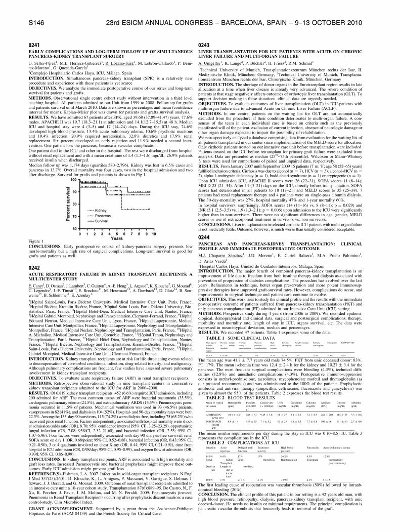
0241EARLY COMPLICATIONS AND LOG-TERM FOLLOW UP OF SIMULTANEOUSPANCREAS-KIDNEY TRANSPLANT SURGERY
G. Seller-Perez1, M.E. Herrera-Gutierrez1, R. Lozano-Saez1, M. Lebron-Gallardo1, P. Benı-tez-Moreno1, G. Quesada-Garcıa1
1Complejo Hospitalario Carlos Haya, ICU, Malaga, Spain
INTRODUCTION. Simultaneous pancreas-kidney transplant (SPK) is a relatively newprocedure and experience with these patients is yet scarce
OBJECTIVES. We analyze the immediate postoperative course of our series and long-termsurvival for patients and grafts.
METHODS. Observational single centre cohort study without intervention in a third levelteaching hospital. All patients admitted to our Unit from 1999 to 2008. Follow up for graftsand patients survival until March 2010. Data are shown as percentages and mean (confidenceinterval for mean). Kaplan–Meier plot was drawn for patients and grafts survival analysis.
RESULTS. We have admitted 67 patients after SPK, aged 39.68 (37.89–41.47) years, 77.6%males. APACHE II was 19.7 (18.3–21.1) at admission and 14.1(12.7–15.5) at 48 h. MedianICU and hospital stay were 4 (3–5) and 17 (14–24) days. During the ICU stay, 74.6%developed high blood pressure, 13.4% acute pulmonary edema, 10.6% psychotic reactionsand 10.4% infection; 20.9% required noradrenalin, 32.8% diuretics and 17.9% renalreplacement. Six percent developed an early rejection and 11.9% needed a second inter-vention. One patient loss the pancreas, because a vascular complication.
One patient died in the ICU and other in the hospital. The rest were discharged from hospitalwithout renal replacement and with a mean creatinine of 1.4 (1.3–1.6) mgr/dL. 26.9% patientsreceived insulin when discharged.
Median follow up was 1,174 days (quartiles 580–2,796). Kidney was lost in 6.5% cases andpancreas in 13.7%. Overall mortality was four cases, two in the hospital admission and twoafter discharge. Survival for grafts and patients is shown in Fig 1.
Figure 1
CONCLUSIONS. Early postoperative course of kidney-pancreas surgery presents lowmorbi-mortality but a high rate of surgical complications. Long-term survival is good forgrafts and patients as well.
0242ACUTE RESPIRATORY FAILURE IN KIDNEY TRANSPLANT RECIPIENTS: AMULTICENTER STUDY
E. Canet1, D. Osman2, J. Lambert3, C. Guitton4, A.-E. Heng5, L. Argaud6, K. Klouche7, G. Mourad8,C. Legendre9, J.-F. Timsit10, E. Rondeau11, M. Hourmant12, A. Durrbach13, D. Glotz14, B. Sou-weine15, B. Schlemmer1, E. Azoulay1.
1Hopital Saint-Louis, Paris Diderot University, Medical Intensive Care Unit, Paris, France,2Hopital Bicetre, Kremlin-Bicetre, France, 3Hopital Saint-Louis, Paris Diderot University, Bio-statistics, Paris, France, 4Hopital Hotel-Dieu, Medical Intensive Care Unit, Nantes, France,5Hopital Gabriel Montpied, Nephrology and Transplantation, Clermont-Ferrand, France, 6HopitalEdouard Herriot, Medical Intensive Care Unit, Lyon, France, 7Hopital Lapeyronnie, MedicalIntensive Care Unit, Montpellier, France, 8Hopital Lapeyronnie, Nephrology and Transplantation,Montpellier, France, 9Hopital Necker, Nephrology and Transplantation, Paris, France, 10HopitalA. Michallon, Medical Intensive Care Unit, Grenoble, France, 11Hopital Tenon, Nephrology andTransplantation, Paris, France, 12Hopital Hotel-Dieu, Nephrology and Transplantation, Nantes,France, 13Hopital Bicetre, Nephrology and Transplantation, Kremlin-Bicetre, France, 14HopitalSaint-Louis, Paris Diderot University, Nephrology and Transplantation, Paris, France, 15HopitalGabriel Montpied, Medical Intensive Care Unit, Clermont-Ferrand, France
INTRODUCTION. Kidney transplant recipients are at risk for life-threatening events relatedto decompensation of co-morbid conditions, infection, drug-related toxicity, and malignancy.Although pulmonary complications are frequent, few studies have assessed severe pulmonaryinvolvement in kidney transplant recipients.
OBJECTIVES. To evaluate acute respiratory failure (ARF) in renal transplant recipients.
METHODS. Retrospective observational study in nine transplant centers in consecutivekidney transplant recipients admitted to the ICU for ARF in 2000–2008.
RESULTS. Of 6,819 kidney transplant recipients, 452 (6.6%) required ICU admission, including200 admitted for ARF. The most common causes of ARF were bacterial pneumonia (35.5%),cardiogenic pulmonary edema (24.5%), and extrapulmonary ARDS (15.5%). Pneumocystis pneu-monia occurred in 11.5% of patients. Mechanical ventilation was used in 93 (46.5%) patients,vasopressors in 82 (41%), and dialysis in 104 (52%). Hospital- and 90-day mortality rates were both22.5%. Among the 155 day-90 survivors, 115 (74.2%) were dialysis-free, including 75 (65.2%) whorecovered prior renal function. Factors independently associated with hospital mortality were shockat admission (odds ratio [OR], 8.70; 95% confidence interval [95% CI], 3.25–23.29), opportunisticfungal infection (OR, 7.08; 95%CI, 2.32–21.60), and bacterial infection (OR, 2.53; 95% CI,1.07–5.96). Four factors were independently associated with day-90 dialysis-free survival: renalSOFA score on day 1 (OR, 0.68/point; 95% CI, 0.52–0.88), bacterial infection (OR, 0.43; 95% CI,0.21–0.90), 3 or 4 quadrants involved on chest X-ray (OR, 0.44; 95% CI, 0.21–0.91), time fromhospital to ICU admission (OR, 0.98/day; 95% CI, 0.95–0.99), and oxygen flow at admission (OR,0.93/l; 95% CI, 0.86–0.99).
CONCLUSIONS. In kidney transplant recipients, ARF is associated with high mortality andgraft loss rates. Increased Pneumocystis and bacterial prophylaxis might improve these out-comes. Early ICU admission might prevent graft loss.
REFERENCE(S). Fishman, J. A. 2007. Infection in solid-organ transplant recipients. N EnglJ Med 357(25):2601–14. Klouche, K., L. Amigues, P. Massanet, V. Garrigue, S. Delmas, I.Szwarc, J. J. Beraud, and G. Mourad. 2009. Outcome of renal transplant recipients admitted toan intensive care unit: a 10-year cohort study. Transplantation 87(6):889–95. De Castro, N., F.Xu, R. Porcher, J. Pavie, J. M. Molina, and M. N. Peraldi. 2009. Pneumocystis jiroveciiPneumonia in Renal Transplant Recipients occurring after prophylaxis discontinuation: a casecontrol-study. Clin Microbiol Infect.
GRANT ACKNOWLEDGMENT. Supported by a grant from the Assistance-PubliqueHopitaux de Paris (AOM 04139) and the French Society for Critical Care.
0243LIVER TRANSPLANTATION FOR ICU PATIENTS WITH ACUTE ON CHRONICLIVER FAILURE AND MULTI-ORGAN FAILURE
A. Umgelter1, K. Lange2, P. Buchler2, H. Friess2, R.M. Schmid1
1Technical University of Munich, Transplantationszentrum Munchen rechts der Isar, II.Medizinische Klinik, Munchen, Germany, 2Technical University of Munich, Transplanta-tionszentrum Munchen rechts der Isar, Chirurgische Klinik, Munchen, Germany
INTRODUCTION. The shortage of donor organs in the Eurotransplant region results in lateallocation at a time when liver disease is already very advanced. The severe condition ofpatients at that stage negatively affects outcomes of orthotopic liver transplantation (OLT). Tosupport decision-making in these situations, clinical data are urgently needed.
OBJECTIVES. To evaluate outcomes of liver transplantation (OLT) in ICU-patients withmulti-organ failure due to advanced Acute on Chronic Liver Failure (ACLF).
METHODS. In our centre, patients on the waiting list for OLT are not automaticallyexcluded from the procedure, if their condition deteriorates to multi-organ failure. A con-sensus of the team in each individual case is based on criteria such as the previouslymanifested will of the patient, exclusion of current infection, absence of neurologic damage orother organ damage expected to impair the possibility of rehabilitation.
We retrospectively analyzed a database comprising data from evaluation for the waiting list ofall patients transplanted in our center since implementation of the MELD-score for allocation.Only cirrhotic patients treated on our intensive care unit before transplantation were included.Patients treated on the ICU before retransplant for primary graft failure were excluded fromanalysis. Data are presented as median (25th–75th percentile). Wilcoxon or Mann–WhitneyU tests were used for comparisons of paired and unpaired data, respectively.
RESULTS. From January 2007 until September 2009 15 patients (7 m, 7f; age 58 (52–65) years)fulfilled inclusion criteria. Cirrhosis was due to alcohol (n = 7), HCV (n = 3), alcohol+HCV (n =2), alpha-1-antitrypsin deficiency (n = 1), budd-chiari-syndrome (n = 1) or cryptogenic (n = 1).
Upon ICU admission ICU, APACHE II scores were 26 (22–31), SOFA scores 11 (8–14);MELD 25 (21–34). After 14 (3–21) days on the ICU, directly before transplantation, SOFAscores had deteriorated in all patients to 18 (17–21) and MELD scores to 35 (25–38). 7patients had renal replacement therapy and 4 patients were on single-pass albumin dialysis.
The 30-day-mortality was 27%, hospital mortality 47% and 1-year mortality 60%.
In hospital survivors, surprisingly, SOFA scores (14 (11–16) vs. 8 (8–11); p = 0.029) andINR (3.1 (2.5–3.5) vs. 1.9 (1.3–2.1); p = 0.006) upon admission to the ICU were significantlyhigher than in non-survivors. There were no significant differences in age, gender, MELDscores or use of extracorporeal treatment in survivors vs. non-survivors.
CONCLUSIONS. Liver transplantation in selected cirrhotic ICU-patients with multi-organ failureis not medically futile. Outcome, however, is much worse than usually considered acceptable.
0244PANCREAS AND PANCREAS-KIDNEY TRANSPLANTATION: CLINICALPROFILE AND IMMEDIATE POSTOPERATIVE OUTCOME
M.J. Chaparro Sanchez1, J.D. Moreno1, E. Curiel Balsera1, M.A. Prieto Palomino1,
D. Arias Verdu1
1Hospital Carlos Haya, Unidad de Cuidados Intensivos, Malaga, SpainINTRODUCTION. The major benefit of combined pancreas-kidney transplantation is animprovement of life due to freedom from both insuline therapy and dialysis associated withprevention of progression of diabetes complications. The procedure has evolved over the lastyears. Refinements in technique, better organ preservation and more potent immunosup-pressive therapies have improved graft-survival rates. However, complications do occur, andimprovements in surgical technique and patient care continue to evolve.OBJECTIVES. This work tries to study the clinical profile and the results with the immediatepostoperative outcome of patients suffered from pancreas-kidney transplantation (PKT) andonly pancreas transplantation (PT) admitted in our Intensive Care Unit (ICU) setting.METHODS. Prospective study during 4 years (from 2006 to 2009). We recorded epidemi-ological, demographical and clinical data, surgical and postsurgical complications, therapy,morbidity and mortality rate, length of stay in ICU, organs survival, etc. The data wereexpressed in mean±typical deviation, median and percentages.RESULTS. We recorded 47 patients. Table 1 expresses some of the data.
TABLE 1 SOME CLINICAL DATAMean age of
diabetes
diagnostic
(mean)
Previous dialysis
in months
(median)
Previous
kidney
transplantation
Previous
pancreas
transplantation
High
blood
pressure
Ischemic
cardiopathy
Cerebrovascular
disease
Periferic
vasculopathy
Neuropathy RETINOPATHY
15 ± 8 8 (1–24) 25% 19% 78.7% 12.8% 2.1% 12.8% 38.3% 78.7%
The mean age was 41.8 ± 7.7 years old male 74.5%. PKT from unic deceased donor: 83%.PT: 17%. The mean ischemia time was 12.4 ± 2.4 h for the kidney and 10.27 ± 3.4 for thepancreas. The most frequent surgical complications were bleeding (4.3%), technical diffi-culties (12.8%) and anesthetic complications (4.3%). Postoperative immunosuppressionconsists in methyl-prednisolone, tacrolimus, mycophenolate mofetil and thymoglobulin (asour protocol recommends) and was administered to the 100% of the patients. Prophylacticantibiotic and antiviral therapy (ampicillin, ceftriaxone, fluconazole and gancyclovir) wasgiven to almost the 95% of the patients. Table 2 expresses the blood test results.
TABLE 2 BLOOD TEST RESULTSMean ± typical
deviation
Hemoglobin
(g/dl)
Platelets
(91,000/
ll)
Leukocytes
(91,000/ll)
Urea
(mg/dl)
Creatinine
(mg/dl)
Calcium
(mg/dl)
Amylase
(U/L)
Glucose
(mg/dl)
Albumin
(g/dl)
ADMISSION
INTO ICU
10.3 ± 1.6 188 ± 63 9.49 ± 3.8 68 ± 27 3.8 ± 2.2 7.2 ± 0.9 269 ± 200 115 ± 33 3.3 ± 0.6
DISCHARGE
FROM
ICU
9.9 ± 1.4 149 ± 65 7.1 ± 5.2 63 ± 33 1.8 ± 1.3 7.7 ± 0.8 106 ± 96 131 ± 40 2.7 ± 0.6
The mean insulin requirements per day during the stay in ICU was 0 (0–8.5) IU. Table 3represents the complications in the ICU.
TABLE 3 COMPLICATIONS AT ICUInfection Acute
rejection
Delayed graft
function
Treatment
toxicity
High blood
pressure
Pancreatitis Acute pulmonary edema
14.9% 6.4% 17% 17% 36.2% 4.3% 12.8%
Renal failure Graft thrombosis Reintervention Transplant nephrectomy
Transplant pancreatectomy
Death at
icu
Length of
stay at
icu in
days
(median)
10.6% 17% 21.3% 2.1% 14.9% 2.1% 5 (4–5)
The first leading cause of reoperation was vascular thrombosis (50%) followed by intraab-dominal bleeding (20%).CONCLUSION. The clinical profile of this patient in our setting is a 42 years old man, withhigh blood pressure, retinopathy, dialysis, pancreas-kidney transplant recipient, with unicdecesed-donor. He needs no insulin or minimal requirements. The principal complication ispancreatic vascular thrombosis that frecuently leads to removal of the graft.
S146 23rd ESICM ANNUAL CONGRESS – BARCELONA, SPAIN – 9–13 OCTOBER 2010

0245PROCALCITONINE AND PLASMA DESAPPEARED RATE (PDR) AS MARKERSOF LIVER REPERFUSION DAMAGE IN THE TRANSPLANTED PATIENTS
M. Lugano1, M. Centis1, C. Barbarino1, A. Rossetto1, P. Chiarandini1, G. Della Rocca1
1Clinica di Anestesia e Rianimazione, Udine, Italy
INTRODUCTION. High plasma procalcitonin (PCT) levels have been reported in severesystemic infection (1). In a clinical practice, a specific marker to evaluate and predict ische-mic-reperfusion injury in liver transplantation (OLT) is not available. Poor organ perfusionand high PCT levels appeared to predict early graft failure only in the cardiac donor (2).
OBJECTIVES. We evaluated PCT as a predictor of ischemic-reperfusion injury in livertransplantation and PDR, as a predictor of complication and graft outcome.
METHODS. Prospective study. 16 patients (age, Child-Pugh score, aetiology of liver cir-rhosis) undergo liver transplant. Bilirubin levels and PDR (0.5 mg/kg of IG in a centralcatheter with LIMON System�, Pulsion Medical System, Munchen, Germany) was measuredonce a day for 3 postoperative days (POD). On the same day, Aspartate AminoTransferase(AST/GPT) and Alanine AminoTransferase (ALT/GOT) were measured. SOFA score was asa patient severity score. Serum level of PCT, C-Reactive Protein (CRP) was collected at theliver reperfusion time and from the 1st to the 3rd POD. Warm and cold ischemia time wascollected. Statistical analysis was performed with Wilcoxon, Spearman tests (P \ 0.05).Linear correlation was performed too.
RESULTS. A small rise on PCT levels were observed early after OLT, with a peak in the 1stday 1 after OLT. It was associated neither with hepatic post-OLT dysfunction nor with othernon infective complications. PCT increased significantly after liver reperfusion (p \ 0.01)and correlate with PDR on the 2nd POD, but not correlation were found with CRP, whiteblood cells, or liver enzymes after OLT. CRP levels increased rapidly after OLT. PCTincreasing after liver reperfusion correlated with Child-Pugh OLT (R = 0.4 at 2nd POD) inthe recipients. The cold ischemia time did not correlate with PCT serum levels after liverreperfusion as well as the warm ischemia time. A negative correlation was found betweenPDR and liver function in the recipient. PDR did not correlate with Child-Pugh score. Thecold ischemia time well correlated with AST and ALT on the first day after transplant(R = 0.4). It negatively correlate with PDR (R = -0.6) at the same time. The warm ischemiatime did not correlate with PDR, AST and ALT. The same results were found between PDRand liver enzymes and lactates. No correlation was found between PDR and SOFA score.
CONCLUSIONS. PCT peak in the recipient at reperfusion and early post operative was notpredictive of graft dysfunction or other non infective complication. It may represent a markerof ischemia–reperfusion injury. CRP levels increased rapidly after OLT and it could be anexpression of surgical procedure, and it doesn’t correlate with PCT.
REFERENCE(S). 1) D.Eyraud et al. Critical Care 2008
2) Levesque E et al. Liver tranplantation 2009;15:1358–64.
0246THE IMMEDIATE POSTOPERATIVE DECREASE OF PLASMATIC NEUTROPHILGELATINASE-ASSOCIATED LIPOCALIN (NGAL) PREDICTS AN EARLYRECOVERY OF RENAL FUNCTION AFTER KIDNEY TRANSPLANTATION
A. Bataille1, S. Abbas1, O. Semoun1, E. Bourgeois1, O. Marie1, B. Francine1, R.-R. Matthieu1,
I. Abboud1, M.-R. Losser1, L. Jacob1
1Hopital Saint Louis, Paris, France
INTRODUCTION. NGAL is a ubiquitous protein already evaluated in several clinical sit-uations as a biomarker in the early diagnosis and prognosis of impaired renal function (1, 2).
OBJECTIVES. The aim of this prospective observational study was to describe the kineticsof NGAL following renal transplantation and to assess its ability to predict delayed graftfunction.
METHODS. Plasmatic NGAL was assayed (Triage � NGAL Test, Biosite Inc., InvernessMedical) in 41 patients undergoing renal transplantation. The samples were collectedimmediately before and after surgery, and at H12, day 1, day 3 and day 7. The demographicand metabolic data were simultaneously collected. A delayed graft function (DGF) wasdefined as the need for dialysis during the first week. The results were expressed as median[Q1, Q3]. Comparing the values of NGAL was performed with a Wilcoxon test and thepredictive value of NGAL was analyzed by ROC curves. This study has been accepted by anethic committee and all patients were informed.
RESULTS. Of the 41 patients enrolled, 15 (36.6%) patients presented a DGF. All patientshad a high preoperative NGAL: 453 ng/mL [382, 595]. In patients with DGF, the meanplasmatic NGAL was significantly higher at H12 (571 [467, 634] vs. 242 [158, 299] ng/mL,p \ 0.0001) and at day 1 (466 [356, 627] vs. 165 [91, 248] ng/mL, p \ 0.0001). A plasmaticNGAL higher than 400 ng/mL at 12 h of transplantation predicts DGF with a sensitivity of93.3%, a specificity of 88.5% and an odds ratio of 63.2 (p = 0.0004). This statistic perfor-mance is greater than plasmatic creatinine.
Evolution of plasmatic NGAL and ROC curves
CONCLUSIONS. Plasmatic NGAL early predicts delayed graft function after renal trans-plantation. The need for dialysis after kidney transplantation is indicated by conventionalmetabolic criteria (creatinine, urea and kaliemia). Plasmatic NGAL was not influenced bydialysis (data not shown), it allows monitoring of renal function in this situation. The decreaseof NGAL in early graft function can not be explained by a single effect of clearance by thegraft, because the NGAL/creatinine ratio decreased during the study. Early identification ofpatients at risk of DGF opens the field of a precise monitoring of renal aggression and themonitoring of the impact of future therapeutics.
REFERENCE(S). 1. Am J Kidney Dis. (2009);54:1012–1024.2. Intensive Care Med. (2010) 36:444–451.
0247OUTCOME OF LUNG TRANSPLANT RECIPIENTS REQUIRING READMISSIONTO THE INTENSIVE CARE UNIT
J. Cohen1, P. Singer1, Y. Raviv2, I. Bakal2, D. Shitrit2, S. Lev1, M. Kramer2
1Rabin Medical Center, Beilinson Hospital, General Intensive Care, Petach Tiqva, Israel,2Rabin Medical Center, Beilinson Hospital, Pulmonology, Petach Tiqva, Israel
INTRODUCTION. Lung transplantation is the recognized therapy for end-stage respiratoryfailure. Many serious medical complications have been described occurring from months toyears after lung transplantation, often necessitating admission to an intensive care unit (ICU),which has been associated with a high mortality. We examined the factors associated withmortality.
METHODS. All patients admitted to the general intensive care unit between 2000 and 2009following lung transplantation were included in this retrospective study. Data was collectedregarding demographic parameters, intensive care unit stay and outcome.
RESULTS. Over the study period, Forty patients were admitted to the ICU. The main pre-transplant diagnosis was idiopathic pulmonary fibrosis followed by chronic obstructive pul-monary disease. The majority (93%) of patients required mechanical ventilation during theirICU stay. The main reason for ICU admission was septic shock in 22 patients (55%) of cases.An organism was isolated from 19 of these patients; in 11 cases, the organism was shown tobe multi-drug resistant. The ICU mortality was 62.5%. Non-survivors were characterized by ahigher admission SOFA score (P = 0.02), an admission diagnosis of sepsis (87.5 vs. 37.5%for all other diagnoses, P \ 0.001), and a requirement for mechanical ventilation (P = 0.02).In addition, the incidence of chronic rejection was significantly higher in the non-survivors(P = 0.02).
CONCLUSIONS. Severe sepsis remains the most important factor associated with a pooroutcome. New strategies are required to alter the course of this common complication of lungtransplantation.
0248LUNG TRANSPLANTATION. ANALYSES OF 129 CASES
J. Sacanell1, C. Laborda1, L. Tenorio1, J.R. Masclans1, O. Roca1,2, V. Monforte2,3, M. Deu4,
J. Rello1,2
1Vall d0Hebron University Hospital, Critical Care Department, Barcelona, Spain, 2Instituto deSalud Carlos III, CibeRes (Ciber Enfermedades Respiratorias), Madrid, Spain, 3Vall d0HebronUniversity Hospital, Respiratory Medicine Department, Barcelona, Spain, 4Vall d0HebronUniversity Hospital, Thoracic Surgery Department, Barcelona, Spain
INTRODUCTION. Lung transplantation is an established viable treatment to prolong andincrease the quality of live in patients with end-stage lung disease.
OBJECTIVES. To analyse clinical characteristics, postoperative complications and mortalityof a large cohort of lung transplant patients.
METHODS. A prospective observational analysis was performed. All adult patients under-going lung transplantation in Vall d’Hebron University Hospital from January 2007 toDecember 2009 were included. Patients were followed until they were discharged from theCritical Care Department (CCD). Results are presented as median (range) or frequency(percentage). Using (SPSS 18.0, Inc, Chicago, IL).
RESULTS. During the period of study, 129 patients were included. The indications for lungtransplantation were chronic obstructive pulmonary disease (COPD) in 58 (45%) patients,idiophatic pulmonary fibrosis (IPF) in 43 (33.3%) cases, 6 (5.4%) patients with cystic fibrosis,4 (3.1%) with primary pulmonary arterial hypertension, 2 (1.5%) were re-transplantedpatients and 16 (12.5%) due to other diseases. At the moment of the transplant, the medianage was 52 (18–64) years. In 80 (62%) patients a bilateral lung trasplantation was performed.During the operation, 33 (25.6%) patients precised extracorporeal circulatory suport. At theadmission to the CCD, the median APACHE III score was 49 (21–148) with a PaO2/FiO2
ratio of 257 (41–600) mmHg. Moreover, 74 (57.3%) patients developed primary graft dys-function grade 2–3. During CCD evolution, tracheostomy was performed in 55 (42.6%)patients. Most common complications were gastroparesis in 46 (35.8%) patients, 41 (31.8%)presented myopathy, 33 (25.7%) cases of diaphragmatic paresis, infection in 40 (31%) cases(the 80% of the infections were respiratory) and 13 (10%) patients presented pulmonaryallograft rejection. According to our immunosuppressive protocol, we started with methyl-prednisolone and tacrolimus and we added mycophenolate later. The CCD length of stay was9 (2–102) days and the median days of mechanical ventilation were 4 (1–92). Thirteen (10%)patients died, basically due to refractory respiratory failure, multiple organ dysfunctionsyndrome and haemorragic shock.
CONCLUSIONS. In our large series of lung transplantation a remarkable incidence ofcomplications has been observed. Despite this complications, lung transplanted patientspresented an excellent short term outcomes.
23rd ESICM ANNUAL CONGRESS – BARCELONA, SPAIN – 9–13 OCTOBER 2010 S147
0249INFLAMMATORY CYTOKINE RESPONSE AND APOPTOSIS IN THE TRANS-PLANTED KIDNEY GRAFT: A PORCINE MODEL
N. Secher1, P. Søndergaard2, K. Ravlo2, C. Sølling1, A. Granfeldt1, B. Jespersen2,
E. Tønnesen1
1Aarhus University Hospital, NBG, Department of Anaesthesiology and Intensive Care,Aarhus C, Denmark, 2Aarhus University Hospital, SKS, Department of Nephrology, AarhusN, Denmark
INTRODUCTION. Acute kidney injury (AKI) poses a massive challenge after kidneytransplantation, especially when kidneys from brain dead adult donors are transplanted intosmall paediatric recipients. Inflammation mediated by cytokines is a key event in experi-mental models of ischaemic AKI.
OBJECTIVES. The aim of this study was to investigate whether remote ischaemic pre-conditioning (rIPC) reduced the inflammatory cytokine load and apoptosis in the kidney aftertransplantation.
METHODS. Kidneys were harvested from eight 65-kg brain dead donor pigs and trans-planted into two groups of 15-kg recipient pigs after 21 h of cold ischaemia. In one group(+rIPC, n = 8) rIPC was performed before the 10-h reperfusion period, while no rIPC wasperformed in the other group (-rIPC, n = 6). Non transplanted kidneys from 7 brain deadpigs served as controls. Concentrations of TNF-a, IL-6, IL-8, and IL-10 in renal tissue weredetermined by an immuofluorometric assay. Renal apoptosis was quantified by immunohis-tochemistry for activated caspase-3.
RESULTS. High concentrations of TNF-a, IL-6, IL-8, and IL-10 were detected in renalcortex in all three groups. No statistical differences between the two transplanted groups werefound for any of the cytokines. Compared to controls higher cortical levels of IL-10 (controlvs. -rIPC, p = 0.02, control vs. +rIPC, p = 0.006) and lower levels of IL-6 (control vs. -rIPC, p = 0.03, control vs. +rIPC, p = 0.01) were found in transplanted kidneys. No dif-ferences were detected for TNF-a or IL-8. Transplantation significantly increased the numberof apoptotic cells in both glomeruli and tubuli (control vs. -rIPC, p = 0.0005, control vs.+rIPC, p = 0.0001). No difference was found between recipients, (p = 0.26).
CONCLUSIONS. In transplanted kidneys from brain dead donors exposed to 21 h of coldischemia and ±rIPC, we found increased tubular and glomerular apoptosis, but no increase inpro-inflammatory cytokines. The levels of IL-10 were higher in transplanted kidneys com-pared to controls. Remote ischaemic preconditioning did not modify cytokine load orapoptosis in the kidney graft.
0250HEPARIN-INDUCED THROMBOCYTOPENIA (HIT): THE MANAGEMENTBEFORE AND AFTER THE CARDIAC TRANSPLANTATION OF A PATIENTWITH POSITIVE ANTI-PF4/HEPARIN ANTIBODIES. CASE REPORT
M.-D. Cristinar1, J.-C. Thiranos1, J.-P. Dupeyron1
1Nouvel Hopital Civil, Anesthesie-Reanimation, Strasbourg, France
INTRODUCTION. Heparin-induced thrombocytopenia (HIT) is a difficult diagnostic in postcardiac surgery. HIT appears as an antibody-mediated adverse drug reaction associated withincreased thrombotic risk. The antigens are located on platelet factor 4 (PF4)/heparin com-plexes. Not all the PF4/heparin-reactive antibodies cause HIT.
OBJECTIVES. We present the case of a patient with confirmed HIT and the management ofits status during the perioperative period of the cardiac transplantation.
METHODS AND RESULTS. A 45 years old patient with a cardiac myxoma was operatedunder heparin anticoagulation. Thrombocytopenia is noted at day 5 after surgery. An enzyme-linked immunosorbent assay (ELISA) was performed and since the result was positive, thetreatment was changed to Lepirudin. The HIT was confirmed by a heparin-induced plateletactivation (HIPA) test. The internal jugular vein thrombosis was observed. The post operativeevolution was marked by the necessity of the implantation of a ventricular assisted device.The patient was submitted to two sessions of plasmapheresis which turned the antibodiesnegative. The patient underwent heparin anticoagulation during the surgery time and Biva-lirudin as the post operative treatment. The antibodies remained negative. Two months later,the cardiac transplantation was performed; heparin was used for anticoagulation during sur-gery. Due to a restored renal function, Danaparoid was used postoperatively.
CONCLUSIONS. HIT is a serious complication of heparin therapy. The diagnosis is diffi-cult. When HIT is strongly suspected, a non-heparin anticoagulant is recommended. Thechoice of the anticoagulant depends on the hepatic and renal function. Plasmapheresis is asolution for the antibody purging prior to cardiac surgery.
REFERENCE(S). 1. T. Warkentin et al. - When is HIT really HIT? Ann Thorac Surg2007;83:21–3.
2. S. Christiansen et al. - Anticoagulative management of patients requiring left ventricularassist device implantation and suffering from heparin-induced thrombocytopenia type II.Ann Thorac Surg 2000;69:774–7
3. A. Greinacher et al. - HIT happens: diagnosis and evaluating the patient with heparin-induced thrombocytopenia. Anesth Analg 2008; 107 (2):354–8.
Education and training 1: 0251–02600251INTENSIVE CARE MEDICINE TRAINEES’ PERCEPTION OF PROFESSIONAL-ISM: A QUALITATIVE STUDY
W.N.K.A. van Mook1,2, W.S. De Grave2, S.L. Gorter3, J.H. Zwaveling4, L.W. Schuwirth2,
C.P.M. van der Vleuten2
1Maastricht University Medical Centre, Departments of Intensive Care and Internal Medicine,Maastricht, Netherlands, 2Maastricht University, Department of Educational Developmentand Research, Maastricht, Netherlands, 3Maastricht University Medical Centre, Departmentof Internal Medicine, division of Rheumatology, Maastricht, Netherlands, 4Maxima MedicalCentre, Board of Directors, Veldhoven, Netherlands
INTRODUCTION. Under the aegis of the ESICM, the Competency-BasedTraining programin Intensive Care in Europe (CoBaTrICE) identified 12 competency domains. Professionalismwas given a prominence equal to technical ability. However, little information pertaining tofellows’ views on professionalism is available.
OBJECTIVES AND METHODS. A nationwide qualitative study investigating their perceptionof the meaning of professionalism,and how they learn to behave professionally was performed. Alleight Dutch ICM training centres participated. The moderator asked participants to clarify theterms professionalism and professional behaviour. Next, participants were asked to explore thequestions ‘How do you learn the mentioned items?’ and ‘What ways of learning do you find usefulor superfluous?’ Qualitative data analysis software (MAXQDA2007) facilitated analysis: aninductive approach applying open, axial and selective coding principles was used.
RESULTS. 35 fellows across eight groups participated. Results relating to the subtopics ‘Elementsof professionalism’ and ‘Teaching and learning of professionalism’ are described consecutively.
Elements of professionalism relevant to intensivists: The elements most frequently addressed werecommunication, keeping distance and boundaries, medical knowledge and expertise, respect, teamwork,leadership and organization and management. Medical knowledge, expertise and technical skills seem tobecome more tacit when training progresses, and relate to ethical, cultural and legal dilemmas originatingin the specific ICU context, and working as a multidisciplinary ICU team member.
Teaching and learning professionalism: Topics can be categorised into the themes workplace-based learning, by gathering practical experience, by following examples and receiving feedbackon their actions, including learning from own and others’ mistakes. Formal teaching courses (e.g.communication) and scheduled sessions addressing professionalism aspects were also valued.
CONCLUSIONS. The emerging elements considered most relevant for intensivists were ade-quate communication skills, and keeping boundaries with patients and relatives. The specific ICMcontext, and working as multidisciplinary ICU team member substantially influenced the ICMfellows’ perception of professionalism. Whereas medical knowledge, expertise and technicalskills seem to become more tacit when training progresses, professionalism issues continue to belearned during ICM training. Professionalism is herein mainly learned ‘on the job’ from rolemodels. Formal teaching courses and sessions addressing professionalism aspects were never-theless valued, and learning from own and others’ mistakes was considered especially useful. Self-reflection as a starting point for learning professionalism was stressed. The latter can e.g. bestimulated by means of assessment, structured feedback and use of portfolios, for which guidelinesare now being developed within the CoBaTrICE project.
0252CHARITE SUMMER ACADEMY 2010 ‘‘DISASTER MEDICINE’’: AN INTERDIS-CIPLINARY AND INTERNATIONAL CONCEPT TO IMPROVE EDUCATION INDISASTER MEDICINE
S. Kramer1, J. Rau1, T. Schroeder1, U. Arnold2, C. Spies1
1Charite-Universitatsmedizin Berlin, Department of Anesthesiology, Berlin, Germany,2Charite-Universitatsmedizin Berlin, Charite International Cooperation, Berlin, Germany
Introduction: During the past decades there has been an increase in mass casualty eventswith changing geopolitical and climate situations. In a mass casualty event comprehensivecare for the individual is expected [4]. To meet these obligations further education in disastermedicine seems obligate [1]. Therefore the German Home Department responsible for masscasualties passed a concept for student education in disaster medicine [2].
OBJECTIVES. The introduction of a Summer Academy ‘‘Disaster Medicine’’ (SADM) is afirst approach at Charite University of Berlin to establish a curriculum for disaster medicine.The SADM is sponsored by the German Academic Exchange Service (DAAD) for 3 years.The enhancement of student education in disaster medicine is supposed to raise the level ofskills and knowledge of future physicians in the face of mass casualties [5].
METHODS. International participants and an interdisciplinary approach are keystones of theSADM concepts. In a globalized world international networking should enable students toexchange knowledge about the handling of mass casualties in different parts of the world.Disaster medicine needs an interdisciplinary approach [3, 4]. Psychological aspects arealways a key factor in the successful handling of mass casualties.
The teaching concept of SADM consists of four parts: e-learning ahead of a 2 week training session,emergency medicine training, disaster medicine training and excursions to evaluate already existingdisaster concepts. The concept will be evaluated [3] using a knowledge test, a skills test and astructured written interview concerning motivation and satisfaction of the students.
CONCLUSION. The support of the DAAD for three consecutive years allows a furtherevolution of this concept by integrating the evaluation results. The SADM should enablefuture physicians to meet the challenges of mass casualties with greater confidence and skills.
REFERENCES. 1. Ashkenazi I et al. (2009) Inadequate mass-casualty knowledge baseadversely affects treatment decisions by trauma care providers: survey on hospital responsefollowing a terrorist bombing. Preshop Disaster Med 24:342–3472. Gemeinsame Empfehlung der Schutzkommission beim Bundesministerium ders Inneren
(2006) Konzept zur Katastrophenmedizinischen Ausbildung im studentischen Unterrichtan deutschen Hochschulen. DGKM e. V.
3. Murray et al. (2006) Disaster health education and training: a pilot questionnaire tounderstand current status. Prehosp Disaster Med 21:156–167
4. Schutzkommission beim Bundesminister des Innern (2006) Katastrophenmedizin-Leitfadenfur die arztliche Versorgung im Katastrophenfall. 4. uberarbeitete Auflage, Berlin
5. Vincent et al. (2009) Mass-casualty triage training for international healthcare workers inthe asia-pacific region using manikin-based simulations. Prehosp Disaster Med24:206–213.
GRANT ACKNOWLEDGMENT. German Academic Exchange Service
S148 23rd ESICM ANNUAL CONGRESS – BARCELONA, SPAIN – 9–13 OCTOBER 2010
0253MEASURING PATIENT SAFETY CULTURE: THE IZEP AND COMPAZ TOOL
T. van Galen1, E.G.M. Smit1
1VU University Medical Center, Intensive Care, Amsterdam, Netherlands
INTRODUCTION. Patient safety is a critical healthcare issue worldwide after IOM publi-cation To Err Is Human1. Many hospital care intervention programs have been startedafterwards to reduce preventable adverse events. Beside direct patent care activities,improvement of patient safety culture (PSC) is an important objective of overall patientsafety2.
In the Netherlands two safety culture surveys are available and applicable in an inpatientenvironment: COMPaZ and IZEP. Both surveys are currently applied in VU Universitymedical center (VUmc).
METHODS. The COMPaZ survey is based on the AHRQ Hospital Survey on Patient SafetyCulture (HSPS)3 and is a quantitative survey. With COMPaZ 11 dimensions of patient safetyare measured during a 41 question survey. With a total of 50 items it takes up to 15 min tocomplete the survey. The IZEP survey is based on Ron Westrum’s theory on organizationalsafety and made applicable by the NHS (MaPSaF)4. IZEP is based on a qualitative approachand is performed in a workshop format. Nine dimensions are assessed on their level on thesafety culture ladder. After the assessment is diagnosed, results are discussed within a mul-tidisciplinary setting. The workshop format is limited to 20 participants and lasts up to90 min. Both surveys are applicable in various hospital care settings and offered to hospitalsfrom the Dutch Patient Safety Management Program (VMS zorg5).
RESULTS. COMPaZ and IZEP, are applied in VUmc. Due to their format differences, bothsurveys can be seen as complementary to each other. Outcomes of COMPaZ on a High Carelevel ward can be compared with institutional and nationwide data.
Patient safety and PSC on the VUmc High Care ward is rated more positive compared toinstitutional an national outcomes. IZEP survey data on two institutional wards shows dif-ference between wards and aetiology of the health care professional. Physicians and staffmembers position patient safety and PSC on a higher level compared to nursing staffmembers.
CONCLUSIONS. IZEP and COMPaZ are valid PSC measurement and interventions tools.
Outcomes of the COMPaZ survey provides a quick insight. Learning opportunities are con-nected to low and high scoring dimensions. IZEP provides comprehension of PSC on a wardlevel. It improves awareness of PSC and it’s importance. Evokes discussion about strengthsand weaknesses and improvement measures are more steady due to local ownership. Toimprove PSC inserting only COMPaZ and IZEP is not enough. More culture interventions arenecessary like corporate governance, full disclosure, safety walk rounds and crew resourcemanagement.
REFERENCE(S). 1. IOM. To err is human: Building a safer health system. NationalAcademy Press, Washington D.C., 2000.
2. Sorra JS, Nieva VF. Hospital survey on patient safety culture. AHRQ publication 04-0041.September 2004.
3. http://www.ahrq.gov
4. http://www.npsa.nhs.uk
5. http://www.vmszorg.nl.
0254DEVELOPMENT OF INTENSIVE CARE MEDICINE IN BOSNIA ANDHERZEGOVINA
P. Kovacevic1, J. Vidovic1, M. Krunic1, B. Zlojutro1, M. Jandric1, S. Kovacevic1, M.
Stanetic2
1University Hospital Banja Luka, Medical Intensive Care Unit, Banja Luka, Bosnia andHerzegovina, 2University Hospital Banja Luka, Clinic for Lung Diseases, Banja Luka, Bosniaand Herzegovina
INTRODUCTION. Intensive care medicine is a relatively new specialty, which was createdin the 1950s, after invent of mechanical ventilation, which allowed caring for critically illpatients who otherwise would have died. Over the years, intensive care medicine developedfurther and became a truly multidisciplinary speciality, encompassing patients from variousfields of medicine and involving specialists from a range of base specialties, with additional(subspeciality) training in intensive care medicine. In Bosnia and Herzegovina, the intro-duction of non invasive ventilation in 2007, and opening of first multidisciplinary ICU inBanja Luka heralded a new age of intensive care medicine.
OBJECTIVES. The main aim of the study is to analyze the number of admissions, highseverity scores, needs for mechanical ventilation and outcomes of treatment during the firstyear in medical ICU in Banja Luka University hospital.
METHODS. This study is designed as a retrospective study.
RESULTS. Results are presented below:
1. Number of admitted patients in MICU in 2009 is 3142. Number of admitted patients with SAPS II [ 50 is 161 (51%)3. Number of patients who died in first 24 h is 63 (20.1%)4. Mortality in MICU is 81 (25.7%)5. Number of patient treated with mechanical ventilation is 215 (68.4%)6. Number of patients treated with non invasive ventilation is 34 (10.8%)
CONCLUSIONS. These results confirmed the need of these kinds of units in the country. Inspite of still suboptimal personnel training, creation of ICU in Banja Luka may serve asexample for other developing countries in the region. However, in order to achieve modernICU standards and follow European trends toward harmonisation of medicine, Bosnia andHerzegovina needs to take up this challenge by recognizing intensive care medicine as adistinctive specialty, by implementing a specific training program and by setting up multi-disciplinary ICUs in acute care hospitals.
REFERENCE(S). 1. Amoateng-Adjepong Y. Caring for the critically ill in developingcountries-our collective challenge. Critical Care Medicine 2006; 34: 1288–1289.2. Besso J, Bhagwanjee S, Takezawa J, Prayag S & Moreno R. A global view of education
and training in critical care medicine. Critical Care Clinics 2006; 22: 539–546.3. Bhagwanjee S. Critical care in Africa. Critical Care Clinics 2006; 22: 433–438.4. Baker T. Critical care in low-income countries. Trop Med Int Health 2009; 14: 143–148.
0255EVALUATION OF DENTAL PRACTIONER SKILL AND TRAINING EFFICACY INMEDICAL EMERGENCIES AND PAIN TREATMENT IN DENTAL PRACTICE
V. Garcia1, G. Ajob1, M. Simon1, O. Collange1, R. Schaeffer1, A. Meyer1, A. Feki2, B. Calon1
1Hopital de Hautepierre, Reanimation Chirurgicale, Strasbourg Cedex, France, 2Faculte deChirurgie Dentaire, Departement de Medecine Buccale et Chirurgie Buccale, Strasbourg,France
INTRODUCTION. Dentists are confronted with medical emergencies and pain treatment.Educational programs are being set up to provide training and skills in these core subjects fordental care professionals.
OBJECTIVES. To evaluate dental practitioner (DP) skills and knowledge prior to a 1 daycontinuous medical education (CME) training session, and assess training efficacy at the endof the session.
METHODS. Nine (9) multiple choice questions concerning medical emergencies and paintreatment were handed out to DPs at the beginning of CME training sessions over a 1 yearperiod in metropolitan France. After the 1 day training session, the same multiple choice wastaken again and collected for statistical analysis (Kruskall–Wallis test).
RESULTS.EXAMINATION BEFORE AND AFTER CME, P \ 0.001
Item Answer Correct answersbefore CME
Correct answersafter CME
Emergency telephone number (France) 15 (France) 91% 91%
Emergency telephone number (Europe) 112 (Europe) 50% 86%
Vasovagal syncope Associated with bradycardia 72% 88%
Recovery position Left side in pregnancy 58% 92%
Choking (severe) Heimlich manoeuver 90% 91%
Choking (severe) Back slaps first 44% 89%
Anaphylaxis (severe) Epinephrine as first drug 69% 94%
Malaise in the diabetic patient Sugar as first treatment 56% 82%
Tooth ache during late pregnancy Avoiding treatment with NSAIDs 76% 83%
We evaluated 494 DPs and obtained a 100% answering rate. Before the CME session, thecorrect answer rate was below 50% for several items, like the 112 european emergencytelephone number or performing back blows before the Heimlich manoeuver for severechoking, and below 75% for identifying vasovagal malaise by bradycardia, giving insulin fordiabetic malaise or treating anaphylactic shock by epinephrine. More worrisome still is thefact that nearly 1out of 4 DPs would prescribe non steroidal anti inflammatory drugs(NSAIDs) during late pregnancy.
The overall impact of CME was highly significant (p \ 0.001), showing real efficacy butcorrect answering rates after CME still remained between 82 and 94%, which leaves room forimprovement. Further studies are under way to evaluate long term memorization of CMEsessions in order to determine their optimal frequency.
CONCLUSIONS. Medical skill and proficiency evaluation before CME training sessions forDPs allows to target the training sessions and to evaluate their efficacy in the short run.
0256ECHOCARDIOGRAPHY ONLINE: A SOUND MEANS OF TRAINING IN TODAY’SWORLD?
N. Jones1, G. Abuella1
1Royal Brompton Hospital, London, UK
INTRODUCTION. The positive impact of immediate bedside echocardiography for rapiddiagnosis and management of acute hemodynamic disturbances in the critically ill patient iswell established. It is advocated that peri-resuscitation echocardiography should be an integralpart of training for all intensivists. However, a major challenge for the intensive care clinicianis access to appropriate echocardiography training outside of specific fellowship programmes.
OBJECTIVES. One suggestion to meet this training need is to combine supervised practicalinstruction with self-learning through the use of on-line educational tools. The internet isideally suited to studying echocardiography as e-tutorials serve to convey theoretical prin-ciples whilst stills/video clips aid image recognition and interpretation. Here we reviewcurrently available web based learning resources.
METHODS. An online search was performed using Google� and Yahoo� search engineswith the following key words: echocardiography, TTE, TOE, education, training, programme,courses, on-line, web-based, critical care. The resulting hits were screened to identify relevantsites and these were then evaluated independently by each author before an overall consensuswas reached. One author had no previous echocardiography training whilst the other hadpassed the American National Board of Echocardiography Perioperative TransesophagealEchocardiography examination.
RESULTS. A total of 16 sites were identified for evaluation (see Table 1); these are listedbelow with a brief description.
TABLE 1 NATIONAL SOCIETIESWebsite Educational
contentGraphicsquality
Navigation Selfassessment
Targetaudience
Experiencerequired
Exampreparation
Otherinformation
Fee
BritishSocietyof Echocardi-
ographyhttp://www.bsecho.org
3 3 3 Y Cardiology,Criticalcareand Emergencymed
Intermediate BSEAccreditation
Doesn0 tcoverbasics
Free
EuropeanAssociationof Cardiologyhttp://www.escardio.org
5 5 5 N Cardiologists Novice EACAccreditation
Basiccontent
Freeto ESEmembers
Americansocietyof echocar-
diographyhttp://www.asecho.org
5 Unableto assess
Unableto assess
N CardiologistsAnaesthetistsCardiacSurgeons
Novice toadvanced
NationalBoardExaminations
Ultrasoundbasicsnotcovered
$525
CONCLUSIONS. Our search demonstrated a number of sites dedicated to facilitatingechocardiography training. These varied from those which were essentially atlases, to thosewith a modular learning programme supported by interactive discussions and self assessment.Some were targeted at the beginner seeking a basic understanding of echo whilst others wereaimed at the enthusiast preparing for examinations. With growing interest in critical careultrasound it is likely that we will see the use of such resources increasing. However echo-cardiography is a practical skill and it is essential that on-line learning is conducted in parallelwith supervised bedside training in a process of ‘blended learning’.
23rd ESICM ANNUAL CONGRESS – BARCELONA, SPAIN – 9–13 OCTOBER 2010 S149
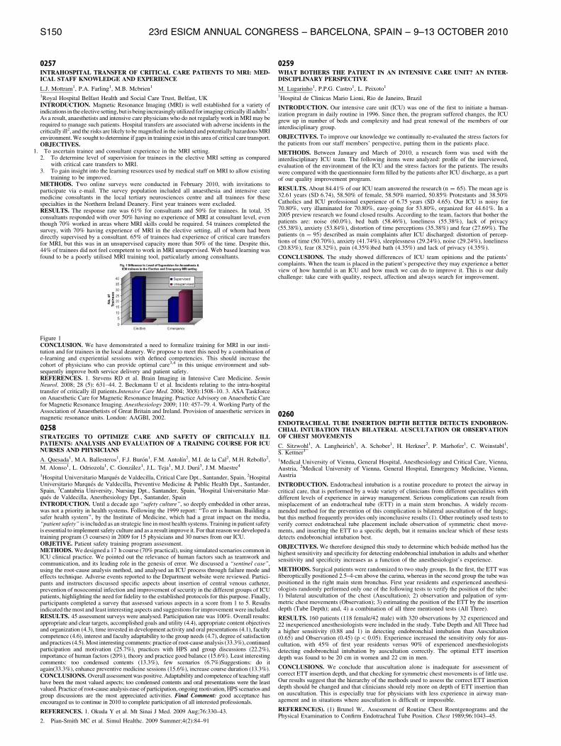
0257INTRAHOSPITAL TRANSFER OF CRITICAL CARE PATIENTS TO MRI: MED-ICAL STAFF KNOWLEDGE AND EXPERIENCE
L.J. Mottram1, P.A. Farling1, M.B. Mcbrien1
1Royal Hospital Belfast Health and Social Care Trust, Belfast, UKINTRODUCTION. Magnetic Resonance Imaging (MRI) is well established for a variety ofindications in the elective setting, but is being increasingly utilized for imaging critically ill adults1.As a result, anaesthetists and intensive care physicians who do not regularly work in MRI may berequired to manage such patients. Hospital transfers are associated with adverse incidents in thecritically ill2, and the risks are likely to be magnified in the isolated and potentially hazardous MRIenvironment. We sought to determine if gaps in training exist in this area of critical care transport.OBJECTIVES.
1. To ascertain trainee and consultant experience in the MRI setting.2. To determine level of supervision for trainees in the elective MRI setting as compared
with critical care transfers to MRI.3. To gain insight into the learning resources used by medical staff on MRI to allow existing
training to be improved.METHODS. Two online surveys were conducted in February 2010, with invitations toparticipate via e-mail. The survey population included all anaesthesia and intensive caremedicine consultants in the local tertiary neurosciences centre and all trainees for thesespecialties in the Northern Ireland Deanery. First year trainees were excluded.RESULTS. The response rate was 61% for consultants and 50% for trainees. In total, 35consultants responded with over 50% having no experience of MRI at consultant level, eventhough 70% worked in areas where MRI skills could be required. 54 trainees completed thesurvey, with 70% having experience of MRI in the elective setting, all of whom had beendirectly supervised by a consultant. 65% of trainees had experience of critical care transfersfor MRI, but this was in an unsupervised capacity more than 50% of the time. Despite this,44% of trainees did not feel competent to work in MRI unsupervised. Web based learning wasfound to be a poorly utilised MRI training tool, particularly among consultants.
Figure 1CONCLUSION. We have demonstrated a need to formalize training for MRI in our insti-tution and for trainees in the local deanery. We propose to meet this need by a combination ofe-learning and experiential sessions with defined competencies. This should increase thecohort of physicians who can provide optimal care3,4 in this unique environment and sub-sequently improve both service delivery and patient safety.REFERENCES. 1. Stevens RD et al. Brain Imaging in Intensive Care Medicine. SeminNeurol. 2008; 28 (5): 631–44. 2. Beckmann U et al. Incidents relating to the intra-hospitaltransfer of critically ill patients.Intensive Care Med. 2004; 30(8):1508–10. 3. ASA Taskforceon Anaesthetic Care for Magnetic Resonance Imaging. Practice Advisory on Anaesthetic Carefor Magnetic Resonance Imaging. Anesthesiology 2009; 110: 457–79. 4. Working Party of theAssociation of Anaesthetists of Great Britain and Ireland. Provision of anaesthetic services inmagnetic resonance units. London: AAGBI, 2002.
0258STRATEGIES TO OPTIMIZE CARE AND SAFETY OF CRITICALLY ILLPATIENTS: ANALYSIS AND EVALUATION OF A TRAINING COURSE FOR ICUNURSES AND PHYSICIANS
A. Quesada1, M.A. Ballesteros1, F.J. Buron1, F.M. Antolın2, M.I. de la Cal2, M.H. Rebollo2,
M. Alonso1, L. Odriozola1, C. Gonzalez1, J.L. Teja1, M.J. Dura3, J.M. Maestre4
1Hospital Universitario Marques de Valdecilla, Critical Care Dpt., Santander, Spain, 2HospitalUniversitario Marques de Valdecilla, Preventive Medicine & Public Health Dpt., Santander,Spain, 3Cantabria University, Nursing Dpt., Santander, Spain, 4Hospital Universitario Mar-ques de Valdecilla, Anesthesiology Dpt., Santander, SpainINTRODUCTION. Until a decade ago ‘‘safety culture’’, so deeply embedded in other areas,was not a priority in health systems. Following the 1999 report: ‘‘To err is human. Building asafer health system’’, by the Institute of Medicine, which had a great impact on the media,‘‘patient safety’’ is included as an strategic line in most health systems. Training in patient safetyis essential to implement safety culture and as a result improve it. For that reason we developed atraining program (3 courses) in 2009 for 15 physicians and 30 nurses from our ICU.OBJETIVE. Patient safety training program assessment.METHODS. We designed a 17 h course (70% practical), using simulated scenarios common inICU clinical practice. We pointed out the relevance of human factors such as teamwork andcommunication, and its leading role in the genesis of error. We discussed a ‘‘sentinel case’’,using the root-cause analysis method, and analysed an ICU process through failure mode andeffects technique. Adverse events reported to the Department website were reviewed. Partici-pants and instructors discussed specific aspects about insertion of central venous catheter,prevention of nosocomial infection and improvement of security in the different groups of ICUpatients, highlighting the need for fidelity to the established protocols for this purpose. Finally,participants completed a survey that assessed various aspects in a score from 1 to 5. Resultsindicated the most and least interesting aspects and suggestions for improvement were included.RESULTS. 45 assessment surveys were analysed. Participation rate was 100%. Overall results:appropriate and clear targets, accomplished goals and utility (4.4), appropriate content objectivesand organization (4.3), time invested in development activity and oral presentations (4.1), facultycompetence (4.6), interest and faculty adaptability to the group needs (4.7), degree of satisfactionand practices (4.5). Most interesting comments: practice of root-cause analysis (33.3%), continuedparticipation and motivation (25.7%), practices with HPS and group discussions (22.2%),importance of human factors (20%), theory and practice good balance (15.6%). Least interestingcomments: too condensed contents (13.3%), few scenarios (6.7%)Suggestions: do itagain(33.3%), enhance preventive medicine sessions (15.6%), increase course duration (13.3%).CONCLUSIONS. Overall assessment was positive. Adaptability and competence of teaching staffhave been the most valued aspects; too condensed contents and oral presentations were the leastvalued. Practice of root-cause analysis ease of participation, ongoing motivation, HPS scenarios andgroup discussions are the most appreciated activities. Final Comment: good acceptance hasencouraged us to continue in 2010 to complete participation of all interested professionals.
REFERENCES. 1. Okuda Y et al. Mt Sinai J Med. 2009 Aug;76:330–43.
2. Pian-Smith MC et al. Simul Healthc. 2009 Summer;4(2):84–91
0259WHAT BOTHERS THE PATIENT IN AN INTENSIVE CARE UNIT? AN INTER-DISCIPLINARY PERSPECTIVE
M. Lugarinho1, P.P.G. Castro1, L. Peixoto1
1Hospital de Clinicas Mario Lioni, Rio de Janeiro, Brazil
INTRODUCTION. Our intensive care unit (ICU) was one of the first to initiate a human-ization program in daily routine in 1996. Since then, the program suffered changes, the ICUgrew up in number of beds and complexity and had great renewal of the members of ourinterdisciplinary group.
OBJECTIVES. To improve our knowledge we continually re-evaluated the stress factors forthe patients from our staff members’ perspective, putting them in the patients place.
METHODS. Between January and March of 2010, a research form was used with theinterdisciplinary ICU team. The following items were analyzed: profile of the interviewed,evaluation of the environment of the ICU and the stress factors for the patients. The resultswere compared with the questionnaire form filled by the patients after ICU discharge, as a partof our quality improvement program.
RESULTS. About 84.41% of our ICU team answered the research (n = 65). The mean age is32.61 years (SD 6.74), 58.50% of female, 58.50% married, 50.85% Protestants and 38.50%Catholics and ICU professional experience of 6.75 years (SD 4.65). Our ICU is noisy for70.80%, very illuminated for 70.80%, easy-going for 53.80%, organized for 44.61%. In a2005 preview research we found closed results. According to the team, factors that bother thepatients are: noise (60.0%), bed bath (58.46%), loneliness (55.38%), lack of privacy(55.38%), anxiety (53.84%), distortion of time perceptions (35.38%) and fear (27.69%). Thepatients (n = 95) described as main complaints after ICU discharged: distortion of percep-tions of time (50.70%), anxiety (41.74%), sleeplessness (29.24%), noise (29.24%), loneliness(20.83%), fear (8.32%), pain (4.35%)bed bath (4.35%) and lack of privacy (4.35%).
CONCLUSIONS. The study showed differences of ICU team opinions and the patients’complaints. When the team is placed in the patient’s perspective they may experience a betterview of how harmful is an ICU and how much we can do to improve it. This is our dailychallenge: take care with quality, respect, affection and always search for improvement.
0260ENDOTRACHEAL TUBE INSERTION DEPTH BETTER DETECTS ENDOBRON-CHIAL INTUBATION THAN BILATERAL AUSCULTATION OR OBSERVATIONOF CHEST MOVEMENTS
C. Sitzwohl1, A. Langheirich1, A. Schober1, H. Herkner2, P. Marhofer1, C. Weinstabl1,S. Kettner1
1Medical University of Vienna, General Hospital, Anesthesiology and Critical Care, Vienna,Austria, 2Medical University of Vienna, General Hospital, Emergency Medicine, Vienna,Austria
INTRODUCTION. Endotracheal intubation is a routine procedure to protect the airway incritical care, that is performed by a wide variety of clinicians from different specialities withdifferent levels of experience in airway management. Serious complications can result frommisplacement of an endotracheal tube (ETT) in a main stem bronchus. A widely recom-mended method for the prevention of this complication is bilateral auscultation of the lungs;but this method frequently provides only inconclusive results (1). Other routinely used tests toverify correct endotracheal tube placement include observation of symmetric chest move-ments, and inserting the ETT to a specific depth, but it remains unclear which of these testsdetects endobronchial intubation best.
OBJECTIVES. We therefore designed this study to determine which bedside method has thehighest sensitivity and specificity for detecting endobronchial intubation in adults and whethersensitivity and specificity increases as a function of the anesthesiologist’s experience.
METHODS. Surgical patients were randomized to two study groups. In the first, the ETT wasfiberoptically positioned 2.5–4-cm above the carina, whereas in the second group the tube waspositioned in the right main stem bronchus. First year residents and experienced anesthesi-ologists randomly performed only one of the following tests to verify the position of the tube:1) bilateral auscultation of the chest (Auscultation); 2) observation and palpation of sym-metric chest movements (Observation); 3) estimating the position of the ETT by the insertiondepth (Tube Depth); and, 4) a combination of all three mentioned tests (All Three).
RESULTS. 160 patients (118 female/42 male) with 320 observations by 32 experienced and22 inexperienced anesthesiologists were included in the study. Tube Depth and All Three hada higher sensitivity (0.88 and 1) in detecting endobronchial intubation than Auscultation(0.65) and Observation (0.45) (p \ 0.05). Experience increased the sensitivity only for aus-cultation, with 45% of first year residents versus 90% of experienced anesthesiologistsdetecting endobronchial intubation by auscultation correctly. The optimal ETT insertiondepth was found to be 20 cm in women and 22 cm in men.
CONCLUSIONS. We conclude that auscultation alone is inadequate for assessment ofcorrect ETT insertion depth, and that checking for symmetric chest movements is of little use.Our results suggest that the hierarchy of the methods used to assess the correct ETT insertiondepth should be changed and that clinicians should rely more on depth of ETT insertion thanon auscultation. This is especially true for physicians with less experience in airway man-agement and in situations where auscultation is difficult or impossible.
REFERENCE(S). (1) Brunel W,. Assessment of Routine Chest Roentgenograms and thePhysical Examination to Confirm Endotracheal Tube Position. Chest 1989;96:1043–45.
S150 23rd ESICM ANNUAL CONGRESS – BARCELONA, SPAIN – 9–13 OCTOBER 2010
Technology assessment: 0261–02710261COMPARISON OF TWO DIFFERENT GENERATION NIRS DEVICES ANDTRANSDUCERS (15 VS. 25 MM) FOR THENAR EMINENCE MICRO-OXYGENA-TION ASSESSMENTC. Luengo1,2, C. Damoisel1, M. Resche-Rigon3, S. Kerever1, D. Payen1
1Lariboisiere Hospital, AP-HP, Paris Diderot University, Anesthesiology and Critical CareDepartment, Paris, France, 2University of Chile Clinical Hospital, Intensive Care Unit, CriticalCare Patient Unit, Department of Medicine, Santiago, Chile, 3Saint Louis Hospital, AP-HP andParis 7 University, Department of Biostatistic and Medical Informatic, Paris, FranceINTRODUCTION. Technological development of the NIRS device, InSpectra Model 650instead of Model 325 (Hutchinson Technology, Min USA), uses a new probe measuring hemo-globin saturation at a lesser depth (15 vs. 25 mm before), with more data output (1value/2 s vs.1value/3.5 s). The new device contains automated software to compute parameters such asocclusion and reperfusion slopes of StO2 obtained during and after a vascular occlusion test (VOT).OBJECTIVES. To compare NIRS parameters obtained with the 2 devices used simultaneously inhealthyvolunteersandcritical carepatients to test if thenewdevicegavesimilar results than theolderone.METHODS. Micro-oxygenation parameters were collected simultaneously with the 2 differentNIRS models, one on each thenar eminence, before (baseline) and during a 3 min upper arm(brachial artery) VOT in 25 patients (15 septic shock (G1), 10 trauma (G2)), compared to 21healthy volunteers (HV)(G3). NIRS probes were then shifted to the contra lateral thenar emi-nence and a second VOT was performed. StO2 occlusion and reperfusion slopes from bothdevices were calculated in all groups by the same software, using linear adjustment (R2 C 0.90to be valid); p \ 0.05 was considered significant. Following parameters were collected inpatients: SAPS II and SOFA scores, macrohemodynamic (heart rate (HR), mean arterial pressure(MAP), central venous pressure (CVP), cardiac output (CO) and SvO2 (mixed venous O2
saturation) or ScVO2), and metabolic parameters (pH, Base Excess, and lactate).RESULTS. Median ± IQR. Patients (G1and G2) did not differ for macrohemodynamic ormetabolic data, except MAP (74 (67–86)mmHg vs. 92 (78–102)mmHg; p = 0.05). BaselineNIRS StO2 values were similar for both groups and for both devices, but were lower than inHV. During VOT, reperfusion slopes were also lower in patients than in HV regardless thedevice used. The minimum StO2 during VOT, occlusion and reperfusion slopes were sig-nificantly different between the 2 devices: Intraclass correlation coefficient (ICC) 0.18, 0.16and 0.26, respectively, and Bland and Altman poor agreement and large bias.CONCLUSIONS. Data obtained with Model 650 largely differ from those obtained withModel 325, regardless of the studied population for both StO2 baseline and slopes. Thesedifferences appeared more pronounced in HV than in patients. Such differences may resultfrom muscle depth, number of data output allowing to more precise linear adjustment, or theminimum value reached during occlusion. It becomes hazardous to compare data obtainedwith these 2 devices either in HV or in critically ill patients.REFERENCE(S). Payen D. et al. Crit Care 2009.GRANT ACKNOWLEDGMENT. Plan quadriennal EA35092009-2013; Hutchinson Tech.
0262CIRCUIT LIFE SPAN ANALYSIS AND COST EFFECTIVENESS OF CONTINUOUSRENAL REPLACEMENT THERAPY (CRRT) WITH REGIONAL CITRATE ANTI-COAGULATION IN CRITICALLY ILL PATIENTS
M. Wepler1, W. Stahl1, K. Traeger1, T. Fels1, R. Meierhenrich1, H. Suger-Wiedeck1,M. Georgieff1, S. Hafner1, CRRT1University Hospital Ulm, Department of Anaesthesiology, Ulm, GermanyINTRODUCTION. CRRT is used increasingly for the management of acute renal failure incritically ill patients. One major problem with CRRT is coagulation of the filters, leading todecreased efficacy and increased costs. Regional anticoagulation with citrate is an effectiveand established form of anticoagulation during CRRT in critically ill patients (1, 2).OBJECTIVES. The aim of this study was to investigate the filter life span during regionalanticoagulation with citrate and regarding cost effectiveness.METHODS. This observational, retrospective study was performed in a mixed surgical andtrauma ICU in a university hospital. Clinical characteristics are shown in Table 1. CitrateCRRT was performed using commercially available equipment and fluid solutions (Multi-filtrate� with integrated CiCa�-system; Fresenius Medical Care; Germany). To maintainstable metabolic and hemodynamic conditions we used an internal standard protocol forcitrate CRRT. Reimbursement for CRRT is calculated on procedure related rates (accordingto German DRG). Data are shown as mean or median and standard deviation.RESULTS. f50 patients treated with citrate CRRT from April 2008 through December 2009were evaluated (Table 1). The mean circuit lifetime of CRRT for all Patients was 49±24 h(Fig. 1). Mean daily costs per patient were calculated as 264 EUR and mean benefit for CRRTas 3095 EUR (Table 2).
Fig. 1 overall filter life span
TABLE 1 STUDY POPULATIONClinical characteristics Total number or mean ± standard deviation
Number of patients 50
Age (years) 68 ± 11
Weight (kg) 87 ± 21
Treatment Days 9 ± 9
Number of filters (n) 226
Filter Life-Span (h) 49 ± 24
Dialysate flow (ml/h) 2,027 ± 295
SAPS II/SOFA 43 ± 12/9 ± 3
TABLE 2 COST ANALYSIS (EUR)Mean costs per patient (SD) 2,410 ± 2,243
Mean daily costs (SD) 264 ± 35
Mean benefit for CRRT (SD) 3,095 ± 3,024
CONCLUSIONS. Regional anticoagulation with citrate is a cost effective therapy of CRRTin acute renal failure. Due to extended filter lifetime it is an efficient anticoagulation strategyduring CRRT in critically ill patients.REFERENCE(S). 1. Morgera et al., Nephron Clin Pract 97:c131–c136, 2004 2. Brophyet al., Nephrol Dial Transplant 20: 1416–1421, 2005
0263CONTINUOUS GLUCOSE MONITORING IN THE ICU: THE EFFECT OF ACOMMONLY ADMINISTERED MEDICATION ON A NOVEL SENSORMEMBRANE
M. Ramachandran1, Y. Luo1, C. Chiu1, J. Rose1, R. Gottlieb1
1Medtronic Diabetes, Northridge, CA, United States
INTRODUCTION. The need for continuous glucose monitoring in the intensive care unit(ICU) has been widely cited.1 Commercially available interstitial glucose sensors havealready been evaluated for this purpose with promising results.2 However, because of therange of medications administered in the ICU, potential interference with sensor performancemust be characterized. To minimize the undesired offset caused by these medications, aninterference rejection membrane (IRM) was uniquely developed for a new subcutaneousglucose sensor for in-hospital monitoring. The novel IRM was studied within the ICU settingto gain a realistic picture of its performance in clinical use.
OBJECTIVES. Acetaminophen is known to be an interfering agent for electrochemicalsensors.3 To study the functionality of the new IRM, the effect of acetaminophen on sensorsworn by critically ill patients was assessed. Sensor signals were characterized to identify anyundesired response from the medication.
METHODS. ICU patients simultaneously wore 2–3-day sensors that were connected toiProTM (Medtronic Diabetes, Northridge, CA) recorders to gather blinded sensor glucosevalues. Patients were given acetaminophen or a mix of hydrocodone and acetaminophenduring their ICU stays; staff charted the exact time of each medication administration.
To assess whether a signal offset would be introduced by the acetaminophen, 60 min ofsensor signals before and after medication administration were compared. The period of60 min was chosen based on acetaminophen’s pharmacokinetic profile and the time to reachmaximal plasma concentration.4 The normalized medians for the signal segments before andafter each acetaminophen delivery were calculated. The medians formed 2 vectors, each withelements representing signal characteristics before and after the medication deliveries. Apaired t test was used to compare the vectors and assess for any effect (p \ 0.05) on sensorperformance.
RESULTS. Across the 8 patients evaluated for this study, acetaminophen and a mix ofhydrocodone and acetaminophen were administered a total of 72 times. One sensor was notavailable during a medication delivery; thus, 143 occurrences of acetaminophen adminis-tration were analyzed.
No effect on sensor signals could be identified in the 143 instances of acetaminophendelivery. No statistically significant difference was observed between the signal segmentsbefore and after administration (p [ 0.57).
CONCLUSIONS. This study demonstrates that a novel IRM effectively reduces the unde-sired interference of acetaminophen on a continuous glucose sensor signal during clinical use.
Although this analysis was focused on acetaminophen, the outcome suggests that the IRMmay also effectively suppress interferences from other medications administered in the ICU.
REFERENCES. 1. Goldberg, et al. Diab Tech Ther 2004;6(3).2. Holzinger, et al. Diab Care 2010;33(3).3. Zhang, et al. Anal Chem 1994;66(7).4. www.tylenolprofessional.com
0264TIME VARYING ELASTANCE ESTIMATION IN AN 8 CHAMBER CARDIOVAS-CULAR SYSTEM MODEL
T. Desaive1, J.G. Chase2, C.E. Hann2, D. Stevenson2, J. Revie2, B. Lambermont1,P. Morimont1, A. Ghuysen1, P. Kolh1, P.C. Dauby1, G.M. Shaw3
1University of Liege, Liege, Belgium, 2University of Canterbury, Christchurch, New Zealand,3Christchurch Hospital, Christchurch, New Zealand
INTRODUCTION. Time-varying elastance curves are a common means of characterizingleft and right ventricle function [1]. However, assessing elastance requires a highly invasivevena-cava occlusion maneuver and left and right ventricle pressure/volume waveforms, whichare not typically available in an intensive care unit (ICU) and may raise ethical issues inregular use. A validated, lumped-parameter 8 chamber cardiovascular system (CVS) model isused to evaluate a time-varying elastance estimate at the bedside using standard clinicalmeasurements.
OBJECTIVES. To assess time-varying elastance at the bedside for the left and right ven-tricles using available ICU data, and prove the concept on a porcine model of pulmonaryembolism.
METHODS. Five pigs had pulmonary embolism (PE) induced via injection of blood clotsover 4 h, developing full PE in stages from a healthy state. At each state several data sets weretaken (46 in total over 5 pigs), measuring aortic and pulmonary artery pressure waveforms(Pao(t), Ppa(t)), left and right ventricular volume and pressure waveforms (Vlv(t), Vrv(t),Plv(t), Prv(t)).
At each cardiac state in inducing PE, the time-varying elastances are estimated as Elv = Plv/Vlv and Erv = Prv/Vrv. These values are correlated to readily measured quantities (Pao andGEDV). These correlations are used to approximate time-varying elastances Erv* and Elv*for use in a clinically validated 8-chamber CVS model. Note these approximations are loaddependent and thus change with cardiac state.
A fivefold cross validation was used to validate the model. A time-varying elastance isgenerated from data from 4 pigs and used to simulate the fifth pig. Simulated PV loops arecompared to the originally measured PV loops to validate the approach.
RESULTS. Pao(t) and Elv were highly correlated over the 46 data sets (R = 0.81 toR = 0.99). Pao(t) and Elv, GEDV and Erv are also well correlated (R = 0.73 to R = 0.98).Using the fivefold cross validation, the median errors (of 46) in the correlation-based esti-mates (Elv*, Erv*) of Elv and Erv to the measured ratios are 1.32 and 2.5% [90th percentile:3.67, 9.43%]. Simulated left and right ventricle PV loops have median errors of 8.3 and 7.0%[90th percentile: 11.5, 13.1%] from measured values.
CONCLUSIONS. Left and right ventricle elastance curves can be estimated using typicalICU measurements of aortic pressure and global end-diastolic volume at any time or givencardiac state. With the addition of SV and pulmonary artery pressure measurements, this CVSmodel can accurately predict ventricular PV loops and thus ventricular function at the bed-side. These results are the basis of an ongoing clinical trial in human subjects.
REFERENCE(S). [1] Segers P, Steendijk P, Stergiopulos, N, Westerhof N (2001). Predictingsystolic and diastolic aortic blood pressure and stroke volume in the intact sheep. Journal ofBiomechanics 34: 45–50.
23rd ESICM ANNUAL CONGRESS – BARCELONA, SPAIN – 9–13 OCTOBER 2010 S151
0265FEASIBILITY OF NONINVASIVE CONTINUOUS BLOOD PRESSURE MONITOR-ING USING THE FINOMETER IN HAEMODIALYSIS PATIENTS
S.R. Panday1, M.J. Wells1, K.A. Hakim1, C. Byrne2, A. Ferraro2, V.G. Wilson3, R.P. Mahajan1
1University of Nottingham, Division of Anaesthesia and Intensive Care, Nottingham, UK,2Nottingham City Hospital, Dialysis Unit, Nottingham, UK, 3University of Nottingham,Medical School, Nottingham, UKINTRODUCTION. The Finometer is a non-invasive continuous beat-by-beat haemodynamicmonitor operating by means of a finger cuff. This device has previously shown accuracy in termsof blood pressure (BP) monitoring1 and is widely used in cardiovascular research.2,3 However,its role in monitoring haemodynamics during routine haemodialysis is not well established.OBJECTIVES. We assessed the feasibility of using the Finometer in the haemodialysissetting by comparing BP measurements acquired by the conventional intermittent non-invasive BP monitors (NIBP) currently used within haemodialysis practice and the Finometer.METHODS. We compared 50 simultaneous pairs of NIBP and Finometer readings obtainedfrom 25 patients, just prior to and after their routine haemodialysis sessions. The mean age(SD) was 61.3 (18.6) and 13 were male.RESULTS. The mean (SD) values of BP measurements from NIBP compared to the Finometerwere 135 (25) vs. 135 (24) mmHg for SBP, 95 (14) vs. 96 (15) mmHg for MBP and 75 (12) vs. 74 (14)mmHg for DBP. The measurements from NIBP and the Finometer showed significant correlation,and the 95% limits of agreement (bias ± 1.96 SD) for the MBP were 20.5 to -22.75 mmHg.Comparative statistical relationships were obtained regarding systolic and diastolic blood pressures.
Pooled data for the MBP measurementsCONCLUSIONS. The Finometer offers a feasible method of continuous BP monitoring in acohort of haemodialysis patients. This reflects its potential use in a population with a highprevalence of co-morbidities experiencing significant haemodynamic stress, not just healthyindividuals in the context of cardiovascular research.REFERENCE(S). 1. Schutte AE et al. Journal of Human Hypertension, 2004. 18(2):p. 79–84. 2. Verheyden B et al. Clinical Science, 2007. 113(7): p. 329–37. 3. Whinnett ZIet al. Europace, 2006. 8(5): p. 358–66.
0266INFLUENCE OF TEMPERATURE ADJUSTMENT ON THROMBOELASTOGRA-PHY RESULTSI. Cundrle1, V. Zvonicek1, V. Sramek1
1St. Anne’s University Hospital Brno, Department of Anaesthesiology and Intensive Care,
Brno, Czech RepublicINTRODUCTION. Thromboelastography (TEG) and standard coagulation tests are carriedout at temperature of 37�C thus omitting effects of hypothermia. Ramaker et al. tested in vitrodifferent conditions including hypothermia. They found out significantly impaired K andAngle a, whereas maximum amplitude MA significantly increased (1). Rapid TEG has notbeen so far tested in hypothermia.OBJECTIVES. To compare the results of TEG with heparinase and Rapid TEG during mildtherapeutic hypothermia when the blood was analysed at actual temperature (hypothermia)and 37�C (normothermia).METHODS. Twenty patients after CPR where therapeutic hypothermia (32–34�C) was indi-cated for 24 h were included into the study. Patients were observed for 36 h, TEG measurementswere done in 12 h intervals. Standard coagulation tests, blood count and dose of anticoagulants/antiaggregants were also monitored. Data are shown as median (IQR). Wilcox match pair testwas used for the statistical analysis, p \ 0.05 was considered significant.RESULTS. Pooled TEG results when therapeutic hypothermia was reached (n = 40) arepresented in Table for both kaolin-heparinase (KH) and Rapid TEG samples.
KAOLIN–HEPARINASEKH Hypothermia Normothermia p
Median 25% 75% Median 25% 75%
R (min) 10 8 12.6 8.8 7.2 10.6 0.001
K (min) 3.2 2.4 4.8 2.8 2 4.2 0.001
Angle (�) 48.6 37.4 57.8 53.7 45.7 62.7 0.001
MA (mm) 64.3 58.6 69.3 63.5 57.4 70.1 0.933
G (d/sc) 9,022.8 7,077.8 11,305.7 8,707.4 6,738.8 11,727.7 0.955
Ly 30 (%) 0 0 0.6 0.3 0 1.1 0.857
CI -4.2 -8.6 -1.1 -3.1 -5.1 0 0.001
RAPID-TEGRapid-TEG Hypothermia Normothermia p
Median 25% 75% Median 25% 75%
R (min) 1.6 0.8 2.1 1 0.6 1.7 0.020
K (min) 2.7 2.2 3.8 2.5 1.8 3.6 0.153
Angle (�) 58 49.8 61.2 59.5 52.3 68.3 0.080
MA (mm) 63.6 58.7 69 62.9 56.4 66.5 0.336
G (d/sc) 8,733.9 7,105.2 11,145.3 8,490.7 6,473.6 9,914.9 0.210
Ly 30 (%) 0 0 0 0 0 0 0.799
ACT 199 128 254 144 105 206 0.007
In hypothermic KH-TEG all parameters connected to cloth formation were different thanthose measured at 37�C. Cloth strenght parameters were not different. In hypothermic RapidTEG, only parameter R and thereby ACT were slightly different than normothermic values.CONCLUSIONS. The cloth formation parameters of TEG measured during hypothermiawere significantly different from the parameters measured at 37�C. The coagulum strengthwas not influenced by hypothermia. Rapid TEG was less able to track hypothermia influenceon coagulation. Clinical significance of our findings remains to be determined.REFERENCE(S). 1. Ramaker, et al. Effects of acidosis, alkalosis, hyperthermia and hypo-thermia on haemostasis: results of point of care testing with the thromboelastographyanalyser. Blood Coagulation & Fibrinolysis: September 2009, Vol. 20, Issue 6, p436–439GRANT ACKNOWLEDGMENT. IGA MZCR NS 10097-3.
0267WORLDWIDE FIRST USE OF THE NEW DP3-DELTASTREAM ECLS SYSTEM:SUCCESSFUL TREATMENT OF A PATIENT WITH MASSIVE PULMONARYEMBOLISM
A. Thiem1, M.V.D. Brelie1, T. Morschheuser1, M. Weuster1, A. Reinecke1, J. Cremer1, N.
Haake1
1University Hospital of Kiel, Cardiovascular Department, Kiel, Germany
OBJECTIVE. Ongoing technological improvements enlarge the indication spectra for theuse of extracorporal membrane oxygenation (ECMO). In case of severe cardiac failure the useas an extra corporal life support (ECLS) cannulated in veno-arterial manner is possible aswell.
In this case we report the worldwide first use of the novel Deltastream-DP3-System (MedosCorp.) in a patient suffering from acute right heart failure due to pulmonary embolismfollowing cardiac surgery.
MATERIALS AND METHODS. In a 68 year-old male patient 6 days after mitral valvereconstruction cardiac arrest occurred during physiotherapy treatment. After failure ofrestoring circulation, a short-term ECLS system (Lifebridge�) was implanted under cardio-pulmonary resuscitation by inserting cannulas in venous and arterial femoral vessels and thenswitched immediately to the DP3-Deltastream system. CT-scan revealed the diagnosis of amassive central pulmonary embolism. Transesophageal echocardiography showed a dilatedfailing right ventricle. A thrombolytic therapy was carried out by administering 100 mgAlteplase. Following CT-scan showed reduced thrombus burden.
The Deltastream system was carried out for 148 h. PTT was maintained to 1.5-fold under i.v.heparine therapy. Pump blood flow was held at a maximum of 4.5 l/min (7,000–8,000 r/min)for 2 days. Despite transesophageal echocardiography showing improved left ventricularfunction the ECLS flow was maintained for further 2 days focussed on the improvement ofright heart. Further on the pump flow was reduced every 6 h and the system could beexplanted after 6 days in now stable cardiolpulmonary situation. The patient was dischargedon day 65 at home in good state without any neurological dysfunction.
RESULTS. Overall duration of the ECLS Deltastream therapy was 148 h. No system relatedmajor complication occurred during the time. Even in maximum blood flow of 4.5 l/min norelevant hemolysis was measured. LDH level was only slightly elevated to 250–300 U/l.
CONCLUSIONS. In this patient the worldwide first use of the new Deltastream-System as anextra corporal life support (ECLS) led to a successful therapy of a patient suffering from anelse fatal pulmonary embolism. Handling of the system in a cardiothoracic ICU seemed quitefeasible in this case.
0268EARLY EXPERIENCE WITH A NEW WAY OF AIRWAY MANAGEMENT ININTENSIVE CARE: ‘‘VEL 70’’ VIDEOENDOLARYNGOSCOPY WITH EFECTIVE70� ANGLE VIEW
A. Martinelli1, A. Acosta1, E. Hernandez1, F. Perez1, E. Ramos1, L. Sanchez1, L. Fernandez1,
D. Cedeno1
1Centro Medico de Caracas, Caracas, Venezuela
INTRODUCTION. Airway management has progressed dramatically in the last 10 years butthe most significant advance has been video laryngoscopy. Several devices have beenintroduced since, the most important currently available are the GlideScope �, C-MAC �,McGrath�, Pentax Airway Scope�, Airtraq�, among others. A common practice has been toabandon direct laryngoscope intubation (DLI) after 3 attempts and move onto advance airwaydevices such as video laryngoscopy which is becoming the first choice when available.Although DLI is successful in the majority of patients, poor glottic exposure is more likely torequire prolonged or multiple intubations attempts and therefore be associated with compli-cations such as oxygen desaturation or airway and dental injuries. In the intensive careenvironment an airway should always be considered a difficult airway due to scarce time toperform assessment, to make decisions and to act. Should the use of video laryngoscopy beimplemented as a routine for airway management in a critical care setting ?
OBJECTIVES. The purpose in this study was to describe for the first time the use of videolaryngoscopy, specifically the VEL 70, as a routine choice for airway management in theintensive care environment.
METHODS. Single center, prospective observational study, from 19 November 2009 to 18February 2010, was conducted in our intensive care facility, which involved utilization of theVEL 70 for all tracheal intubations, no exclusion criteria, rapid sequence intubation (RSI) wasthe standard procedure. Information was recorded by the operator assistant on the same dayidentifying timings of intubation, number of attempts, success or failure and the difficultiesencountered. VEL 70 was developed in our institution in 2002 and later adopted as a standardairway management by the department of anesthesia. The device has an original McCoy bladewith an attached port that holds a channel for the displacement of an optical shaft 155 mmlong, 4.0 mm in diameter with a 70� angle view (Tekno-Medical� Germany) which isassemble to a video camera (TelecanDX II, Karl Storz, Germany), an external light sourceand to a 24-in monitor.
RESULTS. There were 49 tracheal intubations performed by 5 operators, 17 crash intuba-tions and 32 rapid sequence intubations. All intubation attempts were successful, meannumber of attempts 1. The median time to successful intubation was 22 s with no compli-cations. Subjective assessment post intubation showed that in all cases vocal cords were viewin full, all operators manifested to feel comfortable with the handling of the apparatus but feltdependent on a assistant specially to maintain view while maneuvering the endotracheal tube.
CONCLUSIONS. Routine airway management with VEL 70 in critical care setting iseffective with a high rate of success and most important, with a positive impact for patientsafety.
S152 23rd ESICM ANNUAL CONGRESS – BARCELONA, SPAIN – 9–13 OCTOBER 2010
0269MULTI-DEPTH NEAR-INFRARED SPECTROSCOPY ON FOREARM AND THE-NAR IN COMBINATION WITH A VASCULAR OCCLUSION TEST DURINGLOWER BODY NEGATIVE PRESSURE
B. Bartels1, R. Bezemer1,2, D. Milstein1, C. Ince1,2
1Academic Medical Center at the University of Amsterdam, Amsterdam, Netherlands,2Erasmus Medical Center, Rotterdam, Netherlands
INTRODUCTION. Hypovolemia is a common complication in many clinical scenarios andits detection is considered of prime importance. In previous clinical studies, tissue oxygensaturation (StO2) measured by near-infrared spectroscopy (NIRS) has been explored for thispurpose; however, results are disappointing. It has been suggested that the sensitivity of NIRSfor detection of hypovolemia might be improved when NIRS is applied in combination with avascular occlusion test (VOT). NIRS in combination with a VOT, consisting of a 3-minperiod of arterial occlusion followed by reperfusion, allows quantification of muscle deox-ygenation during ischemia (StO2 downslope; a measure of muscle oxygen consumption rate)and muscle reoxygenation after ischemia (StO2 upslope; a measure of microvascular reper-fusion rate).
OBJECTIVES. In the present study we applied multi-site and multi-depth NIRS in combi-nation with a VOT in a model of simulated central hypovolemia; lower body negativepressure (LBNP).
METHODS. Eight healthy male subjects, with a mean ± SD age of 27 ± 6 years, partici-pated in this study. The LBNP protocol consisted of a stepwise increase of LBNP from 0 to -60 mmHg. Stroke volume (SV), heart rate (HR), cardiac output (CO), and mean arterialpressure (MAP) were continuously measured using near-infrared finger plethysmography(Nexfin). Multi-depth NIRS, with probing depths *15 and *25 mm, was performed onforearm and thenar for the measurement of StO2. Three-min VOTs were performed by rapidlyinflating a pneumatic cuff around the left upper arm before application of LBNP and atLBNP = -60 mmHg. VOT-derived StO2 traces were analyzed for baseline, downslope, andupslope.
RESULTS. From baseline to LBNP = -60 mmHg, SV decreased from 121 ± 18 to77 ± 14 mL (p \ 0.001), HR increased from 59 ± 10 to 83 ± 11 bpm (p \ 0.001) and COand MAP were maintained around baseline level. Forearm StO2 baseline decreased signifi-cantly from 83 ± 6 to 78 ± 7 (p \ 0.01) and 86 ± 6 to 82 ± 8% (p \ 0.01) for the 15 and25 mm probing depth, respectively. Forearms StO2 downslope, measured with the 15 and25 mm probe, decreased from -7.2 ± 1.9 to -8.8 ± 1.9%/min (p \ 0.05) and -8.3 ± 1.3to -11.4 ± 2.9 (p \ 0.05), respectively. Forearm StO2 upslopes remained unchanged duringLBNP. VOT-derived StO2 parameters measured on the thenar did not shown any changes as aresult of LBNP.
CONCLUSIONS. VOT-derived StO2 parameters measured on the forearm seem to be moresensitive to the hemodynamic changes associated with LBNP compared to StO2 parametersmeasured at the thenar.
GRANT ACKNOWLEDGMENT. This project was supported in part by HutchinsonTechnologies Inc.
0270MULTI-DEPTH NEAR-INFRARED SPECTROSCOPY ON FOREARM AND THE-NAR DURING LOWER BODY NEGATIVE PRESSURE
R. Bezemer1,2, B. Bartels2, F. Wallis de Vries2, D. Milstein2, C. Ince1,2
1Erasmus Medical Center, Rotterdam, Netherlands, 2Academic Medical Center at the Uni-versity of Amsterdam, Amsterdam, Netherlands
INTRODUCTION. Hypovolemia is a common complication in many clinical scenarios andits detection is considered of prime importance. In previous clinical studies, near-infraredspectroscopy (NIRS) has been explored for this purpose; however results are conflicting dueto inconsistencies in methodology with respect to NIRS probing depth and site.
OBJECTIVES. In the present study we applied multi-site and multi-depth NIRS in a modelof simulated central hypovolemia; lower body negative pressure (LBNP).
METHODS. Fifteen healthy male subjects, with a mean ± SD age of 28 ± 5 years, par-ticipated in this study. The LBNP protocol consisted of a stepwise increase of LBNP from 0 to-80 mmHg. Stroke volume (SV), heart rate (HR), cardiac output (CO), and mean arterialpressure (MAP) were continuously measured using near-infrared finger plethysmography(Nexfin). Multi-depth NIRS, with probing depths *15 and *25 mm, was performed onforearm and thenar for the measurement of tissue oxygen saturation (StO2).
RESULTS. From baseline to LBNP = -80 mmHg, SV decreased from 117 ± 14 to67 ± 9 mL (p\ 0.001), HR increased from 58 ± 10 to 99 ± 12 bpm (p \ 0.001), and COand MAP were maintained around baseline level. Forearm StO2 decreased significantly from84 ± 6.7 to 78 ± 9.9% (p \ 0.001) and 81 ± 6.7 to 75 ± 9.9% (p \ 0.001) for the 25 and15 mm probing depth, respectively. Thenar StO2 measured with the 15 mm probe remainedunchanged, but measured with the 25 mm probe, a decrease from 86 ± 3.6 to 83 ± 3.3%(p \ 0.01) could be observed.
CONCLUSIONS. Forearm StO2 seems to be more sensitive to (simulated) hypovolemiacompared to thenar StO2 and the sensitivity of NIRS seems to increase for increasing probingdepth.
GRANT ACKNOWLEDGMENT. This project was supported in part by HutchinsonTechnologies Inc.
0271RAPID AUTOMATED QUANTIFICATION OF MICROVASCULAR DENSITY INSIDESTREAM DARK FIELD IMAGES
R. Bezemer1,2, I. Dobbe3, W. Holland1, C. Boerma2, C. Ince1,2
1Erasmus Medical Center, Dept. of Intensive Care, Rotterdam, Netherlands, 2AcademicMedical Center at the University of Amsterdam, Dept. of Translational Physiology,Amsterdam, Netherlands, 3Academic Medical Center at the University of Amsterdam, Dept.of Biomedical Engineering and Physics, Amsterdam, Netherlands
INTRODUCTION. Sidestream dark field (SDF) is a microcirculatory imaging modalityimplemented in a hand-held microscope for the non-invasive bed-side visualization of thehuman microcirculation. Despite the many studies showing the importance of microcircula-tory imaging in intensive care patients the introduction of SDF imaging into routine clinicalpractice remains cumbersome. One of the challenges is the need for automatic analysis of theimages which currently is subjective and time consuming.
OBJECTIVES. In the present study, we introduce a rapid automated software method forautomatic quantification of microvascular density, a key microcirculatory parameter, based onSDF image contrast analysis.
METHODS. Twenty-five sequential SDF images (duration = 1 s, resolution = 720 9 576pixels) were isolated from an SDF movie clip, stabilized, and averaged. Subsequently, themean ± SD gray scale intensity in a sliding 5 9 5 pixel window was calculated and the SD-value was assigned to the window center pixel, creating an SDF contrast image. This is asimple and rapid algorithm for vessel wall detection as a pixel window at a tissue-vesseljunction will have a high SD-value due to the presence of both light tissue cells and dark redblood cells. The Number of Vascular Pixels (NVP), defined as the number of pixels with agray scale value above a threshold of the mean + SD of the SDF contrast image (eliminatingbackground and noise contribution), was considered a measure of microvascular density andwas compared to Total Vessel Density (TVD) [mm/mm2], defined as the total vessel length inthe image [mm] divided by the image surface [mm2], as measured using SDF image analysissoftware that was previously developed and validated by our group (Microscan AnalysisSoftware 2.0, MicroVision Medical) (Dobbe JG et al. Med Biol Eng Comput.2008;46:659–70). A total of 36 SDF movie clips of sufficient quality in terms of image driftand contaminations were analyzed.
RESULTS. In the 36 SDF movie clips, TVD, as measured using the validated MicroscanAnalysis Software, ranged from 10.4 to 18.9 mm/mm2. The NVP, as measured using thenovel software, showed a good correlation (Pearson r = 0.80) and a linear relation(R2 = 0.63, p \ 0.01) with TVD. Using the presented methodology, microvascular densitycould be detected in\30 s, while this took[30 min using the Microscan Analysis Software.
CONCLUSIONS. Here, we introduce and validate a rapid automated method for quantifi-cation of microvascular density in SDF images. As this algorithm detects vessel walls ratherthan vessel lumen, smaller and larger vessels have similar contribution to the microvasculardensity assessment. A limitation, however, is that vessel diameters cannot be detected withthis algorithm. The preliminary results confirm the proof of concept of the SDF imagecontrast analysis software, however, further research is required for its optimization.
Improving family centered care: 0272–02840272PHYSICIANS AND HEALTH CARE SURROGATE FOR PATIENTS SCHEDULEDFOR HEART SURGERY
F. Gigon1,2, P. Merlani1,2, B. Ricou1,2
1Universitary Hospitals of Geneva, APSI, Intensive Care Unit, Geneva, Switzerland, 2Uni-versity of Geneva, Medicine, Geneva, Switzerland
INTRODUCTION. In case of patient’s incompetency, the availability of a Health CareSurrogate (HCS) can be helpful for medical decisions. This concept is being currentlydeveloped in Switzerland and elsewhere, especially in acute care medicine.
OBJECTIVE. To investigate what physicians think about HCS in the context of heartsurgery.
METHOD. Self-administered questionnaires sent to general practitioners and specialistspotentially involved in the care of patients scheduled for heart surgery.
RESULTS. From 409 questionnaires, 172 were returned (42%) and 164(40%) fulfilled.Men:116(71). Specialities: General practitioners:50(31); Internists:73(45); Intensi-vists:22(13); Cardiologists:18(11). Private activity:121(73).
60/164(37) physicians ignored what HCS is, the internists the most(p = .05). The intensivistsknew best(p = .004). More years of practice was related to lesser knowledge aboutHCS(p = .03). Physicians in private practice knew less than those in institution(p = .001).As for advance directives, post-graduate courses were the way the majority had learned aboutHCS{53/148(34)}, followed by scientific literature{33/148(21)}.
125/157(80) physicians thought HCS useful. 107/157(68) would encourage a patient sched-uled for heart surgery to designate a HCS, especially physicians with less years ofpractice(p = .02). The most frequently reported motives were the respect of the patient’sautonomy{104/123(85)}, to enhance reassurance{105/123(85)}, and to allow him expressinghis wish of staying alive at any cost{88/123(71)}. The reasons for refraining encouragementswere too high a responsibility put on the HCS{19/32(59)}, the topic induces fear{14/32(44)}and the HCS may not be representative of the patient’s desires when needed{17/32(41)}.
The criteria believed to be necessary for the implementation of HCS in practice were that hisname would be written{106(66)}, the document dated{96(60)} and signed{93(58)}. Physi-cians in private practice wanted date(p = .04) and signature(p = .004) more often than ininstitution. 96(60) physicians thought that the patient must be competent at the designation’time of HCS, especially those who possess advances directives(p = .003) and aHCS(p = .04) themselves. 58(36) thought the HCS should know about the patient’s wishesregarding treatment and care objectives.
CONCLUSIONS: More than 1/3 of physicians did not know who the HCS is. More than 3/4thought HCS useful and at least 2/3 would encourage a patient to designate one before heartsurgery. About 10% thought that being a HCS is a too high responsibility and that the HCScould not be the best representative when needed. The potential fear this topic might induce isa barrier for this minority. Designation of a HCS, who should know about the patient’s wishestowards cares, should be done on a written, dated, signed document. Comparison with whatpatients think is awaited.
GRANT ACKNOWLEDGMENT. This study is sustained by the FNRS (CR31I3_127135/1).
23rd ESICM ANNUAL CONGRESS – BARCELONA, SPAIN – 9–13 OCTOBER 2010 S153
0273DAILY GOALS AUGMENT EFFECTIVE COMMUNICATION IN THE MULTIDIS-CIPLINARY ICU TEAM
J. Hacon1, D. Hutchins1, J. Paddle1, C. Powell1
1Royal Cornwall Hospital, Intensive Care Unit, Truro, UK
INTRODUCTION. The use of a daily goals chart has been shown to improve communi-cation between the multi-disciplinary team leading to an increase in understanding of dailypatient goals and a decrease in length of patient stay on the intensive care unit (ICU) [1]. Wehave used a daily goals chart on our ICU since 2004. We wanted to assess the value of thisinitiative in a general adult ICU.
METHODS. The Royal Cornwall Hospital is a large UK district general hospital. We con-ducted the survey over a 4 week period in the ICU. Each day, after the morningmultidisciplinary ward round, the consultant in charge was asked to give the main goals foreach patient. These were compared with those written on the daily goal chart, or stated by thehouse medical and nursing staff. They were graded as complete match (100% of consultantgoals matched), partial match (99–50% matched) or non match (\50% matched).
RESULTS. 73 surveys were conducted. The daily goals sheet matched the consultantcompletely on 32 (44%) occasions and partially on 29 (40%) occasions. In comparison, thecombination of house medical and nursing staff had complete match on 47 (67%) occasionsand partial match on 19 (27%) occasions. House medical staff had a 95% complete or partialmatch, house nursing staff had a 88% complete or partial match.
CONCLUSIONS. Overall house staff understanding of the goals set on the ward round is farbetter than that recorded on the goals chart. The goals related by medical and nursing staffshowed differences that reflected their differing clinical priorities. Combining results of allstaff led to higher levels of complete match than either group independently. Low levels ofnon-matches indicate that there is good overall understanding and communication within theteam. Use of daily goals charts is an effective aid to augment communication on the ICUmultidisciplinary ward round.
REFERENCE(S). 1. Pronovost PJ, Berenholtz S, Dorman T, Lipsett PA, Simmons T,Haraden C: Improving Communication in the ICU Using Daily Goals. J Crit Care (2003) 18,71–75.
0274IMPACT OF AN ICU DIARY ON FAMILY AND PATIENT’S PSYCHOLOGICALSYMPTOMS AFTER AN ICU STAY
M. Garrouste-Orgeas1, I. Coquet1, A. Perier2, J.-F. Timsit3, F. Pochard2, F. Philippart1, C.
Bruel1, Y. Blel1, N. Cousin1, S. Angeli1, S. Anselme3, B. Misset1
1Saint Joseph Hospital Network, ICU, Paris, France, 2Cochin Hospital, Maison des Adoles-cents, Paris, France, 3Inserm U-578, Clinical Epidemiology of Critically Ill Patients andAirway Cancer, La Tronche, France
INTRODUCTION. Among the events occurring during the ICU stay, delusional memoriescould be among the multi-factorial factors which lead to anxiety, depression and peri-trau-matic stress disorder (PTS) related symptoms. Previous studies provided conflicting data forthe usefulness of an ICU diary in restoring accurate memories on patient and families well-being.
OBJECTIVES. To assess the effectiveness of an ICU diary on post-ICU psychologicalsymptoms of patients (pts) and their families.
METHODS. Single centre prospective study. Three periods: 1 = control (5 to 11/2008),2 = diary (12/2008 to 04/2009), 3 = control (05/2009 to 09/2009). All the pts admittedC4 days for the first time to our medical-surgical ICU were included. During the interventionperiod, the diary was filled both by the caregivers and the pts’ relatives, without directivesexcept for the first (medical summary) and the last (recovery wishes) ones. At ICU discharge,their families were asked to fill a satisfaction questionnaire (CCFNI) and the hospital anxietyand depression scale (HADS), and to be contacted by phone to assess peri traumatic stressdisorders [dissociation and impact of event scale-revised (IES-R)], HADS at 3 months and1 year after ICU discharge. We excluded pts if they or their family refused to participate,were not fluent in French, or if their family was not present around the day of ICU discharge.The optimal theoretical content of the diary was determined by a Delphi technique involving apanel of ICU and non-ICU caregivers and a voluntary visitor. The content of the diaries wasanalysed and linked to the outcome measurements.
RESULTS. Of the 378 admitted patients, 197 were included. After exclusion of 54 pts, 143formed the basis of the study. The content of diaries and the results of IES-R are underanalysis. The 1 year data is not yet available The SAPS II at admission, ICU and 3 monthspost ICU mortality were not significantly different between the three periods. The familysatisfaction score was high and was not significantly different between the three periods.
HADS AND PERI TRAUMATIC DISSOCIATION
Variables Pre-intervention N = 48 Intervention N = 49 Post intervention N = 46 P-value
Patient age, mean ± SD 67.5 ± 13.5 65.4 ± 16.8 61.8 ± 15.9 0.18
At ICU discharge Family anxiety [8 23/48 (47.9%) 23/49 (50%) 25/46 (59.5%) 0.51
At ICU discharge Family depression [8 12/48 (25%) 15/49 (32.6%) 18/46 (42.9%) 0.20
At 3 months Family anxiety [8 30/48 (62.5%) 18/46 (39.1%) 26/42 (61.9%) 0.03
At 3 months Family depression [8 9/48 (18.8%) 10/46 (21.7%) 11/42 (26.2%) 0.69
At 3 months Family dissociation [22 14/48 (29.2%) 9/46 (19.6%) 9/42 (21.4%) 0.50
At 3 months Patient anxiety [8 6/21 (28.6%) 2/19 (10.5%) 4/12 (33.3%) 0.28
At 3 months Patient depression [8 8/21(38.1%) 3/19 (15.8%) 5/12 (41.7%) 0.21
At 3 months Patient dissociatioin [22 13/21 (61.9%) 7/19 (46.7%) 7/12 (63.6%) 0.58
CONCLUSIONS. The implementation of a diary filled by caregivers and families to provideaccurate memories for the patients during their ICU stay showed little impact on the psy-chological symptoms of patients or relatives during and after the ICU stay. The onlysignificant impact was a reduction of family symptoms of anxiety 3 months after ICUdischarge.
0275RECALLED PERCEPTIONS OF ICU IN PATIENTS WITH HIGH SEVERITYSCORES
S. Rodrıguez Villar1, M. Sanchez Casado1, V. Hortiguela Martın1, C. Marco Shulke1, M.
Quintana Dıaz1, H. Cabezas Martın1
1Hospital Virgen de la Salud, Intensive Care Department, Toledo, Spain
OBJECTIVE. To establish the perceptions that patients are able to recall of their stay in ICU.
METHODS. Included: Patients with failure of two or more organs in the first 24 h, admittedto ICU during 2008. Excluded: neurocritical and politrauma patients. Contact 1 year fol-lowing discharge from; questions were asked about the patients’ different perceptions duringtheir stay in ICU. If it was not possible to contact the patient, the next of kin was asked.
RESULTS. 154 patients included. General characteristics during admittance to ICU: 63%male; age 60.2 ± 17.5; SOFA* 7 ± 2.8; APACHE** II 16.8 ± 6.4; APACHE** IV57 ± 18.8; length of stay in ICU: 5.1 ± 28.4 days; 42.9% on invasive mechanical ventilationand 17.5% on non-invasive mechanical ventilation. Data collection was carried out over aperiod of 18 ± 4.4 months, on average 19 months (range: 12–26 months). 18.2% (28patients) had died at the time of contact. The person interviewed was the patient in 33.3% ofthe cases, the spouse in 13.7% and immediate family (patient0s parent/child/sibling) in 43.1%of the cases. Overall, 43.9% do not have any memory of their stay in ICU. For 25.2%, theexperience was unpleasant and for 1.6% of patients the memory is very unpleasant. 29.4%experienced fear, 28.7% disorientation, 4.4% a feeling of lack of hygiene, 9.7% a feeling ofsuffocation/drowning (with the endotraqueal tubes, etc.), 5.1% a lack of privacy (nudity, etc.)and 14.7% pain during procedures. 94.7% were very grateful for our phone interview. 64.8%were satisfied with the staff.
CONCLUSIONS. In patients with high severity scores during their time in ICU, less thanhalf have memory of their stay after 1 year. In those who do, the feeling of fear and dis-orientation predominates.
*SOFA = Sepsis-related Organ Failure Assessment**APACHE = Acute Physiology and Chronic Health Evaluation
0276PATIENT SAFETY IN INTENSIVE CARE UNITS: THE ISSUE OF COMMUNICA-TION FAILURES
R. Daud-Gallotti1, K. Padilha2, N.D. Silva1, W.B. Figueiredo1, A.P. Batista1, M.H. Marques1,
L.S. Zambom1, T. Vasconcelos3, E.N. Inoue3, F.S.C. Rodrigues3, C.R. Carvalho1, M. Park1,I.T. Velasco3
1Clinics Hospital, University of Sao Paulo School of Medicine, Sao Paulo, Brazil, 2Universityof Sao Paulo, School of Nursing, Sao Paulo, Brazil, 3University of Sao Paulo, School ofMedicine, Sao Paulo, Brazil
INTRODUCTION. The Intensive Care Units (ICUs) are complex environments with amultidisciplinary team assisting critically ill patients and their stressed relatives. This scenariofacilitates the occurrence of communication failures, involving not only patients/relatives ofone side and healthcare providers from the other side, but occurring mainly among the ICUhealthcare team itself.
OBJECTIVES. To determine the occurrence of communication failures in clinical ICUs,identifying their main detection tools and disclosing their effects on patient condition.
METHODS. A prospective cohort was conducted in four ICUs of a 1100-bed academic,tertiary-care urban hospital in Sao Paulo, Brazil, enrolling critical ill patients older than15 years from July to August 2009. Communication failures were identified by daily directobservation of medical and nursing rounds and also by chart reviews. The association betweencommunication failures and adverse event occurence was determined using multivariatelogistic regression.
RESULTS. Among the 54 enrolled admissions, as much as 24 admissions (45%) wereaffected by communication failures, with 53 occurrences. The vast majority of the commu-nication problems was not registered in patient charts, and could only be identified during themedical and nursing direct monitoring. None of the identified communication failures causedpatient harm. Nine out of ten communication issues involved exclusively members of themultidisciplinary ICU health team, patients and their relatives being seldom included in thisscenario. Despite communication failures are considered important adverse event risk factors,no association was identified between these two variables.
CONCLUSIONS. The incorporation of direct observation as a research tool for identifyinguntoward events was essential to the detection of communication failures in our study. Almosthalf of the studied admissions was affected by communication flaws, most of them involvingexclusively the healthcare team. Nevertheless, these figures are underestimated, since theresearch team remained in the studied ICUs for no more than 5 h a day. Although patientswere not harmed fortunately, the presence of these communication issues suggests theexistence of important gaps in the provision of critical care. The issue regarding communi-cation deficiencies in ICUs setting affecting Patient Safety deserves attention.
REFERENCE(S). Rothschild, JM et al. The Critical Care Safety Study: The incidence andnature of adverse events and serious medical errors in intensive care. Crit. Care Med., v. 33,p. 1694–1700, 2005.
Boyle, D et al. Disclosing errors and adverse events in the intensive care unit. Crit. Care Med.,v. 34, p. 1532–7, 2006.
GRANT ACKNOWLEDGMENT. This study was sponsored by Fundacao de Amparo aPesquisa do Estado de Sao Paulo-FAPESP. GRANT 2008/00955-2.
S154 23rd ESICM ANNUAL CONGRESS – BARCELONA, SPAIN – 9–13 OCTOBER 2010
0277COMMUNICATING WITH RELATIVES IN THE ICU AT THE NORFOLK ANDNORWICH UNIVERSITY HOSPITAL (NNUH), UK
M. Liu1, S. Hutchinson1, M. Naik1
1Norfolk and Norwich University Hospital, Critical Care Unit, Norwich, UK
INTRODUCTION. Relatives of patients in the intensive care unit (ICU) are exposed to con-siderable stress1. Effective communication with relatives has been shown to provide support andminimise stress whilst improving their wellbeing and decision making for critically ill patients1.Furthermore, satisfaction is dependent on communication by a senior caregiver2. No publishedguideline or recommendation exists for when relatives should be first spoken to, how often theyshould be updated, or how these conversations should be documented.
OBJECTIVES. To determine how well relatives of patients in the ICU are kept informed andto assess the quality of documentation.
METHODS. We retrospectively analysed data from the Metavision� Clinical InformationSystem of patients staying over 4 days during 01/10/09-01/01/10 on the 20-bed ICU at theNNUH. Data obtained from the ‘Relatives Communication’ page included: when relativeswere first spoken to, how often they were spoken to (according to the number of entries made)and the members of staff involved in the conversations. These variables were analysed inrelation to patient outcome and length of stay on the ICU.
RESULTS. 64 patients were analysed. Communication with relatives was not documented in45% of patients. 60% of communication was carried out by a consultant. Discussions were morelikely to occur with relatives of patients who died; 83% compared to 48% of patients discharged tothe ward. Similarly, relatives of patients who died were spoken to more frequently; 90% weretalked to on more than one occasion compared to 28% of patients surviving to discharge.
Relatives were more likely to be spoken to with an increased duration of admission on the ICU;communication occurred with only half the relatives of patients staying 1–5 days, compared to100% of those staying more than 20 days. Two-thirds of relatives of patients staying more than20 days were not communicated with until after the fourth day of admission, although the majorityof these were spoken to on numerous occasions and all were seen by a consultant.
CONCLUSIONS. Relatives of patients dying on the ICU are more likely to be communicatedwith, and are updated more often than those of patients surviving to discharge. A delay in com-munication with relatives of patients staying more than 20 days on the ICU was noted, butconversations occurred more regularly and involved a consultant. We suspect that our resultsdemonstrate a lack of documentation rather than actual communication; auditing relatives’ sat-isfaction with communication on the ICU may help clarify areas for improvement.
REFERENCES.
1. Curtis JR and White DB. Practical Guidance for Evidence-Based ICU FamilyConferences. Chest 2008; 134: 835–843 2. Damghi N et al. Measuring the satisfactionof intensive care unit patient families in Morocco: A regression tree analysis. Crit CareMed 2008; 36 (7): 2084–2091
0278IMPACT OF A MANUAL WITH INFORMATION FOR THE FAMILY IN THEIMPROVEMENT OF SATISFACTION AND QUALITY OF CARE PROVIDED
C. Teixeira1, J. Keating2, I. Silva2, A. Veloso2, O. Ribeiro3, I. Aragao1
1Centro Hospitalar do Porto-Hospital de Santo Antonio, Department of Anaesthesiae,Intensive Care and Emergency, Porto, Portugal, 2Universidade of Minho, School of Psy-chology, Braga, Portugal, 3Faculty of Medicine of Porto, Services of Biostatıstica andMedical Informatics, Porto, Portugal
INTRODUCTION. One of the aspects of continuous quality improvement in health care isthe assessment of satisfaction with care.
Assessment of satisfaction with the quality of care provided to patients hospitalized inintensive care units, in most cases, is transferred to the relatives of the same, given the contextof the patient himself unable to speak. The diagnosis of the needs of families of critically illpatients has been the subject of several studies. Aiming to assess the needs of relatives ofpatients admitted to the PICU (Polivalent Intensive Care Unit), we conducted studies inparticular through an adapted version of the questionnaire CCFNI (Critical Care FamilyNeeds Inventory) developed from the adaptation made by Johnson and col. (1998) and focusgroup. One of the needs identified in these studies was to improve information about whathappens in the PICU. With this in mind we designed a manual to support relatives in order toimprove communication and understanding in the context of the Intensive Care Unit.
OBJECTIVES. This study aims to assess the impact on the level of family satisfaction of amanual we’ve created.
The manual is available from January 2006 to all visitors at the entrance of that unit.
METHODS. The questionnaire was mailed to all families who had a family member hos-pitalized in the PICU during the year following the introduction of the manual. Togetherfollowed a letter to present the study and a stamped and addressed envelope for their return.
Beyond the satisfaction and access to the manual or not, were collected socio-demographicdata from relatives and socio-demographic and clinical data of patients. We obtained 90responses, representing 28% of all potential families. 14 questionnaires were returned becauseof address failure (6.3%). Statistical analysis was performed using SPSS� v.17.0
RESULTS. The satisfaction of family members who had access to the manual was better inall dimensions tested (support, comfort, information, access, trust), and with a statisticallysignificant difference (p \ 0.05). This difference was clearer in the fields Support (med1, 75/2.75) and Information (1.67/2.33).
CONCLUSIONS. The impact of the Manual on the improvement of family satisfaction waspositive in the various dimensions assessed.
The questionnaire of family satisfaction monitoring and understanding of information giventhrough a manual created by us can contribute to a better understanding of the needs offamilies and hence for the continued improvement of service quality.
REFERENCE(S).
Johnson. D., Wilson. M., Cavanaugh. B., Bryden. C., Gudmundson. D. &Moodley. O. (1998).Measuring the ability to meet family needs in an intensive care unit. Critical Care Medicine.26(2): 266–271.
Azoulay E, et al. (2002) Impact of a family information leaflet on effectiveness of informationprovided to family members of intensive care unit patients.
Am J Respir Crit Care Med; 165: 438–442.
0279BURNOUT IN INTENSIVE CARE UNITS
C. Teixeira1, A. Fonseca2, A.S. Carvalho2, S. Martins3
1Centro Hospitalar do Porto-Hospital de Santo Antonio, Department of Anaesthesiae,Intensive Care and Emergency, Porto, Portugal, 2Portuguese Catholic University, BioethicsInstitute, Porto, Portugal, 3Azoren University, Angra do Heroısmo’s Nursinh School, Porto,Portugal
INTRODUCTION. The burnout can be defined in its multidimensionality: emotionalexhaustion, understood as a feeling of exhaustion and failure of the person to give more ofherself; depersonalization, in which the person’s relationship with patients and with col-leagues becomes cold, distant and guided by some cynicism, lack of personal and professionalcompletion, which may manifest itself, on one hand, by the sense of incompetence andinability to respond to requests or, on the other hand, by the sense of omnipotence. Theprovision of Intensive care can lead to health care provider’s physical, psychological andemotional exhaustion, which may develop to burnout. We notice the absence of specificstudies on this syndrome, in Portuguese Intensive care Units.
OBJECTIVES. The study here presented intend to identify the levels of burnout of physi-cians and nurses working in Portuguese intensive care (Adult polyvalent units in the north ofthe country), and to identify factors that can lead to the development of burnout in thePortuguese physicians and nurses working in that setting.
METHODS. The methodology presented consist of application of a questionnaire for selffulfilment with 3 items: 1, socio-demographic data of the study population; 2, experiences inthe workplace; 3, Maslach Burnout Inventory—General Survey. For the application ofmethodological tools, we requested the authorization by the competent institutional bodies:the board, ethics committee and directors of services. The professionals who participated inthe study were asked informed consent, whether in formal or informal. In addition, eachinstrument was accompanied by a cover sheet of the same. We have also done Observation ofthe work contexts, and Interviews. In this study we will focus on the results of thequestionnaire.
RESULTS. Sample: 6 hospitals with a total of 10 intensive care units. Professionals par-ticipants in the study 300, 82 physicians 218 nurses. The mean ages of respondents 32, ofProfessional experience 8 years and of experience in Intensive Care were 4 years. MBIPreliminary results:Distribution of levels of burnout by occupational category: At themoment, portuguese physicians and nurses who work in intensive Care Units seam to havemedium levels of burnout, obtained through the MBI. Results show higher levels at emotionalexhaustion in nurses never less in general they showed higher personal and professionalcompletion than physicians. Depersonalization were higher in physicians.
CONCLUSIONS. The results presented here underline the importance of promoting theprevention of burnout at intensive care. The development of the burnout syndrome in phy-sicians and nurses in intensive care has serious consequences, both for themselves, or theconsequences that entails for patients and their families.
REFERENCE(S). DELBROUCK, M. (2006). Sındrome de Exaustao (Burnout). Lisboa:Climepsi Editores.
GRANT ACKNOWLEDGMENT. Grunenthal Foundation
Merck Sharpe and Dhome.
0280THE NEEDS OF CARE OF FAMILY WITH PERSONS ADMITTED IN INTENSIVECARE UNIT
K.S. Freitas1, L.D.S. Sena1
1Faculty of Technology and Science, Nursing, Feira de Santana, Brazil
INTRODUCTION. The hospitalization of a member of the family in the Intensive Care Unit(ICU) usually occurs in an acutely and inadvertent way, leaving little time for a familyadjustment. Facing the stressful situation, the family may feel disorganized, helpless and withdifficulties to mobilize themselves, enabling the rise of different types of needs. The scope ofthose needs leads to the alleviation of tension and uncertainties that could provide to thefamily the stability needed to cope with the situation disease1.
OBJECTIVES. To identify the needs of care of family members with persons admitted to theICU.
METHODS. This is a transversal study, held in two ICUs (a public one and a private one) inthe city of Feira de Santana, Bahia, Brazil, after approval by ethics and research committees.The relative person is understood by the person who had consanguinity ties or who wasclosest to the patient, who lived with him and had close relationships. 40 relatives wereinterviewed when his relative was over 24 h of hospitalization. The The Brazilian adaptationof the Critical Care Family Need Inventory (INEFTI) was used for measuring the degree ofimportance, once it has 43 items distributed in five dimensions. Descriptive statistics wereused for analysis. The INEFTI reliability was satisfactory (Cronbach a = 0.843).
RESULTS. The needs of care considered most important by family members were thoserelated to the security dimension, expressed by the items ‘‘to know what are the chances ofimprovement of the patient’’ (3.88 ± 0.33), ‘‘to be informed about everything that relates tothe evolution of the patient’’ (3.85 ± 0.36) and ‘‘to feel that hospital people care about thepatient’’ (3.80 ± 0.40). In the category Information, the item ‘‘be able to talk to the doctoreveryday’’ (3.70 ± 0.36) obtained more average. In the category Proximity was considermore important to ‘‘see the patient frequently’’ (3.73 ± 0.45). The needs of the categoriesSupport and Comfort categories showed lower scores. These results are similar to thosepresented by literature1,2, what confirms the appreciation of the family to the aspects related tothe recovery of the hospitalized relative, in detriment of their own needs.
CONCLUSIONS. Having security, information and being around its ill relative is what thefamilies need. The security is provided by the conviction that the person receives the best carein the pharmacological, technological and human aspects, and can be perceived by theinformation transmitted by the team and by the proximity established in the interaction withthe sick relative.
REFERENCE(S). 1.Leske JS. Needs of relatives of critical care patients: a follow up. HeartLung.1986; 15(2):189–93; 2. Bijttebier P, Vanoost S, Delva D, Ferdinand P, Frans E. Needsof relatives of critical care patients: perceptions of relatives, physicians and nurses. IntensiveCare Med. 2001; 27(1):160–5.
23rd ESICM ANNUAL CONGRESS – BARCELONA, SPAIN – 9–13 OCTOBER 2010 S155
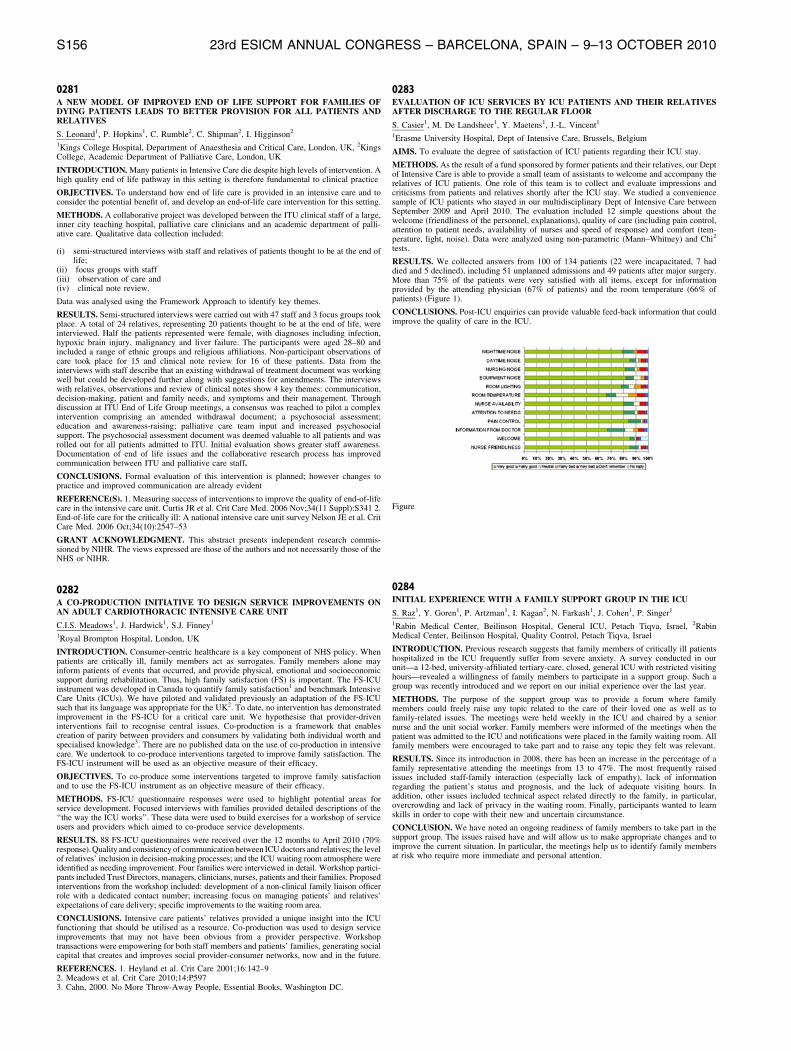
0281A NEW MODEL OF IMPROVED END OF LIFE SUPPORT FOR FAMILIES OFDYING PATIENTS LEADS TO BETTER PROVISION FOR ALL PATIENTS ANDRELATIVES
S. Leonard1, P. Hopkins1, C. Rumble2, C. Shipman2, I. Higginson2
1Kings College Hospital, Department of Anaesthesia and Critical Care, London, UK, 2KingsCollege, Academic Department of Palliative Care, London, UK
INTRODUCTION. Many patients in Intensive Care die despite high levels of intervention. Ahigh quality end of life pathway in this setting is therefore fundamental to clinical practice
OBJECTIVES. To understand how end of life care is provided in an intensive care and toconsider the potential benefit of, and develop an end-of-life care intervention for this setting.
METHODS. A collaborative project was developed between the ITU clinical staff of a large,inner city teaching hospital, palliative care clinicians and an academic department of palli-ative care. Qualitative data collection included:
(i) semi-structured interviews with staff and relatives of patients thought to be at the end oflife;
(ii) focus groups with staff(iii) observation of care and(iv) clinical note review.
Data was analysed using the Framework Approach to identify key themes.
RESULTS. Semi-structured interviews were carried out with 47 staff and 3 focus groups tookplace. A total of 24 relatives, representing 20 patients thought to be at the end of life, wereinterviewed. Half the patients represented were female, with diagnoses including infection,hypoxic brain injury, malignancy and liver failure. The participants were aged 28–80 andincluded a range of ethnic groups and religious affiliations. Non-participant observations ofcare took place for 15 and clinical note review for 16 of these patients. Data from theinterviews with staff describe that an existing withdrawal of treatment document was workingwell but could be developed further along with suggestions for amendments. The interviewswith relatives, observations and review of clinical notes show 4 key themes: communication,decision-making, patient and family needs, and symptoms and their management. Throughdiscussion at ITU End of Life Group meetings, a consensus was reached to pilot a complexintervention comprising an amended withdrawal document; a psychosocial assessment;education and awareness-raising; palliative care team input and increased psychosocialsupport. The psychosocial assessment document was deemed valuable to all patients and wasrolled out for all patients admitted to ITU. Initial evaluation shows greater staff awareness.Documentation of end of life issues and the collaborative research process has improvedcommunication between ITU and palliative care staff.
CONCLUSIONS. Formal evaluation of this intervention is planned; however changes topractice and improved communication are already evident
REFERENCE(S). 1. Measuring success of interventions to improve the quality of end-of-lifecare in the intensive care unit. Curtis JR et al. Crit Care Med. 2006 Nov;34(11 Suppl):S341 2.End-of-life care for the critically ill: A national intensive care unit survey Nelson JE et al. CritCare Med. 2006 Oct;34(10):2547–53
GRANT ACKNOWLEDGMENT. This abstract presents independent research commis-sioned by NIHR. The views expressed are those of the authors and not necessarily those of theNHS or NIHR.
0282A CO-PRODUCTION INITIATIVE TO DESIGN SERVICE IMPROVEMENTS ONAN ADULT CARDIOTHORACIC INTENSIVE CARE UNIT
C.I.S. Meadows1, J. Hardwick1, S.J. Finney1
1Royal Brompton Hospital, London, UK
INTRODUCTION. Consumer-centric healthcare is a key component of NHS policy. Whenpatients are critically ill, family members act as surrogates. Family members alone mayinform patients of events that occurred, and provide physical, emotional and socioeconomicsupport during rehabilitation. Thus, high family satisfaction (FS) is important. The FS-ICUinstrument was developed in Canada to quantify family satisfaction1 and benchmark IntensiveCare Units (ICUs). We have piloted and validated previously an adaptation of the FS-ICUsuch that its language was appropriate for the UK2. To date, no intervention has demonstratedimprovement in the FS-ICU for a critical care unit. We hypothesise that provider-driveninterventions fail to recognise central issues. Co-production is a framework that enablescreation of parity between providers and consumers by validating both individual worth andspecialised knowledge3. There are no published data on the use of co-production in intensivecare. We undertook to co-produce interventions targeted to improve family satisfaction. TheFS-ICU instrument will be used as an objective measure of their efficacy.
OBJECTIVES. To co-produce some interventions targeted to improve family satisfactionand to use the FS-ICU instrument as an objective measure of their efficacy.
METHODS. FS-ICU questionnaire responses were used to highlight potential areas forservice development. Focused interviews with families provided detailed descriptions of the‘‘the way the ICU works’’. These data were used to build exercises for a workshop of serviceusers and providers which aimed to co-produce service developments.
RESULTS. 88 FS-ICU questionnaires were received over the 12 months to April 2010 (70%response). Quality and consistency of communication between ICU doctors and relatives; the levelof relatives’ inclusion in decision-making processes; and the ICU waiting room atmosphere wereidentified as needing improvement. Four families were interviewed in detail. Workshop partici-pants included Trust Directors, managers, clinicians, nurses, patients and their families. Proposedinterventions from the workshop included: development of a non-clinical family liaison officerrole with a dedicated contact number; increasing focus on managing patients’ and relatives’expectations of care delivery; specific improvements to the waiting room area.
CONCLUSIONS. Intensive care patients’ relatives provided a unique insight into the ICUfunctioning that should be utilised as a resource. Co-production was used to design serviceimprovements that may not have been obvious from a provider perspective. Workshoptransactions were empowering for both staff members and patients’ families, generating socialcapital that creates and improves social provider-consumer networks, now and in the future.
REFERENCES. 1. Heyland et al. Crit Care 2001;16:142–92. Meadows et al. Crit Care 2010;14:P5973. Cahn, 2000. No More Throw-Away People, Essential Books, Washington DC.
0283EVALUATION OF ICU SERVICES BY ICU PATIENTS AND THEIR RELATIVESAFTER DISCHARGE TO THE REGULAR FLOOR
S. Casier1, M. De Landsheer1, Y. Maetens1, J.-L. Vincent1
1Erasme University Hospital, Dept of Intensive Care, Brussels, Belgium
AIMS. To evaluate the degree of satisfaction of ICU patients regarding their ICU stay.
METHODS. As the result of a fund sponsored by former patients and their relatives, our Deptof Intensive Care is able to provide a small team of assistants to welcome and accompany therelatives of ICU patients. One role of this team is to collect and evaluate impressions andcriticisms from patients and relatives shortly after the ICU stay. We studied a conveniencesample of ICU patients who stayed in our multidisciplinary Dept of Intensive Care betweenSeptember 2009 and April 2010. The evaluation included 12 simple questions about thewelcome (friendliness of the personnel, explanations), quality of care (including pain control,attention to patient needs, availability of nurses and speed of response) and comfort (tem-perature, light, noise). Data were analyzed using non-parametric (Mann–Whitney) and Chi2
tests.
RESULTS. We collected answers from 100 of 134 patients (22 were incapacitated, 7 haddied and 5 declined), including 51 unplanned admissions and 49 patients after major surgery.More than 75% of the patients were very satisfied with all items, except for informationprovided by the attending physician (67% of patients) and the room temperature (66% ofpatients) (Figure 1).
CONCLUSIONS. Post-ICU enquiries can provide valuable feed-back information that couldimprove the quality of care in the ICU.
Figure
0284INITIAL EXPERIENCE WITH A FAMILY SUPPORT GROUP IN THE ICU
S. Raz1, Y. Goren1, P. Artzman1, I. Kagan2, N. Farkash1, J. Cohen1, P. Singer1
1Rabin Medical Center, Beilinson Hospital, General ICU, Petach Tiqva, Israel, 2RabinMedical Center, Beilinson Hospital, Quality Control, Petach Tiqva, Israel
INTRODUCTION. Previous research suggests that family members of critically ill patientshospitalized in the ICU frequently suffer from severe anxiety. A survey conducted in ourunit—a 12-bed, university-affiliated tertiary-care, closed, general ICU with restricted visitinghours—revealed a willingness of family members to participate in a support group. Such agroup was recently introduced and we report on our initial experience over the last year.
METHODS. The purpose of the support group was to provide a forum where familymembers could freely raise any topic related to the care of their loved one as well as tofamily-related issues. The meetings were held weekly in the ICU and chaired by a seniornurse and the unit social worker. Family members were informed of the meetings when thepatient was admitted to the ICU and notifications were placed in the family waiting room. Allfamily members were encouraged to take part and to raise any topic they felt was relevant.
RESULTS. Since its introduction in 2008, there has been an increase in the percentage of afamily representative attending the meetings from 13 to 47%. The most frequently raisedissues included staff-family interaction (especially lack of empathy), lack of informationregarding the patient’s status and prognosis, and the lack of adequate visiting hours. Inaddition, other issues included technical aspect related directly to the family, in particular,overcrowding and lack of privacy in the waiting room. Finally, participants wanted to learnskills in order to cope with their new and uncertain circumstance.
CONCLUSION. We have noted an ongoing readiness of family members to take part in thesupport group. The issues raised have and will allow us to make appropriate changes and toimprove the current situation. In particular, the meetings help us to identify family membersat risk who require more immediate and personal attention.
S156 23rd ESICM ANNUAL CONGRESS – BARCELONA, SPAIN – 9–13 OCTOBER 2010
Disease-related outcome: 0285–02980285OUTCOME OF CANCER PATIENTS ADMITTED TO CRITICAL CARE OVER A 5YEAR PERIOD FROM A UK ONCOLOGY TERTIARY REFERRAL CENTRE
P. Prashast1, A. Raithatha1, D.C. Bryden1
1Sheffield Teaching Hospitals NHS Foundation Trust, Critical Care Department, Sheffield,UK
INTRODUCTION. The SOAP study suggested outcomes of cancer patients admitted to ICUare similar to those without cancer in contrast to other reports .1 We wanted to compare thiswith our own experience.
OBJECTIVES. (1) To determine critical care and hospital outcome of patients withmalignancy referred to critical care in the previous 5 years. (2) To identify any factorsinfluencing treatment decisions and survival after admission.
METHODS. Retrospective chart review of patients undergoing treatment for malignancyadmitted to ICU for medical or surgical reasons from May 2006 to Feb 2010. Leukaemiapatients were not included as they are treated at a different hospital by a different group ofclinicians. Demographic information, tumour/treatment related factors e.g neutropenia, pre-admission status and critical care diagnoses e.g. sepsis, were collected in addition to patientoutcomes.
RESULTS. 38 patients were identified (2.8% of all critical care admissions). ITU mortalitywas 39.5% (n = 15), however only 31.6% (n = 12) survived to hospital discharge (compa-rable overall unit mortality: 20–25%, hospital mortality:30%). Hospital survivors wereyounger (median 54.5 vs. 62 years), and more non-survivors had pre-existing comorbidities,sepsis, ALI and required more organ support (all ns). There was no difference between thegroups regarding cancer treatment. Non-survivors had a longer stay in critical care andtreatment withdrawal/limitation decisions were more common suggesting these were oftenbased on lack of medical progress whilst on ICU rather than diagnostic nihilism.
CONCLUSIONS. Hospital mortality in patients with malignancy is higher in our specialistcentre than reported for a Europe wide cohort (68.4 vs. 27% overall and 41% in the medicalsubgroup). The majority of our patients were medical and not post-surgical unlike in theSOAP study. This may account for the greater mortality as a larger proportion of our patientshad ALI, sepsis, neutropenia and required inotropes. Numbers admitted to critical care aremuch smaller than the 15.1% reported in the SOAP study, suggesting some referral andadmission triaging by the oncologists and the ICU team. Our results are similar to singlecentre French (hospital survival rate 31.6 vs. 21.8%) and Brazilian studies (31.6 vs. 42%),although a majority of our patients did not receive mechanical ventilation.2,3 A diagnosis ofcancer or active treatment for it should not be the major determinant of critical care support,but the patient’s general premorbid status and the extent of organ failures appear to beimportant factors in decision making as for any other critical care patient. Figures from theSOAP study for non specialist centres do not appear to reflect the experience of specialistoncology centres.
REFERENCE(S). 1. Taccone et al. (2009) Critical Care, 13: R15 2.Lecuyer L (2007). Crit.Care med. 35:808–814.3. Marcio S (2010). Crit care med. 38(1), pp 9–15.
0286OUTCOMES AND PROGNOSTIC FACTORS IN PATIENTS WITH HAEMATO-LOGICAL MALIGNANCIES IN A SPECIALIST CANCER INTENSIVE CARE UNIT
G. Bird1, P. Gruber1, P. Farquhar-Smith1, R. Forrest1, R. Saini1
1The Royal Marsden NHS Foundation Trust, London, UK
INTRODUCTION. Historically there has been a negative perception of the prognosis forpatients with haematological malignancies requiring admission to the intensive care unit(ICU). However, advances in chemotherapeutic regimes and haematopoietic stem celltransplantation (HSCT), along with improved monitoring and supportive measures havesuggested that outcomes for these patients have improved [1]. Establishing key prognosticindicators predictive of outcome may be useful in identifying patients most likely to benefitfrom ICU therapy. The aim of this study was to describe clinical outcomes and identifyprognostic factors in patients with haematological malignancy requiring admission to ICU.
METHODS. Following research approval, a retrospective cohort study was undertaken in a12-bedded specialist cancer ICU over a 5-year period (October 2004–September 2009).Recorded patient variables included demographics, haematological diagnosis, reason for ICUadmission, HSCT, APACHE II, admission laboratory data, number of organ failure, use ofinvasive mechanical ventilation, renal replacement therapy (RRT) and vasopressors. Theprimary outcome was in-hospital mortality. Key prognostic variables in determining in-hospital mortality were identified using univariate and multivariate analysis.
RESULTS. 199 patients with haematological malignancies were admitted to the ICU duringthe study period: mean age 55.3 (SD 15.6); 43.7% female; haematological diagnosis (45.7%leukaemia, 40.2% lymphoma, and 13.6% myeloma); 54.2% emergency admissions and 43.9%were post-HSCT. Mean APACHE II was 21.6 (SD 7.4), mean number of organ failures 2.3(SD 1.7), 52% required invasive mechanical ventilation, 41.4% RRT and 51.6% vasopressortherapy in the first 24 h of ICU admission. ICU, in-hospital and 6-month mortality were 32.7,45.2 and 60% respectively. Significantly higher mortalities were seen in patients who weremechanically ventilated (65 vs. 25% non-ventilated patients p \ 0.001), on vasopressorsupport (57 vs. 25% no vasopressor support p \ 0.001), neutropenic (52 vs. 45% non-neu-tropenic p \ 0.001) and in multi-organ failure defined as C2 organ failures (84 deaths vs. 15deaths in patients with B1 organ failure, p \ 0.001). Univariate analysis revealed mechanicalventilation, vasopressor support, albumin \20 g/l, neutropenia, platelet count \50 9 109/land multi-organ failure were all significant with p values \0.001, \0.001, 0.003, \0.001,0.015 and\0.001 respectively. Multivariate analysis revealed that multi-organ failure was theonly independent prognostic predictor of in-hospital mortality.
CONCLUSION. Mechanical ventilation, APACHE II, vasopressor support, albumin\20 g/l,neutropenia, platelets \50 9 109/l and multi-organ failure all had a significant associationwith mortality; however multi-organ failure was the only independent factor that predictedpoor outcome.
REFERENCE. 1. Hampshire PA, Welch CA, McCrossan LA, et al. Crit Care2009;13(4):R137.
0287GENDER DIFFERENCES IN RISK FACTORS, ANGIOGRAPHIC SEVERITY,TREATMENT AND IN-HOSPITAL MORTALITY OF PATIENTS WITH ST ELE-VATION MYOCARDIAL INFARCT (STEMI)
C.Y.C. Michael1, A. Vasu1, S. Eillyne1
1Tan Tock Seng Hospital, Emergency Department, Singapore, SingaporeINTRODUCTION. Coronary heart disease is the leading cause of mortality and morbidityfor both women and men. Although men are affected in greater numbers, women have beenshown to have worse outcomes and higher mortality.OBJECTIVES. This study aims to examine gender differences in risk factors, angiographicseverity, treatment and in-hospital mortality after STEMI.METHODS. In this retrospective study, the medical records of 619 patients with an admittingdiagnosis of STEMI from Tan Tock Seng Hospital, Emergency Department (TTSH ED)between 1st January 2009 and 31st December 2009 were reviewed. We extracted the datafrom the electronic records of the emergency case notes and inpatient discharge summaries.RESULTS. Of the 619 patients studied, 123 (19.9%) were women and 496 (80.1%) men. Fourhundred and forty-nine (72.5%) patients underwent coronary angiography. One hundred andseventy (27.5%) patients did not undergo coronary angiography, majority (65.3%) were elderlyaged C65 years (men 34.1% and women 31.2%). Between women and men, there was nosignificant difference between the number and distribution of diseased coronary vessels(including triple vessel and left main stem diseases). Regardless of age, men were frequentlytreated with a coronary artery stent (67.9%). Elderly women (aged C65 years) were more oftentreated conservatively (67%) while those younger women (aged B64 years) were frequentlytreated with a coronary artery stent (62.1%). In-hospital mortality rate was significantly higherfor women than men (6.5 vs. 3.8%, p = 0.013). Amongst the patients treated conservatively,elderly women had the highest in-hospital mortality when compared to the other patients(women C 65 years 19.2 vs. women B64 years 2.7%; men C65 years 13 vs. men B64 years6.8%). Compared to men, women were significantly older (p \ 0.001; 95% CI 12.6–17.6), morelikely to have a history of hypertension (73.2 vs. 50.2%; p \ 0.001), diabetes (45.5 vs. 28.2%;p \ 0.001), hyperlipidemia (63.4 vs. 46.6%; p = 0.001), peripheral vascular (5.7 vs. 1.2%;p = 0.006) or ischemic heart diseases (28.5 vs. 15.7%; p = 0.002) and less likely to be smokers(12.2 vs. 57.7%; p \ 0.001) or consume alcohol (0 vs. 10.1%; p \ 0.001).
CONCLUSIONS. Elderly women who were treated conservatively had the highest in-hos-pital mortality during the early management of STEMI.
0288EARLY ADMISSION TO THE INTENSIVE CARE UNIT IN HIGH RISK ACUTEMYELOID LEUKEMIA (AML) PATIENTS
E. Lengline1,2, E. Raffoux2, V. Peigne1, V. Lemiale1, N. Boissel2, M. Darmon1, B.
Schlemmer1, H. Dombret2, E. Azoulay1
1Hopital Saint-Louis, AP-HP, Paris Diderot University, Medical Intensive Care Unit, Paris, France,2Hopital Saint-Louis, AP-HP, Paris Diderot University, Hematology Department, Paris, FranceINTRODUCTION. AML is considered as an oncology emergency as a proportion of patientsexperience life threatening complications within the first hours or days after diagnosis. Earlydeath had been shown to be statistically related to high white blood cell (WBC) andmonoblastic leukemia1-3, with leukostasis and lysis syndrome as the most deadful events.OBJECTIVES. To evaluate the relationship between timing of ICU admission and outcomesin high risk AML patients at the earliest phase of the malignancy (before any chemotherapy)METHODS. Retrospective study in a tertiary care teaching hospital. Adult patients with newlydiagnoses AML from 1998 to 2008 were included. Patients admitted for an immediate life sus-taining therapy (ventilation, vasopressors or renal replacement therapy) were excluded. 42 patientsadmitted directly to the ICU (Early admission) were matched for age, WBC and FAB subtype with42 patients primarily admitted in hematology ward. Datasets were extracted from medical charts.RESULTS. 84 patients were included (42 early admitted to the ICU and 42 admitted first to thewards). Median follow up was 10.3 months. Median age was 46.5 years (36–57). FAB M4 or M5was retrieved in 58% of the patients. Karyotype was favorable for 30% and poor for 19%. MedianWBC was 103 9 109 L-1. No statistical difference was seen for demographic and hematologicalparameters between early admitted patients and matched controls. Among the 42 patients admittedfirst to the wards (controls), 20 were subsequently admitted to the ICU (Lately admitted) and 22remained in ward during the entire treatment course (Never admitted). The median time betweendiagnostic and ICU admission of this last group was 4 (1–9) days. Strikingly, patients latelyadmitted had more frequently dyspnea,oxygen requirement, high respiratory rate, low diastolicarterial pressure and lower first 24 h urine output. Lately admitted patients were less likely toreceive the complete dose of induction chemotherapy (68 vs. 88%) Furthermore, Late admissionresulted in increased use of invasive mechanical ventilation (60 vs. 33%) and vaso-active drugs (60vs. 16%). These differences resulted in longer stay in ICU and decreased survival.CONCLUSION. Patients at the earliest phase of High risk AML who are lately admitted tothe ICU experience worse outcomes, with increased use of life-sustaining therapies and highermortality, compared to patients early admitted to the ICU. Physiologic parameters at the timeof AML diagnosis such as respiratory rate, diastolic blood pressure, SpO2, or oxygen need arelikely to help clinicians distinguish those patients at risk of late ICU admission and sub-sequent adverse outcomes. Studies are needed to assess the right place for newly diagnosedAML with physiological abnormalities but no organ dysfunction.
REFERENCES.1. Creutzig U et al. (2004) J Clin Oncol 22:4384–932. Thakkar SG et al. (2008) Cancer 112:2233–403. Azoulay E et al. (2003) Am J Respir Crit Care Med 167:1329–33
23rd ESICM ANNUAL CONGRESS – BARCELONA, SPAIN – 9–13 OCTOBER 2010 S157
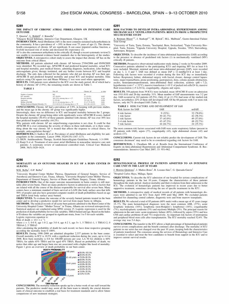
0289THE IMPACT OF CHRONIC ATRIAL FIBRILLATION ON INTENSIVE CAREOUTCOME
T. Quasim1, L. Riddell1, J. Kinsella1
1Glasgow Royal Infirmary, Intensive Care Department, Glasgow, UKINTRODUCTION. Atrial fibrillation (AF) is the most common sustained tachyarrhythmia inthe community. It has a prevalence of *10% in those over 75 years of age (1). The chronichealth consequences of chronic AF are significant. It can cause impaired cardiac function, afivefold increased risk of stroke and decreased life expectancy (2).AF is also the commonest arrhythmia in the critically ill, though a recent systematic review(3)was unable to recommend evidence based standards due to the heterogeneity of the studies.OBJECTIVES. A retrospective cohort study to assess the impact that chronic AF has on theoutcome from critical illness.METHODS. All patients admitted with chronic AF between 27/04/2006 and 05/09/2009were identified. We recorded age, APACHE II and predicted hospital mortality, actual ICUand hospital mortality, past medical history, admitting diagnosis, medication, echo findings,anticoagulants given, therapy instituted, and any further events between ICU and hospitaldischarge. The only data collected for the patients who did not develop AF was their age,APACHE II and predicted hospital mortality and actual ICU and hospital mortality. Dataanalysis using Chi square test and Mann–Whitney U test were used where appropriate.RESULTS. 1168 patients were admitted to the ICU over the study period, 46 of which had ahistory of chronic AF (3.9%), the remaining results are shown in Table 1.
TABLE 1Non AF Chronic AF p-value
Number of patients 965 46
ICU mortality 257 (26.6%) 11 (23.9%) 0.683
Hospital mortality 309 (32%) missing data n = 27 14 (30.4%) 0.724
Age(years) 52 (38–66) 70.5 (62–76) 0.000
Median APACHE 18 (13–24) missing data n = 79 25 (17.75–28.25) 0.001
Median Predicted Hospital Mortality (%) 27.4 (10.52–51.33) missing data n = 79 44.2 (26.1–63.25) 0.001
CONCLUSIONS. Chronic AF had a prevalence of 3.9%, in keeping with previous studies,and the mean age in the chronic AF group was significantly higher.Interestingly, there was no difference in ICU and hospital mortality between the 2 groups.Despite the chronic AF group being older with significantly worse APACHE II scores. Indeedthe hospital mortality (30.4%) of those patients admitted with chronic AF was over 30% lessthan predicted hospital mortality (44.2%).Why patients with chronic AF are outperforming expectation is not clear. It could be thatAPACHE II is over estimating the severity of illness in these individuals, or is there some-thing about the way chronic AF is treated that affects the response to critical illness, forexample, anticoagulation therapy?REFERENCES.(1) Sudlow M et al. Prevalence of atrial fibrillation and eligibility for anti-coagulants in the community. Lancet 1998; 352(9135):1167–1171.(2) SIGN. Antithrombotic Therapy. Scottish Intercollegiate Guidelines Network. 1-3-1999.(3) Kanji S et al. Treatment of new-onset atrial fibrillation in noncardiac intensive care unit
patients: A systematic review of randomized controlled trials. Critical Care Medicine2008; 36(5):1620–1624.
0290MORTALITY AS AN OUTCOME MEASURE IN ICU OF A BURN CENTER INALBANIA
M.K. Belba1, G.P. Belba2
1University Hospital Center Mother Theresa, Department of General Surgery, Service ofAnesthesia and Intensive Care, Tirana, Albania, 2University Hospital Center Mother Theresa,Department of General Surgery, Service of Burns and Plastic Surgery, Tirana, AlbaniaINTRODUCTION. One of the major outcome measurements in burns centers is still mor-tality after severe burns. There are many predictive factors in admission as well as factors thatare related with all the course of the disease responsible for survival after severe burn. Manycenters have a minimum standard of burn survival or LA50 (the body surface area that kills50% of people) and also have generated computer models of death probabilities based on ageand TBSA (total body surface area) burned.OBJECTIVES. To evaluate the outcome of the severely burned patients treated in the burncenter and to develop a predictive model for survival from major burns in Albania.METHODS. The medical records of all acute burn patients admitted to the Burn Center of theUniversity Hospital Center ‘‘Mother Teresa’’ in Tirana, Albania are reviewed retrospectively.Statistical analyses are conducted using SPSS version 15. Logistic regression is used for theprediction of death probability for two risk variables, TBSA burned and age. Based on the Indexof Evidence the variables are grouped in significant strata, from 1 to 5 for each variable.Logistic regression equation is:Risk of death = 1/(1 + e-z)where z = 3, 0-0 8, age 1-0 2, age 3-0 4, age 4-2 1, age 5+ 1, 5 TBSA1-1, 1 TBSA3-2, 0TBSA4-2, 5 TBSA5After calculating the probability of death for each record, we have done respective groupingaccording the mortality from 0–100%.RESULTS. During 1998–2008 are admitted altogether 2,337 patients in the burn center.Overall mortality in ICU is 10.5% with a significant reduction during the years, up to 4.4 in2008. Row burn mortality is 0.8 for 100,000 persons per year. LA 50 for children is 60%TBSA; for adults 65% TBSA and for aged 45% TBSA. Based on probability of death, wenotice that older age and larger burn size are associated with a higher like hood of mortality.Figure 1 gives an overview of death probability in our burn center.
CONCLUSIONS. The mortality reduction speaks up for a better work of our staff toward thepatients. The predictive model may assist all the burn team to identify the crucial determi-nants of clinical outcome to establish a real basis for treatment standards and to allow futurecomparisons of new treatment strategies.
0291RISK FACTORS TO DEVELOP INTRA-ABDOMINAL HYPERTENSION AMONGMECHANICALLY VENTILATED PATIENTS: RESULTS FROM A PROSPECTIVEMULTICENTRE STUDY
A. Reintam Blaser1,2, J. Starkopf1,3, M. Bjorck4, M.L. Malbrain5, Gastro-Intestinal FailureTrial Group1University of Tartu, Tartu, Estonia, 2Inselspital, Bern, Switzerland, 3Tartu University Hos-pital, Tartu, Estonia, 4Uppsala University Hospital, Uppsala, Sweden, 5ZNA Stuivenberg,Antwerpen, Belgium
OBJECTIVES. To describe the incidence of intra-abdominal hypertension (IAH) accordingto the presence or absence of predefined risk factors (1) in mechanically ventilated (MV)critically ill patients.
METHODS. Prospective observational multicenter study during 2 weeks in November 2009.Consecutive patients admitted to the participating ICUs and requiring MV for at least 6 hwere included. Maximal, minimal and mean intra-abdominal pressure (IAP), were recordedon day 1, 2, 4 and 7. IAH was defined as mean IAP C 12 mmHg/24 h at least 1 day.Following risk factors were recorded if evident during the first ICU day or immediatelybefore: Respiratory failure, abdominal surgery with fascial closure, damage control lapara-tomy, major trauma/burns, prone positioning, gastroparesis, ileus, colonic pseudo-obstruction,ascites, hemo/pneumoperitoneum, intra-abdominal fluid collection, acidosis (pH \ 7.2),hypothermia (core t� \ 33�C), massive transfusion ([10 U of packed red cells/24 h), massivefluid resuscitation ([5 L/24 h), coagulopathy, oliguria and sepsis.
RESULTS. 358 patients from 39 ICUs were included; mean APACHE II score on admissionwas 19.8 (8.0) and 28-day mortality 31%. Mean number of IAP measurements was 3.2 perday. IAH occurred in 163 patients (45.5%). Only 41 pt (11.5%) had none of the studied riskfactors, nevertheless 22% of them still developed IAH. Of the 68 patients with 5 or more riskfactors, only 64.7% developed IAH (Table 1).
TABLE 1 RISK FACTORS AND DEVELOPMENT OF IAH
Risk factors for IAH IAH noIAH
0 risk factors 9 (22%) 32 (78.0%)1 risk factor 30 (41.7%) 42 (58.3%)2 risk factors 37 (43.0%) 49 (57.0%)3–4 risk factors 43 (47.3%) 48 (52.7%)5 risk factors 44 (64.7%) 24 (35.3%)
The most common risk factors associated with IAH were acute respiratory failure (present in85 patients with IAH), sepsis (77), coagulopathy (43), tight abdominal closure (42) andacidosis (37).
CONCLUSIONS. Current risk factors do not reliably predict the development of IAH. Theconsensus document1 may need to be re-considered regarding risk factors for IAH.
REFERENCE(S). 1. Cheatham ML et al. Results from the International Conference ofExperts on Intra-abdominal Hypertension and Abdominal Compartment Syndrome. II. Rec-ommendations. Intensive Care Med 2007; 33: 951–962.
0292HEMATOLOGICAL PROFILE OF PATIENTS ADMITTED TO AN INTENSIVECARE UNIT IN THE LAST 10 YEARS
J. Moreno-Quintana1, J. Munoz-Bono1, R. Lozano-Saez1, G. Quesada-Garcıa1
1Hospital Carlos Haya, Malaga, Spain
OBJECTIVES. To describe the ICU admission of our hospital for serious complications ofhematology patients in the last 10 years. Compare the characteristics of these patientsthroughout the study period. Analyze mortality and their evolution from their admission to theICU. The evolution of hematologic patients has improved in recent years due to bettersupportive treatment, sometimes involving the use of specific treatments in the ICU.
METHODS. A retrospective study of medical records of all patients with hematologic dis-eases were admitted to our ICU from April 1999 until May 2009. We excluded patientsadmitted for channeling central catheter, diagnostic tests and bone marrow transplants.
RESULTS. We selected a total of 85 patients (60% male) with a mean age of 45 years (range15–77). The main hematological diagnoses were the most common AML (37%), acutelymphatic leukemia (16%), lymphoma (non-Hodgkin’s lymphoma) (16%), coagulopathy(7%), myelodysplastic syndrome (5%) and myeloma Multiple (5%). The principal reason foradmission in the unit were: acute respiratory failure (40%), followed by sepsis (35%) and lessCNS and cardiac problems (8 and 7%) respectively. As important risk factors of neutropeniaand peripheral blood stem cells after transplantation. The ICU mortality reached 52.6%. Theaverage stay was 5.4 days.
CONCLUSIONS. The transfer to the ICU allows a high percentage of hematological patientssurvive severe complications and the benefit continues after discharge. The mortality of ICUpatients in our series has not changed over the past 10 years, keeping both the characteristicsof patients transferred. The consensus among the services of Hematology and Intensive Careis essential to select and treat the best candidates to benefit from support in the ICU and toimprove current survival results.
S158 23rd ESICM ANNUAL CONGRESS – BARCELONA, SPAIN – 9–13 OCTOBER 2010
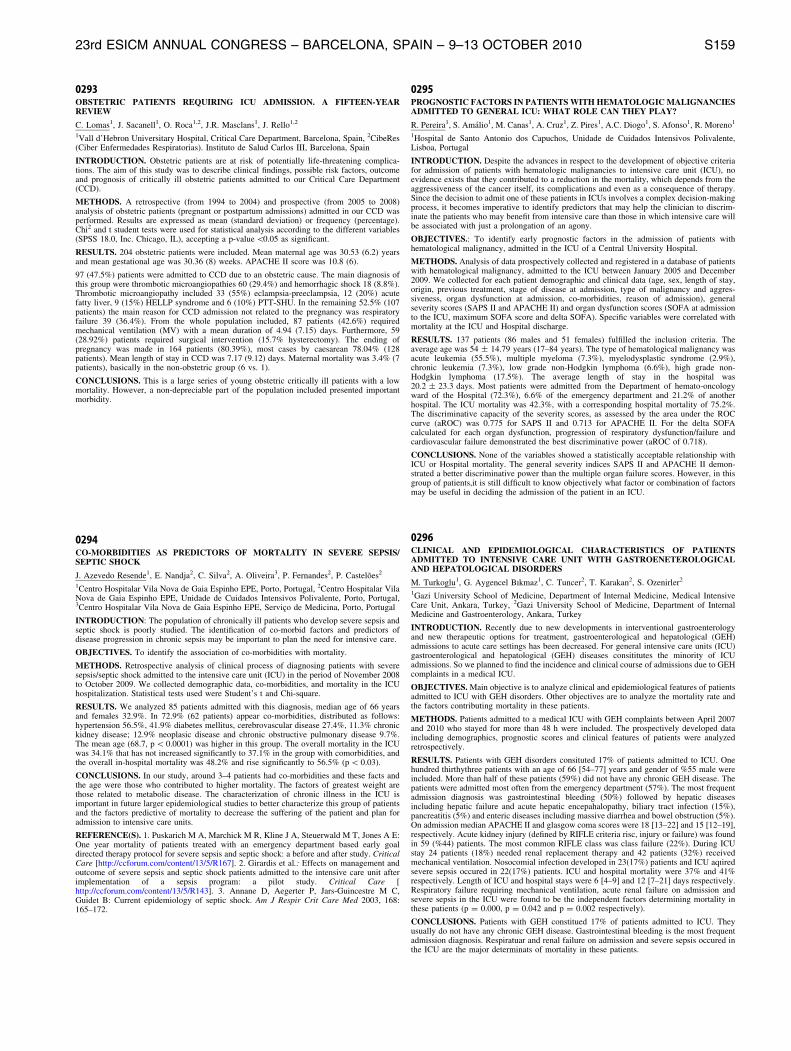
0293OBSTETRIC PATIENTS REQUIRING ICU ADMISSION. A FIFTEEN-YEARREVIEW
C. Lomas1, J. Sacanell1, O. Roca1,2, J.R. Masclans1, J. Rello1,2
1Vall d’Hebron Universitary Hospital, Critical Care Department, Barcelona, Spain, 2CibeRes(Ciber Enfermedades Respiratorias). Instituto de Salud Carlos III, Barcelona, Spain
INTRODUCTION. Obstetric patients are at risk of potentially life-threatening complica-tions. The aim of this study was to describe clinical findings, possible risk factors, outcomeand prognosis of critically ill obstetric patients admitted to our Critical Care Department(CCD).
METHODS. A retrospective (from 1994 to 2004) and prospective (from 2005 to 2008)analysis of obstetric patients (pregnant or postpartum admissions) admitted in our CCD wasperformed. Results are expressed as mean (standard deviation) or frequency (percentage).Chi2 and t student tests were used for statistical analysis according to the different variables(SPSS 18.0, Inc. Chicago, IL), accepting a p-value \0.05 as significant.
RESULTS. 204 obstetric patients were included. Mean maternal age was 30.53 (6.2) yearsand mean gestational age was 30.36 (8) weeks. APACHE II score was 10.8 (6).
97 (47.5%) patients were admitted to CCD due to an obstetric cause. The main diagnosis ofthis group were thrombotic microangiopathies 60 (29.4%) and hemorrhagic shock 18 (8.8%).Thrombotic microangiopathy included 33 (55%) eclampsia-preeclampsia, 12 (20%) acutefatty liver, 9 (15%) HELLP syndrome and 6 (10%) PTT-SHU. In the remaining 52.5% (107patients) the main reason for CCD admission not related to the pregnancy was respiratoryfailure 39 (36.4%). From the whole population included, 87 patients (42.6%) requiredmechanical ventilation (MV) with a mean duration of 4.94 (7.15) days. Furthermore, 59(28.92%) patients required surgical intervention (15.7% hysterectomy). The ending ofpregnancy was made in 164 patients (80.39%), most cases by caesarean 78.04% (128patients). Mean length of stay in CCD was 7.17 (9.12) days. Maternal mortality was 3.4% (7patients), basically in the non-obstetric group (6 vs. 1).
CONCLUSIONS. This is a large series of young obstetric critically ill patients with a lowmortality. However, a non-depreciable part of the population included presented importantmorbidity.
0294CO-MORBIDITIES AS PREDICTORS OF MORTALITY IN SEVERE SEPSIS/SEPTIC SHOCK
J. Azevedo Resende1, E. Nandja2, C. Silva2, A. Oliveira3, P. Fernandes2, P. Casteloes2
1Centro Hospitalar Vila Nova de Gaia Espinho EPE, Porto, Portugal, 2Centro Hospitalar VilaNova de Gaia Espinho EPE, Unidade de Cuidados Intensivos Polivalente, Porto, Portugal,3Centro Hospitalar Vila Nova de Gaia Espinho EPE, Servico de Medicina, Porto, Portugal
INTRODUCTION: The population of chronically ill patients who develop severe sepsis andseptic shock is poorly studied. The identification of co-morbid factors and predictors ofdisease progression in chronic sepsis may be important to plan the need for intensive care.
OBJECTIVES. To identify the association of co-morbidities with mortality.
METHODS. Retrospective analysis of clinical process of diagnosing patients with severesepsis/septic shock admitted to the intensive care unit (ICU) in the period of November 2008to October 2009. We collected demographic data, co-morbidities, and mortality in the ICUhospitalization. Statistical tests used were Student’s t and Chi-square.
RESULTS. We analyzed 85 patients admitted with this diagnosis, median age of 66 yearsand females 32.9%. In 72.9% (62 patients) appear co-morbidities, distributed as follows:hypertension 56.5%, 41.9% diabetes mellitus, cerebrovascular disease 27.4%, 11.3% chronickidney disease; 12.9% neoplasic disease and chronic obstructive pulmonary disease 9.7%.The mean age (68.7, p \ 0.0001) was higher in this group. The overall mortality in the ICUwas 34.1% that has not increased significantly to 37.1% in the group with comorbidities, andthe overall in-hospital mortality was 48.2% and rise significantly to 56.5% (p \ 0.03).
CONCLUSIONS. In our study, around 3–4 patients had co-morbidities and these facts andthe age were those who contributed to higher mortality. The factors of greatest weight arethose related to metabolic disease. The characterization of chronic illness in the ICU isimportant in future larger epidemiological studies to better characterize this group of patientsand the factors predictive of mortality to decrease the suffering of the patient and plan foradmission to intensive care units.
REFERENCE(S). 1. Puskarich M A, Marchick M R, Kline J A, Steuerwald M T, Jones A E:One year mortality of patients treated with an emergency department based early goaldirected therapy protocol for severe sepsis and septic shock: a before and after study. CriticalCare [http://ccforum.com/content/13/5/R167]. 2. Girardis et al.: Effects on management andoutcome of severe sepsis and septic shock patients admitted to the intensive care unit afterimplementation of a sepsis program: a pilot study. Critical Care [http://ccforum.com/content/13/5/R143]. 3. Annane D, Aegerter P, Jars-Guincestre M C,Guidet B: Current epidemiology of septic shock. Am J Respir Crit Care Med 2003, 168:165–172.
0295PROGNOSTIC FACTORS IN PATIENTS WITH HEMATOLOGIC MALIGNANCIESADMITTED TO GENERAL ICU: WHAT ROLE CAN THEY PLAY?
R. Pereira1, S. Amalio1, M. Canas1, A. Cruz1, Z. Pires1, A.C. Diogo1, S. Afonso1, R. Moreno1
1Hospital de Santo Antonio dos Capuchos, Unidade de Cuidados Intensivos Polivalente,Lisboa, Portugal
INTRODUCTION. Despite the advances in respect to the development of objective criteriafor admission of patients with hematologic malignancies to intensive care unit (ICU), noevidence exists that they contributed to a reduction in the mortality, which depends from theaggressiveness of the cancer itself, its complications and even as a consequence of therapy.Since the decision to admit one of these patients in ICUs involves a complex decision-makingprocess, it becomes imperative to identify predictors that may help the clinician to discrim-inate the patients who may benefit from intensive care than those in which intensive care willbe associated with just a prolongation of an agony.
OBJECTIVES.: To identify early prognostic factors in the admission of patients withhematological malignancy, admitted in the ICU of a Central University Hospital.
METHODS. Analysis of data prospectively collected and registered in a database of patientswith hematological malignancy, admitted to the ICU between January 2005 and December2009. We collected for each patient demographic and clinical data (age, sex, length of stay,origin, previous treatment, stage of disease at admission, type of malignancy and aggres-siveness, organ dysfunction at admission, co-morbidities, reason of admission), generalseverity scores (SAPS II and APACHE II) and organ dysfunction scores (SOFA at admissionto the ICU, maximum SOFA score and delta SOFA). Specific variables were correlated withmortality at the ICU and Hospital discharge.
RESULTS. 137 patients (86 males and 51 females) fulfilled the inclusion criteria. Theaverage age was 54 ± 14.79 years (17–84 years). The type of hematological malignancy wasacute leukemia (55.5%), multiple myeloma (7.3%), myelodysplastic syndrome (2.9%),chronic leukemia (7.3%), low grade non-Hodgkin lymphoma (6.6%), high grade non-Hodgkin lymphoma (17.5%). The average length of stay in the hospital was20.2 ± 23.3 days. Most patients were admitted from the Department of hemato-oncologyward of the Hospital (72.3%), 6.6% of the emergency department and 21.2% of anotherhospital. The ICU mortality was 42.3%, with a corresponding hospital mortality of 75.2%.The discriminative capacity of the severity scores, as assessed by the area under the ROCcurve (aROC) was 0.775 for SAPS II and 0.713 for APACHE II. For the delta SOFAcalculated for each organ dysfunction, progression of respiratory dysfunction/failure andcardiovascular failure demonstrated the best discriminative power (aROC of 0.718).
CONCLUSIONS. None of the variables showed a statistically acceptable relationship withICU or Hospital mortality. The general severity indices SAPS II and APACHE II demon-strated a better discriminative power than the multiple organ failure scores. However, in thisgroup of patients,it is still difficult to know objectively what factor or combination of factorsmay be useful in deciding the admission of the patient in an ICU.
0296CLINICAL AND EPIDEMIOLOGICAL CHARACTERISTICS OF PATIENTSADMITTED TO INTENSIVE CARE UNIT WITH GASTROENETEROLOGICALAND HEPATOLOGICAL DISORDERS
M. Turkoglu1, G. Aygencel Bıkmaz1, C. Tuncer2, T. Karakan2, S. Ozenirler2
1Gazi University School of Medicine, Department of Internal Medicine, Medical IntensiveCare Unit, Ankara, Turkey, 2Gazi University School of Medicine, Department of InternalMedicine and Gastroenterology, Ankara, Turkey
INTRODUCTION. Recently due to new developments in interventional gastroenterologyand new therapeutic options for treatment, gastroenterological and hepatological (GEH)admissions to acute care settings has been decreased. For general intensive care units (ICU)gastroenterological and hepatological (GEH) diseases consititutes the minority of ICUadmissions. So we planned to find the incidence and clinical course of admissions due to GEHcomplaints in a medical ICU.
OBJECTIVES. Main objective is to analyze clinical and epidemiological features of patientsadmitted to ICU with GEH disorders. Other objectives are to analyze the mortality rate andthe factors contributing mortality in these patients.
METHODS. Patients admitted to a medical ICU with GEH complaints between April 2007and 2010 who stayed for more than 48 h were included. The prospectively developed dataincluding demographics, prognostic scores and clinical features of patients were analyzedretrospectively.
RESULTS. Patients with GEH disorders consituted 17% of patients admitted to ICU. Onehundred thirthythree patients with an age of 66 [54–77] years and gender of %55 male wereincluded. More than half of these patients (59%) did not have any chronic GEH disease. Thepatients were admitted most often from the emergency department (57%). The most frequentadmission diagnosis was gastrointestinal bleeding (50%) followed by hepatic diseasesincluding hepatic failure and acute hepatic encepahalopathy, biliary tract infection (15%),pancreatitis (5%) and enteric diseases including massive diarrhea and bowel obstruction (5%).On admission median APACHE II and glasgow coma scores were 18 [13–22] and 15 [12–19],respectively. Acute kidney injury (defined by RIFLE criteria risc, injury or failure) was foundin 59 (%44) patients. The most common RIFLE class was class failure (22%). During ICUstay 24 patients (18%) needed renal replacement therapy and 42 patients (32%) receivedmechanical ventilation. Nosocomial infection developed in 23(17%) patients and ICU aqıiredsevere sepsis occured in 22(17%) patients. ICU and hospital mortality were 37% and 41%respectively. Length of ICU and hospital stays were 6 [4–9] and 12 [7–21] days respectively.Respiratory failure requiring mechanical ventilation, acute renal failure on admission andsevere sepsis in the ICU were found to be the independent factors determining mortality inthese patients (p = 0.000, p = 0.042 and p = 0.002 respectively).
CONCLUSIONS. Patients with GEH constitued 17% of patients admitted to ICU. Theyusually do not have any chronic GEH disease. Gastrointestinal bleeding is the most frequentadmission diagnosis. Respiratuar and renal failure on admission and severe sepsis occured inthe ICU are the major determinats of mortality in these patients.
23rd ESICM ANNUAL CONGRESS – BARCELONA, SPAIN – 9–13 OCTOBER 2010 S159
0297ANALYSIS OF MORTALITY IN ICU BY MULTICENTER STUDY SPANISH FORHEMATOLOGICAL PATIENTS ADMITTED TO ICU (EMEHU-GTEI)
M. Borges1, S. Sancho2, G. Muniz3, R. Granada4, J.I. Ayestaran5, J. Bonastre6, R. Zaragoza2,
J.R. Irureta7, Y. Diaz8, J.C. Ballesteros9, E. Quintana10, L. Gutierrez1, B. Balsera11, EMEHU-
GTEI1H. Son Llatzer, Palma de Mallorca, Spain, 2H. Peset, Valencia, Spain, 3H. UniversitarioCentral, Asturias, Spain, 4H. Belvitge, Barcelona, Spain, 5H. Son Dureta, Palma de Mallorca,Spain, 6H. La Fe, Valencia, Spain, 7H. Cruces, Vizcaya, Spain, 8H. del Mar, Barcelona, Spain,9H. Salamanca, Salamanca, Spain, 10H. San Pau, Barcelona, Spain, 11H. Arnau Vilanova,Lleida, Spain
INTRODUCTION. In recent years described series with hematological patients in ICU, butthese studies are often limited because they are retrospective, single center in a few patientsdivided over many years. To determine the characteristics of mortality in this group is veryimportant to assess their management in the ICU.
OBJECTIVE. To analyze prognostic factors associated with ICU mortality of patients (pts)with hematologic malignancies admitted to the intensive care unit (PTU).
METHOD. An observational, transversal, prospective, multicenter OCT conducted betweenJune 2007 and October 2008. We conducted a descriptive analysis, Chi-square, bivariate andlogistic regression including variables with a value of p \ 0.2 with the SAS statistical Pauetto assess the factors influencing mortality in ICU.
RESULTS. We included 450 patients from 34 ICUs. The mean age was 54 years. TheAPACHE II at admission was 22.89 ± 8.24 (CI 22.10–23.69) and the first day SOFA ± 4.349.24 (8.84–9.64). The crude mortality ICU was 51.6% (232 pts). We divide related mortalityby infectious etiology (61%) versus other causes (39%). In univariate analysis the variablessignificantly associated with mortality were: males p \ 0.01, hematology plant from 0.007,0.04 multiple myeloma, respiratory failure at admission \0.0001, tachycardia, 0.002, 0.001hypothermia, tachypnea 0 02, APACHE II C 20, more than two organ failures \0.0001,presence of ARDS \ 0.0001, invasive mechanical ventilation (IMV) \ 0.0001, NIV 0.0003,0.0002 transfusion (of all products: red cells, platelets and plasma), acquisition intraUCIinfection 0.009, 10 days longer stay in ICU 0.002. The presence of neutropenia was notassociated (p = 0.0533) at a significantly higher mortality, or personal history, septic shock,bone marrow transplant or other reasons for admission to the ICU. Table 1 describe theindependent factors associated with mortality in logistic regression analysis.
TABLE 1 MULTIVARIATE ANALYSIS OF MORTALITY
Variables Hazard ratio 95% CI p
Mechanical ventilation 11.71 6.16–22.25 \0.0001
ARDS Criteria (LIS [ 2.5) 2.44 1.23–4.83 0.0109
More than 2 organ failures 2.29 1.18–4.46 0.0146
Need for transfusion 1.94 1.06–3.55 0.0307
APACHE II C 20 1.81 1.03–3.19 0.0397
CONCLUSIONS. Variables classics such as septic shock or neutropenia not associated withmortality. And the independent variables associated with increased ICU mortality were: VM,ARDS, severity and need for transfusion of blood products.
REFERENCE(S). 1. Taccone et al. Crit Care. 2009; 2. Azoulay E et al. Medicine. 2004
0298A REVIEW OF CRITICAL CARE OUTCOMES IN ALL HAEMATOLOGYONCOLOGY PATIENTS ADMITTED TO THE JAMES COOK UNIVERSITYHOSPITAL INTENSIVE CARE UNIT
L. O’Connor1, D. Monkhouse1
1The James Cook University Hospital, Anaesthesia and Critical Care, Middlesbrough, UK
INTRODUCTION. Due to advances in haematology treatments such as bone marrowtransplant and intensive chemotherapy, haematology oncology patients have an improved lifeexpectancy. They are frequently subject to profound immunosuppression and physiologicalderangement requiring critical care support. With studies suggesting outcomes similar tothose of critically ill non-cancer patients, factors such as disease prognosis, chemotherapy andrenal replacement therapy (RRT) may play only minor predictive roles. The question stillremains: who benefits from ICU?
OBJECTIVES. To review the effect of haematological disease, organ failure and organsupport on mortality.
METHODS. A retrospective search was conducted of all adult level 3 admissions withhaematological malignancy from 2003 to 2010 using the Ward Watcher database.
RESULTS. 41 patients were identified. Major haematology diagnoses were acute leukaemia32%, NHL 22% and lymphoma 12%. Mean time to ICU admission was 13.4 days with 31%admitted within 24 h. The commonest reasons for ICU admission were respiratory failure49%, sepsis 33% and acute renal failure 7%. The mean number of organs supported was 2.9.65% of patients had C 3 organ failure. Mean APACHE II score was 24.5. Increasing organfailure correlated with increasing mortality. Patients with 4 or 5 organ failure had 100%mortality. Mean ICU stay was 10 days with 35% having an ICU stay of less than 48 h. ICUmortality rate was 62.7%. 46.5% received invasive ventilation, 16.3% failed non-invasiveventilation (NIV) and required invasive ventilation, 16.3% had NIV only and 21% receivedno respiratory support. Vasoactive support was given to 74.3% and RRT to 23.3%. Invasiveventilation and NIV were associated with a higher mortality; 90 and 60% versus 40% inspontaneously ventilating patients. Vasoactive support was associated with more organ fail-ure, longer ICU stay and higher mortality. RRT was associated with a higher mortality 90versus 54%. Patients with a documented poor haematological prognosis had a higher mor-tality but also more organs supported.
CONCLUSIONS. From this study invasive ventilation, cardiovascular support and multi-organ failure are strongly associated with increased mortality. The need for RRT was not anindependent predictor of mortality. Close collaboration is needed between the specialities toallow early resuscitation and critical care support to avoid delayed admissions with multiorgan failure.
REFERENCE(S). 1. Cuthbertson BH, Rajalingam Y, Harrison S, et al.The outcome ofhaematological malignancy in Scottish intensive care units. JICS 2008 9(2) 135–140. 2. Hampshire PA, Welch CA, McCrossan LA, et al. Admission factors asso-ciated with hospital mortality in patients with haematological malignancy admitted to UKadult, general critical care units: a secondary analysis of the ICNARC Case Mix ProgrammeDatabase. Critical Care 2009, 13:R137.
Cost and cost-effectiveness: 0299–03110299STRESS ULCER PROPHYLAXIS IN CRITICALLY ILL PATIENTS. A FINANCIALPERSPECTIVE
B.A. Oliveira1, A. Moghal2
1Mayday University Hospital, Intensive Care Medicine, London, UK, 2Mayday UniversityHospital, Intensive Care, London, UK
INTRODUCTION. The prescription of stress ulcer prophylaxis (SUP) in critically illpatients is relatively commonplace due to the association between physiological stress andgastrointestinal (G.I.) bleeding. However, recent guidelines recommend that only patientswho are mechanically ventilated, and/or have a coagulopathy warrant prophylaxis. They alsostate that histamine receptor blockers (H2RB’s) can be used primarily for SUP (1).
OBJECTIVES. The aim of this audit was to compare prescription practice of SUP at theITU/HDU at Mayday Hospital with those set out in the guidelines and to calculate thepotential cost savings resulting from following these guidelines.
METHODS. Data prospectively collected from consecutive admissions to Mayday HospitalITU/HDU between October and November 2009.
RESULTS. Data was collected on 56 patients, 3 had a G.I. bleed on admission, and 8 werealready on SUP so were excluded. 27 of 29 (96%) patients with major risk factors wereprescribed SUP, compared with 11 of 15 (73%) patients with no major risk factors. Protonpump inhibitors (PPI’s) were prescribed preferentially to H2RB’s; 31 versus 14. The cost ofSUP during the audit period was £5,646. If we had only prescribed it to those at high risk thecost would have been £5,079.86, and if we had only used H2RB’s the cost would have been£745.07.
CONCLUSIONS. Prescribing of SUP in our unit does not reflect Quenot’s guidelines. Thisnot only represents an increased cost but there are increased rates of nosocomial pneumoniaand C. Difficile diarrhoea associated with SUP (2,3). There is little evidence showing thesuperiority of PPI’s over H2RBs in the prophylaxis of bleeds; and there is evidence of anincreased rate of the aforementioned infections with PPI’s as compared to H2RB’s (3). Weprescribed PPI’s to a significant majority of our patients; however, it is our opinion that ourcurrent unit practice is not dissimilar to that of the rest of the UK. We would encourage allcritical care units to review their SUP prescribing as our results show that significant savingscan be made with judicious prescription of these drugs.
REFERENCE(S). 1. Quenot JP, Nadiejda T, Barbar, Saber. When should stress ulcer pro-phylaxis be used in the ICU? Current opinion in critical care 2009; 15 (2):139–1432. Dial S, Alrasadi K, Manoukian C, et al. Risk of Clostridium difficile diarrhea among
hospital inpatients prescribed proton pump inhibitors: cohort and case–control studies.CMAJ 2004; 171:33–38
3. Miano T, Reichert M, Houle T, MacGregor D, Kincaid E, Bowton D, Nosocomial Pneu-monia Risk and Stress Ulcer Prophylaxis A Comparison of Pantoprazole vs. Ranitidine inCardiothoracic Surgery patients. Chest 2009;136(2):440–447.
0300ANALYSIS OF DAILY DRUG COSTS ON THE CRITICAL CARE UNIT (CCU)
R. Shulman1, S. Ong2, I.P. Bates3
1University College London Hospitals NHS Foundation Trust, Pharmacy, London, UK,2Utrecht University, Faculty of Science, Pharmaceutical Sciences, Utrecht, Netherlands,3School of Pharmacy, University of London, London, UK
INTRODUCTION. Surprisingly little is published on the cost of drug treatment for criticallyill patients. Critical care is expensive, mainly due to the high staff ratio, expensive equipmentbut also due to a significant reliance on pharmacological management, which is usuallyfunded with a limited drug budget.
OBJECTIVES. To explore the relationship between drug expenditure, patient acuity andoutcome.
METHODS. Data was generated by retrospective analysis of consecutive patients admitted toour 35 bedded general adult ICU/HDU in a London teaching hospital, during February 2009.Patients were excluded from analysis if they were present in ICU for less than 24 h. The first andfinal CCU days stay were not included so that only full days were analysed. Daily drug-use perpatient was manually extracted from the computerized ICU management system (CIS, QS, GEMedical). Costs of prescribed drugs, fluids and parenteral nutrition (PN) were calculated fromthe pharmacy computer system and analyzed using regression analysis (SPSS ver18).
RESULTS. The patient characteristics and outcomes of the 82 patients are described inTable 1
TABLE 1 PATIENT CHARACTERISTICS AND OUTCOMESAge 63 (50–75)*
Gender (male n and %) 37 (45%)
APACHE II score 17.9 (8.5)**
TISS score/patient 15.9 (6.1)**
Length of CCU stay (days) 3 (2–6)*
CCU survival (n and %) 71 (86.6%)
Daily drug cost for each patient’s stay £28.2 (£17.9–61.5)*
Daily drug cost for CCU and hospital survivors n = 61 £24.5 (13.8–47.8)*
Daily drug cost for CCU survivors who died in hospital n = 10 £38.0 (25.2–73.6)*
Daily drug cost for CCU non-survivors n = 11 £74.1 (39.7–80.3)*
*Median (interquartile range), **mean (standard deviation)
The median daily drug cost was £28.2. Of note, the drug cost was highest for CCU non-survivors compared with survivors and also compared with patients who died in hospital afterCCU discharge (p = 0.01). Multivariate regression analysis demonstrated that median dailydrug cost/patient = -4.2 + 1.1 (mean TISS score) + 1.4 (APACHE II score), R2 = 35.6%;i.e median daily drug cost/patient was positively associated with TISS and APACHE II scoreexplaining 36% of the variation in cost seen.
CONCLUSIONS. This is the first study to show that daily drug expenditure in all generalCCU adult patients correlates with patient acuity. Median daily drug costs/patient were foundto be £28.2. This parameter would make an interesting comparison with other units bothnationally and internationally. Daily drug costs can be predicted on the basis of APACHE IIand TISS scores. Furthermore, this may be further refined to develop a quality marker of dailydrug cost in relation to survivors and non-survivors.
S160 23rd ESICM ANNUAL CONGRESS – BARCELONA, SPAIN – 9–13 OCTOBER 2010
0301BEDSIDE TRACHEOSTOMIES IN A NEUROLOGICAL ICU VS. OPERATINGROOM TRACHEOSTOMIES. IS THIS A SAFE PROCEDURE IN THE ICU?
E. Castillo-Lorente1, J.-F. Brea-Salvago1, F. Acebal-Balnco2, M.I. Ruiz-Garcıa1, A. Tejero-
Pedregosa1, L. Rucabado-Aguilar1
1Hospital Neurotraumatologico, Complejo Hospitalario de Jaen, Intensive Care Unit, Jaen,Spain, 2Hospital Neurotraumatologico, Complejo Hospitalario de Jaen, Maxillofacial Sur-gery, Jaen, Spain
INTRODUCTION. Prospective analysis of tracheostomies performed in patients admitted toa neurological ICU, comparing the group of tracheostomies performed at the bedside and thegroup of tracheostomies performed in the operating room (for difficult anatomy), analyzingintraoperative complications and complications at the first week.
MATERIAL, METHODS AND RESULTS. All patients admitted to the ICU of neuro-trauma, which underwent a tracheostomy after admission. Data were collected: affiliation,Cause of admission, average stay, indication of tracheostomy, tracheostomy time delay fromits indication, place of performance of the procedure (ICU or operating room), perioperativecomplications (event during transfer to operating room, event during surgery: hypoxia,hypotension, arrhythmia, bleeding, premature extubation, false cannulation, cardiac arrest,pneumothorax or death), and postoperative complications in the first week (bleeding, diffi-culty in changing cannula, stomal infection, pneumothorax, death). In 24 consecutivesmonths, we included 54 patients, age 51.98 ± 19.48 years, 70.47% male, mean APACHE IIat admission 16.74 ± 8.93 points, GCS on admission 8 ± 5 points, 25.29 ± 13.10 averagestay, cause of ICU admission (29.63% Brain Trauma, 22.22% Stroke, Multiple Trauma16.67%, 7.41% other causes of coma, respiratory failure 14.81%, 9.62% other), reason forindication of the trach (59.26% low level of consiousness, 16.67% prolonged mechanicalventilation, failed extubation 16.67, 3.7%, airway obstruction, 3.65% others). Of the 54tracheostomies, 35 were done in ICU. During the transfer of the 19 patients to the operatingroom, there was no serious event. There were no cases of arrhythmia, premature extubation,false cannulation, pneumothorax, or perioperative death. The tracheostomized patients whowere in the ICU had a higher incidence of hypotension during surgery (15/35), and thisdifference was statistically significant (p = 0.04). There is a tendency to have higher inci-dence of stomal infection in the operating room tracheostomies (4/19 vs. 3/35 in ICU, p 0.5),although these differences are not SS. The average time delay of tracheostomies performed inthe operating room was greater (3.45 ± 2.95 vs. 1.88 ± 3.88 days in ICU), although thisdifference was not SS.
CONCLUSIONS. Tracheostomie is a simple surgical technique and 64.81% of trache-ostomıes could be safely performed in the ICU, saving hours of scheduled interventions in theoperating room. There were no serious event during the transfer to the operating room orduring the performance of tracheostomy. Tracheostomized patients in ICU, had a higherincidence of hypotension during surgery, although this complication in any case was seriousor required treatment with vasoactive amines. When the tracheostomy is performed in theoperating room, the delay shows a tendency to be higher, although this difference is not SS
0302QUALITY OF GLYCEAMIC CONTROL IN TWO INTENSIVE CARE UNITS
V. Seppi1, J. Harper2, R. Nelson3, N. Scawn1, L. McCrossan2
1Liverpool Heart and Chest Hospital, Intensive Care Department, Liverpool, UK, 2The RoyalLiverpool and Broadgreen University Hospitals, Intensive Care Department, Liverpool, UK,3Countess of Chester NHS Foundation Trust, Intensive Care Department, Chester, UK
INTRODUCTION. The concept of tight glyceamic control in critically ill has led to the riseof number of insulin infusion protocols designed to keep the blood sugar (BS) in predefinedrange. At the same time monitoring practices and patients populations vary greatly betweenintensive care units and thus so do the results. The matter is complicated by the absence of awidely agreed common glyceamic control indicators against which protocols can be evaluatedand compared (1).
OBJECTIVES. To establish the quality of glyceamic control in two different intensive careunits. To compare the quality of glyceamic control between two intensive care units, withdifferent glyceamic protocols and blood sugar measurement practices.
METHODS. We conducted retrospective non-randomized population study comparingquality of glyceamic control in two independent and non-related intensive care units. Timespend in pre-defined glyceamic range was chosen as a quality indicator for both units (1, 2).Data was collected from the electronic database and point of care BS measuring devices. Thefrequency distribution was analyzed to establish the patient-to-patient variability and a degreeof BS deviation from the target value.
RESULTS. Units were different in method of sampling, frequency of sampling, target foroptimal glyceamic range and instigated insulin protocol. Data was collected on 104 patients(5,234 BS measurement) in ITU1 and 120 patients (3,232 BS measurements) in ITU2. MeanBS was 7.2 (SD = 1.9) mmol/l in ITU1 and 7.6 (SD = 2.3) mmol/l in ITU2.
CONCLUSIONS. The performance of both protocols were satisfactory—92.8 and 89.24% ofthe time patients spend with BS less than 10 mmol/l in ITU1 and ITU2 respectively. Thequality of glyceamic control on both ITUs is similar in terms of proportion of time spend indifferent glyceamic bands, with the exception of the longer time spend in a hyperglyceamicstate in ITU2. This study confirms the notion for a need of unified approach for evaluatingquality of glyceamic control for in-patient populations.
PERCENTAGE OF TIME SPENT IN DIFFERENT GLYCEAMIC BA
Band Value ITU1 ITU2
Hyperglyceamia [11.1 mmol/l 2.9% 4.85%Liberal 10–11 mmol/l 3.12 3.97Intermidiate 8–10 mmol/l 18.87 17.85Normal 6.1–8 mmol/l 48.16 46.68Stringent 4.4–6.1 mmol/l 25.8 24.71Hypoglycaemia \4.4 mmol/l 2.02 2.02
REFERENCE(S). 1. Saeid Eslami et al.: A systemic review on quality indicators for tightglycaemic control in critically all patients: need for an unambiguous indicator referencesubset. Critical Care Medicine 2008, 12:R139 2. Simon J Finney et al.: Glucose control andmortality in critically ill. JAMA, October 15, 2003, vol 290, 15:2041–47
GRANT ACKNOWLEDGMENT. The authors have no conflict of interest to declare.
0303RETROSPECTIVE ANALYSIS OF PLATELET TRANSFUSION PRACTICE OVER10 YEARS IN AN ICU IN THE UNITED KINGDOM
D.M. Connor1, M.R. Duffy1
1Derriford Hospital, Department of Intensive Care Medicine, Plymouth, UK
INTRODUCTION. Thrombocytopaenia is a predictor of mortality in adult intensive care1.Optimal management remains controversial with a current trend to a more conservativeplatelet transfusion strategy2. We aimed to assess the platelet transfusion practice on a 26bedded ICU over 10 years and to determine compliance with the British Society of Hae-matology guidelines, published in 20033.
METHODS. Blood bank transfusion records were used to identify patients receiving platelettransfusions on the ICU from January 2000 to December 2009. Review of patient records andlaboratory data was performed to determine demographics, diagnoses, illness severity scoresand platelet transfusion thresholds.
RESULTS. Between January 2000 and December 2009, there were 13,197 ICU admissions,of which 696 (5.3%) received platelet transfusions. A total of 1912 units (each unit*220–310 mls) were transfused with each patient receiving a mean of 2.8 units. Medianpatient age was 63.0 (IQR 49.6–72.8) and 61.9% were male. The median Apache II score was19 (IQR 14–23) with an ICU mortality of 36.8% and in-hospital mortality of 47.3%. Themedian transfusion threshold was a platelet count of 35 9 109/L with yearly medians rangingfrom 21.5 9 109/L to 40 9 109/L. The % of platelet transfusions complying with BCSHguidelines increased from 53.1 to 68.8% during the 10 year study period.
The specialties with the highest platelet requirement were General Surgery (27.6%), Hae-matology (12.1%) and General Medicine (10.9%) as a % of total units transfused. The yearlymedian threshold for haematology patients fell from 31.5 9 109/L in 2000 to 6 x10⁹/L in2009, increasing guideline compliance from 16.1 to 69.2%. Compliance remained highthroughout for neurosurgical patients (varying from 57.1 to 100%), whilst a modestimprovement was observed over the 10 years in other specialties (61.2–69.2%).
The overall demand for platelet transfusions declined from 7.0 to 4.3% of ICU admissionsbetween 2000 and 2009. At the same time, the mean number of units transfused per patientfell from 4.5 to 2.4.
CONCLUSIONS. Our series shows that clinicians are using more appropriate thresholds totrigger platelet transfusion, particularly in the haematology sub-group. It also demonstrates adecline in platelet use and that, when required, transfusions are administered sparingly.Figures for guideline compliance are comparable to national UK data4 although in ourpopulation only one unit was transfused without a preceding platelet count. Platelets are anexpensive, limited resource and regular audit of their administration should be performed toencourage optimal transfusion practice.
REFERENCE(S). 1. Vanderschueren S et al. Crit Care Med. 28(6): 1871–6, 2000. 2. Gajic Oet al. Crit Care Med. 34(5): S170–3, 2006. 3. BCSH, BTTF. Brit J Haem. 122(1): 10–23,2003. 4. Qureshi H et al. Transfusion Clinique et Biologique. 14(6): 509–513, 2007.
0304CRITICAL CARE READMISSIONS: THE CLINICAL AND ORGANISATIONALCOSTS
C.E. Miller1, A. Chana1, C. Matejowsky1, J.R. Welch1, P. Breen1
1University College Hospital, Critical Care, London, UK
INTRODUCTION. Almost 1 in 10 patients who initially survive ICU will be readmittedduring the same hospitalization. 1 Such ICU readmissions are associated with worse clinicaloutcome: hospital mortality rates are 2–10 times higher for readmitted patients than for thosewho survived an ICU admission and were never readmitted.2
OBJECTIVES.
1. To determine the ICU readmission rate at 48 h and 7 days post initial discharge.2. To compare the ICU mortality rate for readmitted patients and non-readmitted patients.3. To describe bed day utilisation and costs associated with readmission patients.
METHODS. University College Hospital London has a mixed medical and surgical (non-cardiac) ICU. In this retrospective audit the computerised case notes of all patients readmittedto ICU between January 1st and December 31st 2009 were studied. Exclusion criteria werepatients discharged home from first admission prior to a second admission, patients whoreturned to ICU following further elective surgery during the same hospitalisation andpatients who had multiple readmissions following their first readmission.
RESULTS. In 2009, of a total of 2,127 ICU admissions, 134 were readmissions. 19 of the 134patients were excluded from our analysis (planned readmissions post further surgery (n = 5)and multiple readmissions (n = 14). Thus, data from 115 readmitted patients was analysedyielding a corresponding readmission rate of 5.7%
23/115 (20%) of all readmissions occurred within 48 h of initial ICU discharge, whilst 33/115(28.7%) occurred between day 2 and day 7. The ICU mortality rate for non-readmittedpatients was 171/1,993 (8.5%). The 30 day ICU mortality rate for readmitted patients was 16/115 (14%).
Out of a total of 11,145 ICU bed days in 2009, 847 (7.6%) were accounted for by read-missions. At a daily ICU bed charge of £1587, the total cost of care of readmitted patients was£1,344,189. 164/2,127 (7.7%) of all discharges were out of hours (22:00–07:00). 12 (10.4%)of the readmitted patients were discharged out of hours.
CONCLUSIONS. The reasons for readmissions are complex and multi-factorial. We need todevelop predictive models to help prevent readmissions. Our study identified that readmittedpatients had more commonly been discharged out of hours. The ICU mortality rate forreadmitted patients fell from 28% in 2008 to 14% in 2009. The cost of readmission bed daysin our unit was over £1 million for 2009. Our 48 h readmission rate of 1.26% (23/1,822) islower than the ICNARC Case Mix Programme average of 1.8% 3. Our audit reaffirms patientsreadmitted to ICU have a higher mortality compared to those not readmitted.
REFERENCE(S). 1 Frost SA et al. ‘Severity of illness and risk of readmission to intensivecare: a meta-analysis.’ Resuscitation 2009 (80) 505–510.
2 Rosenberg AL, Watts C. ‘Patients readmitted to ICUs: a systematic review of risk factorsand outcomes.’ Chest 2000 118 (2): 492–502.
3 ICNARC Case Mix Programme.
23rd ESICM ANNUAL CONGRESS – BARCELONA, SPAIN – 9–13 OCTOBER 2010 S161
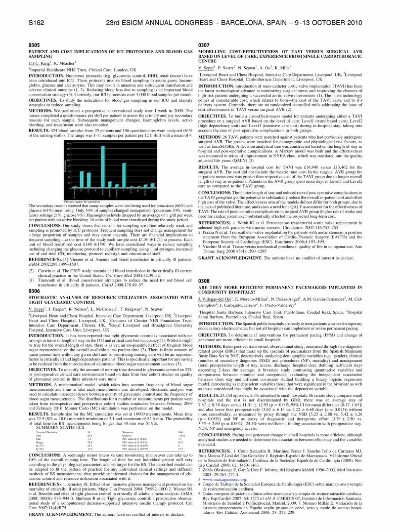
0305PATIENT AND COST IMPLICATIONS OF ICU PROTOCOLS AND BLOOD GASSAMPLING
H.J.C. King1, R. Meacher1
1Imperial Healthcare NHS Trust, Critical Care, London, UK
INTRODUCTION. Numerous protocols (e.g. glycaemic control, HHH, renal rescue) havebeen introduced into ICU. These protocols involve blood sampling to assess gases, haemo-globin, glucose and electrolytes. This may result in anaemia and subsequent transfusion andadverse clinical outcome (1, 2). Reducing blood loss due to sampling is an important bloodconservation strategy (3). Currently, our ICU processes over 4,000 blood samples per month.
OBJECTIVES. To study the indications for blood gas sampling in our ICU and identifystrategies to reduce sampling.
METHODS. We performed a prospective, observational study over 1 week in 2009. Thenurses completed a questionnaire per shift per patient to assess the primary and any secondaryreasons for each sample. Subsequent management changes, haemoglobin levels, activebleeding, and transfusion were also recorded.
RESULTS. 424 blood samples from 25 patients and 106 questionnaires were analysed (61%of the nursing shifts). The range was 1–11 samples per patient per 12 h shift with a mean of 4.
The secondary reasons showed that many samples were also being used for potassium (66%) andglucose (61%) monitoring. Only 54% of samples changed management (potassium 24%, venti-latory settings 21%, glucose 9%). Haemoglobin levels dropped by an average of 1 g/dl per weekper patient with no active bleeding. 10 units of blood were transfused during the study period.
CONCLUSIONS. Our study shows that reasons for sampling are often relatively weak andsampling is promoted by ICU protocols. Frequent sampling does not change management fora large proportion of samples and may cause anaemia. There are financial implications tofrequent sampling—at the time of the study each sample cost £1.50 (€1.71) to process. Eachunit of blood transfused cost £140 (€159). We have considered ways to reduce samplingincluding changing the glucose protocol to capillary sampling, using 1 ml syringes, increaseduse of end tidal CO2 monitoring, protocol redesign and education of staff.
REFERENCE(S). [1] Vincent et al. Anemia and blood transfusion in critically ill patients.JAMA 2002;288:1499–1507.
[2] Corwin et al. The CRIT study: anemia and blood transfusion in the critically ill-currentclinical practice in the United States. Crit Care Med 2004;32:39–52.
[3] Tinmouth et al. Blood conservation strategies to reduce the need for red blood celltransfusion in critically ill patients. CMAJ 2008;178:49–57.
0306STOCHASTIC ANALYSIS OF RESOURCE UTILIZATION ASSOCIATED WITHTIGHT GLYCEAMIC CONTROL
V. Seppi1, J. Harper2, R. Nelson3, L. McCrossan4, T. Ridgway1, N. Scawn1
1Liverpool Heart and Chest Hospital, Intensive Care Department, Liverpool, UK, 2LiverpoolHeart and Chest Hospital, Liverpool, UK, 3Countess of Chester NHS Foundation Trust,Intensive Care Department, Chester, UK, 4Royal Liverpool and Broadgreen UniversityHospital, Intensive Care Unit, Liverpool, UK
INTRODUCTION. It has been reported that tight glyceamic control is associated with netsavings in terms of length of stay on the ITU and critical care bed occupancy (1). Whilst it mightbe true for the overall length of stay, there is as yet, an un-quantified effect of frequent bloodsugar measurement on the overall available nurse-patient time (2). There is finite amount ofnurse-patient time within any given shift and so prioritizing nursing care will be an importantfactor in critically ill and high dependency patients. This is specifically important for any savingto be realized from the introduction of automated blood sugar measurement devices (3).
OBJECTIVES. To quantify the amount of nursing time devoted to glyceamic control on ITUor post-operative critical care environment based on data from four cohort studies on qualityof glyceamic control in three intensive care units.
METHODS. A mathematical model, which takes into account frequency of blood sugarmeasurements and time to take each measurement was developed. Stochastic analysis wasused to calculate interdependency between quality of glyceamic control and the frequency ofblood sugar measurements. The distributions for a number of measurements per patient weretaken from retrospective and prospective cohort studies conducted between February 2009and February 2010. Monter Carlo (MC) simulation was performed on the model.
RESULTS. Sample size for the MC simulation was set at 10000 measurements. Mean timewas 32.5 (SD = 18.0) min with minimum of 3.1 and maximum of 82.6 min. The probabilityof total time for BS measurements being longer that 30 min was 51.9%.
SUMMARY STATISTICSStandard Deviation 18 Skewness 0.46
Max 82.6 Kurtosis -0.52
Min 3.1 90% interval Q (0.05) 7.5
Range 78.8 90% interval Q (0.95) 65.3
Q (75) 44.5 95% interval Q (0.025) 6
Q (25) 17.9 95% interval Q (0.975) 71.7
CONCLUSIONS. A seemingly minor intensive care monitoring manoeuver can take up to10% of the overall nursing time. The length of time for any individual patient will veryaccording to the physiological parameters and set target for the BS. The described model canbe adapted to fit the pattern of practice for any individual clinical settings and differentmethods of BS measurements, thus providing informed choices for the management of gly-ceamic control and resource utilization associated with it.
REFERENCE(S). 1. Krinsley JS: Effect of an intensive glucose management protocol on themortality of critically ill adult patients. Mayo Clin Proceed 2004; 79:992–1000 2. Wiener RSet al. Benefits and risks of tight glucose control in critically ill adults: a meta-analysis. JAMA2008; 300(8): 933–944 3. Shulman R et al. Tight glycaemic control: a prospective observa-tional study of a computerised decision-supported intensive insulin therapy protocol. CritCare 2007;11(4):R75
GRANT ACKNOWLEDGMENT. The authors have no conflict of interest to declare.
0307MODELLING COST-EFFECTIVENESS OF TAVI VERSUS SURGICAL AVRBASED ON LEVEL OF CARE: EXPERIENCE FROM SINGLE CARDIOTHORACICCENTRE
V. Seppi1, P. Sastry2, N. Scawn1, A. Oo2, K. Mills1
1Liverpool Heart and Chest Hospital, Intensive Care Department, Liverpool, UK, 2LiverpoolHeart and Chest Hospital, Cardiothoracic Department, Liverpool, UK
INTRODUCTION. Introduction of trans-catheter aortic valve implantation (TAVI) has beenthe latest technological advance in minimizing surgical stress and improving the chances ofhigh-risk patient undergoing a successful aortic valve intervention (1). The latest technologycomes at considerable cost, which relates to both—the cost of the TAVI valve and to it’sdelivery system. Currently, there are no randomized controlled trails addressing the issue ofcost-effectiveness of TAVI versus surgical AVR (2).
OBJECTIVES. To build a cost-effectiveness model for patients undergoing either a TAVIprocedure or a surgical AVR based on the level of care: Level1 (ward based care), Level2(high dependency unit) and Level3 (intensive care unit) during in-hospital stay, taking intoaccount the rate of post-operative complications in both groups.
METHODS. 26 TAVI patients were matched against patients who had previously undergonesurgical AVR. The groups were matched for demographic and physiological risk factors, aswell as EuroSCORE. A decision analytical tree was constructed based on the length of stay inhospital and post-operative complications. A Markov model was built and the effectivenesswas measured in terms of improvement in NYHA class, which was translated into the qualityadjusted life years (QALY) (3).
RESULTS. The average in-hospital cost for TAVI was £16,940 versus £13,462 for thesurgical AVR. The cost did not include the theatre time cost. In the surgical AVR group thein-patient mean cost was greater than respective cost of the TAVI group due to longer overalllength of stay as in-patients. Patients in the AVR group spent more days in Level3 and Level2care as compared to the TAVI group.
CONCLUSIONS. The shorter length of stay and reduced rate of post-operative complications inthe TAVI group has got the potential to substantially reduce the overall in-patient cost and offsethigh cost of the valve. The effectiveness arm of the models did not differ for both groups, due tothe lack of published literature, and raises a need for a QALY assessment for the effectiveness ofTAVI. The rate of post-operative complications in surgical AVR group (higher rate of stroke andneed for cardiac pacemaker) substantially affected the projected long-term cost.
REFERENCE(S). 1. Webb JG et al. Percutaneous transarterial aortic valve replacement inselected high-risk patients with aortic stenosis. Circulation. 2007;116:755–7632. Piazza N et al. Transcatheter valve implantation for patients with aortic stenosis: a position
statement from the European Association of Cardio-Thoracic Surgery (EACTS) and theEuropean Society of Cardiology (ESC). EuroInterv 2008;4:193–199.
3. Vicchio M et al. Tissue versus mechanical prostheses: quality of life in octogenarians. AnnThorac Surg 2008 85(4):1290–1295
GRANT ACKNOWLEDGMENT. The authors have no conflict of interest to declare.
0308ARE THEY MORE EFFICIENT PERMANENT PACEMAKERS IMPLANTED INCOMMUNITY HOSPITALS?
J. Villegas-del Ojo1, E. Moreno-Millan1, N. Parias-Angel1, A.M. Garcia-Fernandez2, M. Cid-
Cumplido2, J. Carbajal-Guerrero2, F. Prieto-Valderrey2
1Hospital Santa Barbara, Intensive Care Unit, Puertollano, Ciudad Real, Spain, 2HospitalSanta Barbara, Puertollano, Ciudad Real, Spain
INTRODUCTION. The Spanish public hospitals are ready to treat patients who need temporaryendocavitary electrocatheter, but not all hospitals can implement or revise permanent pacing.
OBJECTIVES. To determine if interventions for permanent pacing (PPM) and change ofgenerator are more efficient in small hospitals.
METHODS. Retrospective, transversal, observational study, measured through five diagnosisrelated groups (DRG) that make up the casemix of pacemakers from the Spanish MinimumBasic Data Set in 2007, descriptively analyzing demographic variables (age, gender), clinical(number of secondary diagnoses (NSD) and procedures (NP), mortality) and management(total, preoperative length of stay, access, discharge, hospital size), defining inefficient staysexceeding 2 days the average. A bivariate study contrasting quantitative variables andcomparisons between nominal and categorical, evaluating the independent associationbetween short stay and different covariates studied building a binary logistic regressionmodel, introducing as independent variables those that were significant in the bivariate as wellas those considered that might be associated with the dependent variable.
RESULTS. 23,154 episodes, 5.3% admitted to small hospitals. Bivariate study compare smallhospitals and the rest is not discriminated by GDR, there was an average stay of7.87 ± 8.78 days versus 11.01 ± 12.95 (p = 0.005, 95% CI for mean difference [0.17, 1.65])and also lower than preoperatively (3.62 ± 6.14 vs. 4.22 ± 6.68 days (p = 0.015)) withoutmore comorbidity, as measured by proxy through the NSD [5.23 ± 2.88 vs. 5.42 ± 3.28(p = 0.055)] and NP as proxy of diagnostic and therapeutic effort [3.79 ± 2.50 vs.3.55 ± 2.69 (p = 0.002)]. 24.1% were inefficient, finding association with preoperative stay,NDS, NP and emergency access.
CONCLUSIONS. Pacing and generator change in small hospitals is more efficient, althoughanalytical studies are needed to determine the association between efficiency and the variablesevaluated.
REFERENCE(S). 1. Coma Samartın R, Martınez Ferrer J, Sancho-Tello de Carranza MJ,Ruiz Mateas F,Leal del Ojo Gonzalez J. Registro Espanol de Marcapasos. VI Informe Oficialde la Seccion de Estimulacion Cardıaca de la Sociedad Espanola de Cardiologıa (2008). RevEsp Cardiol 2009; 62: 1450–1463.2. Zubıa Olaskoaga F, Garcıa Urra F. Informe del Registro MAMI 1996–2003. Med Intensiva
2005; 29:265–271.5.3. www.marcapasossec.org.4. Grupo de Trabajo de la Sociedad Europea de Cardiologıa (ESC) sobre marcapasos y terapia
de resincronizacion cardıaca.5. Guıas europeas de practica clınica sobre marcapasos y terapia de resincronizacion cardıaca.
Rev Esp Cardiol 2007; 60: 1272 e1-e51.8. CMBD 2007, Instituto de Informacion Sanitaria,Ministerio de Sanidad y PolıticaSocial, Madrid, 2009. 7. Moreno Millan E. Variacion de laestancia preoperatoria en Espana segun grupos de edad, sexo y modo de acceso hospi-talario. Rev Calidad Asistencial 2008; 23: 222–229.
S162 23rd ESICM ANNUAL CONGRESS – BARCELONA, SPAIN – 9–13 OCTOBER 2010
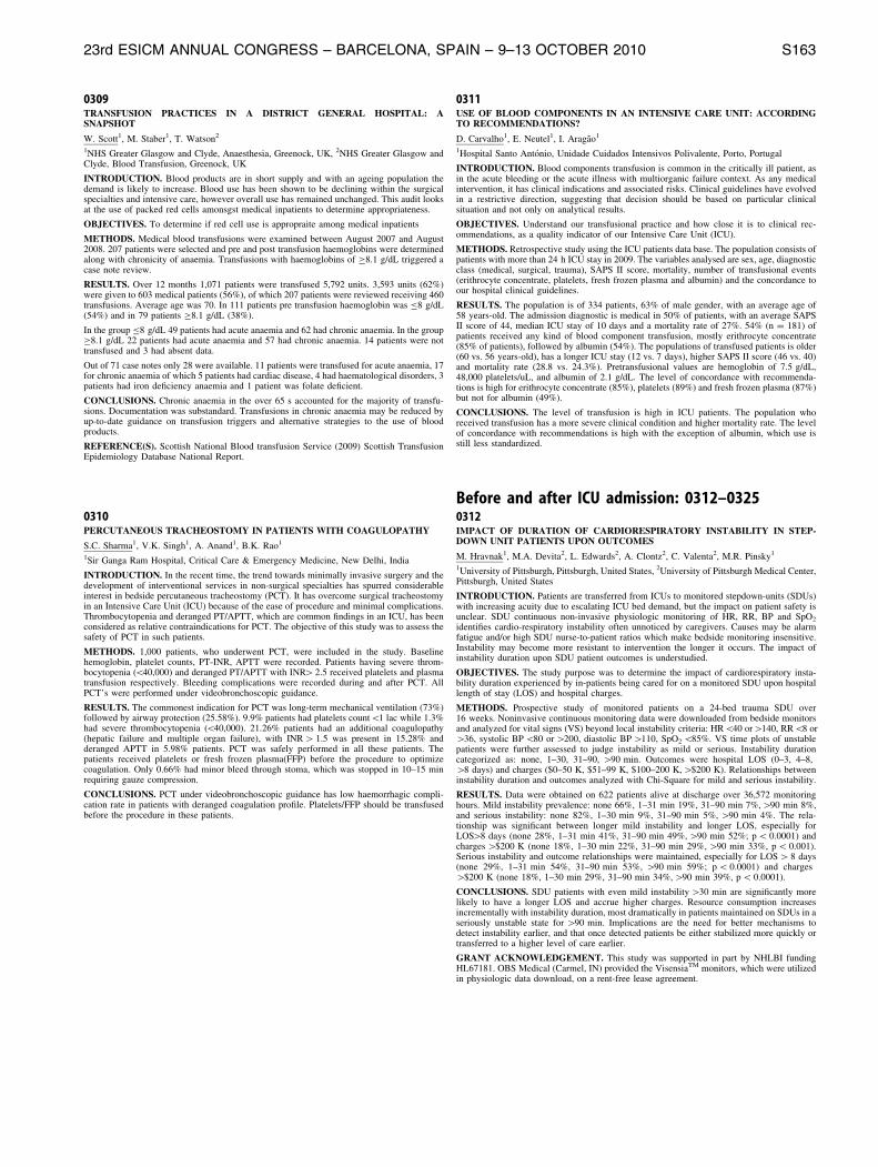
0309TRANSFUSION PRACTICES IN A DISTRICT GENERAL HOSPITAL: ASNAPSHOT
W. Scott1, M. Staber1, T. Watson2
1NHS Greater Glasgow and Clyde, Anaesthesia, Greenock, UK, 2NHS Greater Glasgow andClyde, Blood Transfusion, Greenock, UK
INTRODUCTION. Blood products are in short supply and with an ageing population thedemand is likely to increase. Blood use has been shown to be declining within the surgicalspecialties and intensive care, however overall use has remained unchanged. This audit looksat the use of packed red cells amonsgst medical inpatients to determine appropriateness.
OBJECTIVES. To determine if red cell use is appropraite among medical inpatients
METHODS. Medical blood transfusions were examined between August 2007 and August2008. 207 patients were selected and pre and post transfusion haemoglobins were determinedalong with chronicity of anaemia. Transfusions with haemoglobins of C8.1 g/dL triggered acase note review.
RESULTS. Over 12 months 1,071 patients were transfused 5,792 units. 3,593 units (62%)were given to 603 medical patients (56%), of which 207 patients were reviewed receiving 460transfusions. Average age was 70. In 111 patients pre transfusion haemoglobin was B8 g/dL(54%) and in 79 patients C8.1 g/dL (38%).
In the group B8 g/dL 49 patients had acute anaemia and 62 had chronic anaemia. In the groupC8.1 g/dL 22 patients had acute anaemia and 57 had chronic anaemia. 14 patients were nottransfused and 3 had absent data.
Out of 71 case notes only 28 were available. 11 patients were transfused for acute anaemia, 17for chronic anaemia of which 5 patients had cardiac disease, 4 had haematological disorders, 3patients had iron deficiency anaemia and 1 patient was folate deficient.
CONCLUSIONS. Chronic anaemia in the over 65 s accounted for the majority of transfu-sions. Documentation was substandard. Transfusions in chronic anaemia may be reduced byup-to-date guidance on transfusion triggers and alternative strategies to the use of bloodproducts.
REFERENCE(S). Scottish National Blood transfusion Service (2009) Scottish TransfusionEpidemiology Database National Report.
0310PERCUTANEOUS TRACHEOSTOMY IN PATIENTS WITH COAGULOPATHY
S.C. Sharma1, V.K. Singh1, A. Anand1, B.K. Rao1
1Sir Ganga Ram Hospital, Critical Care & Emergency Medicine, New Delhi, India
INTRODUCTION. In the recent time, the trend towards minimally invasive surgery and thedevelopment of interventional services in non-surgical specialties has spurred considerableinterest in bedside percutaneous tracheostomy (PCT). It has overcome surgical tracheostomyin an Intensive Care Unit (ICU) because of the ease of procedure and minimal complications.Thrombocytopenia and deranged PT/APTT, which are common findings in an ICU, has beenconsidered as relative contraindications for PCT. The objective of this study was to assess thesafety of PCT in such patients.
METHODS. 1,000 patients, who underwent PCT, were included in the study. Baselinehemoglobin, platelet counts, PT-INR, APTT were recorded. Patients having severe throm-bocytopenia (\40,000) and deranged PT/APTT with INR[2.5 received platelets and plasmatransfusion respectively. Bleeding complications were recorded during and after PCT. AllPCT’s were performed under videobronchoscopic guidance.
RESULTS. The commonest indication for PCT was long-term mechanical ventilation (73%)followed by airway protection (25.58%). 9.9% patients had platelets count\1 lac while 1.3%had severe thrombocytopenia (\40,000). 21.26% patients had an additional coagulopathy(hepatic failure and multiple organ failure), with INR [ 1.5 was present in 15.28% andderanged APTT in 5.98% patients. PCT was safely performed in all these patients. Thepatients received platelets or fresh frozen plasma(FFP) before the procedure to optimizecoagulation. Only 0.66% had minor bleed through stoma, which was stopped in 10–15 minrequiring gauze compression.
CONCLUSIONS. PCT under videobronchoscopic guidance has low haemorrhagic compli-cation rate in patients with deranged coagulation profile. Platelets/FFP should be transfusedbefore the procedure in these patients.
0311USE OF BLOOD COMPONENTS IN AN INTENSIVE CARE UNIT: ACCORDINGTO RECOMMENDATIONS?
D. Carvalho1, E. Neutel1, I. Aragao1
1Hospital Santo Antonio, Unidade Cuidados Intensivos Polivalente, Porto, Portugal
INTRODUCTION. Blood components transfusion is common in the critically ill patient, asin the acute bleeding or the acute illness with multiorganic failure context. As any medicalintervention, it has clinical indications and associated risks. Clinical guidelines have evolvedin a restrictive direction, suggesting that decision should be based on particular clinicalsituation and not only on analytical results.
OBJECTIVES. Understand our transfusional practice and how close it is to clinical rec-ommendations, as a quality indicator of our Intensive Care Unit (ICU).
METHODS. Retrospective study using the ICU patients data base. The population consists ofpatients with more than 24 h ICU stay in 2009. The variables analysed are sex, age, diagnosticclass (medical, surgical, trauma), SAPS II score, mortality, number of transfusional events(erithrocyte concentrate, platelets, fresh frozen plasma and albumin) and the concordance toour hospital clinical guidelines.
RESULTS. The population is of 334 patients, 63% of male gender, with an average age of58 years-old. The admission diagnostic is medical in 50% of patients, with an average SAPSII score of 44, median ICU stay of 10 days and a mortality rate of 27%. 54% (n = 181) ofpatients received any kind of blood component transfusion, mostly erithrocyte concentrate(85% of patients), followed by albumin (54%). The populations of transfused patients is older(60 vs. 56 years-old), has a longer ICU stay (12 vs. 7 days), higher SAPS II score (46 vs. 40)and mortality rate (28.8 vs. 24.3%). Pretransfusional values are hemoglobin of 7.5 g/dL,48,000 platelets/uL, and albumin of 2.1 g/dL. The level of concordance with recommenda-tions is high for erithrocyte concentrate (85%), platelets (89%) and fresh frozen plasma (87%)but not for albumin (49%).
CONCLUSIONS. The level of transfusion is high in ICU patients. The population whoreceived transfusion has a more severe clinical condition and higher mortality rate. The levelof concordance with recommendations is high with the exception of albumin, which use isstill less standardized.
Before and after ICU admission: 0312–03250312IMPACT OF DURATION OF CARDIORESPIRATORY INSTABILITY IN STEP-DOWN UNIT PATIENTS UPON OUTCOMES
M. Hravnak1, M.A. Devita2, L. Edwards2, A. Clontz2, C. Valenta2, M.R. Pinsky1
1University of Pittsburgh, Pittsburgh, United States, 2University of Pittsburgh Medical Center,Pittsburgh, United States
INTRODUCTION. Patients are transferred from ICUs to monitored stepdown-units (SDUs)with increasing acuity due to escalating ICU bed demand, but the impact on patient safety isunclear. SDU continuous non-invasive physiologic monitoring of HR, RR, BP and SpO2
identifies cardio-respiratory instability often unnoticed by caregivers. Causes may be alarmfatigue and/or high SDU nurse-to-patient ratios which make bedside monitoring insensitive.Instability may become more resistant to intervention the longer it occurs. The impact ofinstability duration upon SDU patient outcomes is understudied.
OBJECTIVES. The study purpose was to determine the impact of cardiorespiratory insta-bility duration experienced by in-patients being cared for on a monitored SDU upon hospitallength of stay (LOS) and hospital charges.
METHODS. Prospective study of monitored patients on a 24-bed trauma SDU over16 weeks. Noninvasive continuous monitoring data were downloaded from bedside monitorsand analyzed for vital signs (VS) beyond local instability criteria: HR\40 or[140, RR\8 or[36, systolic BP \80 or [200, diastolic BP [110, SpO2 \85%. VS time plots of unstablepatients were further assessed to judge instability as mild or serious. Instability durationcategorized as: none, 1–30, 31–90, [90 min. Outcomes were hospital LOS (0–3, 4–8,[8 days) and charges ($0–50 K, $51–99 K, $100–200 K, [$200 K). Relationships betweeninstability duration and outcomes analyzed with Chi-Square for mild and serious instability.
RESULTS. Data were obtained on 622 patients alive at discharge over 36,572 monitoringhours. Mild instability prevalence: none 66%, 1–31 min 19%, 31–90 min 7%, [90 min 8%,and serious instability: none 82%, 1–30 min 9%, 31–90 min 5%, [90 min 4%. The rela-tionship was significant between longer mild instability and longer LOS, especially forLOS[8 days (none 28%, 1–31 min 41%, 31–90 min 49%, [90 min 52%; p \ 0.0001) andcharges [$200 K (none 18%, 1–30 min 22%, 31–90 min 29%, [90 min 33%, p \ 0.001).Serious instability and outcome relationships were maintained, especially for LOS [ 8 days(none 29%, 1–31 min 54%, 31–90 min 53%, [90 min 59%; p \ 0.0001) and charges[$200 K (none 18%, 1–30 min 29%, 31–90 min 34%, [90 min 39%, p \ 0.0001).
CONCLUSIONS. SDU patients with even mild instability [30 min are significantly morelikely to have a longer LOS and accrue higher charges. Resource consumption increasesincrementally with instability duration, most dramatically in patients maintained on SDUs in aseriously unstable state for [90 min. Implications are the need for better mechanisms todetect instability earlier, and that once detected patients be either stabilized more quickly ortransferred to a higher level of care earlier.
GRANT ACKNOWLEDGEMENT. This study was supported in part by NHLBI fundingHL67181. OBS Medical (Carmel, IN) provided the VisensiaTM monitors, which were utilizedin physiologic data download, on a rent-free lease agreement.
23rd ESICM ANNUAL CONGRESS – BARCELONA, SPAIN – 9–13 OCTOBER 2010 S163

0313RECORDING OF THE SCOTTISH EARLY WARNING SCORE: AN IMPROVE-MENT IN PRACTICE
D.J. Moore1, N. Young1, J.S. McNeill1, C. Davidson1, A. Howie2, S. Dummer2
1New Royal Infirmary of Edinburgh, Critical Care, Edinburgh, UK, 2New Royal Infirmary ofEdinburgh, Edinburgh, UKINTRODUCTION. Most cardiopulmonary arrests are preceded by deterioration in physiologicalparameters1. Physiological scoring systems have been developed to attempt to identify patients at riskof deterioration, who may require medical intervention or an increased level of care2. The ScottishEarly Warning Score (SEWS) requires the measurement of respiratory rate, oxygen saturation,temperature, systolic blood pressure, heart rate and neurological status. If the score is 4 or more thenmedical assessment should occur. The National Institute of Clinical Excellence recommends that allhospital inpatients should be monitored using a physiological scoring system at least every 12 h3.OBJECTIVES. To examine the SEWS chart of every hospital inpatiet in the Royal Infirmaryof Edinburgh during one day and to assess whether the necessary physiological parametershad been recorded at last assessment. To determine if practice had improved following anidentical study 2 years previously.METHODS. Data were collected from SEWS charts of 601 patients from 20 wards in theRoyal Infirmary of Edinburgh during 1 day. Patients in the Emergency Department, CriticalCare and Day Admissions were excluded. We recorded whether or not each parameter hadbeen documented at the last entry, whether the total SEWS score had been calculated cor-rectly, and whether appropriate action had been taken.RESULTS. All physiological parameters required to calculate SEWS were recorded in 504/601 (83.86%) of patients. In 2007, only 231/595 (38.8%) of patients had all of the SEWScomponents recorded. Blood pressure 597/601(99.3%) and heart rate 593/601(98.6%) wererecorded more frequently than other physiological parameters. Respiratory rate was the leastwell recorded 533/601 (92%), although there was an improvement from the previous studywhere it had only been recorded on 289/595 (48.6%) of occasions.CONCLUSIONS. There has been a marked improvement in the overall recording of SEWS sincethe previous study. It is of concern that respiratory rate was again the least well recorded parameteras this has been shown to be the best physiological predictor of impending cardiopulmonaryarrest1,4. This may be because respiratory rate is not provided by the automated monitoring devicesavailable on the general wards in our hospital, and must be calculated manually. The improvementin the recording of SEWS in the last 2 years is likely to be the result of nurse training initiatives andself-auditing implemented in response to the previous study in 2007.REFERENCE(S). 1 Schein RM et al. Clinical antecedents to in-hospital cardiopulmonaryarrest. Chest 1990; 98: 1388–92
2 Morgan RJM et al. An Early Warning System for detecting developing critical illness.Clin. Intens Care 1997; 8:100.
3 http://www.nice.org.uk/nicemedia/pdf/CG50Full Guidance.pdf4 Fieselmann JF et al. Respiratory rate predicts cardiopulmonary arrest for internal medicine
patients. J Gen Intern Med 1993; 8: 354–60.
0314CHARACTERISTICS, PRACTICE PATTERNS AND OUTCOMES IN PATIENTSWITH ACUTE HYPERTENSION. EUROPEAN REGISTRY FOR STUDYING THETREATMENT OF ACUTE HYPERTENSION (EURO-STAT)
A. Vuylsteke1, on behalf of the Euro-STAT Investigators1Papworth Hospital NHS Trust, Cambridge, UKOBJECTIVES. The aim of the Euro-STAT registry is to describe current clinical manage-ment of acute hypertension (HTN) treated with intravenous (IV) antihypertensive drugs.METHODS. We identified 548 consecutive patients, who received an IV drug for the treatmentof HTN in emergency departments (EDs), operating/recovery rooms (PeriOp) or intensive careunits (ICUs) in 11 hospitals in 7 European countries (Austria, Belgium, Germany, Italy, Spain,Sweden, UK). Selected physiological variables, antihypertensive drugs and outcomes wererecorded until discharge or 30 d of hospitalization (whichever was earlier).RESULTS. Median age was 70 years and 35% were women; average mean arterial pressure atstart of treatment 107 mmHg. In each group,[90% achieved a 10% decrease in systolic bloodpressure at a median of 24 min of starting the antihypertensive drug. In ED patients, the mostfrequent presenting symptoms were chest pain (28%) and dyspnoea (26%); 6.9% experienced anepisode of hypotension requiring discontinuation of antihypertensive drug, administration ofvasopressors, fluids or reverse Trendelenburg positioning within 72 h of starting treatment;3.5% relapsed (required resumption of anti-HTN IV treatment) within 72 h of discontinuinginitial IV treatment. Most PeriOp patients had undergone cardiac (49%) or gastrointestinal(14%) surgery; 13% experienced an episode of hypotension within 72 h of starting treatment and6% relapsed within 72 h of discontinuing initial IV treatment. In ICU patients, 11% experiencedan episode of hypotension within 72 h of starting treatment and 7.2% relapsed within 72 h ofdiscontinuing initial IV treatment. Baseline factors and the proportional use of each antihy-pertensive drug for all patients and the three treatment areas are shown in the Table 1.
TABLE 1 BASELINE CHARACTERISTICS, TREATMENTS AND OUTCOMES]Variable All patients
(n = 548)
ED patients (n = 87) PeriOp patients
(n = 182)
ICU patients
(n = 279)
Median age, years
(IQR)
70 (60, 78) 69 (61, 79) 70 (58, 77) 71 (60, 78)
History of HTN 64% 70% 65% 61%
History of diabetes 26% 30% 24% 25%
History of chronic
kidney disease
9.1% 9.2% 5.0% 12%
Median SBP/DBP at
start of treatment
162/79 mmHg 200/100 mmHg 160/80 mmHg 159/74 mmHg
Number of IV
antihypertensive
drugs during 1st
3 h, median (IQR)
1 (1, 2) 1 (1, 2) 1 (1, 2) 1 (1, 1)
Most frequent 1st IV
antihypertensive
Nitroglycerin (42%),
urapadil (24%),
clonidine (14%),
furosemide (6%)
Furosemide (30%),
nitroglycerin (23%),
urapadil (18%),
esmolol (14%)
Urapadil (39%),
nitroglycerin (22%),
clonidine (18%), Na
nitroprusside (7%)
Nitroglycerin (60%),
urapadil (16%),
clonidine (14%),
labetalol (4%)
New/worsened end-
organ damage
within 30 days
30% 48% 20% 30%
30-day mortality 5.0% 11% 2.9% 4.5%
CONCLUSIONS. Acute HTN is currently managed with numerous IV agents in the variousclinical settings where it may arise; treatment is associated with hypotension in approximately11% of patients. These patients have substantial concomitant morbidity and mortality.GRANT ACKNOWLEDGMENT. Euro-STAT is supported by a research grant from theMedicines CompanyTM.
0315CONSEQUENCES OF TIME OF DISCHARGE FROM INTENSIVE CARE ONMORTALITY AND READMISSION RATES IN A UK UNIVERSITY TEACHINGHOSPITAL
R.C. Barker1, N.J. Flint1
1University Hospitals of Leicester NHS Trust, Leicester Royal Infirmary, Leicester, UK
INTRODUCTION. In 2001 an Intensive Care National Audit and Research Centre reviewdemonstrated a link between overnight discharge and increased patient mortality1; studiesfrom Canada have provided similar results.2,3 The most recent UK based was published in2002 it demonstrated an increase in mortality even when TISS scores were taken into accountas an independent risk factor.4 Since these publications critical care outreach and the use ofearly warning scores have become common place; however it was felt that time of dischargewas still impacting on patient outcome.
OBJECTIVES. To review our post-unit mortality and readmission rate, with particular focuson the time of discharge.
METHODS. Data was extracted from our database of all admissions to the Leicester RoyalInfirmary critical care unit between 2002 and 2009. Calculations of the risk of death andreadmission after discharge were made for three time periods (8.00 to 15.59, 16.00 to 23.59and 0.00 to 7.59).
RESULTS. There were 6,409 admissions, 6,393 were analysed (16 having essential datamissing), 6,092 were first admissions, 947 died on the unit leaving 5,145 discharges. Mortalityrates were greater in those discharged between 16.00 and 23.59, there was a trend towards anincrease rate of readmission amongst the same group. Unit mortality after readmission was26.2% compared with 15.6% for first admissions.
POST DISCHARGE MORTALITY AND READMISSION
Time period Discharges Deaths post discharge Readmissions
00.00–07.59 99 4 (4%) 5 (5%)08.00–15.59 2580 149 (5.8%) 144 (5.6%)16.00–23.59 2466 210 (8.5%) 152 (6.2%)Grand Total 5145 363 (6.6%) 301 (5.1%)
CONCLUSIONS. Our mortality and readmission data compare favourably with a recentpublication.5 There is a clear difference in mortality related to time of discharge; however thisis for evening discharges as compared to night discharges in previous papers.1-4 The time ofdischarge may represent logistical issues of planned discharges or early discharge decisionsdue to pressure for beds. Overnight discharge is an uncommon occurrence in our unit; thisevidence suggests that previous concern about night discharges should be extended to eveningdischarges.
REFERENCE(S). 1. Goldfrad, C, Rowan, K. Consequences of discharges from intensivecare at night. Lancet 2000; 355: 1138–42.2. Priestap, F. A. Impact of intensive care unit discharge time on patient outcome. CriticalCare Medicine 2006; 34(12): 2946–51.3. Hanane, T et al. The association between nighttime transfer from the intensive care unit andpatient outcome. Critical Care Medicine 2008; 36(8): 2232–37.4. Beck, D. H et al. Waiting for the break of dawn? Intensive Care Medicine 2002; 28:1287–93. 5. Campbell, A. J et al. Predicting death and readmission after intensive caredischarge. British Journal of Anaesthesia 2008; 100(5): 656–662.
0316TRANSFER OF CRITICALLY ILL PATIENTS: TRAINEE’S PERSPECTIVE
R. Sadasivan1, M. Ranganathan2
1University Hospital of Coventry and Warwickshire, Coventry, UK, 2George Eliot Hospital,Anaeshtetics and Critical Care, Nuneaton, UK
INTRODUCTION. Transferring critically ill patients is a challenging task in the day to dayactivities of the critical care team. Safe accomplishment of these transfers relies on skills ofthe persons accompanying and the resources available. Guidelines have been produced byvarious professional bodies [1, 2] to safely accomplish these transfers. The competency doc-ument released by the Royal College of Anaesthetists, UK requires that junior trainees haveappropriate knowledge, skills, attitude and behaviour in the principles of safe transfer ofcritically ill patients [3, 4].
OBJECTIVES. To obtain information about trainee’s perspective, experience and knowl-edge in transfer of critically ill.
METHODS. A web based online survey was sent to all the anaesthetic/ITU trainees in theWest Midlands region of the UK.
RESULTS. Total number of respondents were 72. Of these, 12.5% had less than 3 months ofanaesthetic training before undertaking a transfer. Only 33% had formal training on transferof critically ill patients. 79% of the trainee’s didn’t have any competency based formalassessment of their skills, attitudes and behaviour in transfer of critically ill patients. Majorityof them (86%) felt that every one should undergo formal training before undertaking transfers.While 91% of the respondents have undertaken transfers during their training, only 33% haveexperienced some form of critical incident during these transfers. More than 50% of theseadverse events were related to equipment failures while 27% were due to patient deteriora-tion. Nearly 75% of the trainees were not aware of terms and conditions of the insurancecover for these transfers.
CONCLUSIONS. This survey highlights the deficiencies involved in training the trainee’sfor transfer and the transfer itself. The results demonstrates that majority of the trainees wouldprefer to attend specific transfer courses before venturing out on an actual transfer. We hencerecommended the following for implementation: improvement of training process for thoseundertaking transfers; regular monitoring of this process; regular analysis of critical incidentsand acting upon it; making the insurance compulsory for those undertaking the transfers.
REFERENCE(S). 1. Association of Anaesthetists of Great Britain & Ireland. SafetyGuideline on Interhospital transfer. February 2002.2. Intensive Care Society: Guidelines for the transport of the critically ill adult. 2002.3. CCT II Anaesthesia: Competency-based ST1 ST2 training and assessment. A manual fortrainees and trainers. Royal College of Anaesthetists. Jan. 2007.4. Spencer C, Watkinson P, McCluskey A. Training and assessment of competency of traineesin the transfer of critically ill patients. Anaesthesia 2004; 59:1248–49.5. Gillman L, Leslie G, Williams T, Fawcett K, Bell R, McGibbon V. Adverse eventsexperienced while transferring the critically ill patient from ED to the ICU. Emerg Med J2006; 23:858–61.
S164 23rd ESICM ANNUAL CONGRESS – BARCELONA, SPAIN – 9–13 OCTOBER 2010
0317MEDICAL EMERGENCY TEAM, CHANGING SURVIVAL IN PATIENTS WITHHEMATO-ONCOLOGIC MALIGNANCIES
W.Y. Kim1, S.B. Hong1, Y.J. Chang1, T. Park1, H. Seo1, S.H. Choi1, J.W. Huh1, C.M. Lim1,
Y. Koh1
1Asan Medical Center, University of Ulsan College of Medicine, Department of Pulmonologyand Critical Care Medicine, Seoul, Republic of Korea
INTRODUCTION. Patients with hemato-oncologic malignancies who receive intensivechemotherapy and/or bone marrow transplantation often experience life-threatening situationssuch as infection or cardiopulmonary events. Over the decade, treatment outcomes and sur-vival rates in critically ill cancer patients have improved dramatically1. There are reportsmedical emergency team (MET) reduced unexpected in-hospital cardiac arrests and mortal-ity2 by early detection of clinically deteriorating status, yet no study clarified the effectivenessof MET in hemato-oncology patients.
OBJECTIVES. To evaluate the effectiveness of MET in patients with hemato-oncologicmalignancies.
METHODS. Administrative data of 89 hemato-oncology patients admitted to this hospital’sintensive care unit (ICU) between 2000 and 2001 were retrospectively analyzed and com-pared to data of 64 patients of similar characteristics admitted to same ICU in 2008, afterimplementation of MET. The baseline characteristics were reviewed and compared. ICU/hospital mortality and 1–2 month mortality were measured to compare survival outcomes.Univariate analysis was done to find out situations associated with improved outcomes afterMET implementation.
RESULTS. The group analyzed after MET implementation was consisted of older patients(mean age, 58.3 ± 12.3 vs. 45.7 ± 14.8; p \ .05) and worse performance status than thegroup before (p \ .05). There were no significant differences between two groups in sex,disease status, and history of recent chemotherapy. Main reasons for ICU admission wererespiratory failure (48.4 vs. 56.2%) and hypotension (15.6 vs. 29.2%), showing no significantdifferences between two groups. ICU mortality decreased after MET implementation (52.6 vs.82.4%; p \ .05), same for hospital mortality and short-term survival. In patients with sepsis,ICU mortality significantly decreased after MET implementation (OR, 6.00; 95% CI,1.89–19.08). Subgroup analysis was done for patients with sepsis and revealed lower meanAPACHE II score (24.3 ± 5.7 vs. 29.3 ± 5.8; p \ .05) and higher mean blood pressure at thetime of ICU admission (77.2 ± 18.2 vs. 53.1 ± 13.1; p \ .05) in the group after METimplementation.
CONCLUSIONS. After implementation of MET in this hospital, early detection and man-agement improved survival in critically ill hemato-oncology patients, especially with sepsis.
REFERENCE(S). 1. Thiery G, et al. Outcome of cancer patients considered for intensivecare unit admission: A hospital-wide prospective study. J Clin Oncol 2005;23:4406–4413.2. Santamaria J, et al. Changing cardiac arrest and hospital mortality rates through a medical
emergency team takes time and constant review. Crit Care Med 2010;38:445–450.
0318MORTALITY IN CARDIOVASCULAR SURGERY IS HIGUER IN PATIENTS WITHLONG LENGHT OF PREOPERATIVE STAY AT HOSPITAL
M.N. Parias Angel1, P. Font Ugalde2, R. Guerrero Pabon3
1Hospital Universitario Reina Sofıa de Cordoba, Intensive Care Medicine, Cordoba, Spain,2Hospital Universitario Reina Sofıa de Cordoba, Doctorado y Masteres Oficiales, Cordoba,Spain, 3Hospital Universitario Reina Sofıa de Cordoba, Intensive Care Unit, Cordoba, Spain
INTRODUCTION. In hospitalized patients, long lenght of stay is associated with a higherrate of complications.
OBJECTIVES. To determine the effect on survival and outcome of long preoperative stayingat hospital before cardiac surgery.
METHODS. Prospective, observational cohort study of 124 consecutive patients, betweenNovember of 2007 and February of 2008, were eligible. Younger than 18 years old, inmu-nocompromised and emergency surgery patients were excluded for analysis. Relationshipswith mortality were evaluated for the following variables: age, gender, lifestyle risk factorsand pre-existing comorbidities (cardiac heart failure,diabetes mellitus, dyslipemia, hyper-tension, obesity (defined as CMI [ 30 kg/m2), chronic kidney failure, NYHA clasification),farmacological treatments (betablockers, statins, digital, ACE, diuretics), Parsonnet score,type of cardiac surgery (valvular or coronary), time of ischemia, time of extracorporealcirculation. The time of monitoring was 90 days. Discrete variables were expressed as counts(%) and continuous variables as mean and standard deviation (SD). Differences in categoricalvariables were calculated using two-sided likehood ratio v2 test or Fisher‘s exact test, and theMann–Withney U test or Student t test was used for continuous variables, when appropiate.Cox proportional-hazards regression analysis was used to assess the impact of independentvariables on mortality across the time. Variebles significantly associate with mortality in theunivariate analysis were entered in the model. In order to avoid sprurious associations,variables entered in the regression models were those wih a relationship in univariate analysis(p \ 0.05) or a plausible relationship with the dependent variable. Data analysis was per-formade using SPSS for Windows 15.0.0 (SPSS, Chicago, IL).
RESULTS. A total of 124 patients were included. The 48.4% (60) were women, the mean agewas 65 ± 11 years. The frecuency of comorbidities and farmacological treatments were:hypertension (61%), chronic kidney failure (8%), diabetes (25%), dyslipemia (40.5%), obesity29%; betablockers 39.5%, statins 40.3%, digital 19.4%, ACE 46.8%, Parsonnet score was16.8 ± 9.7 points. Valvular (aortic and mitral) surgical replacement was 61.2%. The mor-tality after 90 days was 13 patients of all. A Cox regression analysis identified that long lenghtof preoperative stay at hospital remained associated with lower mortality (HR 1.054, 95% CI1.017–1.092, p 0.004). Mortality in patients who were staying at hospital less than 7 daysbefore the surgery was zero.
CONCLUSIONS. A long lenght stay at hospital before cardiac surgery is a independent riskfactor of mortality. It could be useful to reduce the preoperative time at hospital.
REFERENCE(S). 1. Ryan TA, et al. Predictors of outcome in cardiac surgical patients withprolonged intensive care stay. Chest 1997;112:1035–1042.
0319IMPACT OF CRITICALLY ILL PATIENTS IN REGARDS TO THE BOARDINGTIME FROM THE EMERGENCY DEPARTMENT TO THE INTENSIVE CAREUNIT
G. Intas1, M. Pitsoli1, P. Stergianis1, G. Fildissis1
1University of Athens, Nurse, Athens, Greece
INTRODUCTION. Emergency departments (ED) serve many functions in Greek hospitals,including initial management of critically ill patients and primary care for a growing pro-portion of the population. The impact of ED length of stay (LOS) on patient outcome has notbeen covered adequately by existing surveys so far.
OBJECTIVES. The aim of this study was to determine the association between ED over-crowding and outcomes for critically ill patients.
METHODS. In the present study, we included medical and surgical pts that all of them wereintubated promptly to ED of 2 general hospitals of Athens GR, for 12 months. Pts survived[24 h were divided into 2 groups: ED boarding \6 h (group A) and ED boarding C6 h(group B). Demographics, APACHE II, diagnosis, LOS, and ICU and hospital mortality wererecorded. ED boarding time was measured in min. Groups were compared using Chi-square,Mann–Whitney, unpaired Student‘s t tests and stepwise regression analysis. The collection ofdata lasted 12 months.
RESULTS. In the ED, 196 critically ill patients with a mean age 59.6 ± 20.9 years andAPACHE II score 24.6 ± 12.7 were intubated. 144 pts were males and 52 were females witha mean age 54.5 ± 20.1 and 72.1 ± 17.5 years, and Apache II score 23.8 ± 13.2 and26.4 ± 11.5 respectively. Main diagnosis was multi trauma (72) stroke (32), respiratoryfailure (32), cardiac (20), head injury (16), coma (16) and infections (8). Group A = 76 pts(mean time ED boarding, LOS, mortality): 28 pts were admitted to CCU (30.7 ± 20.4,7.7 ± 9.3, 42.9%), 32 to ICU (231.25 ± 103.1, 11.9 ± 7.8, 37.5%) and 16 to intermediateunit (191.3 ± 33.5, 7.8 ± 5.7, 0%). Group B = 120 pts (mean time ED boarding, LOS,mortality): 28 pts were admitted to ICU (559.28 ± 101.6, 15.6 ± 10.3, 58.7%) and 92 tointermediate unit (3094.4 ± 3273.4, 19.1 ± 14.5, 78.3%). Hospital LOS was19.2 ± 20.4 days for group A vs. 41.5 ± 62.7 days for group B (P\ 0.001), and mortality26.8 vs. 45.7% respectively (P\ 0.001). In the stepwise regression analysis, delayed admis-sion, higher Apache II, age [61 years, female gender and direct admission to ICU wereassociated with lower hospital survival (odds ratio 0.815; 95% 0.612–0.976).
CONCLUSIONS. Critically ill patients with ED boarding C6 h have increased hospital LOSand higher mortality. Higher Apache II, age[61 years, female gender and direct admission toICU are associated with lower hospital survival.
REFERENCE(S). Engoren M. Crit Care Med 2005; 33:727-732, Goldhill DR, et al. Inten-sive Care Med 2004; 30:1908–13.
0320THE EPIDEMIOLOGY OF MEDICAL EMERGENCY TEAM CALLS PRECIPI-TATED BY ATRIAL FIBRILLATION IN A TEACHING HOSPITAL
A.G. Schneider1, P. Calzavacca1, R. Bellomo1, D. Jones1
1Austin Health, Heidelberg, Australia
INTRODUCTION. Atrial fibrillation (AF) is common within hospitalized patients andrepresents a frequent cause for activation of the Medical Emergency Team (MET). It is oftenregarded as easily controlled and requiring limited intervention, usually in the form ofmedications.
OBJECTIVES. We sought to assess the baseline characteristics and outcomes of the patientspresenting AF as a cause of MET call activation.
METHODS. Using the MET database of one tertiary teaching hospital, we retrospectivelyreviewed all patients for which the MET diagnosis was atrial fibrillation. We reviewed theirclinical history, immediate treatment and outcome. These data were compared to those of acontrol group of randomly selected MET Calls with patients being matched for age, genderand ward of origin (surgical or medical).
RESULTS. Between January 2005 and August 2007, there were 2237 medical emergencycalls, and 137, concerning 113 patients, were precipitated by atrial fibrillation. The patientswere, on average, 76 year old, 54% were female and 52% on a surgical ward. 25% had writtenlimitations of medical therapy orders (30% in the control group). Antiarrhythmic drugs wereprescribed in 50% of cases. Amongst the 85 patients without documented treatment limita-tions, 5% were transfered to the Intensive Care (ICU) or Coronary Care Unit (CCU) comparedwith 18% in the control group. Overall mortality was 20% in the AF group versus 43% in thecontrol group. For patients left on the ward, the mortality rate was 17% in the AF group and25% in the control-group.
CONCLUSION. MET calls triggered by AF are associated with an in-hospital mortality rateof 20%. For patients left on the ward, the mortality rate was 17%. AF should be regarded as amarker of illness severity and ICU/CCU admission considered for patients without limitationsof medical therapy.
23rd ESICM ANNUAL CONGRESS – BARCELONA, SPAIN – 9–13 OCTOBER 2010 S165
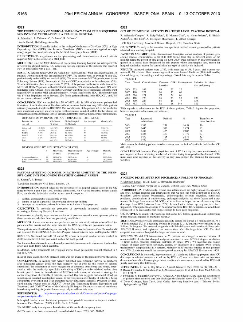
0321THE EPIDEMIOLOGY OF MEDICAL EMERGENCY TEAM CALLS REQUIRINGNON INVASIVE VENTILATION IN A TEACHING HOSPITAL
A. Schneider1, P. Calzavacca1, D. Jones1, R. Bellomo1
1Austin Health, Heidelberg, Australia
INTRODUCTION. Normally limited to the setting of the Intensive Care Unit (ICU) or HighDependency Units (HDU), Non Invasive Ventilation (NIV) is sometimes applied as respi-ratory support for ward patients by the Medical Emergency Team (MET).
OBJECTIVES. We sought to review the epidemiology and clinical outcome of ward patientsrequiring NIV in the setting of a MET Call.
METHODS. Using the MET database of one tertiary teaching hospital, we retrospectivelyreviewed the clinical history, ICU admission rate and outcome of the patients who receivedNIV in association with a MET call.
RESULTS. Between January 2005 and August 2007, there were 2237 MET calls and 150 calls (138patients) were associated with the application of NIV. The patients were, on average 71 year old,predominantly males (68%) and medical (66%). The most commons MET diagnosis, were AcutePulmonary Edema (49%), Pneumonia (11%) and COPD exacerbation or bronchospasm (7%).Treatment limitation plans were present in 55 (37%) of the patients had before or at the time of theMET Call. Of the 95 patients without treatment limitation, 51% remained on the ward, 31% weretransferred to the ICU and 13% to the HDU or Coronary Care Unit. 6% of the patients left on the wardrequired NIV on another MET call and ultimately 1% were transferred to HDU. The mortality ratewas 17% for the patients left on the ward, 22% for the patients admitted to the HDU/CCU and 38%for the patients admitted to ICU.
CONCLUSION. NIV was applied in 6.7% of MET calls. In 37% of the cases, patients hadlimitations of medical treatment. For those without treatment limitations, only 50% of the patientsultimately required a transfer to HDU/ICU. The mortality rate of the patients left on the ward aftersuch a treatment was high and even higher for those patients admitted to ICU. Further studies areneeded to assess the antecedents to MET calls in an attempt to improve the outcome of these patients.
OUTCOME OF PATIENTS WITHOUT TREATMENT LIMITATION
Transfer after
met call
n Male/female
(% Male)
Medical/surgical
(% Medical)
Age (average) Mortality (%)
Left on ward 48 (51%) 35/13 (72%) 25/23 (52%) 69 8/48 (17%)
To ICU 29 (31%) 21/8 (72%) 20/9 (41%) 63 11/29 (38%)
To HDU 9 (9%) 5/4 (55%) 7/2 (78%) 70 4/9 (45%)
To CCU 4 (4%) 1/3 (25%) 4/0 (100%) 84 0/4 (0%)
Other 5 (5%) 2/3 (40%) 2/3 (40%) 68 0/5 (0%)
Total 95 64/31 (67%) 58/37 (61%) 68 23/95 (24%)
DEMOGRAPHIC BY RESUSCITATION STATUS
n Male/female
(% Male)
Medical/surgical
(% Medical)
Age (average) Mortality
(%)
No treatment limitation 95 64/31 (67%) 58/37 (61%) 68 23/95 (24%)
Written treatment limitations 55 38/17 (69%) 41/14 (74%) 76 38/55 (69%)
Total 150 102/48 (68%) 99/51 (66%) 71 61/150 (40%)
0322FACTORS AFFECTING OUTCOME IN PATIENTS ADMITTED TO THE INTEN-SIVE CARE UNIT FOLLOWING IN-PATIENT CARDIAC ARREST
H. Khetani1, R. Breeze1
1Lewisham Healthcare NHS Trust, London, UK
INTRODUCTION. Quoted values for the incidence of In-hospital cardiac arrest in the UKrange between 1 and 5 per 1,000 hospital admissions. An NHS-led initiative, Patient SafetyFirst, has divided in-hospital cardiac arrests into 3 categories:
1. sudden, unpredictable catastrophic events2. failure to act on a patient’s deteriorating physiology in time3. attempts to resuscitate patients in whom resuscitation is inappropriate.
OBJECTIVES. To ascertain the proportion of preventable in-hospital cardiac arrestsoccurring at University Hospital Lewisham.
Furthermore, to identify any common predictors of poor outcome that were apparent prior tothose arrests and whether these are potentially modifiable.
METHODS. A case note review was performed on the cohort of patients who suffered in-patient cardiac arrests and who were admitted for ICU (level-3) care post-resuscitation.
These patients were identified using our quarterly feedback from the Intensive Care National Auditand Research Centre (ICNARC) Case Mix Program dataset between April and September 2009.
RESULTS. We found that half (11 out of 21) of our in hospital cardiac arrests resulted indeath despite level-3 care post arrest within the audit period.
9 of these in-hospital arrests were deemed preventable from case note review and trust cardiacarrest call audit forms when available.
In addition, in the preventable sub-group an arterial blood gas sample was not obtained in 7out of 9, 77%.
In all of these cases, the ICU outreach team was not aware of the patient prior to the arrest.
CONCLUSIONS. In keeping with widely published data regarding survival to dischargeafter in-hospital cardiac arrest, the high mortality rate of 50% for this cohort of patientsemphasises the importance of early recognition of abnormal physiology and timely inter-vention. With the sensitivity, specificity and validity of EWS yet to be validated and no clearbenefit proved from the introduction of MET/outreach teams, an alternative strategy forearlier recognition of critically ill patients is needed. Our data suggests that arterial blood gassampling, an essential investigation central to the recognition of critically ill patients is beingconsistently overlooked and is an important factor influencing outcome. Provision of dedi-cated training courses such as ALERT� (Acute Life Threatening Events: Recognition andTreatment) and CCrISP� (Care of the Critically Ill Surgical Patient) as a part of mandatoryintroductory training for junior doctors may help to address this issue.
REFERENCE(S). http://www.patientsafetyfirst.nhs.uk/Content.aspx?path=/Campaign-support/countyourcalls/
In-hospital cardiac arrest: incidence, prognosis and possible measures to improve survival.Intensive Care Medicine (2007) Vol 33, No 2; 237–245
The MERIT study investigators. Introduction of the medical emergency team
(MET) system: a cluster-randomised controlled trial. Lancet 2005; 365: 2091–7.
0323OUT OF ICU MEDICAL ACTIVITY IN A THIRD LEVEL TEACHING HOSPITAL
R. Abizanda-Campos1, R. Reig-Valero1, E. Moreno-Clari1, A. Heras-Javierre1, E. Bisbal-
Andres1, S. Mas-Font1, A. Belenguer-Muncharaz1, L. Albert-Rodrigo1
1ICU Dpt, University Associated General Hospital, ICU, Castellon, Spain
OBJECTIVE. To analyze the intensive care specialist medical request generated by patientsadmitted to a teaching hospital.
PATIENTS AND METHODS. Observational descriptive cohort analysis of patients gen-erating medical consultations to the ICU staff during their stay in different wards of thehospital during the period of time going sin 2004–2009. Data collection by ICU physicians isquoted on a special form designated for this purpose where demographic data, reason forhospital admission, reason for consultation and type of activity are included.
RESULTS. Attended patients were 2,787, with mean age of 58, 7 years, and women rep-resent 41, 2% of them. Most demanding services were Internal Medicine (31%) followed byGeneral Surgery, Haematology and Nephrology. Global data may be seen in Table 1.
TABLE 1Year Global Consultations Catheter
insertionCPR Management Sedation in diges-
tive endoscopy
2004 273 143 69 25 222005 410 227 84 33 612006 364 171 89 27 652007 488 193 83 24 52 1282008 500 224 64 25 77 1552009 752 332 62 28 146 182
With regards to admissions to the ICU of these patients, Table 2 depicts the proportionbetween requested admissions, and refusals.
TABLE 2Year Requested
admissionsRefusals % Transfers to
other hospitals
2004 1393 231 16.58 302005 1445 372 25.74 932006 1497 435 29.05 682007 1428 453 31.72 872008 1477 425 28.77 522009 1462 394 26.94 90
Main reason for deriving patients to other centres was the lack of available beds in the ICU(87, 6%).
CONCLUSIONS. Intensive Care physicians out of ICU activity increases continuously inour hospital, with an increasing number of activities trying to respond to the demand. ICUsmust keep strict registers of this activity as they may support the planning for increasingfacilities.
0324AVOIDING DEATH AFTER ICU DISCHARGE: A FOLLOW UP PROGRAM
P. Martinez-Lopez1, R.D.F. Luis1, J. Hernandez-Rodriguez1
1Hospital Universitario Virgen de la Victoria, Critical Care Unit, Malaga, Spain
INTRODUCTION. Tradicionally, critical care interventions are highly intensive, expensiveand brief. Critical illnesses and interventions that we use, can both contribute to postICUdisability: catheter-related bacteraemia, polineuropathy, resistant organism, nutricionalproblems, complications of tracheostomy, prolonged analgesic. All these factors and a pre-mature discharge from an ever full ICU, can even have an impact on occult mortality afterdischarge from ICU (between 6 and 20%). In our Unit a follow up program have beenimplanted. When patients are about to be discharged from ICU, ICU clinicians selected thoseconsidered to be recoverable but fragile enough to have poor prognosis.
OBJECTIVES. To quantify the workload that a after ICU follow-up entails, and to determineif this program impacts on mortality postICU.
METHODS. Prospective and interventional study carried out during a 7 months period. At a18 beds medical UCI of a teaching hospital in Malaga ICU, 55 patients were enrolled in theFollow Up Program. We assessed prognosis with Sabadell score and severity of illness withAPACHE II score; and registered our interventions after discharge from ICU. The finalendpoint was status at hospital discharge: survivant or dead.
RESULTS. We did 129 interventions in 55 patients: we changed a venous catheter 17ocasions (40% of patients), changed analgesic schedule 15 times (27.2%), stopped antibiotics13 times (24%), modified parenteral nutrition 25 times (45%). We searched and treatedsources of sleep deprivation (delirium, anxiety or insomnio) in 5 patients (9%); treatedtracheostomy complications in 3 patients. Mortality of 55 patients enrolled in this programwas 3.7% (2 patients) even if the mean expected mortality by APACHE II score was[30%.
CONCLUSIONS. In our study, implementation of a continued follow-up program after ICUdischarge in selected patients, carried out by ICU staff, was associated with an importantdecrease of mortality. Encouraging clinical results and a non-excesive workload for ICU staffjustify continuing this follow-up.
REFERENCES. 1. Lee K, Hog SB et al. Anaesth Intensive Care. 2008; 36 (4): 528–-342. Rivera-Fernandez R, Sanchez-Cruz J, Abizanda-Campos R. et al. Crit Care Med 2001; 29
(9): 1701–93. Fernandez R, Baigorri F, Navarro G, Artigas A. A modified McCabe score for stratification
of patients after intensive care unit discharge: the Sabadell score. Crit Care 2006; 10: R1794. Derek C Angus, Jean Carlet, Jean Carlet. Surviving intensive care. 18Edicion. Berlin:
Springer-Verlag;2003.
S166 23rd ESICM ANNUAL CONGRESS – BARCELONA, SPAIN – 9–13 OCTOBER 2010
0325THE INTERFACE BETWEEN MEDICAL ADMISSIONS UNIT, CRITICAL CAREOUTREACH TEAM AND INTENSIVE CARE UNIT IN A DISTRICT GENERALHOSPITAL
H. Altemimi1, S. Altaf1, J. Brown1, S. Al-Juboori1, V. Jadhav1
1Queen Elizabeth Hospital, Kings Lynn, Medical Assessment Unit, Kings Lynn, UKINTRODUCTION. The Medical Assessment Unit (MAU) in our District General Hospital(with 500 beds) provides assessment and treatment of acute medical patients from GeneralPractice (GP) referals and the Emergency Department (ED). Patients arriving in MAU are firsttriaged by the nurses before assessment by junior doctors. The Early Warning Score (EWS) isan indepenently verified scoring systems used to prioritise patient assessment. Appropriatereferal to Critical Care Outreach teams and Intensive Care Units can be triggered by nurses ordoctors according to the EWS score. Clear and seemless referal pathways and communicationbetween those involved with acutely unwell patients is essential.OBJECTIVES. To measure key performance indicators in the MAU and identify specificareas for improvement.METHODS. Retrospective audit of all admissions to Medical Assessment Unit during1 week in 2009. The following parameters were recorded for 149 patients admitted. Patientages ranged from 16 to 94:1. Source of admission2. Time to A&E triage3. Time to A&E doctor4. Time to MAU doctor5. Documentation of Early Warning Score6. Decision to escalate treatment for patients with high EWS.7. Outreach review
RESULTS.1. ED 51%, GP 47%, Outpatient clinic 2%2. 30% within 1 h, 50% within 3 h3. 20% within 1 h, 45% within 3 h4. 36% within 1 h, 60% within 3 h5. 70% of patients had EWS documented on admission.6. 13 (9%) patients had EWS [3. Of these, only 4 were reviewed within 1 h.7. 100% of patients reviewed within 15 min.CONCLUSIONS. In the UK, early warning scores have been developed to trigger early review.The most sophisticated intensive care becomes unnecessarily expensive terminal care when the pre-ICU system fails to refer in a timely manner. Our results showed that when a patient was recognisedas being unwell, they were seen rapidly by the outreach team. However, not all new admissions hadtheir EWS documented, overlooking an important opportunity to risk stratify patients before formalmedical clerking. Our Critical Care Outreach team have prominent role in education to identifyabnormal physiology early, and take action as appropriate.REFERENCE(S). 1. ‘‘The right person, in the right setting-first time’’ Report of the AcuteMedicine Task Force, Royal College of Physicians. October 20072. Subbe C.P., Kruger M., Gemmel L.-‘‘Validation of a modified Early Warning Score in
medical admissions.’’ Quarterly Journal of Medicine 2001; 94; 521–6.3. Lee A, Bishop G, Hillman KM, Daffurn K: ‘‘The medical emergency team.’’ Anaesth
Intensive Care 1995, 23:183–1864. Carol Ball, Margaret Kirkby, Susan Williams. ‘‘Effect of the critical care outreach team on
patient survival to discharge from hospital and readmission to critical care: non-randomisedpopulation based study’’ BMJ 2003;327:1014
GRANT ACKNOWLEDGMENT. We wish to thank the Queen Elizabeth Hospital, King’sLynn Audit Department for analysing the data, especially Alka Anoop.
Basic research in sepsis: New therapeuticapproaches: 0326–03390326THE ROLE OF IMMUNOMODULATION ON IMMUNE FUNCTION AND PROG-NOSIS IN SEPSIS
S.-W. Huang1, Xiang-Dong Guan1The First Affiliated Hospital of Sun Yat-sen University, Surgical Intensive Care Unit (SICU),Guangzhou, ChinaINTRODUCTION. Sepsis is defined as the systemic inflammatory response to infection.Immunological host reactions are primarily believed to determine the clinical course of thisdisease. An overwhelming inflammatory response to microbial invasion may be involved inthe pathogenesis of SIRS, sepsis and multiple organ failure. It is important to block theinflammatory response and stop or alleviate sepsis injuries. Immunotherapy is regarded aseffective approach to improve the immunological function.OBJECTIVES. Immunomodulation in the critically ill is an appealing notion because of theabnormal immune responses. The aim of this study is to evaluate the immunomodulation andits mechanism to improve immune function and prognosis in sepsis.METHODS. Experimental part:CLP model were divided into three groups including sham group,control group and experimental group. Control group only used antibiotic and experimental group usedantibiotic plus immunomodulation. Blood collection were made after CLP model in 3, 12, 24 and 48 h.Lymphocyte counting, CD4+, CD8+ T lymphocyte and CD4/CD8 ratio were checked. The apoptosis oflymphocyte in thymus and spleen and survival rate were checked.Clinical part: Prospective analysis seventy patients conformed to the enrolled standard. It wasdivided into two groups at random. One was control group with regular therapy, the therapygroup with Ulinastatin plus Thymosin-a1 in a week.The immune index before and after therapy in the 0, 1st, 3rd, 7thday was observed.RESULTS. Experimental part: Lymphocyte, CD4+ T lymphocyte and CD4/CD8 ratio inexperimental group increased more significant than in control group (P \ 0.05). Lymphocyteapoptosis index of thymus and spleen in control group increased more significant than inexperimental group (P \ 0.05).72 h-survival rate in experimental group was higher than incontrol group. Clinical part: CD4
+T lymphocyte, Lymphocytes and HLA-DR CD14+ had
more significant increase in therapy group than in control group (P \ 0.05). Twenty patientsdied in the control group and thirteen patients died in the therapy group (P \ 0.05).CONCLUSIONS. Immunomodulation in sepsis can improve immune function and alleviatethe lymphocyte apoptosis and extend the survival time.REFERENCE(S). Ying Zhang, Hao Chen, Yu-min Li, et al. Thymosin a1- and Ulinastatin-Based Immunomodulatory Strategy for Sepsis Arising from Intra-Abdominal Infection Due toCarbapenem-Resistant Bacteria[J]. The Journal of Infectious Diseases, 2008, 198:723–730.Li Yumin, Chen Hao, Li Xun, et al. A New Immuno-modulatory Therapy for Severe Sepsis:Ulinastatin plus Thymosina1[J]. Journal of Intensive Care Medicine, 2009, 24(1): 47–53.GRANT ACKNOWLEDGMENT. Sun Yat-sen University funds (5010) and Techpoolfunds(01200909).
0327NIACIN ATTENUATES ACUTE LUNG INJURY AND IMPROVES SURVIVALDURING SEPSIS BY DOWN-REGULATION OF NUCLEAR FACTOR-JBPATHWAY
W.Y. Kwon1, G.J. Suh1, K.S. Kim1
1Seoul National University Hospital, Emergency Medicine, Seoul, Korea, Republic of
INTRODUCTION. In recent years, the antiinflammatory effects of niacin by reducingnuclear factor jB (NF-jB) activation have attracted attention. However, the protective effectsof niacin on the development of acute lung injury and systemic inflammatory response duringsepsis have not been studied.
OBJECTIVES. We performed this study to determine whether niacin attenuates acute lunginjury (ALI) and improves survival during sepsis and the beneficial effects of niacin areassociated with the down-regulation of NF-jB pathway.
METHODS. Lipopolysaccharide (LPS) at a dosage of 10 mg/kg was injected into the tailvein to induce endotoxemia in rats. Then, vehicle, low dose niacin (360 mg/kg), or high doseniacin (1180 mg/kg) diluted in distilled water with same volume (3 ml/kg) was administeredonce to the rats through orogastric tube, respectively. We observed the survival of the sub-jects. At 6 h post-LPS injection, we measured serum tumor necrosis factor-a (TNF-a),interleukin-6 (IL-6) levels, lung cytoplasmic phosphorylated inhibitor jB-a (p-IjB-a), IjB-a,nuclear NF-jB p65 expressions, NF-jB p65 DNA-binding activity, and ALI occurrence.
RESULTS. High dose niacin improved survival during sepsis. Niacin induced dose-depen-dent reduction of serum TNF-a, IL-6 levels, lung cytoplasmic p-IjB-a, nuclear NF-jB p65expressions, NF-jB p65 DNA-binding activity, and ALI occurrence in endotoxemic rats. Incontrast, niacin preserved lung cytoplasmic IjB-a expression dose-dependently in endo-toxemic rats.
CONCLUSIONS. Niacin attenuated acute lung injury and improved survival during sepsis inrats. The protective effects of niacin were associated with the down-regulation of NF-jBpathway. Niacin can be considered as a therapeutic agent for sepsis.
REFERENCE(S). 1. Gurujeyalakshmi G, Wang Y, Giri SN (2000) Taurine and niacin blocklung injury and fibrosis by down-regulating bleomycin-induced activation of transcriptionnuclear factor-kappaB in mice. J Pharmacol Exp Ther 293:82–90. 2. Ganji SH, Qin S, ZhangL, Kamanna VS, Kashyap ML (2009) Niacin inhibits vascular oxidative stress, redox-sen-sitive genes, and monocyte adhesion to human aortic endothelial cells. Atherosclerosis202:68–75. 3. Schwartz MD, Moore EE, Moore FA, Shenkar R, Moine P, Haenel JB,Abraham E (1996) Nuclear factor-kappa B is activated in alveolar macrophages from patientswith acute respiratory distress syndrome. Critical Care Med 24:1285–1292.
0328THE EFFECTS OF RECOMBINANT HUMAN SOLUBLE THROMBOMODULIN INTHE EXPERIMENTAL ENDOTOXEMIC MOUSE INDUCED BYLIPOPOLYSACCHARIDE
A. Sawamura1, M. Hayakawa1, S. Gando1, N. Kubota1, S. Uegaki1, M. Sugano1
1Hokkaido University Graduate School of Medicine, Division of Acute and Critical CareMedicine, Department of Anesthesiology and Critical Care Medicine, Sapporo, Japan
OBJECTIVES. Thrombomodulin is an endothelial anticoagulant factor that plays animportant role in the regulation of disseminated intravascular coagulation. In addition,thrombomodulin may suppress the production of inflammatory cytokines, such as TNF-a andIL-6.
DESIGN. Prospective, comparative, experimental study.
SETTING. Laboratory animal research center at Hokkaido University.
SUBJECTS. Male balb/c mice (20–25 g)
INTERVENTIONS. Endotoxemic mouse models were induced by a bolus intravenousinjection of lipopolysaccharide at a dose of 4 mg/kg. Recombinant human soluble throm-bomodulin (rhTM) was administered as a bolus injection at a dose of 1, 2, 4 mg/kg as soon aspossible after the administration of lipopolysaccharide. As a control, an equal volume ofphysiologic saline was administered.
MEASUREMENTS AND MAIN RESULTS. Mice were randomly devided into 4 groups.Four groups were consisted with physiologic saline group, rhTM 1 mg/kg group, 2 mg/kggroup and 4 mg/kg, respectively. Four hours after the injection of rhTM, the levels ofinflammatory cytokines in the lung, liver and kidney were exmined. TNF-a and IL-6 werepredominantly reduced in the liver and kidney at 4 mg/kg group. (TNF-a: liver; p = 0.0126,kidney; p = 0.0195, IL-6: liver; p = 0.0015, kidney; p = 0.0418)
CONCLUSIONS. This study demonstrated that rhTM inhibits the expression of inflamma-tory cytokines in experimental endotoxemic mice. Administration of rhTM markedly reducedliver and kidney dysfunction. Therefore, rhTM have a beneficial treatment for sepsis.
23rd ESICM ANNUAL CONGRESS – BARCELONA, SPAIN – 9–13 OCTOBER 2010 S167
0329COMPARISON OF THE EFFECTS OF HYPERBARIC OXYGEN TREATMENTAND OZONE THERAPY ON LUNG INJURY IN SEPTIC RATS
U. Kaldirim1, S.K. Tuncer1, A. Ozturk2, Y.E. Eyi1, M. Durusu1, B. Uysal3, L. Yamanel4
1Gulhane Military Medical Faculty, Emergency Medicine, Ankara, Turkey, 2SarıkamısMilitary Hospital, Internal Medicine, Kars, Turkey, 3Gulhane Military Medical Faculty,Physiology, Ankara, Turkey, 4Gulhane Military Medical Faculty, Intensive Care Unite,Ankara, Turkey
OBJECTIVES. Various therapeutic protocols were used for the management of sepsisincluding hyperbaric oxygene (HBO) therapy. It has been shown that ozone therapy (OT)reduced inflammation in several entities and exhibits some similarity with HBO in regard tomechanisms of action. Thus, we designed a study to evaluate the efficacy of OT in anexperimental rat model of sepsis and to compare these effects with HBO.
METHODS. Forty male Wistar albino rats were divided into sham, sepsis+cefepime (con-trol), sepsis+cefepime+HBO (HBO), and sepsis+cefepime+OT (OT) groups. Sepsis wasinduced by an intraperitoneal injection of 2.1 9 109 cfu Escherichia coli; HBO wasadministered twice daily at 2.8-atm pressure for 90 min; OT was set as intraperitonealinjections of 0.7-mg/kg ozone/oxygen gas mixture once a day. The treatments were continuedfor 5 days after the induction of sepsis. At the end of experiment the lung tissues and bloodsamples of the study animals were harvested for biochemical and histopathologic analyses.
RESULTS. Lung tissue myeleperoxidase activities and oxidative stress parameters, andserum proinflammatory cytokine levels, IL-1b and TNF-a, were found to be ameliorated bythe adjuvant use of HBO and OT when compared with the antibiotherapy alone group.Histopathologic evaluation of the lung tissue samples confirmed the biochemical outcome.Some measures indicated significantly more efficacy of OT than HBO.
CONCLUSIONS. Our data presented that both HBO and OT reduced inflammation andinjury in the septic rats’ lungs; a greater benefit was obtained for OT. These findings suggestthat it may be possible to improve the outcome of sepsis by using OT as an adjuvant therapy.
0330THE PROTECTIVE EFFECTS OF STATINS ON VASCULAR ENDOTHELIUM INSEPTIC RATS
G.L. Cai1, J. Yan1, Y.H. Yu1, Z.C. Zhang1, C.B. Hu1
1Zhejiang Hospital, Hangzhou, China
INTRODUCTION. Endothelial cells are activated by inflammatory mediators. Severalclinical studies seem to support the concept that statins therapy may be beneficial in patientswith endotoxemia.
OBJECTIVES. To investigate the regularity for change of PAF, TM and vWF in septic rat,and the protective effects of statins on vascular endothelium.
METHODS. Fifty-four male SD rats were randomized into simvastatin with LPS group(group A, n = 24) and LPS group (group B, n = 24) and control group(n = 6). They wererespectively accepted 10 ml/kg normal saline (NS) abdominal injection for both control groupand group B, 10 ml/kg simvastatin abdominal injection for group A, then 15 h later, total 48male SD rats from group A and group B were respectively accepted LPS (10 mg/kg weight)abdominal injection to establish sepsis model and 10 ml/kg NS abdominal injection forcontrol group. Thereafter, detected the serum concentration of von Willebrand factor (vWF),thrombomodulin(TM) and antithrombin (AT-III) at different point of time (1, 3, 6 and 12 hafter LPS abdominal injection) in both group A and group B by ELISA, the endothelial cellsfrom thoracic aorta was observed with electron microscope.
RESULTS. Under electron microscope scanning, endothelial cells in septic rats from group Bwere found disarranged. Under transmission electron microscope, endothelial cells werefound to be in prophase of apoptosis characterized by unclear cell membrane, thickened cell–cell conjunction, disappeared desmosome and microfilament, dissolved or vacuolizedorganelles and agglutinated and evaporated chromatin gathering under the karyolemma, butthe karyorrhexis were not found. No similar changes were found in group A. (1):Comparedwith the control group, the serum concentration of vWF and TM gradually increased afterLPS stimulation, reached to peak in 6 h (79.04 ± 12.02 vs. 46.7 ± 7.1 ng/ml, P \ 0.001;2.85 ± 0.93 vs. 0.85 ± 0.27 ng/ml, P \ 0.001), then gradually decreased, compared with thegroup B, the serum concentration of vWF in group A was significantly decreased in 6 h(79.04 ± 12.02 vs. 60.16 ± 12.02 ng/ml, P \ 0.05; 2.85 ± 0.93 vs. 1.57 ± 0.69 ng/ml,P \ 0.05); (2):Compared with the control group, the serum concentration of PAF graduallyincreased after LPS stimulation, reached to peak in 6 h (28.1 ± 4.9 vs. 7.4 ± 1.9 pg/ml,P \ 0.001), then gradually decreased, compared with the group B, the serum concentration ofPAF in group A was significantly decreased in 1, 3 and 6 (13.61 ± 3.01 vs. 22.38 ± 5.1,P \ 0.005; 14.6 ± 2.1 vs. 26.1 ± 5.1, P \ 0.001; 15.2 ± 6.2 vs. 28.1 ± 4.9, P \ 0.005).
CONCLUSIONS. Simvastatin can decrease the serum concentration of TM, VWF and PAFafter LPS stimulation, and have protective effects on vascular endothelium,statins therapycould well be the next step in the search for adjuvant therapy for sepsis.
REFERENCE(S). Armand MD, Christian BB. Statins: the next step in adjuvant therapy forsepsis? Intensive Care Med.2006; 32:11–14
0331ERYTHROPOIETIN DOES NOT ATTENUATE LYMPHOCYTE APOPTOSIS INPORCINE ENDOTOXEMIA
C. Solling1,2, U. Nygaard1,2, A.T. Christensen1,2, J. Krog1,2, E.K. Tonnesen1,2
1Aarhus University Hospital, Aarhus Sygehus NBG, Dept. Anaesthesiology and IntensiveCare, Aarhus C, Denmark, 2Aarhus University, Institute of Clinical Medicine, Aarhus N,Denmark
INTRODUCTION. Sepsis induced lymphocyte apoptosis is believed to play an importantrole in the pathogenesis of sepsis and in the development of the immunesuppresion observedin septic patients. Lymphocyte apoptosis not only decreases the number of functional lym-phocytes but may also modify the immune response towards an anti-inflammatory state.Erythropoietin (EPO) has recently been recognized as a multifunctional cytokine with anti-inflammatory, antioxidative, and antiapoptotic properties.
OBJECTIVES. This study aimed to test whether EPO could mitigate peripheral bloodmononuclear cell (PBMC) apoptosis and whether EPO could modify the dynamic changes inlymphocyte-subsets in a porcine model of acute endotoxemia.
METHODS. Twenty-eight anesthetized and mechanical ventilated pigs were randomized toone of three groups:
1) EPO group, EPO administered 1 h prior to endotoxemia (n = 9);2) Placebo group, vehicle administered 1 h prior to endotoxemia (n = 9);3) Sham group, animals only anesthetized and mechanical ventilated. Endotoxemia was
induced by an infusion of lipopolysaccharide (LPS).
After 2 h the LPS infusion was reduced to a maintenance dose and the animals were fluidresuscitated. PBMC were isolated at time 0, 60, 240, and 540 min of endotoxemia. Apoptosisin PBMC and relevant lymphocyte subsets were assessed by staining with 7-amino-actino-mycin D (7AAD) and annexin V using multicolor flow cytometry. Apoptotic lymphocytes inspleen were quantified by immunohistochemical staining for activated caspase-3.
RESULTS. Endotoxemia increased the number of apoptotic mononuclear cells in both blood(P = 0.002) and in spleen (P = 0.03), but with no significant modifying effects of EPO. Thenumbers of both CD4+ (T-helper) and CD8+ (cytotoxic) T-cells declined during endotoxe-mia. CD21+ cells, defining B-lymphocytes, demonstrated a biphasic response with animmediate decline followed by an increase in number of B-cells. The dynamic changes in thelymphocyte subsets were not modified by EPO.
CONCLUSIONS. Endotoxemia significantly altered the frequencies of T-helper cells,cytotoxic T-cells, and B-cells in peripheral blood, and reduced the number of circulatingleucocytes. EPO had no modifying effects on these dynamic changes. Furthermore, EPO didnot mitigate apoptosis in PBMCs analyzed by flow cytometry or in spleen lymphocytesanalyzed by immunohistochemistry. This study does not support that EPO confer protectionagainst lymphocyte apoptosis.
0332COMBINED RHAPC AND CEFTAZIDIME REDUCE PULMONARY MYELOPER-OXIDASE LEVELS IN OVINE SEPTIC SHOCK AND ARDS
M.O. Maybauer1,2, D.M. Maybauer1,2, J.F. Fraser3, C. Szabo1, L.D. Traber1, D.L. Traber1
1University of Texas Medical Branch, Anesthesiology, Galveston, TX, United States, 2Phi-lipps University of Marburg, Anaesthesia and Intensive Care, Marburg, Germany, 3Universityof Queensland, Intensive Care Medicine, Brisbane, Australia
INTRODUCTION. Animal models for sepsis studies should receive adequate fluid resus-citation and effective antibiotic therapy in order to mirror current clinical practice.
OBJECTIVES. Aim of this study was to investigate the effects of combined, recombinanthuman activated protein C (rhAPC) and ceftazidime (CEF) in our established model of acuterespiratory distress syndrome (ARDS) and septic shock
METHODS. Thirty sheep (35–40 kg) were operatively prepared for chronic study, and wererandomly allocated either to sham, control, rhAPC, CEF, or rhAPC/CEF groups (n = 6 each).After tracheostomy, acute lung injury and sepsis was produced in all groups, following anestablished protocol (1,2), except the sham group that received the vehicle. The sheep werestudied for 24 h in an awake state and were ventilated with 100% oxygen. PaO2/FiO2 ratiowas determined intermittently. CEF (3 g) was administered intravenously 1 and 13 h postinjury. RhAPC was given as a continuous infusion (24 mcg/kg/h), starting 1 h post injury.The animals were resuscitated with Ringer’s Lactate Solution to maintain filling pressures andhematocrit. Lung tissue was obtained during necropsy and analyzed for myeloperoxidase(MPO) using a commercially available kit. Statistical analysis: two-way ANOVA and Stu-dent–Newman–Keuls post hoc comparison. Data are expressed as mean ± SEM. SignificanceP \ 0.05.
RESULTS. MPO levels (mU/mg protein) were 70 ± 8 in sham and significantly increased inthe control group (140 ± 15*). The rhAPC (118 ± 20*) and CEF group (120 ± 18*)increased significantly vs. sham and tended to be lower than controls, but not statisticallysignificant. MPO levels of combined rhAPC/CEF (90 ± 20*) showed no difference to sham,but were significantly lower than controls or rhAPC or CEF alone. The PaO2/FiO2 ratioremained stable in sham (BL: 518 ± 13 vs. 24 h: 500 ± 19) animals. The control groupshowed a significant decrease in PaO2/FiO2 ratio (BL: 497 ± 21 vs. 24 h: 76 ± 5). The CEFgroup also showed a fall in PaO2/FiO2 ratio (BL: 524 ± 11 vs. 24 h: 165 ± 47), as well as therhAPC (BL: 541 ± 12 vs. 24 h: 118 ± 17) group. Both goups showed a significantimprovement of the PaO2/FiO2 ratio compared to the control group. The PaO2/FiO2 ratio ofthe rhAPC/CEF group (525 ± 10 vs. 24 h: 280 ± 20) was significantly higher than in allother injured groups.
CONCLUSIONS. Combined administration of rhAPC and ceftazidime in ARDS associatedwith septic shock improved oxygenation more than CEF or rhAPC alone, and prevented theonset of ARDS.
REFERENCE(S). (1) Maybauer MO et al., Crit Care Med 2006; 34(9):2432–3; (2) May-bauer MO et al., Intensive Care Med 2007; 33(7):1219–27.
S168 23rd ESICM ANNUAL CONGRESS – BARCELONA, SPAIN – 9–13 OCTOBER 2010
0333EFFECT OF SODIUM SELENITE ON LPS ENDOTHELIAL INJURY
J. Schaper1, R. Samapaty2, A. Zakrzewicz2, A.R. Pries2, X. Forceville3
1Charite-Universitatsmedizin Berlin, Dept. of Anesthesiology and Critical Care Medicine, Berlin,Germany, 2Charite-Universitatsmedizin Berlin, Institute of Physiology, Berlin, Germany,3Groupe Hospitalier de l’Est Francilien-Meaux, Reanimation Polyvalente (ICU), Meaux, France
INTRODUCTION. Septic shock resulting from an overwhelming systemic inflammatory hostresponse to infection, leads to widespread endothelial activation and microcirculatory dysfunction.Seleno-compounds, such as sodium selenite (Na2SeO3) show conflicting clinical results in thetreatment of sepsis. Efficacy, as well as mechanism of action of Na2SeO3, are unclear, withprevailing opinion that it acts as an anti-oxidant. However, Na2SeO3 has also oxidant propertiesthat could have a paradoxical therapeutic role in septic shock by reducing over-activated phag-ocytic cells. Indeed, in septic sheep, high dose Na2SeO3 injection as bolus rather than continuousadministration resulted in a beneficial effect on survival time, macro and microcirculation (1).
OBJECTIVES. To investigate at the endothelial level the mechanism of action of a bolusinjection of a high oxidative dose of Na2SeO3.
METHODS. In 17 Male Wistar rats, lipopolysaccharide (LPS, 55 mg/kg) or normal salinewere injected intraperitoneally, followed 1 h later by either an intravenous bolus injection ofNa2SeO3 (corresponding to 0.4 mg/kg Se) or normal saline. After 5 h of LPS, extravasation offluoroisothiocyanate-dextran and leukocyte-endothelium interaction in venules of the cre-master muscle were quantified by intravital microscopy.
RESULTS. Na2SeO3 did not alter systemic haemodynamic variables as compared to LPSrats. There were no intergroup differences in fluoroisothiocyanate-dextran extravasation. LPSsignificantly decreased leukocyte rolling when compared to control animals (p \ 0.05). Bolusinjection of Na2SeO3 did not alter leukocyte rolling but decreased leukocyte adhesion andextravasation levels to control values.
CONCLUSIONS. Our results in endotoxemic rats suggest that a toxic dose of Na2SeO3 mayhave a beneficial effect of on leukocyte-endothelium interaction without a significant effect onplasma extravasation.
REFERENCE. (1) Wang, Forceville et al. Shock. 2009 32 (2):140-–6.
0334EFFECT OF DIFFERENT RESUSCITATION PROTOCOLS ON HEMODYNAMICSAND RENAL FUNCTION IN EXPERIMENTAL SEPSIS
Y. Rojas-Vega1, P. Cardinal1, N. Nin1, L. Martinez-Caro1, A. Ferruelo1, M. El Assar1, A.
Sanchez-Ferrer1, J.A. Lorente1, A. Esteban1
1Hospital Universitario de Getafe, CIBERES, Madrid, Spain
INTRODUCTION. Animal models of sepsis usually manifest a hypodynamic cardiovascularresponse. The study of renal hemodynamics and function is thus confounded by the effects ofthe decreased systemic blood flow (QT).
OBJECTIVES. To design a model of sepsis in pigs characterized by an unchanged QT over time.
METHODS. After a 12 h fasting, pigs (weight 27–30 kg) were sedated with ketamine (10 mg/kg) and midazolam (0.5 mg/kg) i.m. Animals were tracheostomized and anesthetized (propofol15 mg/Kg iv bolus, followed by 10 mg/kg/h), atracurium (0.5 mg/kg/h) and fentanil (5 lg/Kg/h). The internal jugular vein, carotid artery and pulmonary artery were catheterized for iv fluidadministration and monitoring. A lumbotomy was performed and an ultrasonic blood flow and aLaser-Doppler microvascular flow probes were placed in the left renal artery and on the kidneysurface to measure renal artery blood flow (RABF) and renal cortical blood flow (RCBF),respectively. A cystostomy was performed to collect and measure urine output (UO). Sepsis wasinduced by the iv administration of live E. coli (1.5 9 108 CFU/Kg over 30 min).
Different groups were studied (n = 7 per group): (1) Control: normal saline 0.9% (NS) 10 ml/Kg/h; (2) Sep-10: sepsis+NS 10 ml/Kg/h; and (3) Sep-17: sepsis+NS 17 ml/Kg/h.
Mean systemic (MAP), central venous (CVP), pulmonary artery (PAP) and pulmonary arteryocclusion (PAOP) pressures, as well as QT, RABF, RCBF and UO were monitored for 5 hafter the initiation of the E. coli infusion. Means were compared by ANOVA (factorial orepeated measures) and the post hoc Scheffe’s test.
RESULTS. In the control group, no significant hemodynamic changes were observed,whereas Sep-10 showed a fall in MAP, QT, RABF, RCBF and UO, as well as an increase inPAP, as compared to the control group. Sep-17 showed a less marked fall in MAP, RABF,RCBF and UO as compared to Sep-10, and QT was similar to the control group.
CONCLUSIONS. This sepsis model is characterized by profound changes in systemic andrenal hemodynamics. Aggressive fluid resuscitation prevents the sepsis-induced fall in QT, sothat the renal function and hemodynamics in sepsis can be studied under conditions of normalsystemic blood flow.
GRANT ACKNOWLEDGMENT. Instituto de Salud Carlos III, FIS 09/02644, FundacionMutua Madrilena.
0335CITRULLINE SUPPLEMENTATION RESCUES ENDOTHELIAL NITRIC OXIDESYNTHASE DERIVED NO PRODUCTION IN SEPSIS
K.A. Wijnands1, N.M. van den Akker2, M. Gosh3, W.A. Buurman1, W.H. Lamers4, M.A. van
Zandvoort3, D.G. Molin5, M. Poeze6
1Maastricht University Medical Centre, General Surgery and Nutrim School for Nutrition andToxicology, Maastricht, Netherlands, 2Maastricht University Medical Centre, CardiovascularResearch Institute Maastricht (CARIM), Cardiology, Maastricht, Netherlands, 3MaastrichtUniversity Medical Centre, Cardiovascular Research Institute Maastricht (CARIM), Bio-medical Engineering, Maastricht, Netherlands, 4Maastricht University Medical Centre,Anatomy & Embryology, Maastricht, Netherlands, 5Maastricht University Medical Centre,Cardiovascular Research Institute Maastricht (CARIM), Physiology, Maastricht, Netherlands,6Maastricht University Medical Centre, General Surgery, Division of Traumatology andNutrim School for Nutrition and Toxicology, Maastricht, Netherlands
INTRODUCTION. Our previous study showed that citrulline (CIT) supplementation duringendotoxemia improved microcirculatory flow and endothelial function, and prevented gly-cocalyx degradation as a consequence of increased arginine (ARG)-dependent vascular NitricOxide (NO) production. During sepsis the availability of ARG, the substrate for endothelialNO production, is tempered as a consequence of increased inflammatory NO synthase (iNOS)activity. The reduced endothelial NOS (eNOS) activity and vascular NO production isbelieved to result in endothelial and vascular dysfunction. A shortage of ARG availability foreNOS is considered the main cause of the dysfunction. Previous studies have indicated CIT asan important, if not exclusive, mediator for eNOS-derived NO production. CIT is a substratefor Argininosuccinate synthetase, an ARG-producing enzyme that co-localizes with eNOS inthe caveolae, thus directly and exclusively supplying ARG to eNOS.
OBJECTIVES. We investigated whether CIT supplementation during an ongoing endotox-emia rescues the eNOS-derived NO production in endothelial cells, thereby providing amechanistic explanation for its positive in vivo effects.
METHODS. Mice received a continuous intravenous endotoxin (LPS, 200 lg total) infusionfor 18 h alone or an 18 h LPS infusion with CIT (37.5 mg total) during the last 6 h ofendotoxin infusion. After the 18 h infusion, the mice were sacrificed, arterial blood wassampled and the carotid arteries were removed. NO production in the carotid arteries wasmeasured ex vivo with 2-photon fluorescence microscopy, using a fluorescent copper-basedNO probe. Amino-acid concentrations in plasma were measured by HPLC.
RESULTS. Both CIT and ARG plasma concentrations were significantly increased in theLPS-CIT group compared with mice treated with LPS alone (p \ 0.05). In vivo CIT sup-plementation led to detectable levels of NO production ex vivo in carotid smooth muscle cells(SMC) and endothelial cells (EC) by using the NO-probe with 2-photon fluorescencemicroscopy. While EC-derived NO production was absent in the carotid arteries of micetreated with only LPS, the SMC-related NO signal was undisturbed. NO production in the ECof the LPS-CIT group was not blocked by the iNOS inhibitor 1,400 W, suggesting eNOS tobe responsible for the observed effect. Furthermore, ex vivo incubation of the carotid arteriesof the LPS-CIT mice for 30 min with extra CIT (75 mg/mL) resulted in prominentlyincreased NO production in the carotid EC, whilst this effect was not observed in the carotidarteries of LPS without CIT treated mice.
CONCLUSIONS. CIT supplementation during murine sepsis rescues the eNOS-derived NOproduction in carotid artery endothelial cells, providing a mechanistic base for the positiveeffect of CIT supplementation on endothelial NO synthase during endotoxemia.
GRANT ACKNOWLEDGMENT. Elli Lily Sepsis Elite Award 2008.
0336PAF-AH IMPROVED BACTERIAL CLEARANCE IN SEPSIS MODEL: SIGNALINGINVOLVING MCP-1/CCL2 AND NITRIC OXIDE PRODUCTION
R.N. Gomes1, M.T. da Cunha1, R.T. Figueiredo2, N.E. Roehrs1, S.C. Alves1, F.A. Bozza3, G.
Zimmerman4, M. Bozza2, P. Bozza1, H.C. Castro-Faria-Neto1
1Oswaldo Cruz Foundation-IOC/FIOCRUZ, Immunopharmacology Laboratory, Rio deJaneiro, Brazil, 2Federal University of Rio de Janeiro-UFRJ, Imunity and InflammationLaboratory, Rio de Janeiro, Brazil, 3IPEC-FIOCRUZ, Rio de Janeiro, Brazil, 4Prog. HumanMolecular Biology and Genetics, UTAH, United States
INTRODUCTION. Current evidence indicates that dysregulation of the host inflammatoryresponse to infectious agents is central to the mortality in sepsis. Strategies to blockinflammatory mediators, such as PAF are currently being investigated as new adjuvanttherapies for sepsis. PAF-AH the enzyme responsible for PAF degradation was shown to havemarked anti-inflammatory effect. We previous demonstrated that treatment with rPAF-AHprotects animals from CLP, reducing the inflammatory response.
OBJECTIVES. Investigated the mechanism involved in the clearance of bacteria observedafter rPAF-AH treatment in sepsis model.
METHODS. Mice were subjected to CLP model, after 15 min, the mice were treated withrPAF-AH. The CFU counts and measured of mediators were determined.
RESULTS. The numbers of bacteria (CFU) recovered in the peritoneal fluid was inhibited inrPAF-AH treated group (140.0/7.35), suggesting a more efficient clearance of bacteria afterrPAF-AH treatment. Direct incubation of S. Typhimurium, E. coli and S. aureus failed toaffect bacterial growth indicating lack of a direct effect of PAF-AH on bacteria. Adminis-tration of rPAF-AH in CCR2 (receptor for MCP-1/CCL2) deficient mice failed to increasebacterial clearance after CLP, suggesting that MCP-1 signaling is involved in this phenom-enon. rPAF-AH treatment also failed to increase bacterial clearance in iNOS deficient miceand NO levels were found to be elevated (14.07 ± 5.5/22.57 ± 7.5) in peritoneal fluid of themice treated with rPAF-AH after CLP surgery. Synergism for NO production was also seenwhen macrophages stimulated with E. coli were treated with rPAF-AH+MCP-1 and corre-lated with better bacterial killing by macrophages. Peritoneal macrophages from knockoutmice for MCP-1, stimulated from LPS+IFN inhibited NO levels when compared to WT mice(60.18 ± 0.46/21.92 ± 0.12). This results indicating that, excessive MCP-1 favors macro-phage production of NO and hence the ability of macrophages to deal with invading bacteria.
CONCLUSIONS. We conclude that the increase in bacterial clearance is important for theprotective effect of rPAF-AH in sepsis and that exist a signaling involving MCP-1/CCL2 andNO in this system.
REFERENCE(S). Gomes et al., 2006; Castro-Faria-Neto, HC et al., 2005.
23rd ESICM ANNUAL CONGRESS – BARCELONA, SPAIN – 9–13 OCTOBER 2010 S169
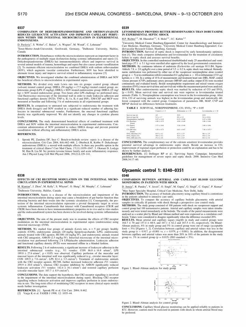
0337COMBINATION OF DEHYDROEPIANDOSTERONE AND ORTHOVANADATEREDUCES LEUKOCYTE ACTIVATION AND IMPROVES CAPILLARY PERFU-SION WITHIN THE INTESTINAL MICROCIRCULATION IN EXPERIMENTALENDOTOXEMIA
D. Pavlovic1, S. Weber1, C. Balzer1, A. Wegner1, M. Wendt1, C. Lehmann2
1Ernst-Moritz-Arndt-Universitat, Greifswald, Germany, 2Dalhousie University, Halifax,Canada
INTRODUCTION. Disturbances within the microcirculation represent an important factor inthe pathogenesis of multiple organ dysfunction during systemic inflammation and sepsis [1].Dehydroepiandosterone (DHEA) has immunomodulatory effects and improves survival inseveral animal models of trauma, hemorrhage and sepsis but also causes potent vasodilatation[2]. To maintain efficient microcirculation we combined DHEA with sodium orthovanadate(SOV), which augments vascular contraction. Furthermore, SOV has been identified toattenuate tissue injury and improve survival related to inflammatory response [3].
OBJECTIVES. We investigated whether the combined administration of DHEA and SOVhas beneficial effects to microcirculation in experimental sepsis.
METHODS. We divided sixty male Lewis rats into six groups: control group; ethanol(solvent) treated control group; DHEA (50 mg/kg) + (7.5 mg/kg) treated control group; en-dotoxemic group (LPS 15 mg/kg); DHEA + SOV treated endotoxemic group; DHEA (25 mg/kg) + SOV treated endotoxemic group. Two hours after LPS challenge we performed intra-vital fluorescence microscopy of the intestinal wall in order to study leukocyte adhesion andfunctional capillary density (FCD). TNF-a, IL-1a, IL-4 and INFc, GM-CSF and MCP weremeasured at baseline and following 3 h of endotoxemia in all experimental groups.
RESULTS. In comparison to untreated rats subjected to endotoxemia the treatment withDHEA (both dosages) and SOV resulted in a significant reduced number of adhering leu-kocytes in intestinal submucosal venules. Furthermore, the mucosal functional capillarydensity was significantly improved. We did not identify any changes in cytokine plasmalevels.
CONCLUSIONS. The study demonstrated beneficial effects of combined treatment withDHEA and SOV within the intestinal microcirculation in experimental endotoxemia. Con-comitant administration of SOV permitted to reduce DHEA dosage and prevent potentialvasodilation without affecting anti-inflammatory DHEA action.
REFERENCES.
1. Spronk PE, Zandstra DF, Ince C: Bench-to-bedside review: sepsis is a disease of themicrocirculation. Crit Care 2004, 8(6):462–468. 2. Oberbeck R, Kobbe P: Dehydroepi-androsterone (DHEA): a steroid with multiple effects. Is there any possible option in thetreatment of critical illness? Curr Med Chem, 17(11):1039–1047. 3. Okutani D, LodygaM, Han B, Liu M: Src protein tyrosine kinase family and acute inflammatory responses.Am J Physiol Lung Cell Mol Physiol 2006, 291(2):L129–141.
0338EFFECTS OF CB2 RECEPTOR MODULATION ON THE INTESTINAL MICRO-CIRCULATION IN EXPERIMENTAL SEPSIS
M. Kianian1, J. Zhou1, M. Kelly1, S. Whynot1, O. Hung1, M. Murphy1, C. Lehmann1
1Dalhousie University, Halifax, Canada
INTRODUCTION. Sepsis is a disease of the microcirculation and impairment of theintestinal microcirculation during sepsis may cause a breakdown of gut barrier function thusreleasing bacteria and their toxins into the systemic circulation [1]. Consequently, the pro-tection of the intestinal microcirculation represents a pivotal therapeutic target in severesystemic inflammation. Cannabinoids that interact with Cannabinoid receptors (CB1R andCB2R) have been shown to have immunomodulatory properties in in vivo and in vitro studiesand the endocannabinoid system has been shown to be involved during systemic inflammation[2].
OBJECTIVES. The aim of the present study was to examine the effects of CB2 receptormodulation on the intestinal microcirculation in experimental sepsis (endotoxemia) usingintravital microscopy (IVM).
METHODS. We studied four groups of animals (Lewis rats, n = 5 per group): healthycontrols (CON), endotoxemic animals (20 mg/kg lipopolysaccharide; LPS), endotoxemicanimals treated with CB2 agonist, HU308 (10 mg/kg IV), and endotoxemic animals treatedwith CB2 antagonist, AM630 (2.5 mg/kg IV). Intravital microscopy of the intestinal micro-circulation was performed following 2 h LPS/placebo administration. Leukocyte adhesionand functional capillary density (FCD) were measured offline in a blinded fashion.
RESULTS. Following 2 h of endotoxemia, a significant increase of leukocyte adhesion in theintestinal submucosal venules (e.g., V1 venules: CON 86.8 ± 16.8 n/mm2, LPS189.8 ± 27.6 n/mm2, p \ 0.05) was observed. Capillary perfusion of the muscular andmucosal layers of the intestinal wall was significantly reduced (e.g., circular muscular layer:CON 109.9 ± 7.8 cm/cm2, LPS 82.4 ± 3.5 cm/cm2). Treatment of endotoxemic animalswith the CB2 receptor agonist, HU308, further increased leukocyte adhesion (V1 venules:259.9 ± 20.0 n/mm2), whereas CB2 receptor inhibition by AM630 significantly reducedleukocyte activation (V1 venules: 142.4 ± 16.1 n/mm2) and restored capillary perfusion(circular muscular layer: 107.7 ± 8.9 cm/cm2).
CONCLUSIONS. The data support the hypothesis, that CB2 receptor signalling is involvedin the impairment of the intestinal microcirculation during sepsis. Blocking CB2 receptorsignalling reduces leukocyte activation and improves capillary perfusion in acute endotoxe-mia in rats. The long-term effect of modulating CB2 receptors in more clinical sepsis modelsneeds further investigation.
REFERENCES. [1] Spronk PE et al. Crit Care. 2004; 8:462.[2] Varga K et al. FASEB J 1998; 12: 1035–44.
0339LEVOSIMENDAN PROVIDES BETTER HEMODYNAMICS THAN DOBUTAMINEIN EXPERIMENTAL SEPTIC SHOCK
H.P. Richter1,2, M. Ehrenfort1,2, S. Holst1,2, J.C. Kubitz1,2
1University Medical Center Hamburg-Eppendorf, Center for Anaesthesiology and IntensiveCare Medicine, Hamburg, Germany, 2University Medical Center Hamburg-Eppendorf, Car-diovascular Research Center, Hamburg, GermanyINTRODUCTION. Survival of septic shock is improved by early hemodynamic optimiza-tion [1]. This study compares dobutamine and levosimendan for the treatment of circulatoryfailure in septic shock and assesses survival benefits.OBJECTIVES. In this controlled randomized doubleblinded study 25 anaesthetized and venti-lated pigs (37.1 ± 5.1 kg) were enrolled after approval by the local governmental commission.METHODS. By continuous infusion of endotoxin (Escherichia coli serotype 0111:B4, Sigma-Aldrich; 7.8 ± 2.0 lg/kg/h) over a time period of 3.5 ± 0.2 h, septic shock was induced. Hemody-namic stabilization was performed by either use of the vasopressor norepinephrine alone (controlgroup; n = 5)or in combination with levosimendan (0.4 lg/kg/min; n = 10)or dobutamine (15.0 lg/kg/min; n = 10). In a setting of 10 h of measurements and treatment heart rate (HR), MAP, centralvenous pressure (CVP), pulmonary artery pressure (MPAP) and cardiac output (CO) were recordedcontinuously and evaluated hourly. Beside norepinephrine requirement and mixed venous oxygensaturation (SVO2) mean survival time and survival rate within the measurement period were analysed.RESULTS. After endotoxinemia septic shock was marked by reduction of CO and SVO2
[p \ 0.05]. Mean survival time and survival rate were superior in levosimendan treatedanimals (Table 1). Norepinephrine consumption was lowest in the levosimendan group. After10 h, CO of surviving animals was highest in the levosimendan group and statistically dif-ferent compared with the control group. Comparison of parameters HR, MAP, CVP andMPAP showed no differences between treatments.
TABLE 1 SURVIVAL, NOREPINEPHRINE, CO, SVO2; *P \ 0.05Control; n = 5 Dobutamine; n = 10 Levosimendan; n = 10
Survival rate [n] 4/5 5/10 8/10
Survival time [h] 9.2 ± 1.7 8.0 ± 2.2 9.6 ± 0.8 h
Norepinephrine [mg/kg/h] 0.25 ± 0.10 0.33 ± 0.09 0.15 ± 0.06
CO (Baseline)
[l/min]
4.98 ± 0.73 4.90 ± 0.49 5.08 ± 0.46
CO (Sepsis)
[l/min]
3.73 ± 0.64 3.36 ± 0.46 3.60 ± 0.46
CO (600 min)
[l/min]
4.20 ± 0.73 6.62 ± 0.67 7.46 ± 0.52 *
SVO2 (Baseline) [%] 63.9 ± 4.2 67.5 ± 2.9 68.4 ± 2.9
SVO2 (Sepsis) [%] 50.8 ± 4.2 52.5 ± 2.9 45.8 ± 2.9
SVO2 (600 min) [%] 57.4 ± 4.8 71.3 ± 4.9 63.3 ± 3.4
CONCLUSIONS. The complementary use of the calcium sensitizer levosimendan providespotential survival advantage in endotoxemic septic shock. Beside an increase in CO,improvement of regional organ perfusion or protection could be an explanation and has to beshown by further analysis.REFERENCE(S). [1] Dellinger RP et al.; Surviving Sepsis Campaign; Internationalguidelines for management of severe sepsis and septic shock: 2008. Intensive Care Med2008;34:17–60.
Glycaemic control 1: 0340–03510340COMPARISON BETWEEN ARTERIAL AND CAPILLARY BLOOD GLUCOSEMONITORING IN PATIENTS WITH SHOCK
D. Juneja1, R. Pandey1, Y. Javeri1, O. Singh1, M. Gupta1, G. Singh1, C. Gupta1, P. Kumar1
1Max Super Specialty Hospital, Critical Care Medicine, New Delhi, IndiaINTRODUCTION. The accuracy of bedside glucometry using capillary blood is contentiousin the patients admitted to intensive care units.OBJECTIVES. To compare the accuracy of capillary bedside glucometry with arterialsamples in critically ill patients with shock through a prospective case control study.METHODS. The study group consisted of 100 patients with shock on vasopressor support andcontrol group had 100 normotensive patients. Arterial and capillary samples were taken simulta-neously and were tested immediately at the bedside. The results of the paired measurements wereanalysed as a scatter plot by Bland and Altman method and were expressed as a correlation coef-ficient. Values were considered to disagree significantly when the difference exceeded 20%.RESULTS. Mean arterial and capillary sugars (mg/dl) in study and control groups were164.7 ± 70 and 157.4 ± 68.9, and 167.1 ± 62.2 and 167.5 ± 61, respectively. On Bland–Altman analysis, 6% in study group and 5% in control group were out of range (acceptablelimit \ 5%) [Figures 1, 2]. Correlation between capillary and arterial values was less in thestudy group (r = 0.917, p \0.001 vs. r = 0.979, p \ 0.001). In addition, the disagreementbetween capillary and arterial values was more than 20% in 18% of the patients in the studygroup vs. 3% in control group (p = 0.015) (ISO standard \ 5%).
Figure 1. Bland–Altman analysis for study group
Figure 2. Bland–Altman analysis for control group
CONCLUSIONS. Capillary blood glucose monitoring can be applied reliably to patients inICU. However, caution must be exercised in patients with shock in whom arterial blood maybe preferred.
S170 23rd ESICM ANNUAL CONGRESS – BARCELONA, SPAIN – 9–13 OCTOBER 2010
0341HIGH-DOSE VITAMIN D3 IN THE MEDICAL ICU: A RANDOMIZED, DOUBLE-BLIND, PLACEBO-CONTROLLED PILOT STUDY
K. Amrein1, G. Wagner2, H. Sourij1, S. Amrein3, T.R. Pieber1, K. Smolle1, H. Dobnig1
1Medical University of Graz, Department of Internal Medicine, Graz, Austria, 2MedicalUniversity of Graz, Department of Medical Informatics, Statistics and Documentation, Graz,Austria, 3Medical University of Graz, Department of Anaesthesiology and Intensive CareMedicine, Graz, Austria
INTRODUCTION. Vitamin D deficiency is highly prevalent in critically ill patients. A fastcorrection of low vitamin D status is attractive in this setting because vitamin D is thought toexert pleiotropic effects including those on immune function, the musculoskeletal, cardio-vascular and the metabolic system.
OBJECTIVES. Our primary objective was to evaluate the safety and efficacy of a single oralhigh dose vitamin D3 supplementation in an intensive care setting over a one-week obser-vation period.
METHODS. 540,000 IU (corresponding to 13.5 mg) of cholecalciferol (D) dissolved in45 ml herbal oil or matched placebo (PBO) were given enterally (via nasogastric feeding tubeor swallowed) to 25 patients with vitamin D deficiency [25(OH)D B 20 ng/ml] in the medicalICU.
RESULTS. Baseline characteristics including age, sex and SAPS II were balanced betweenthe two groups (mean age 62 ± 16 years, 76% male, SAPS II 36 ± 18). Mean serum25(OH)D levels at baseline were 14 ± 4 ng/ml in both groups. The mean serum 25(OH)Dincrease in the intervention group was 25 ng/ml (range 1–47 ng/ml). Two patients showed asmall (7 ng/ml) or no response (1 ng/ml) attributable to gastrointestinal dysfunction afterprolonged hypoxia and gastrointestinal GVHD after allogeneic stem cell transplantation. Thetime course of the 25(OH)D response is given in Figure 1. The highest 25(OH)D levelreached was 63 ng/ml. Hypercalcaemia did not occur in any patient. Total serum calciumlevels increased by 0.11 (PBO) and 0.15 mmol/L (D; p \ 0.05 for both), probably attributableto acute immobilisation. Ionised calcium levels increased significantly in the D group only(1.07 mmol/L on day 0–1.18 on day 7). There was no increase in triglyceride levels or orcalciuria in either group. Parathyroid hormone (PTH) levels decreased by 19 and 27 pg/ml((PBO and D, ns) over the 7 days, while 1.25(OH)D showed a non-significant increase (46–74in the PBO and 69–111 pmol/l in the D group).
Figure 1
CONCLUSIONS. This pilot study shows that a single oral ultra-high dose of 540,000 IUcholecalciferol is able to restore normal 25(OH)D levels within 2 days in most patientswithout causing adverse effects. Further research is needed to establish whether 25(OH)Dsupplementation can affect the clinical outcome of vitamin D deficient critically ill patients.
0342ACCURACY AND RELIABILITY OF A SUBCUTANEOUS CONTINUOUS GLU-COSE MONITORING SYSTEM (CGMS) IN CRITICALLY ILL PATIENTS
R. Brunner1, R. Kitzberger1, W. Miehsler1, H. Herkner2, C. Madl1, U. Holzinger1
1Medical University of Vienna, General Hospital, Dept. of Medicine III, Vienna, Austria,2Medical University of Vienna, General Hospital, Dept. of Emergency Medicine, Vienna,Austria
INTRODUCTION. Continuous glucose monitoring (CGM) has been proposed to optimizeglucose control in critically ill patients. To achieve strict glucose regulation, accurate andreliable CGM systems (CGMS) are essential.
OBJECTIVES. Evaluation of a subcutaneous CGMS for use in critically ill patients.
METHODS. 2045 CGMS sensor glucose values of 174 critically ill patients were comparedto arterial reference blood glucose levels, determined by a blood gas analyzer. CGM data wasrecorded continuously for up to 72 h using a subcutaneous CGM sensor (CGMS GoldTM;Guardian�; Medtronic, Northridge, CA, USA). The correlation of both methods and thedifferences between CGMS and reference values were calculated, as well as the conformity ofCGM values with ISO criteria. Influence of covariates on the absolute value of the bias wasexamined by multivariable random coefficient modeling.
RESULTS. The Pearson correlation coefficient was 0.92, showing strong correlation betweenthe two methods. Mean difference ± SD between CGMS and reference values was-0.100 ± 0.641 mmol/L (CGMS minus reference) and absolute difference ± SD0.441 ± 0.475 mmol/L. According to the insulin titration error grid analysis 99.1% of CGMSvalues were in the acceptable treatment zone. No CGMS measurements were found in thelife-threatening zone. 92.9% of the CGMS glucose values met the ISO criteria. Multivariablerandom coefficient modeling revealed that only the covariates age (0.003 mmol/L per addi-tional year), glucose level (0.014 mmol/L per additional mmol/L glucose) and rank ofmeasurement per patient (-0.015 mmol/L per rank) had a significant influence on the dif-ference of CGMS to the reference value.
CONCLUSIONS. The subcutaneous CGMS is reliable for use in critically ill patients andshowed glucose values with a strong correlation to arterial reference blood glucose levels,determined by a blood gas analyzer.
0343A TEST BENCH FOR TIGHT GLYCEMIC CONTROL ALGORITHMS BASED ONA SET OF VIRTUAL ICU PATIENTS
A. Guerrini1,2, M. Sorine2, P. Kalfon3
1LK2, Saint Avertin, France, 2I.N.R.I.A, Paris-Rocquencourt, France, 3C.H. Chartres, Char-tres, France
INTRODUCTION. Considerable controversy has emerged as to whether tight glucosecontrol (TGC) is warranted in all critically ill adult patients. Recently, a new blood glucoseupper limit (10 mmol/L) has been assessed as more appropriate. Rather than blood glucosetarget ranges, algorithms used to achieve TGC should be numerically evaluated before ini-tiating clinical trials (preclinical validation test). Our purpose was to assess performances ofTGC algorithms in realistic virtual ICU patients.
METHODS. We compared numerically the NICE-SUGAR algorithm (N–S)1 and the CGAOsystem (CGAO) used in the ongoing CGAO-REA study [ClinicalTrials.gov, ID:NCT01002482]. Aset of virtual patients constituting the test bench was built with 1) real data coming from patientscontrolled with CGAO before starting CGAO-REA and 2) a non-linear pharmaco-dynamic glucose-insulin system model2 where patient endogenous glucose clearance and insulin-sensitivity were timevarying parameters. In order to anticipate how algorithms would manage glycaemic control inclinical settings, delayed controls and inaccuracy of glucometers were implemented. The overallperformance of each algorithm over the whole stay was assessed according to standard scores.
RESULTS. The percentage of time in the target range [4.4–6.1 mmol/L] with N–S was lessthan 50% for almost all patients. In insulin-sensitive patients, glycemic fluctuations andsometimes severe hypoglycemia are induced by N–S (Fig. 1). The mean time in the targetrange with CGAO was about 60% and variability scores were significantly lower than with N–S. Mean glucose and standard deviations were always lower with CGAO than with N–S.
CONCLUSIONS. A numerical test bench constituted of realistic virtual ICU patients, whosefeatures were defined from real data obtained in patients under glycemic control, enabled todetermine the best algorithms candidate for further evaluation in clinical settings. Accordingto this approach, the algorithm used to achieve TGC in NICE-SUGAR would not have beenselected for such a large clinical trial while CGAO reached the first validation step in sim-ulation. We recommend that further glucose control studies focus not only on the target rangebut also on the algorithmic properties.
REFERENCE(S). [1] https://studies.thegeorgeinstitute.org/nice[2] C.E. Hann, et al., Integral-based parameter identification for long-term dynamic veri-
fication of a glucose-insulin system model, Comput. Methods Programs Biomed. 2005.
Figure 1 Performances of algorithms
0344IS HYPOGLYCAEMIA RELATED TO INSULIN THERAPY IN INTENSIVE CAREPATIENTS?
S. Nair1, C. Ferguson1
1Plymouth Hospitals NHS Trust, Plymouth, UK
INTRODUCTION. There has been much debate in recent years about the appropriate levelof blood glucose for intensive care patients with proposals of different levels of glucosecontrol using insulin infusions. One risk of intensive glucose control is hypoglycaemia andthis has been proposed as a measure of quality of care given by delivering the protocol safely.The NICE-SUGAR1 trial found that intensive glucose control increased mortality amongadults in intensive care.
OBJECTIVES. The aim of our study was to record hypoglycaemia and study it’s relation toinsulin therapy. Insulin therapy on our unit follows the recommendations of the NICE-SUGAR trial.
METHODS. Hypoglycaemia was recorded as a blood glucose level\3 mmol/L. Levels weredetected using the blood gas analyser (Radiometer 825 M). Data was recorded at the time ofhypoglycaemia to provide an explanation using the Innovian system which is the paperlesspatient record system on our unit. Data was obtained over a period of 3 months betweenOctober 2009 and December 2009. Data recorded included adverse events which were definedas worsening shock and/or increasing inotropic support. Feeding status at the time of hypo-glycaemia was recorded.
RESULTS. There were a total of 375 admissions over this period and there were a total of13,304 blood glucose measurements.
24 incidents of hypoglycaemia were recorded, of which 13 patients were on insulin and 11 werenot. Of the 13 patients who were on insulin, 5 had adverse events at the time of hypoglycaemia.All these 5 patients died within 24 h of the adverse event. All except one was on full feed. Theothers had minimal feed due to poor absorption. Of the 8 patients who did not have adverseevents, 7 were discharged and one died 2 days after the hypoglycaemic event due to worseningsepsis. Of the 13 patients on insulin, there were 3 iatrogenic errors where feeding was stoppedand the insulin was left on. None resulted in any adverse outcome for the patients.
Of the 11 patients who were not on insulin therapy, 8 had adverse events at the time. 7 diedwithin 24 h of the adverse event and 1 died 2 days later. The remaining 3 patients weredischarged. None of the 11 patients were on full feeding protocol.
CONCLUSIONS. Our findings suggest that hypoglycaemia in our unit is not primarily relatedto insulin therapy. It is related to adverse events and possibly inappropriate feeding at the time ofhypoglycaemia. Hypoglycaemia, in the absence of insulin therapy, is associated with a pooroutcome. Use of hypoglycaemia as a quality indicator should be interpreted with caution.
REFERENCE(S). 1. Intensive versus conventional glucose control in critically ill patients.N Engl J Med. 2009 Mar 26;360(13):1283–97.
23rd ESICM ANNUAL CONGRESS – BARCELONA, SPAIN – 9–13 OCTOBER 2010 S171
0345PREVALENCE OF VITAMIN D DEFICIENCY AND CORRELATION WITHOUTCOME IN INTENSIVE CARE PATIENTS IN WINTER AND SUMMER
J.J. Weenink1,2, K.T.K.H. Yap3, P.H.J. van der Voort1, E.H. Slaats4, H.M. Oudemans-vanStraaten1
1Onze Lieve Vrouwe Gasthuis, Intensive Care Medicine, Amsterdam, Netherlands, 2TweeSteden Ziekenhuis, Intensive Care/Internal Medicine, Tilburg, Netherlands, 3University ofAmsterdam, Amsterdam, Netherlands, 4Onze Lieve Vrouwe Gasthuis, Laboratory of ClinicalChemistry, Amsterdam, Netherlands
INTRODUCTION. Vitamin D deficiency seems increasingly prevalent. Pleiotropic effectsof vitamin D like immunomodulation and effects on muscle strength may be of specialimportance to critically ill patients [1]. However, vitamin D deficiency has only been studiedin small and selected groups of ICU patients [2].
OBJECTIVES. To prospectively determine the prevalence of vitamin D deficiency in winterand summer and relate vitamin D status to outcome in cohorts of critically ill patients.
METHODS. In a prospective observational cohort study performed in a 20-bed mixed ICUwe measured 25-hydroxyvitamin D (Vit D) on admission in all consecutive patients admittedin March/April 2009 and in an equally sized cohort in July/August/September 2009. Patientsreceived enteral feeding. Additional vitamin D was not supplied. Vitamin D status wasdefined as:Adequate:[75, insufficient: 50–75, deficient: 25–50, severely deficient:\25 nmol/L (to convert values to ng/ml, divide by 2.50). We compared observed and predicted mortality(APACHE IV) between the cohorts.
RESULTS. Vit D was measured in 111 patients admitted in winter and 112 patients admittedin summer (Table 1). Mean Vit D was significantly lower in winter than in summer. In winter,85% was deficient, 49% severely deficient. In summer, 50% was deficient, 9% severelydeficient. Predicted mortality was higher in winter and higher in Vit D deficient patients.Observed mortality was lower than predicted in all groups, but not different between groups.Including both Vit D and season in a Multiple Regression Analysis, winter (p = 0.004) andnot vit D (p = 0.94) was related to predicted mortality.
TABLE 1All Winter Summer p Vit D B 25 [25 p
Nr 223 111 112 66 157
Age 66 67 65 NS 67 66 NS
Vit D, 95%CI 40.2, 37–43 29.4, 26–33 50.8, 47–55 \0.001*
Observed
Mort., 95%CI
13%, 9–18% 16%, 9–23% 11%, 5–17% 0.25� 18%, 9–28% 11%, 6–17% 0.20�
APIVPM,
95%CI
0.25, 0.21–0.29 0.31, 0.25–0.37 0.18, 0.13–0.24 0.001* 0.33, 0.25–0.40 0.21, 0.17–0.26 0.01*
CI = confidence interval, * t test, � Chi2 testCONCLUSIONS. Vitamin D was significantly lower in patients admitted in winter comparedto summer. Half of the winter patients were severely deficient. Hospital mortality was notsignificantly different between cohorts. Predicted mortality was higher in the winter cohortand in patients with Vit D deficiency. In a multiple regression analysis, winter and not vitaminD level was related to predicted mortality. However, vitamin D deficiency might increase thesusceptibility to disease. Further studies are needed.
REFERENCE(S). 1. Lee P. ICM 2009:35;2028 2. Lee P. NEJM 2009:360;1912
0346EXOGENOUS GLUCAGON-LIKE PEPTIDE-1 ATTENUATES THE GLYCAEMICRESPONSE TO ENTERAL NUTRIENT IN CRITICALLY ILL PATIENTS WITHPRE-EXISTING TYPE-2 DIABETES MELLITUS
A. Deane1,2, M.J. Chapman1,2, R.J. Fraser3, M.J. Summers4, A.V. Zaknic4, A. DiBartolomeo2,
M. Horowitz3
1Royal Adelaide Hospital, Intensive Care Unit, Adelaide, Australia, 2University of Adelaide,Discipline of Acute Care Medicine, Adelaide, Australia, 3University of Adelaide, Disciplineof Medicine, Adelaide, Australia, 4Royal Adelaide Hospital, Department of Gastroenterology,Adelaide, Australia
INTRODUCTION. Glucagon-like peptide-1 (GLP-1) lowers blood glucose via stimulationof insulin and suppression of glucagon secretion, as well as slowing gastric emptying. Wehave previously shown that exogenous GLP-1 attenuates hyperglycaemia in non-diabeticcritically ill patients [1, 2]. However, islet cell function in critically ill diabetic patients maybe so disturbed that pharmacological doses of GLP-1 have no effect in this group.
OBJECTIVES. The aim of this study was to evaluate the effect of exogenous GLP-1 onglycaemic excursions during intraduodenal nutrient infusion in critically ill patients with pre-existing type-2 diabetes mellitus.
METHODS. Nine critically ill, mechanically ventilated, patients with pre-existing type-2diabetes (8 M:1F, Age 56 ± 6 years, HbA1C 8.7% ± 0.7%, BMI 35 ± 3 kg/m2, APACHEII on day 1 of study 17 ± 3, days in ICU on day 1 of study 5 ± 1) received IV infusions ofGLP-1 (1.2 pmol/kg/min), and placebo, from t = 0–270 min on separate days in a random-ised, double-blind, fashion. Between t = 30–270 min a liquid nutrient (Ensure) was infusedintraduodenally at a rate of 1 kcal/min via a naso-enteric feeding catheter. Blood glucoseconcentrations were measured by glucometer at 15 min intervals. Data are mean±SEM andcomparisons are using Student’s t test.
RESULTS. Prior to the commencement of IV infusions there was no difference in bloodglucose between the groups (at t-0 min: GLP-1: 8.1±0.7 mmol/l vs. placebo: 8.8 ±1.1 mmol/l; P = 0.38). During fasting, GLP-1 had no effect on glycaemia (at t = 30 min: GLP-1:7.7 ± 0.6 mmol/l vs. placebo: 8.9 ± 1.1 mmol/l; P = 0.18). However, GLP-1 attenuated theoverall glycaemic response to the nutrient (AUC30–270 min: GLP-1: 2,208 ± 158 mmol/l.minvs. placebo: 2,647 ± 247 mmol/l.min; P \ 0.01), as well as the peak blood glucose (GLP-1:11.1 ± 0.9 mmol/l vs. placebo: 12.5 ± 1.1 mmol/l; P \ 0.04)
CONCLUSIONS. Exogenous GLP-1 is effective in reducing the glycaemic excursions thatoccur with enteral nutrient critically ill patients with pre-existing type 2 diabetes mellitus.These data indicating that further studies using GLP-1, or its analogues, are warranted in thisgroup.
REFERENCE(S). [1] Deane AM, et al.: Critical Care 2009 May 13; 13(3):R67[2] Deane AM, et al.: Critical Care Medicine 2010 May; 38(5):1261–1269
GRANT ACKNOWLEDGMENT. Supported by a National Health & Medical ResearchCouncil of Australia Grant (NHMRC 508081)
0347TIGHT GLYCAEMIC CONTROL: ARE WE REALLY PUTTING OUR PATIENTSAT RISK?
G. Barton1, K. Davies2, L. Gibson3, D. Hughes4
1St Helens and Knowsley Teaching Hospitals NHS Trust, Pharmacy, Liverpool, UK, 2MidCheshire NHS Trust, Liverpool, UK, 3Southport and Ormskirk NHS Trust, Liverpool, UK,4Wirral NHS Trust, Chester, UK
INTRODUCTION. Hyperglycaemia and insulin resistance are recognised as an almostuniversal complication of the septic patient and lead to a myriad of further complications. In2001 Greet Van Den Berghe et al. reported that normalisation of blood glucose levels incritically ill post surgical patients improved Intensive Care (ICU) mortality, particularly inpatients who stayed on ICU longer than 5 days1. More recent studies have failed to show thisbenefit2,3,4,5, whilst raising concerns regarding an increased risk of hypoglycaemia.3,4,5.Locally most units adopt a protocol that reflects the practice of the original 2001 study.
OBJECTIVES. This study was conceived due to concerns around the safety of tight gly-caemic control (TGC). Our objectives were to measure adherence to our local policies andascertain our true rates of hypoglycaemia.
METHODS. This study was designed as a retrospective audit on four critical care units in theCheshire and Mersey Critical Care Network. Each site used the same audit tool but adapted itto allow for differences in local practice and protocols. Data pertaining to the prescribing andadministration of insulin was collected daily over a 1 week period (the time of data collectionvaried from day to day). The doctors and nursing staff were unaware of the audit and the datawas collected by the ward pharmacist who suggested modifications to therapy if it wasdeemed inappropriate or unsafe.
RESULTS. 109 patient days worth of data was collected with 897 blood glucoses checked inthis period. 100% of patients receiving insulin had insulin prescribed. Only 23% of bloodglucoses were within the target range set by the local protocol. However, of all the resultsonly 3.5% were ‘‘low’’ as defined by the local protocol, and only 0.1% (1/897) were hypo-glycaemic episodes as defined in the Greet Van Den Berghe paper of 2001 (\2.2 mmol/L).Conversely, 73.5% were above the target range. In the 2 trusts that recorded how many ofthese levels were[8 mmol/L (a proposed alternative upper limit), the rates were 43 and 46%.In response to a blood glucose the policies suggest dosage adjustments/maintenance. On only46% of occasions were the adjustments made correct.
CONCLUSIONS. Insulin infusions appeared to be managed safely by nursing staff. Insulin,if given, was always prescribed and hypoglycaemia (blood glucose \2.2 mmol/L) occurredon only one occasion. Although safe, adustments often didn’t follow the protocols and thepatients’ blood glucose were within the target range only 23% of the time, potentiallynegating many of the perceived benefits of TGC. Reasons for non-compliance with theprotocols was difficult to objectively establish
REFERENCE(S). 1. Van Den Berghe G et al. N Engl J Med 2001;345:1359–67.2. Van Den Berghe G et al. N Engl J Med 2006;354:449–61.3. Devos P et al. Intensive Care Med 2007;33:suppl 2:S1894. Brunkhorst FM et al. N Engl J Med 2008;358:125–395. The NICE-SUGAR Study Investigators. N Engl J Med 2009;360:1283–97.
0348DOES HEMOGLOBIN A1C LEVEL WHICH INDICATES LONG TERM DIABETıCCONTROL EFFECT SURGICAL OUTCOMES AFTER CORONARY ARTERYBYPASS GRAFT SURGERY?
Y. Bicer1, S. Ay1, E. Hakko2, C. Arıturk3, A.H. Aslan3, S. Cicek3
1Anadolu Medical Center, Anestesiology and Reanimation, Kocaeli, Turkey, 2AnadoluMedical Center, Infectious Disease and Clinical Microbiology, Kocaeli, Turkey, 3AnadoluMedical Center, Cardiovascular Surgery, Kocaeli, Turkey
INTRODUCTION. Diabetes mellitus has been associated with an increased risk of adverseoutcomes after coronary artery bypass grafting. Hemoglobin A1c is a reliable measure oflong-term glucose control. It is unknown whether adequacy of diabetic control, measured byhemoglobin A1c, is a predictor of adverse outcomes after coronary artery bypass grafting.
MATERIAL AND METHOD. We evaluated 123 consecutive diabetic patients whounderwent primary, elective coronary artery bypass grafting at the Anadolu Medical Center.HbA1c levels of all patients with diabetes mellitus were measured and value of 7% or greaterwas used as a threshold for uncontrolled hyperglycemia. All the peroperative variables wererecorded and then, statistically evaluated. The statistical analysis was realised by t test forparametric variables and Chi-square test for nonparametric variables.
RESULTS. There were 305 consecutive patients that underwent elective coronary arterybypass graft surgery between January 2009 and April 2010. Among them, 123 patients haddiabetes mellitus and others not. There were no significant differences between groupsregarding each adverse outcomes (Table 1). Although, 13 (68.5%) of total 19 surgical siteinfection in 305 patients had been seen in 123 diabetic patients, there were also no significantdifferences between groups regarding the rate of infections (Table 2). There was no earlypostoperative mortality in 123 diabetic patients.
TABLE 1 ADVERSE OUTCOME IN 123 DIABETIC PATIENTSComplications (n/%) HbA1c [ %7(65/53) HbA1c \ %7 (58/47) p value
Postoperative atriyal fibrillation 5/7.7 6/10.3 ns
Cerebrovascular complications 3/4.6 4/6.9 ns
Renal dysfunction 3/4.6 2/3.4 ns
Early myocardial dysfunction 4/6.2 2/3.4 ns
ICU stay [ 3 days 6/10.3 9/13.8 ns
Prolonged hospital stay [10 days 12/16.9 14/24.1 ns
Readmittion to hospital 11/16.9 10/17.2 ns
TABLE 2 _INFECTIONS AND TYPE OF INFECTIONS IN 123Infections (n/%) HbA1c [ %7 (65/53) HbA1c \ %7 (58/47) p value
Total infections 9/13.8 6/10.3 ns
SSSI (Superfisial surgical site infection) 3/4.6 3/5.2 ns
DSSI (Deep surgical site infection) 5/7.7 2/3.4 ns
DAI (Device assocaited infection) 1/1.5 1/1.7 ns
CONCLUSION. Although, diabetes mellitus is accepted as significant risk factor for out-comes after coronary artery bypass graft surgery; HbA1c level which indicates the level ofglucose control in diabetic patients did not significantly effect the outcomes after coronaryartrey bypass graft surgery.
REFERENCES. Halkos ME, Puskas JD, Lattouf OM, Kilgo P, Kerendi F, Song HK, GuytonRA, Thourani VH. Elevated preoperative hemoglobin A1c level is predictive of adverseevents after coronary artery bypass surgery. J Thorac Cardiovasc Surg. 2008Sep;136(3):631–40
S172 23rd ESICM ANNUAL CONGRESS – BARCELONA, SPAIN – 9–13 OCTOBER 2010
0349LOW RISK OF HYPOGLYCEMIA DURING INTENSIVE INSULIN THERAPY
W.P. Kingma1, A.J. Bakker2, H. Buter1, M. Koopmans1, E.C. Boerma1, M.A. Kuiper1
1Medical Center Leeuwarden, Intensive Care Medicine, Leeuwarden, Netherlands, 2MedicalCenter Leeuwarden, Dept of Clinical Chemistry, Leeuwarden, Netherlands
INTRODUCTION. In 2004 in Medical Center Leeuwarden (MCL) we introduced IntensiveInsuline Treatment (IIT), by implementing a completely nurse driven protocol as in theLeuven I study, to achieve tight glucose control in our 16-bed medical(cardio-)surgical ICUand 6 non-ventilator beds. In the last year, the benefit of IIT and the possible detrimentaleffects of hypoglycemia on survival have been heavily debated.
OBJECTIVES. The goal is to analyze our daily practice in all ICU patients and compare thiswith the intensive treated groups from the Leuven 1, VISEP en NICE-SUGAR trial.
METHODS. We compared mean morning blood glucose levels and the percentage ofpatients who had a hypoglycaemia, defined as glucose below 2.3 mmol/l, from 2006 to 2009.The frequency of control and the insulin dosage was comparable to the Leuven 1 study.Enteral or parenteral feeding was started at admission. No standard intravenous glucose wasused. Glucose was measured with arterial blood samples on the ABL800flex Radiometer asPOCT.
RESULTS. In our patients, the mean morning blood glucose was higher than in the Leuven 1study and comparable to the VISEP and Nice-Sugar. The percentage of hypoglycemia on ourICU was lower in comparison with the VISEP and Nice-Sugar. This may be explained by theavailability of a POCT on our ICU which allows quick adjustments of the insulin dosage.
Table 1MCL
2006
MCL
2007
MCL
2008
MCL
2009
VISEP Nice_Sugar Leuven 1
Patients 973 1151 1438 1459 247 3016 755
Mean morning
blood
glucose
6.08 6.22 6.27 6.44 6.22 6.55 5.71
SD 2.17 2.16 1.95 2.06 1.0 1.39 1.05
95% CL-mean 5.9–6.2 6.1–6.4 6.2–6.4 6.3–6.6 6.1–6.3 6.5–6.6 5.6–5.8
% patients with hypoglycaemia
\2.3 mmol/l
5.3 5.2 3.6 3.6 17 6.8 5.1
CONCLUSIONS. IIT with adequate control of blood glucose and a low rate of hypogly-caemia using the Leuven nurse driven protocol is feasible in daily ICU practice for alladmitted patients.
0350REDUCED ORGAN FAILURE WITH EFFECTIVE GLYCEMIC CONTROL
J.-C. Preiser1, J.G. Chase2, C.G. Pretty2, L. Pfeifer3, G.M. Shaw3, A.J. Le Compte2, J. Lin2, D.
Hewett2, T. Desaive1
1University of Liege, Liege, Belgium, 2University of Canterbury, Christchurch, New Zealand,3Christchurch Hospital, Christchurch, New Zealand
INTRODUCTION. Mortality in the intensive care unit (ICU) after the first few days isstrongly associated with organ failure rate and severity. This study uses the sequential organfailure assessment (SOFA) score to evaluate the impact of a successful tight glycemic control(TGC) intervention (SPRINT) on organ failure, morbidity, and thus mortality.
OBJECTIVES. To determine if effective TGC reduces the rate, level or number of organfailures in providing a platform for reduced ICU mortality.
METHODS. A retrospective analysis of clinical data from 2003 to 2007 totalling 784 ICUpatients and 6574 patient days. The data includes 371 patients (3358 patient days) on theSPRINT TGC protocol (August 2005–April 2007) and 413 matched retrospective patients(3,216 patient days) from the 2 years prior to the implementation of SPRINT in 2005, mat-ched by APACHE III. Cohort details including glycemic and mortality outcome are in [1].SOFA score was calculated daily for each patient. The effect of the SPRINT TGC inter-vention is assessed by the percentage of patients each day with a total SOFA score B5, and bytrends over time. Organ-failure free days (all component SOFA scores B2) and total numberof organ failures in each cohort (SOFA component scores [2) are examined.
RESULTS. Admission SOFA scores were similar (p = 0.20). Patients reached similarmaximum SOFA scores (p = 0.76) in similar times (median: 1 day; IQR: [1, 3]) for bothcohorts (p = 0.99). Median length of stay was similar (p = 0.94) with 4.1 days (SPRINT)and 3.8 days (pre-SPRINT). The percentage of patients with total SOFA B 5 is significantlydifferent over time (p \ 0.001), rising over the first 21 days to 85% for the Pre-SPRINTcohort and *93% for the SPRINT cohort, with clear separation from days 1–2 onward(p \ 0.001). SPRINT had 41.6% of patient days free of organ failure versus 36.6% for Pre-SPRINT (p \ 0.0001). The number of organ failures, as a percentage of the total possible foreach cohort, was lower for SPRINT (16.0%) than pre-SPRINT (19.0%) (p \ 0.0001). Trendsfor survivors and non-survivors were similar within and between cohorts, with non-survivorshaving higher admission, mean daily, and maximum SOFA scores (p \ 0.05).
CONCLUSIONS. Effective TGC with SPRINT resolved organ failure faster, and for agreater percentage of patients who had similar admission and maximum SOFA scores,compared to a matched retrospective conventional control cohort. These morbidity reductionsmirror the reduced mortality seen with SPRINT. These results suggest that reduced organfailure, assessed by SOFA, is a fundamental element in reduced mortality when TGC isimplemented effectively.
REFERENCE. [1] Chase, JG et al. ‘‘Implementation and Evaluation of the SPRINT Protocolfor Tight Glycaemic Control in Critically Ill Patients: A Clinical Practice Change,’’ CriticalCare, 12(2):R49, 2010.
0351INCREASED BLOOD GLUCOSE VARIANCE IS ASSOCIATED WITH HYPOGLY-CAEMIA DURING TIGHT GLYCAEMIC CONTROL
T. Szakmany1, D. Jones1, E. Jiwanmall1
1Royal Glamorgan Hospital, Anaesthetics, Theatres and Critical Care, Llantrisant, UK
INTRODUCTION. Tight glycaemic control was reported to reduce mortality in selectedsurgical critically ill patients and lowering of blood glucose (BG) levels was recommended asa means of improving patient outcomes (1). However, this approach has been linked withsignificant risk of hypoglycaemia. Recently, several studies have confirmed significantassociations between variability of BG levels and patient outcomes (2).
OBJECTIVES. To evaluate the association between BG variability and hypoglycaemia in amixed adult ICU.
METHODS. Retrospective analysis of the prospectively collected and stored BG measure-ments over a 1 year period, during which tight glycaemic control was targeted in all patients.Every day we have calculated the BG coefficient variation as expressed by SD/mean BGlevel. We have divided the patients into Low, Medium and High variability groups (0–15,16–50 and[50, respectively). Hypoglycaemia was determined if BG was below 4.4 mmol/L.For statistical analysis Chi-square test and Pearsons correlation test was used.
RESULTS. 584 patients were admitted over the 1-year period, providing 2357 daily datapoints. BG variability was high in 16 daily measurements (0.7%), medium in 570 (24.2%) andlow in 1771 (75.1%). Hypoglycaemia occurred in 401 measurement points (17.0%). Hypo-glycaemia was observed at all points (100%) when BG variability was high vs. 32.8% whenBG variability was medium and 11.2% when BG variability was low and this difference wasstatistically significant (p = 0.001). We observed a significant correlation between increasedBG variability and hypoglycaemia (R = 0.280, p = 0.001).
CONCLUSIONS. Increased BG variability as expressed by coefficient variation is associatedwith hypoglycaemia, when measured daily in a mixed ICU population employing tight glu-cose control. Decreasing the variability of the BG concentration may be an importantdimension of glucose management. If reducing swings in the BG concentration is a majormechanism behind the beneficial effects of glucose control, it may not be necessary to pursuelower glucose levels with the associated risk of hypoglycemia.
REFERENCE(S). 1. van den Berghe G, Wouters P, Weekers F, Verwaest C, Bruyninckx F,Schetz M, Vlasselaers D, Ferdinande P, Lauwers P, Bouillon R.: Intensive insulin therapy inthe critically ill patients. N Engl J Med. 2001 Nov 8;345(19):1359–67. 2. Hermanides J,Vriesendorp TM, Bosman RJ, Zandstra DF, Hoekstra JB, Devries JH.: Glucose variability isassociated with intensive care unit mortality. Crit Care Med. 2010 Mar;38(3):838–42.
Acute kidney injury: 0352–03650352HEPATIC METABOLISM OF MIDAZOLAM IS INHIBITED IN CRITICALLY ILLPATIENTS WITH ACUTE KIDNEY INJURY
C.J. Kirwan1, I.A. MacPhee2, T. Lee3, D. Holt3, B.J. Philips1
1St Georges University of London, Intensive Care Medicine, London, UK, 2St GeorgesUniversity of London, Renal and Transplant Medicine, London, UK, 3St Georges Universityof London, Analytical Unit, London, UK
INTRODUCTION. Chronic Kidney Disease (CKD) has been shown to cause a decrease inthe hepatic metabolism of drugs in vivo and in vitro. The impact of acute kidney injury (AKI)on hepatic drug metabolism is untested. A single time point determination of midazolamconcentration, 4 h after intravenous administration, accurately predicts total midazolamexposure and thus metabolism in critically ill patients. Midazolam metabolism is a measure ofhepatic activity of cytochromes P450 (CYP3A) 3A4 and 3A5 that are involved in themetabolism of [50% of widely used drugs.
METHODS. All patients admitted to the adult intensive care unit were considered unlessthey had received a benzodiazepine in the previous 24 h, had acute or chronic hepatic failure,were pregnant or were being administered major CYP3A inhibitors i.e. macrolide antibioticsor an imidazole anti-fungal. 1 mg midazolam was given intravenously at time 0 and serumcollected at 4 h. Serum midazolam concentration was determined by mass spectrometry:T4[mid]. Patients were categorised using the AKIN/RIFLE criteria. Glomerular filtration ratewas assessed by simultaneous 4 h creatinine clearance (4CrCl) measurements.
RESULTS. 72 patients (53 with AKI) were recruited. Mean age 72 (23–90); 45 male. 19 hadnormal renal function with 16, 20, 17 in groups 1(R), 2(I) and 3(F) respectively. In criticallyill patients the median (range) T4 [midazolam] concentration N v AKI was 3.15 (1.29–10.27)and 5.57 (0.59–113.6) ng mL-1 respectively (p\0.005). There were two major outliers whichmay skew the results in favour of the hypothesis. If these two results are removed (Fig. 1) thestatistical significance remains strong (N v * p \ 0.008; **p = 0.003; ***p = 0.009)).Standard multiple regression analysis found the most useful predictors of T4[mid] were ‘timewith AKI’ and ‘serum urea’ (beta coefficient 0.33 and 0.31 (p \ 0.05) respectively). 4CrCl,Serum creatinine and urine output did not add further predictive statistical power.
Figure 1
CONCLUSIONS. This study demonstrates a reduction in the hepatic metabolism ofmidazolam associated with AKI. This effect is related most strongly to the length of time thepatient has suffered with AKI.
23rd ESICM ANNUAL CONGRESS – BARCELONA, SPAIN – 9–13 OCTOBER 2010 S173
0353AN AUDIT OF COMPLIANCE OF CARE OF PATIENTS WITH ACUTE KIDNEYINJURY COMPARED TO THE NCEPOD 2009 REPORT ‘‘ADDING INSULT TOINJURY’’
A. Roberts1, K. Hall1, H. Black1
1Wirral University Teaching Hospital, Intensive Care Unit, Liverpool, UK
INTRODUCTION. The National Confidential Enquiry into Patient Outcome and Death(NCEPOD) report entitled ‘‘Adding Insult To Injury’’ 1 reviewed the care of inpatients whodied from Acute Kidney Injury (AKI). Several recommendations were published.OBJECTIVES. To audit compliance with the key recommendations in patients at risk of AKIadmitted to our ICU.METHODS. With a questionnaire based on the NCEPOD report, all patients at risk of AKI,admitted to our ICU, had a retrospective case note review. A RIFLE (Risk, Injury, Failure,Loss, Endstage) score, a classification for AKI, triggered inclusion over a 2 month period.RESULTS. Classification: 51 patients were included. 80% scored R or worse at admission.The breakdown of RIFLE scores, R, I, and F was 49%, 24%, and 27%, respectively. In 10patients the onset of risk was as an inpatient, including 2 in the ICU. Deterioration of RIFLEscore occurred in 31%.Risk Assessment: 59% had the risk of AKI documented within 24 h. 29% had no writtenRIFLE classification and 84% had no evidence of assessment of risk factors potentiallycausing AKI.
Monitoring and Investigations: Monitoring compliance was good but there was variabilitydepending on clinical area (Table 1). 71% did not have renal imaging. 24% were reviewed bya nephrologist.
TABLE 1 PATIENT MONITORING PRE AND POST ICU %
Monitoring/investigation Pre ICU ICU
Admission electrolytes 100 100
Catheter 96 100
Hourly urine volume measurement 59 100
Physiological monitoring plan 8 43
Dipstick urinalysis 23 4
Acid base evaluation 84 100
Basic observations 100 100
Senior review within 12 h of AKI onset 68 100
CONCLUSIONS. Our results are similar to the NCEPOD report. Even with multiple rec-ommendations by NCEPOD and the National Institute for Clinical Excellence (NICE)2
recognition of the critically ill remains poor. Detection of organ failure risk is vital toimplement preventative strategies. We found a delay in AKI recognition and a lack of riskassessment. Observations, included in admission protocols, were recorded, but investigationsoutside of these, were often absent. NICE2 suggest management should be physiologicallyand not diagnosis based but few patients had a documented physiological plan.
We suggest improving under and postgraduate education to increase awareness of AKI. Thiscould occur as an extension to the national, Acute Life-Threatening Events Recognition andTreatment Course. An AKI admission protocol may allow identification of at risk patients andinstigate appropriate monitoring, investigation and management. Improved ward based fluidmonitoring and management would reduce deterioration. Incorporation of a physiologicalmonitoring plan on the ICU observation chart may reduce preventable AKI.
REFERENCES. 1. NCEPOD Adding Insult to Injury 2009 2. NICE. Clinical Guideline 50.Acutely ill patients in hospital. 2007
0354VOLUME THERAPY AS A RISK FACTOR OF KIDNEY INJURY IN PATIENTSWITH INTRACRANIAL HEMORRHAGES
J.V. Titova1, S.S. Petrikov1, A.A. Solodov1, V.V. Krylov1, S.V. Efremenko1,I.V. Alexandrova1, S.I. Rey1, E.B. Ryabov1, J.V. Puras1
1Sklifosovsky Research Institute for Emergency Medicine, Moscow, Russian Federation
INTRODUCTION. The possibility of kidney function impairment by artificial colloidsolutions was shown in a few recent investigations. One of the probable mechanisms of acuterenal failure (ARF) is hyperoncotic kidney injury during administration of hyperoncoticsolutions in patients with unstable renal perfusion pressure.
OBJECTIVES. To determine the incidence and risk factors of ARF required renalreplacement therapy (RRT) in patients with intracranial hemorrhages (ICH).
METHODS. A retrospective analysis of 274 patients with ICH admitted to the neurosurgicalICU from 01/10/2008 to 01/04/2009 was performed. 118 pts treated in ICU more than 3 dayswere selected for subsequent analysis. All patients had risk factors of ARF (vancomycin andaminoglycosides administration, hyperosmolar solutions infusion, vasopressors). Patientswere divided into 3 groups depending on colloid solution they were treated with (85–6% HES130/0.4/9:1; 24–6% HES 130/0.42/6:1; 9–10% HES 200/0.5). Average amount of infusedHES did not exceed safe recommended daily dose. Acute Kidney Injury Network (AKIN)staging system was used for ARF determination. AKIN stage III was the indication for RRT.
We used multiple linear regression analysis and odds ratio (OR) to determine rick factors ofmortality and ARF.
RESULTS. We did not find significant differences in SOFA score, artificial lung ventilationnecessity, vasopressors use, serum creatinine and urea levels during 28 days of observation in ptstreated with different types of HES. The incidence of ARF required RRT was 5.9%. Vancomycinadministration was the main rick factor of ARF (regression coefficient b = 0.146; sb = 0.058;P = 0.01). Risk of ARF was lower in pts treated with tetrastarches (6% HES 130/0.4/9:1 vs. 10%HES 200/0.5, OR 0.01 (95% CI 0.0005; 0.23, P = 0.004), 6% HES 130/0.42/6:1 vs. 10% HES 200/0.5, OR 0.09 (95% CI 0.008; 0.99, P = 0.049)). ARF development was associated with significantincrease of length of stay in ICU (pts without ARF-8 (5; 20) days; pts with ARF-28 (15;43) days,P = 0.013) and inhospital mortality (pts without ARF-24%; pts with ARF-100%, P\ 0.05). Themain risk factors of inhospital mortality were ARF (b = 0.449; sb = 0.138; P = 0.001), pneu-monia (b = 0.413; sb = 0.076; P\ 0.01) and hyperosmolar solutions administration (b = 0.233;sb = 0.075; P = 0.002). We didn’t find any correlation between inhospital mortality and type ofHES (6% HES 130/0.4/9:1 vs. 10% HES 200/0.5, OR 0.29 (95% CI 0.07; 1.2, P = 0.09); 6% HES130/0.42/6:1 vs. 10% HES 200/0.5, OR 0.4 (95% CI 0.08; 1.91, P = 0.25); 6% HES 130/0.4/9:1 vs.6% HES 130/0.42/6:1, OR 0.74 (95% CI 0.28; 1.97, P = 0.74).
CONCLUSIONS. The incidence of ARF required RRT in patients with ICH is 5.9%. ARF isassociated with significant increase of length of stay in ICU and inhospital mortality. Van-comycin and 10% HES 200/0.5 are the main rick factors of ARF required RRT.
0355CONTINUOUS HEMODIAFILTRATION IN PATIENTS WITH HYPEROSMOLARNONDIABETIC ENCEPHALOPATHY AFTER CARDIAC SURGERY
T. Mukhoedova1, Z. Unarokov1
1State Research Institute of Circulation Pathology, Department of Extracorporeal Detoxifi-cation, Novosibirsk, Russian Federation
INTRODUCTION. Hypernatremia in critically ill patients associates with severe neuro-logical disorders and increasing mortality. Continuous hemodiafiltration (CVVHDF) can be amethod of choice when traditional therapy is noneffective. The gradual slow correction ofsodium (0.5–1 mmol/l/h) and serum osmolarity is very important to avoid cerebral edema.High-sodium solutions for CVVHDF have been suggested to be used in recent years.
OBJECTIVES. To investigate the effect of CVVHDF with high-sodium solutions in patientswith hypernatremic hyperosmolar nondiabetic encephalopathy after nardiac surgery.
METHODS. Fourteen patients after nardiac surgery with hypernatremia were treated from April2005 to November 2009. Mean age 56.6 ± 2.7 years. Twelve patients were on vasopressorsupport, 13 patients on mechanical ventilation. All patients had Acute Kidney Injury in the stageInjury or Failure. APACHE II severity was 28.2 ± 0.9. All patients had the severe cerebraldisorder, severity of a Glasgow score was 8.3 ± 0.7 (from 4 to 12). Four patients had cerebralinfarction, identified by tomography. The mean pre-CVVHDF urea was 26.4 ± 2.4 mmol/l,sodium -160.2 ± 1.2 (to 175) mmol/l, and glucose level between 5.2 and 14.1 (10.2 ± 0.7)mmol/l. Calculated osmolarity was 366.2 ± 2.1 mosm/l. CVVHDF was performed on Multifiltrat(Fresenius Medical Care) and Prismaflex (Gambra Medical). 10% sodium chloride was added toreplacement and dialysate solutions to avoid concentration of natrium less than serum one to5–10 mmol/l. The concentration sodium in solutions was changing in CVVHDF.
RESULTS. CVVHDF was started on 2.3 ± 0.2 postoperative days; mean time was60.3 ± 9.1 h. The reduction rate of sodium level -0.5 to 1 mmol/l/h. Twelve patients withcorrection of sodium concentration had recovery cerebral functions after 12–56 h CVVHDF.There was no effect in 2 patients with extensive stroke and high severity of a Glasgow score(4–6 points). There were complications of therapy in no cases.
CONCLUSIONS. CVVHDF with high-sodium replacement solutions may be effective andsafe in patients with hyperosmolar nondiabetic encephalopathy associated with hypernatre-mia. Slow graded controlled correction of sodium may prevent the fatal cerebral edema.
0356ACUTE KIDNEY INJURY IN A MIX IRANIAN CRITICAL CARE UNITE, BASEDON AKI NETWORK
S.M.R. Hashemian1,2, H. Jamaati1, B. Farzanegan1, F. Rashidfarrokhi1, A. Bagheri1,M. Malekmohammad1, G. Radmand1, S. Hossein Ardehali1, K. Noorizadeh1, M. Chitsazan1,
H. Mahdi Malekpour1
1NRITLD, Tehran, Iran, Islamic Republic of, 2Tracheal Disease Research Center, Tehran,Iran, Islamic Republic of
INTRODUCTION. Acute kidney injury is a compex disorder for intensivists and patients. AKINdefinition is a new diagnostic criteria based on alternation in serum creatinin or urine output.
OBJECTIVES AND METHODS. In an observational prospective study, a total of 200patients who admitted during 6 months in a medical and surgical intensive care unit and didn0thave any recent history of renal replacement therapy were included in the study. 66(33%) of allpatients was in AKI (acute Kidney Injury) group according to the AKIN (Acute Kidney InjuryNetwork) definition. The mean of age in AKI group was more than Non-AKI (54.5 ± 19.1,43.9 ± 18.7 respectively; P \ 0.001); and had worse condition according to APACHE II (AcutePhysiology and Chronic Health Evaluation II) score (12.3 ± 5.6 vs. 6.9 ± 3.6; P \ 0.001). TheAKI patients stayed longer in ICU rather than Non-AKI patients (7.6 ± 7.6 vs. 3.7 ± 2.8 daysrespectively; P \ 0.001); with more mortality rate (19.7 vs. 1%; P \ 0.001). Also themechanical ventilation days, time of vasoacive drugs and the use of dobutamin were more in AKIgroup (P \ 0.001; P = 0.007 and P = 0.007 respectively). The AKI was a significant predictorfor mortality using the multivariate logistic regression (ORadj = 14.8; 95%CI: 1.8–121.98); andhad the same sensitivity as the APACHE II score in prediction of mortality (Sen. = 0.93).
AKI mortality
Dobutamin
CONCLUSION. The AKIN criteria for acute kidney injury, can be a good predictor ofmortality beside the APACHE II score.
REFERENCE(S). Mehta R, Kellum J, Shah S, Molitoris B, Ronco C, Warnock D, Levin A,AKINetwork:Acute Kidney Injury Network: Report of an Initiative to Improve Outcomes in Acute KidneyInjury Critical Care 2007, 11:R31Hoste EA, Clermont G, Kersten A, Venkataraman R, Angus DC, De Bacquer D, Kellum JA:RIFLE criteria for acute kidney injury are associated with hospital mortality in critically illpatients: a cohort analysis. Crit Care 2006, 10:R73.GRANT ACKNOWLEDGMENT. No
S174 23rd ESICM ANNUAL CONGRESS – BARCELONA, SPAIN – 9–13 OCTOBER 2010
0357ACUTE KIDNEY INJURY PREDICTS MORTALITY IN CRITICALLY ILLPATIENTS: A COMPARISON OF RIFLE AND AKIN CLASSIFICATIONS
M. Kompoti1, A. Paridou1, E. Stefanatou1, I. Koutsodimitropoulos1, O. Iordanidou1,
N. Markou1, F. Tsidemiadou1
1Thriassio General Hospital of Eleusis, Intensive Care Unit, Latsion Burn Center, Athens,Greece
INTRODUCTION. The impact of acute kidney injury (AKI) on the outcome of ICU patientshas well been recognized. However, there is no consensus on the definition of AKI. The mostwidely used classifications are RIFLE (Risk, Injury, Failure, Loss, End-stage kidney disease)and AKIN (Acute Kidney Injury Network).
OBJECTIVES. This study investigated AKI as predictor of ICU mortality, comparing thecorresponding predictive ability of RIFLE and AKIN classifications as well.
METHODS. Prospective cohort study of critically ill patients admitted in a multidisciplinaryICU. Patients were daily evaluated for the presence of AKI using RIFLE (1) and AKINcriteria (2). Data recorded were: age, gender, admission category (medical, elective surgical,emergency surgical, trauma, burn), Acute Physiology and Chronic Health Evaluation(APACHE) II score at admission, daily Sequential Organ Failure Assessment (SOFA) score,implementation of continuous renal replacement therapy (CRRT), use of nephrotoxic agents,ICU length of stay (LOS) and ICU mortality. Data were analyzed with logistic regression andreceiver operating characteristic (ROC) analysis with statistical significance set at p \ 0.05.
RESULTS. One hundred fourteen patients (72 male, 42 female) were included in the study.Age (mean ± SD) was 51.7 ± 20.5 years, APACHE II score 17.6 ± 8.3. AKI was recordedin 41 patients (36.0%) with RIFLE criteria (23.7% risk, 7.0% injury, 4.4% failure and 0.9%loss) and in 46 (40.4%) with AKIN criteria (21.1% stage 1, 6.1% stage 2 and 13.2% stage 3).ICU mortality was independently associated with APACHE II score at admission [odds ratio(OR) = 1.2, p = 0.001] and with AKI [OR = 9.2 (95% confidence interval 3.1–27.5) in theRIFLE-AKI model and OR = 9.1 (95% confidence interval 3.2–27.5) in the AKIN-AKImodel]. The predictive ability of RIFLE and AKIN scores at the beginning of AKI and that ofthe maximum RIFLE and AKIN scores in the ICU was poor [areas under curve (AUCs) \0.7]. However, bivariable models with APACHE II score and the initial RIFLE or AKINscore showed a very good predictive ability for ICU mortality (both AUCs = 0.872,p \ 0.001). AKI based on RIFLE criteria displayed 71.4% sensitivity and 85.2% specificityfor predicting ICU mortality, while the corresponding estimates for AKI based on AKINcriteria was 65.7% sensitivity and 83.6% specificity.
CONCLUSIONS. AKI is an independent risk factor for ICU mortality. Development of AKI,defined by either RIFLE or AKIN criteria, is significantly associated with a ninefold increaseof ICU mortality. AKI defined with the RIFLE criteria was a more sensitive predictor of ICUmortality compared with AKIN-defined AKI.
REFERENCES. 1. Bellomo et al. Crit Care 2004;8:R204–R212 2. Mehta et al. Crit Care2007;11:R31
0358RENAL FUNCTIONS IN CHILDREN AFTER OPEN HEART SURGERY
J. Krastins1, Z. Straume1, J. Auzins1
1University Children’s Hospital, Pediatric Intensive Care, Riga, Latvia
INTRODUCTION. Cardiac surgery with cardiopulmonary bypass (CPB) is commonlyperceived as a risk factor for decline in renal function, especially in infants and childrenhaving immature kidney. Hypothermia, hypoxia, hypotension, use of inotropic and (or)vasoactive substances and diuretics affects kidney and contributes to the development acutekidney injury (AKI).
OBJECTIVES. The purpose of this study was to evaluate renal function in children withcongenital heart disease (CHD) undergoing cardiac surgery with CPB.
METHODS. We conducted prospective, non randomized observational study at the tertiarycare University Children’s hospital 12-bed surgical ICU. Study protocol was approved byHospital Ethics commission. The study included 17 patients with CHD with body weight from2.8 to 17 kg (Mean 9.41 ± 5.02 kg) and age from 14 days to 4 years (Mean age 20 months).There were 8 patients with ventricular septal defect (VSD), 4 patients had atrioventricularseptal defect (AVSD), two had total anomalous pulmonary venous drainage (TAPVD), onehad Tetrology of Fallot (TOF), one had transposition of great arteries (TGA), and one hadaortic stenosis, requiring Ross operation. Urine was collected in the postoperative periodduring the first 24 h after surgery for determination of ClCr. The serum creatinine (SCr) levelwas determined by Jaffe0s method (Cobas 6000 analyzer, Roche). Harrison AM et al. [1]shows that estimated creatinine clearence (ClCr) using Schwartz formula does not accuratelypredict ClCr. Therefore we used standard formula for ClCr calculations. Urine output, ino-trope score, duration of aortic cross clamping and cardiopulmonary bypass was recorded. Weapplied RIFLE criteria to assess renal functions, using ClCr as a variable reflecting glomerularfiltration rate (GFR).
RESULTS. Mean ClCr was 94.65 ± 69.33 ml/min/1.73 m2, range 3.74–177.48 ml/min/1,73 m2. Duration of aortic cross-clamping was 84.38 ± 43.35 min., range 25–200 min.Mean CPB time was 127.5 ± 67.86 min. In seven of our patients AKI developed according tothe RIFLE criteria. Three patients comply with R (risk) category, in which GFR wasdecreased by 25%. Four patients (23.5%) comply with I (injury) category, where GFRdecreased more then 50%. One of them had preoperatively established renal disease. How-ever, their urine output and SCr level remains normal. None of our patients required renalreplacement therapy postoperatively.
CONCLUSIONS. ClCr is sensitive test reflecting degree of kidney injury in children aftersurgical repair of CHD.
REFERENCE(S). 1 Harrison AM, Davis S, Eggleston S, Cunningham R, Mee RBB, Bok-esch PM (2003) Serum creatinine and estimated creatinine clearance do not predictperioperatively measured creatinine clearance in neonates undergoing congenital heart sur-gery. Ped Crit Care Med 4:55–59.
GRANT ACKNOWLEDGMENT.
0359IMPACT OF KNOWLEDGE OF RIFLE STATUS ON THERAPY FOR AKI AND AKIPROGRESSION
K. Colpaert1, E. Hoste1, K. Steurbaut2, D. Benoit1, F. De Turck2, J. Decruyenaere1
1Ghent University Hospital, Intensive Care Unit, Ghent, Belgium, 2Ghent University, Infor-mation Technology (INTEC), Ghent, Belgium
INTRODUCTION. Since 2004, acute kidney injury (AKI) is defined by the RIFLE classi-fication, which defines 3 increasing severity classes Risk (R), Injury(I) and Failure (F), onrelative increases of serum creatinine (Cr) or on duration of a decrease of urine output (UO).As the RIFLE calculation can be cumbersome, we configured an electronic alert on top of ourIntensive Care Information System, which continuously evaluates RIFLE class of everypatient based upon data input of UO and Cr.
OBJECTIVES. Evaluate whether a real-time alert of worsening of RIFLE class, through thephysicians’ DECT telephone system, would affect therapeutic interventions for AKI andprogression of RIFLE class.
METHODS. Single centre, prospective intervention study during a 6-month period in our 36-bed surgical and medical ICU. Three study phases were compared: a 1.5-month control phase(CON1) where physicians were blinded for the electronic alerts, a 3-month intervention phasewhere electronic alerts of worsening RIFLE class were made available to the physiciansthrough the DECT telephone system (INT), followed by a second 1.5 month control period(CON2). PASW Statistics 18 was used for statistical analysis and a double sided P value of\0.05 was considered as significant.
RESULTS. A total of 974 different patients with a total of 1103 patient episodes wereadmitted during the study period (CON1:242, INT: 623 and CON2: 238 patient episodes).There were no differences between the three study groups regarding demographic data,severity of illness (APACHE II, SAPS II), co-morbidity, Creatinine upon admission, radio-contrast or vasopressor use, admission diagnosis or ICU length of stay (LOS). A total of 2686alerts (266 Cr and 2420 UO based) were registered (CON1: 582, INT:1,452, CON2:652alerts). There were 1572 Risk, 899 Injury and 215 Failure alerts, no sign difference betweenstudy groups was noted. Overall, physician actions increased in the INT group (11.9 vs. 35.5vs. 9.7%, P \ 0.001). A higher proportion of patients received fluid therapy (4.4 vs. 21.8 vs.8%, P \ 0.01), diuretics (2.6 vs. 4.1 vs. 0.8%, P \ 0.001) or were started on vasopressors (1vs. 3.8 vs. 0.8%, P \ 0.001). Furthermore, administration of fluids and diuretics occurredmore readily in the INT group (P \ 0.05). There was no difference in change of RIFLE classbetween groups. However, there was a more frequent decrease in RIFLE class after UOalerting in the INT group (P \ 0.001). Also, maximum RIFLE was lower in the INT group(P \ 0.05). We found no significant difference in number of patients treated with renalreplacement therapy, creatinine upon ICU discharge, or ICU mortality.
CONCLUSIONS. The real-time alerting of worsening RIFLE class by the DECT telephonesystem increased the number of early therapeutic interventions and resulted in in a lessprogression of RIFLE class in patients with oliguria.
GRANT ACKNOWLEDGMENT. K. Colpaert received a Clinical Research Fund fromGhent University Hospital
0360THE INFLUENCE OF N-ACETYLCYSTEINE AND DEFEROXAMINE ON PLASMACYTOKINE AND OXIDATIVE DAMAGE IN PATIENTS WITH PROLONGEDHYPOTENSION AS PROPHYLAXIS FOR ACUTE RENAL FAILURE
C. Ritter1, C. Mazon1, C.D. Tomasi1, L.D.S. Constantino1, F. Vuollo1, F. Petronilho1, F.Felisberto1, V. Giombelli1, F. Dal Pizzol1
1Universidade do Extremo Sul Catarinense, UNESC, Criciuma, Brazil
INTRODUCTION. Acute renal failure (ARF) is long recognized as a severe disorder. Themost common etiology of ARF in ICU is acute tubular necrosis. Hypotension, as a result ofsepsis, heart failure, or severe hypovolemia, is a common cause of acute tubular necrosis.Reactive oxygen species (ROS) have been implicated as important effectors of tubule cellinjury in both isquemic and toxic acute tubular necrosis. ROS have several deleterious effectson cells including lipid peroxidation, oxidation of cell proteins and damage to DNA.
OBJECTIVES. We hypothesized that the association of NAC and DFX decrease oxidativestress and inflammatory markers, therefore attenuates the acute kidney injury in patients withshock admitted to an ICU.
METHODS. Prospective, randomized, double-blinded, placebo-controlled study design wasapproved by ethics committee. Informed consent were obtained from patients or their legalsurrogate. Were included patients who had new onset of at least 30 min of hypotension orvasopressor medications use (March–December of 2009). Patients were randomized by aresearch nurse to receive NAC + DFX or placebo, based on a randomization scheme. A doseof NAC was 50 mg/kg by 4 h followed by 100 mg/kg/day for 48 h. A single dose of DFX(1 g) was administered intravenously at a rate of 15 mg/kg/h, both doses diluted in glucose5%. At study entry, before and after to receive antioxidant or placebo concentration the bloodwas drawn, to posterior determination of thiobarbituric acid reactive species (TBARS),protein carbonyls, total nitrite concentration and IL-6.
RESULTS. The use of NAC+DFX decreases oxidative damage parameters. Patients atantioxidant arm have, despite not reaching statistical significance, a decrease on plasma IL-6levels 24 h after the start of treatment. As observed to oxidative damage parameters, IL-6returned to the placebo levels after the end of antioxidant administration. The nitrite levelsincreased 24 h after NAC+DFX, returning to placebo levels 48 and 72 h. The incidence ofARF using the RIFLE criteria was not significantly different in the two arms, and this wasalso true to a number of secondary end points, none of which showed significant differencesbetween the treatment arms. Analyzing the subgroups of the SOFA score we observed at day2 a worse cardiovascular SOFA in the NAC+DFX arm (3.5 ± 1.1 vs. 1.6 ± 1.9, p = 0.01)and a better renal SOFA in antioxidant treated patients (0.57 ± 1.0 vs. 1.6 ± 1.2, p = 0.02).
CONCLUSIONS. We demonstrated that NAC + DFX administration was able to decreaseplasma markers of oxidative damage and to a minor extend IL-6 plasma levels. We believethat the use of antioxidants could be an alternative adjuvant therapy to prevent ARF in criticalill patients with hypothension.
REFERENCE(S). Kellum JA: Acute Kidney injury. Crit Care Med 2008;36:S141–5. Ritter Cet al. Antioxidants and sepsis: Can we find the ideal approach? Crit Care Med2004;32:1445–6.
GRANT ACKNOWLEDGMENT. CNPq, CAPES, FAPESC, UNESC.
23rd ESICM ANNUAL CONGRESS – BARCELONA, SPAIN – 9–13 OCTOBER 2010 S175
0361RISK FACTORS FOR ACUTE KIDNEY INJURY (AKI) AFTER CARDIAC SUR-GERY. A 3-YEARS EXPERIENCE
G.S. Juliano1, S. Salis1, N. Piazzoni1, C. Beverini1, G. Tichepev1, E. Sisillo1
1IRCCS Centro Cardiologico Monzino, Department of Anaesthesia and Intensive Care, Mi-lano, Italy
INTRODUCTION. Acute Kidney Injury (AKI) is a well-known, serious complication aftercardiac surgery. Despite the magnitude of the problem and the fact that cardiac surgery hasbeen a feature of modern hospital practice for several decades, the mortality of postoperativeAKI remains high, ranging 40–60%.
OBJECTIVES. We aimed to evaluate pre and perioperative risk factors of an impairment ofrenal function in patients undergoing cardiac surgery with Cardiopulmonary Bypass (CPB)
METHODS. For this retrospective analysis we enrolled 2339 consecutive patients undergonecardiac surgery with CPB in a 3-years period. To assess risk factors in this population fordeveloping postoperative AKI, defined as a postoperative serum creatinine[2.0 mg/100 ml,we studied the influence of 20 perioperative variables, that were analysed with v2 test, andrelative risk calculated by logistic regression analysis and expressed with 95% confidenceinterval. A p value \0.05 was considered significant.
RESULTS. A postoperative AKI as above defined was found in 197 patients (8.42%).Mortality rate in these patients was 11.16%, and 44 patients (22.33%) required a RenalReplacement Treatment (RRT). Risk factors identified with the univariate analysis were:combined Coronary Artery Bypass Graft (CABG) and Aortic Valve Replacement (AVR)(p \ 0.0001), CPB length (p \ 0.0001), operation length (p \ 0.0001), intra- and postoper-ative use of noradrenaline (p \ 0.0001), number of packed red cells units transfused(p \ 0.0001), perioperative Acute Miocardic Necrosis (p 0.0003), use of inotropic drugs inIntensive Care Unit (p \ 0.0001). When these factors were entered in a multivariate logisticregression model, only those shown in Table 1 were found to be independent risk factors forpostoperative AKI:
TABLE 1Odds ratio 95% Confidence interval p value
CABG+AVR 2.245 1206–4.113 0.0096Use of inotropic drugs in ICU 2.321 1592–3379 \0.0001Perioperative red cells transfusion 1.237 1.089–1.403 0.0010Postoperative red cells transfusion 1.320 1.189–1.471 \0.0001Age 1.051 1.027–1.077 \0.0001
CONCLUSIONS. The results of our retrospective analysis suggest that both in univariate andin multivariate analysis risk factors for postoperative AKI are related with the complexity ofthe operation (combinated procedures, length of CPB), and with peri-and postoperativecomplications requiring vasoactive drugs administration and red cells transfusion. Thereforerenal function, expecially in aged patients scheduled for combined operations requiring CPB,should be strictly monitored.
REFERENCE(S). Wijeysundera D.N., Karkouti K., Scott Beattie W., Rao V.and al.Improving the identification of patients at risk of postoperative Renal Failure after cardiacsurgery. Anesthesiology 2006; 104: 65–72Hoste E.A., Cruz D.N.,Davenport A., Mehta R.L., Piccinni P., Tetta C., Viscovo G., Ronco CThe epidemiology of cardiac surgery-associated kidney injury. Intern J of Artif Organs. 2008;31: 158–165
0362ISOVOLEMIC CONTINUOUS HEMODIAFILTRATION IN REFRACTORYOLIGURIC PATIENTS WITH ACUTE DECOMPENSATED HEART FAILURE
Y. Hosokawa1, T. Yamamoto1, T. Aokage1, H. Suzuki1, M. Suzuki1, A. Ueno1, R. Munakata1,Y. Tokita1, K. Akutsu1, N. Sato1, S. Takeda1, K. Tanaka1
1Nippon Medical School, Intensive and Cardiac Care Unit, Tokyo, Japan
BACKGROUND. Cardiorenal syndrome is associated with prolonged hospitalizations, sig-nificant morbidity and mortality. Recently continuous renal replacement therapy (CRRT) hasbeen increasingly utilized for critically ill patients with acute kidney injury. However, whenand how to implement CRRT remains unclear in those patients, especially in patients withacute decompensated heart failure (ADHF). We hypothesized that isovolemic continuoushemodiafiltration expecting less hemodynamic compromise might be beneficial in refractoryoliguric patients with ADHF.
AIMS. To evaluate the effects of isovolemic continuous hemodiafiltration on clinical coursein refractory oliguric patients with ADHF.
METHODS. We retrospectively reviewed 135 consecutive patients treated with CRRTadmitted to Intensive and Cardiac Care Unit, Nippon Medical School Hospital between 2006and 2009. Patients who complicated cardiogenic shock, who were preventive dialysis forcontrast-induced nephropathy or who were maintenance dialysis for end-stage kidney diseasewere excluded.
RESULTS. Of the 135 study patients, ten underwent isovolemic continuous hemodiafiltra-tion. 60% were women, mean age was 75 years, mean left ventricular ejection fraction was44%, and ischemic etiology was 70%. Before continuous hemodiafiltration, mean serumcreatinine was 3.7 mg/dl and mean estimated glomerular filtration rate (eGFR) was 12.5 ml/min/1.73 m2. Average dialystate and replacement fluid was 996.9 ± 180.6 ml/h. Significantdiuresis was observed at 2.2 ± 1.5 h after initiation of isovolemic continuous hemodiafil-tration. Six of 8 survived patients did not need any mechanical fluid removal and anyincrement of injected vasopressors and were successfully weaned from continuous hemo-diafiltration. Median length of duration using continuous hemodiafiltration was 4.6 days andlength of stay in ICU was 20 days. Among the survived patients, mean serum creatininebefore discharge was 2.9 mg/dl and mean eGFR was 23.0 ml/min/1.73 m2.
CONCLUSIONS. Isovolemic continuous hemodiafiltration for ADHF patients with refrac-tory oliguria might produce beneficial effects of dieresis without hemodynamic compromiseand renal dysfunction.
0363PROGRESSION OF ACUTE KIDNEY INJURY IN CRITICALLY ILL ICUPATIENTS. RISK FACTORS AND IMPACT ON MORTALITY
P. Kontou1, A. Giakamozis1, A. Lavrentieva1, G. Vasiliadou1, M. Bitzani1
1G. Papanikolaou Hospital, Thessaloniki, Greece
INTRODUCTION. Acute kidney injury (AKI) occurrence rate, etiological factors andimpact on morbidity and mortality in critically ill patients are evaluated in numerous recentstudies. The impact of progression of acute kidney injury on mortality and the risk factors forthe progression have not been sufficiently analysed in prospective clinical studies.
OBJECTIVES. To apply the RIFLE (Risk, Injury, Failure, Loss, End-stage kidney disease)criteria to critically ill ICU patients and to evaluate the overall incidence of AKI, the pro-gression of AKI and the risk factors that influence the progression of renal injury.
METHODS. The predisposing factors for AKI development and for progress of non-failureAKI classes (Risk and Injury) to Failure class were evaluated. Intraabdominal pressure andlactate levels at the first diagnosis of AKI were recorded for each patient. The APACHE IIand SAPS II scores were used to grade the illness severity. Sepsis was diagnosed according tocurrent definition criteria. For the diagnosis of multiple organ dysfunction or failure, theSequential Organ Failure Assessment score (SOFA) was calculated. A corresponding organSOFA subscore C3 was considered as organ failure.
RESULTS. Data of 364 patients were studied. The incidence of AKI was 32.9% (120patients). Patients with AKI had higher mortality than those without AKI (53.3 vs.14.3%,p = 0.001). Patients who progressed to higher RIFLE class (n: 40) had higher mortality ratecompared to those who improved their renal function (n: 69), (77.5 vs. 34.8%, p = 0.01).Logistic regression analysis indicated that the independent risk factors for progression of AKIwere the severity of organ dysfunction assessed by SOFA (OR 1.167, 95% CI 1.3–1.04,p = 0.008), the cardiac subscore of SOFA (OR 1.645, 95% CI 2.26–1.2, p \ 0.001), thenumber of organ failures (OR 1.543, 95% CI 2.1–1.1, p = 0.003), the presence of septicshock (OR 1.3, 95% CI 1.61–1.23, p = 0.02), the high intraabdominal pressure (OR 1.22,95% CI 1.36–1.09, p \ 0.001) and the high lactate levels (OR 1.2, 95% CI 1.36–1.09,p \ 0.001) at the onset of AKI.
CONCLUSIONS. We observed a high incidence of AKI in our patients. Presence of AKI andprogression to more severe RIFLE classes influenced mortality of ICU patients. Sepsisassociated multiple organ failure, abdominal hypertension and hyperlactatemia seem tocontribute to the negative progression of AKI in critically ill patients.
0364THE EFFECT OF ANTIOXIDANTS IN RENAL FUNCTION OF CRITICAL ILLPATIENTS UNDERGOING IMAGING STUDIES WITH INTRAVENOUS RADIO-OPAQUE AGENTS
E. Palli1, P. Zigoulis1, D. Karampekos1, N. Tzovaras1, D. Nikoulis2, D. Makris1, E.Zakynthinos1
1University Hospital of Larissa, ICU, Larissa, Greece, 2University Hospital Larissa, Immu-nology, Larissa, Greece
INTRODUCTION. Data for contrast-induced nephropathy in critical-ill patients are scarce.Previous studies underlined the effect of antioxidant treatment in patients who undergoangiographic imaging studies.
OBJECTIVES. We aimed to access prospectively whether the use of antioxindants hasbeneficial effects in renal function of critical-ill patients undergoing imaging studies withintravenous radio-opaque agents (IVCA).
METHODS. Patients were recruited from those hospitalized in a tertiary intensive care unitbetween 2009 and 2010. Inclusion criteria were: a) requirement for imaging studies withIVCA b) no use of renal replacement therapy. Patients were randomized to receive before andafter imaging, either antioxidants (N-acetyl-cysteine 1,200 mg and ascorbic acid 2 g and200 ml NS0.9%) (SG) or 200 cc NS0.9% (CG). Renal function was assessed by serum levelsof creatinine and cystatin C assessed before and at 24, 48 h following administration ofIVCA. Patients were followed until discharge. Systatin C was measured by Elisa.
RESULTS. Median (IQR) age of patients was 56(42–67), baseline Apache score was19(11–24). Systatin C levels (mg/L) in SG and CG at baseline, 24 h and 48 h were1.20(0.91–1.7) and 1.14(0.92–1.4), 1.08(0.95–1.5) and 1.20(0.87–1.49), 1.22(1.1–1.5) and1.2(0.9–1.4), respectively. The median(IQR)%change from the baseline between SG and CGwas significantly different at 24 h [-7 (-11.2, 7.6) vs. 3.8 (-6.41, 11.7), Wilcoxon-pairedtest p = 0.045]. In contrast creatinine changes were not clinically significant during the studyperiod.
CONCLUSIONS. The results of this study suggest that the use of preventive antioxidanttherapy may protect critical-ill patients from contrast-induced nephropathy. Our preliminaryresults have to be confirmed in larger cohorts.
S176 23rd ESICM ANNUAL CONGRESS – BARCELONA, SPAIN – 9–13 OCTOBER 2010
0365PROGNOSTIC VALUE OF DOPPLER-BASED RENAL ARTERIAL RESISTIVEINDEX IN CRITICALLY ILL PATIENTS WITH ACUTE KIDNEY INJURY:PRELIMINARY RESULTS
F. Barbani1, M. Boddi2, R. Cammelli3, A. Cecchi3, E. Spinelli3, M. Bonizzoli3, A. Peris3
1University of Florence, Postgraduate School of Anesthesia and Intensive Care, Florence,Italy, 2University of Florence, Department of Critical Care Medicine and Surgery, Florence,Italy, 3Careggi Teaching Hospital, Anesthesia and Intensive Care Unit of EmergencyDepartment, Florence, Italy
INTRODUCTION. Acute Kidney Injury (AKI) is a frequent complication in critically illpatients. There is emerging evidence that AKI can lead to chronic kidney disease and thateven only partial renal recovery after AKI is associated with a higher long-term mortality.
OBJECTIVES. We investigated whether measurement of renal resistive index (RRI) byultrasound could predict renal function recovery after AKI.
METHODS. RRI has been determined in patients who had been admitted (Jan 2008–Feb2010) to our mixed Intensive Care Unit (ICU) and referral trauma center (Careggi TeachingHospital, Florence, IT) and developed AKI. RRI measurements were performed within 24 hfrom AKI diagnosis, according to RIFLE criteria. Renal recovery was defined as the return tothe normal renal function parameters.
RESULTS. Patients studied were 38 (29 male vs. 9 female) with a mean age of58.9 ± 20.9 years. AKI was due to sepsis (n = 11), shock (n = 8), rhabdomyolysis (n = 7),abdominal compartment syndrome (n = 8) and major surgery (n = 4). Mean length of ICUstay was 17 ± 12 days. At discharge 18 patients showed a complete recovery of renalfunction, while 20 patients had persistent altered renal function parameters or needed renalreplacement therapy (RRT); mortality rate was 18.4% (7/38). RRI measured at AKI onset wassignificantly higher in patients with persistence of renal failure than in patients with completerenal recovery (0.89 ± 0.13 vs. 0.72 ± 0.14, p \ 0.001). RRI [ 0.75 had a sensitivity of81% (95% CI 67–90%), a specificity of 70% (95% CI 53–82%) and a positive likelihood ratioof 2.75 (95% CI 1.44–5.13) for persistent renal dysfunction at the discharge.
CONCLUSIONS. Our findings suggest that Doppler-based determination of RRI at the onsetof AKI can identify those patients who are at greater risk for no complete recovery of renalfunction. Further studies on larger populations are required to confirm these preliminaryresults so to promote therapies dedicated to this high risk population.
REFERENCE(S). R. Bellomo and the ADQI workgroup. Critical Care 2004, 8: R204–R212SG Coca et al. Am J of Kidney Dis 2009, 53 (6): 961–973E. Macedo et al. Current Opinion in Critical Care 2008, 14:660–665N. Lerolle et al. Intensive Care Med (2006) 32:1553–1559
Cardiac arrest 1: 0366–03790366IMMEDIATE PERCUTANEOUS CORONARY INTERVENTION IS ASSOCIATEDWITH BETTER SURVIVAL AFTER OUT-OF HOSPITAL CARDIAC ARREST.INSIGHTS FROM THE PROCAT REGISTRY
F. Dumas1,2, C. Spaulding1,3,4, S. Manzo Silberman3, D. Grimaldi5, B. Vivien6, J. Rosencher3,J.P. Empana1, P. Carli4,6, J.P. Mira4,5, X. Jouven1,4, A. Cariou1,4,5
1INSERM U970-PARCC, Paris, France, 2Hotel Dieu APHP, Emergency Department, Paris,France, 3Cochin Hospital, Cardiology, Paris, France, 4Paris Descartes University, Paris,France, 5Cochin Hospital, Medical Intensive Care Unit, Paris, France, 6Necker APHP, SAMU75, paris, France
INTRODUCTION. Acute coronary occlusion is the leading cause of cardiac arrest. Becauseof limited data, the indications and timing of coronary angiography and angioplasty in sur-vivors of out of hospital cardiac arrest are controversial.
OBJECTIVES. Using data from the Parisian Region Out of hospital Cardiac ArresT(PROCAT) prospective registry, we performed an analysis to assess the impact of an invasivestrategy on hospital survival.
METHODS. Between January 2003 and December 2008, 714 survivors of out of hospitalcardiac arrest were referred to a tertiary center in Paris, France. In 435 survivors with noobvious extra-cardiac cause of arrest, an immediate coronary angiogram followed if indicatedby coronary angioplasty was performed at admission. The prognostic value of pre-hospitaland in-hospital characteristics on in-hospital mortality was evaluated using logistic regressionanalysis.
RESULTS. At least one significant coronary artery lesion was found in 304 (70%) patients, in128/134 (96%) of patients with ST segment elevation on the ECG performed after the returnof spontaneous circulation and in 176/301 (58%) of patients without ST segment elevation.The hospital survival rate was 40%. Multivariable analysis showed successful coronaryangioplasty to be an independent predictive factor of survival, regardless of the post-resus-citation ECG pattern (OR = 2.06; 95% CI [1.16–3.66]).
CONCLUSIONS. Successful immediate coronary angioplasty is associated with improvedhospital survival in patients with or without ST segment elevation. Our findings thereforesupport the use of immediate coronary angiography in survivors of out of hospital cardiacarrest with no obvious non cardiac cause of arrest regardless of the ECG pattern.
0367THERAPEUTIC HYPOTHERMIA POST CARDIAC ARREST: A SURVEY OFCURRENT PRACTICE IN NORTHERN IRELAND INTENSIVE CARE UNITS
D. Trainor1, N. Nagdeve1, J. McCaffrey2, S. Austin1
1The Mater Hospital, Belfast Health and Social Care Trust, Belfast, UK, 2Belfast CityHospital, Belfast Health and Social Care Trust, Belfast, UKINTRODUCTION. Therapeutic hypothermia has been shown to improve survival andneurological outcome in patients who have suffered out-of-hospital cardiac arrest and inwhom the initial rhythm was ventricular fibrillation (VF) [1, 2]. International guidelines nowrecommend the use of therapeutic hypothermia as part of post-resuscitation care in patientsfulfilling the above criteria [3].OBJECTIVES. We surveyed current practice regarding the use of therapeutic hypothermiafor post resuscitation care in Northern Ireland (NI) intensive care units.METHODS. A questionnaire was devised, reviewed and agreed by each author prior toposting to the lead clinician in each of Northern Ireland’s 10 adult intensive care units. A100% response rate was obtained. We asked about the existence of a protocol for cooling,which patients were cooled, duration of cooling, by what particular method(s) cooling wasachieved and how temperature was monitored during cooling.RESULTS. 8 out of 10 (80%) adult ICUs in NI institute therapeutic hypothermia routinely as partof their post-resuscitation care. Only 2 out of the 8 units (25%) have a protocol for institution andmaintenance of hypothermia. All units that utilise hypothermia do so regardless of the initialcardiac rhythm. 5 out of 8 (62.5%) ICUs target a temperature of 32–34�C with 3 out of 8 (37.5%)targeting a temperature of 34–36�C. All units utilise surface cooling methods with 2 out of 8 (25%)also using cold intravenous fluids occasionally. 6 out of 8 (75%) units cool for 12–24 h, 1 (12.5%)unit 12–48 h and 1 unit 24–36 h. All units use more than one method of temperature monitoringduring cooling. All units sedate patients during cooling and 5 out of 8 (62.5%) also routinelycurarise patients during cooling. The 2 units that do not currently use therapeutic hypothermia citedlack of resources/funding as the main obstacle to adopting this evidence based practice.CONCLUSIONS. The practice of therapeutic hypothermia post cardiac arrest has beenembraced by the majority of ICUs in NI. There appears however to be variation in the targettemperature and duration of hypothermia once instituted. ICUs that cool patients appear to doso regardless of initial cardiac rhythm. Regional protocolisation of this therapeutic modalitymay help standardise practice across NI ICUs.REFERENCE(S). [1] Hypothermia after Cardiac Arrest Study Group. Mild therapeutichypothermia to improve the neurological outcome after cardiac arrest. NEJM 2002;346:549–56[2] Bernard SA, Gray TW, Buist MD et al. Treatment of comatose survivors ofout-of-hospital cardiac arrest with induced hypothermia. NEJM 2002; 346:557–63 [3] NolanJP, Morley PT, Hoek TL, Hickey RW. Therapeutic hypothermia after cardiac arrest. Anadvisory statement by the Advancement Life Support Task Force of the International LiaisonCommittee on Resuscitation. Resuscitation 2003; 57:231–5
0368THERAPEUTIC HYPOTHERMIA AFTER OUT OF HOSPITAL CARDIACARREST: IMPROVEMENT OF PRACTICE AND NEUROLOGICAL OUTCOMEFOLLOWING THE INTRODUCTION OF AN INTRAVASCULAR COOLINGDEVICE
M. Wakatsuki1, A. Keeton1, D. Pogson1
1Queen Alexandra Hospital, Department of Critical Care, Portsmouth, UKINTRODUCTION. International Liaison Committee on Resuscitation (ILCOR) guidelines[1] recommend therapeutic hypothermia (TH) for patients with out of hospital (OOH) ven-tricular fibrillation or pulseless ventricular tachycardia (VF/VT) to improve neurologicaloutcome. Intravascular cooling device (IVCD), Icycath� was introduced in 2009 in ourhospital in addition to protocolised care of TH.OBJECTIVES. To audit the effectiveness of IVCD in adhering to the local [2] and ILCORguidelines for therapeutic hypothermia.METHODS. A retrospective audit of the patients admitted with cardiac arrest to theDepartment of Critical Care (DCC) at Portsmouth Hospital in 2009 was conducted. Ourstandards included 100% of eligible patients after OOH VF/VT should undergo TH. DuringTH, 100% should achieve: target temperature (34�C) within 4 h, to be maintained in targetrange (32–34�C) for at least 12 h, and re-warmed at a rate of 0.25–0.5�C/h. Furthermore, nopatient should have temperature\31�C during TH and C38�C within 48 h after the return ofspontaneous circulation (ROSC).RESULTS. 70 patients (26(37%) OOH VF/VT, 7(10%) in-hospital (IH) VF/VT, 14(20%) OOHnon-VF/VT, 23(33%) IH non-VF/VT) were admitted to DCC in 2009 after cardiac arrest.100% of eligible OOH VF/VT patients underwent TH. In total, 28 patients received TH (23 OOHVF/VT, 3 OOH non-VF/VT and 2 IH VF/VT). Of those, IVCDs were used in 23(82%) patients.We compared our data with those from 2008 retrospective audit [3]. The method of TH wasvia surface cooling technique together with cold intravenous saline infusion but not IVCD.Total 73 patients presented with cardiac arrest: 31 underwent TH (22 OOH VF/VT and 9OOH non-VF/VT). In 2009, there was an overall improvement in adherence to the auditstandards, as shown in Table 1:
TABLE 1 *THIS TARGET WAS INTRODUCED IN 2009
Target tem-peraturereachedwithin 4 h
Target tem-peratureachieved forC12 h
Rate ofrewarmingat \0.5�C/h
Temperature\31�C dur-ing TH
TemperatureC38�C withinfirst 48 h afterROSC
2008 32.2% 32.2% N/A* 0% 53%2009 64.3% 82.1% 92% 0% 50%
The outcome of TH patients in terms of hospital survival and Glasgow Coma Scale (GCS) atICU discharge is shown in Table 2:
TABLE 2Hospital survival rate of TH % GCS 15 of the survivors at ICU discharge
2008 12/31 (39%) 7/12 (58%)2009 12/28 (43%) 11/12 (92%)
CONCLUSIONS. Introduction of IVCD has led to an improved compliance with local andILCOR TH guidelines. Although the total numbers are small, there has been an increase in thepatients discharged with GCS 15 from our ICU using IVCD. There are areas that require furtherimprovement, notably the time to reach target temperature and prevention of rebound hyper-thermia. Work continues on protocolised evaluation of neurologically damaged survivors.REFERENCE(S). 1. Nolan JP et al. (2003) Resuscitation 57:231–235. 2. Pogson D. Hypo-thermia after Cardiac Arrest Clinical Guideline. DCC, PHT NHS Trust 2009. Available from thedepartment. 3. Harris and Pogson (2009) Abstract number 0539 presented ESICM Viennameeting 2009.
23rd ESICM ANNUAL CONGRESS – BARCELONA, SPAIN – 9–13 OCTOBER 2010 S177
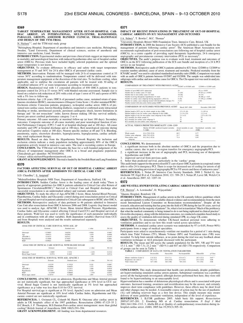
0369TARGET TEMPERATURE MANAGEMENT AFTER OUT-OF-HOSPITAL CAR-DIAC ARREST, AN INTERNATIONAL, MULTI-CENTRE, RANDOMISED,PARALLEL GROUPS, ASSESSOR BLINDED CLINICAL TRIAL-RATIONALEAND DESIGN OF THE TTM-TRIAL
N. Nielsen1,2, and the TTM-trial study group1Helsingborg Hospital, Department of anesthesia and intensive care medicine, Helsingborg,Sweden, 2Lund University, Department of clinical sciences, section of anesthesia andintensive care medicine, Lund, SwedenINTRODUCTION. Experimental studies and previous clinical trials suggest an improvementin mortality and neurological function with induced hypothermia after out-of-hospital cardiacarrest (OHCA). Previous trials have included highly selected populations and the optimaltarget temperature is not known.OBJECTIVES. To evaluate differences in efficacy and safety with target temperaturemanagement at 33 and 36�C for 24 h after OHCA of presumed cardiac cause.METHODS. Intervention: Patients will be managed with 24 h of temperature control at 33versus 36�C according to randomisation. Temperature control will be delivered with tem-perature management equipment at the discretion of the trial sites. To facilitate cooling, whenapplicable, and to stabilise the circulation all patients will be treated with 30 ml/kg ofcrystalloid infusion (4�C or room temperature according to treatment arm).DESIGN. Randomised trial with 1:1 concealed allocation of 850 OHCA patients to tem-perature control for 24 h at 33 versus 36�C with blinded outcome assessment. Sample size isbased on a relative risk reduction of 20% with a risk of type-1 error of 5% and a power of 90%with a 5% loss to follow-up.Inclusion criteria: Age C18 years, OHCA of presumed cardiac cause, sustained return of spon-taneous circulation (ROSC), unconsciousness (Glasgow Coma Score\8) after sustained ROSC.Exclusion criteria: Conscious patients, pregnancy, in-hospital cardiac arrest, OHCA of pre-sumed non-cardiac cause, known bleeding diathesis, suspected or confirmed acute intracranialbleeding or stroke, unwitnessed asystole, persistent cardiogenic shock, known limitations intherapy and ‘‘do not resuscitate’’ order, known disease making 180 day survival unlikely,known pre-arrest cerebral performance category 3 or 4.Primary outcome: All-cause mortality at maximal follow-up (at least 180 days). Secondaryoutcomes: Composite outcome of all-cause mortality and poor neurological function (Cere-bral Performance Category (CPC) 3 and 4) at hospital discharge and at 180 days. All-causemortality and CPC at hospital discharge and at 180-days. Best neurological outcome duringtrial period. Cognitive status at 180 days. Neuron specific enolase at 48 and 72 h. Bleeding,pneumonia, sepsis, electrolyte disorders, hyperglycaemia, hypoglycaemia, cardiac arrhyth-mia, renal replacement therapy.RESULTS. Based on data from the Hypothermia Network Registry the inclusion andexclusion criteria indicate a study population including approximately 80% of the OHCApopulation actively treated in intensive care units. The trial is recruiting centres in Europe.CONCLUSION. The TTM-trial will broaden the base for a well-founded judgement of theefficacy of temperature management after OHCA in a broad and pragmatic population.Clinicaltrials.gov identifier: NCT01020916.Contact: [email protected] ACKNOWLEDGMENT: This trial is funded by the Swedish Heart andLung Foundation
0370FACTORS AFFECTING SURVIVAL OF OUT OF HOSPITAL CARDIAC ARREST(OHCA) PATIENTS AFTER ADMISSION TO CRITICAL CARE UNIT
S.D. Chaudhry1, S. Anipindi1
1Midstaffordshire Hospitals NHS Trust, Department of Anaesthesia, Stafford, UKINTRODUCTION. Sudden Cardiac Arrest is the leading cause of death today.1There ispaucity of appropriate guidelines for OHCA patients admitted to Critical Care after Return ofSpontaneous Circulation(ROSC).2 Survival to Critical Care and Hospital discharge aftersuccessful resuscitation is an understudied subject and warrants attention.OBJECTIVES. To study the effect of Age,APACHE 2 Score, Mean Arterial Blood Pressure,Hypothermia, Cardiac Index and Glycaemic control during post resuscitation period on CriticalCare and Hospital survival in patients admitted to Critical Care subsequent to ROSC after OHCA.METHODS. Retrospective analysis of data pertinent to 46 patients admitted to IntensiveCare Unit after resuscitation from OHCA between 2006 and 2008. Logistic (log linear) modelwas used to analyse Age, APACHE 2 Score, Mean Arterial Blood Pressure, Hypothermia,Cardiac Index and Glycaemic control affecting Critical Care(CCU) and Hospital survival ofthese patients. Wald test was used to verify the significance of each parameter individuallyand in combination with all other variables. Both dependent variables (Survival from CCUand from Hospital) were analyzed and the results summarized separately.RESULTS.
ANALYSIS OF CRITICAL CARE SURVIVAL WITH AGE, AP
Estimate Standard error z value Pr([|z|)
(Intercept) -0.30007 4.66394 -0.064 0.9487
Age 0.04313 0.02582 1.670 0.0949
APACHE 2 0.10273 0.07411 1.386 0.1657
Hypothermia 0.14006 0.79487 0.176 0.8601
MAP -0.05021 0.04808 -1.044 0.2963
ANALYSIS OF CRITICAL CARE SURVIVAL WITH BLOOD SUGAR
Estimate Standard error z value Pr([|z|)
(Intercept) -1.8869 1.3154 -1.434 0.1514
Blood Sugar Control 0.2974 0.1649 1.803 0.0714
ANALYSIS OF HOSPITAL SURVIVAL WITH AGE, APACHE 2
Estimate Standard error z value Pr([|z|)
(Intercept) 1.63339 6.74524 0.242 0.8087
Age 0.09896 0.04453 2.223 0.0262
APACHE 2 0.21247 0.12571 1.690 0.0910
Hypothermia 0.53469 1.24974 0.428 0.6688
MAP -0.16595 0.08660 -1.916 0.0553
Blood sugar control 0.34753 0.31805 1.093 0.2745
CONCLUSIONS. APACHE2 score on admission, Hypothermia and Mean Arterial pressureare not statistically significant, but Age approached significance at 10% level in CCU sur-vival. Blood Sugar Control is not statistically significant at 5% level but approachedsignificance as p value was less than 0.10 for CCU survival.For Hospital survival,age is significant at 5% level; Apache2 score on admission and MeanArterial Pressure are significant at 10% level while Cardiac Index, Hypothermia and Gly-caemic control are not statistically significant .
REFERENCE(S). 1. Gwinnutt CL, Columb M, Harris R. Outcome after cardiac arrest inadults in UK hospitals: effect of the 1997 guidelines. Resuscitation (2000) 47:125–35 2.D.C.Bouch, J. P. Thompson, M.S.Damian.Post-cardiac arrest management: more than globalcooling? British Journal of Anaesthesia 2008 100(5):591–594.GRANT ACKNOWLEDGMENT. All funding was from departmental resources.
0371IMPACT OF RECENT INNOVATIONS IN TREATMENT OF OUT-OF-HOSPITALCARDIAC ARRESTS ON ICU MANAGEMENT AND OUTCOMES
J.A. Sidney1, T. Bowles1, M.C. Thomas1
1University Hospitals Bristol NHS Foundation Trust, Intensive Care, Bristol, UKINTRODUCTION. In 2008 the Intensive Care Society (ICS) published a care bundle for themanagement of patients following cardiac arrest1. The American Heart Association nowrecommends regionalisation of post-resuscitation care following out of hospital cardiac arrest(OHCA), in a centre capable of providing rapid therapeutic hypothermia, 24 h emergencyangiography and percutaneous coronary intervention (PCI) as necessary2.OBJECTIVES. The audit’s purpose was to evaluate work load, treatment and outcomes ofOHCA on the ICU following publication of the ICS care bundle and inception of a 24 h PCIservice in our hospital.METHODS. Retrospective audit of OHCA patients admitted to ICU from 12/2008 to 12/2009 toexamine number admitted, cause of arrest, treatment and mortality. Predicted mortality from theICNARC model3 was used to calculated standardised mortality ratio (SMR). Comparison was madewith an audit of OHCA patients between 03/2007 and 03/2008. The sample was subdivided intosubgroups with cardiac and non-cardiac cause for OHCA. The Chi-square test was used in analysis.RESULTS.
COMPARISON OF TREATMENT WITH PREVIOUS AUDIT03/2007 to 03/2008 audit 12/2008 to 12/2009 audit
Number of post-OHCA ICU admissions 25 43 p = 0.029
Number due to cardiac cause (% total) 11 (44) 29 (67) p = 0.0021
Number due to non-cardiac cause (% total) 14 (56) 14 (33)
Number receiving therapeutic hypothermia (% total) 17 (77) 38 (88) p = 0.07
Number in cardiac cause subgroup having angiogram ± PCI (%) 4 (36) 17 (59) p = 0.0062
Number in cardiac cause subgroup treated with thrombolytic therapy (%) 3 (27) 0 (0)
Number treated with intra-aortic baloon pump (IABP) 0 4
COMPARISON OF OUTCOMES WITH PREVIOUS AUDIT March 2007–2008 December 2008–2009
Median length of ICU stay for whole sample in days (IQR) 2.5 (1.9–10) 3 (1.85–4)
Median length of stay for cardiac cause subgroup in days (IQR) 2 (1.5–3.25) 3 (2.1–4)
Median length of stay for non-cardiac cause subgroup in days (IQR) 3 (1–4.5) 2.25 (1.02–3.9)
ICU mortality for whole sample % 76 53
ICU mortality for cardiac cause subgroup % 72 48
ICU mortality for non-cardiac cause subgroup % 80 64
ACTUAL VERSUS PREDICTED MORTALITY 12/08–12/09
Median ICNARC predicted hospital mortality
% (IQR)
Actual mortality in sam-
ple %
Standardised mortality ratio
(SMR)
Whole sample 81.4 (70.0–92.0) 60.5 0.77
Cardiac cause subgroup 80.8 (70.5–92.0) 51.7 0.64
Non-cardiac cause
subgroup
83.2 (61.2–92.6) 71.4 0.95
CONCLUSIONS. We demonstrated:1. a significant increase both in the absolute number of OHCA and the proportion due to
cardiac causes, due in part to in-region transfers for emergency angiography/PCI.2. a significant increase in the use of angiography and PCI, increasing use of therapeutic
hypothermia and IABP.3. improved survival from previous audit.4. better than predicted survival, particularly in the ‘cardiac‘ group.This audit lends support for the protocolised ICU care of post-OHCA patients in a regional centreable to offer 24 h emergency PCI. There is scope for increased use of cooling for arrests of allcauses, and extending provision of angiography/PCI to VF arrests and those of unknown cause2.REFERENCE(S). 1. Nolan JP. Intensive Care Society Standards. 2008 2. Nichol G, Au-fderheide TP, Eigel B et al. Circulation 2010; 121: 709–29 3. Nolan JP, Laver SR, Welch CAet al. Anaesthesia 2007; 62: 1207–16
0372ARE WE STILL HYPERVENTILATING CARDIAC ARREST PATIENTS IN THE UK?
P.B. Sherren1, A. Lewinsohn1, D. Wijayatilake1
1Queens Hospital, Romford, UKINTRODUCTION. Management of cardiac arrests in the UK currently follows guidelines whichare updated regularly to reflect best available clinical evidence and recommendations from the mostrecent International Liaison Committee on Resuscitation recommendations1. Despite all theresearch, education and training that has gone into the field of CPR during the last 50 years, survivalrates remain bleak for the majority of patients2. So, what is lost in translation? Much of the problem isthat what the medical community is being trained to do is not being replicated in clinical practice3.Given this discrepancy, along with the deleterious outcomes, we conducted a manikin-based study toassess the quality of ventilation delivered during simulated CPR, in a large UK centre.OBJECTIVES. To demonstrate whether UK-based medical personal were adhering toventilation guidelines and to see how this result varied across specialities.METHODS. A simulated cardiac arrest scenario was undertaken by 97 (a-0.05, Power-90%)participants from a range of medical specialties.Participants were asked to asynchronously ventilate our manikin for a period of 1 min duringwhich time Tidal Volumes (TV), Minute Volume (MV) and Ventilation rates (VR) wererecorded. To help limit outside influence, at no point during the trial was any feedback aboutventilation technique or ALS principals discussed with the participant.RESULTS. The mean and SD across the sample population for the MV, VR and TV were13.1 L min-1 (SD 7.3), 21.2 min-1 (SD 9.1) and 647 ml (SD 170) respectively. Comparisonof groups can be seen in Table 1.
TABLE 1 COMPARISON OF DATA ON VENTILATION PARAMETERS
Doctors(SD) Nurses(SD) Pre-hospital(SD) Others(SD) p-Value
Minute ventilation (Lmin-1) 11.8 (6.2) 15.4 (7.1) 15.6 (6.7) 18.3 (12.2) 0.03 *
Respiratory rate (min-1) 19.2 (7.3) 24.6 (10.6) 21.5 (6.9) 25.4 (14.6) 0.06
Tidal volume (ml) 627 (166) 644 (188) 748 (143) 731 (154) 0.06
CONCLUSION. This study demonstrated that health care professionals, despite guidelines,are hyperventilating simulated cardiac arrests patients. Suboptimal ventilation was a problemacross all the backgrounds investigated; although doctors performed best here, they were stillfound to be hyperventilating to an unacceptable level.Hyperventilation has a number of deleterious physiological effects and is associated with pooroutcomes. Increased training, awareness and recertification may be the answer, and certainlyimproves short term compliance with guidelines. However, these effects may be short livedand other changes may be needed. A reasonable course of action may be the use of paediatric(1L) self inflating reservoir bags as a first line device. This simple measure may ensuredelivery of more guideline consistent ventilation, independent of the level of experience.REFERENCES. 1. ILCOR guidleines 2005. Adult basic life support. Resuscitation2005;67:187–201. 2. Eisenberg MS et al. Cardiac resuscitation. N Engl J Med2001;344:1304–1313. 3. Abella BS et al. Quality of cardiopulmonary resuscitation during in-hospital cardiac arrest. JAMA. 2005 Jan 19;293(3):305–10.
S178 23rd ESICM ANNUAL CONGRESS – BARCELONA, SPAIN – 9–13 OCTOBER 2010
0373USEFULNESS OF ROUTINE LABORATORY PARAMETERS IN THE DECISIONTO TREAT REFRACTORY CARDIAC ARREST WITH EXTRACORPOREAL LIFESUPPORT
B. Megarbane1, M. Aout2, N. Deye1, I. Malissin1, P. Brun1, W. Haik1, S. Voiccu1, F.J. Baud1
1Hopital Lariboisiere, Reanimation Medicale et Toxicologique, Paris, France, 2Hopital Lar-iboisiere, Departement de Biostatistiques, Paris, France
INTRODUCTION. Extracorporeal life support (ECLS) has been proposed as the ultimateheroic rescue measure in prolonged cardiac arrest unresponsive to conventional cardiopul-monary resuscitation. ECLS effectiveness in out-of-hospital cardiac arrest remains to beaddressed. Decision to discontinue CPR due to medical futility is based upon presumedprolonged anoxia, with existing guidelines for termination. However, even when ECLS isimplemented, failure to maintain stable hemodynamic conditions due to marked capillary leakfrequently results in patient’s death.
OBJECTIVES. To evaluate the usefulness of routine laboratory parameters in the decision totreat refractory cardiac arrest patients with ECLS .
METHODS. Sixty-six adults with witnessed cardiac arrest of cardiac origin unrelated topoisoning or hypothermia undergoing cardiopulmonary resuscitation without return ofspontaneous circulation (duration: 155 min [120–180], median, [25–75%-percentiles]) wereincluded in a prospective cohort-study. ECLS was implanted under cardiac massage, using acentrifugal pump connected to a hollow-fiber membrane-oxygenator, aiming to maintainECLS flow C2.5 l/min and mean arterial pressure C60 mmHg.
RESULTS. Forty-seven of 66 patients died within 24 h from multiorgan failure and massivecapillary leak. Of 19/66 patients who survived C24 h with stable circulatory conditionspermitting neurological evaluation, four became conscious and were transferred for furthercardiac assistance, while three became organ donors. Ultimately, one patient survived withoutneurologic sequelae after cardiac transplantation. Using multivariate analysis, only pre-can-nulation peripheral venous oxygen saturation (SpvO2, 28% [15–52]) independently predictedinability to maintain targeted ECLS conditions C24 h (odds ratio for each 10%-decrease[95%-confidence interval]: 1.65 [1.21; 2.25], p = 0.002). The area under the receiver-oper-ating-characteristics curve was 0.78 [0.63; 0.93]. SpvO2 cut-off value of 33% was associatedwith a sensitivity of 0.68 [0.50; 0.83] and specificity of 0.81 [0.54; 0.96]. SpvO2 B8%, lactateconcentration C21 mmol/l, fibrinogen B0.8 g/l, and prothrombin index B11% predictedpremature ECLS discontinuation with a specificity of 1.
CONCLUSIONS. SpvO2 is useful to predict the inability of maintaining refractory cardiacarrest victims on ECLS without detrimental capillary leak and multiorgan failure until neu-rological evaluation.
0374PROGNOSIS OF ELDERLY PATIENTS AFTER SUCCESSFUL RESUSCITATIONFOR CARDIAC ARREST
D. Grimaldi1, F. Dumas2, O. Varenne3, J. Charpentier1, J. Fichet1, J.-P. Mira1, C. Spaulding3,
A. Cariou1
1Hopital Cochin APHP, Medical ICU, Paris, France, 2Hopital Hotel Dieu APHP, EmergencyDepartment, Paris, France, 3Hopital Cochin APHP, Cardiovascular Department, Paris, France
INTRODUCTION. Due to the human lifespan increasing, people are living longer. Cardiacarrest (CA) in old people could be seen as a natural end of life process and cardio-pulmonaryresuscitation (CPR) in this setting as a disturbance. Therefore, the question of prognosis inpatients has been raised when performing CPR in the elderly. Data from the 1980s found 6-month mortality in 94% of patients over 80 suggesting that CPR in elderly people could befutile (1). The recent progresses in the management of resuscitated patients, such as mildhypothermia, were not evaluated in patients older than 75 years (2, 3). In a recent study, wefound that age[60 years was an independent pejorative prognostic factor (4). Hence there isvirtually no data of prognosis factor of elderly patients after CA. Our aim was to determinethe prognosis factors in patients older than 75 years successfully resuscitated.
METHODS. All patients admitted to ICU for CA with successful ROSC were consecutivelyincluded between 2000 and 2009. CA data were prospectively entered in a registry accordingto Utstein recommendations. Patients were managed following standardized procedures.Good prognosis was defined as CPC 1 or 2 at ICU discharge. Factor associated with a goodoutcome were identified using multivariate analysis.
RESULTS. Among 1,210 patients admitted for CA, 225 were older than 75 years. Medianage was 79.5 years (75–97), CA was from cardiac origin in 50% of patients and 57.3% had aVT/VF initial rhythm. Mean no flow (NF) and low flow (LF) were 4.5 (±6.5) and 16.3 min(±13.4). Mean blood lactate and creatinine level at admission were 6.5 mmol/L (±4.3) and132 lmol/L (±49). 47.5% of patients presented post-resuscitation shock (PRS) and 67.5%were treated with hypothermia.
At ICU discharge, 24.9% patients were CPC 1 or 2. By univariate analysis, diabetes mellitus,the location of CA at home, NF [3 min, LF [15 min, PEA/asystole initial rhythm, need of[3 mg of epinephrine before ROSC, lactate level [5.1 mmol/L, creatinine [125.5 lmol/L,and PRS were associated with bad outcome. Interestingly, the rate of good outcome wassimilar between the half youngest patients compared to the half oldest (55 vs. 45%,P = 0.44). By multivariate analysis, absence of PRS, blood lactate level \5.1 mmol/L, NFB3 min and LF B15 min were associated with good outcome (OR 3.4 [1.4–8.3] P = 0.009;OR 5.5 [2.1–14.3] P = 0.0001; OR 3.18 [1.32–7.62] P = 0.01 and OR 2.96 [1.11–7.91]P = 0.03 respectively).
CONCLUSIONS. CA in elderly patients is associated with an in-ICU 25% good outcomerate. This should promote the CPR in non-severely disable elderly patients with CA regardlessof their age. We plan to collect the 6-month mortality and the functional status of survivors.
REFERENCES. 1. Rogove CCM 19952. Holzer M et al. 2002 N Engl J Med 346:549–5563. Bernard SA et al. 2002 N Engl J Med 346:557–563 4. Dumas F et al. Circ Cardiovasc
Interv in press
0375MICROCIRCULATORY ALTERATIONS AFTER CARDIAC ARREST: A PILOTSTUDY
K. Donadello1, R. Favory1, D. Salgado Ribeiro1, J. Creteur1, D. De Backer1, L. Gottin2, J.-L.
Vincent1, F.S. Taccone1
1Erasme University Hospital, Brussels, Belgium, 2Policilinico Universitario G.B.Rossi, Ve-rona, Italy
INTRODUCTION. Post-resuscitation phase is often characterized by a ‘‘sepsis-like’’ syn-drome, which may be associated with the development of organ dysfunction.Microcirculatory abnormalities play a key role in sepsis-related organ failure; however nodata are available on microvascular function after cardiac arrest (CA).
OBJECTIVES. The aim of this study was to investigate peripheral microcirculation duringand after therapeutic hypothermia (TH) in CA patients.
METHODS. This prospective, observational study included 10 patients treated by TH afterCA. Sublingual microcirculation was evaluated using Sidestream Dark-Field (SDF, Micro-scan, The Netherlands) videomicroscopy at hypothermia and normothermia in all patients. Atleast 5 images of 20 s each from separate areas were recorded at each time point and storedunder a random number to be analyzed, using a semi-quantitative method, by an investigatorblinded to time and condition. Thenar oxygen saturation (StO2) was measured using a tissuespectrometer (InSpectra 650; Hutchinson, USA). A vaso-occlusive test was performed athypothermia and normothermia by rapid inflation of a pneumatic cuff around the arm toevaluate StO2 reperfusion rate, reflecting microvascular reactivity.
RESULTS. Compared to hypothermia, measurements at normothermia showed a significantincrease in functional capillary density (FCD) from 7.2 ± 1.9 to 10.1 ± 1.4 n/mm(p = 0.002), the proportion of small perfused vessels (PPV) from 76 ± 13 to 92 ± 3%(p = 0.006) and mean flow index (MFI) from 2.1 ± 0.5 to 2.8 ± 0.2 (p = 0.01). FCD andPPV values were significantly correlated with body temperature. StO2 reperfusion rate waslargely decreased when compared to healthy volunteers, but it did not change over the studyperiod (from 0.94 ± 0.38 to 1.13 ± 0.54%/sec) and showed large inter-individual variability.The same was found for StO2 (from 78 ± 9 to 82 ± 8%).
CONCLUSIONS. Mild hypothermia is associated with decreased FCD and PPV in thesublingual area when compared to normothermia. Microvascular reactivity is decreased butchanges are unpredictable.
0376ACUTE POSTHYPOXIC MYOCLONUS AFTER CARDIOPULMONARYRESUSCITATION
D. van Poppelen1, A. Bouwes1,2, M.A. Tijssen1, J.H. Koelman1, M. Kuiper3, D.F. Zandstra4,
S. Tromp5, E. Zandbergen6, J. Horn1
1Academic Medical Center at the University of Amsterdam, Amsterdam, Netherlands,2SLAZ, Amsterdam, Netherlands, 3MCL, Leeuwarden, Netherlands, 4OLVG, Amsterdam,Netherlands, 5St Antonius Hospital, Nieuwegein, Netherlands, 6Rynstate Hospital, Arnhem,Netherlands
INTRODUCTION. Acute posthypoxic myoclonus (PHM) occurs in deeply comatosepatients, soon after a hypoxic episode. It is characterized by generalized, severe body jerkswith violent flexor movements, but more focal myoclonus is reported too(1). Acute PHM andstatus myoclonus are considered to have a poor prognostic outcome(1, 2). Although thecerebral cortex is known to be the most common origin of myoclonus in ambulant patients(3), the origin of acute PHM is uncertain (2).
OBJECTIVES. To determine whether acute PHM originates from damage in cortical orsubcortical structures.
METHODS. For this study patients with myoclonus in the first 72 h after admission wereselected from the PROPAC II study, a prospective cohort study including patients admittedafter CPR and treated with hypothermia. Exclusion criteria: pre-existing disease with lifeexpectancy\6 months and severely disability before CPR. Baseline characteristics were usedfrom the main database. Additional data of EEG and SSEP recordings made after rewarmingwere collected. EEGs were evaluated for presence of epileptic activity, status epilepticus,generalized periodic discharges, burst suppression pattern, iso-electric or low voltageamplitudes and reactivity of the background pattern. Data collected from SSEPs: N20potential, giant potential (defined as a potential five times the size of a normal potential) andP27/N35 amplitudes (done by JHK). The Glasgow Outcome Scale (GOS) was used to assessoutcome after 6 months, poor outcome was defined as a GOS of 1–3 (death, vegetative state,severe disability), good outcome as a GOS of 4–5 (moderate disability, good recovery).
RESULTS. From a total of 391 patients included in the PROPACII study, 68 (17%) patientsdeveloped myoclonus. Baseline characteristics of this group: age 67, 79% male, time toROSC 20 min, primary cardiac arrest in 61 patients, hypoxic arrest in 7. SSEP recordingswere available from 45 patients. N20 potentials were present bilaterally in 39% (23) and giantpotentials were seen in 9% (2) of the patients with a present N20 potential. 32 EEGs weremade, epileptic activity was seen in 34% (11) and a status epilepticus in 22% (7), thus 44% ofthe EEGs did not show any type of epileptic activity. Good outcome was seen in 10% of thepatients, poor outcome in 90%. Mortality was 88%.
CONCLUSIONS. The results of this study show that acute PHM is found in 17% of patientsadmitted after CPR and treated with hypothermia. It did not necessarily lead to a pooroutcome, but we did not have information about the type of myoclonus. The available dataseem to support the idea that the myoclonus originates mainly from subcortical structures,given the low number of patients with EEGs showing epileptiform activity and SSEPs withgiant potentials, which can be seen in cortical myoclonus (3).
REFERENCES. 1. Young, Neurology 1990. 2. Venkatesan, Neurol Clin 2006 3. Caviness,Neurol Clin, 2009
GRANT ACKNOWLEDGMENT. Dutch Heartfoundation 2007B039.
23rd ESICM ANNUAL CONGRESS – BARCELONA, SPAIN – 9–13 OCTOBER 2010 S179
0377MILD THERAPEUTIC HYPOTHERMIA AFTER CARDIAC ARREST REGARD-LESS OF INITIAL RHYTHM
A. Damelou1, K. Tsikritsaki2, M.K. Fragou2, P. Zotos1, G. Koukoulitsios2, D. Karakitsos3,A. Karabinis2
1General Hospital of Athens ‘G Gennimatas’, ICU, Athens, Greece, 2General Hospital ofAthens ‘G Gennimatas’, Athens, Greece, 3General Hospital of Athens, Athens, Greece
INTRODUCTION. The International Liaison Committee on Resuscitation, the AmericanHeart Association and the European Resuscitation Council recommend that mild therapeutichypothermia improve neurological outcome in unconscious adult patients with return ofspontaneous circulation (ROSC) after out-of-hospital cardiac arrest (OHCA) due to ventric-ular fibrillation (VF) or ventricular tachycardia (VT). In our Intensive Care Unit (ICU) we usemild hypothermia in all patients following CPR with successful ROSC regardless of initialrhythm. In this study we compared the effect of mild therapeutic hypothermia at neurologicaloutcome and mortality between the patients who had OHCA due to VF or VT and them whohad OHCA due to a different initial cardiac rhythm as asystole or pulseless electrical activity.
METHODS. The study protocol was approved by the local ethics committee on humanresearch. A total of 23 patients were admitted to our ICU with ROSC after OHCA betweenMay 2008 and December 2009. Therapeutic hypothermia was initiated after admission in ICUby intravenous infusion of cold saline (4�C 1,000 ml bolus) followed by intravenous coolingdevice (CoolLine Catheter, CoolGard Alsius Corporation Irvine, CA, USA). The targettemperature was 34�C maintained for 48 h followed by slow active re-warming over aminimum period of 24 h (0.1�C per hour). Intravenous anesthesia was induced in all patientsby a combination of propofol and remifentanyl with dose adjustment as needed. To preventshivering, patients received muscle relaxation by iv administration of sisatracurium every 2 h.The primary end point was the neurological outcome at 6 months according to the Pittsburghcerebral performance category (CPC). Secondary end point was mortality at 6 months. Pre-hostital cooling procedures were not applied.
RESULTS. Nine of the 14 patients (64%) of the group of the patients who had OHCA due toVF or VT had favourable neurological outcome CPC 1 or 2 as compared with 3 of 9 (33%) ofthe group of the patients who had OHCA due to a different initial cardiac rhythm. Mortality at6 months was 14% (2 of 14 patients died) in the group of the patients who had OHCA due toVF or VT as compared with 55% (5 of 9 patients died) in the group of the patients who hadOHCA due to a different initial cardiac rhythm.
CONCLUSIONS. In patients who had OHCA due to VF or VT mild therapeutic hypothermiainproves the neurological outcome and reduces mortality as compared with the patients whohad OHCA due to a different initial cardiac rhythm.
0378THERAPEUTIC HYPOTHERMIA AFTER CARDIAC ARREST: A RETROSPEC-TIVE COMPARISON OF SURFACE AND ENDOVASCULAR COOLINGTECHNIQUES
M. Gillies1,2, R. Pratt1, C. Whiteley1, I. Mccullough2, R. Beale1, S. Tibby1
1Guys and St Thomas’ NHS Foundation Trust, Critical Care, London, UK, 2Royal Infirmaryof Edinburgh, Critical Care, Edinburgh, UK
INTRODUCTION. Therapeutic hypothermia (32–34�C) is recommended for comatosesurvivors of cardiac arrest; however the optimal technique for cooling is unknown. TheInternational Liaison Committee on Resuscitation identifies ‘‘optimal cooling technique:internal versus external’’ as a critical knowledge gap.
OBJECTIVES. We aimed to compare therapeutic hypothermia using either surface or en-dovascular techniques in terms of efficacy, complications and outcome at our institution, a 30bedded tertiary referral ICU.
METHODS. A local research ethics committee reviewed the proposed study and waived theneed for a full ethics submission, as the study met the national criteria for service evaluation.Data were collected from 83 patients undergoing therapeutic hypothermia following cardiacarrest over a 2.5 year period by retrospective casenote review and interrrogation of theCarevue (Phillips UK) database. Therapeutic hypothermia was initiated in the ICU using icedHartmann’s solution, followed by either surface (n = 41) or endovascular (n = 42) cooling;choice of technique was based upon endovascular device availability. The target temperaturewas 32–34�C for 12 to 24 h, followed by rewarming at a rate of 0.25 deg h-1.
RESULTS. The mean age was 61 ± 16 years; 88% of arrests occurred out of hospital, and64% were ventricular fibrillation/tachycardia. Endovascular cooling provided a longer timewithin the target temperature range (p = 0.02), less temperature fluctuation (p = 0.003),better control during rewarming (0.04), and a lower 48-h temperature load (p = 0.008).Endovascular cooling also produced less cooling-associated complications in terms of bothovercooling (p = 0.05) and failure to reach the target temperature (p = 0.04). After adjust-ment for known confounders, there were no differences in outcome between the groups interms of ICU or hospital mortality, ventilator free days and neurological outcome.
CONCLUSIONS. Endovascular cooling provides better temperature management than sur-face cooling, as well as a more favorable complication profile. The equivalence in outcomesuggested by this small study requires confirmation in a randomized trial.
REFERENCE(S). 1. Nolan JP, Neumar RW, Adrie C et al. Post-cardiac arrest syndrome:epidemiology, pathophysiology, treatment, and prognostication. A Scientific Statement fromthe International Liaison Committee on Resuscitation. Resuscitation 2008; 79:350–3792. Merchant RM, Abella BS, Peberdy MA et al. Therapeutic hypothermia after cardiac arrest:
unintentional overcooling is common using ice packs and conventional cooling blankets.Crit Care Med 2006; 34:S490–494.
0379CEREBRAL BLOOD FLOW DURING PROLONGED MILD HYPOTHERMIA (72 H)AND PASSIVE REWARMING IN CARDIAC ARREST PATIENTS
L.L.A. Bisschops1, C.W.E. Hoedemaekers1, J.G. van der Hoeven1
1Radboud University Nijmegen Medical Centre, Department of Intensive Care, Nijmegen,Netherlands
INTRODUCTION. Mild hypothermia reduces cerebral blood flow (CBF) without concurrentincrease of cerebral oxygen extraction rate in the first 24 h after cardiac arrest, indicating alower cerebral metabolic activity with a preserved metabolic coupling.
OBJECTIVES. The aim of this study was to assess the cerebral blood flow and cerebraloxygen extraction in patients treated with prolonged hypothermia, as previously was per-formed in newborn infants with perinatal asphyxial encephalopathy1.
METHODS. Patients were included after restoration of spontaneous circulation (ROSC) afterasystole, pulseless electrical activity based circulatory arrest or after ROSC after ventricularfibrillation based prolonged resuscitation. In this prospective observational study 9 comatosepatients after cardiac arrest were treated with prolonged hypothermia for 72 h. After 72 hpatients were passively rewarmed. Mean flow velocity in the middle cerebral artery(MFVMCA), reflecting CBF, was measured after 0, 12, 24, 36, 48, 60, 72, 84, 96 and 108 hafter admission to the ICU by transcranial Doppler (TCD). Jugular bulb oxygenation (SjbO2)and arterial oxygenation were measured at intervals of 6 h.
RESULTS. MFVMCA was low (27.8 ± 12.4 cm/s) on admission, and gradually increased to57.1 cm/s during mild hypothermia in the first 72 h (p \ 0.05). After passive rewarmingMFVMCA further increased to 70.7 ± 20.5 (p \ 0.05) cm/s at 84 h after admission andremained relatively stable after 96 and 108 h (67.0 ± 18.4 cm/s and 64.6 ± 17.1 cm/srespectively, (p [ 0.05). SjbO2 at the start of the study was 56.4 ± 9.4% and graduallyincreased to 82.0 ± 7.3% after 72 h and 78.7 ± 4.0% after 114 h (p \ 0.05).
CONCLUSIONS. In cardiac arrest patients CBF was lower during hypothermia compared tophysiological values and showed a gradual increase during prolonged mild hypothermia whilecerebral oxygen extraction remained normal. This increase of CBF occurred during prolongedhypothermia suggesting a time dependent effect besides a temperature dependent increase ofthe cerebral metabolism after cardiac arrest.
REFERENCE. 1. Azzopardi DV, Strohm B, Edwards AD et al.: Moderate hypothermia totreat perinatal asphyxial encephalopathy. N Engl J Med. 2009 Oct 1;361(14):1349–58.
Subarachnoid hemorrhage and cerebrovascularevents: 0380–03930380NORMOBARIC HYPEROXIA EFFECTS ON THE INTRACRANIAL PRESSURE,CEREBRAL OXYGENATION AND METABOLISM
A.A. Solodov1, S.S. Petrikov1, Y.V. Titova1, V.V. Krylov2
1Sklifosovsky Research Institute, Neurosurgical Intensive Care Unit, Moscow, RussianFederation, 2Sklifosovsky Research Institute, Neurosurgery Department, Moscow, RussianFederation
INTRODUCTION. It’s well known that normobaric hyperoxia (NH) increases arterial andbrain oxygen tension. However the influence of NH on intracranial pressure (ICP) andcerebral metabolism in patients with intracranial hemorrhages (ICH) is a discussable question.
OBJECTIVES. To investigate the dynamics of ICP, jugular bulb saturation (SvjO2), brainoxygen tension (PbrO2) and cerebral metabolism during NH in patients with ICH.
METHODS. 8 patients with ICH and GCS B 9 enrolled in the study. ICP monitoring andtissue microdialysis were used in all patients, SvjO2 monitoring - in 6, PbrO2 - in 5.Microdialysis and PbrO2 catheters were placed in lesioned (les) and intact (int) brain tissue.ICP, SvjO2, PbrO2, glucose and glycerol levels, lactate/pyruvate ratio dynamics during FiO2
increase from 0.3 (n = 10) to 0.5 (n = 13) and 1.0 (n = 14) were analyzed.
RESULTS. NH was accompanied by increase of PaO2 from 102 ± 37 mmHg (FiO2 0.3) to171 ± 28 mmHg (ð \ 0.05) (FiO2 0.5) and 350 ± 64 mmHg (ð \ 0.05) (FiO2 1.0). PbrO2
increased as well (PbrO2 (int) - 31.4 (26.3; 38.1) mmHg (FiO2 0.3), 56.8 (45.5; 65) mmHg(ð \ 0.05) (FiO2 0.5), 79.3 (53.9; 151.4) mmHg (ð \ 0.05) (FiO2 1.0); PbrO2 (les) - 26.3(23.1; 39.3) mmHg (FiO2 0.3), 45 (26.8; 55.9) mmHg (ð \ 0.05) (FiO2 0.5), 91.8 (57.1;163.3) mmHg (ð \ 0.05) (FiO2 1.0)). Cerebral metabolism didn0t change significantly, exceptglycerol level increase in lesioned brain tissue from 97 (32; 233) mlmol/l (FiO2 0.3) to 234(32; 306) mlmol/l (FiO2 0.5) and 205 (23; 233) mlmol/l (FiO2 1.0). FiO2 0.3: glucose (int) -1.1 (0.3; 1.2) mmol/l, glucose (les) - 1.35 (0.4; 1.9) mmol/l, lactat/piruvat (int) - 20.2 (18.5;29.3), lactat/piruvat (les) - 20.9 (16.4; 27.4), glycerol (int) - 108 (59; 150) mlmol/l. FiO2 0.5:glucose (int) - 1 (0.2; 1.3) mmol/l, glucose (les) - 1 (0.5; 1.8) mmol/l, lactat/piruvat (int) - 19.4(18.2; 23.4), lactat/piruvat (les) - 22.6 (18.7; 28.7), glycerol (int) - 68 (41; 195) mlmol/l. FiO2
1.0: glucose (int) - 1.2 (0.15; 1.4) mmol/l, glucose (les) - 1 (0.3; 1.4) mmol/l, lactat/piruvat(int) - 19.3 (18; 31.5), lactat/piruvat (les) - 23.8 (19.6; 26.6), glycerol (int) - 82 (42; 155)mlmol/l. All patients had normal VO2/DO2 relationships initially (SvjO2 - 75 ± 1%). NHwas accompanied by significant increase of SvjO2 (FiO2 0.5 - 81 ± 5%, FiO2 1.0 - 88 ± 9%).ICP was stable during investigation (12.1 ± 2.9 mmHg (FiO2 0.3), 10.7 ± 2.5 mmHg (FiO2
0.5), 11.2 ± 5.8 mmHg (FiO2 1.0)).
CONCLUSIONS. NH is accompanied by PaO2 and PbrO2 increase and doesn’t influence ICPand cerebral metabolism in ICH patients with normal VO2/DO2 relationships.
S180 23rd ESICM ANNUAL CONGRESS – BARCELONA, SPAIN – 9–13 OCTOBER 2010
0381CEREBROVASCULAR COMPLICATIONS OF VENTRICULAR ASSIST DEVICES:A SYSTEMATIC REVIEW
W.M. van den Bergh1, D. Backes2, A.L. van Duijn2, D. van Dijk2, J.R. Lahpor2, A.J. Slooter2
1Academic Medical Center, Amsterdam, Netherlands, 2UMC Utrecht, Utrecht, Netherlands
INTRODUCTION. Ventricular assist devices (VADs) are successfully used in patients withend stage heart failure, usually as a bridge to transplantation or recovery, but increasingly asdestination therapy as well. A major threat for patients with a VAD is the frequent occurrenceof, mainly thromboembolic, stroke, with a reported incidence of up to 50%. Manufactureguidelines for anticoagulation therapy are based on relatively small observational studies andcommon sense rather than evidence, and as a consequence anticoagulation protocols varywidely between centers.
OBJECTIVES. The aim of this systematic review was to provide more evidence in order todetermine the optimal anticoagulation protocol to prevent stroke in patients supported with aVAD.
METHODS. A systematic search in Pubmed and Embase was performed in which weincluded all types of VADs. All types of anticoagulation drugs applied to prevent thrombo-embolism were included and divided in three categories; heparin, coumarins and antiplatelets.We included references with a full text available, written in English, Dutch, German orFrench, and which described patients with a stroke or TIA. Our primary outcome measure wasdefined as the onset of any type of stroke. Two authors evaluated independently the results ofthis search; doubtful references were evaluated by two other authors.
RESULTS. After critical appraisal 50 articles were selected as relevant, which include 29cohort studies, 6 case–control studies and 15 trials, totaling 3194 patients with VAD supportbetween 1982 and 2008. The mean age was 50 years (range 7–79) and the mean duration ofsupport was 116 days (0–1309). Stroke occurred in 0–50% patients supported by VAD, withan incidence of 0–23/patient-year. The majority of strokes occurred within the first year. Sixtypes of anticoagulation protocols were used that combines drugs from one or all threecategories. Most protocols used a combination of all 3 categories (1,162 patients with a totalfollow up of 115,082 days) and had an average stroke incidence of 1.28 events/patient-years.The lowest average stroke incidence was reported in studies that used only antiplatelets (0.62events/patient-years) and the highest in which only heparin was used (22 events/patient-years).
CONCLUSIONS. We could not detect a decreased risk for stroke in patients with VADsupport when coumarines or heparin were used instead of or in addition to antiplatelets.Antiplatelets should be part of an anticoagulation protocol to prevent stroke in patientssupported by VAD.
0382CAN TRANSCRANIAL DOPPLER SONOGRAPHY PREDICT ISQUEMIC NEURO-LOGICAL DEFICIT AFTER SPONTANEOUS ACUTE SUBARACHNOIDHEMORRAGE ?
J.J. Egea-Guerrero1, M.A. Munoz-Sanchez1, F. Murillo-Cabezas1, A. Cayuela2, P. Cancela3,M.D. Rincon-Ferrari1, C. Garcıa-Alfaro1, R. Amaya-Villar1, M. Gascon-Castillo1,J.M. Flores-Cordero1
1H.H.U.U.Virgen del Rocıo, Neurocritical Care Unit, Seville, Spain, 2H.H.U.U.Virgen delRocıo, Department of Clinical Documentation, Seville, Spain, 3H.H.U.U.Virgen del Rocıo,Department of Neurosurgery, Seville, Spain
OBJECTIVE. The aim of this research has been to ascertain the predictive value of trans-cranial Doppler sonography (TCD) to detect patients who are at high risk of delayed ischemicneurological deficit (IND).
METHODS. A descriptive and observational study was carried out for 3 years in theemergency department. 123 patients with SAH in a grade I–III of Hunt and Hess scale wereincluded. The mean velocity (MV) of middle cerebral artery (MCA) from both cerebralhemispheres was recorded. Sonography vasospasm pattern (SVP) was considered if MV was[120 cm/s and a hemispheric index [3. Mean arterial pressure, heart rate, temperature,haematocrit, intracranial hypertension signs and ulterior development of IND were registered.
RESULTS. SVP was detected in 24 patients (19.5%) however high velocities pattern (HVP)were presented in 38 patients (30.9%). IND developed in 21 patients (MV183 ± 49 cm/s), allwith a previous SVP except one. No patients with IND showed MV below 120 cm/s. In thisgroup MV were increasing 22 ± 15 cm/s/day during the first 3 days. The Group without HVP(84 patients/MV of 67 ± 16.6 cm/s) compared with IND group, showed differences inhighest MV (p \ 0.001), and in the DMV/day (8.30 ± 9.45 cm/s vs. 22 ± 15 cm/s) duringthe first 3 days (p \ 0.009).
CONCLUSION. TCD is a useful tool for an early detection of patients at risk of delayed INDafter SAH. During the first 3 days an increase of 22 cm/s/24 h in MV was strongly associatedwith the development of symptomatic vasospasm.
0383INHALATIONAL SEDATION WITH ISOFLURANE IN SUBARACHNOID HEM-ORRHAGE (SAH) PATIENTS: PRELIMINARY DATA ON ITS EFFECTS ONREGIONAL CEREBRAL BLOOD FLOW (RCBF)
F. Villa1, A. Coppadoro1, C. Iacca1, M.G. Abate1, A. Pesenti2,3, G. Citerio1
1Ospedale San Gerardo, Anesthesia and Critical Care, Unit of Neuro Intensive Care, Monza,Italy, 2Ospedale San Gerardo, Anesthesia and Critical Care, Monza, Italy, 3Universita Milano-Bicocca, Medicina Sperimentale, Milan, Italy
INTRODUCTION. Isoflurane is a volatile anesthetic known for its direct vasodilating effecton cerebral vessels, producing a CBF increase. Moreover, it has been shown in animal studiesthat Isoflurane has neuroprotective properties, inducing tolerance to ischemia when used as apreconditioning agent. At the present time, Isoflurane is not used as a sedative agent inNeuroICU because of the fear of an increase in Intracranial Pressure (ICP) caused by theincrease in CBF. However, SAH patient at risk for vasospasm may benefit from an increase inCBF. The ischemic risk will peak at day 5–7, leaving a window of opportunity forneuroprotection.
OBJECTIVES. We decided to measure rCBF during traditional intravenous sedation withPropofol and during volatile sedation with Isoflurane in SAH patients not having intracranialhypertension.
METHODS. The clinical trial was approved by the hospital IRB and registered on trial.gov(NCT00830843). We enrolled 4 SAH (Fisher 3–4, WFNS B3) patients, monitored with anintraparenchimal ICP device and a Thermal Diffusion Probe (TDP, Hemedex) for theassessment of rCBF. An ICP [18 mmHg was an exclusion criteria. Cerebral and haemody-namic parameters were assessed at 3 steps: Step 1 during sedation with Propofol 3–4 mg/kg/h; Step 2: after 1 h of sedation with Isoflurane 0.8–1% MAC administered through anAnesthetic Conserving Device; Step 3 after 1 h of Propofol re-started at the same infusionrate.
RESULTS. In all 4 patients sedation with Isoflurane produced an increase of rCBF althoughnot yet significant in the population (Step 1: 23 ± 18 ml/100 g/min; Step 2: 58 ± 62 Step 3:19 ± 8). Jugular Vein Oxygen Saturation, SjO2, was significantly higher at the end of theIsoflurane step (Step 1: 74 ± 4%; Step 2: 83 ± 5; Step 3: 73 ± 4). ICP did not changesignificantly and remained below the pathological threshold (Step 1: 6.7 ± 2; Step 2:7.7 ± 3; Step 3: 7 ± 4 mmHg).
CONCLUSIONS. The small population of this pilot study phase causes a lack in statisticalsignificance, however our data already suggest that Isoflurane induces a marked cerebralvasodilatation and an increase in rCBF compared to Propofol. Patients with a normal ICP didnot develop an intracranial hypertension.
REFERENCE(S). 1. Patel PM, Drummond JC: Effects of anesthetics oc Cerebral BloodFlow and Cerebral Metabolic Rate. In Miller’s Anesthesia, Elsevier 2005.2. Zhao P, Peng L, Li L, Xu X, Zuo Z: Isoflurane preconditioning improves long-term
neurologic outcome after hypoxic-ischemic brain injury in neonatal rats. Anesthesiology.2007 Dec;107(6):963–70.
GRANT ACKNOWLEDGMENT. Grant: This no-profit trial has been supported by Depuy/Hemedex, providing the probes. No other economical support has been received.
0384CEREBRAL FIBRINOLISIS IN A DISTRICT HOSPITAL
J.P. De Rojas Roman1, M.J. Morales Acedo2, M. Zaheri Bernayaki2, M.D.M. Ruestes Saiz2,P. Munoz Sanchez2, M.L. Mendez Pozo2, Grupo de Implantacion PLACA Antequera1Hospital Comarcal de Antequera, Unidad de Cuidados Criticos y Urgencias, Antequera,Spain, 2Hospital Comarcal de Antequera, Antequera, Spain
OBJECTIVE. To assess the feasibility of fibrinolysis in cerebral ischemic stroke in a districthospital.
METHODS. Retrospective analysis of cases of cerebral fibrinolysis performed at our hos-pital, third level, which treats 112,000 habitants, an average of 60,000 annual emergencyroom, 112 beds for hospitalization, with an intensive care unit with 7 beds, staffed by medicalspecialists in intensive care. The maximum time arrival at hospital is 45 min. The transfertime to our reference center is 60 min. NMR, trascraneal doppler or neurologist presence,aren’t available. We analyze the fibrinolysis undertaken since the beginning of the ‘‘ischemicstroke code’’. They require a rigorous diagnostic criteria. Tac support and less than 4 h ofevolution, approved by the European Stroke Association (Appendix 1). The fibrinolysis isperformed in the intensive care unit. At 24 h a control TC and evolution, angioRMN anddoppler supraaortic trunks are performed.
RESULTS. From October 2007 to March 2010, we have administered fibrinolytic to 7patients who had all the criteria of our protocol. Four patients have presented immediatereperfussion criteria, the others three non-reperfussed, only in a case of ischemia turned intoan hemorrhage/internal bleeding.
CASE 1: Woman of 73 years old, NIHSS at admission 16. Delay of 150 min. Reperfussioncriteria. Discharged home with minimal hemiparesis.
CASE 2: Man of 75 years old, NIHSS at admission 19. Delay of 150 min. Non reperfussioncriteria. Non complications from administration of fibrinolytic.
CASE 3: Man of 76 years old, NIHSS at admission 22. Delay of 120 min. Immediate rep-erfussion and reappearance of the clinic at 12 h.
CASE 4: Man of 54 years old, NIHSS at admission 7. Delay of 165 min. Full recovery.
CASE 5: Woman of 52 years old, NIHSS at admission 18. Delay of 90 min. Non criteria forimpact and neurological impairment at 72 h with malignant brain edema, intracranealhypertension and exitus.
CASE 6: Woman of 75 years old, NIHSS at admission 9. Delay of 150 min. Total recovery.
CASE 7: Man of 77 years old, NIHSS at admission 11. Delay of 180 min. Non initial criteriaof impact and hemorrhagic transformation on control TAC at 24 h.
CONCLUSION. We consider totally feasible the realization of fibrinolysis of ischemicstroke in our environment. The results are satisfactory and encouraging. We consider it a safepractice with low complication rate.
23rd ESICM ANNUAL CONGRESS – BARCELONA, SPAIN – 9–13 OCTOBER 2010 S181

0385OUTCOME OF ANEURYSMAL SPONTANEOUS SUBARACHNOID HEMORRHAGEAT SHORT AND LONG TERM AND ITS CORRELATION WITH SEVERITY SCALES,SCALES OF QUALITY OF LIFE AND ENDOVASCULAR TREATMENTM. Argueso1, A. Mesejo1, A. Tellez1, R. Vento1, M. Blasco1, J.R. Pineda1, M. Garcia1, A. Serrano1
1Hospital Clinico Universitario de Valencia, Intensive care, Valencia, SpainINTRODUCTION. Subarachnoid hemorrhage (SAH) is a medical emergency in all patients.There is not a consensus about which scales should be used to define the outcome afteraneurysmal subarachnoid hemorrhage.OBJECTIVES. The purpose of this study was to determine the risk factors and impact onfunctional outcome and quality of life 6 months after spontaneous SAH due to rupturedintracranial aneurysms.METHODS. We performed a prospective study of 46 patients with aneurysmal spontaneousSAH admitted to our centre from July 2008 to August 2009. Diagnosis of SAH was done byCT and ethiological diagnosis by brain angiography. We paid attention to previous patho-logical history, clinical characteristics at admission and radiological characteristics. Theseverity was measured by the Hunt-Hess (HH), WFNS, GRAEB and Fisher scales. The6 months outcome was assessed by the Glasgow Outcome Scale (GOS) and modified Rankinscale. The basic activities of daily living were evaluated with the Barthel Index. Moreover,patients were asked about their subsequent incorporation to their previous occupation. Wedivided our population into two groups depending on the clinical grade at admission (Hunt-Hess scale): Group I, HH 1, 2 or 3; and group 2, HH 4 or 5. Long-term followed up continues.RESULTS. During the year of study a total of 46 patients have been included. The mean age ofpatients was 55 years with a prevalence of 65% in women, being arterial hypertension andsmoking history the main factors of related risk. The angiography was performed in 95.6% of thepatients. An aneurysm was confirmed as the origin of the bleeding in 78.3%. Poor clinical gradeat admission (HH 4 or 5) was associated with APACHE II, SOFA, glucose, more SAH computedtomography on admission, and infection and ICU stay. There is 29% mortality in the series. Aftera 6 months period, group 2 patients (HH 4 or 5 grade) had a severe disability and functionaldependence to perform instrumental and basic activities of daily living (p = 0.001). Only 35%of patients were able to return to their previous occupation 6 months after the initial bleeding.
Scales and outcome by clinical grade at admissionGroup I Group II p
Mortality 21.2% 50% 0.07
Glucosa (admission) 138.67 ± 34.74 186.25 ± 45.3 0.000
GCS admission 14.15 ± 1.6 6.08 ± 3.4 0.000
SOFA 1.47 ± 1.6 6.67 ± 3.4 0.000
APACHE II 7.55 ± 3.3 19.08 ± 4.2 0.000
Modified Rankin 6 months 1.7 ± 2.1 4.3 ± 1.9 0.001
GOS 6 months 4.1 ± 1.4 2.6 ± 1.6 0.009
Barthel 90.77 ± 23.6 62.5 ± 42.8 0.03
Ability to return to the previous occupation at 6 months 46.2% 0% 0.03
Return to the previous occupation at 6 months
CONCLUSIONS. Spontaneous SAH continues to be a disease with high morbidity andmortality. Poor Hunt-Hess grade is related to unfavorable outcome and less ability to performinstrumental and basic activities of daily living, as well as the inability to return to previousoccupation before the SAH.
0386INVASIVE VASOSPASM TREATMENT AND EARLY ANEURYSM REPAIR: ABETTER WAY TO MANAGE SPONTANEOUS SUBARACHNOID HEMORRHAGE?M. Martinez1, D. Canovas2, A. Carvajal3, J. Perendreu4, J. Cabiol5, S. Lopez6, J. Branera4
1Hospital of Sabadell, Critical Care, Sabadell, Spain, 2Hospital of Sabadell, Neurology,Sabadell, Spain, 3UDIAT, Neuroradiology, Sabadell, Spain, 4UDIAT, Interventional Radi-ology, Sabadell, Spain, 5Hospital of Sabadell, Neurosurgery, Sabadell, Spain, 6Hospital ofSabadell, Anaesthesiology, Sabadell, SpainINTRODUCTION. Rebleeding and cerebral vasospasm are the most important prognosticfactors associated with spontaneous subarachnoid haemorrhage (SAH).OBJECTIVES. To analyze a multidisciplinary protocol implementation of SAH manage-ment that focused in early aneurysm repair and aggressive treatment (endovascular approach)of cerebral vasospasm.METHODS. Prospective observational study, Patients with SAH admitted in our critical careunit between January 2007 and December 2009. All patients were managed following amultidisciplinary protocol that includes an agreed by consensus decision of treatment, earlyaneurysm treatment (\48 h), an invasive and urgent management (endovascular techniques)of associated cerebral vasospasm. We collected epidemiological variables, SAH severity(WFNSS and CT Fisher classification), diagnosis methods (CT angiography/cerebral angi-ography), type of aneurysm treatment (coiling or clipping), presence of vasospasm and itstreatment, related complications and clinical outcome (Glasgow outcome scale) at 3 months.RESULTS. We have treated 53 patients (29 males, 24 females), aged 56 ± 12 years. We havefound a cerebral aneurysm in 39 patients and an arteriovenous malformation in 4 patients as a causeof SAH. Cerebral aneurysms were treated with coiling in 22 patients (56%) and surgical clipping in15 (39%). In two patients none treatment were performed due to their devastating neurological status.None of the patients with active treatment rebleed. Coiling was performed 1.5 ± 1.1 days afterinitial hemorrhage and surgical clipping 2.2 ± 1.2 days. Symptomatic cerebral vasospasm wasdiagnosed in 11 patients (21%), all cases were immediately managed using endovascular techniques(total of 25 sessions): intra-arterial nimodipine in 10 patients (23 sessions) and balloon transluminalangioplasty in one patient (2 treatments), all of them without related complications. Only one patientdied due to a vasospasm without response to treatment. Is important to note that 39 patients (74%)had a good neurological outcome (GOS 4–5) at 3 months and only 5 patients (9%) died.CONCLUSIONS. The application of a strict protocol of SAH management with earlyaneurysm repair and invasive cerebral vasospasm treatment seems to allow a good results inpatients with SAH (better than reported ones for the literature).REFERENCE(S). Diringer MN. Management of aneurysmal subarachnoid hemorrhage. CritCare Med 2009; 37:432–440. Zwienenberg-Lee M et al. Endovascular management ofcerebral vasospasm. Neurosurgery 2006. 59: S3-139–S3-147.
0387EFFECT OF ATORVASTATIN ON CEREBRAL VASOSPASM AND SHORT-TERMOUTCOMES AFTER SPONTANEOUS AND TRAUMATIC SUBARACHNOIDHEMORRHAGE
H. Mangoyan1, R. Hakobyan2
1Medical Center ‘Erebuni’, Yerevan, Armenia, 2Medical Center ‘Zeytun’, Anesthesiology,Yerevan, Armenia
INTRODUCTION. Aneurysmal subarachnoid hemorrhage (aSAH) remains a therapeuticchallenge due to unacceptably high levels of mortality and morbidity. For survivors of theinitial insult, cerebral vasospasm and related delayed ischemic deficits are the major deter-minants of final outcomes. Traumatic SAH (tSAH) occurs in as high as 60% of patients withTBI, and is associated with a twofold increase in risk of death. Statins may be an alternativefor conventional treatments of vasospasm due to their beneficial pleiotropic effects oncerebral vasomotor reactivity as well as absence of negative haemodynamic influence.
OBJECTIVES. The goal of our study was to examine the effects atorvastatin therapy oncerebral vasospasm after aSAH and tSAH as well as on short term outcomes of ICU patients(length of stay and severity of condition upon discharge from ICU).
METHODS. After institutional approval 80 adult patients (25–70 years of age) with aSAHand tSAH were randomly allocated to receive either atorvastatin 80 mg/day for 14 days(n = 50) or nimodipine 0.05 mg/kg/h i/v (n = 30) within first 72 h after the incident. Groupswere identical in demographics, clinical severity of insult, radiological evidence and man-agement. Cerebral vasospasm was assessed by transcranial Doppler ultrasonography and CT-scan data according to standard criteria. Systolic (Vs), diastolic (Vd), mean (Vm) flowvelocities (FV), and Lindegaard ratio (LR) in middle cerebral arteries were measured beforetreatment (basal FV), then daily and on 14-th day (final FV). Short-term outcomes wereassessed by comparing length of stay (LOS) in ICU and modified Rankin Score (RS) upondischarge from ICU. Repeated measurements were analysed by Friedman’s Chi square withsubsequent Wilcoxon sign test. Between groups variables were analysed using Mann–Whit-ney U test. All p values were two sided. Parameters are presented as median (interquartilerange).
RESULTS. Groups did not differ significantly by basal FV: Vs in atorvastatin group - 166(58.35) vs. 149.15(49.30) in nimodipine group (p = 0.642). We detected significant decreasein repeated measurements of all monitored parameters in both groups (p \ 0.05). There wasalso significant between group differences in final parameters: Vs in atorvastatin group 123.5(30.9) vs. 157(46.8) in nimodipine group (p = 0.003), LR in atorvastatin group 3.55 (1.02) vs.3.45 (2.02) in nimodipine group (p = 0.004). LOS in atorvastatin group was 5.00 (2) vs. 5.50(4) in nimodipin treated patients (p = 0.012). RS values were lower in atorvastatin group 2.00(1) vs. 3.30 (4) (p = 0.006)
CONCLUSIONS. We detected a trend to better control of cerebral vasospasm and improvedshort term outcomes with acute atorvastatin treatment after aSAH and tSAH compared totraditional regimens with nimodipine. Further studies with more power and extended followup are required for assessment of statins influence on final outcomes after SAH.
0388EFFECTS OF HYPERTONIC SALINE ON WHOLE BLOOD COAGULATION INVITRO ASSESSED BY THROMBOELASTOMETRY
T. Luostarinen1, T. Niiya2, A. Schramko1, P. Rosenberg1, T. Niemi1
1Helsinki University Central Hospital, Department of Anesthesiology and Intensive CareMedicine, Helsinki, Finland, 2Sapporo Medical University, Department of Anesthesiology,Sapporo, Japan
INTRODUCTION. Hypertonic saline infusion is an alternative to mannitol to decreaseintracranial pressure before craniotomy [1, 2]. Previous studies have demonstrated that bothhypertonic saline and mannitol interfere negatively with various components of bloodcoagulation [3, 4]. Normal blood coagulation capacity is essential during craniotomy and,therefore, we created an in vitro model to examine the effects of hypertonic sodium chlorideand mannitol solution on whole blood coagulation.
METHODS. Fresh citrated whole blood, withdrawn from 10 volunteers, was diluted with 0.9,2.5 or 3.5% sodium chloride solution or 15% mannitol to make 10 vol.% and 20 vol.%hemodilution in vitro. The diluted blood and undiluted control samples were analyzed withthromboelastometry (ROTEM�) using two activators, tissue thromboplastin without(ExTEM�) or with cytochalasin (FibTEM�).
RESULTS. All the study solutions with the same vol.% hemodilution induced comparabledecrease in hematocrit. In ExTEM� analysis, alpha-angle was smaller in the mannitol groupthan in the 0.9% or 3.5% sodium chloride group after 10 vol.% dilution (p\0.05). In 20 vol.%hemodilution, alpha-angle was also more decreased, and clot formation time more delayed inthe mannitol group than in the 0.9, 2.5 or 3.5% sodium chloride groups in ExTEM� analyses(p \ 0.05). Maximum clot firmness (MCF) in ExTEM� analysis was similar with all thestudy solutions after 10 vol.% dilution, but after 20 vol.% dilution MCF was weaker in themannitol group than in the other groups. MCF was also weaker in 3.5% than in 0.9% sodiumchloride group after 20 vol.% dilution. In FibTEM� analysis, MCF was stronger in the 2.5%sodium chloride group than in the mannitol group after both dilutions (p \ 0.05).
CONCLUSIONS. A 10 or 20 vol.% dilution of 15% mannitol disturbs whole blood coag-ulation more than equiosmolar 2.5% sodium chloride. This disturbance seems to be attributedto overall clot formation and strength but also to pure fibrin clot firmness. 2.5% sodiumchloride might be more favorable than mannitol before craniotomy in patients with highbleeding risk.
REFERENCE(S). 1. Rozet I, Tontisirin N, Muangman S et al. Effect of equiosmolar solu-tions of mannitol versus hypertonic saline on intraoperative brain relaxation and electrolytebalance. Anesthesiology 2007;107:697–7042.
2. Wu CT, Chen LC, Kuo CP et al. A comparison of 3% hypertonic saline and mannitol forbrain relaxation during elective supratentorial brain tumor surgery. Anesth Analg2010;110:903–73.
3. Wilder DM, Reid TJ, Bakaltcheva IB. Hypertonic resuscitation and blood coagulation: invitro comparison of several hypertonic solutions for their action on platelets and plasmacoagulation. Tromb Res 2002;107:255–614.
4. Lindroos AC, Schramko A, Tanskanen P et al. Effect of the combination of mannitol andRinger acetate or hydroxyethyl starch on whole blood coagulation in vitro. J NeurosurgAnesthesiol 2010;22:16–20
S182 23rd ESICM ANNUAL CONGRESS – BARCELONA, SPAIN – 9–13 OCTOBER 2010
0389INDUCED HYPERTENSION FOR TREATMENT OF DELAYED CEREBRALISCHAEMIA AFTER ANEURYSMAL SUBARACHNOID HAEMORRHAGE
C.S. Gathier1, W.M. Van den Bergh2, G.N. Beute3, C.M.F. Dirven4, J. Horn2, M. Van derJagt5, F. van Kooten6, A.W. Oldenbeuving7, L. Regli8, G. Roks9, W.P. VanderTop10,B.H. Verweij8, A. Algra11, G.J.E. Rinkel1, A.J.C. Slooter12
1University Medical Center Utrecht, Neurology, Utrecht, Netherlands, 2Academic MedicalCenter at the University of Amsterdam, Intensive Care Medicine, Amsterdam, Netherlands, 3St.Elisabeth Hospital Tilburg, Neurosurgery, Tilburg, Netherlands, 4Erasmus Medical Center,Neurosurgery, Rotterdam, Netherlands, 5Erasmus Medical Center, Intensive Care Medicine,Rotterdam, Netherlands, 6Erasmus Medical Center, Neurology, Rotterdam, Netherlands, 7St.Elisabeth Hospital Tilburg, Intensive Care Unit, Tilburg, Netherlands, 8University MedicalCenter Utrecht, Neurosurgery, Utrecht, Netherlands, 9St. Elisabeth Hospital Tilburg, Neurol-ogy, Tilburg, Netherlands, 10Academic Medical Center at the University of Amsterdam,Neurosurgery, Amsterdam, Netherlands, 11University Medical Center Utrecht, Utrecht, Neth-erlands, 12University Medical Center Utrecht, Intensive Care Unit, Utrecht, NetherlandsINTRODUCTION. Delayed cerebral ischemia (DCI) is a major complication after aneu-rysmal subarachnoid haemorrhage (SAH), occurring in around 30% of patients and increasingcase fatality 1.5–3-fold. Induced hypertension, alone or in combination with haemodilutionand hypervolemia (Triple-H), is used around the world as a therapy in the treatment of DCI,but its efficacy in improving outcome is not based on a randomised clinical trial.OBJECTIVES. To investigate the outcome of induced hypertension versus no inducedhypertension in patients with DCI after aneurysmal SAH.METHODS.Study design
A multi-centre, single blinded, randomized controlled trial.Study population
Patients admitted after recent SAH with a treated aneurysm and DCI based on the onset of anew focal deficit and/or a decrease of the level of consciousness of at least 1 point on theGlasgow Coma Scale with exclusion of other causes of deterioration, will be randomized toeither hypertension (n = 120) or no hypertension (n = 120).Interventions
In the intervention group, blood pressure will be raised with norepinephrine or additionaldobutamin in case of low cardiac output. When clinical improvement occurs, hypertensionwill be continued for 48 h, after which the dose norepinephrine will be tapered daily, butresumed in case of clinical deterioration. When no clinical improvement occurs within 24 h,induced hypertension will not be continued.Patients in the reference group will be treated according to the standardised SAH treatmentprotocol of the participating centre including oral nimodipine and normovolaemia withouthaemodilution.Main outcome measurement
The modified Rankin scale at 3 months after the SAH, will be compared between patientswho were randomized to induced hypertension and patients who were randomized to noinduced hypertension.REFERENCE(S). 1. Sen J, Belli A, Albon H, Morgan L, Petzold A, Kitchen N. Triple-Htherapy in the management of aneurysmal subarachnoid haemorrhage. Lancet Neurol 2003;Oct;2(10):614–21. 2. Ekelund A, Reinstrup P, Ryding E, Andersson AM, Molund T, Kris-tiansson KA, et al. Effects of iso- and hypervolemic hemodilution on regional cerebral bloodflow and oxygen delivery for patients with vasospasm after aneurysmal subarachnoid hem-orrhage. Acta Neurochir (Wien) 2002; Jul;144(7):703,12; discussion 712–3. 3. Raabe A, BeckJ, Keller M, Vatter H, Zimmermann M, Seifert V. Relative importance of hypertensioncompared with hypervolemia for increasing cerebral oxygenation in patients with cerebralvasospasm after subarachnoid hemorrhage. J Neurosurg 2005; Dec;103(6):974–81. 4. Mu-ench E, Horn P, Bauhuf C, Roth H, Philipps M, Hermann P, et al. Effects of hypervolemia andhypertension on regional cerebral blood flow, intracranial pressure, and brain tissue oxy-genation after subarachnoid hemorrhage. Crit Care Med 2007; Aug;35(8):1844,51; quiz 1852.
GRANT ACKNOWLEDGMENT. Netherlands Heart Foundation.
0390ASSOCIATION OF ANEMIA AND BLOOD TRANSFUSION WITH THE OUTCOMEOF PATIENTS WITH ANEURYSMAL SUBARACHNOID HEMORRHAGE
E.Festic1,A.A. Rabinstein2, W.D. Freeman1, S.A. Dupont2, M.T. Robinson2, G.D. Divertie1, O. Gajic2
1Mayo Clinic, Jacksonville, United States, 2Mayo Clinic, Rochester, United States
INTRODUCTION. Anemia in critically ill is frequent and may lead to worse outcomes inthose with subarachnoid hemorrhage (SAH). Transfusion of blood has been associated withcomplications in critically ill, as well as in patients with SAH, including cerebral infarction,ALI/ARDS and death. While conservative transfusion strategy is justified in majority ofcritically ill, this strategy may not apply to patients with SAH. No well defined threshold fortransfusion or target hemoglobin (Hb) level for these patients exists.OBJECTIVES. To examine associations of anemia and transfusion with adverse outcomes inpatients with SAH, and better define Hb level thresholds associated with these.METHODS. Retrospective, observational study of consecutive patients with SAH admittedto ICUs at Mayo Clinic in Jacksonville and Rochester, USA, over 6-year period. Dataincluded demographics, nadir Hb, blood transfusion, ALI/ARDS, vasospasm, radiographi-cally confirmed cerebral infarction, APACHE and WFNS. Primary outcome was associationof anemia and transfusion with death and secondary outcomes were vasospasm, cerebralinfarction and ALI/ARDS.RESULTS. We identified 333 patients, mean age was 56 (±14), 206 (62%) were females.Mortality was 17%. Seventy-two (22%) of patients were transfused. 155 (46%) patients hadvasospasm and 108 (33%) cerebral infarction. There was a strong association betweentransfusion and increased mortality (p = 0.001), vasospasm (p = 0.007), cerebral infarction(p \ 0.001) and ALI/ARDS (p \ 0.001). Median nadir Hb for transfused patients was 8.8 g/dl (8.1, 9.6). Nadir Hb threshold of 9 g/dl or less was associated with increased mortality(p \ 0.001), vasospasm (p = 0.002) and cerebral infarction (p \ 0.0001). When adjusted forage, gender, nadir hemoglobin, APACHE (201/333) and WFNS (231/333), RBC transfusion(p = 0.006) remained independently associated with increased mortality.CONCLUSIONS. Significant associations between both transfusion and anemia with adverseoutcomes among SAH patients were confirmed. Mortality and all secondary adverse out-comes were higher among transfused patients. In terms of anemia, Hb threshold of 9 g/dl orlower was associated with higher mortality and adverse outcomes. Prospective studies areneeded to determine risk–benefit ratio of transfusion in patients with SAH.REFERENCES. 1. Kramer AH et al. Critical Care Medicine. 36(7):2070–5, 2008 Jul. 2.Naidech AM et al. Critical Care Medicine. 35(10):2383–9, 2007 Oct. 3. Corwin HL et al.Critical Care Medicine. 32(1):39–52, 2004 Jan. 4. Hebert PC et al. NEJM. 340(6):409–17,1999 Feb 11. 5. Vincent JL et al. JAMA. 288(12):1499–507, 2002 Sep 25.GRANT ACKNOWLEDGMENT. Mayo Foundation.
0391DESCRIPTIVE COMPARITIVE STUDY ABOUT THE EVOLUTION OF PRYMARYINTRACEREBRAL HEMORRHAGES (ICH) IN AN INTENSIVE CARE UNITDURING 10 YEARS
A.M. de la Torre1, C. Marco1, D.J. Palacios1, A. Pedrosa1, I. Lopez de Toro1, V.A.
Hortiguela1
1Hospital Virgen de la Salud, Toledo, Spain
OBJECTIVES. To analyze the difference between two groups of patients with ICH con-sidering severity, treatment, evolution and mortality.
METHODS. Description retrospective study of admitted patients between 1st of January of1999 to 31st of December 2001 and 1st of January of 2007 to 31st of December 2009 in theICU of Virgen de la Salud Hospital (Toledo) with the diagnosis of ICH.
RESULTS. There are 85 patients in the group of 99-01 and 124 patients in the groups of 07-09 without any difference of age. Regarding comorbidity between the two groups no dif-ferences can be found regarding the previous presence of HTA, but regarding diabetes anddislipemy, we do find a higher prevalence on the second group (11.8 vs. 23.4%, p \ 0.05 and8.2 vs. 24.2%, p \ 0.01 respectively). Reviewing the presence of anticoagulated patients, nodifferences of significance can be found, but a trend (14.1 vs. 23.4%, p \ 0.1). Regarding thelocation of the hemorrhage, the most frequent is the basal ganglia (31.8 vs. 25%), existing nodifferences amongst the two groups. We don’t find differences either in the presence of aintraventricular component, whether it is neither supratentorial nor infratentorial. There aredifferences between the ICH score of both groups with p \ 0.05: with a score of 0 (9.5 vs.2.7%), 1 (32.1 vs. 16.4%), 2 (23.8 vs. 30.9%), 3 (25 vs. 30%), 4 (9.5vs. 16.5%) y 5 (0 vs.3.6%). In closing, there exists a higher percentage of hemorrhages greater than 30 cc in thesecond sample (45 vs. 62%, p \ 0.05). Regarding the time of the surgical intervention thereare no differences of significance, being intervened mostly in the first 24 h. Finally, ananalysis is made of the patients’ evolution once determined that there exists no differencesregarding the Glasgow Outcome Score at the time of discharge from the ICU: 1 (29.4 vs.36.3%), 2–3 (47.1 vs. 31.5%) and 4–5 (23.5 vs. 32.3%) neither regarding the mortality (41.2vs. 38.7%) nor the need for mechanical ventilation which there can be observed differences inthe days of stay in the ICU [\5 days (56.5 vs. 51.6%), 5–10 days (23.5 vs. 12.1%),11–15 days (3.5 vs. 9.7%) and [16 (16.5 vs. 26.6%), p \ 0.05].
CONCLUSIONS. An increase in the comorbidity can be observed in the included patients,which can be due to a better screening of these pathologies. We also find that the ICH score ishigher than in the 07-09 sample, which is attributed to the admittance in the ICU of patientswith the ultimate goal of organ donations. In the evolution, it can only be observed a longerstay in patients from the second sample, likely because they are more serious patients with ahigher ICH score. The analysis of the two groups has not been conclusive when it comes toassessing the improvements that might have come out in these last years, although it isnecessary a deeper analysis of the data.
REFERENCE. Hemphill JC, Bonovich DC, Besmertis L, et al. The ICH Score: A Simple,Reliable Grading Scale for Intracerebral Hemorrhage. Stroke 2001;32;891–897.
0392OUTCOME OF SODIUM DYSBALANCES IN SUBARACHNOID HEMORRHAGE:THE RESULTS OF A FIVE-YEAR PROSPECTIVE STUDY
V. Spatenkova1, A. Kazda2, P. Suchomel3
1Regional Hospital, Neurocentre, Neurointensive Care Unit, Liberec, Czech Republic, 21stFaculty of Medicine, Charles University, Department of Clinical Biochemistry and Labora-tory Medicine, Prague, Czech Republic, 3Regional Hospital, Neurocentre, Department ofNeurosurgery, Liberec, Czech Republic
INTRODUCTION. Sodium dysbalances are frequent medical complications in patients withsubarachnoid hemorrhage (SAH). Hyponatremia is more frequent but it is associated withbetter outcome than hypernatremia. The aim of this study was to observe differences inoutcome between hyponatremic and hypernatremic patients with SAH.
METHODS. We performed the prospective 5 years study in incidence of hyponatremia(serum sodium \135 mmol/l) and hypernatremia (serum sodium [150 mmol/l) in 211patients (pts) with SAH. We compared the incidence of cerebral complications, GlasgowOutcome Scale (GOS) upon discharge from the neurointensive care unit (NICU) and inmortality NICU.
RESULTS. There were 82 (39%) pts with dysnatremia, more patients had hyponatremia (46,22%), less hypernatremia (36, 17%). Between these groups there were no diferences in stay inNICU (p = 0.197), duration of dysnatremia (p = 0.055), fluid intake (p = 0.155, ml/day),daily sodium intake (p = 0.440, mmol/day) and fluid output (p = 0.055, ml/day). Hypona-tremia was more frequent on admission (p \ 0.001) and connected with higher diuresis(p \ 0.001). Hypernatremic pts received more antiedematic therapy (p \ 0.001). Hyperna-tremia was arised in pts with significantly lower Glasgow Coma Scale (p = 0.001). These ptshad more cerebral complications (p = 0.018), worse Glasgow Outcome Scale upon dischargefrom NICU (p \ 0.001) and higher mortality in the NICU (p = 0.002).
CONCLUSION. Dysnatremia is frequent in patients with SAH (39%). Hyponatremia occursmore often, but hypernatremia is connected with worse outcome.
23rd ESICM ANNUAL CONGRESS – BARCELONA, SPAIN – 9–13 OCTOBER 2010 S183
0393SERUM AND BRAIN GLUCOSE LEVELS RELATIONSHIPS IN PATIENTS WITHANEURISMAL SUBARACHNOID HEMORRHAGE
S.S. Petrikov1, A.A. Solodov1, J.V. Titova1, V.V. Krylov1
1Sklifosovsky Research Institute for Emergency Medicine, Neurosurgical ICU, Moscow,Russian Federation
INTRODUCTION. Relationships between serum and brain glucose levels in patients withaneurismal subarachnoid hemorrhage (SAH) are still unknown.
OBJECTIVES. To investigate brain metabolism at different serum glucose levels.
METHODS. Six patients with aneurismal SAH and vasospasm were enrolled in the study(age 47.5 ± 18.1; male/female 1/5; GCS 4–8). Cerebral microdialysis was used in allpatients. Microdialysis catheters were placed into ‘‘lesioned’’ (brain tissue perfused byinvolved artery) and ‘‘intact’’ brain tissue. Glucose levels in arterial blood (GLUart) and inbrain interstitial fluid were compared. We analyzed brain glucose levels, intracranial pressure(ICP), lactate/pyruvate (L/P) ratio, PaO2, PaCO2 and cerebral perfusion pressure (CPP) atblood glucose levels B6 mmol/l (Group I, n = 50), 6.1–10 mmol/l (Group II, n = 112),10.1–14 mmol/l (Group III, n = 56), C14 mmol/l (Group IV, n = 21).
RESULTS. We found out tight correlation between glucose levels in arterial blood and in‘‘lesioned’’ brain tissue (n = 239; r = 0.71; p \ 0.0001) and weak correlation betweenGLUart and glucose levels in ‘‘intact’’ brain tissue (n = 177; r = 0.43; p \ 0.0001). PaO2,PaCO2, ICP and CPP were comparable between groups. GLUart was 5.3 ± 0.6 mmol/l inGroup I, 7.5 ± 1.1 mmol/l in Group II, 11.8 ± 1.2 mmol/l in Group III and16.6 ± 2.9 mmol/l in Group IV. Normal glucose levels in arterial blood (Group I) wereaccompanied by low glucose levels in ‘‘intact’’ (0.52 ± 0.4 mmol/l) and ‘‘lesioned’’(0.49 ± 0.28 mmol/l) brain tissue. Arterial blood glucose elevation has led to significantincrease in brain glucose levels. But brain glucose levels were in normal ranges duringhyperglycemia. Glucose levels in ‘‘intact’’ brain tissue: 1.16 ± 0.79 mmol/l (Group II),1.76 ± 1 mmol/l (Group III), 1.24 ± 0.53 mmol/l (Group IV). Glucose levels in ‘‘lesioned’’brain tissue: 0.99±0.7 mmol/l (Group II), 1.58±0.7 (Group III), 2.7±1.4 (Group IV). Wedidn’t find out any significant differences in L/P ratio at different blood glucose levels.However, low glucose levels in ‘‘intact’’ brain tissue (B1.4 mmol/l; 0.7±0.4 mmol/l;n = 123) were accompanied by significant increase of L/P ratio (34.2±15.8 vs. 27.6±11.4 atnormal brain glucose levels (C1.5 mmol/l; 2.3±0.7 mmol/l; n = 54)). We didn’t notice anysignificant differences in L/P ratio in ‘‘lesioned’’ brain tissue at different brain glucose levels,opposite to intact brain tissue. L/P ratio was 29.5 (24.2; 43.1) at glucose levels in ‘‘lesioned’’brain tissue B1.4 mmol/l (0.73 ± 0.4 mmol/l; n = 175) vs. 31(26.3;37) at glucose levels in‘‘lesioned’’ brain tissue C1.5 mmol/l (2.4 ± 0.9; n = 64).
CONCLUSIONS. Hyperglycemia is not accompanied by high glucose levels and brainmetabolism impairment in ‘‘intact’’ and ‘‘lesioned’’ brain tissue in severe patients with an-eurismal SAH. Low glucose levels in ‘‘intact’’ brain tissue are related with significant increaseof L/P ratio.
Traumatic brain injury: 0394–04070394INTRACRANIAL PRESSURE MONITORING IS A VALUABLE AND SAFE INTER-VENTION IN THE MANAGEMENT OF SEVERE TRAUMATIC BRAIN INJURY INA NON-NEUROSURGICAL CENTRE
K.B. Bauchmuller1, K.A. Sharpe1, J.J. Paddle1
1Royal Cornwall Hospital, Intensive Care Unit, Truro, UK
INTRODUCTION. The 2007 National Confidential Enquiry into Patient Outcome and Deathreport found that less than 50% of patients with a severe head injury received a standard ofcare that was judged to be good [1]. Our Intensive Care unit (ICU) at the Royal CornwallHospital, a large District General Hospital, is one of the few non-neurosurgical centres in theUK to use Intracranial Pressure (ICP) monitoring.
OBJECTIVES. We aimed to assess if the practice of ICP monitoring in our non-neurosur-gical centre was valuable and safe.
METHODS. Retrospective audit of case notes and charts of all patients admitted withtraumatic brain injury receiving ICP monitoring over a 6 year period from 1st January 2004until 1st January 2010.
RESULTS. A total of 23 patients who had ICP monitoring for traumatic brain injury wereidentified. The CodmanTM strain-gauge catheter was used in all cases, 20 (87%) were male, 3(13%) were female. Mean age was 36 years (range 14–86). Median reported GCS at the sceneof injury was 3 (range 3–15), median GCS prior to intubation was 7 (range 3–15). MedianAPACHE score was 16 (range 5–36). All patients were discussed with the neurosurgicalreferral centre. Patients were monitored for a median of 2 days (range 1–4). ICP was raised in15 patients (65%). Elevation of ICP triggered the following interventions: 13 patients (87%)received hyperosmolar agents, 8 patients (53%) were treated with therapeutic hypothermia, 2patients (13%) with barbiturate coma and none of the patients received steroids. 5 patientswere transferred to a neurosurgical centre (33% of patients with elevated ICP, 22% of total).Complications comprised one minor intracranial haematoma and one monitor failure. Reviewof documented care bundles for head-injury patients revealed: Head-up tilt in 96%, gastricprotection in 91%, normoglycaemia in 91%, early enteral nutrition in 96%, appropriatethromboprophylaxis in 87%, appropriate sedation in 96% and appropriate analgesia in 96%.87% survived to hospital discharge.
CONCLUSIONS. In our audit the majority of patients received specific treatment for raisedICP, which might have gone undetected without invasive ICP monitoring. At the same timemost patients were managed without neurosurgical intervention. In light of the paucity ofneurocritical care beds [2] this approach appears to help to select a suitable subgroup ofpatients needing transfer to a specialist centre. Our data adds weight to the evidence that ICPmonitoring is a valuable and safe monitoring modality in a non-neurosurgical ICU if usedappropriately within established guidelines and in collaboration with a neurosurgical referralcentre.
REFERENCE(S). 1 NCEPOD. Trauma: Who cares? National Confidential Enquiry intoPatient Outcome and Death, London 2007.
2 http://www.dh.gov.uk/en/Publicationsandstatistics/Statistics/Performancedataandstatistics/Beds/DH_077451 accessed 27/03/10
0395BRAIN TISSUE OXYGENATION AND PUTATIVE BIOMARKERS IN SEVERETRAUMATIC BRAIN INJURY
E. Ezer1, S. Mondello2, S.A. Robicsek2, A. Gabrielli2, G. Brophy2, L. Papa3, J. Tepas3, C.Robertson4, G. Robinson2, E. Czeiter5, A. Buki5, R.L. Hayes2, K.K.W. Wang2
1University of Pecs, Pecs, Hungary, 2Banyan Biomarkers and University of Florida, Alachua,United States, 3Department of Emergency Medicine, Orlando Regional Medical Center,Orlando, United States, 4Neurosurgery, Baylor College of Medicine, Houston, United States,5Neurosurgery, Pecs University, Pecs, Hungary
INTRODUCTION. Assessment of injury severity and prediction of outcome represent majorchallenges following severe traumatic brain injury (TBI).
OBJECTIVES. This study was aimed to evaluate relationships between cerebrospinal fluid(CSF) levels of purported biomarkers of TBI including glial fibrillary acidic protein (GFAP),ubiquitin C-terminal hydrolase (UCH-L1) and alpha-II spectrin breakdown product(SBDP145), and partial pressure of brain tissue oxygen (ptiO2) as well as brain temperatureduring the first 24 h and up to 10 days post-injury.
METHODS. Twenty-seven severe TBI patients with CSF drainage and invasive monitoring(Licox probe) have been studied with quantitative CSF-ELISA for SBDP145, UCH-L1 andGFAP on admission and every 6 h up to 10 days.
RESULTS. In the first 24 h, biomarker levels decreased while those of ptiO2 increased. Allthree biomarkers correlated with ptiO2 (P \ 0.0001, P = 0.016 and P = 0.023, respectively).After the first 24 h, there were statistically significant changes in levels of the three markers aswell as in levels of ptiO2 (P = 0.025, P \ 0.0001, P = 0.033, P \ 0.0001, respectively).However, the correlation between biomarkers and brain tissue oxygenation was sustained and,for UCH-L1 improved (P \ 0.0001). No significant correlations between biomarker levelsand brain temperature were found.
CONCLUSIONS. The results indicate that CFS levels of SBDP145, UCH-L1 and GFAP arerelated to ptiO2 following severe TBI. Future studies are on their way to unravel whetherthese biochemical markers and ptiO2 measurement could serve the better care of the headinjured.
0396TRAUMATIC BRAIN INJURY AND TIGHT GLYCEMIC CONTROL. A MICRODI-ALYSIS STUDY
R. Badenes1, A. Lopez2, L. Alcover2, A. Gutierrez1, S. Ferri1, A. Maruenda1, F.J. Belda1
1Hospital Clinic Universitari, Anesthesiology and Critical Care, Valencia, Spain, 2Hospital LaFe, Anesthesiology and Critical Care, Valencia, Spain
INTRODUCTION. This was a pilot study to compare the cerebral neurochemical changes inpatients with traumatic brain injury (TBI) who underwent conventional blood glucose level(BGL) control and intensive BGL control with continuous titrated insulin.
OBJECTIVES. To demostrate that Intensive glycaemic control using insulin induced adecrease of cerebral glucose.
METHODS. This prospective, randomized study was conducted in 30 traumatic brain injurypatients in a surgical and trauma intensive care unit. Patients admitted over an 18-monthperiod with TBI were prospectively divided into two groups according to the method used forBGL control: the0intensive0 group consisted of patients who underwent continuous titratedinsulin infusion to maintain a lower normoglycemic level of 4–8 mmol/L, and the 0conven-tional0 group consisted of patients whose BGL was maintained at between 8.1 and 10.0 mmol/L using conventional 0sliding scale0 bolus subcutaneous insulin administration. Data oncerebral haemodynamics, interstitial brain oxygenation (PtiO(2)) and neurochemical moni-toring were collected via microcatheters inserted in the penumbral region.
RESULTS. We analyzed 2,844 cerebral microdialysis samples. In 15 patients treated withintensive insulin therapy, there was a reduction in microdialysis glucose by 77% of baselineconcentration compared with a 8% reduction in 15 patients treated with a conventional bloodglucose level control. Intensive insulin therapy was associated with increased incidence ofmicrodialysis markers of cellular distress, elevated glutamate (44 ± 34 vs. 8 ± 14%,p \ .01), elevated lactate/pyruvate ratio (40 ± 37 vs. 14 ± 22%, p \ .03) and low glucose(26 ± 17 vs. 11 ± 15%, p \ .05), and increased global oxygen extraction fraction. Cerebralmicrodialysis glucose was lower in nonsurvivors than in survivors (0.46 ± 0.23 vs.1.04 ± 0.56 mmol/L, p \ 0.05).
CONCLUSIONS. Intensive glycaemic control using insulin induced a decrease of cerebralglucose and an increase in microdialysis markers of cellular distress. In patients with severebrain injury, tight systemic glucose control is associated with increased mortality.
REFERENCE(S). 1. Curr Opin Crit Care 2009;15:1107, 2.Crit Care. 2010;14:R13.
S184 23rd ESICM ANNUAL CONGRESS – BARCELONA, SPAIN – 9–13 OCTOBER 2010
0397CLINICAL OUTCOME AND PREDICTORS OF MORTALITY AT DISCHARGEFROM INTENSIVE CARE UNIT AFTER TRAUMATIC BRAIN INJURY
E. Curiel Balsera1, M.A. Prieto Palomino1, O. Perez2, G. Quesada Garcıa1, M. Delange Van
der Kroft3, A. Munoz Lopez1
1Carlos Haya Regional University Hospital, Intensive Care Unit, Malaga, Spain, 2Carlos HayaRegional University Hospital, Fundacion IMABIS, Malaga, Spain, 3Axarquia Hospital,Intensive Care Unit, Velez Malaga, Spain
OBJECTIVES. To analyze clinical, epidemiologic and outcome differences and to identifypredictor factors of mortality at discharge from ICU of patients admitted after craneoence-phalic trauma (CET) according to Glasgow Coma Scale (GCS) score.
METHODS. Observational prospective study of patients admitted in the Intensive Care Unit(ICU) after CET. We classified the traumatic brain injury, according to GCS score of B8 orC9 points groups. We analyzed clinical and demographic data during ICU stay, as well asphysiological, functional and emotional data measured with PAECC scale (Project for theEpidemiological Analysis of Critical Patients) at discharge from ICU. Qualitative variablesare expressed as a percentage and quantitative variables as mean and standard deviation. Weused Chi2 test, t-Student and multivariate analysis as required with a maximum alpha error of5%.
RESULTS. We analyzed 447 patients, 81.2% male. At admission 67.3% had GCS B 8.Traffic accidents (45.2% in GCS C 9 and 58.5% in GCS B 8) were the most frequent causeof CET. There was a higher rate of out hospital hypotension in the GCS B 8 group(p = 0.002, OR 6.01, 95% CI 1.7–20.1). Cranial computed tomography (CT) scan findings inthe GCS C 9 group were diffuse injury I and II (58.9%) after Marshall classification versus28.6% in the GCS B 8 group (p = 0.0001). Fewer complications were detected in patients inthe GCS C 9 vs. GCS B 8 group (36.3 vs. 66.8%, p = 0.0001, OR 0.82, 95% CI0.047–0.14). ICU mortality rate was significantly lower in the GCS C 9 group than in theGCS B 8 group (10.3 vs. 36.2, p \ 0.01). Predictors of mortality were GCS at admission(p = 0.05), CT findings type III, IV and V (p = 0.014, 0.001 and 0.028 respectively),complications (p = 0.001), tracheotomy (p = 0.025), days on ventilator (p = 0.001),APACHE II (p = 0.001) and the length of ICU stay (p = 0.001). Best overall score inPAECC questionnaire at discharge from ICU was better in the GCS C 9 vs. GCS B group.(26.5 ± 5.8 vs. 19.22 ± 7.7, p = 0.0001).
CONCLUSIONS. Patients with lower GCS at admission presented higher rate of prehospitalhypotension, more severe CT findings, more complications at ICU, worse physiologic,functional and emotional outcome, and higher rate of mortality, than patients with GCS C 9.Factors associated with increased mortality were coma level, type of findings in CT, com-plications, prolonged mechanical ventilation, length of ICU stay, APACHE II, and the needfor a tracheotomy.
REFERENCE(S). Jacobs B, Beems T, Stulemeijer M, van Vugt AB, van der Vliet TM, BormGF, Vos PE. Outcome prediction in mild traumatic brain injury: age and clinical variables arestronger predictors than CT abnormalities. J Neurotrauma 2010;27:655–68
0398EARLY BRAIN TISSUE OXYGENATION IS RELATED TO SELECTIVE NEURO-NAL LOSS FOLLOWING TRAUMATIC BRAIN INJURY
V. Newcombe1, I. Timofeev2, C. Williams1, D. Chatfield1, P. Hutchinson2, J. Coles1, J.
Pickard1, G. Williams1, A. Gupta1, D. Menon1, Neuro-Intensive Care and Emergency Med-
icine, Trauma (Including Brain Trauma)1University of Cambridge, Division of Anaesthesia, Cambridge, UK, 2University of Cam-bridge, Academic Department of Neurosurgery, Cambridge, UK
INTRODUCTION. We have previously used 11C-flumazenil positron emission tomographyto show that selective neuronal loss in the thalamus is pervasive after traumatic brain injury(TBI) and correlates with functional outcome,1 findings that are concordant with previouspost-mortem data.2 The mechanisms responsible are unclear, but may involved globalhypoxia/ischaemia as well as retrograde degeneration.
OBJECTIVES. We hypothesised that early brain tissue oxygenation would correlate withlate diffusion tensor imaging (DTI) abnormalities in the thalamus, and therefore, help toprovide an explanation for late neuronal loss.
METHODS. Nine patients underwent brain tissue oximetry (PbO2) following acute TBI,using a Licox PbO2 probe, sited in structurally normal frontal white matter. Mean PbO2 wascalculated for the duration of their intensive care admission. At a median of 11.6 months(range 237–702 days) they underwent magnetic resonance imaging, including DTI. Apparentdiffusion coefficient ADC (maps) were created, ADC calculated in regions of interest in thefrontal lobes, splenium of the corpus callosum and thalami, and correlated with mean PbO2
using Spearman’s Rho. Ethical approval was obtained from the Local Research EthicsCommittee, and assent from next-of-kin was obtained in all cases.
RESULTS. Mean PbO2 was inversely related to ADC in both frontal lobes (r = -0.73 and-0.72; p = 0.025 and 0.031), and with the ADC in the thalamus bilaterally (r = -0.87 and-0.80; p = 0.002 and p = 0.010). In contrast, no correlation was seen between mean PbO2
and ADC in the splenium of the corpus callosum, a common site of traumatic axonal injury(TAI; p = 0.265).
CONCLUSIONS. The inverse correlation of mean PbO2 associated with ADCs in themonitored brain region is unsurprising, but the correlations observed with contralateralregions and deep grey matter suggest that the burden of tissue hypoxia has a significant impacton secondary neuronal loss across the brain. In contrast, the lack of correlation with ADCchanges in an area at risk of TAI suggests a less significant impact of hypoxia on theprogression or maturation of TAI. The correlations with measures of thalamic microstructuralinjury are particularly significant, since they establish a clear link between acute physiology,tissue fate in key brain regions, and clinical outcome.
REFERENCE(S). 1.Coles J, et al. Evidence for selective neuronal loss following clinicalhead injury (abstract) J Neurotrauma 2009 26:A2–A1012. Maxwell WL, et al. Differentialresponses in three thalamic nuclei in moderately disabled, severely disabled and vegetativepatients after blunt head injury. Brain 2004;127:2470–8
GRANT ACKNOWLEDGMENT. MRC, NIHR, Royal College of Anaesthetists, HealthFoundation, Academy of Medical Sciences, Gates Cambridge Trust.
0399BARORECEPTOR SENSITIVITY AND HEART RATE VARIABILITY IN EARLYTRAUMATIC BRAIN INJURY
P. Lowhagen1, S. Sondergaard2, B. Rydenhag3, T. McKelvey4, C. Eriksson-Ritzen3,
A. Aneman5
1Sahlgrenska University Hospital, Intensive Care Unit, Gothenburg, Sweden, 2Sahlgrenska University Hos-
pital, Department of Anaesthesia, Gothenburg, Sweden, 3Sahlgrenska University Hospital, Department of
Neurosurgery, Gothenburg, Sweden, 4Chalmers University, Gothenburg, Sweden, 5University of New South
Wales, Sydney, Intensive Care Unit, Liverpool Hospital, Australia
INTRODUCTION. Earlier studies have suggested that autonomic dysfunction is associated with poor out-
come in traumatic brain injury (TBI).
OBJECTIVES. This study was performed to assess changes in baroreceptor sensitivity and heart rate vari-
ability as indices of autonomic dysfunction in relation to ICU management variables during early TBI.
METHODS. Ten patients (3 females/7 males) with a median age of 31 (interquartile range, IQR, 20–51)
admitted to ICU following TBI were prospectively studied for the first consecutive 6 days. All patients were
sedated and mechanically ventilated with ICP monitoring in place. High fidelity signals at 300 Hz were
sampled for ECG and invasive arterial pressure (radial) to monitor baroreceptor sensitivity (BRS) based on
three or more consecutive beats in which successive systolic pressure and RR intervals increased or decreased
with the threshold set at 1 mmHg and 5 ms, respectively. Heart rate variability (HRV) was analysed by Fourier
transformation in the low (LF, 0.04–0.15 Hz) and high frequency (HF, 0.15–0.4 Hz) domains. The LF/HF ratio
and total power (0.01–0.6 Hz) were also calculated for HRV. Management variables included use of inotropes,
vasopressors, ICP, use of decompressive craniectomy, ICU length of stay (LOS) and mortality. Statistical
significance was set at p \ 0.05 using Mann–Whitney one-way ANOVA and Spearman correlation tests.
RESULTS. Median days (and IQR) were 7.5 (3–9.8) for inotropes, 3.5 (0–9) for vasopressors and 19.5
(17–24.5) for ICU LOS. BRS and LF, HF, LF/HF ratio and total power were all depressed throughout early
ICU management with no significant changes in the first week. No significant correlations were found between
BRS/HRV and days on inotropes/vasopressors or ICU LOS. Four patients underwent decompressive crani-
ectomy and one patient died while in ICU. No correlation was found between these events and BRS or HRV
changes
BRS, HRV and ICP data
Day 1 Day 2 Day 3 Day 4 Day 5 Day 6 p-value
(ANOVA)
BRS
(ms/
mmHg)
1.46
(1.0–1.6)
1.34
(0.9–2.3)
1.18
(0.9–2.7)
1.21
(0.97–1.9)
1.91
(1.2–4.8)
4.47
(1.2–5.4)
0.19
LF
(ms2)
2.04
(1.4–6.8)
1.02
(0.3–2.7)
1.52
(0.6–4.6)
0.92
(0.5–1.8)
0.90
(0.7–2.5)
1.91
(0.5–5.4)
0.18
HF
(ms2)
2.67
(1.3–5.5)
3.21
(1.1–6.1)
3.67
(1.5–7.3)
3.82
(1.1–6.6)
2.75
(2.3–5.1)
2.69
(3.0–5.4)
0.92
LF/HF 1.61
(0.19–1.9)
0.33
(0.01–1.4)
0.50
(0.04–0.9)
0.10
(0.02–0.5)
0.13
(0.03–0.6)
0.15
(0.02–0.8)
0.05
Total
power
(ms2)
14.1
(5.9–22)
11.5
(3.0–53)
8.3
(4.9–27)
12.6
(6.2–21)
10.6
(6.7–16)
6.4
(4.6–19)
0.93
ICP
(mmHg)
13
(9–19)
15
(12–20)
15
(14–22)
19
(14–22)
15
(13–19)
17
(11–20)
0.68
CONCLUSIONS. Both BRS and HRV were depressed early following TBI but did notcorrelate to ICU management variables. While autonomic dysfunction is evident early in theICU treatment of TBI, no evidence to support an association between severity of impairmentand ICU outcome was found in this pilot study.
GRANT ACKNOWLEDGMENT. The LUA-project at Gothenburg University 2009.
0400ULTRASOUND METHODS TO EVALUATE INTRACRANIAL PRESSURE INBRAIN INJURY PATIENTS
D.K. Radolovich1, C. Saracci1, A. Corona2, M. Pagani1, G. Castellani1, C. Lo Coco1,A. Bottazzi1, M. Belliato1, A. Amatu1, F. Sciutti1, M. Ferrari1, L. Carnevale1, G.A. Iotti1
1IRCCS Fondazione Policlinico San Matteo, Intensive Care Unit, Pavia, Italy, 2AziendaOspedaliera L. Sacco, Intensive Care Unit, Milano, Italy
INTRODUCTION. In neurocritical care, raised intracranial pressure (ICP) is associated to apoor outcome and its detection still leads the therapeutic management of the patients sufferingfrom head pathologies.
OBJECTIVES. Although invasive devices are recommended to detect intracranial pressure(ICP), we investigated the correlation and the reliability with non-invasive ultrasound tech-niques as the optic nerve sheath diameter (ONSD) assessed by ultrasonography and thepulsatility index (IP) measured by transcranial Doppler sonography (TCD).
METHODS. We included 13 patients suffering from intracranial hypertension, sedated and mechanically
ventilated and 10 control individuals, chosen among healthy people. All the patients had ICP measured
invasively either by external ventricular drains (EVD) or intraparenchymal catheter. Everyone underwent 3
non-invasive measurements of ONSD bilaterally and simultaneous medial cerebral artery IP assessment at the
side of the best window.
RESULTS. ONSD had a significant difference between volunteers (4.50 ± 0.36 mm) and patients
(5.60 ± 0.77 mm). IP’s were 0.81 ± 0.16 for the control individuals and 1.27 ± 0.43 for the patients (media
ICP = 19 ± 12 mmHg) and also revealed a significant difference. ONSD strongly correlated with ICP
(y = 0.026x + 5.005, p \ 0.020, R2 = 0.66) whereas IP had some correlation but without statistical signifi-
cance (y = 0.008x + 1.117, p = 0.188, R2 = 0.59). ONSD was found wider (p = 0.047) within patients with
both ICP and IP abnormal. A strong correlation was found between ONSD and IP (y = 0.708x + 4.819,
p = 0.004, R2 = 0.51). However, we could not find the best cut-off values of ONSD and IP for predicting an
ICP [20 mmHg.
CONCLUSIONS. IP and ONSD correlated with ICP with a stronger reliability results of the latter, suggesting
the possibility to integrate their use in the case of ICP invasive monitoring would be contraindicated or not
available.
REFERENCE(S). Geeraerts T et al. (2007) Intensive Care Med 33: 1704–1711
Bellner J et al. (2004) Surg Neurol. 62(1):45–51; discussion 51.
23rd ESICM ANNUAL CONGRESS – BARCELONA, SPAIN – 9–13 OCTOBER 2010 S185

0401HYPERHAES INFUSION AS A METHOD OF THE BAROREGULATION RESTOREIN SEVERE BRAIN TRAUMA
I. Bubnova1, I. Dobrynin1, S. Busygin1, N. Grachev1, A. Manoilov1
1Urals Medical Academy of Postgraduate Education, Chelyabinsk, Russian Federation
INTRODUCTION. Cerebral perfusion pressure in brain trauma depends on both ICP and BPregulation. The influence of HyperHAES on ICP is well known, but it is important to knowwhat role can HyperHAES play in haemodynamic regulation.
OBJECTIVES. The aim of our study was to estimate the influence of HyperHAES infusionon haemodynamic regulation.
METHODS. We examined 25 patients with severe brain trauma (GCS score \8, sedation,artificial ventilation). The BP, stroke volume (SV) and their variability (BPV, SVV) weredetermined by the bioimpedance method. Additionally we determined the peripheral pulse(PP) and its variability (PPV) using plethysmography curve registration. Special attentionwere paid to the P3 (0.1–0.15 Hz) and P4 (0.2–0.5 Hz) bands of BPV, SVV and PPV,connected with volume state, breathing, and autonomic regulation, especially baroregulation.All the comparisons were made before and after 10 min. of the infusion of HyperHAES(250 ml).
RESULTS. All patients had the autonomic dysfunction: the waves of baroregulation (P3) hadlow power compared with healthy. Hypovolemia was moderate: SV (50 ± 4.5) and PP(22 ± 2.6) were slightly decreased, but BP were 135 ± 5.2 mmHg. The BP increasing wasregistered as the first reaction on the infusion of HyperHAES (151 ± 4.1 mmHg). The sametime PP increased more then twice and became 54 ± 3.3, and P4 of PPV decreased from30 ± 2.8 to 19 ± 3.9, that reflected the improving of the volume state. Although SV did notrise significantly, the increasing of P3 were estimated in SVV, as a marker of the barore-gulation restoration.
CONCLUSIONS. The infusion of HyperHAES in severe brain trauma not only decrease therange of hypovolemia, but restore the baroregulation as a significant part of autonomicregulation, needed for cerebral perfusion support.
0402ACCURACY OF INTRAPARENCHYMAL PRESSURE SENSOR PLACEMENT
B. Fisher1, A. Bacon2, E. Thomas2
1Peninsulla Medical School, Plymouth, UK, 2Southwest Neurosurgical Centre, Plymouth, UK
INTRODUCTION. Continuous measurement of intracranial pressure (ICP) using intrapa-renchymal sensors has become part of the standard management of patients at risk ofdeveloping intracranial hypertension. Whilst reliability has been explored previously little ispublished on the accuracy of depth to which the sensor is placed. A difference in sensorlocation has been shown to affect the reliability of ICP readings and could impact negativelyon patient management (1).
OBJECTIVES. To determine whether ICP sensors (Codman, J&J) placed by Neurosurgicalstaff were within the optimum depth of 2 cm from the cortex.
METHODS. 50 consecutive patients were identified from a prospectively collected NeuroICU database who had had a CT of the head (CTH) performed whilst an ICP sensor was insitu during 2009. The depth of the sensor tip on CTH was measured and patients werestratified according to whether they had surgery or no surgery to determine any differencesbetween open and percutaneous placement.
RESULTS. Of the 50 ICP sensors 36 (72%) were placed deeper than 2 cm. The greatestincidence of deep placement was in surgical patients 26/33 (79%) and in craniotomy patients71% of these were deeper than 3 cm
TABLE 1 DEPTH OF ICP MONITOR
Total number Within 2 cm Deeper than 2 cm Deeper than 3 cm
All Cases 50 (100%) 14 (28%) 36 (72%) 24 (48%)Craniotomy 14 (28%) 1 (7%) 13 (93%) 10 (71%)Craniectomy 19 (38%) 6 (32%) 13 (68%) 7 (37%)Percutaneous 17 (34%) 7 (41%) 10 (59%) 7 (41%)
CONCLUSIONS. This investigation has shown that the majority of patients admitted to ourNeuroICU have less than optimally placed ICP sensors. Inappropriately deep sensors appearmore common in surgical patients, particularly following craniotomy. The impact of this onpatient outcome is unknown and would require further study. This study has highlighted weneed to implement new methods to improve the accuracy of our ICP sensor placement.Graduated marks on the sensor sheath during manufacture may assist accurate placement.
REFERENCE(S). Comparison of simultaneous continuous intracranial pressure (ICP) sig-nals from a Codman and a Camino ICP sensor. Med Eng & Physics, 28 (2006) 542–549.
0403MULTIMODALITY MONITORING IN SEVERE TRAUMATIC BRAIN INJURY:CLINICAL PRACTICE AND GUIDELINES IMPLEMENTATION IN PARIS,FRANCE ICU’S
J. Mateo1, A.C. Lukaszewicz1, T. Lescot2, I. Ghout3, J.J. Weiss4, B. Vigue5, Paris SevereTraumatic Brain Injury Study Group (Paris’S TBI)1Lariboisiere University Hospital, Anesthesiology and Critical Care, Paris, France, 2Pitie Uni-versity Hospital, Anesthesiology and Critical Care, Paris, France, 3Ambroise Pare UniversityHospital, CRU, Paris, France, 4Centre Francilien Ressource Traumatisme Cranien, CFRTC, Paris,France, 5Bicetre University Hospital, Anesthesiology and Critical Care, Kremlin Bicetre, FranceINTRODUCTION. Secondary injury after severe trauma brain injury (TBI) related to brainhypoxia and/or ischemia results from different mechanisms that can be better prevented ordetected by a multimodal neuromonitoring. Therapeutic actions may correct abnormal valuesand perform potentially Cerebral Blood Flow/Oxygen Extraction Coupling.OBJECTIVES. Aim of this study was to describe the incidence and the type of bedside ICUmonitoring devices used in the management of patients with severe TBI in French ICU’s.METHODS. Multicentric observational study including patients having severe TBI (GlasgowComa Scale (GCS) \ 8) picked on scene by a mobile medical unit for pre-hospital care(SAMU). Inclusion period: 18 months (2005–2006). Clinical data collected: ISS: Injuryseverity score, LOS: length of stay in ICU, SAPS 2: Severity score. Neuromonitoring tech-niques in ICU: Intracranial Pressure (ICP), End Tidal CO2 (EtCO2), Transcranial Doppler(TCD), Continuous jugular venous oxygen saturation (SvjO2).
RESULTS. Expressed in median and 25–75 interquartile. 481 mechanically ventilated patients wereincluded, with 78% directly admitted to a neurosurgical centre. Minimum monitoring performedincluded ECG, SaO2, invasive arterial blood pressure, temperature. ICU mortality rate was 43%.
DEMOGRAPHIC DATA AND INTENSITY OF NEUROMONITORINGMedian Interquartile
25–75
Type of
neuromonitoring
% of
use in
patients
Number of
neuromonitoring
device
% of
patients
Age (years) 39 24–56 ICP 49 None of them 18
GCS 4 3–6 EtCO2 65 1 neuromonitoring 18
ICU LOS (days) 10 1–23 TCD 64 2 neuromonitoring 24
ISS 30 25–41 SvjO2 7.6 C3 neuromonitoring 40
SAPS 2 49 39–63
CONCLUSIONS. If ICP is largely used, it remains below 50%. 40% of the cases have beenmonitored with at least 3 techniques suggesting an acceptance tendency for a multimodalmonitoring. Among these techniques, the control of PCO2 seemed considered important as theuse of TCD to assess perfusion and vessel tone. On-going analysis of these data will provideinformation on the therapeutic strategy performed and the impact on outcome.
0404IMPACT OF VARIATIONS IN THE MEASUREMENT OF OPTIC NERVE SHEATHDIAMETER ON THE DIAGNOSIS OF RAISED INTRACRANIAL PRESSURE: ANAPPRAISAL OF QUANTITATIVE AND QUALITATIVE METHODS
K.K. Pichamuthu1, J.V. Peter1, I.J. Prithishkumar2, P. Samuel3, M. Joseph4, G. John1
1Christian Medical College Hospital, Medical Intensive Care unit, Vellore, India, 2ChristianMedical College, Department of Anatomy, Vellore, India, 3Christian Medical College,Department of Biostatistics, Vellore, India, 4Christian Medical College Hospital, NeuroIntensive care unit, Vellore, India
INTRODUCTION. Raised intracranial pressure (RICP) can be evaluated sonographically bymeasuring the optic nerve sheath diameter (ONSD). A wide variation in the threshold mea-surement of ONSD for RICP has been reported in literature. It is likely that exaggeration ofthe hypoechoic edge artifact around the dura by high frequency ([10 MHz) linear transducersand uncertainty over whether to measure 3-mm behind the papilla or behind the globe(posterior margin of sclera) could have resulted in such differences.
OBJECTIVES. To determine the optimal site of measurement of ONSD by correlating it withRICP determined clinico-radiologically. We also evaluated if different sonographic appear-ances of the ONSD could be used qualitatively to determine the presence or absence of RICP.
METHODS. Initially, in order to precisely delineate the anatomical dura sonographically andassess optimal cursor placement, 2 cadaver orbital preparations were studied before and aftersubarachnoid fluid insufflation. 60 scans were then done by a single sonographer using a13–6 MHz linear transducer on 8 healthy volunteers and 46 patients admitted to the medical/neurosurgical intensive care units. ONSD was measured at 2 locations; 3-mm behind the papillaeand 3-mm behind the globe. In each location 1 measurement was made within the anatomicaldura and another between the echogenic margins of retrobulbar fat as described in literature.Four patterns (Fig. 1) of the nerve sheath were identified based on the appearance of the CSFspace and edge artifact. RICP was diagnosed by clinically correlated computed tomography ofthe brain. An independent 2 sample t test was done to correlate measurements with RICP.
Classification of Optic nerve sheath appearances
RESULTS. 14/60 (23%) scans were done on patients with RICP. All 4 measurements inde-pendently correlated with RICP (P \ 0.02); albeit cut-offs differed substantially (Table 1). Thepresence of a Type 3 pattern (Fig.) in both eyes strongly suggested RICP (100% positivepredictive value) whilst its absence in both eyes ruled out RICP (89% negative predictive value).
TABLE 1 OPTIMAL ONSD CUTOFFS AT DIFFERENT LOCATIONS3 mm behind
papilla, within
anatomic dura
3 mm behind
papilla, within
echogenic fat
3 mm behind
globe, within
anatomic dura
3 mm behind
globe, within
echogenic fat
Cutoff 5.4 mm 6.05 mm 5.2 mm 5.95 mm
CONCLUSIONS. Variations in cursor placement result in a systematic error in ONSDmeasurement. The optimal ONSD cut-off for RICP depends on the measurement technique.Qualitative assessment of patterns may be a quick and reliable screening tool for RICP.
S186 23rd ESICM ANNUAL CONGRESS – BARCELONA, SPAIN – 9–13 OCTOBER 2010
0405BENEFICIAL EFFECT OF NORMOTHERMIA IN BRAIN INJURED PATIENTS
G. Koukoulitsios1, A. Damelou1, P. Zotos1, K. Tsikritsaki1, M.-K. Fragou1, A. Zacharaki1, C.Tsakalakis1, D. Karakitsos1, A. Karabinis1
1G. Gennimatas General Hospital of Athens, Department of Intensive Care Unit, Athens,Greece
INTRODUCTION. We studied the effects of normothermic therapy during the acute phaseof traumatic brain injury.
METHODS. Twenty patients (10 males, 45 ± 18 years old) who were admitted in theIntensive Care Unit due to traumatic brain injury (Glasgow Coma Scale upon admission 5–8,Marshall Scale II to IV) were studied. If patients’ core temperature was above 38.5�C, a CoolLine�, ALSIUS Corporation, Irvine, CA. USA intravascular heat exchange central venouscatheter (Coolgard 3000� device) was inserted via the femoral or the subclavian route, inorder to achieve and maintain a target temperature of 36�C. Intracranial pressure (ICP) wasmeasured (a) invasively by means of a intraparenchymal catheter (Camino, Camino Labo-ratories, San Diego, CA, USA) and (b) noninvasively by means of transcranial Dopplersonography (TCD), using a Philips HD11XE (Philips Medical Systems; Bothell, WA, USA)equipped with a 2 MHz transducer and a 12 MHz linear transducer. Estimated ICP wascalculated by TCD using the equation proposed by Czosnyka and colleagues1 and the pul-satility index (PI = Vs - Vd/Vm) was assessed in the middle cerebral artery bilaterally. Allmeasurements were conducted at baseline and repeatedly on a daily basis for the next 7 daysand the average values were used in the statistical analysis.
RESULTS. Target temperature (t: 36.2 ± 0.3�C) was achieved within the next 6 h of thecatheter insertion. At baseline (t: 38.5�C) Pulsatility Index (PImean) was 1.4 ± 0.18, ICP-mean/invasive was 24 ± 4.5 mmHg and ICPmean/noninvasive was 25.2 ± 5.1 mmHg.Invasive and noninvasive ICP values correlated significantly (r = 0.8, p \ 0.05). Following24 ± 5 h from the insertion of the catheter the above parameters were significantly decreasedas compared to baseline values (PImean = 1.1 ± 0.1, ICPmean/invasive = 21 ± 3.4 mmHg,ICPmean/noninvasive = 22.1 ± 2.9 mmHg, respectively; all p \ 0.05). Out of 20 patients, 1progressed towards brain tamponade, 10 remained in a persistent vegetative state and 9 weredischarged with normal consciousness and motor deficits. However, due to the small numberof patients no analysis could be performed to estimate the possible impact of normothermictherapy on the survival of these patients.
CONCLUSIONS. These preliminary results suggest that normothermia during the acutephase of traumatic brain injury may decrease ICP and ameliorate cerebral blood flow.
REFERENCE(S). 1Czosnyka M, Matta BF, Smielewski P, Kirkpatrick PJ, Pickard JD:Cerebral perfusion pressure in head-injured patients: a noninvasive assessment using trans-cranial Doppler ultrasonography. J Neurosurg. 1998 May;88(5):802–8.
0406EXTRACRANIAL ORGAN DYSFUNCTION AFTER TRAUMATIC BRAIN INJURY:EFFECT OF PLASMA FROM BRAIN INJURED PATIENTS ON ENDOTHELIALFUNCTION
I.M. Mastromauro1, B. Assenzio1, O. Akide1, E.L. Martin1, C. Bagna1, S. Viberti1, L. DelSorbo1, V. Fanelli1, M. Berardino1, M. Fontanella1, A. Ducati1, L. Mascia1
1University of Turin, Turin, Italy
INTRODUCTION. Extracranial organ dysfunctions are extremely common in patients withsevere traumatic brain injury (TBI). Although it has been shown that development of TBI-induced multiple organ failure (MOF) is associated with a poor outcome, the underlyingmechanisms leading to MOF after TBI have not been investigated.
OBJECTIVES. To investigate in vitro the effect of plasma from TBI patients developingdifferent degrees of organ failure on human endothelial cells.
METHODS. Ten consecutive severe TBI patients were included. GCS, ISS and SOFA scorewere recorded at admission; SOFA was also recorded after 2 days. Plasma samples wereobtained at the same time points. Adhesion of freshly isolated human neutrophils on spon-taneously transformed human umbelical vein endothelial cells (ECV 304) was evaluated after4 h in vitro stimulation with 10% of plasma collected from TBI patients 2 days afteradmission. Expression of intercellular adhesion molecule-1 (ICAM-1) was assessed byimmunofluorescence. To determine the impairment of the endothelial cell barrier function,trans-endothelial electrical resistance (TEER) and permeability to FITC-dextran were mea-sured. Plasma from healthy volunteers were used as control. Data are expressed asmean ± SD. Results from different experiments were compared by unpaired t test.
RESULTS. Ten male patients were enrolled, mean age 35 ± 15, GCS 7 ± 1, ISS 32 ± 9,SOFA was 5.3 ± 1.8 on admission and 8 ± 1.6 on day 2. Plasma derived from TBI patientsincreased the adhesion of neutrophils on ECV 304 cells (80 ± 15 vs. 48 ± 7 cells/field,p \ 0.01), induced a significant reduction of TEER (130.1 ± 1.8 ohm/cm2 vs.139.1 ± 1.9 ohm/cm2, p \ 0.001) and caused a concomitant increase of endothelial perme-ability to dextran (132.5 ± 27.2 vs. 97.5 ± 7.2%, p \ 0.05) compared to control (healthyplasma). A visual up-regulation of ICAM-1 expression was also observed. There was asignificant correlation between delta SOFA score (day 2–day 0), calculated excluding GCS,and neutrophil adhesion on endothelial cells exposed to plasma from TBI patients (p = 0.009,R2 = 0.70).
CONCLUSIONS. Plasma from patients with TBI causes the increase of endothelial per-meability and neutrophil adhesion, which correlates with the development of extracranialorgan dysfunction expressed by SOFA. These results suggest a mechanism potentiallyresponsible for the development of MOF after traumatic brain injury.
GRANT ACKNOWLEDGMENT. Fondi Universitari Ex-60%, Regione Piemonte-RicercaSanitaria Finalizzata.
0407DECOMPRESSIVE CRANIECTOMY IN THE TREATMENT OF INTRACTABLEINTRACRANIAL HYPERTENSION
K. Elghadhioui1, N. Baffoun1, A. Naffati1, K. Baccar1, Z. Zaghdoudi1, C. Kaddour1
1National Institute of Neurology, Anaesthesiology & Critical Care Unit, Tunis, Tunisia
INTRODUCTION. Decompressive craniectomy (DC) improves vital prognosis in intracta-ble intracranial hypertension (ICHT) but its contribution in neurological outcome is uncertain.
OBJECTIVES. The aim of this study is to describe indications of DC and outcomes in ourunit.
METHODS. It is a retrospective revue collecting patients who had a DC for severe ICHTbetween January 1996 and July 2009. Inclusion criteria were: ICHT refractory to medicalmanagement due to cerebral oedema. We define intractable ICHT as intracranial pression(ICP) over 20 cmH2O, pupil modification, neurologic deterioration persisting more than30 min despite medical management or CT scan findings. Exclusion criteria were imminentcerebral death, ICHT due to an acute hematoma, tumour, hydrocephalus and prediction ofshort life expectancy.
RESULTS. 19 patients had a DC aged 29 ± 16[6–61] years. It was done in 12(63.2%) severetraumatic brain injury (TBI), 4(21.1%) cerebral infarction, 2(10.5%) cerebral venousthrombosis (CVT) and 1(5.25%) subarachnoidal bleeding. The operative indication occurs inthe three-first days in 31.6% cases. The sign showing severe ICHT was a refractory ICP in 1case, asymmetric pupils in 8 cases (42.1%), bilateral unreactive pupils in 3 cases (15.8%),neurological alteration in 2 cases (10.5%), per operative observation of a non-beating brain in1 case, CT scan showing brain oedema or shifting in 3 cases (15.8%) and in 1 case following aperioperative brain trauma during the evacuation of a extradural hematoma (EDH). Com-plications of DC were: 7(36.8%) cerebral herniation, 5(26%) hygroma, 2(15.8%) woundinfections, 1(5.3%) meningitis, 1(5.3%) EDH and 1(5.3%) seizure. For survivors the meanlength stay in ICU was 37 ± 27 [11–101] days; 67 days in cerebral infarction group, 24 daysin TBI group and 39 days in CVT group. At discharge, GCS was 12 ± 4 [3–15], respectively11, 13 and 15 in the groups of TBI, cerebral infarction and CVT. Seven (36.8%) patients hadsevere neurologic sequela. 11 patients (57.9%) died in the ICU after 12 ± 11[0–31] days;81.8% were due to ICHT and 18.2% to a sepsis. These results are poor in comparison withauthors using intracranial pression monitoring [1, 2].
CONCLUSIONS. DC saves lives but functional outcome benefit is less certain at that time ofevaluation. Using clinical criteria may affect outcome by delaying surgery. A further eval-uation after a longer period is needed to know if clinical criteria can be used to indicate DC.
REFERENCE(S). 1. Chibbaro S. Role of decompressive craniectomy in the management ofsevere head injury with refractory cerebral oedema and intractable intracranial pressure. Ourexperience with 48 cases. Surgycal Neurology 2007; 68: 632–638.2. Vahedi K. Early decompressive surgery in malignant infarction of the middle cerebral
artery: a pooled analysis of three randomised controlled trials. Lancet Neurology 2007; 6:215–222.
Patient and family experience: 0408–04200408DO WE NEED EARLIER LONG TERM FOLLOW UP FOR CRITICALLY ILLPATIENTS?
E. Kain1, Long Term Follow Up Service1Good Hope NHS Hospital, Heart of England Foundation Trust, Critical Care, Birmingham,UK
INTRODUCTION. There is 20 years worth of data to show the extent of physical andpsychological problems long stay critical care patients may suffer. It is no longer acceptableto merely save patients’ lives following a critical illness. The NICE guidelines ‘Rehabilitationafter critical illness’ March 2009, state we must restore them back to as ‘near normal health’as possible. Follow up services are deemed the appropriate support system for these patients.However, a study by Cuthbertson et al., published in 2009 challenged the effectiveness andcost effectiveness of nurse led follow up.
OBJECTIVES. Historically, research has targeted problems experienced several months postdischarge from critical care, namely using data from follow up clinics \12 months after thecritical illness. In contrast, this study aimed to review data from patients recently transferredfrom critical care to the wards. This data can then be compared with patients’ progress afterearlier rehabilitation.
METHODS. This was a prospective, longitudinal study, involving long stay critical carepatients ([4 days), between April 2008–2010. This cohort included all patients nursed withinour ITU/HDU, regardless of speciality, age or gender. All reviews took place whilst thepatients were still hospitalised. The study used qualitative data, using specifically designedquestionnaires which highlighted the type of long term problems our patients suffered. Thesewere developed using data from previous informal patient interviews and the HAD (HospitalAnxiety and Depression) questionnaire. The latter was considered too intrusive for the wardenvironment. A data base was used to list the frequency of these problems to providequantitative data.
RESULTS. A total of 116 patients were reviewed over 2 years. The qualitative data high-lighted 16 different, common morbidities, in addition to disease specific morbidities.Between, April 08–09, 66% of patients demonstrated at least 4 physical or psychologicalproblems and 95% displayed 2 or more. The quantitative data showed the most prevalentmorbidities were: poor mobility at 60%, nightmares 53%, loss of appetite 46% and insomnia35%. Year 2 showed similar findings, plus global weakness at 30%.
CONCLUSIONS. This research showed significant post critical illness morbidities in thiscohort which appeared to impact on the patients’ recovery on the wards. The challenge for ourfollow up is to target support and rehabilitation whilst the patients are in critical care and thenre-evaluate the results. If this expedites a return to a near normal quality of life, it will haveimproved the efficacy of our service and may impact on follow up care in general.
REFERENCE(S). Cuthbertson B.H. et al. (2009) ‘The PRaCTICal study of nurse led,intensive care follow up programmes for improving long term outcomes from critical illness’cited BMJ 2009.339:b3723.
NICE (2009) Guideline 83 ‘Rehabilitation after critical illness’.
23rd ESICM ANNUAL CONGRESS – BARCELONA, SPAIN – 9–13 OCTOBER 2010 S187
0409IMPLEMENTING BASIC STIMULATION IN A DANISH ICU
K. Damborg1, B. Smith1
1ICU, Regional Hospital West Jutland, Holstebro, Holstebro, Denmark
INTRODUCTION. Basic stimulation (BS) is a concept for the advancement, care andaccompaniment of severely disabled patients in a holistic and individualized approachwherein the focus in nursing has shifted from nursing interventions to the patients and theirindividual needs. It enables a higher nursing performance and a better communication withthe severely impaired patients1,2. This study describes how nurses dealt with the imple-menting of BS in an ICU of a general hospital in Denmark.
METHODS. Three years after the introduction of the concept of BS in our ICU, a ques-tionnaire was sent to 40 people directly involved in the nursing of critically ill patients tostudy the current practice during the previous month.
RESULTS. Responses were received from 26 persons, a 65% response rate. 25 (96%) per-sons had practiced BS. Only 10 (38%) employees used calming bath and 5 (19%) aninvigorating bath. Guided oral care and guided suction was performed respectively by 18(69%) and 10 (38%) nursing persons. Orientative positioning was used by 12 (46%) nurses.Everybody is very satisfied with the instruction manual which can be found in each room. 14(54%) persons estimated to be well informed about BS. Others wanted more practice courseand training at the bedside.
CONCLUSION. Pattern suggested an increasing interest in BS since its introduction. Initialtouch is well implemented. More effort is needed for continuous education and training.
REFERENCES. 1. Frohlich, A.: Basale Stimulation-das Konzept. Verlag SelbsbestimmtesLeben, Dusseldorf 19982. Egerod I et al. A descriptive study of basic stimulation in Danish ICUs in 2006. Scand J
Caring Sci; 2009; 23; 697–704.
0410PATIENTS’ EXPERIENCES WITH PREOPERATIVE INFORMATION
A.K. Gabrielsen1, R. Nord2
1Oslo University Hospital, Division of Emergency Medicine, Postoperative Unit, Oslo,Norway, 2University of Oslo, Nursing Science, Oslo, Norway
INTRODUCTION. Admittance to hospital for surgical treatment, is often linked withinsecurity and anxiety for many patients. To most patients, the postoperative care unit con-stitutes an unknown environment, and can represent a frightening experience. Research hasshown that preoperative information leads to subjective outcome as anxiety reduction, andobjective outcome as shorter hospital stay and less intake of pain medication. Few studies,however, have addressed patients’ experiences with preoperative information about the earlypostoperative phase.
OBJECTIVES. The purpose of this study was to describe patients’ experiences with pre-operative information about events they may experience during their stay in the postoperativeunit. Patients’ experiences may contribute to increased knowledge about this topic.
METHODS. The study design was exploratory-descriptive, and a semi-structured interviewbased on thematic guide was used. Nine patients met the inclusion criteria, and they had anaverage age of 65 years. They were admitted to elective surgery for cancer and their stay inthe postoperative unit varied from 1 to 3.5 days. The interviews were conducted 5–11 daysafter surgery and transcribed verbatim. The data material was subjected to qualitative contentanalysis.
RESULTS. Experience with information before surgery and in the early postoperative phase,was categorized into four themes: being prepared before surgery, reactions to differingexperience, discomfort and pain, management of some self-care activities and experienceswith the environment of the postoperative unit.
CONCLUSIONS. The patients received a fair amount of information before surgery, butonly limited information concerning what to expect while in the postoperative unit. Thepatients’ information needs differed and patients with former experience with surgery weremore prepared for what to expect. The patients got mainly verbal information and most of thiswas given the day before surgery.
0411NOISE POLLUTION IN THE INTENSIVE CARE AN OBSERVATIONAL STUDY
O.M. Peters-Polman1, M. van Roosmalen2, J.E. Tulleken3, J.G. Zijlstra3
1UMC Groningen, Intensive Care, Groningen, Netherlands, 2ARUP Nederland, Amsterdam,Netherlands, 3UMC Groningen, Groningen, Netherlands
BACKGROUND. WHO guidelines concerning sound levels in hospitals requires a maximumof 35 dB in daytime, a nighttime sound level below 30 dB to allow good sleep quality andpeak levels that do not exceed 40 dB at all times. In an ICU environment, apart from beingcritically ill, patients are exposed to typical ICU environmental noise. Sleeping cycles dis-turbed by illness are further disrupted by care providers performing procedures and takingvital signs and alarms with delirium as a common result. We describe the acoustic envi-ronment in our mixed ICU.
METHODS. We conducted an observational study in which we continuously measured thesound level (dB), frequency and repetition of sound in our ICU. We used a SPLnet micro-phone and noise monitoring system which was placed, after informed consent, at the ear levelof the patient.
RESULTS. Noise sources are numerous and consist of human voices, doors slamming,pagers, telephones, shoes and equipment alarms. There is a round the clock continuousbackground noise of 45–68 dB, staff conversation with levels of 70–75 dB and peak levels upto 90–110 dB, mainly due to alarms. These levels of sound are comparable to loud conver-sation at 1 meter distance (45–68 dB), walking along a motorway (70–90 dB) or standingnext to a roaring engine (110 dB).
CONCLUSION. WHO guidelines clearly state maximum sound levels of 35 dB in daytime,30 dB at night and peak levels of 40 dB. The sound level in our unit is exceptionally andunacceptably high throughout day and night and requires a behavioral intervention. Furtherresearch on the influence of these noise levels on our patients is necessary.
0412THE COMPARISON OF EFFICIENCY OF MANUAL AND MECHANICAL COM-PRESSION METHODS USED IN FEMORAL SHEATH REMOVAL
B. Armutcu1, V. Unver1, H. Kursaklıoglu1, A. Badır2, N. Akbayrak1
1Gulhane Military Medical Academy, Ankara, Turkey, 2Koc University School of Nursing,Istanbul, Turkey
INTRODUCTION. Diagnostic techniques, requiring femoral artery puncture, such as cardiaccatheterisation, and revascularization techniques, such as percutaneous transluminal coronaryangioplasty, are commonly used in interventional cardiology.
OBJECTIVES. This study has been achieved in order to realize the comparison of efficiencyof manual and mechanical compression techniques used for the maintenance of haemostasisafter femoral sheath removal.
METHODS. This study was planned and applied as a randomized controlled trial. The studywas executed at a military education and research hospital in Turkey between January andMarch 2010. Data collecting form was prepared by the investigators after the literatureexamination. The form consists of 21 questions which are evaluating the demographic data ofpatients, the compression time, the pain level (before sheath removal, during the manual/mechanical compression and after the sheath removal), the complications occurring in fem-oral zone, the mobilization/discharge period and the problems (bruise, oedema, hemorrhageand etc.) occurring the 5th day after the discharge. The patients have been called up in order toevaluate the problems occurring on the 5th day. The 55 persons that were applied mechanicalcompression have constituted the experimental group and the 59 patients that were appliedmanual compression have formed the control group. The patients volunteer to participate tothe study have been informed in regard with the implementing procedures before the appli-cation. Descriptive statistics were shown in numbers and percentages for the variablesobtained by counting and in mean ± standard deviation for variables obtained bymeasurement.
RESULTS. The average of age is 52.70 ± 16.38 in the experimental group and is47.62 ± 14.88 in the control group. The average of compression time is 39.66 ± 19.65 minin the experimental group and is 13.5 ± 5.03 min in the control group (p \ 0.05). Theaverage of mobilization time after sheath removal is 103.36 ± 40.99 min in the experimentalgroup and is 379.32 ± 50.37 min in the control group (p \ 0.05). It has been observed alower pain level during compression and after sheath removal in patient that were appliedmechanical compression in comparison to the patients that were applied manual compres-sion(p \ 0.05). When the groups were compared in terms of femoral zone complicationswhile no haematoma was observed in the experimental group, haematoma has been occurredin the 10.2% (n = 6) of the control group.
CONCLUSIONS. The mechanical compression provides an earlier mobilization and earlierdischarge of the patient. This study shows that mechanical compression is a method as safetyas the manual method in order to obtain a haemostasis
REFERENCE(S). Jones T. and McCutcheon H, A Randomised Controlled Trial Comparingthe Use of Manual Versus Mechanical Compression to Obtain Haemostasis Following Cor-onary Angiography, Intensive Care Nursing, 2003.
S188 23rd ESICM ANNUAL CONGRESS – BARCELONA, SPAIN – 9–13 OCTOBER 2010
0413TO HAVE A RELATIVE IN STATE OF LIFE THREAT: DISCOMFORT IN THEDAILY LIFE OF FAMILY
K.S. Freitas1, F.C. Mussi2, I.G. Menezes2, Interdisciplinary Group on the CardiovascularHealth Care1Federal University of Bahia, Nursing School, Salvador, Brazil, 2Federal University of Bahia,Nursing school, Salvador, BrazilINTRODUCTION. The illness and hospitalization of a relative in intensive care unit (ICU)may be experienced such as abrupt events, unexpected, frightening and problematic by thefamily, resulting in changes and new challenges for it1. It may affect the balance of family andchange their roles and responsibilities, mainly due to the separation from their relativeimposed by hospitalization. This admission may also generate other discomforts to the family,as relationship problems, emergence of diseases, lack of financial resources for expendituresnow installed, anxiety, depression, fear and irritability2.OBJECTIVE. To get to know the discomfort that characterizes the changes experienced inthe daily life of families who have a relative in ICU.METHOD. This is a qualitative study, conducted in a general ICU of a large hospital in thecity of Salvador, Bahia, Brazil, from August to October of 2009. Nine relatives of peoplehospitalized for at least 24 h in ICU were interviewed. The data were analyzed by the use ofprocedures of analysis of Grounded Theory.RESULTS. The hospitalization of a family member in ICU produced discomfort for her, as theuncertainty of recovery, the fear of loss and the sudden physical separation of the relative. Theinteraction between the huge discomfort of life threat and the separation from the relative, inturn, spawned other discomforts in the daily lives of families, expressed by two categories. Thefirst one, having difficulties to answer psychobiological needs, meant for the family member theexperience of sleep deprivation, loss of appetite and constant concern with the relative. Thesecond one, suffering a discontinuity in the daily life, meant to the family the disruption of dailyactivities with the relative, irritability front other family members, removal of the routine ofhome, loss on work performance and studies and difficulties to enjoy the leisure and recreation.CONCLUSION. The coping of illness and hospitalization of a relative in intensive care produceddiscomfort for family members, characterized by changing of the routine of the daily life and carefor oneself. The family’s attention is primarily directed toward the ill relative. It starts to experiencethe routine of a hospital and disrupts the organization of familiar and personal life, it finds itselfsuffering a break from daily life. The discomfort experienced by the family can be minimized bypractices of the healthcare team, which provide care, information, support, safety and convenience.REFERENCES. 1. Silveira AO, Angelo M. Interaction experience for families who liveswith their child’s disease and hospitalization. Rev Latino-am Enfermagem 2006 novembro-dezembro; 14(6):on line; 2. Horn EV.; Tesh A. The effect of critical care hospitalization onfamily members stress and responses. Dimens Crit Care Nurs. 2000; 19 (4): 40–9.
0414QUALITY AND QUANTITY OF SLEEP IN MULTIPLE VERSUS SINGLE PATIENTROOM INTENSIVE CARE UNITS
M. van Eijk1, R. van den Bossche2, M. Nouwen1, F. Leijten1, A. de Weerd2, M. Schneider1,J. Kesecioglu1, A. Slooter1
1University Medical Center Utrecht, Utrecht, Netherlands, 2SEIN Zwolle, Zwolle,NetherlandsINTRODUCTION. Sleep fragmentation and deprivation is common in Intensive Care Unit(ICU) patients1. It is generally assumed that the ICU environment, including over-exposure tosound and light in the night-time, is an important cause for disturbed sleep1, but well-designedstudies are lacking. In the University Medical Centre, Utrecht, the Netherlands, a new ICU wasbuilt with quiet, single-patient rooms with much daylight (Fig. 1). This is a unique opportunity tostudy the effects of nursing environment on sleep quality and quantity in ICU patients.
OBJECTIVES. To study the effects of nursing environment on sleep quality and quantity inICU patients.METHODS. A total of 20 patients will be included in this ongoing study: ten subjects whowere admitted to the old, ward-like ICU (Fig. 2), and ten patients who will be admitted to thenew, single-room ICU (Fig. 1).
To increase homogeneity, we include patients undergoing elective cardio-thoracic surgeryonly. A 15-lead polysomnography is recorded from 07:00 p.m. to 07:00 a.m. Concomitantmedication and medical procedures (e.g. extubation or type of operation) are registered. 24 hbefore and 48 h after the registration, subjects are asked to fill in a questionnaire concerningthe subjective quality of sleep. Light and sound exposure in both the old and the new ICU arerecorded for several weeks.RESULTS. At the ESICM meeting, we will present data on total sleep time, number ofminutes awake, number en frequency of naps and frequency and duration of the differentsleep stages in the old, ward-like ICU and in the new, single-room ICU.CONCLUSIONS. Polysomnography is feasible in ICU patients. This is the first study on therole of different types of ICUs and sleep. Data will be discussed at the ESICM meeting.REFERENCE(S). 1. Freedman NS et al. Abnormal sleep/wake cycles and the effect ofenvironmental noise on sleep disruption in the intensive care unit. Am J Respir Crit Care Med2001; 163(2):451–457.
0415FAMILY ROLE IN AN INTENSIVE CARE UNIT
R. Leonardo1, S. Nascimento1, A. Mendes1, H. Jose2
1Hospital Faro, EPE, UCIP, Faro, Portugal, 2University from Algarve, Faro, Portugal
INTRODUCTION. The admission in an Intensive Care Unit is a time of crisis and transitionfor the family of the critically ill patient, and that should be considered in the process ofnursing care.OBJECTIVES. In order to understand the family’s perception of nursing care, the authorsundertook this study, assessing the family satisfaction and use the results to increase thequality of care.METHODS. This is a qualitative study of inductive nature. Data was collected along the timeof the study by in-depth interviews, to six relatives of each patient, after discharge theintensive care unit. For data analysis we followed the steps of the phenomenological method,according to Max van Manen (1990).RESULTS. From data analysis, the results were divided in 5 categories: 1) Relative needs; 2)ICU environment; 3) relative’s Feeling; 4) Nurses role and 5) Suggestions. All this categorieswere grouped in a central theme called ‘‘Being a relative in a general intensive care unit’’. Theresults show that relatives of patients admitted to the intensive care unit often require com-plete knowledge of the medical condition of the patient, a specific area for that purpose and acomfortable waiting room. They express the need to be near the patient and participate incare. They feel fear and anger with the situation that inevitably are forced to live and think,mostly, that nurses are effective and efficient in meeting the needs of the patient and family.CONCLUSIONS. It is essential to promote attitudes and behaviors that provide comfort,safety and privacy for relatives, and acknowledge that they have a role in the process of care,reinforcing their importance in decision making process.REFERENCE(S). Benner, P. (2001). De Iniciado a Perito. Coimbra: Quarteto Editora;Chick, N.; Meleis, A. I. (1986) Transitions: a Nursing Concern. Chinn, P. L. Nursing Resarchmethodology. Rockerville: Aspen. Gandara, M. (1998). O poder na relacao enfermeiro-utente.Revista Servir. 4 (46) 162–170; Hesbeen, W. (2000). Cuidar no Hospital. Loures: Luso-ciencia; Ordem do Enfermeiros (2003). Codigo deontologico dos enfermeiros anotacoes ecomentarios. Ordem dos Enfermeiros; Siqueira, A. B., Filipini, R., Posso, M. B. S., Fiorano,A. M. M. & Goncalves, S. A. (2006) Relacionamento enfermeiro, paciente e famılia: factorescomportamentais associados a qualidade da assistencia. Arq Med ABC, 31, (2), 73–77;Streubert, H. J. & Carpenter, D. R. (2002). Investigacao qualitativa em enfermagem:avancando o imperativo humanista (2a ed.). Loures: Lusociencia.
0416ASSESSMENT OF NURSES’ BELIEFS AND ATTITUDES TOWARDS VISITING INGREEK INTENSIVE CARE SETTINGS
A. Athanasiou1, E. Papathanassoglou2, E. Patiraki3, X. Lemonidou4, M. Giannakopoulou4
1401 General Military Hospital of Athens, Intensive Care Unit, Athens, Greece, 2University ofCyprus, Faculty of Nursing, Leukosia, Cyprus, 3University of Athens, Athens, Greece,4University of Athens, Faculty of Nursing, Athens, Greece
INTRODUCTION. There is evidence that the policy of a flexible and open visiting inintensive care units benefit both patient and family1,2. In Greece intensive care units favourrestricted visiting policies resulting in conflicts with the relatives while raising ethicalconsiderations3.
OBJECTIVES. To assess the beliefs and attitudes of intensive care unit nurses towardvisiting and open visiting policies and their effect on patients, family and nurses.
METHODS. A correlational survey was conducted in 6 public hospitals located in Athens.143 critical care nurses completed anonymous questionnaires4,5, yielding a response rate of63%.
RESULTS. Greek critical care nurses believe that open visiting increases family’s satisfac-tion (84.6%), exhausts family members (69.9%) and provides emotional support to the patient(89.5%); nevertheless the effects of visiting depend both on patient and family (91.6%).Furthermore open visiting hampers the planning of adequate nursing care (75.5%) and is not ahelpful support for the caregivers (84.6%) while increases their physical and psychologicalburden (87.5%). Critical care nurses’ attitudes toward visiting hours were rather negative andthey didn’t want to liberalize the visiting policy of their unit (94.4%). There was a positivecorrelation between nurses’ beliefs and attitudes regarding visiting (r = 0.42, p \ 0.001,r = 0.45, p \ 0.001). The factors ‘‘working experience’’, ‘‘adequacy in staff’’ and ‘‘thenumber of shifts’’ were found to be independently correlated and they predicted the score ofthe 3 scales of the questionnaire.
RESULTS OF MULTIPLE REGRESSION ANALYSIS
1st ‘‘beliefs’’ scale b SE P
Number of shifts in 15 days -0.41 0.20 0.040
Adequacy in staff in each shift 0.92 0.46 0.048
2nd ‘‘beliefs’’ scale b SE P
Years of experience in intensive care nursing 0.32 0.15 0.035
Adequacy in staff in each shift 1.63 0.66 0.014
‘‘Attitudes’’ scale b SE P
Years of experience in intensive care nursing 0.25 0.11 0.023
Number of shifts in 15 days -0.46 0.20 0.021
CONCLUSIONS. Greek ICU nurses have rather negative beliefs and attitudes toward vis-iting and open visiting policy. This will be a challenging barrier to overcome when imposingnew flexible policies in ICUs6.
REFERENCE(S). 1. Lee LYK & Lau YL (2003). Immediate needs of adult family membersof adult intensive care patients in Hong Kong Journal of Clinical Nursing; 12: 490–500. 2.Gonzalez CE, Carroll DL, Elliott JS, Fitzgerald PA, Vallent JH (2004). Visiting preferencesof patients in the intensive care unit and in a complex medical unit. Am J CritCare;13(3):194–197. 3. Plakas S, Cant B, Taket A. (2008). The experiences of families ofcritically ill patients in Greece: A social constructionist grounded theory study. Intensive andCritical Care Nursing; 25(1):10–20. 4. Marco L, Bermejillo I, Garayalde N, et al. (2006).Intensive care nurses’ beliefs and attitudes towards the effect of open visiting on patients,family and nurses. Nurs Crit Care; 11:33–41. 5. Berti D, Ferdinande P, Moons P (2007).Beliefs and attitudes of intensive care nurses toward visits and open visiting policy. IntensiveCare Med;33:1060–1065. 6. Lee MD, Friedenberg AS, Mukpo DH, Conray K, Palmisciano A,Levy MM (2007). Visiting hours policies in New England intensive care units: strategies forimprovement. Crit Care Med; 35(2):497–501.
GRANT ACKNOWLEDGMENT. This study was supported by a post graduate programmein nursing.
23rd ESICM ANNUAL CONGRESS – BARCELONA, SPAIN – 9–13 OCTOBER 2010 S189
0417QUALITY OF LIFE AND SELF-CARE BEHAVIORS OF PATIENTS WITH HEARTFAILURE
V. Unver1, B. Armutcu1, A. Badır2, A. Iyisoy1
1Gulhane Military Medical Academy, Ankara, Turkey, 2Koc University School of Nursing,Istanbul, Turkey
INTRODUCTION. Heart failure (HF) is a complex syndrome that commonly affects elderlypatients in whom it has a major impact upon longevity and quality of life. It is usuallyassociated with symptoms such as dyspnoea, fatigue, and fluid retention, and results in fre-quent episodes of hospitalisation.
OBJECTIVES. This study was planned to examine the relationship between self-carebehaviors and quality of life in patients with HF.
METHODS. This study was planned and applied as a descriptive and a cross-sectional study.Study was conducted at Cardiology Department of Gulhane Military Medical Academy fromApril 2009 to January 2010. The data were collected by using a data collection form involvingquestions on the patients’ specific characteristics, The Left Ventricular Dysfunction Ques-tionnaire (LVD-36) and Evaluation Form of Self-Care Behaviors of Patients with HF. Thevalidity and reliability study for the Turkish version of LVD-36 were assessed by Ozer in2002 (Cronbach’s alpha = 0.90). Evaluation Form of Self-Care Behaviors were developed byBadır in 1997 (Cronbach’s alpha = 0.95). This study was carried out using 62 participantswith HF. Permission was obtained for the study by applying to the local ethics committee ofthe Gulhane Military Academy. Informed consent was obtained from all participants. Theapplication was realized by surveying on volunteer patients after making necessary expla-nations about the aim of the study. The survey was filled by the patients in the room and took30–45 min. Means and standard deviations for continuous data and frequencies and per-centages for nominal data were descriptive statistics.
RESULTS. The mean age was 69.97 (SD = 11.51) for participants with HF. 42.2% of theparticipants with HF were women (n = 28) and married (96.4%; n = 27). 54.8% of theparticipants with HF were men (n = 34). The majority of participants had graduated fromelementary school (45.2%; n = 28). The mean quality of life score was 44.08 (SD = 8.34).The mean Self-Care Behaviors score was 53.11 (SD = 10.59). Relation between quality oflife and Self-Care Behaviors is positive, but not significant.
CONCLUSIONS. Quality of life was found low patient with HF. Nurses are taken importantrole in patient education. Self-Care Behaviors should be developed with patient education.
REFERENCE(S). Leary CJO, Jone PW. The left ventricular Dysfunction Questionnaire(LVD 36): Reliability, validity and Responsiveness, Heart, 83(6), June 2000,634–639.
0418EXPERIENCES OF COMFORT OF RELATIVES OF PERSONS IN A CRITICALSTATE OF HEALTH
K.S. Freitas1, F.C. Mussi1, I.G. Menezes1
1Federal University of Bahia, Nursing School, Salvador, Brazil
INTRODUCTION. The hospitalization of a relative in ICU, especially when it’s unexpected,is considered a too stressful experience for the family, usually compounded by the disruptionof daily life. From the relative’s hospitalization at the ICU, the family will necessarily interactwith health practices, the rationality that underlie it and institutional objects that may be asource of comfort or discomfort1,2.
OBJECTIVES. To understand the situations defined as comfort to relatives of people incritical state of health and the sense of comfort in this situation.
METHODS. This is a qualitative study conducted in the general ICU of a large hospital in thecity of Salvador, Bahia, Brazil, from June to October of 2009. Fourteen family members ofpersons hospitalized for at least 24 h in ICU were interviewed by a specific questionnaire. Allrecorded interviews were transcribed and the data were analyzed by the use of procedures toencode the data of Grounded Theory.
RESULTS. Seven categories expressed the experience and sense of comfort for the relativeswho had a person hospitalized in the ICU:
1) Security: confidence of relatives in technical-scientific and humanistic team and in thepossibility of recovery of the person who is hospitalized,
2) Reception: comfort experienced by the family by being treated as a person by theprofessional of ICU when they interact with a supportive attitude,
3) Information: comfort experienced by the family when it feels conscious of the reality ofthe health condition of their relative and to receive guidance about the unit0s routine,
4) Social and spiritual support: comfort experienced by the receiving of help and support ofthe family, friends and religion,
5) Proximity: the comfort of being close of the relative and being able to enjoy theinteraction established between them,
6) Convenience: comfort experienced in interacting with pleasant elements and support ofbasic needs of the family, offered by the environment and physical structure of thehospital;
7) Integration with itself and the daily: the possibility of the family member to take care ofhimself, to help the relative and to give continuity to the family routine as it did beforethe hospitalization.
CONCLUSIONS. The comfort is a positive, subjective and dynamic experience, whichchanges in time and space, which is the result of the interactions that the individual sets withhimself, with those around him and with the situations he faces, without losing view thatevery family is unique and can experience this process in a proper way.
REFERENCES. 1. Mussi FC, Koizumi MS, Angelo M, Lima MS. Undergoing loss ofspontaneous action: the discomfort experienced by men having suffered acute myocardialinfarction. Rev Esc Enferm USP 2002; 36(2): 115–24; 2. Mussi FC. Do we comfort? We dealwith human beings without being aware of our humanness. Rev. Esc. Enf. USP 1999;33(2):113–22.
0419ICU NURSES’ PERCEPTIONS ABOUT THE NEEDS OF FAMILY
K.S. Freitas1, B. Freitas Vital1
1Faculty of Technology and Science, Nursing, Feira de Santana, Brazil
INTRODUCTION. The admission of a family member in the Intensive Care Unit (ICU) is astressful experience for patients and their families, occurs as a result of an unexpected diseaseor trauma1. Generally, family members have no experience with acute illnesses and are notprepared to face this situation2. Nurses use their skills and resources of modern technology toprovide patient care, but reserve little time to care of family members, who often have theirneeds overlooked or forgotten3.
OBJECTIVES. To ascertain the perceptions of ICU nurses’ about the needs of families ofcritically ill patients
METHODS. This is a transversal study. The data were collected in four ICUs in the city ofFeira de Santana, Bahia, Brazil, after approval by ethics and research committees. All 20clinical nurses of ICUs were interviewed. The Brazilian adaptation of the Critical Care FamilyNeed Inventory (INEFTI) was used for measuring the degree of importance of 43 needs offamily members, valued at increasing levels from 1 to 4. Descriptive statistics were used foranalysis, needs with a mean score [3 were defined as having the greatest importance.
RESULTS. The nurses identified 70% of needs as important for family members. The itemsrelated to Security ‘‘to be sure that the best possible treatment is being offered to the patient’’(3.90 ± 0.31), and the Information ‘‘to talk to the doctor every day’’ (3.80 ± 0.41), werepointed out as the most important, with consistent results with the literature. Some needs ofSupport ‘‘to have general guidelines on the ICU at the first visit’’ (3.80 ± 0.41) and Comfort‘‘to have a bathroom near the waiting room’’ (3.20 ± 0.76) were also identified as important.However, the needs of Proximity, like being close to their relative was not identified asimportant to the family for all nurses.
CONCLUSIONS. The nurses identified the need for security and information as important,however the wish that the family has to be near their relative was not considered important, asdescribed in the literature1,2,3. A movement of nursing towards the family can be perceived, sonurses should plan their interventions based on knowledge of the demands of the family inorder to promote care for the relief of immediate distress and anguish, which will conse-quently encourage the recovery of the ill relative
REFERENCE(S). 1. Freitas KS, Kimura M, Ferreira KASL. Family members’ needs atintensive care units: comparative analysis between a public and a private hospital. Rev.Latino-am. enfermagem. 2007;15(1) (on line).2. Neabel B. Family assessment tools: a review of the literature from 1978 to 1997. Heart
Lung. 2000; 29(3):196–209.3. Titler MG, Cohen M, Craft MJ. Impact of adult critical care hospitalization: perceptions of
patients, spouses, children and nurses. Heart Lung 1991; 20(2):174–82.
0420LEADING THE WAY: IMPLEMENTING A PATIENT CLASSIFICATION TOOLFOR MAKING APPROPRIATE, COMPETENCY-BUILDING ASSIGNMENTS FORTHE NOVICE CRITICAL CARE NURSE
I.A. Daley1, E. Gordon1, C.R. Holland1
1University Health Network-Toronto General Hospital, MSICU, Toronto, Canada
INTRODUCTION. The Medical Surgical Intensive Unit (MSICU) at Toronto GeneralHospital, one of Canada’s largest university hospitals, has introduced a clinical orientationtool to assist novice practitioners in their learning progression during their first year of ICUpractice. The MSICU is a 20-bed unit comprising 104 full-time, 23 part-time and 15 casualnurses, with an average of 10 years of critical care nursing experience. The MSICU UnitCouncil, comprised of expert and novice frontline RNs, the Patient Care Coordinator, andNurse Educator, realized that with an ongoing critical care nursing shortage world wide, evenwhen retention is high, some turnover is inevitable necessitating an orientation tool to guidecharge nurses in assigning new hires to critically ill patients. Impetus for this clinical ori-entation tool arose from observations that new hires were often overwhelmed or disengaged atthe bedside, and patient assignments did not consistently foster the development of criticalcare skills.
METHODS. The orientation tool reflected a staged approach to patient assignments; grad-ually exposing the new hire to progressive levels of complexity. Embedded within the toolwere guidelines specifying performance competency expected of new hires at 3 monthintervals in their first year of critical care practice. Evaluation at this stage involved on thespot interviews. Use of the tool began in January 2009.
RESULTS. Since its introduction, this tool has guided patient assignments for 15 newly hirednurses. In six cases, nurses moved through the stages more quickly than anticipated. Reportsfrom staff nurses, Clinical Educator, Patient Care Coordinator and Nurse Manager suggestthat anxiety and stress of novice critical care nurses related to the complexity of patientassignments have decreased, and that the tool’s structure provides clear goals and hasenhanced satisfaction with the consolidation experience.
CONCLUSIONS. Our goal was to ensure the tool facilitated an optimal learning experience,structured around orientation standards and leading to the development of confident, com-petent practitioners. Future plans for on-going evaluation include: formal surveys with presentMSICU staff, and exit interviews with nurses new to critical care who have left their MSICUpositions prior to the introduction of the orientation tool.
REFERENCE(S). Cavanaugh, Debra & Huse, Anita 2004. Surviving the Nursing Shortage:Developing A Nursing Orientation Program to Prepare and Retain Intensive Care UnitNurses. J Contin Educ Nurs; 35, 6. Dracup, Kathleen & Bryan-Brown, Christopher W 2004.From Novice to Expert to Mentor: Shaping the Future. Am J Crit Care; 13: 448–450. Francis,Richard & Batsie, Catherine 1998. Critical Pathways: Not Just for Patients Anymore. NursManage: 29, 10.
S190 23rd ESICM ANNUAL CONGRESS – BARCELONA, SPAIN – 9–13 OCTOBER 2010

Activated protein C & haemoltration in sepsis:0421–04320421SELECTIVE HEMOPERFUSION IN COMPLEX INTENSIVE CARE FOR SEVERESEPSIS AND SEPTIC SHOCK IN PATIENTS AFTER CARDIAC SURGERY
M. Yaroustovsky1, M. Abramyan1, Z. Popok1, D. Popov1, M. Plusch1, E. Nazarova1,O. Stupchenko1
1Center for Cardiovascular Surgery, Moscow, Russian Federation
INTRODUCTION. During the last years extracorporeal methods of blood purification arebeing increasingly used in complex care for sepsis, and a special place among these methodsbelongs to sorption techniques, in particular to selective LPS-adsorption. This method is ableto remove circulating endotoxin from the blood, thus preventing the development ofinflammatory response cascade, severe septic complications and multiorgan failure.
AIM OF STUDY: clinical and laboratory evaluation of the effectiveness of the use ofselective LPS-sorbents: LPS-adsorber (ALTECO Med. AB, Lund, Sweden) and Tora-ymyxin—PMX-F (Toray, Japan) in complex therapy for severe sepsis (SS) and septic shock(SSh) in patients with gram-negative infectious complications after cardiac surgery.
METHODS. Twenty-nine patients were enrolled into the study. Alteco LPS-adsorption was used inGroup I (n = 10), while Toraymyxin—PMX-F in Group II (n = 19). Mean age of patients was53.4 ± 12.8 years in Group I and 55.3 ± 11.1 years in Group II. Each patient underwent 2 proce-dures of 120 min duration. All patients received significant doses of vasopressors for hemodynamicsupport, mechanical ventilation and broad-spectrum antibiotics. Mean APACHE II scores in theGroup I and II were 28.9 ± 4.4 and 27.4 ± 5.9, SOFA scores were 13.4 ± 2.1 and 13.3 ± 2.8respectively. Inclusion criteria were: clinical signs of SS and SSh with microbiologically confirmedfocus of gram-negative infection or/and gram-negative blood culture, elevated blood procalcitonin(PCT, B.R.A.H.M.S AG, Germany)[2 ng/ml, LPS concentration according to LAL-test (Cambrex,USA) C0.72 U/ml, endotoxin activity assay (EAA, Spectral Diagnostics, Canada) C0.6.
RESULTS. During the treatment we noted the improvement of hemodynamic indices,oxygenation index, as well as the normalization of leukocytosis and body temperature,positive dynamics on chest X-ray; bacteriological tests becomes negative. After the proce-dures of LPS-adsorption we found the decrease of LPS concentrations according to LAL-test(from 1.13 to 0.65 EU/ml in Group I and from 1.33 to 0.54 EU/ml in Group II), PCT (from40.49 to 3.95 ng/ml in Group I and from 39.49 to 8.42 ng/ml in Group II), EAA (from 0.69 to0.48 in PMX group). 28-days survival rate was 30% in Group I and 59% in Group II.
CONCLUSIONS. Our experience with clinical use of selective LPS-adsorption suggests thatit is reasonable to include this method of extracorporeal therapy in complex intensive care ofsepsis in patients after cardiac surgery.
REFERENCE(S). None.GRANT ACKNOWLEDGMENT. To ‘‘Toray Medical’’ (Japan), ‘‘ALTECO Medical’’(Sweden) and ‘‘Spectral Diagnostics’’ (Canada) Companies.
0422PREDICTION OF OUTCOME WITH EVOLVING SEQUENTIAL ORGAN FAILUREASSESSMENT IN PATIENTS RECEIVING ACTIVATED PROTEIN C (APC)
J.M. Bruce1, A.D. De Wet1, M. Brockway1
1St Johns Hospital, Department of Anaesthesia and Critical Care, Livingston, UK
INTRODUCTION. Sequential Organ Failure Assessment (SOFA) (1) has previously beenclaimed to show an association between improvement of score (or lack of) over time andsurvival status (2). Severe sepsis in patients is associated with considerable mortality. Acti-vated Protein C (APC) is a mediator of the inflammatory and coagulation systems, which hasshown decreased mortality in severe sepsis (3).OBJECTIVES. We wanted to evaluate SOFA scores after initiation and during infusion ofAPC for patients with severe systemic sepsis and to observe whether any trend of SOFAscoring may be linked to mortality/survival.METHODS. Modified SOFA scores (GCS excluded) were recorded from patients whoreceived APC during period 2002–2009 by retrospective analysis of individual patientrecords. Scores were recorded 24 h pre APC and every subsequent 24 h thereafter untilcessation of APC infusion or death.RESULTS. 43 patients were evaluated. Survivor group, n = 27, mean age = 52, meanAPACHE = 23; Non Survivor group n = 16, mean age = 60, mean APACHE = 26.
SOFA scores are depicted in Fig 1. Mean initial SOFA in both groups was 10. SOFA scoresincreased during first 48 h of APC infusion in the Non Survivor group, whereas SOFA scoresdecreased in the Survivor Group.
Beyond 48 h SOFA scores showed further distinct improvement over the APC infusion periodin the Survivor group, whereas minimal improvement was seen in the Non survivors.
Change in daily SOFA scores
CONCLUSIONS. Our analysis appears to agree with Levy’s previous findings of increasingSOFA (albeit modified) scores and mortality. We, however, looked at a different treatment period,(APC infusion rather than first day of sepsis) but, nonetheless, found the same association ofincreasing score and mortality and decreasing score and survival. The mean age in survivor groupwas considerably less than that in non survivors and this age discrepancy largely accounts for thedifference in mean APACHE score (3 points) between groups. The depicted trend in SOFA scoresis more apparent beyond the initial 48 h of treatment, and suggests that improving SOFA scoresand outcome prediction is possible beyond the previously reported 48 h. Our numbers are small,but lend support to the predictive potential of repetitive SOFA scores and outcome.
REFERENCE(S). 1. Vincent JL et al. Int Care Med 1996; 22, 707–710 2. Levy MM et al.CHEST 2003; 124(4):1208 3. Bernard GR et al. NEJM 2001; 344, 699–707.
0423CLINICAL UTILITY OF APC–PCI (ACTIVATED PROTEIN C–PROTEIN CINHIBITOR) COMPLEX AS PREDICTORS FOR SEVERITY AND PROGNOSISIN SEPSIS: PRELIMINARY STUDY
T. Park1, Y. Koh2, C.-M. Lim2, S.-B. Hong2
1Asan Medical Center, University of Ulsan College of Medicine, Critical Care Medicine,Seoul, Korea, Republic of, 2Asan Medical Center, University of Ulsan College of Medicine,Pulmonology, Seoul, Republic of Korea
INTRODUCTION. Recently human recombinant activated protein C (drotrecogin alfa[activated]) has been shown to reduce mortality in severe sepsis. In severe sepsis, conversionof protein C to APC (activated protein C) is impaired due to endothelial dysfunction. APClevel is related to coagulation cascade known to be important in sepsis pathophysiology.
OBJECTIVES. In this preliminary study, we checked APC–PCI (activated protein C–proteinC inhibitor) complex level to evaluate protein C activation using APC–PCI ELISA kit(BioPorto Diagnostics, Grusbakken, Denmark). And we evaluate the clinical utility of novelbiomarker for diagnosis of sepsis severity and prediction of hospital outcome.
METHODS. 93 patients with sepsis were enrolled. They were admitted Asan Medical center(Seoul, Korea) medical intensive care unit (ICU) between Sep 1, 2007 and April 30, 2008. Weused their stored blood samples in their 1st ICU admission day to check the APC–PCIcomplex level.
RESULTS. APACHE II and SOFA score of enrolled patients was 25.2 and 11.1, respectively.APC–PCI level had no statistically difference among sepsis, severe sepsis, septic shock (5.4,3.0, 2.9 ng/ml, p = 0.104). Between hospital survivor group and non-survivor group therewas also no difference in APC–PCI level (3.1, 2.9 ng/ml, p = 0.673).
CONCLUSIONS. APC–PCI level didn’t show differences as sepsis severity and hospitaloutcome. But APC–PCI level was tended to decrease in severe sepsis, septic shock group ascompared to sepsis group.
REFERENCES. S. Betty Yan, Jean-Francois Dhainaut: Activated protein C versus protein Cin severe sepsis. Crit Care Med 2001; 29[supp.]:S69–S74Patrica C. Y. Liaw: Endogenous protein C activation in patients with severe sepsis. Crit CareMed 2004; 32[suppl.]:S214–S218Bernard GR, Vincent JL, Laterre PF, LaRosa SP, Dhainaut JF, Lopez-Rodriguez A, SteingrubJS, Garber GE, Helterbrand JD, Ely EW, Fisher CJ Jr: Efficacy and safety of recombinanthuman activated protein C for severe sepsis. N Engl J Med 2001; 344:699–709.
0424EXPERIENCE WITH DROTRECOGINE ALFA IN SEPTIC CRITICAL PATIENTS
M.-L. Gomez-Grande1, M.L. Moreno-Perulero2, H. Abdel-Hadi Alvarez1, M.C. Corcobado1,L. Yuste Dominguez3, A. Serrano1
1Hospital General, Unidad Medicina Intensiva, Ciudad Real, Spain, 2Hospital General, Ser-vicio de Farmacia, Ciudad Real, Spain, 3Hospital General, Ciudad Real, Spain
OBJECTIVES. To evaluate the experience with drotrecogine alfa (DA) in septic criticalpatients (SCP) in a intensive care unit (ICU)
METHODS. Analysis of SCP admitted in an ICU unit between 2005 January and 2008December and which received treatment with DA. Patients were included or excluded on theapproved specifications FDA and EMEA. Inclusion criteria were septic patients and APACHEII score C25 and/or two or more organ dysfunction (OD). Exclusion criteria were thesewritten in the drug specification. OD definitions: cardiovascular system dysfunction, arterialsystolic pressure «90 mmHg or mean arterial pressure «70 mmHg for at least 1 h despiteadequate fluid resuscitation, adequate intravascular volume status or the use of vasopressors inan attempt to maintain a systolic blood pressure of »90 mmHg or a mean arterial pressure of»70 mmHg; kidney dysfunction, urine output \0.5 ml/kg of body weight/h for 1 h, despiteadequate fluid resuscitation; respiratory-system dysfunction, PaO2/FiO2«200; hematologicdysfunction, platelet count \80,000/mm3 or to have decreased by 50 percent in the 3 dayspreceding enrolment; metabolic acidosis, the pH «7.30 or base deficit »5.0 mmol/l and plasmalactate level [1.5 times the upper limit of the normal value for the reporting laboratory.
RESULTS. Thirty-eight patients were eligible. Baseline characteristics [mean (SD)] were:58.45 (12.5) years old, man 65.79%, APACHE II 25.95 (3.27), SOFA 11.82 (2.24); PO2/FiO2
119 ± 52. Presumed site of infection: respiratory 42.11%, abdominal 36.84, urologic 13.16%,skin 7.89%; OD number 3.71 (0.73), OD: respiratory 94%, renal 71%, cardiovascular 97%,metabolic 65%, hematologic 36%; SDRA 44.74, VMC 86.84, VNI, 15.79, All patientsreceived empirical antibiotic treatment in the first 6 h according to epidemiologic charac-teristics. MICROBIOLOGY: Some germens were isolated in 26 patients (78%); [bacteraemia26% (E. Coli 5, Streptococcus pyogenes 2, Streptococcus pneumoniae 1, Enterobacter aer-ogenes 1); tracheal aspirate Streptococcus pneumoniae 2; pleural effusion: Streptococcuspyogenes 1, Streptococcus milleri 1; urine E. Coli 5; peritoneal liquid: Enterobacter cloacae4, Enterobacter aerogenes 1, Citrobacter feundii 1, Acinetobacter baumanii 1; positivepneumococcus antigen urine 6, positive legionella antigen urine 1]. MORTALITY (n/%) = day 28: 7/18; day 90: 10/26. Mortality (%) by presumed site of infection day 28/day 90:respiratory 18/25, abdominal 21/28, urologic 20/20; skin 66/66. Average hours drug admin-istration was 13 h. DA was administrated at 70% of the patients between hour 0 and hour 12,the other 30% between hour 13 y 28. Mortality related with administration time was: day28 = B 12 h n = 3 (13%), [12 h n = 3 (27%); day 90: B12 h n = 5 (21%), [12 h n = 5(45%)
CONCLUSIONS. Critical septic patient treated early with empirical antibiotics, the beststandard care and DA have a low mortality rate. More early DA administration was associatedto lower mortality.
23rd ESICM ANNUAL CONGRESS – BARCELONA, SPAIN – 9–13 OCTOBER 2010 S191

0425EVALUATING THE USE OF DROTRECOGIN ALFA (ACTIVATED) IN THETREATMENT OF SEVERE SEPSIS IN FRANCE: OVERVIEW OF A 3-YEARSURVEY PERIOD
A. Cariou1, P. Montravers2, B. Guidet3, C.D. Martin4, F. Fillon5, D. Granados6, H. Sapin5,S. Tcherny-Lessenot5, D. Payen7
1Cochin Hospital, Medical Intensive Care Unit, Paris, France, 2Bichat Hospital, SurgicalIntensive Care Unit, Paris, France, 3Saint Antoine Hospital, Medical Intensive Care Unit,Paris, France, 4North Hospital, Medical Intensive Care Unit, Marseille, France, 5Eli Lilly &Company, Medical Department, Suresnes, France, 6Eli Lilly & Company, PRA Department,Suresnes, France, 7Lariboisiere University Hospital, Medical Intensive Care Unit, Univ Paris7, Paris, France
OBJECTIVES. This real-life registry was implemented to describe clinical characteristics ofpatients treated with drotrecogin alfa (activated) (DAA) in France and the use of this drug.
METHODS. This national multicenter observational study, proposed to intensive care units(ICUs) which used DAA, was conducted by data abstraction from hospital files of patientsadmitted in ICUs and treated by DAA. Two sets of data were obtained: a) retrospective datacollected between January 1st, 2006 and beginning of the prospective phase in each site; b)prospective data for patients treated until November 2009. This current analysis aimed todescribe the patients retrospectively enrolled and treated between January 2006 and April2009. Statistical analysis was mainly descriptive.
RESULTS. 936 patients 62.2 (±15.2) years old were enrolled in 65 ICUs (80% surgical orgeneral, 20% medical) with 62% of male. Sepsis was mainly in the lung (55%), abdomen(25%), and urinary tract (10%). At time of DAA initiation, mean SAPS II score was 60.3(±18.7), number of organ dysfunctions (median (min; max)) was 4 (2;7). The median [Q1–Q3] time from the 1st organ dysfunction and start of DAA infusion was 1 [0–2] days. Themedian [Q1;Q3] effective duration of infusion was 90 [45;96] h and 19% of patients had atleast one interruption of infusion. For sepsis management before DAA administration,patients received antibiotics (99%), vasopressors (97%), corticosteroids (75%), heparin pro-phylaxis (45%), heparin therapeutic (9%). At 28 days follow-up, the Kaplan–Meier survivalrate (IC 95%) was 55% (51–58%) and 2% of patients had at least one severe bleeding eventduring treatment infusion and 3% after treatment infusion.
CONCLUSIONS. This study showed a good concordance between the target population andpopulation treated by DAA in terms of treating patients with higher disease severity. Patientstreated in real-life had a particularly severe sepsis as shown by the SAPS II score of 60 andhigh number of organ dysfunctions at time of infusion initiation. The 1-month observedmortality was lower than the predicted hospital mortality of 68% with this level of SAPS II.
0426PATIENT OUTCOME IN PATIENTS RECEIVING ACTIVATED PROTEIN CBETWEEN 2002 AND 2009
A.D. De Wet1, J.M. Bruce1, M. Brockway1
1St Johns Hospital, Department of Anaesthesia and Critical Care, Livingston, UK
INTRODUCTION. Mortality in Severe sepsis is variously described, but is often up to 50%(1). Activated Protein C (APC), a mediator of the inflammatory and coagulation system hasshown a decrease of hospital mortality from 34.9 to 29.7% (2), however Cochrane Database(3) has claimed no benefit can be demonstrated from using APC. We assessed mortalityoutcomes in patients who received APC in our unit between 2002 and 2009.
OBJECTIVES. Confirmation or refutal of APC survival benefit
METHODS. Data was collected from patients who received APC during the period 2002 to2009, using the our ICU database and retrospective analysis of individual patient records. Theexpected hospital mortality was calculated using a Mortality Prediction Model of APACHE IIscores for the 24 h post ICU admission.
Additionally we ascertained the survival status of patients at 1 year post APC using ourhospital database.
RESULTS. APC was administered to 54 patients. 8 were excluded (4 participated in a phase3 study and 4 due to difficulty retrieving medical notes). This left 46 patients for consider-ation, 24 females and 22 males with a mean age of 55 and a mean APACHE 24. APC wasadministered for 96 h, except for 14 patients (6 died, 4 had resolution of illness and 4developed severe coagulopathy).
Overall, 28 (61%) patients survived to hospital discharge and there were 18 deaths (39%).Predicted mortality was 52.5%, giving a SMR of 0.74. Our previous report of 2006 (4)showed an SMR of 0.79.
The newer patients (recruited 2006–2009, n = 19) had a mean APACHE of 26 (range 19–41)compared to original group (recruited 2002–2005, n = 27) mean APACHE 23 (range 11–37).The SMR for this newer group was 0.71 when considered on it’s own.
At 1 year post APC we found that 23 patients (56%) out of 41 were still alive (5 patients arestill less than 1 year post APC and therefore not eligible for consideration).
CONCLUSIONS. This study is limited in size, but demonstrates further favourable evidenceto support the administration of APC for patients with severe sepsis, and appears to contradictthe Cochrane findings (3). We have shown that our hospital survival has improved since initialreport (4). We attribute this improvement in SMR to better targeting of APC to the moreseverely ill septic patients (as evidenced by the increased APACHE score).
Longer term survival data was also encouraging, 56% of our patients were alive at 1 year,post APC. This should be considered against an initial predicted survival to hospital discharge(never mind 1 year) of 47.5%. We find this result very promising, it would appear that initialsurvival advantage with APC is in fact sustained beyond hospital discharge.
REFERENCE(S). 1. Padkin A et al.CCM 2003;31,2332–382. Bernard GR et al. NEJM 2001; 344, 699–7073. Marti-Carvajal AJ et al., Cochrane database 2008,14. De Wet et al., ICM 2006, 32, S1,S177.
0427EFFECT OF DROTRECOGIN ALPHA (ACTIVATED) ON LEPTIN SERUM LEV-ELS IN BLOOD OF SEPTIC PATIENTS
U. Hoffmann1, M. Behnes1, J. Saur1, A. Tecl1, C. Liebetrau1, S. Lang1, M. Borggrefe1
11st Medical Department, Medical Faculty Mannheim, University of Heidelberg, Mannheim,Germany
INTRODUCTION. Leptin is an adipocyte-derived cytokine regulating energy homeostasis,metabolism as well as immune-inflammatory processes. Leptin also has thermogenic actionsand regulates enzymes of fatty acid oxidation. Leptin is significantly increased in response toacute infection and sepsis and exerts direct effects on CD4+ T-lymphocyte proliferation,macrophage phagocytosis, and secretion of inflammatory cytokines such as IL-1 and tumornecrosis factor (TNF) a. We measured Leptin in blood of septic patients treated with orwithout Drotrecogin alpha (activated).
PATIENTS AND METHODS. We measured Leptin serum levels in septic patients (n = 63)on day 1 of sepsis and healthy controls (n = 56). We compared leptin levels of septic patientstreated with Drotrecogin alpha (activated) (n = 9) and in 9 septic patients without treatment(control group) on day 1, 3 and 5 of sepsis. Statistical analysis was performed with ANOVA.
RESULTS. Leptin levels in septic patients (MW = 30,829.1 ng/ml ± SEM = 4,941.1 ng/ml) are significantly higher compared to healthy controls (MW = 12,115.6 ng/ml ± SEM = 1,944.5 ng/ml, p \ 0.001). Serum levels of leptin were significantly higher onday 3 (MW = 63,291.2 ng/ml ±SEM = 17,882.8 ng/ml) and day 5 (MW = 66,016.5 ng/ml ± SEM = 19,816.1 ng/ml, p \ 0.001) of sepsis compared to day 1 (MW = 17,611.1 ng/ml ± SEM = 2,085.7 ng/ml) in patients treated with Drotrecogin alpha (activated). Leptinlevels of septic patients without treatment shows no significantly changes from day 1 to day 5of sepsis.
CONCLUSIONS. In our study we can show that Leptin levels in septic patients are sig-nificantly higher compared to controls. Moreover we can show that Drotrecogin alpha(activated) can up-regulate Leptin during the time course of sepsis from day 1 to day 5. Thisobservation strongly suggests that Drotrecogin alpha (activated) plays an important role inregulating the secretion of leptin during the inflammatory response.
0428EXPERIENCED USE OF RECOMBINANT HUMAN ACTIVATED PROTEIN C INITALIAN ICUS
L. Rinaldi1, L. Donno1, F. Baratto2, A. Donati3, G. Tulli4, G. Monti5, E. Bertellini6, M.
Girardis1
1University Hospital of Modena, Modena, Italy, 2University Hospital of Padova, Padova,Italy, 3University Hospital of Ancona, Ancona, Italy, 4Hospital S. Giovanni di Dio, Firenze,Italy, 5Hospital Niguarda, Milano, Italy, 6Hospital Ramazzini, Carpi, Italy
OBJECTIVES. The aim of this multi-centre retrospective observational study was todescribe the prescribing practices and clinical outcomes associated to an experienced use ofrecombinant human activated protein C (rhAPC) treatment and to evaluate the impact of theconcomitant recommended interventions.
METHODS. All the patients receiving rhAPC in 6 Italian ICUs from 2004 to 2007 wereincluded in the study. Criteria for institution inclusion were 30 or more treated patients since2003 or at least 6 patients per year in the last 2 years (1). Compliance to 6-h resuscitationbundle, rhAPC contraindications and adverse events, and ICU mortality were collected.
RESULTS. A total of 173 (65% with septic shock) patients were studied with an average of8.7 patients/year per unit. Relative and absolute contraindications to rhAPC therapy werepresent in 64 (36%) patients and mainly consisted in start of therapy [48 h from first organdysfunction (22, 13%) and low platelet count (16/173, 9.2%). Bleeding events during theinfusion period occurred in 23 patients (11.6%) with 5 events (2.9%) considered serious.Resuscitation bundle was completed in 46 patients (27%). The overall ICU mortality rate was44% and the compliance to 6-h resuscitative bundle significantly reduced ICU mortality (23.9vs. 47.3%; p 0.012).
CONCLUSIONS. The above preliminary data indicated that an experienced use of rhAPC,when compared to other survey (2), was associated with a reduction of serious bleedingevents, a more frequent off-label use and a similar mortality rate. The concomitant adherenceto evidence-based guidelines improved significantly the patient survival.
REFERENCE (S). 1) Macias et al. Sources of variability on the estimate of treatment effectin the PROWESS trial: Implications for the design and conduct of future studies in severesepsis. Crit Care Med 2004;32:2385–91.
2) Bertolini et al. Use of Drotrecogin alfa (activated) in Italian intensive care units: theresults of a nationwide survey. Intensive Care Med (2007) 33:426–434.
S192 23rd ESICM ANNUAL CONGRESS – BARCELONA, SPAIN – 9–13 OCTOBER 2010
0429RECOMBINANT HUMAN ACTIVATED PROTEIN C HAS A GLOBAL MODULAT-ING EFFECT ON THE INFLAMMATORY RESPONSE IN MURINE SEPSIS
S. Nymo1, V. Kuklin1,2, S. Zykova3, L. Bjertnæs1,2
1University of Tromsø, Department of Anaesthesiology, Institute of Clinical Medicine,Tromsø, Norway, 2University Hospital of Northern Norway, Department of Anaesthesiology,Tromsø, Norway, 3University Hospital of Northern Norway, Department of ClinicalPathology, Tromsø, Norway
INTRODUCTION. Acute lung injury (ALI) is one of the most frequent complications ofsepsis. Although Coecum ligation and puncture (CLP)-induced sepsis is a frequently usedmodel, we found only three microarray studies of CLP-induced sepsis1-3, of which onelooked at lung tissue2. None of them had examined the effects of recombinant humanActivated Protein C (rhAPC) on ALI.
OBJECTIVES. Our aim was to elucidate potential mechanisms for the beneficial effects ofrhAPC on ALI, as assessed by microarray analysis of lung tissue after sepsis induced by CLPin rats.
METHODS. Sepsis (n = 14) was induced in rats by CLP. A sham-operated group (n = 5)underwent laparotomy and closure without CLP. A CLP group (n = 7) received subcutaneoussaline (30 ml/kg) and a CLP + APC group (n = 7) additionally received 24 mg/kg rhAPC.Twelve hours postoperatively, lung tissue was preserved in mRNA later until mRNA isolationby Promega total RNA kit and analyzed using Illumina Beadarray. Data were Log2 variancestabilized and quantile normalized using the lumi package in R and the limma package wasused for group comparisons and false discovery rate correction. Data were further analyzedusing Panther and DAVID.
RESULTS. The clinical outcomes of this study showed a marked attenuation of the sepsis-induced increase in lung permeability in rats treated with rhAPC4. Although no formalstatistical significance was reached for the gene expression changes between the CLP and theCLP + APC groups, there was a clear attenuating effect of rhAPC on the changes in geneexpression caused by sepsis reflected in generally lower fold change values and fewer sig-nificantly differentially regulated genes in the CLP + APC versus sham group compared tothe non-treated CLP vs. sham group (153 vs. 863 genes). Nevertheless, there were only 18genes of which the fold change difference between CLP versus sham and CLP + APC versussham was more than one, indicating that although there was a large difference in the numberof differentially expressed genes, the difference in fold change between these genes wassmall.
CONCLUSIONS. These data suggest that the rhAPC treatment of septicemic rats does notonly cause a down regulation of specific pathways as argued by previous investigators, butleads to a global reduction in the inflammatory response at a mRNA level.
REFERENCE(S). 1. Checchia P, et al. Myocardial transcriptional profiles in a murine modelof sepsis: Evidence for the importance of age. Pediatric Critical Care Medicine.2008;9(5):530–5.
2. Chinnaiyan A, et al. Molecular Signatures of Sepsis: Multiorgan Gene Expression Profilesof Systemic Inflammation. Am J Pathol. 2001 Oct 1;159(4):1199–209.
3. Cobb J, et al. Sepsis gene expression profiling: Murine splenic compared with hepaticresponses determined by using complementary DNA microarrays. Critical Care Medicine.2002;30(12):2711–21.
4. Kuklin V. Endothelin-1 in endotoxin- and sepsis-induced lung injury. Thesis. University ofTromsø, 2005
0430DROTRECOGIN ALFA (ACTIVATED) IN REAL-LIFE CLINICAL PRACTICE FORMANAGEMENT OF SEVERE SEPSIS IN SURGICAL PATIENTS
F. Lopez Sanchez1, P. Mas-Serrano1, J. Caturla Such1, M. Mignini2, J. Navarro1
1Hospital General Universitario de Alicante, Alicante, Spain, 2Medical Advisor, Lilly, Flor-ence, Italy
INTRODUCTION. Compared to placebo, drotrecogin alfa (activated) (DAA) reduces theabsolute and relative risk of death in adult patients with severe sepsis and multiple organdysfunction. However, its use for severe sepsis in patients who have undergone major surgeryin the last 30 days is controversial due to reports of severe hemorrhagic complications.
OBJECTIVES. In patients with severe sepsis and multiple organ dysfunction, major surgeryis not a contraindication for early (12–24 h after surgery) DAA administration.
METHODS. Retrospective, observational, descriptive cohort study in 75 patients with severesepsis and multiple organ dysfunction treated with DAA.
RESULTS. The principal focus of sepsis differed between the two groups (p \ 0.0001): inthe surgical group it was the abdomen (73.3%) followed by the skin and soft tissues (13.3%);whereas in the medical group it was the lung (53.3%) followed by the urinary tract (24.4%).Polymicrobial infections were more common in the surgical group (33.3 vs. 6.7%;p = 0.041). No lethal or intracranial hemorrhages were observed. In the surgical group, thetotal incidence of severe/moderate hemorrhages was lower and hemorrhages occurred morepredominantly in the perfusion period, although these differences did not reach significance(16.7 vs. 26.7%; RR 0.63; 95% CI 0.25–1.59; p = 0.740 and 16.7 vs. 22.2%; RR 0.75; 95%CI 0.28–1.98; p = 0.455, respectively). In the perfusion period, the distribution of severe/moderate hemorrhages in the surgical group was 0%/16.7 vs. 4.4%/22.2% in the medicalgroup. We found no differences between groups in death from any cause at 28 days (33.3 vs.33.3%; RR 1; 95% CI 0.52–2.19; p = 1.000). Nevertheless, at 90 days overall mortality hadincreased in both groups, although this difference was not statistically significant: the per-centage of increase with respect to the 28-day mortality was 25% in the surgical group and20% in the medical group, with an overall observed mortality of 50 vs. 46.7% (RR 1.07; 95%CI 0.67–1.72; p = 0.777). The overall ICU mortality in the two groups was 36.7 vs. 37.8%,(RR 0.97; 95% CI 0.53–1.77; p = 0.922) and the overall hospital mortality was 13.3 vs. 8.9%(RR 1.50; 95% CI 0.41–5.54; p = 0.706), respectively.
CONCLUSIONS. In patients with severe sepsis and multiple organ dysfunction, majorsurgery is not a contraindication for early (12–24 h after surgery) DAA administration. Givenour findings at 90 days, studies including a wider period from the initiation of DAAadministration are necessary to evaluate the cost-efficacy. Our results need to be validated in alarger study.
REFERENCE(S). 1. Bernard GR, et al. Efficacy and safety of recombinant human activatedprotein C for severe sepsis. N Engl J Med 2001; 344:699–709.
2. Sweeney DA, et al. Recombinant human activated protein C, package labeling and hem-orrhage risk. Crit Care Med 2009; 37(1):327–328.
0431USE OF ACTIVATED PROTEIN C IN A TERTIARY CARE HOSPITAL: FIVEYEAR EXPERIENCE
P. Vidal Cortes1, M. Mourelo Farina1, P. Fernandez Ugidos1, A.V. Aller Fernandez1, P.
Lameiro Flores2, A. Lamigueiro Merino2, P. Cid Silva3, R. Gomez Lopez1, D. Freire Moar1
1CHUAC, Unidad de Cuidados Intensivos, A Coruna, Spain, 2CHUAC, Servicio de Urgen-cias, A Coruna, Spain, 3CHUAC, Servicio de Farmacia, A Coruna, Spain
INTRODUCTION. Septic shock guidelines recommends the use of recombinant humanactivated C protein (ACP) in high risk mortality patients. The aim of our study is to describethe clinical characteristics, and the outcome of patients treated with ACP in our hospital.
METHODS. Retrospective and descriptive study which includes patients with severe sepsis/septic shock treated with PCA in a tertiary hospital Intensive Care Unit (ICU) over 5 years(2005–2009). We analyze epidemiological data, reason for admission, infectious focus andagent, severity scores, organ failure, complications, stay and mortality. We used Chi-squareanalysis to compare categorical data and Student’s t test to compare continuous variables.
RESULTS. We studied 49 patients. 81.6% male, mean age: 60.5 ± 12.3 years. 55.1%patients admitted from emergency department, 20.4% from surgical, 16.3% from medicalwards and 8.2% from other hospitals. Reason for admission: respiratory failure 44.9%,hemodynamic instability 32.7%, postoperative control 12.2%. APACHEII: 21.6 ± 7.3, SOFA9.7 ± 2.3 (39.6% APACHEII [ 25). 100% had mutiorgan dysfunction (MOD) (3 organfailure on average) and needed mechanical ventilation and vasopressors, 49% renalreplacement. Initial serum lactate level was 40.2 mg/dL ± 32.0 (24 h later: 10.9 ± 9.3). Thesource of infection was: 53.1% respiratory, 30.6% abdominal (others: 10.2%). 28.6% of thepatients required urgent surgery. 39.6% had bacteremia. 49% infections were caused by grampositives, 18.4% gram negative and 2% virus (remaining unknown). The mean delay of ACPinfusion was 23 h. 68.8% of treatments were finished without interruption, 8.3% were tem-porarily stopped and 23% were not finished (mortality: 16.7%). 25% patients had bleedingevents. There was no therapy-related death. ICU stay was 20.5 ± 20.1 (1–94) days and44.1 ± 37.1 (2–165) days in hospital. Respiratory failure duration was 10.2 ± 13.8 days(16.4 ± 18.8 days of mechanical ventilation), hemodynamic failure 4.9 ± 3.1 days and renalfailure 9.5 ± 9.9 days. Mortality: 38.8% (ICU), 42.9% (hospital). Statistically significantfactors related to mortality were (univariable analysis): organ dysfunction (2 vs. [2) 4.8 vs.66.7% (p = 0.034), APC indication (MOD vs. MOD + APACHEII [ 25) 38.1 vs. 61.9%(p = 0.005), renal failure 95.2 vs. 4.8% (p = 0.002), PaO2/FiO2 relation (208.8 vs. 152.1,p = 0.05), amount of vasopressors (2.3 vs. 0.6 mcg/kg/min; p = 0.002), pH (7.3 vs. 7.2,p = 0.003), bicarbonate (22.3 vs. 18.6, p = 0.01, base deficits (-3.6 vs. 8.5, p = 0.003),APACHEII (18.9 vs. 25.1, p = 0.03) and ICU stay (25.2 vs. 14.1, p = 0.00).
CONCLUSIONS. In our study most patients were admitted from emergency department,with organic failure caused by pneumonia. We haven’t detected deaths related with ACPcomplications, even in patients undergoing surgery. We found as prognostic factors formortality: organ dysfunction, ACP indication, renal failure, PaO2/FiO2 relation, amount ofvasopressors, bicarbonate and base deficit levels, APACHEII and ICU stay.
0432HIGH VOLUME HEMOFILTRATION VS. VERY HIGH VOLUME HEMOFILTRA-TION. EFFECTS ON HEMODYNAMICS IN PATIENTS WITH SEVERE SEPSIS: ANURSING APPROACH
C. Sanchez1, P. Corbalan1, F. Rodriguez1, A. Sanchez1, S. Palominos1
1H.U. Virgen de la Victoria, Malaga, Spain
INTRODUCTION. Due to the increasing Hemofiltration therapy in our ICU, we decided tostudy its implication in hemodynamics of patients with septic shock.
OBJECTIVES. To analyze changes on hemodynamics in patients with severe sepsis treatedwith high volume hemofiltration (35 ml/kg/h) vs. patients treated with very high volumehemofiltration ([55 ml/kg/h).
METHODS. We conducted a prospective randomized trial from January to November 2009in patients admitted into ICU with a diagnosis of septic shock in which HF was indicated.Patients were randomized to one of each group of therapy. The control group received highvolume hemofiltration therapy and the experimental group received very high volumehemofiltration. The hemodynamic parameters were measured at the admittance in ICU andevery 24 h onwards.
RESULTS. Data of 30 patients were collected (21 men and 9 women) mean age59 ± 13 years old. The hemodynamic parameters registered at the admittance and during thetherapy had no significant difference between the groups. The control group received HFtherapy during 6.4 ± 4.5 days and the intervention group 5.7 ± 3.1 days. There wasn’t anydifference either in the administration of vasoactive drugs between both groups. The mostsignificant difference between the groups was the 28-day survival rate, 86.7% of the exper-imental group against 53.3% of the control group (p = 0.46).
CONCLUSIONS. From these results we can conclude that very high volume hemofiltrationtherapy should be the therapy of election because it improves the survival of patients withsevere sepsis without impairing the hemodynamic parameters. We have to point out theimportance of the nursing staff in the assembly and management of the equipment as well asin the patient care and thus avoiding potential complications.
23rd ESICM ANNUAL CONGRESS – BARCELONA, SPAIN – 9–13 OCTOBER 2010 S193

Preventing adverse events: 0433–04430433THE USAGE OF HERBAL REMEDIES BEFORE SURGERY IN PATIENTS WITHCARDIOVASCULAR DISEASES
A. Koyuncu1, A. Yava2, M. Kurkluoglu1, H. Cicek2, U. Demirkilic1
1Gulhane Military Medical Academy, Cardiovascular Surgery, Ankara, Turkey, 2GulhaneMilitary Medical Academy, School of Nursing, Ankara, Turkey
INTRODUCTION. The use of herbal products is increasing, and may result in increaseddrug-herb interactions or form a potential for adverse reactions in cardiovascular surgerypatients.
OBJECTIVES. The aim of this study was to describe the utilization patterns for herbalproducts in patients with cardiovascular disease.
METHODS. This was a descriptive study which was carried out among adult patients pre-senting to Cardiovascular Surgery Department for elective cardiac surgery betweenSeptember 2009 and April 2010 in a research and training hospital. After giving informedconsent, patients were interviewed by researchers using a structured survey instrument in thepreoperative period.
RESULTS. Interviews were conducted with 289 patients (mean age 68.12 ± 12.85, range:45–79 years), 92% of them were married, 70% were men, and 62% of the patients had highschool or university education. The majority (72%) had coronary artery disease and most ofthe patients (58%) had concomitant diabetes mellitus and hypertension. The most commonused drugs were anti-hypertensives, NSAI’s, and anti-aggregants. Most patients (84%)reported the regular use of drugs.
Eighty-nine (31%) among the surveyed 289 patients reported the use of herbal products. Themost common used herbal products were garlic (43%), apple vinegar (39%), lavandulastoechas (31%), cratageus (29%) and ginger-honey mixture (27%).
The average educational degree of herbal product users was found to be higher when com-pared with the others. Many patients report being informed about those products fromtelevision, internet, newspapers, herbal-stores personal communications, and report the use ofthose products after the diagnosis is made. None of those patients have informed physicians ornursing staff about the use of those herbs. The demographic variables of patients and theherbal-product usage has failed to show a statistically significant difference (p [ 0.05).
CONCLUSIONS. The use of herbal products is common among the patients with cardio-vascular diseases. Health professionals should be aware of the usage of those products inorder to prevent possible adverse reactions and drug-herb interactions.
0434PRACTICE CHANGES ACCORDING TO ERROR-COPYING STRATEGIESAMONG ICU NURSES
M. Karga1, P. Kiekkas2, D. Aretha3, C. Lemonidou4
1Patras University Hospital, Patras, Greece, 2Patras Specialized Hospital for PulmonaryDiseases, Patras, Greece, 3Pyrgos Hospital, Pyrgos, Greece, 4Athens University, NursingDepartment, Athens, Greece
INTRODUCTION. Nurses employed in the ICU operate in a complex environment, undertime pressure, and with limited information available. Thus, errors are inevitable and, besidestheir adverse consequences, they offer the potential for learning1. In this concept, properlycopying with errors is a prerequisite for avoiding their recurrence and improving nursingpractice.
OBJECTIVES. Our aim was to investigate how error-copying strategies are associated withconstructive and defensive changes in ICU nursing practice.
METHODS. 62 questionnaires were completed (a 60% response rate) from nurses employedin the ICUs of adults, children, and the Coronary Care Unit of two Greek hospitals, betweenJanuary-June 2007. ‘‘Ways of copying’’ scale as revised by Wu et al.2 was used for evaluatingcopying strategies. This includes 6 copying subscales, each ranging between 0 and 9. Con-structive changes in response to error included 7 items (paying more attention to detail,keeping better patient records, reading patient notes more carefully, seeking advice, dis-cussing with colleagues about similar situations, devoting more observation on patients,reading for covering knowledge deficiencies), while defensive changes included 5 items(getting more worried, feeling less confident at work, being more likely not to discuss errors,being less trusting of others, thinking to leave profession.). A four-point Likert scale (0–3)was used for evaluating each item, and points were summed to estimate total scores ofconstructive and defensive changes.
RESULTS. 72.6% of participants were female and 87.1% were registered nurses. Mean(±SE) age was 36.2 ± 1.0 years and mean ICU experience was 11.9 ± 0.9 years. Multiplelinear regressions with constructive and defensive changes in practice as dependent variablesand copying strategies as independent variables are summarized in Table 1. Participants weremore likely to make constructive changes if they coped by seeking social support (p = 0.015)and accepting responsibility (p = 0.041). At the same time, they were more likely to makedefensive changes if they coped by escape-avoidance (p = 0.029).
TABLE 1 BETA STANDARDIZED COEFFICIENTS (*P \ 0.05)
Error-copying strategies Constructive changes Defensive changes
Accepting responsibility 2.33* 0.56Planful problem solving 0.76 -0.19Seeking social support 1.97* -0.78Emotional self-control 0.67 -0.25Escape-avoidance -0.48 1.62*Distancing -0.43 0.95
CONCLUSIONS. Health care managers should provide counselling and support to ICUnurses for their errors, and encourage them to accept error responsibility without fear ofrepercussions, improve collegial relationships and discuss ways of error prevention.
REFERENCE(S). 1. Walsh T, Beatty PC. Human factors error and patient monitoring.Physiol Meas. 2002; 23:R111–32.
2. Wu AW, Folkman S, McPhee SJ, Lo B. Do house officers learn from their mistakes? QualSaf Health Care. 2003; 12:221–8.
GRANT ACKNOWLEDGMENT.
0435JACKSON-CUBBIN PRESSURE AREA RISK CALCULATOR: A DEVELOPMENTPROJECT FOR BETTER PRESSURE ULCER CARE
M. Ahtiala1, K. Leivo1, H. Lundgren-Laine2
1Turku University Hospital, Dept. of Anesthesia and Intensive Care, Turku, Finland, 2Uni-versity of Turku, Dept. of Nursing Science, Turku, Finland
INTRODUCTION. According to the previous research ICU patients have a high risk todevelop a pressure ulcer. Reported pressure ulcer incidences have varied between 1% tonearly 60%, and over 100 risk factors have been recognized. Reasons for high risks are e.g.sedation and analgesia, immobility, malnutrition and hemodynamic or oxygenation problemsas well as poor identification or unsystematic assessment of the risks. In our unit, we per-formed a 6 months retrospective review of pressure ulcer risks for all patients (n = 747) usingJackson-Cubbin calculator. The risk point level in this instrument is 29 and below. 314patients of 747 (42%) exceeded the risk limit already in the ICU admission phase.
OBJECTIVES. To change care practices of pressure ulcer prevention and care with actionresearch. With the measurement tool nurses are able to identify patients at risk of developingpressure ulcers during the whole ICU in-patient time, and to prevent risks of pressure ulcer in anearly stage. With the assessment tool, the measurement is systematic which enables thebenchmarking and have effects on material recourse planning and cost caused by pressure ulcers.
METHODS. The study unit is a 24-bed ICU for adults taking care annually circa 1,800 patientswith multiple disorders. Firstly, the Jackson-Cubbin pressure area risk calculator was translatedinto Finnish. A few changes which have an effect on pressure ulcer risk were added; weight limitswere also defined with BMI values, Hyperbaric Oxygen Therapy was added in deducted points aswell as 24 h limit for blood transfusions and limits for hypothermia from 35 Celsius or under.Secondly, standardized guidelines for different risk levels were developed; if patients have a highrisk specialized mattresses should be used and changes of positions and beds should be stressed.Thirdly, an electronic evolution form for patient information system was planned and imple-mented. Finally, a systematic education program for ICU personnel including special lectures,material demonstrations and familiarization by the nurse responsible for wound care was started.
RESULTS. After the development project and systematic education the knowledge of personnelabout pressure ulcer risks and care has increased. Risk points are counted once a day for everypatient using the electronic form. All the mattresses at the ICU have been changed to medium andhigh risk mattresses and are chosen for a patient related to the risk assessment points.
CONCLUSIONS. A reliable assessment scale, systematic measurement and continuousevaluation and education are crucial for identification of high risk pressure ulcer patient at theICUs. With a systematic measurement and recognizing high risk patients we can also improvepatients’ quality of life and reduce the cost caused by pressure ulcers.
REFERENCE(S). Jackson, C. 1999. The revised Jackson/Cubbin Pressure Area Risk Cal-culator. Intensive Crit Care Nurs 15, 169–175.
0436NEW ADSORPTIVE THERAPIES IN SEPSIS: NURSERY CARE
A. Gonzalez Pujol1, J. Rubio Moreno1, J. Sabater Riera1, R. Gasull Perpinya2, G. Martinez
Estalella2, M. Ferrer2
1Hospital Universitario de Bellvitge, Intensive Care, Hospitalet de Llobregat, Barcelona,Spain, 2Hospital Universitario de Bellvitge, Hospitalet de Llobregat, Barcelona, Spain
INTRODUCTION. Endotoxin (membrane lipopolysacaride) is considered to be one of themain agents in gram negative sepsis. In recent years several adsorption dispositives have beendesigned in order to achieve low blood endotoxin levels with a theorical clinical improvement.
Toramyxin (polymixine B fixed to polyesthirene fibers) and Alteco LPS adsorber (polyeth-ylene discs) are two of these dispositives. Both are used with 2 h sessions for several daysuntil patient clinical improvement.
OBJECTIVES.• Design a nursery prothocol with the most important procedures in gram negative septic
shock patients with acute renal failure that undergo adsorption cartridge therapy.• Evaluate initial experience in the use of endotoxin adsorption cartridges.
METHODS. Nursery prothocol with special attention to the settlement and management ofthe cartridge therapy, based on library references and practice guidelines.
Once prothocol was stablised, prospective observational study was started from January 2009till January 2010. Inclusion criteria were: patients admitted to our intensive care departmentwith gram negative confirmed septic shock and acute renal dysfunction requiring continuousrenal replacement therapies (CRRT). Adsorptive cartridge were added and several parameterswere studied: heart rate, mean blood pressure, vasopressor support (norepinephrine), PaO2/FiO2, and Lactate. The information was analyzed in Excel.
RESULTS. Nursery protocol was correctly applied in all patients and showed to be basic inthe maintenance and early complication detection.
Hemodynamic improvement that allowed norepinephrine lowering dose and normalizedlactate levels with no changes in PaO2/FiO2.
Patient 1 (Toraymyxin): 55 years man, acute pancreatitis with septic shock.
Patient 2 (Toraymyxin): 44 years man, pneumococic pneumonia and klebsiella septic shock.
Patient 3(Alteco): 55 years man, acute pancreatitis with enterobacter cloacae septic shock.
Patient 4 (Alteco): 60 years woman, acute peritonitis with E. Coli septic shock.
TABLE 1
Patient Time TAM Heart frequency Norepinephrine PO2/FiO2 Lactate
P-1 t (0) 66 137 0.03 75 1.5
t (48) 75 71 0 64 1
P-2 t (0) 62 115 0.5 327 4
t (48) 78 70 0.06 290 2
P-3 t (0) 64 128 0.2 262 3.5
t (48) 80 75 0.01 412 1.8
P-4 t (0) 64 120 0.2 197 4.8
t (48) 70 118 0.05 223 3.2
CONCLUSIONS. In our own experience, endotoxin adsorption cartridges are safe disposi-tives that can be implemented in CRRT trained departments, with no adverse effects.
REFERENCE(S). 1) Hisataka Shoji, Kazuyoshi Hanasawa, Masashi Kodama Extracorporealendotoxiin removal by Polymyxin-B immobilized fiber cartridge; designing an antiendotoxinefficacy in clinical application. Therapeutic Apheresis 1998; 2:3–12. 2) Opal S. the hostresponse to endotoxin,antilipolysaccharide strategies, and the management of severe sep-sis.Ints.med. microbial.2007, 297(5):365–77 3) Seden, Manual de protocolos yprocedimientos de actuacion de enfermeria nefrologica. 2001 Fernando Anaya, Servicio denefrologıa del Hospital Gregorio Maranon, aferesis terapeutica, 2005.
S194 23rd ESICM ANNUAL CONGRESS – BARCELONA, SPAIN – 9–13 OCTOBER 2010

0437THE HOSPITAL PATIENT SAFETY PROGRAM IN THE NETHERLANDS
T. van Galen1, P. Veerman1
1VU University Medical Center, Amsterdam, Netherlands
INTRODUCTION. Patient safety is a critical healthcare issue worldwide after IOM publi-cation To Err Is Human1. This study showed high mortality in US hospitals related topreventable adverse events (AE). Different studies have stated AE incidence between 2.9%and 16.6% and up to 51.2% may be preventable 2. In the Netherlands it is estimated that75,000 patients annually suffer an AE, 30,000 of which may have been preventable. Anestimated 1,735 patients die annually due to an preventable AE3. The Dutch healthcare systemhas developed a dual pathway system to reduce hospital morbidity and mortality.
METHODS. The Patient Safety Management Program is a dual pathway program4 (Table 1and 2). 1st pathway supports hospital management structure. The 2nd pathway contains 10specific hospital care interventions. Objective is the reduction of preventable AE’s in Dutchhospitals by 50%. Interventions are evidence based and best practice.
TABLE 1 HOSPITAL SAFETY MANAGEMENT PLAN1. Safety strategy 4. Risk analysis
2. Patient safety culture 5. Continuous improvement
3. Reporting and analyzing incidents 6. Patient participation
TABLE 2 VMS PROGRAM, 10 HOSPITAL CARE INTERVENTIONS1. Surgical site infection bundle 6. AMI care reliability
2. Reducing sepsis 7. Pain management bundle
3. Rapid response teams 8. High risk medication
4. Medication reconciliation 9. Preventing wrong site surgery
5. Preventing elderly patients from AE0 s 10. Renal safety bundle
In the VU University medical center interventions are implemented, supported and monitoredby a cooperation between ward staff, medical affairs and patient safety officers.
RESULTS. All the 2nd pathway interventions have been implemented or implementation isplanned. Intervention 7and 9 are best practice at VUmc. The results of the (partial) imple-mentation have not yet been determined. It is difficult to document reliability and results at anearly stage. Although data is often questioned and validity is limited, results from the IHI100 K program5 can be an acceptable predictor for the Dutch program. An early observationat VUmc is that it is difficult to measure outcomes and process reliability.
CONCLUSIONS. The VUmc is currently implementing both pathways. Hospitals canaccelerate patient safety initiatives by integrating patient safety in their organization missionstatement and adaptation by hospital management. Hospital policy must focus on health careprofessionals balancing professional autonomy and teamwork. Multidisciplinary training mustbe integrated into standard training programs to improve outcomes of team based care pro-tocols and patient safety culture.
REFERENCE(S). 1 IOM. To err is human. National Academy Press, Washington D.C.,2000. 2 Brennan TA, Leape LL, et al. Incidence of adverse events and negligence in hospi-talized patients. N Engl J Med 1991. Baker GR, Norton PG, et al. The Canadian adverseevents study. CMAJ 2004. 3 Bruijne, M.C. de, L.H.F. Wagner, C, et al. Onbedoelde schade inNederlandse ziekenhuizen. EMGO Instituut en NIVEL 2007. 4 http://www.vmszorg.nl5 http://www.ihi.org.
0438ANALYZING MEDICATION ERRORS IN AN ICU SETTING, A SYSTEMATICAPPROACH
M. Sleeswijk1, J. Rekker1
1Flevoziekenhuis, ICU, Almere, Netherlands
INTRODUCTION. Medication errors are very common in the ICU setting, 78% of allserious medical errors are due to medication errors1. The large amount of medications pre-scribed and the frequent change in dose or substance in the complex ICU environment mayenhance the risk of medication errors. To improve patients safety and reduce the risk of lifethreatening medication errors, incidents should be systematically analyzed.
OBJECTIVES. Medication errors reported in a self reporting medical incident system weresystematically analyzed to identify root causes and obtain preventive measures
METHODS. All medication incidents received within 1 year in a 8-bed mixed ICU, wereanalyzed by 2 trained persons in analyzing medical adverse events. The systematic approachconsisted of five steps. 1. Description of the incident in a causal tree. 2. All causes were classifiedinto the 5 main categories according to the PRISMA2 incident analysis tool (technical failure,organization failure, human failure, patient related and non classified). 3. All medication errorswere categories into the 5 broad stages of medication process (prescription, transcription,preparation, dispensation and administration). 4. The recovery phase of all near miss wereanalyzed. 5. Development of an action matrix based on the most suitable solution (equipment,procedures, information/communication, training and motivation) for each root cause.
RESULTS. 236 incidents/near miss were recorded. 37% were medication or fluid therapyrelated incidents/near miss. Human intervention (44%), verification (32%) and organizational/protocol (17%) were the most common causes of medication incidents/near miss. 48% of allerrors occurred in the administration phase and 23% in the prescription phase. The mostsuitable solution for the recorded medication errors are shown in Fig. 1.
Figure 1
CONCLUSIONS. This systematic approach reveals that introduction of new equipment, suchas a patient data management system (PDMS), and improvement of the procedures are themost important actions to reduce medication errors in our ICU.
REFERENCES. 1. Rothschild JM, et al.The Critical Care Safety Study:the incidence andnature of adverse events and serious medical errors in intensive care. Crit Care Med2005;33:1694–700. 2. C Snijders, et al. Feasibility and reliability of PRISMA-Medical forspecialty-based incident analysis. Qual Saf Health Care 2009 18: 486–491
0439MEDICATION ERRORS: PERSPECTIVES OF NEWLY GRADUATE AND EXPE-RIENCED NURSES
V. Unver1, S. Tastan1, N. Akbayrak1
1Gulhane Military Medical Academy, Ankara, Turkey
INTRODUCTION. Medication administrations are very important by means of safety of thepatients. Nurses have very important responsibilities in prevention of medication errors asthey play a key role in medication process. Reducing medication errors is an ongoing processof quality improvement. Studies on examining the reasons of medication errors conductedwith nurses will contribute to reduce the medication errors.1
OBJECTIVES. This study is a descriptive study which is carried out in order to determinethe perspectives of newly graduate and experienced nurses on medication errors working in amilitary education and research hospital.
METHODS. This study was planned and applied as a descriptive and a cross-sectional study.Study was executed at a military education and research hospital in Turkey between July andAugust 2009. Totally 169 nurses were involved, 87 of those were newly graduate and 82 wereexperienced nurses. Data collection form which has been prepared by the researches in orderto determine the perspectives of nurses on medication errors consists of two parts. The firstpart consists of 7 questions prepared in order to determine the ages, departments, educationallevels, experiences and some informative characteristics of the nurses. In the second part thereis questionnaire form on perspectives of the nurses on medication errors which was preparedby Gladstone in 1995.2 The study was applied after written ethical approval of the ethicalcommittee of the military education and research hospital and application permission of thenursing department. The application was realized by surveying on volunteer nurses aftermaking necessary explanations about the aim of the study and the application procedures tothe participants. The data were analyzed using percentages, mean ± standard deviation, Chi-squared test and Independent-Samples t test.
RESULTS. The highest three perceived causes of medication errors are drug errors occurwhen nurses are tired and exhausted (=8.89, SD = 1.84); drug errors occur when nurses aredistracted by other patients, coworkers or events on the unit (=8.31, SD = 2.14); and drugerrors occur when the nurse fails to check the patient’s name band with the MedicationAdministration Record (MAR) (=6.73, SD = 3.09).
CONCLUSIONS. In this study among the causes of drug errors; tiredness and exhaustion ofnurses is stated in the first place. It is thought that rearrangement of working hours of thenurses, reduction of long working hours by the nursing administration will be effective onpreventing drug errors.
REFERENCE(S). 1. Armitage G, Knapman H. Advarse events in drug administration: aliteratur review. J Nurs Manag. 2003; 11(2):130–140. 2. Gladstone J. Drug administrationerrors: a study into the factors underlying the occurrence and reporting of drug errors in adistrict general hospital. J Adv Nurs. 1995; 22(4):628–637.
GRANT ACKNOWLEDGMENT. As researches we would like to thank all participantnurses.
0440RESULTS OF A SURVEY ON THE BASIC KNOWLEDGE AND PRACTICALIMPLEMENTATION OF PICCO MONITORING AMONG INTENSIVE CAREPERSONNEL
K. Merveille1, P.-J. Palmers1, C. Cordemans1, A. Van Ingelgem1, N. Van Regenmortel1,
I. De Laet1, K. Schoonheydt1, H. Dits1, M. Malbrain1
1ZNA Stuivenberg, Intensive Care Medicine, Antwerpen, Belgium
INTRODUCTION. Optimal treatment of the critically ill pts demands adequate, precise andcontinuous monitoring of clinical parameters. Recent studies show that application ofhemodynamic monitoring can improve outcome. PiCCO monitoring (Pulsion Medical Sys-tems, Munich, Germany) provides information on fluid status, fluid responsiveness, cardiacoutput (CO), contractility and severity of pulmonary edema (1).
OBJECTIVES. To test the basic knowledge and practical implementation of PiCCO mea-surements by ICU personnel.
METHODS. Descriptive trial in which (para)medical ICU personnel were asked to partici-pate in a written or online survey (25 questions based on the information found in the manualof the PiCCO system).
RESULTS. So far, 125 persons have participated: 95 nurses and 30 medical doctors (8 wereresidents in training), all of them actively working in an ICU. In total, 74% of the respondentsknew that a PiCCO CO measurement is performed intermittently by transpulmonal thermo-dilution and on a continuous basis by arterial pulse contour analysis. About 53% is convincedthat a PiCCO measurement is an invasive procedure, while in fact it is considered minimallyinvasive. Opinions are divided upon the indications for PiCCO measurement. Some partici-pants do not know that the measurement of extravascular lung water provides valuableadditional information in pts with acute hypoxic respiratory failure and some even believethat PiCCO can also measure pulmonary capillary wedge pressures. The basic knowledge onCO calibration is insufficient: 50% do not know that the temperature of the injection fluidshould best be below 8�C and only 6% know that the volume of the calibration fluid dependson the patients’ weight. 63% faulty believe that the patient has to be in the supine position toperform a measurement and 75% is not informed on the fact that the CO obtained should notdiffer more than 15% from the mean CO value. Only 50% of the respondents carry out a rapidflush test before each PiCCO measurement and only 34% know that the calibration fluid hasto be injected in less than 10 s to obtain a correct measurement. Finally, 33% believe that it isnecessary to input the CVP value to calculate a correct CO, although 83% of the respondentscorrectly knew what to do in case the delta T� is too small and 85% could correctly interpretthe 3 thermodilution curves displayed in the survey.
CONCLUSIONS. From these data we can conclude that a big variation exists in theknowledge on the basic principles and the practical implementation of PiCCO measurements.Some confusion exists with regard to the terminology used. We conclude that (as with anynew technique) high quality education on PiCCO measurements is necessary for ICU per-sonnel. This education can be facilitated by a good protocol, that can be implemented bynurses and doctors at the bedside to avoid human errors.
REFERENCE. 1) Malbrain M et al. Yearbook of Intensive Care and emergency Medicine,2005:603–631.
23rd ESICM ANNUAL CONGRESS – BARCELONA, SPAIN – 9–13 OCTOBER 2010 S195

0441SERVICE PROJECT INTENSIVE CARE
A.C. Beers1
1VU University Medical Center, Intensive Care, Amsterdam, Netherlands
INTRODUCTION. VU University Medical Center operates in a highly competitive market.Health insurance companies in the Netherlands sign exclusive contracts with hospitals.Patients are more critical and independent. They consciously choose a particular hospital ortreatment. This is why our management gave high priority to the subject of customer servicein their long term policy plan.
OBJECTIVES. In January 2009 a project group was launched, which aimed for a number ofspecific improvements but also by increasing awareness and enthusiasm for customer expe-rience amongst employees. The project is focused on the experience or perception of patientsand visitors.
METHODS. The first step was a baseline measurement by HCG (Hospitality ConsultingGroup). This measurement included interviews with employees from different ICU locationsand an online survey that was completed by employees, next of kin and other visitors. Thisresulted in a high score for commitment of staff towards patients and visitors. Remarkably,employees thought that aspects for example reliability, professional care and privacy wouldbe valued higher by customers. They attach more importance to how they are received, toempathy and sympathy. Respondents also mentioned other things for improvement forexample better signage, improved telephone access, better information about rules and pro-cedures, unambiguity in approach, a better visibility of staff and a pleasant and hygienicdepartment.1
RESULTS. Several improvements such as product, behavior and environment were achieved:We employed family counsellors, developed an information folder and started a pilot for animproved name badge. We can still make improvements in awareness, behavior andaddressing each other on this subject. This year we plan to come to an agreement on stan-dards, competencies and control by means of several management training sessions andworkshops for employees.
CONCLUSIONS. We can measure changes in patient satisfaction by family evaluationsurveys or an instrument called Netto Promotor Score. The Netto Promotor Score (NPS)indicates how many respondents will recommend our ward or hospital to their family andfriends. To quote Fred Lee: ‘‘If a service is provided as expected, patients or visitors will notremember it, they are merely satisfied. Satisfied patients will forget a service quickly, have nostory to tell to their family and friends and are not really loyal to your organisation. Thereforeyou must create an unforgettable experience, because an experience that remains in memory,is told to others.’’ 2
REFERENCE(S). 1. de Weger, Paul: Rapportage Quick Scan ICV, Hospitality ConsultingGroup 20092. Lee, Fred: If Disney Ran Your Hospital, 9 � things you would do differently, Second RiverHealthcare Press 2004
0442THE TIME BURDEN ASSOCIATED WITH MAINTAINING TIGHT GYLCEMICCONTROL
J. Gartemann1, E. Shaw-Caffrey2, N. Hadker2, G.M. Creed3, S. Terrell1, C. Rausch1
1Roche Diagnostics, Ltd., Rotkreuz, Switzerland, 2Abt Bio-Pharma Solutions, Inc., Lexing-ton, United States, 3Guy’s and St. Thomas’ Hospital NHS Trust, London, UK
INTRODUCTION. Critically ill medical, post-surgical, and trauma patients are at greaterrisk for hyperglycemia with associated increase in mortality and morbidity. Tight GlycemicControl (TGC) has been well documented as a method to control hyperglycemia by managingblood glucose fluctuations through carefully controlled continuous insulin infusion. In orderto determine the amount of time it takes within practice for nurses to implement effectively aTGC protocol within the Critical Care unit, we conducted a pilot time-in-motion study toelucidate the effect on workload.
OBJECTIVES. A time-and-motion study was carried out at a hospital located in London, UKin order to document the time associated with TGC activities.
METHODS. A timing workflow, used to capture the key steps involved with effective TGCimplementation when utilizing blood gas analyzers for the determination of whole bloodglucose (bG) and the time required to complete each step, was designed and then validated byCCU staff. CCU staff was trained on the timing workflow and mechanism. Independentobservers shadowed CCU nurses, observing when a blood glucose measurement was taken,which steps were completed, and the length of time required to complete each step. Other datasuch as time of the previous bG measurement and status of the last bG test was collected foranalysis purposes.
RESULTS. Analysis of the observations showed that the TGC protocol is carried out, onaverage, every 2 h and 4 min and it takes a nurse 7 min and 18 s, on average, to conduct theprotocol. During a standard 12-h shift, nurses conduct the TGC protocol 5.8 times. Thus, onenurse spends 42 min, on average, per shift per patient administering the TGC protocol. Thisequates to 2 h and 48 min per nurse per week.
CONCLUSIONS. This pilot study suggests that at the system level of a hospital, nursing timeassociated with administering the TGC protocol is approximately 145 h and 36 min perpatient, which reflects a significant time burden to the institution. This could impact on thefinancial aspect of an institution and patient care.
REFERENCE(S). None.
GRANT ACKNOWLEDGMENT. The research presented in this abstract was funded byRoche Diagnostics, Ltd.
0443TEAMWORK AND PATIENT SAFETY ATTITUDE IN HONG KONG ICU DOC-TORS AND NURSES
L. Rainbow Lai Ping1, T. Sammei Oi Foon2, C. Claudia Ai Yu2, H. Mei Fung2, H. Ka Man3,
G. Joynt4
1School of Nursing, The Hong Kong Polytechnic University, Hong Kong, China, 2NorthDistrict Hosptial, Intensive Care Unit, Hong Kong, China, 3Prince Wales Hospital, IntensiveCare Unit, Hospital Authority, Hong Kong, China, 4The Chinese University, Department ofAnaesthesia and Intensive Care, Faculty of Medicine, Hong Kong, ChinaINTRODUCTION. During the past few years, the increased incident rate of medical errorsoccurring in hospitals under governing of Hong Kong Hospital Authority has contributed sig-nificant attention from the public and health care policy makers. In such a situation, promotepatient safety culture becomes paramount for all health care professionals and hospital settings.Interdisciplinary teamwork is important in the Intensive Care Units. The benefits of goodteamwork have been well documented in the literatures. They included fewer delays, increase inworking moral, increased in job satisfactory and decreased in medical errors. In relation topatient safety, fewer errors occur when there is strong teamwork because patient care activitiesare planned, well organized and standardized. Therefore, substantial attention should be given todecrease of medical errors and nurture patient safety culture within high-risk areas such as ICUs.OBJECTIVES.• To examine the perception of teamwork and patient safety culture of doctors and nurses
between ICUs and within ICU• To investigate the relationship between teamwork and patient safety culture of ICU
doctors and nursesMETHODS. A cross-sectional surveyed of doctors and nurses in three intensive care unitswith various size, level of care and staff to patient ratio of Hong Kong hospitals. Totally 209ICU doctors and nurses have been included in this study. A modification of Safety AttitudesQuestionnaire developed by Sexton and colleagues in 2006 was adopted.RESULTS. The overall response rate was 64.11%. There were no significant difference ofperception of teamwork and patient safety attitude among 3 studied ICU’s doctors and nurses.However, ICU (A) and ICU (C)’s doctors demonstrated more positively and showed sig-nificant different in perceptions of teamwork (p = 0.014; 0.017) than nurses. Regardingpatient safety attitude, ICU (A)’s doctor also showed significant difference (p = 0.018) andrated more positively than nurses working in the same clinical area. A highly statisticallysignificant association between patient safety attitude and teamwork was shown in theSpearman rho statistics with rs(134) = 0.61, p = 0.000.CONCLUSIONS. The rate of agreement on teamwork and patient safety attitude were higherin ICU doctors. They were more likely to perceive effective teamwork and patient safety inthe working area. Nurses tended to rate both items lower. As teamwork has been shown tohave strongest relationship with patient safety issues, more attention should be given toimprove teamwork for ICU nurses.REFERENCE(S). Sexton, J., et al. (2006). The Safety Attitudes Questionnaire: PsychometricProperties, Benchmarking Data, and Emerging Research. BMC Health Services Research, 6 (44).
Oral SessionsPathophysiology of ventilator-induced lung injury:0444–04480444SPECIFIC INHIBITION OF INTRA-ALVEOLAR P55 TNF RECEPTOR SIGNAL-LING BY A DOMAIN ANTIBODY ATTENUATES VENTILATOR-INDUCED LUNGINJURY IN MICE
S. Bertok1, M.R. Wilson1, P.J. Morley2, R. de Wildt2, A. Bayliffe2, M. Takata1
1Imperial College London, Chelsea and Westminster Hospital, Section of Anaesthetics, PainMedicine and Intensive Care, London, UK, 2GlaxoSmithKline, Biopharmaceutical R&D,Stevenage, UKINTRODUCTION. TNF is upregulated within the alveolar space early in the course ofventilator-induced lung injury (VILI), and plays a major role in the pathogenesis. We pre-viously found in knockout mice that two TNF receptors play opposing roles during VILI, withp55 promoting but p75 preventing pulmonary oedema. This suggests that specific blockade ofthe p55 receptor within the alveoli is a potential therapeutic strategy for VILI. Domainantibodies (dAbs) are the smallest antigen-binding fragments of the IgG molecule, which mayhave advantages over complete antibodies due to their small size (short half life, enablingregional delivery) and monovalent binding (no receptor cross-linking).OBJECTIVES. We tested the effects of an intratracheally (i.t.) delivered dAb that binds toand inhibits the mouse p55 receptor (Biopharmaceutical R&D, GlaxoSmithKline), on pul-monary oedema and inflammation during VILI.METHODS. C57BL6 mice were ventilated with a high-stretch protocol (plateau pressure12.5–13.5cmH2O, tidal volume 20–22 ml/kg, PEEP 3cmH2O, O2 with 2–4% CO2). Imme-diately after the start of high-stretch, mice were given an i.t. bolus of non-specific ‘dummy’dAb or p55-specific dAb (25 lg in 50 ll) and ventilated for up to 4 h (1-hit model). As a 2-hitmodel, 20 ng LPS was included in the dAb bolus. Respiratory elastance (Ers) and blood gaseswere monitored, and BAL performed at termination. In the 2-hit model, lung cell suspensionwas analysed for intravascular margination of neutrophils (PMN), and BAL fluid (BALF)assessed for PMN infiltration and alveolar macrophage (AM) activation using flow cytometry.RESULTS. High-stretch ventilation produced deteriorations in Ers and PO2, and high BALFprotein in both models. Treatment with the p55-specific dAb substantially attenuated all ofthese changes in the 1-hit model (Table 1). In the 2-hit model, p55 blockade preventeddeteriorations in Ers and PO2, and significantly decreased PMN margination, intraalveolarPMN infiltration and ICAM-1 expression on AMs (Table 2).
TABLE 1 Dummy dAb p55-specific dAb
1-hit model After instillation End After instillation End
Ers (ml/cmH2O/kg) 0.91 ± 0.13 1.07 ± 0.21* 0.91 ± 0.16 0.93 ± 0.18
PO2 (mmHg) 444 ± 72 287 ± 180* 450 ± 26 415 ± 86
BALF protein (mg/ml) 2.9 ± 1.7 1.4 ± 0.4�
TABLE 2 Dummy dAb p55-specific dAb
2-hit model After instillation End After instillation End
Ers (ml/cmH2O/kg) 0.94 ± 0.11 1.08 ± 0.2* 0.88 ± 0.08 0.90 ± 0.11
PO2 (mmHg) 486 ± 21 305 ± 168* 497 ± 16 434 ± 114
BALF protein (mg/ml) 3.0 ± 1.6 2.1 ± 1.5
Total lung PMN 2.32 ± 0.57 9 106 1.25 ± 0.34 9 106 �
BALF PMN/ml 1.92 ± 1.69 9 105 0.47 ± 0.41 9 105 �
ICAM-1 on AM (MFI) 80 ± 20 56 ± 13�
Mean ± SD, N = 8–10, *p \ 0.05 vs. After instillation, �p \ 0.05 vs. dummy dAb.CONCLUSIONS. Selective inhibition of intra-alveolar p55 TNF receptor signalling by dAbsmay open new therapeutic approaches for ventilated patients with acute lung injury.GRANT ACKNOWLEDGMENT. Supported by GlaxoSmithKline.
S196 23rd ESICM ANNUAL CONGRESS – BARCELONA, SPAIN – 9–13 OCTOBER 2010

0445MATRIX METALLOPROTEINASE-2 PROMOTES REPAIR AFTER VENTILA-TOR-INDUCED LUNG INJURY
A. Gonzalez-Lopez1, A. Astudillo2,3, E. Garcia-Prieto4, M.S. Fernandez-Garcia2,3, A. Lopez-
Vazquez5, E. Batalla-Solis1, F. Taboada4,6, G.M. Albaiceta1,4
1Universidad de Oviedo, Biologia Funcional, Oviedo, Spain, 2Universidad de Oviedo, Cirugiay Especialidades Medicoquirurgicas, Oviedo, Spain, 3Hospital Universitario Central deAsturias, Anatomia Patologica, Oviedo, Spain, 4Hospital Universitario Central de Asturias,Medicina Intensiva, Oviedo, Spain, 5Hospital Universitario Central de Asturias, Inmunologia,Oviedo, Spain, 6Universidad de Oviedo, Medicina, Oviedo, Spain
INTRODUCTION. Ventilator-induced lung injury (VILI) triggers a variety of molecularresponses within the lungs. However, the contribution of these pathways to lung repair has notbeen identified.
OBJECTIVE. To identify the molecular mechanisms involved in lung repair after VILI.
METHODS. VILI was induced in mice by ventilation using high pressures (25 cmH2O)without PEEP for 90 min. After this, pressure was decreased to 15 cmH2O and PEEPincreased to 2 cmH2O for 4 h more to promote lung repair. We quantified histologicaldamage, protein content in alveolar lavage (BALF) and different molecules in lung tissue(collagen, matrix metalloproteinases-2 and -9, TNFa, IFNc, IL-4, IL-10, MIP-2 and LIX) inthe different conditions (baseline, after injury and after repair). Additionally, survivors andnon-survivors to the repair phase were compared. The effects of the differentially releasedmediators were studied in a wound model using murine alveolar cells cultured in presence ofBALF obtained from ventilated animals, and human alveolar cells and BALF from ventilatedpatients. Results were compared using an ANOVA, with a significance level of p = 0.05.
RESULTS. 80 mice were studied (21 at baseline, 27 after injury and 32 after repair). Highpressure ventilation caused lung tissue injury, with significant increases in BALF proteincontent, MMP-2, MMP-9, TNFa and MIP-2, and a significant decrease in IL-10. During therepair phase, tissue injury was partially reverted, BALF proteins and levels of TNFadecreased, but MMPs and MIP-2 persisted elevated. Mortality during the repair phase was50%. Survivors showed lower levels of collagen and higher levels of MMP-2 (995 ± 143 vs.446 ± 70 units, p \ .05) and MIP-2 (721 ± 196 vs. 135 ± 13 pg/mg protein, p \ .05).Blockade of MMP-2, but not MIP-2, delayed wound closure in both murine and humanalveolar cells cultured in presence of BALF from ventilated mice or patients respectively.
CONCLUSIONS. VILI is partially reversible by decreasing airway pressures and increasingPEEP. MMP-2 promotes epithelial repair.
GRANT ACKNOWLEDGMENT. Universidad de Oviedo (UNOV-09-BECDOC), FICYT(COF-08-20).
0446REGIONAL BRAIN ACTIVATION INDUCED BY DIFFERENT VENTILATORYPATTERNS IN A RAT EXPERIMENTAL MODEL
M.E. Quılez1,2, L. Blanch1,2, G. Fuster1,2, A. Artigas1,2, O. Marti-Sistac1, C. Flores2,3,J. Villar2,4, J. Lopez-Aguilar1,2
1Corporacio Parc Taulı, Critical Care Center, Sabadell, Spain, 2CIBER de EnfermedadesRespiratorias, Instituto de Salud Carlos III, Madrid, Spain, 3Hospital Universitario N.S. deCandelaria, Research Unit, Tenerife, Spain, 4Hospital Universitario Dr. Negrin, Multidisci-plinary Organ Dysfunction Evaluation Research Network, Research Unit, Las Palmas de GranCanaria, Spain
INTRODUCTION. Critically ill survivors present significant long-term brain-related mor-bidity. Excessive end-inspiratory stretch during mechanical ventilation (MV), even in healthylungs, may promote alterations in the local and the systemic inflammatory cascade. Theeffects of ventilator-induced systemic inflammation on brain structures are unknown.
OBJECTIVES. To characterize the role of the ventilatory pattern (high vs. low tidal volume(VT)) in the development of local or systemic inflammatory response and regional neuronalbrain activation in rats.
METHODS. 3 groups of male Sprague-Dawley rats (8 per group): 1) Control: non ventilatedanimals (exsanguinated immediately after the anesthesia), 2) Low VT (LVT): MV for 3 hwith VT 8 ml/kg and 0 cmH2O PEEP (ZEEP) and 3) High VT (HVT) for 3 h with VT 30 ml/kg and ZEEP. Measurements: Lung mechanics, mean arterial pressure (MAP), arterial bloodgases, and plasma and lung protein levels of pro and anti-inflammatory mediators (ELISA).Brains were processed for c-fos immunohistochemistry, as a cellular marker for activatedneurons, in the following regions: thalamus, cerebral cortex, amygdala, hippocampus,hypothalamus, and caudal striatum. Data were analyzed using one-way ANOVA (p \ 0.05).
RESULTS. MAP and lung compliance remained stable and in the normal range in bothgroups. PaO2 decreased and PaCO2 increased at 3 h in LVT. MV animals presented highlevels of systemic and lung inflammatory mediators compared with baseline levels. HVTsignificanly increased TNFa and IL-6 in plasma when compared with LVT group. In the lungsMV increased IL-6, IL-10, IL-1b and MIP-2 proteins, irrespective of the VT level (LVT orHVT). MCP-1 only increased in HVT lungs, while TNFa lung levels are similar in ventilatedand non-ventilated animals. A significant increase in the number of c-fos immunopositiveneurons was only found in retrosplenial cortex and thalamus in HVT animals as a sign ofneuronal activation of those areas. None of these two areas were activated in LVT or incontrol animals.
CONCLUSIONS. Mechanical ventilation produced a moderated systemic and lung inflam-mation in the context of a preserved lung function. High tidal volume ventilation promoteddifferential neuronal activation in the brain compared with LVT animals. These findingssuggest a novel cross-talking mechanism between lung and brain in the context of experi-mental acute lung injury.
GRANT ACKNOWLEDGMENT. MEC BFU2006-07124/BFI, Fundacio Parc Taulı. JL-Ais senior researcher Program I3 ISCIII, and CIBERES.
0447MESENCHYMAL STEM CELLS ENHANCE REPAIR FOLLOWING VENTILATORINDUCED LUNG INJURY
G.F. Curley1, M. Contreras1, B. Ansari1, C. Masterson1, P. McHale1, J. Devaney1,B.D. Higgins1, D. O’Toole1, J.G. Laffey1
1National University of Ireland, Department of Anaesthesia, Galway, Ireland
INTRODUCTION. Bone-marrow derived mesenchymal stem cells (MSCs) have demon-strated promise for the treatment of Acute Lung Injury (ALI). MSCs reduced pulmonaryoedema and pro-inflammatory cytokines, and improved survival in murine E. coli inducedALI (1) and systemic sepsis (2).
In an ex vivo perfused human lung preparation injured by E. coli endotoxin, allogeneic humanMSCs or the conditioned medium restored normal fluid balance (3).
OBJECTIVES. We wished to evaluate the potential for MSCs to modulate inflammation andenhance repair after Ventilator Induced Lung Injury (VILI).
METHODS. Adult male Sprague-Dawley rats were anaesthetised, orotracheally intubatedand subjected to injurious mechanical ventilation. Following the development of VILI, ani-mals were recovered and extubated. Thereafter the animals received two intravenousinjections of MSCs (2.5 million cells) or vehicle immediately post injury and at 24 h. Theextent of the inflammatory response and recovery from VILI, as measured by systemicoxygenation, respiratory static compliance, lung wet:dry ratio and lung lavage inflammatorycell infiltration, was assessed at 48 h.
RESULTS. MSCs reduced inflammation and enhanced repair following VILI. MSC treat-ment improved respiratory static compliance, reduced total lung water as assessed by wet:dryratio, and decreased bronchoalveolar lavage total inflammatory cell and neutrophil counts,from 73,772 cells/ml to 11,828 cells/ml (CI 4.827–119.060) (Fig. 1). There was a trendtowards better oxygenation in the MSC group.
CONCLUSIONS. These findings demonstrate the potential of MSCs to modulate inflam-mation and enhance repair following VILI. Further analysis of our work, including BALcytokine assay and histological assessment of injury, will provide insight into the utility ofMSCs to enhance repair in the lung.
REFERENCE(S). 1. Gupta, J Immunol, 2007. 2. Jianguo Xu Am J Physiol Lung Cell MolPhysiol, 2007. 3. Jae W. Lee PNAS 2009.
GRANT ACKNOWLEDGMENT. GC is a Clinician Scientist Fellow funded by MolecularMedicine Ireland and The Higher Education Authority of Ireland under the Framework 7Programme.
Figure 1
0448NEUROIMMUNE REGULATION OF VENTILATOR INDUCED LUNG INJURY
C.C. dos Santos1, Y. Shan2, A. Akram2, A.S. Slutsky1, J.J. Haitsma2
1Keenan Research Centre at the Li Ka Shing Knowledge Institute, Toronto, Canada,2St. Michaels Hospital, Toronto, Canada
INTRODUCTION. Ventilator induced lung injury (VILI) contributes to the mortality inpatients with acute respiratory distress syndrome. Cyclic stretch injury promotes lunginflammation. Recent evidence suggests that stimulation of the cholinergic anti-inflammatorypathway (CAP) may be an attractive way to attenuate inflammatory injury.
OBJECTIVES. To determine the role of vagus nerve signaling in VILI and establish whetherstimulation of the vagus reflex can mitigate lung injury from high volume ventilation.
METHODS. First we demonstrate that disruption of the CAP reflex by bilateral vagotomyresults in worsening lung injury in a mouse model of high-volume-induced lung injury. In aclinically relevant rat model of injurious ventilation following hemorrhagic shock/resuscita-tion (HS; model of lung ischemia/reperfusion injury), we then tested the hypothesis thatelectrical and pharmacological stimulation of the vagus nerve can attenuate injurious effectsof VILI. Finally, to determine the molecular mechanisms by which stimulation of the cho-linergic response mitigates VILI, we exposed human bronchial epithelial cells (Beas2b) tocyclic stretch (30 cycles/min, 25 Pka) in the presence of specific agonist or antagonist of the 7subunit of the acetylcholine nicotine receptor.
RESULTS. Vagotomy exacerbates lung injury from high volume ventilation in mice asdemonstrated by increased wet-to-dry ratio, infiltration of neutrophils in bronchoalveolarlavage fluid and lung tissues, and increased tissue levels of interleukin-6. Vagotomy exac-erbated while vagus stimulation attenuates lung injury in rats after ischemia reperfusion injuryventilated with either high or low volume strategies. Treatment of both mice and rats with thevagus mimetic drug, semapimod, resulted in decreased lung injury. Vagotomy also increasedpulmonary apoptosis whilst vagus stimulation (electrical and pharmacological) attenuatedVILI-induced apoptosis. In vitro studies suggest that vagus-dependent effects on inflammationand apoptosis are mediated via the a7nAchRc-dependent effects on cyclic stretch-dependentsingling pathways c-jun N-terminal kinase (JNK) and Fas (TNF receptor superfamily,member 6).
CONCLUSIONS. Stimulation of the cholinergic anti-inflammatory reflex may represent apromising alternative for the treatment of VILI.
REFERENCE(S). 1. dos Santos, C. C. and A. S. Slutsky. 2006. Annu.Rev.Physiol68:585–618. 2. Tracey, K. J. 2002. Nature 420:853–859.
GRANT ACKNOWLEDGMENT. Supported by the Parker B. Francis foundation, Physi-cian Services Incorporate, Ontario Thoracic Society and Canadian Lung Association.
23rd ESICM ANNUAL CONGRESS – BARCELONA, SPAIN – 9–13 OCTOBER 2010 S197
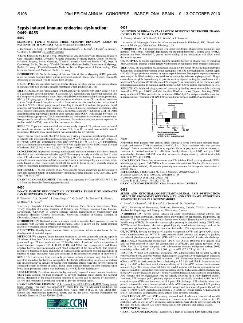
Sepsis-induced immuno-endocrine dysfunction:0449–04530449SELECTIVE TYPE-II MUSCLE FIBRE ATROPHY DEVELOPS EARLY INPATIENTS WITH NON-EXCITABLE MUSCLE MEMBRANE
J. Bierbrauer1, S. Koch1, C. Olbricht1, M. Braunschmidt1, C. Kleber2, J. Fielitz3, S. Spuler4,C. Spies1, J. Spranger5, S. Weber-Carstens1
1Charite-University Medicine Berlin, Department of Anesthesiology and Operative IntensiveCare Medicine, Berlin, Germany, 2Charite-University Medicine Berlin, Center for Muscu-loskeletal Surgery, Berlin, Germany, 3Charite-University Medicine Berlin, CVK, Dept. ofCardiology, Berlin, Germany, 4Charite-University Medicine Berlin, Department of Myology,Berlin, Germany, 5Charite-University Medicine Berlin, Department of Endocrinology, Dia-betes and Nutritional Medicine, Berlin, Germany
INTRODUCTION. So far, histological data on Critical Illness Myopathy (CIM) primarilyrefers to muscle biopsies taken during protracted critical illness (after weeks), repeatedlydescribing pronounced type-II muscle fibre atrophy.
OBJECTIVES. We speculate that type-II fibre atrophy develops during early critical illnessin patients with non-excitable muscle membrane which predicts CIM. (1)
METHODS. Due to their elevated risk for CIM, critically ill patients with SOFA scores C8 on 3of 5 consecutive days within the first 8 days after ICU admission were eligible for inclusion intothis prospective, observational study. Preexisting IDDM or neuromuscular disorder, pregnancy,BMI C 35 kg/m2, age \ 18 years, or pretreatment[4 days on other ICU constituted exclusioncriteria. Surgical muscle biopsies were taken from vastus lateralis muscles between day 5 and 8after first SOFA C 8 and postprocessed according to standard procedures (isopentane, liquidnitrogen, ATPase/toluidineblue staining). We assessed muscle membrane excitability afterdirect muscle stimulation, abnormal muscle membrane excitabilty indicating CIM (1). Afterquantifying fibre-type specific median cross sectional areas (CSA) with ImageJ-Software, wecompared fibre-type specific CSA in patients with and without non-excitable muscle membrane.Nonparametric tests (Mann–Whitney U) were used for statistical analyses, results expressed asmedian and (25th/75th) percentiles for continuous variables.
RESULTS. 33 patients were enrolled and subsequently biopsied. 29 patients were evaluatedfor muscle membrane excitability, of whom 62% (n = 18) showed non-excitable musclemembrane. Reliable CSA quantification was obtainable for 27 patients.
Type-II but not type-I muscle fibre CSA during early critical illness was significantly decreasedin patients with non-excitable muscle membrane (2,839 lm2 compared to 4,285 lm2 for type-II,p = 0.028; 3,504 lm2 compared to 3,683 lm2 for type-I, p = 0.197; n = 23). Furthermore,non-excitable muscle membrane was associated with significantly lower MRC scores after endof sedation (3.00 (2.94/3.41) vs. 3.75 (3.31/4.75), p = 0.023, n = 20).
CONCLUSIONS. In patients showing non-excitable muscle membrane after direct musclestimulation we could observe selective type-II fibre atrophy as early as within the first 10 daysafter ICU admission (day 5–8 after 1st SOFA C 8). Our findings demonstrate that non-excitable muscle membrane indeed is associated with a histomorphological correlate previ-ously linked to CIM. These results highlight the need to focus on early critically illness inorder to investigate pathophysiological aspects of CIM.
REFERENCE. 1. Weber-Carstens et al., Nonexcitable muscle membrane predicts intensivecare unit-acquired paresis in mechanically ventilated, sedated patients. Crit. Care Med. 2009Sep;37(9):2632–2637
GRANT ACKNOWLEDGMENT. This study was supported by Grant KFO192, WE 4386/1-1 from the Deutsche Forschungsgemeinschaft.
0450INNATE IMMUNE DEFICIENCY OF EXTREMELY PREMATURE NEONATESCAN BE REVERSED BY INTERFERON-c
P. Tissieres1,2,3, A. Ochoda1,3, I. Dunn-Siegrist1,3, G. Drifte1,3, M. Morales4, R. Pfister2,
M. Berner2, J. Pugin1,3
1University Hospitals of Geneva, Division of Intensive Care, Geneva, Switzerland, 2Uni-versity Hospitals of Geneva, Division of Pediatric and Neonatal Intensive Care, Geneva,Switzerland, 3University of Geneva Medical School, Department of Microbiology andMolecular Medicine, Geneva, Switzerland, 4University Hospitals of Geneva, Division ofObstertrics, Geneva, Switzerland
INTRODUCTION. Bacterial sepsis is a major threat in neonates born prematurely, and isassociated with elevated morbidity and mortality. Little is known on the innate immuneresponse to bacteria among extremely premature infants.
OBJECTIVES. Identify innate immune defect in premature infants as risk factor for thedevelopment of neonatal sepsis.
METHODS. We compared innate immune functions to bacteria commonly causing sepsis in21 infants of less than 28 wks of gestational age, 24 infants born between 28 and 32 wks ofgestational age, 25 term newborns and 20 healthy adults. Levels of surface expression ofinnate immune receptors (CD14, TLR2, TLR4, and MD-2) for Gram-positive and Gram-negative bacteria were measured in cord blood leukocytes at the time of birth. The cytokineresponse to bacteria of those leukocytes as well as plasma-dependent opsonophagocytosis ofbacteria by target leukocytes were also measured in the presence or absence of interferon-c.
RESULTS. Leukocytes from extremely premature infants expressed very low levels ofreceptors important for bacterial recognition. Leukocyte inflammatory responses to bacteriaand opsonophagocytic activity of plasma from premature infants were also severely impairedcompared to term newborns or adults. These innate immune defects could be corrected whenblood from premature infants was incubated ex vivo 12 h with interferon-c.
CONCLUSIONS. Premature infants display markedly impaired innate immune functions,which likely account for their propensity to develop bacterial sepsis during the neonatalperiod. Maturation of the innate immune response to bacteria can be induced by interferon-cex vivo and represents a promising strategy to prevent neonatal sepsis.
GRANT ACKNOWLEDGMENT. P.T. received the 2008 ECCRN-ESICM Young Inves-tigator Award. This study was supported by grants from the von Meissner Foundation, theUniversity Hospital of Geneva Research Fund (Grant PRD-08-II-2), an Ada Marek-Rut-kowski and Maksymilian Rutkowski Grant, and by the Swiss National Foundation forScientific Research (Grant 320080-122034).
0451INHIBITION OF RHO-A BY C5A LEADS TO DEFECTIVE NEUTROPHIL PHAGO-CYTOSIS IN CRITICALLY ILL PATIENTS
A. Conway Morris1, A.G. Rossi1, T.S. Walsh2, A.J. Simpson1
1University of Edinburgh, Centre for Inflammation Research, Edinburgh, UK, 2Royal Infir-mary of Edinburgh, Critical Care, Edinburgh, UK
INTRODUCTION. The anaphylotoxin C5a impairs neutrophil phagocytosis in animals1 andhumans2 with sepsis. Although dependency on the phosphoinositol 3-kinase delta (PI3Kd)pathway has been identified2,3, greater understanding of the mechanism will allow noveltherapeutic options.
OBJECTIVES. To test the hypotheses that C5a mediates its effect on phagocytosis by impairingRhoA activation, and that similar defects will be found in neutrophils from critically ill patients.
METHODS. The mechanism was dissected using an in vitro model of C5a-mediated neutrophildysfunction, treating healthy human donor neutrophils with C5a at concentrations found in sepsis(100 nM). Phagocytosis was assessed by zymosan particle uptake. Neutrophils exposed to zymosanwere assayed for RhoA activity, a key mediator of actin polymerisation in phagocytosis4. Phago-cytosis by neutrophils from critically ill patients was investigated, looking for correlations with amarker of C5a exposure (CD88, the main C5a receptor)2, an examination of the RhoA and actinpolymerisation response to zymosan, and the ability of GM-CSF to restore phagocytosis ex-vivo.
RESULTS. C5a inhibited phagocytosis of zymosan by healthy donor neutrophils (reducingfrom 67 to 27%, p \ 0.0001) and also impaired RhoA activation (Figure). Blocking PI3Kd,using inhibitor IC87114, prevented the inhibition of RhoA by C5a, and prevented the reductionin phagocytosis. Treatment with GM-CSF restored phagocytosis and RhoA activation (Fig. 1).
Figure 1
Neutrophils from patients with critical illness showed a strong correlation between phago-cytosis and surface CD88 expression (r = 0.80, P \ 0.001), consistent with our previousfindings2. Patient neutrophils failed to up regulate RhoA or polymerise actin in response tozymosan, in marked contrast to cells from healthy donors (p = 0.002 and p = 0.006respectively). Ex-vivo GM-CSF was able to improve phagocytosis by patient neutrophilsfrom 32 to 69%, p \ 0.001.
CONCLUSIONS. These data demonstrate that C5a inhibits RhoA activity through PI3Kd,inhibiting phagocytosis. GM-CSF is able to reverse this inhibition. Similar effects are seen inneutrophils from critically ill patients, providing new avenues for therapeutic intervention incritical illness.REFERENCES. 1 Huber-Lang M, et al. J Immunol. 2002;169:3223–312 Conway Morris A, et al. AJRCCM. 2009;180:19–283 Conway Morris A et al. JICS. 2010;11:614 Caron E, et al. Science 1998:282: 1717–21GRANT ACKNOWLEDGMENT. Chief Scientist Office CAF/08/13.
0452SEPSIS AND HYPOTHALAMO-PITUITARY-ADRENAL AXIS DYSFUNCTION:IMPACT OF ARGININE-VASOPRESSIN (AVP) AND APELIN (APL) EXOGENOUSADMINISTRATIONS IN A RODENT MODEL
O. Lesur1, F. Chagnon1, J.-F. Roussy1, L. Chouinard2, N. Gallo-Payet2
1MICU, University of Sherbrooke, Medicine, Sherbrooke, Canada, 2CHUS, University ofSherbrooke, Physiology and Biophysics, Sherbrooke, Canada
INTRODUCTION. Severe sepsis induces an acute hypothalamo-pituitary-adrenal axisdysfunction which is prevalent, impacts shock and vasopressor-dependency, and possibly theoutcome. This dysfunction was recently denominated Critical Illness-Related CorticosteroidInsufficiency (CIRCI). Because the corticotrophic pathway is disturbed during CIRCI, withACTH-cortisol dissociation, alternative physiologically nondominant pathways such as thevasopressinergic/apelinergic axis, become essential to the HPA adaptation to stress.
OBJECTIVES. Seeking the impact of arginine-vasopressin (AVP) and apelin (APL) exog-enous administrations on: ACTH & corticosterone blood contents, and respective pituitaryand adrenal gland receptor expression (V1b, APJ) in a rodent model of endotoxin challenge.
METHODS. A rodent model of endotoxin (LPS E. Coli 055:B5, 10 mg/kg I/P)-induced HPAaxis has been selected to study the committment of AVP/APL and related receptors (V1b/APJ). Rats (n = 6) were equipped with subcutaneous osmotic minipumps (Alzet, 2001)containing: saline, APL-13 (10, 100, 1,000 lg), or AVP (37.5, 75 lg) for 18 h.
RESULTS. Without LPS challenge, exogenous APL administration did not alter ACTH &corticosterone blood contents whereas high dosage of exogenous AVP significantly increasedcorticosterone blood content (p \ 0.05 vs. control). LPS I/P challenge induced a huge increasedof blood ACTH & corticosterone, both culminating at 1.5 h (6- and 23-fold increases respec-tively, p \ 0.05 vs. baseline), which was normalizing at 18 h for ACTH whereas corticosteroneremained high. This dissociation validates the model, matchs with human observations, andsuggests non ACTH-dependent corticosterone release after LPS challenge. After LPS challenge,blood AVP slightly increased and AVP pituitary content decreased, whereas blood and pituitaryAPL contents did not significantly vary (as already described in a water-deprived model).However, APL receptor (APJ) pituitary expression was drastically quenched while the AVPcentral receptor (V1b) was increased, at 18 h post LPS challenge. APL administration com-pletely reversed the above down-regulation while AVP also partially restored APJ pituitaryexpression by almost 50% in a dose-dependent manner, and to a lesser degree in the adrenalgland (p \ 0.05 vs. LPS). Selective non-peptide V1b (SSR149415) and peptide APL antagonists(F13A) substitutions confirmed the above effects were directely mediated.
CONCLUSIONS. APL and AVP pituitary neuronal and bloodstream contents behave dif-ferently, and blood ACTH & corticosterone contents were dissociated, after acute LPSchallenge. APL as well as AVP exogenous administrations were able to reverse (partially forthe later) the LPS-induced APJ down-regulation in both pituitary and adrenal glands.
REFERENCE(S). None.
GRANT ACKNOWLEDGMENT. Support by a Dept of Medicine UDS fellowship grant.
S198 23rd ESICM ANNUAL CONGRESS – BARCELONA, SPAIN – 9–13 OCTOBER 2010

0453TOLL-LIKE RECEPTOR 2 (TLR2) IMPAIRS LUNG DEFENCE TOWARDS PSEU-DOMONAS AERUGINOSA IN THE SETTING OF SEPSIS-INDUCED IMMUNEDYSFUNCTION
F. Pene1,2,3, B. Zuber1,2,3, A. Laubier2, B. Sauneuf2, D. Grimaldi1,2,3, C. Rousseau2,
J.-P. Mira1,2,3, J.-D. Chiche1,2,3
1Hopital Cochin, Medical ICU, Paris, France, 2Institut Cochin, Inserm U1016, CNRSUMR8104, Paris, France, 3University Paris Descartes, Paris, France
INTRODUCTION. Host infection triggers an innate immune response leading to a systemicinflammatory response, often followed by an immune dysfunction which impairs the lungdefence mechanisms in mice and increases susceptibility to secondary P. aeruginosa pneu-monia. Activation of the Toll-like receptor (TLR)-dependent signalling pathways influencesthe magnitude of the initial pro-inflammatory phase of sepsis. Contribution of TLR signalingto the subsequent development of post-infective immunosuppression has been poorly studied.
OBJECTIVES. To investigate the relative contribution of TLR2 and TLR4 in lung defencetowards P. aeruginosa in the setting of sepsis-induced immune dysfunction.
METHODS. We used wild-type (WT) C57BL6/J mice and littermates deficient for TLR2(TLR2 KO), TLR4 (TLR4 KO) or both TLR2 and TLR4 (TLR2 9 4 KO). These animalswere subjected to a sublethal polymicrobial sepsis (cecal ligature and puncture, CLP) fol-lowed by a secondary P. aeruginosa pneumonia at day 8 post-CLP 1. We evaluated 8-daysurvival and the lung response 4 and 24 h after instillation through lung histology, quanti-fication of protein level, cell recruitment and myeloperoxydase (MPO) activity. Lungexpression of TLR2, TLR4 and TLR5 was assessed through quantitative RT-PCR. Bacteriallung clearance was evaluated through quantitative culture of bronchoalveolar lavage fluid(BALF). Bacteremic dissemination was assessed through quantitative blood cultures. Finally,we measured cytokines in the BALF and in the whole lung.
RESULTS. Post-septic WT and TLR4 KO mice displayed high susceptibility (mortality rate80%) towards secondary pneumonia. In contrast, post-septic TLR2-deficient mice (eitherTLR2 KO or TLR2 9 4 KO), survived the secondary pulmonary infection (mortality rate\20%). In WT mice, CLP increased lung expression of TLR2, but neither of TLR4 norTLR5. TLR2 KO mice displayed improvement in lung bacterial clearance and reduction inbacteremic dissemination as compared to WT mice. With regard to pulmonary inflammation,TLR2 KO mice displayed decreased alveolar damage. Furthermore WT and TLR2 KO micedisplayed differences in the pulmonary release of cytokines. Thus TLR2 KO mice exhibitedincreased production of TNF-a and IFN-c and a decreased production of IL-10.
CONCLUSIONS. In a model of polymicrobial sepsis followed by P. aeruginosa pneumonia,TLR2 deficiency improves survival by promoting efficient bacterial clearance and decreasingpulmonary inflammation. TLR2-dependent mechanisms that specifically contribute to lungdefence in the setting of sepsis-induced immune dysfunction are currently investigated.
REFERENCE(S). Pene et al., J Immunol (2008); 181 (12): 8513–20
GRANT ACKNOWLEDGMENT. Supported by the ESICM Young Investigator Award2007
From infection diagnosis to therapy: 0454–04580454EVALUATION OF THE INTRODUCTION OF AN ANTIMICROBIAL STEWARD-SHIP PROGRAM TO A MEDICAL–SURGICAL INTENSIVE CARE UNIT
L. Burry1,2, S. Howie1, S. Lapinsky2, T. Khory2, B. Minnema2, M. Christian2, T. Stewart2, R.Wax2, S. Mehta2, M. Thomson2, C. Bell3, A. Morris2
1Mount Sinai Hospital, Pharmacy, Toronto, Canada, 2Mount Sinai Hospital, Medicine, Tor-onto, Canada, 3St Michael’s Hospital/University of Toronto, Medicine, Toronto, Canada
INTRODUCTION. An antimicrobial stewardship program (ASP) is a method of optimizingantimicrobial prescribing, altering antimicrobial resistance, reducing costs, and improvingpatient care. The intensive care unit (ICU) is an ideal environment for the application of anASP given the complexity of the patient population, the ecology of resistant organisms, andthe fact that selection of inappropriate antimicrobials or delays in initiating appropriatetherapy has measurable effects on outcomes.
OBJECTIVE. To determine the impact of the introduction of an ICU ASP on prescribing,patient outcomes, resistance, and costs.
METHODS. We implemented an ASP in a 16-bed medical surgical ICU of a university-affiliatedhospital. The ASP team used prospective audit with interaction and feedback providing suggestedchanges in therapy (e.g. antibiotic choice, dose, duration) on a daily basis. ASP provided con-sultation on all ICU patients not followed by the Infectious Diseases Consult service. Parameterscollected included demographic data, details of antimicrobial regimens, culture results, defineddaily doses (DDD)/100 patient days and antimicrobial costs. Descriptive statistics were applied tocompare pre-ASP (Feb/08–Jan/09) and post-ASP (Feb/09–Jan/10) data.
RESULTS. There was no significant difference in age distribution, sex, APACHE II score, ortypes of ICU admissions (medical vs. surgical) between the pre-ASP (n = 840) and post-ASPgroups (n = 834). The ASP team provided prospective audit and feedback for C80% of patientseach day. Mean monthly antibacterial DDD/100 patient-days post-ASP was reduced by 15.6%(146.4 vs. 123.6, p \ .05). The implementation of the ASP was associated with a 28.4% decreasein mean monthly antibiotic costs/100 patient-days ($3916 vs. $2805) for a total cost reduction of$53,635. In terms of prescribing and resistance, the introduction of ASP was associated with areduction in the prescribing of anti-Pseudomonal agents (compared with antibiotics not coveringPseudomonas) (mean monthly ratio 2.0 vs. 1.6), MRSA vs. MSSA covering antibiotics (meanmonthly ratio 2.0 vs. 1.4) and improved susceptibility pattern for Pseudomonas aeruginosa asdemonstrated by increases of 17, 9 and 7% to tobramycin, meropenem, and piperacillin-tazo-bactam susceptibilities, respectively. There was no difference in ICU mortality rate (19.1% pre-ASP vs. 18.9% post-ASP), mean ICU length of stay (5.6 days pre-ASP vs. 5.9 days post-ASP),or the % ventilation days (64.5% pre-ASP vs. 57.6% post-ASP).
CONCLUSIONS. The ASP team worked collaboratively with the ICU team to improveantimicrobial therapy and as a result improved overall antibiotic usage and resistance patternsof important ICU microorganisms, and decreased antimicrobial costs. Appropriate and judi-cious antimicrobial use guided by an ASP is beneficial to the ICU.
0455CAN PCT AND/OR CRP HELP IDENTIFY ASSOCIATED BACTERIAL INFECTIONIN PATIENTS WITH INFLUENZA PNEUMONIA?
E.J. Cuquemelle1, C. Brun-Buisson1, REVA Group1Hopital Henri Mondor, Reanimation Medicale, Creteil, France
INTRODUCTION. During the 2009 winter, a new pandemic influenza A H1N1 virus emergedand spread globally, primarily in younger age groups, with a high rate of in intensive care units(ICU) admission and death in 7% of the patients. Causes of death included an overwhelmingviral infection and primary or secondary bacterial infection. Bacterial pneumonia cannot bedifferentiated from viral pneumonia on the basis of the patients characteristics, chest radiographsor biologic results. Procalcitonin (PCT) is a recognized marker of bacterial infection and mightbe a prognostic marker in lower respiratory tract infections.
OBJECTIVES. To determine if PCT and/or C-Reactive protein (CRP) levels at admission inIntensive Care Unit can help identify associated bacterial infection in patients with influenzaepneumonia.
METHODS. A nationwide registry (REVA) was set up in France during the 2009 H1N1influenza pandemic. Levels of PCT and CRP at ICU admission were compared betweenpatients presenting with Influenzae pneumonia associated or not with a bacterial coinfection.
RESULTS. 137 patients were included, 48 of whom received antibiotics prior to hospitaliza-tion. Of the remaining 89 patients, 43 (48%) had documented bacterial co-infection. The bacteriainvolved in the co-infections were Streptococcus pneumoniae (n = 22, 16.5%), Staphylococcusaureus (n = 11, 8.3%), Streptococcus A group (n = 6, 4.5%). Figure 1 shows the initial valuesof procalcitonin and CRP. Median values for both PCT (28.9 vs. 0.74 ng/ml, p \ 0.001) andCRP (263 vs. 95 mg/l, p = 0.008) were significantly higher in patients with bacterial co-infection. The area under the ROC curve was 0.88 and 0.79, respectively for PCT and CRP. Acutoff of[0.8 ng/ml for PCT (sensitivity 91% and specificity 59%) best identified patients withbacterial co-infection. For CRP, a cutoff of[220 mg/l (sensitivity 75%, specificity 78%) bestidentified patients with bacterial co-infections. Comparison of the 2009 H1N1 pneumonia andbacterial pneumonia group revealed no differences except for a higher SAPS 3 in the latter (45vs. 55 respectively, p = 0.004). However, there was no difference between the two groups forlength of stay in the ICU, mechanical ventilation (54 vs. 75%) or death (17 vs. 21%) rates.
Figure 1: Procalcitonin and CRP values
CONCLUSIONS. Procalcitonin may help to discriminate isolated viral pneumonia frombacterial associated infection in patients with Influenzae pneumonia in the ICU. Bacterial co-infection was not associated with a higher mortality.
0456IMPACT OF NEWS BIOMARKERS FOR THE DIAGNOSIS OF INFECTION INPATIENTS WITH SEVERE ACUTE DYSPNEA
C. Ara Somohano1,2,3, C. Bard1,2, A. Vesin3, A. Bonadona1, R. Hamidfar1, P. Pavese4, C.Minet1, Y. Dubois1, A. Tabah1,3, C. Schwebel1, J.P. Brion4, F. Carpentier2, J.F. Timsit1,3, BIOIRA1University Hospital A. Michallon, Medical Intensive Care Unit, Grenoble, France, 2Uni-versity Hospital A Michallon, Emergency Department, Grenoble, France, 3Institut AlbertBonniot, INSERM, U823 Team ‘‘Outcome of cancer and critical illnesses’’, Grenoble, France,4University Hospital A Michallon, Infectious Diseases Department, Grenoble, FranceINTRODUCTION. The cause of severe acute dyspnoea might be difficult to ascertain inemergency. Biomarkers may help in this setting but the respective contribution of multiplebiomarkers is unclear in this setting.OBJECTIVES. To measure the diagnostic contributions of routinely used (NT B typeNatriuretic Peptide, cardiac troponin I,DDimeres, C-reactive protein (CRP) and procalcitonin(PCT)) and new biomarkers (Mid-regional Pro-Atrial natriuretic peptide (MR Pro-ANP), ProAdrenomedulin (Pro ADM), Pro Endothelin (Pro ET) and Copeptin (Pro vasopressin CP)) fordiagnosing infection in patients with severe acute dyspnea.METHODS. We designed a prospective study of patients admitted in Emergency Department(ED) and Medical Intensive Care Unit (ICU) in a University Hospital. Inclusion criteria were acutedyspnea with SpO2 B 92% and/or respiratory rate C25 b/min. Patients with obvious myocardialinfarction or pneumothorax were excluded. All clinical and biological data were recorded andbiomarkers sampled. An independent blinded expert panel classified the patients according to allthe available data including response to treatment and outcomes blindly to biomarkers’ results.The roles of biomarkers were assessed quantitatively and then using terciles of the distribution. Thecontribution of the biomarkers in the diagnosis was assessed using AUC-ROC curves and bymultiple logistic regression taking into account other clinical and biological explanatory variables.
RESULTS. 342 consecutive patients (52% male, med age 74 years, 28 day mortality 17%)were enrolled. The final diagnosis was severe sepsis for 126 (36.9%) (pulmonary: n = 104,non-pulmonary n = 22). The parameters independently associated with infection lead to aclinico-biological model with an AUC = 0.806 and a good calibration (P (HLchi2) = 0.55)and included temperature, arterial pressure, cyanosis, stupor and coma, orthopnea, localizedchest sound abnormalities, PaO2/FiO2 ratio and localized infiltrate on chest X ray.New biomarkers were weakly but significantly associated with a sepsis AUC-ROC(SD): MidPro-ANP: 0.579 (0.034), Pro ADM: 0.596 (0.034), Pro ET: 0.607 (0.034)) and Pro vaso-pressin CP: 0.571 (0.035) whereas the AUC-ROC for PCT was PCT 0.792 (0.027)Adjusted onselected clinico-biological covariates, both PCT (AUC 0.858; p \ 0.0001) and CRP (AUC0.843; p \ 0.001) were significantly associated with the diagnosis of sepsis. A model with allthe clinico-biological variables and both biomarkers discriminated accurately (AUC 0.882,p \ 0.001). Among new biomarkers only Mid Pro-ANP added a significant (p = 0.013)contribution to the previous model (final AUC-ROC: 0.891, p \ 0.001).
CONCLUSIONS. Although new biomarkers were different between septic and non septicpatients with severe acute dyspnea, only Mid Pro-ANP may add a significant contribution.Further analysis about the prognostic value of these biomarkers is ongoing.
GRANT ACKNOWLEDGMENT. Grenoble University Hospital and Brahms diagnostic.
23rd ESICM ANNUAL CONGRESS – BARCELONA, SPAIN – 9–13 OCTOBER 2010 S199
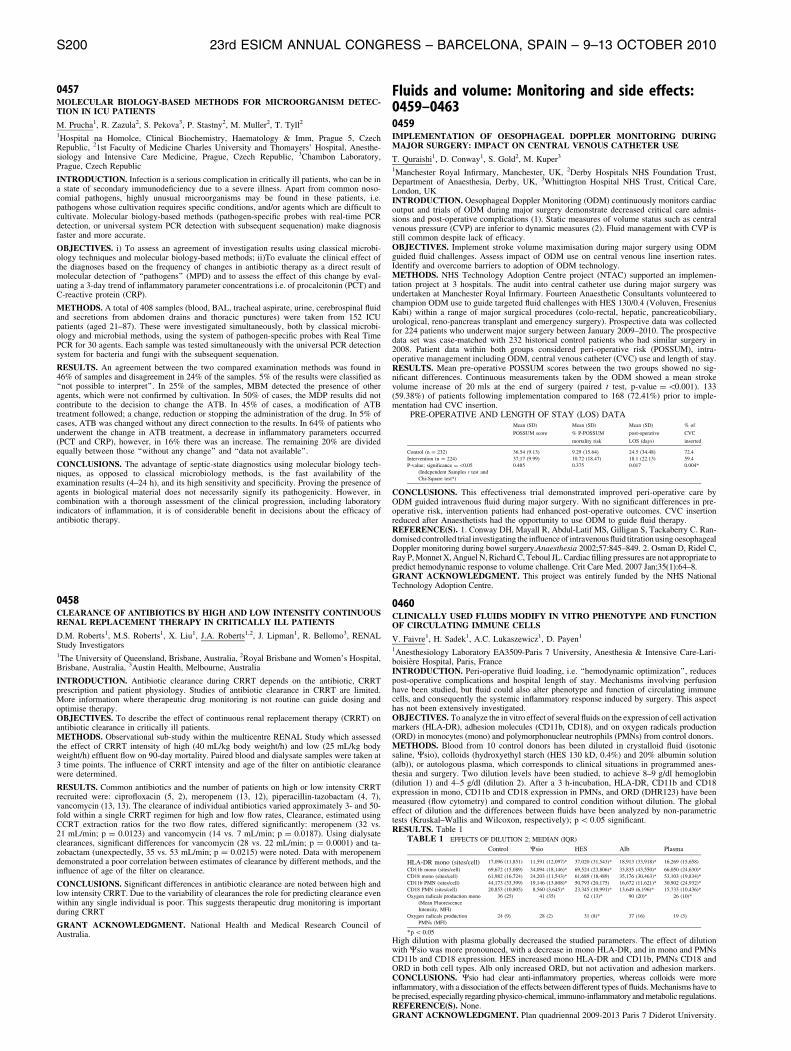
0457MOLECULAR BIOLOGY-BASED METHODS FOR MICROORGANISM DETEC-TION IN ICU PATIENTS
M. Prucha1, R. Zazula2, S. Pekova3, P. Stastny2, M. Muller2, T. Tyll2
1Hospital na Homolce, Clinical Biochemistry, Haematology & Imm, Prague 5, CzechRepublic, 21st Faculty of Medicine Charles University and Thomayers’ Hospital, Anesthe-siology and Intensive Care Medicine, Prague, Czech Republic, 3Chambon Laboratory,Prague, Czech Republic
INTRODUCTION. Infection is a serious complication in critically ill patients, who can be ina state of secondary immunodeficiency due to a severe illness. Apart from common noso-comial pathogens, highly unusual microorganisms may be found in these patients, i.e.pathogens whose cultivation requires specific conditions, and/or agents which are difficult tocultivate. Molecular biology-based methods (pathogen-specific probes with real-time PCRdetection, or universal system PCR detection with subsequent sequenation) make diagnosisfaster and more accurate.
OBJECTIVES. i) To assess an agreement of investigation results using classical microbi-ology techniques and molecular biology-based methods; ii)To evaluate the clinical effect ofthe diagnoses based on the frequency of changes in antibiotic therapy as a direct result ofmolecular detection of ‘‘pathogens’’ (MPD) and to assess the effect of this change by eval-uating a 3-day trend of inflammatory parameter concentrations i.e. of procalcitonin (PCT) andC-reactive protein (CRP).
METHODS. A total of 408 samples (blood, BAL, tracheal aspirate, urine, cerebrospinal fluidand secretions from abdomen drains and thoracic punctures) were taken from 152 ICUpatients (aged 21–87). These were investigated simultaneously, both by classical microbi-ology and microbial methods, using the system of pathogen-specific probes with Real TimePCR for 30 agents. Each sample was tested simultaneously with the universal PCR detectionsystem for bacteria and fungi with the subsequent sequenation.
RESULTS. An agreement between the two compared examination methods was found in46% of samples and disagreement in 24% of the samples. 5% of the results were classified as‘‘not possible to interpret’’. In 25% of the samples, MBM detected the presence of otheragents, which were not confirmed by cultivation. In 50% of cases, the MDP results did notcontribute to the decision to change the ATB. In 45% of cases, a modification of ATBtreatment followed; a change, reduction or stopping the administration of the drug. In 5% ofcases, ATB was changed without any direct connection to the results. In 64% of patients whounderwent the change in ATB treatment, a decrease in inflammatory parameters occurred(PCT and CRP), however, in 16% there was an increase. The remaining 20% are dividedequally between those ‘‘without any change’’ and ‘‘data not available’’.
CONCLUSIONS. The advantage of septic-state diagnostics using molecular biology tech-niques, as opposed to classical microbiology methods, is the fast availability of theexamination results (4–24 h), and its high sensitivity and specificity. Proving the presence ofagents in biological material does not necessarily signify its pathogenicity. However, incombination with a thorough assessment of the clinical progression, including laboratoryindicators of inflammation, it is of considerable benefit in decisions about the efficacy ofantibiotic therapy.
0458CLEARANCE OF ANTIBIOTICS BY HIGH AND LOW INTENSITY CONTINUOUSRENAL REPLACEMENT THERAPY IN CRITICALLY ILL PATIENTS
D.M. Roberts1, M.S. Roberts1, X. Liu1, J.A. Roberts1,2, J. Lipman1, R. Bellomo3, RENALStudy Investigators1The University of Queensland, Brisbane, Australia, 2Royal Brisbane and Women’s Hospital,Brisbane, Australia, 3Austin Health, Melbourne, Australia
INTRODUCTION. Antibiotic clearance during CRRT depends on the antibiotic, CRRTprescription and patient physiology. Studies of antibiotic clearance in CRRT are limited.More information where therapeutic drug monitoring is not routine can guide dosing andoptimise therapy.OBJECTIVES. To describe the effect of continuous renal replacement therapy (CRRT) onantibiotic clearance in critically ill patients.METHODS. Observational sub-study within the multicentre RENAL Study which assessedthe effect of CRRT intensity of high (40 mL/kg body weight/h) and low (25 mL/kg bodyweight/h) effluent flow on 90-day mortality. Paired blood and dialysate samples were taken at3 time points. The influence of CRRT intensity and age of the filter on antibiotic clearancewere determined.
RESULTS. Common antibiotics and the number of patients on high or low intensity CRRTrecruited were: ciprofloxacin (5, 2), meropenem (13, 12), piperacillin-tazobactam (4, 7),vancomycin (13, 13). The clearance of individual antibiotics varied approximately 3- and 50-fold within a single CRRT regimen for high and low flow rates, Clearance, estimated usingCCRT extraction ratios for the two flow rates, differed significantly: meropenem (32 vs.21 mL/min; p = 0.0123) and vancomycin (14 vs. 7 mL/min; p = 0.0187). Using dialysateclearances, significant differences for vancomycin (28 vs. 22 mL/min; p = 0.0001) and ta-zobactam (unexpectedly, 35 vs. 53 mL/min; p = 0.0215) were noted. Data with meropenemdemonstrated a poor correlation between estimates of clearance by different methods, and theinfluence of age of the filter on clearance.
CONCLUSIONS. Significant differences in antibiotic clearance are noted between high andlow intensity CRRT. Due to the variability of clearances the role for predicting clearance evenwithin any single individual is poor. This suggests therapeutic drug monitoring is importantduring CRRT
GRANT ACKNOWLEDGMENT. National Health and Medical Research Council ofAustralia.
Fluids and volume: Monitoring and side effects:0459–04630459IMPLEMENTATION OF OESOPHAGEAL DOPPLER MONITORING DURINGMAJOR SURGERY: IMPACT ON CENTRAL VENOUS CATHETER USE
T. Quraishi1, D. Conway1, S. Gold2, M. Kuper3
1Manchester Royal Infirmary, Manchester, UK, 2Derby Hospitals NHS Foundation Trust,Department of Anaesthesia, Derby, UK, 3Whittington Hospital NHS Trust, Critical Care,London, UKINTRODUCTION. Oesophageal Doppler Monitoring (ODM) continuously monitors cardiacoutput and trials of ODM during major surgery demonstrate decreased critical care admis-sions and post-operative complications (1). Static measures of volume status such as centralvenous pressure (CVP) are inferior to dynamic measures (2). Fluid management with CVP isstill common despite lack of efficacy.OBJECTIVES. Implement stroke volume maximisation during major surgery using ODMguided fluid challenges. Assess impact of ODM use on central venous line insertion rates.Identify and overcome barriers to adoption of ODM technology.METHODS. NHS Technology Adoption Centre project (NTAC) supported an implemen-tation project at 3 hospitals. The audit into central catheter use during major surgery wasundertaken at Manchester Royal Infirmary. Fourteen Anaesthetic Consultants volunteered tochampion ODM use to guide targeted fluid challenges with HES 130/0.4 (Voluven, FreseniusKabi) within a range of major surgical procedures (colo-rectal, hepatic, pancreaticobiliary,urological, reno-pancreas transplant and emergency surgery). Prospective data was collectedfor 224 patients who underwent major surgery between January 2009–2010. The prospectivedata set was case-matched with 232 historical control patients who had similar surgery in2008. Patient data within both groups considered peri-operative risk (POSSUM), intra-operative management including ODM, central venous catheter (CVC) use and length of stay.RESULTS. Mean pre-operative POSSUM scores between the two groups showed no sig-nificant differences. Continuous measurements taken by the ODM showed a mean strokevolume increase of 20 mls at the end of surgery (paired t test, p-value = \0.001). 133(59.38%) of patients following implementation compared to 168 (72.41%) prior to imple-mentation had CVC insertion.
PRE-OPERATIVE AND LENGTH OF STAY (LOS) DATAMean (SD)
POSSUM score
Mean (SD)
% P-POSSUM
mortality risk
Mean (SD)
post-operative
LOS (days)
% of
CVC
inserted
Control (n = 232) 36.54 (9.13) 9.29 (15.64) 24.5 (34.48) 72.4
Intervention (n = 224) 37.17 (9.99) 10.72 (18.47) 18.1 (22.13) 59.4
P-value; significance = \0.05
(Independent Samples t test and
Chi-Square test*)
0.485 0.375 0.017 0.004*
CONCLUSIONS. This effectiveness trial demonstrated improved peri-operative care byODM guided intravenous fluid during major surgery. With no significant differences in pre-operative risk, intervention patients had enhanced post-operative outcomes. CVC insertionreduced after Anaesthetists had the opportunity to use ODM to guide fluid therapy.REFERENCE(S). 1. Conway DH, Mayall R, Abdul-Latif MS, Gilligan S, Tackaberry C. Ran-domised controlled trial investigating the influence of intravenous fluid titration using oesophagealDoppler monitoring during bowel surgery.Anaesthesia 2002;57:845–849. 2. Osman D, Ridel C,Ray P, Monnet X, Anguel N, Richard C, Teboul JL. Cardiac filling pressures are not appropriate topredict hemodynamic response to volume challenge. Crit Care Med. 2007 Jan;35(1):64–8.GRANT ACKNOWLEDGMENT. This project was entirely funded by the NHS NationalTechnology Adoption Centre.
0460CLINICALLY USED FLUIDS MODIFY IN VITRO PHENOTYPE AND FUNCTIONOF CIRCULATING IMMUNE CELLS
V. Faivre1, H. Sadek1, A.C. Lukaszewicz1, D. Payen1
1Anesthesiology Laboratory EA3509-Paris 7 University, Anesthesia & Intensive Care-Lari-boisiere Hospital, Paris, FranceINTRODUCTION. Peri-operative fluid loading, i.e. ‘‘hemodynamic optimization’’, reducespost-operative complications and hospital length of stay. Mechanisms involving perfusionhave been studied, but fluid could also alter phenotype and function of circulating immunecells, and consequently the systemic inflammatory response induced by surgery. This aspecthas not been extensively investigated.OBJECTIVES. To analyze the in vitro effect of several fluids on the expression of cell activationmarkers (HLA-DR), adhesion molecules (CD11b, CD18), and on oxygen radicals production(ORD) in monocytes (mono) and polymorphonuclear neutrophils (PMNs) from control donors.METHODS. Blood from 10 control donors has been diluted in crystalloid fluid (isotonicsaline, Wsio), colloids (hydroxyethyl starch (HES 130 kD, 0.4%) and 20% albumin solution(alb)), or autologous plasma, which corresponds to clinical situations in programmed anes-thesia and surgery. Two dilution levels have been studied, to achieve 8–9 g/dl hemoglobin(dilution 1) and 4–5 g/dl (dilution 2). After a 3 h-incubation, HLA-DR, CD11b and CD18expression in mono, CD11b and CD18 expression in PMNs, and ORD (DHR123) have beenmeasured (flow cytometry) and compared to control condition without dilution. The globaleffect of dilution and the differences between fluids have been analyzed by non-parametrictests (Kruskal–Wallis and Wilcoxon, respectively); p \ 0.05 significant.RESULTS. Table 1
TABLE 1 EFFECTS OF DILUTION 2; MEDIAN (IQR)
Control Wsio HES Alb Plasma
HLA-DR mono (sites/cell) 17,096 (11,851) 11,591 (12,097)* 37,020 (31,543)* 18,913 (33,918)* 16,269 (15,658)
CD11b mono (sites/cell) 69,672 (15,089) 34,094 (18,146)* 69,524 (23,806)* 33,835 (43,550)* 66,050 (24,630)*
CD18 mono (sites/cell) 61,982 (16,724) 24,203 (11,543)* 61,689 (18,489) 35,176 (30,463)* 53,103 (19,834)*
CD11b PMN (sites/cell) 44,173 (33,399) 19,146 (13,808)* 50,793 (20,175) 16,672 (11,621)* 30,902 (24,932)*
CD18 PMN (sites/cell) 20,853 (10,805) 8,560 (3,645)* 23,345 (10,991)* 13,649 (6,196)* 15,733 (10,436)*
Oxygen radicals production mono
(Mean Fluorescence
Intensity, MFI)
36 (25) 41 (35) 62 (13)* 90 (20)* 26 (10)*
Oxygen radicals production
PMNs (MFI)
24 (9) 28 (2) 31 (8)* 37 (16) 19 (3)
*p \ 0.05
High dilution with plasma globally decreased the studied parameters. The effect of dilutionwith Wsio was more pronounced, with a decrease in mono HLA-DR, and in mono and PMNsCD11b and CD18 expression. HES increased mono HLA-DR and CD11b, PMNs CD18 andORD in both cell types. Alb only increased ORD, but not activation and adhesion markers.CONCLUSIONS. Wsio had clear anti-inflammatory properties, whereas colloids were moreinflammatory, with a dissociation of the effects between different types of fluids. Mechanisms have tobe precised, especially regarding physico-chemical, immuno-inflammatory and metabolic regulations.REFERENCE(S). None.GRANT ACKNOWLEDGMENT. Plan quadriennal 2009-2013 Paris 7 Diderot University.
S200 23rd ESICM ANNUAL CONGRESS – BARCELONA, SPAIN – 9–13 OCTOBER 2010
0461WORLD INITIATIVE ON ABDOMINAL HYPERTENSION EPIDEMIOLOGY, AUNIFYING PROJECT (WAKE-UP): A METABASE ANALYSIS ON INTRA-ABDOMINAL HYPERTENSION IN CRITICALLY ILL PATIENTS
M. Malbrain1, A. Reintam Blaser1, M. Sugrue1, P. Pelosi1, D. Chiumello1, G. Hernandez1, N.
Brienza1, A. Kirkpatrick1, A. Schachtrupp1, E. Estenssoro1, I. De Laet1, for the Wake-UpStudy Group (www.wsacs.org)1ZNA Stuivenberg, Intensive Care Medicine, Antwerpen, Belgium
INTRODUCTION. Intra-abdominal pressure (IAP) is a prognostic factor for ICU morbidityand mortality (1)
OBJECTIVES. To perform a meta-analysis on incidence and outcome of intra-abdominalhypertension (IAH) in different ICU populations, the evolution of IAP over time and thecorrelation with organ failure and fluid balance (FB)
METHODS. Pts admitted to ICU with IAP measurements (gastric or bladder) were included.Data from existing databases were collected on 2,645 pts from 24 centers (10 countries).Lowest (IAPlow) and highest IAP (IAPhigh) and lowest abdominal perfusion pressure(APP = MAP-IAP) were recorded each day. Mean (IAPmean) and maximal IAP (IAPmax)during the stay was calculated
RESULTS. BMI was 26.3 ± 10.5, M/F ratio 3/2, age 60.5 ± 18.3, APACHE-II 19 ± 9.7,SAPS-II 42.8 ± 17.7, SOFA score 5.6 ± 4.5. On admission, IAPlow was 11.3 ± 6.6 andIAPhigh 13.3 ± 5 while the lowest APP was 64.1 ± 19 mmHg. 12,909 IAP measurementswere performed. The overall IAPlow was 10.1 ± 5.6 and IAPhigh 12.1 ± 5.9 mmHg.IAPmean during stay was 11.8 ± 6.5 while IAPmax was 16 ± 8 occurring after2.6 ± 2.1 days. There were no differences in IAP between medical and surgical pts althoughthe IAPmax occurred later in medical pts. IAPmax above 12 mmHg (IAH) was present in1,585 pts (72.2%) and above 20 mmHg (ACS) in 538 (24.5%), while IAPmean above12 mmHg was present in 994 pts (45.3%) and above 20 mmHg in 199 (9.1%). The incidenceof IAH was significantly higher in emergency surgery (48.2%) versus elective surgery(30.7%) versus medical (28.8%) versus trauma (23.2%) (p \ 0.0001). This was also true forACS: 6.5 versus 2.5 versus 2.3 versus 2.2%. The incidence of IAH ranged within countriesfrom 6.5 to 87.9%. The IAPmean (8.1–17.9) and IAPmax (12.5–22.2) differed significantlybetween countries and centres. Overall ICU mortality was 42.5%. IAPhigh correlated wellwith (cumulative) FB and SOFA scores. The IAPhigh was significantly higher from day 2onwards in nonsurvivors while APP was already lower on admission and remained lowerduring the first week, suggesting better discriminatory power. The (cumulative) FB and SOFAscore remained also higher during the first week in nonsurvivors. The ICU and hospitalmortality was significantly higher in pts with IAH and ACS, defined according to an IAPmeanabove 12 or 20 mmHg respectively, this was not the case when IAH and ACS were definedaccording to IAPmax
CONCLUSIONS. The preliminary results of a metabase-analysis show that IAH can beexpected in about 45% of cases and ACS in 9%. The incidence differs according to the ICUpopulation studied and was the highest in emergency surgical pts and occurred later during thestay in medical pts (secondary IAH). The occurrence of IAH (defined according to mean andnot maximal values), together with (cumulative) FB and organ failure (defined by SOFAscore) are outcome predictors and suggest a relation between capillary leak, fluid loading,increased IAP and end-organ failure
REFERENCE. 1.Malbrain M et al. Crit Care Med 2005,33:315–22.
0462IS THE STROKE VOLUME VARIATION (SSV) AND/OR THE PULSE PRESSUREVARIATION (PPV) CALCULATED WITH THE LIDCO RAPID SUITABLE FORPREDICTION OF FLUID RESPONSIVENESS?
P. Brass1, J. Latza2, J. Peters2, M. Hellmich3, E. Berendes1
1Helios Klinikum Krefeld, Klinik fur Anasthesie, Operative Intensivmedizin und Schmerz-therapie, Krefeld, Germany, 2Klinikum Duisburg, Klinik fur Anasthesie, Intensivmedizin,Schmerztherapie und Notfallmedizin, Duisburg, Germany, 3Universitatsklinikum Koln,Institut fur Medizinische Statistik, Informatik und Epidemiologie, Koln, Germany
INTRODUCTION. Both hypovolemia and hypervolemia during and after major surgery, aswell as in patients on critical care units could lead to circulatory instability and organ dys-function and therefore increases the rate of complications and mortality. A balancedperioperative fluid management targeting normal volemia and adequate oxygen deliveryimproves outcome. In contrast to heart rate, blood pressure, central venous pressure the SVVand PPV are valid parameters to predict volume responsiveness (VR). Passive leg raisingprior to volume administration is an available test (PLRT) that shows reversible response tovolume loading.
OBJECTIVES. The purpose of our investigation was to evaluate whether LiDCO rapidgenerated SVV and/or PPV are as accurate as PiCCO2 generated SVV, to predict VR. Inaddition we investigated reliability of the PLRT.
METHODS. After ethical approval patients undergoing major abdominal surgery, neuro-surgery (n = 20) and critical Care patients (n = 10) fluid management was driven byPiCCO2. PiCCO2 was calibrated at the following points: Induction (M1), PLRT (M2), 500 mlHAES volume challenge (M3). Simultaneously the LiDCO rapid calculated the nominalparameters. All hemodynamic parameters were continuously documented and compared attime of thermodilution.
RESULTS. LiDCO rapid determined SVV corresponds with an average deviation of 15% toPiCCO2 generated SVV. SVV \ 8% at M1 no patient responded to PLRT and fluid challengewith an increase in CO. SVV [ 10% 20 patients after PLRT and 28 patients after volumechallenge responded with an increase in CO. The cut off values (SVV \8% respectively[10%), the amount of patients responding to fluid challenge and the resulting changes in COwere the same in both methods.
CONCLUSIONS. SVV and PPV measured by LiDCO rapid are reliable and can be used aspredictors for CO changes after fluid challenge. PLRT is not suitable for all patients to predictVR.
0463EFFECTS OF DIFFERENT HYDROXYETYL STARCH SOLUTIONS ON APOPTO-SIS AND PROLIFERATION IN MURINE KIDNEY CELLS
S. Doemming1, R.A. Claus2, G. Wolf3, T. Schuerholz1, G. Marx1
1University Hospital Aachen, Intensive Care, Aachen, Germany, 2University Hospital Jena,Anesthesology and Intensive Care, Jena, Germany, 3University Hospital Jena, InternalMedicine III, Jena, Germany
INTRODUCTION. Volume replacement with hyperoncotic hydroxyethyl starch (HES) isassociated with acute kidney failure in sepsis [1]. Recently, it was demonstrated that 10%HES 200/0.5 induces increased inflammation and leads to more tubular damage compared to6% HES 130/0.42 in an ex-vivo kidney perfusion model [2].
OBJECTIVES. We investigated whether different HES solutions lead to disturbed cellproliferation or to increased apoptosis in murine kidney cells.
METHODS. Proximal murine tubulus cells (mouse tubulus cells, MTC) and podocytes wereincubated either with 10% HES 200/0.5 (Hemohes 10%, B. Braun, Melsungen, Germany) or6% HES 130/0.42 (Venofundin 6%, B. Braun) for 12 h in culture medium. For both solutionsa 40% (HES 40; n = 12), 80% (HES 80; n = 12) and a 90% dilution (HES 90; n = 12) wasprepared. Control group was incubated with NaCl 0.9% (B. Braun; n = 6). The ability of cellsto proliferate was measured with a MTT-assay. To evaluate apoptosis we performed a cas-pase-3 assay. Results were analysed as percent deviation from control group. Statistics wereperformed with 1-way-ANOVA and Dunnett t test or t test where appropriate. P \ 0.05 wasconsidered significant.
RESULTS. Proliferation of MTC’s was significantly reduced only by high amounts of HES200/0.5 (HES 90: 39 ± 8%; p \ 0.001). Comparable results were obtained for HES 130/0.42(HES 90: 34 ± 13%; p \ 0.001). HES 200/0.5 significantly increased apoptosis in tubuluscells in HES 90 group (286 ± 145%; p = 0.002). Smaller amounts of HES 200/0.5 non-significantly affected apoptosis. All HES 130/0.42 dilutions increased apoptosis of MTC notsignificantly. Apoptosis was significantly lower in the HES 90 group after the addition of HES130/0.42 compared to HES 200/0.5 in MTC (p = 0.036). Proliferation of podocytes wasinhibited after addition of HES 200/0.5 in HES 80 (39 ± 5%; p \ 0.001) and HES 90 group(51 ± 3%; p \ 0.001) compared to control. All dilutions of HES 130/0.42 inhibited podo-cytes (HES 40: 7 ± 8%, p = 0.039; HES 80: 27 ± 4%, p \ 0.001; HES 90: 44 ± 4%,p \ 0.001). Compared with HES 130/0.42 podocytes were inhibited significantly strongerafter incubation with HES 200/0.5 (HES 80: p = 0.012; HES 90: p = 0.017). HES 130/0.42increases apoptosis not significantly in podocytes. Apoptosis of podocytes was significantlylower in HES 90 group after addition of HES 130/0.42 compared with HES 200/0.5(p = 0.006).
CONCLUSIONS. Hydroxyethyl starch leads to a concentration dependent impairment ofproliferation and induces apoptosis in murine tubulus cells and podocytes. Higher doses ofHES 200/0.5 induce a significant increase in apoptosis and inhibition of proliferation com-pared to HES 130/0.42.
REFERENCE(S). 1. Brunkhorst FM, Engel C, Bloos F, Meier-Hellmann A, Ragaller M,Weiler N, et. al. NEJM 2008;358:125–1392. Huter L, Simon TP, Weinmann L, Schuerholz T, Reinhart K, Wolf G, Amann KU,Marx G. Critical Care 2009;13(1): R23.
Outcome-related factors: 0464–04680464DELIRIUM EPIDEMIOLOGY IN CRITICAL CARE (DECCA) STUDY: AN INTER-NATIONAL STUDY
J. Salluh1, M. Soares1, J.M.M. Teles2, D.H. Ceraso3, N. Raimondi3, V.S. Nava3, P. Blasquez1,
S. Ugarte3, C.I. Guzman3, J.V. Centeno3, M. Laca3, G. Grecco4, E. Jimenez3, S. Arias-Rivera3, C. Duenas3, M.G. Rocha5
1Instituto Nacional de Cancer, Rio de Janeiro, Brazil, 2Hospital da Bahia, Salvador, Brazil,3DECCA Study Group, Federacion Panamericana e Iberica de Sociedades de Medicina Cri-tica y Terapia Intensiva (FPIMCTI), Buenos Aires, Argentina, 4DECCA Study Group,Federacion Panamericana e Iberica de Sociedades de Medicina Critica y Terapia Intensiva(FPIMCTI), Rio de Janeiro, Brazil, 5Pavilhao Pereira Filho-Santa Casa de Misericordia dePorto Alegre, Porto Alegre, Brazil
INTRODUCTION. Delirium is a frequent source of morbidity in intensive care. Presently,most data on its epidemiology is from single center studies.
OBJECTIVES. Our aim was to conduct a multicenter study to evaluate the epidemiology ofdelirium in intensive care units (ICU).
METHODS. A 1-day point-prevalence study was performed with the aim of describing in104 ICUs from 11 countries in South and North America and Spain.
RESULTS. A total of 975 patients were screened and 497 fulfilled inclusion criteria and wereenrolled (median age 62 years, 52.5% men, 16.7 and 19.9% for ICU and hospital mortality),64% were admitted to the ICU due to medical causes and sepsis was the main diagnosis(n = 76, 15.3%). 265 patients were sedated and only 232 (46.6%) patients could be evaluatedwith the CAM-ICU. The prevalence of delirium was 32.3% (n = 75). As compared topatients without delirium, those with the diagnosis of delirium had a higher severity of illnessat admission as demonstrated by higher SOFA (p = 0.004) and SAPS3 scores (p \ 0.0001).Delirium was associated with increased ICU (20 vs. 5.7%, p = 0.002) and hospital mortality(24 vs. 8.3%, p = 0.0017), longer ICU (p \ 0.0001) and hospital LOS (22 (11–40) vs. 7(4–18) days, p \ 0.0001). Previous use of midazolam (p = 0.009) was more frequent inpatients with delirium. Increased use of invasive devices such as central venous catheter(p \ 0.0001), arterial catheter (p = 0.004) and urinary catheter (p = 0.0001) were morefrequent in patients with delirium. On multivariate analysis, delirium was independentlyassociated with increased ICU mortality (OR = 3.14 [1.26–7.86], 95% CI) and hospitalmortality (OR = 2.5 (1.1–5.7) 95% CI).
CONCLUSIONS. In this one-day international study, delirium was frequent in ICU patientsand associated with increased mortality and ICU LOS. The main modifiable risk factorsassociated with the diagnosis of delirium were the use of invasive devices and sedatives,namely midazolam.
GRANT ACKNOWLEDGMENT. The study was funded by the Federacion Panamericana eIberica de sociedades de Medicina Critica y Terapia Intensiva (FPIMCTI). The FPIMCTI hasused in part an educational grant from Hospira.
23rd ESICM ANNUAL CONGRESS – BARCELONA, SPAIN – 9–13 OCTOBER 2010 S201

0465THE AGEING ICU POPULATION: EXPERIENCE FROM A UK TEACHINGHOSPITAL 2002–2009
M. Trivedi1, M. Loftus1, K. Gunning1
1Addenbrooke’s Hospital, John Farman Intensive Care Unit, Cambridge, UK
INTRODUCTION. The UK population is ageing and by 2,033 5% of the population (3.2million) will be aged 85 or more. This is likely to be reflected in the ICU population and it isimportant to study this age group further to see who benefits from intensive therapy.AIMS. To see whether the number of elderly medical patients admitted to our ICU haschanged over the past 8 years and to study their healthcare requirements and outcome.METHOD. We have retrospectively studied admissions to our unit between January 2002and December 2009 and selected a cohort of non-surgical patients aged 80 years or more (396patients in total).RESULTS. In 2002, we admitted 22 medical patients aged 80 or more (3.6% of all ICUadmissions). By 2009 there were 80 admissions (9.4% of ICU admissions). The most commonreasons for admission were pneumonia (22%), septic shock (29%) and myocardial infarction(26%). Respiratory failure occurred in 38% of elderly medical patients admitted, cardio-vascular failure in 27% and renal failure 9%. Median APACHE II scores varied each yearranging from 21 to 28. Median length of stay for the cohort was 2.7 days.Over the whole study period 75% of elderly medical patients survived to ICU discharge and53% survived to hospital discharge. By comparison, of 351 surgical patients aged over80 years admitted over the same period 90% of survived to ICU discharge and 66% survivedto hospital discharge.On examining the trends in mortality, we found that in 2002, 60% of medical patients agedover 80 years survived to ICU discharge, and 29% survived to hospital discharge (Fig. 1). By2009, 76% of similar patients survived to ICU discharge and 53% to hospital discharge. Thisdemonstrates a clear improvement in outcome, despite similar APACHE scores.
CONCLUSIONS. Elderly medical patients constitute a significant and increasing proportionof ICU admissions. The reasons for admission are often reversible, and median length of stayrelatively short. Organ support (primarily respiratory) is frequently needed but outcome goodwith median ICU LOS 2.7 days and 53% surviving to hospital discharge. In our institutionthere has been a trend to improving survival over the last 8 years. Such information is helpfulin making informed decisions regarding ICU admission in the elderly, but more information isneeded about the quality of life in survivors.
0466TRENDS IN OUTCOME OF SEPTIC SHOCK PATIENTS WITH MALIGNANCIESIN FRENCH INTENSIVE CARE UNITS
B. Zuber1,2, D. Grimaldi1,2, P. Aegerter3,4, B. Guidet5,6, J.-D. Chiche1,2, J.-P. Mira1,2,
F. Pene1,2
1Hopital Cochin, Medical ICU, Paris, France, 2University Paris Descartes, Paris, France,3Hopital Ambroise Pare, Biostatistics, Boulogne, France, 4University Paris Ile-de-FranceOuest, St Quentin-en-Yvelines, France, 5Hopital St-Antoine, Medical ICU, Paris, France,6University Pierre et Marie Curie, Paris, FranceINTRODUCTION. Septic shock is a frequent and dreaded complication among patients withsolid tumors or hematological malignancies, and remains affected with a poor prognosis. Forpatients without malignancies, survival from septic shock has improved with new advances incare.OBJECTIVES. To evaluate if outcome of septic shock similarly improved for cancerpatients.METHODS. We performed a large cohort study on prospectively collected data over a 12-year period (1997–2008). Data were extracted from the CUB-Rea Network 1, a Frenchregional database in which 35 ICUs from Paris and its suburb prospectively record data usingstandardized coding methods. Cancer patients with septic shock were selected through key-words including the malignancy status on one hand (‘‘hematological malignancy’’, ‘‘cancer’’,‘‘cancer with metastasis’’), and ‘‘septic shock’’ or the combination of ‘‘septicemia’’ and‘‘shock’’ on the other hand. Extracted variables included demographic characteristics, type ofmalignancy, requirements for organ failure supports (vasopressor therapy, mechanical ven-tilation, renal replacement therapy) and severity-of-illness score (SAPS2). Vital status wascollected at ICU and hospital discharge.RESULTS. Between 1997 and 2008, 36 595 patients with cancer or hematological malig-nancy were admitted to the 35 ICUs participating in the database. Among them, 4 487(12.2%) were admitted for septic shock. The mean age (62.2 ± 14.6 years) slightly increasedover the study period, that was consistent with the growing proportion (7.2% in 1997 to 10.7%in 2008) of patients older than 80. Whereas requirements of vasopressor therapy (85.3%) andmechanical ventilation (74.6%) were similar over the study period, renal replacement therapywas more frequently used during the recent period (17.5% in 1997 to 28.5% in 2008).However, the severity of patients as assessed by the SAPS2 score (63 ± 24.9) was similaralong the study period. Between 1997 and 2008, ICU and hospital mortality rates significantlydeclined (65.4–50% and 70.1–55%, respectively), corresponding to a reduction of relative riskof death of 23.5 and 21.5% respectively.CONCLUSIONS. In this large cohort study, we report that mortality of cancer patients withseptic shock considerably dropped over time. Factors that accounted for this phenomenonmay include a better selection of patients likely to benefit from intensive care, an early ICUadmission policy for such patients and implication of recent advances in the care and man-agement of sepsis.
REFERENCE(S). 1 Annane et al. Am J Respir Crit Care Med (2003); 168 (2): 165–72
0467EXTREMES OF BMI ARE INCREASING AND SHOW INCREASED MORBIDITYAND MORTALITY FOLLOWING CARDIAC SURGERY
R. Smith1, H. Murdoch1, S. Linter1, C. Monk1
1University of Bristol Hospitals NHS Foundation Trust, Anaesthesia and Intensive Care,Bristol, UK
INTRODUCTION. Extremes of Body Mass Index (BMI \18.5 and 40–44.9) may showdifferences in complication rate, length of stay and mortality for patients undergoing cardiacsurgery.1, 2, 3, 4, 5 However, some studies have suggested little effect on morbidity or mor-tality.6, 7 Clarification of any differences would improve pre-operative risk assessment,providing more information for the clinician and patient. It would also aid resource planningin the critical care unit. We hypothesized a proportional increase in the extremes of BMIoccurring over the 13-year study period.
METHODS. We analysed data collected prospectively between April 1996 and April 2009on patients undergoing cardiac surgery in our unit using the Patient Analysis and TrackingSystem (PATS�) database. The patients were grouped according to BMI. For each group wecalculated organ specific complication rates, re-operation and readmission rates, ITU andpostoperative length of stay and overall mortality. We studied the change in mean BMI ofpatients over the 13 year period.
RESULTS. In comparison with normal BMI range (18.5–24.9), patients with BMI range\18.5 had significantly increased rates of peri-operative myocardial infarction (MI) (6 vs.1.4% p \ 0.001) and post-operative stroke (4.5 vs. 1.3% p = 0.003). Those patients with aBMI range 40–44.9 had more arrhythmias (31.6 vs. 19.5%) peri-operative MI (4.3 vs. 1.4%)renal complications (12.3 vs. 6.6%) re-intubations (4.4 vs. 3%), re-operations (11.4 vs. 6%)and readmissions (5.3 vs. 2.1%). Overall mortality in this group was 7.9 vs. 2.4% in normalBMI range. Post-operative length of stay was slightly increased in the BMI \18.5 group by2 days. The 13 year study period shows a gradual increasing trend in all BMIs [30.
CONCLUSIONS. Both the high and low extremes of BMI range show significant increasesin complication rates compared to normal BMI patients undergoing cardiac surgery. The BMIgroup 40–44.9 shows significant increase in re-operation, readmission and mortality rates.There is a year on rear increase in the BMI group [40. This suggests a greater demand forresources particularly in the intensive care unit as the population with a higher morbidity andmortality increases. We would welcome an opportunity to present a detailed analysis of ourfindings.
REFERENCE(S). 1. Prabhakar et al., Ann Thorac Surg; 74:1125–1131. 20022. Manoj Kuduvalli et al., Eur J Cardiothorac Surg; 22:787–793. 20023. Barnaby C Reeves et al., J Am Coll Cardiol; 42:668–676. 20034. Evgenij et al., European Heart Journal; 24 (21):1933–1941. 20035. Brandle et al., Ann Thorac Surg; 84:10–16. 20076. Ruyun Jin, et al. Circulation; 111:3359–3365. 20057. Gokcen Orhan, et al. Eur J Cardiothorac Surg; 25:212–217. 2004.
0468POSTTRAUMATIC STRESS DISORDER AND DEPRESSION IN INTENSIVE CARESURVIVORS
M. Campos1, R. Frada1, A. Dagge1, C. Dias2, E. Neutel1, T. Cardoso1, E. Gomes1
1Hospital de Santo Antonio, Porto, Portugal, 2Universidade do Porto-Faculdade de Medicina,Biostatistics, Porto, Portugal
INTRODUCTION. With advances in critical care medicine, more patients are survivingintensive care units stays. Patients admitted to critical care may experience morbidity thataffects their life after discharge1. In addition to any physical morbidity, treatment in criticalcare may also be stressful and psychologically traumatic for patients2.
OBJECTIVES. To describe the psychological outcome of patients surviving ICU admissionat 6 months. A second end-point was to find possible relationships between patients’ back-ground and intensive care variables, post-traumatic stress and depression disorders.
METHODS. Retrospective analysis of data from ICU follow-up clinic. Data were collectedin questionnaires (PTSS-14 and Beck inventory) during an interview at 6 months after dis-charge of ICU in the last 5 years. Statistics analysis: Pearson’s Chi-square or Fisher’s exacttest, significance for p \ 0.05.
RESULTS. 770 patients were interviewed, mean age was 50 ± 19, 67% were male and49.5% were trauma victims. Mean ICU length of stay was 10 ± 10 days and SAPSII score45 ± 16. 55.8% of patients had less than 4 years of education. Concerning previous healthstatus 49.3% were healthy and 5.2% were dependent for daily living activities. 34.5% wereretired. More than 60% had new complaints after discharge. Only 39.6% of the previouslyprofessionally active patients resumed their work and only 49% of the retired were able tomaintain their normal activity. Almost 40% of patients had new psychiatrics symptoms andonly 8.7% were being followed by Psychiatry: 17% were on benzodiazepines and 9.5% onanti-depressants. 11.4% of the patients had symptoms suggestive of diagnosis of post-trau-matic stress disorder (PTSD) having a PTSS-14 superior to 49. Regarding Beck Inventory,38% were considered to have a depression, with 20.9% having moderate to severe depression.After the interview 21% were oriented to a psychiatric consultation. 72% of depressivepatients had new symptoms (p \ 0.01). Of the previously active patients who did not resumenormal activities, a significant (p \ 0.01) part was depressed (73.8%). The same is true for theretired patients (59.2%), p = 0.002. Longer Hospital and ICU lengths of stay were relatedwith development of depression (p = 0.048 and p = 0.028, respectively). Patients withhigher SAPSII were more prone to develop PTSD. Women had more PTSD than men(p = 0.003). Patients in risk of PTSD and depression were younger (p = 0.011; p = 0.009,respectively). As with depression these patients also had more new complains and did notreturn to work.
CONCLUSIONS. Psychiatric diseases are frequent in patients after ICU1. Depression andPTSD risk contributed to new complains and was related to less return to the normal dailyactivities. Future investigations could develop early screening interventions in the ICUregarding these risk factors, facilitating appropriate prevention and treatment.
REFERENCE(S). 1. Gen Hosp Psyc 30(2008)421–434, 2. Int Care Med Jan 09 35:796–809.
S202 23rd ESICM ANNUAL CONGRESS – BARCELONA, SPAIN – 9–13 OCTOBER 2010
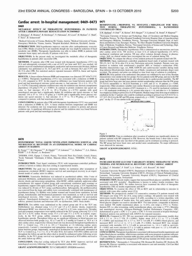
Cardiac arrest: In-hospital management: 0469–04730469METABOLIC EFFECT OF THERAPEUTIC HYPOTHERMIA IN PATIENTSAFTER CARDIOPULMONARY RESUSCITATION NCT00500825
U. Holzinger1, R. Brunner1, R. Kitzberger1, V. Fuhrmann1, H. Losert2, H. Herkner2, C. Madl1,
F. Sterz2, B. Schneeweiß3
1Medical University of Vienna, Medicine III, Vienna, Austria, 2Medical University of Vienna,Emergency Medicine, Vienna, Austria, 3Hospital Kirchdorf, Medicine, Kirchdorf, Austria
INTRODUCTION. Mild hypothermia improves outcome after cardiopulmonary resuscita-tion (CPR). Modes of action for it are manifold, though one way might be reduction of basalmetabolic rate (BMR). Therapeutic hypothermia was able to reduce BMR in patients withtraumatic brain injury and critically ill patients with fever.
OBJECTIVES. In the present study we investigated the metabolic effect of therapeutichypothermia in patients after successful CPR.
METHODS. 25 patients after CPR were treated with therapeutic hypothermia (33�C) for24 h and subsequently rewarmed with a rate of 0.4�C per hour until 36�C was reached. Allpatients received standardized sedoanalgetic medication and neuromuscular blockers. Indirectcalorimetry was performed at 33, 34.5 and 36�C, as well as between 36.5 and 37.5�C and48–72 h after CPR. For statistical analysis repeated measures ANOVA, linear and logisticregression were used.
RESULTS. A linear relation between BMR and temperature was detected (297 kJ/m2/1.5�C;p \ 0.0001). Therapeutic hypothermia (33�C) was associated with a reduction of BMR by22 ± 6% compared to 36�C. In this regard no difference was found between patients withgood and bad neurological outcome (good outcome vs. bad outcome: 24 ± 4 vs. 19 ± 7%;p = 0.479). Concerning substrate oxidation rates only fat oxidation rate showed a temperaturedependency (10 g/day/1.5�C; p \ 0.0001). In contrast to protein oxidation rate (good out-come vs. bad outcome: 45 ± 51 vs. 49 ± 70 g/day; p = 0.751) patients with goodneurological outcome had a significantly higher fat oxidation rate (good outcome vs. badoutcome: 144 ± 57 vs. 110 ± 47 g/day; p = 0.039) and a significantly lower glucose oxi-dation rate (good outcome vs. bad outcome: 21 ± 77 vs. 74 ± 99 g/day; p = 0.029) ascompared to patients with bad neurological outcome.
CONCLUSIONS. In patients after CPR mild therapeutic hypothermia (33�C) was associatedwith a reduction of BMR by 22%. A linear relation between temperature and BMR wasdetected. Fat oxidation rate was temperature dependent in contrast to protein and glucoseoxidation rate. A significant difference in glucose and fat oxidation rates was found betweenpatients with good and bad neurological outcome.
0470HYPOTHERMIC TOTAL LIQUID VENTILATION IMPROVES SURVIVAL ANDNEUROLOGICAL RECOVERY IN AN EXPERIMENTAL MODEL OF CARDIACARREST IN RABBITS
R. Tissier1,2,3, M. Chenoune1,2,3, B. Ghaleh1,2,3, F. Lidouren1,2,3, L. Darbera1,2,3, J.-L. Dubois-Rande1,2, J.-D. Ricard4, A. Berdeaux1,2,3
1INSERM, U955, Equipe 3, Creteil, France, 2Universite Paris Est Creteil, Creteil, France,3Ecole Nationale Veterinaire d’Alfort, Maisons-Alfort, France, 4INSERM, U722, Paris,France
INTRODUCTION. Total liquid ventilation (TLV) with temperature-controlled perfluoro-carbons is known to induce ultra-fast cooling in experimental models.
OBJECTIVES. Our goal was to determine whether its institution after resumption ofspontaneous circulation (ROSC) improves survival and neurological recovery in an experi-mental model of cardiac arrest in rabbits.
METHODS. Ventricular fibrillation was induced in anesthetized rabbits. After 5-min ofuntreated fibrillation, cardiopulmonary resuscitation was attempted using external massage,electric shocks and intravenous epinephrine. After ROSC, rabbits randomly underwent eithernormothermic life support (Control group with conventional ventilation until weaning) orhypothermic support with rapid cooling (TLV group). In this last group, a 32�C hypothermiawas induced by 20-min of TLV using a perfluorocarbon. Subsequently, the perfluorocarbonwas removed from the lungs and rabbits were conventionally ventilated with maintenance ofhypothermia during 3 h. Rabbits were further warmed and weaned from ventilation. In bothgroups, hemodynamic and biochemical parameters were monitored, as well as survival andneurological recovery. After 7 days, survivors were finally euthanized for post-mortemanalyses. Neurological dysfunction was assessed by a 0–100% scoring system evaluatingreflexes, postural reactions and behaviour (0%: no dysfunction; 100%: brain death).
RESULTS. Ten rabbits were randomized to the control group and 10 to the TLV one.Defibrillation was obtained using 1.4 ± 0.3 and 1.5 ± 0.3 electric shocks, respectively.Subsequent ROSC was observed after 2.3 ± 0.3 and 2.4 ± 0.3 min, respectively. Oesopha-geal and tympanic temperatures were rapidly reduced in the TLV group, achieving 32.5 ± 0.2and 32.5 ± 0.3�C within 30-min versus 37.5 ± 0.5 and 37.1 ± 0.3�C in Control, respec-tively. In the TLV group, rabbits returned to normothermia within 2–3 h after thehypothermic episode. Throughout follow-up, no significant difference in blood pressure wasobserved between both groups (e.g., 82 ± 3 and 82 ± 5 mmHg in Control vs. TLV at 1 hafter cardiac arrest, respectively) whereas heart rate was decreased throughout hypothermia inTLV vs. Control (e.g., 174 ± 6 vs. 221 ± 7 beats/min at 1 h following cardiac arrest,respectively). Lactates’s concentration and epinephrine dosages were not significantly dif-ferent between groups. Importantly, neurological dysfunction was significantly attenuated inTLV vs. Control (e.g., 5 ± 3 vs. 32 ± 9% after 48 h). In Control, 5/10 (50%) rabbits survivedthroughout the follow-up and the others died or should be euthanized earlier following severedisability. In the TLV group, survival was significantly increased as 9/10 rabbits survived tothe entire follow-up (90%).
CONCLUSION. Ultra-fast cooling induced by TLV after ROSC improves survival andneurological recovery following 5-min of experimental cardiac arrest in rabbits.
GRANT ACKNOWLEDGMENT. TLV-CARDAREST (INSERM-transfert).
0471REMIFENTANIL + PROPOFOL VS. FENTANYL + MIDAZOLAM FOR SEDA-TION DURING THERAPEUTIC HYPOTHERMIA: A RANDOMISEDCONTROLLED TRIAL
T.W. Bjelland1, O. Dale1,2, K. Kaisen3, B.O. Haugen1,4, S. Lydersen5, K. Strand3, P. Klepstad1,2
1Norwegian University of Science and Technology, Dept. of Circulation and Medical Imaging,Trondheim, Norway, 2St. Olavs Hospital Trondheim University Hospital, Dept. of Anaesthesiologyand Emergency Medicine, Trondheim, Norway, 3Stavanger University Hospital, Dept. of Anes-thesia and Intensive Care, Stavanger, Norway, 4St. Olavs Hospital Trondheim University Hospital,Dept. of Medicine, Trondheim, Norway, 5Norwegian University of Science and Technology, Dept.of Cancer Research and Molecular Medicine, Trondheim, NorwayINTRODUCTION. Therapeutic hypothermia (TH) is administered to unconscious survivorsafter cardiac arrest (CA). TH involves at least 12 h of induced hypothermia (33–34�C),mechanical ventilation and sedation. TH may affect sedation through changes in pharma-cology. Still, no clinical studies have investigated the use of sedation during TH.OBJECTIVE. To compare the efficacy of two sedation protocols for patients treated with TH.METHODS. Open, randomised, controlled, population based study of patients treated withTH (33–34 C for 24 h) after CA in two Norwegian university hospitals. Patients were ran-domised to sedation with remifentanil + propofol (RP) or fentanyl + midazolam (FM).Baseline characteristics (age, sex, BMI, SAPS-II) and cardiovascular variables during studydrug infusion (blood pressure, heart rate, use of fluids, vasopressors and inotropic drugs) wererecorded. The primary end point was defined as time from stop of sedation to extubation.RESULTS. Sixty patients were randomised. One patient was withdrawn by next of kin. Baselinecharacteristics were similar in the two groups. For two patients in the RP group, and one in the FMgroup, study drugs were stopped shortly after allocation due to cardiovascular instability. The RPgroup had lower heart rates and more patients needed noradrenaline infusions than the FM group(57.3(14.4) (mean(SD)) vs. 66.2(15) beats/min, p = 0.05, and 21 vs. 10 patients, p = 0.004,respectively). Other circulatory variables were similar. Reasons to not stop sedation or not extubateafter stop of sedation were; cessation of ICU treatment (n = 15), need for mechanical ventilation(n = 10), inadequate awakening (n = 6), seizures after stop (n = 4), and other (n = 4). Sedationwas stopped according to protocol in 35 of 59 patients. Median (range) time from stop of sedation toextubation for the 17 patients who could be extubated according to protocol was 0.25 (0–0.72) vs.12.7 (0.5–26.4) h in the RP and FM group, respectively, p = 0.002. ‘‘Cerebral performance cate-gory’’ on day 13–15 was similar in the two groups.
Figure 1
CONCLUSIONS. Time to extubation after cessation of sedation was significantly shorter inpatients sedated with RP compared to FM. However, the benefit from a short time to extu-bation is limited by that only one-third of the patents can be extubated according to protocol.The RP group had lower heart rates and needed more noradrenaline. No major differenceswere observed for outcome.
GRANT ACKNOWLEDGMENT. NTNU.
0472INCREASED BLOOD GLUCOSE VARIABILITY DURING THERAPEUTIC HYPO-THERMIA AND NEUROLOGICAL RECOVERY AFTER CARDIAC ARREST
N. Villoz1, F. Delodder1, F. Feihl2, L.A. Urbano1, A.O. Rossetti3, M. Oddo1
1Lausanne University Hospital (CHUV), Department of Intensive Care Medicine, Lausanne,Switzerland, 2Lausanne University Hospital (CHUV), Division of Clinical Pathophysiology,Lausanne, Switzerland, 3Lausanne University Hospital (CHUV), Department of ClinicalNeurosciences, Lausanne, SwitzerlandINTRODUCTION. Recent studies suggest that increased blood glucose variability (BGV) isassociated with ICU mortality1. Hypothermia is known to induce insulin resistance, thuspotentially increasing BGV. No studies however have examined the effect of therapeutichypothermia (TH) on insulin requirements and BGV.OBJECTIVES. To examine the effect of TH on BGV and its relationship to outcome inpatients with coma after cardiac arrest (CA).METHODS. We prospectively studied 132 consecutive comatose CA patients treated withTH (target core temp 33�C for 24 h, using surface cooling). All patients were treated withintravenous insulin (blood glucose target 6–8 mM), according to a written algorithm, withnurse-driven adjustment of insulin dose. For each patient, standard deviation of repeatedblood glucose samples was used to calculate BGV. Two time-points, comparable in duration,were studied: TH (stable maintenance phase, i.e. 6–24 h, core temp ± 33�C) vs. Normo-thermia (NT, i.e. after rewarming, stable normothermic phase, core temp ± 37�C). Mortalityand neurological recovery (Glasgow-Pittsburgh Cerebral Performance Categories, CPC,dichotomized as good = CPC 1–2 vs. poor = CPC 3–5) were assessed at hospital discharge.Statistical analysis was performed with ANOVA for repeated measures.RESULTS. Compared to NT, TH was associated with increased intravenous insulin dose(0.8 ± 1.1 vs. 1.6 ± 2 U/h, P \ 0.0001), higher mean (6.9 ± 1.3 vs. 7.7 ± 1.8 mM,P \ 0.0001) and maximum (9.1 ± 3.7 vs. 10.9 ± 3.6 mM, P \ 0.0001) blood glucose, andincreased BGV (1.3 ± 1.2 vs. 1.7 ± 1.1 mM, P = 0.004). Increased BGV was stronglyassociated with mortality (2.5 ± 1.5 mM in non-survivors vs. 1.6 ± 1 mM in survivors,P \ 0.001) and worse outcome (2.3 ± 1.4 mM in patients with poor vs. 1.5 ± 0.8 mM inthose with good neurological recovery, P \ 0.0001).CONCLUSIONS. Therapeutic hypothermia is associated with increased insulin requirements andhigher blood glucose variability, which in turn correlates with worse prognosis in patients with post-CA coma. Strategies aimed to maintain stable glycemic profile and avoid blood glucose variabilitymight contribute to optimize the management of TH and may translate into better outcome.
REFERENCE(S). 1 Hermanides J, Vriesendorp TM, Bosman RJ, Zandstra DF, Hoekstra JB,Devries JH. Glucose variability is associated with intensive care unit mortality. Crit Care Med2010 Mar;38(3):838–42.
23rd ESICM ANNUAL CONGRESS – BARCELONA, SPAIN – 9–13 OCTOBER 2010 S203
0473CARE BUNDLE IMPLEMENTATION AND THERAPEUTIC HYPOTHERMIAFOLLOWING CARDIAC ARREST
R. Gray1, L.C. Evans1, P. Anderson1, P.G. Morgan2
1Royal Sussex County Hospital, Brighton, UK, 2Surrey and Sussex NHS Trust, Redhill, UK
INTRODUCTION. Therapeutic hypothermia post cardiac arrest has been shown to improvesurvival and neurological outcome1. There are approximately 50,000 treated cardiac arrests inthe UK each year with one-eighth admitted to an Intensive Care Unit (ICU).2
OBJECTIVES. We aimed to establish if implementation of an agreed care bundle includingtherapeutic hypothermia reduced mortality and improved neurological outcome.
METHODS. Patients were divided into two groups consisting of out-of-hospital arrests(OOHA) and in-hospital arrests (IHA). Patients were categorized according to initial cardiacrhythm, ventricular fibrillation/tachycardia (VF/VT) or non-VF/VT. We recorded the degreeof implementation of the post cardiac arrest care bundle, comprising; coronary reperfusion,haemodynamic optimisation, control of ventilation, blood glucose, temperature and seizures.Data was compared with survival to discharge and neurological function.
RESULTS. We identified 239 patients that were admitted to our ICU post cardiac arrestbetween February 2006 and 2010. There were 111 IHAs, 73 male (mean age 67.3 years) and37 female (69.9 years) and 128 OOHAs, 94 male (61.4 years) and 34 female (62.6 years).The predominant presenting rhythm in OOHAs was VF/VT (58.8%) compared to IHAs whichwas non VF/VT (89.3%). Underlying co-morbidities included; ischaemic heart disease(42.8%), drug/alcohol abuse (14.8%) and respiratory disease (10.2%).
Blood glucose control was achieved in 91.1% of patients. Target temperature (32–34�C) wasachieved in 75% of VF/VT OOHAs compared to 14.3% of VF/VT IHAs. The complete carebundle was delivered to 56.3% of OOHAs and 3.6% of IHAs. In total 31.8% of all patientsreceived the complete care bundle.
Survival rates were higher in all patients with complete bundle of care versus those with anincomplete bundle independent of location or rhythm (53.9 vs. 38.7% p = 0.01). This im-proval in survival was also demonstrated in VF/VT arrests receiving the complete bundle(61.8 vs. 39.4%, p = 0.034).
Of those patients who had a VF/VT arrest, survival with full neurological recovery (GCS 15on discharge) was higher in those receiving therapeutic hypothermia 53.2 versus 17% wheretherapeutic hypothermia was not achieved.
CONCLUSIONS. Adherence to the post cardiac arrest care bundle led to significantlyimproved outcome following both VF/VT and non VF/VT arrests. There was a trend towardstherapeutic hypothermia improving neurological outcome on discharge.
REFERENCE(S). 1. The hypothermia after cardiac arrest study group. Mild therapeutichypothermia to improve the neurological outcome after cardiac arrest. NEJM346(8):549–556. 2002. 2. Standards for the management of patients after cardiac arrest.Intensive Care Society 2008.
GRANT ACKNOWLEDGMENT. None. Royal Sussex County Hospital Intensive CareUnit, Brighton, UK for patient data.
S204 23rd ESICM ANNUAL CONGRESS – BARCELONA, SPAIN – 9–13 OCTOBER 2010