Embed Size (px)
Citation preview

BIOPAC Blood Flow Monitor
LDF100C‘Blood Perfusion’
‘Blood Flow’
‘Blood Flux’
Technique Technique --
Laser Doppler Laser Doppler FlowmetryFlowmetry
(LDF)(LDF)The LDF100C is a laser Doppler blood perfusion monitor used to measure real-time micro-vascular red blood cell perfusion in tissue.
Laser Doppler signals from the tissue are recorded in BPU (Blood Perfusion Units) which is a relative units scale defined using a carefully controlled motility standard comprising a suspension of latex spheres undergoing Brownian motion.
Perfusion is sometimes also referred to as micro-vascular blood flow or red blood cell flux.

What does LDF measure ?What volume of tissue does the LDF100C measure?LDF defines a perfusion parameter from information contained in the optical spectrum of light remitted from the tissue. The actual measurement sampling volume or depth can only be determined by identifying precisely which blood vessels and erythrocytes have interacted with the remitted light, which in turn, is principally dependant on two parameters; namely the optical scattering and optical absorption coefficients of the tissue under observation. Since both of these coefficients are entirely dependent on the site of observation and perfusion of the microvasculature at the time of measurement, it is impossible to
determine the actual sampling volume/depth at any tissue site. Generally speaking however, we have estimated that for well-perfused
tissue such as muscle, the mean sampling depth is in the region 0.5-1.0 mm with a concomitant sampling volume in the region 0.3-0.5 mm3. For cutaneous
measurements, the sampling depth is likely to be in the range 1.0 –
1.5 mm. These estimates have been obtained heuristically through many years of experience and are based on both in vitro
observations and mathematical modelling of photon diffusion through 'imaginary tissues’
using Monte-Carlo techniques
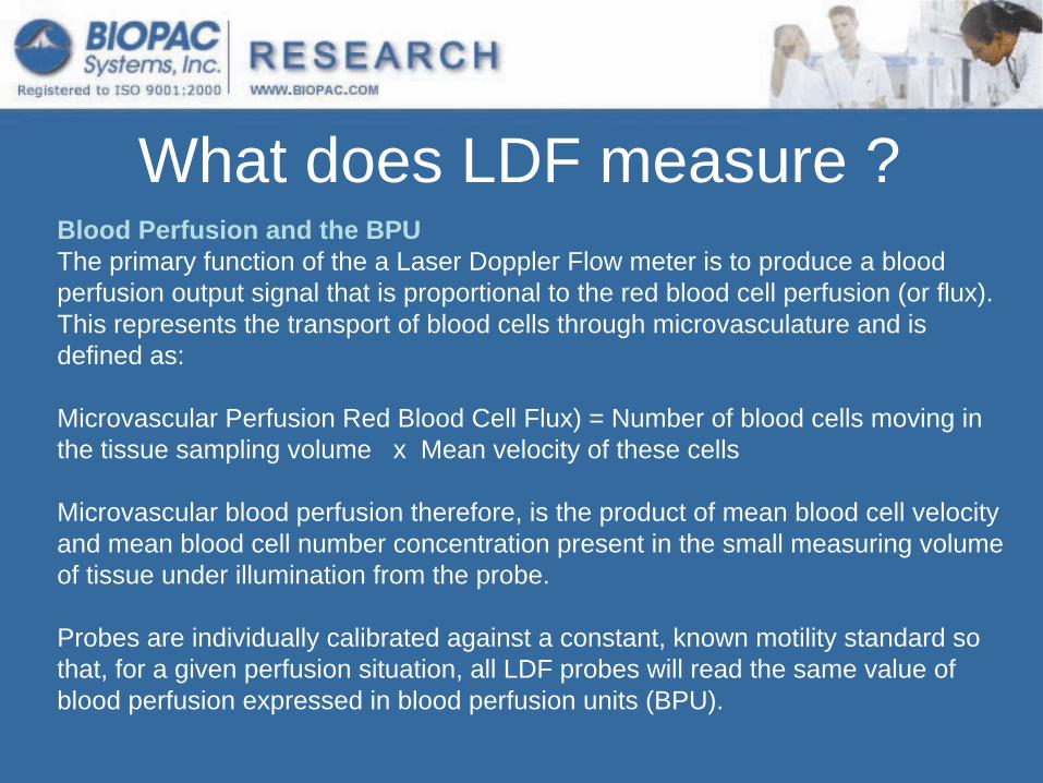
What does LDF measure ?Blood Perfusion and the BPUThe primary function of the a Laser Doppler Flow meter is to produce a blood perfusion output signal that is proportional to the red blood cell perfusion (or flux). This represents the transport of blood cells through microvasculature and is defined as:
Microvascular
Perfusion Red Blood Cell Flux) = Number of blood cells moving in the tissue sampling volume x Mean velocity of these cells
Microvascular
blood perfusion therefore, is the product of mean blood cell velocity and mean blood cell number concentration present in the small measuring volume of tissue under illumination from the probe.
Probes are individually calibrated against a constant, known motility standard so that, for a given perfusion situation, all LDF probes will read the same value of blood perfusion expressed in blood perfusion units (BPU).

What does LDF measure ?•
Microvascular
Blood Perfusion (BPU) =
•
Volume of tissue sampled
0.5 -
1 mm3
•
No measure of volume in LDF —
BPU is an arbitrary unit (not
ml/min/g tissue)
•
Used to measure RELATIVE changes in perfusion
Mean velocity of Mean velocity of moving cellsmoving cells
XXNumber of moving blood Number of moving blood cells in volume sampledcells in volume sampled

How does LDF work ?•
Tissue is illuminated by
low power laser light; the light is scattered by the static tissue structures and moving blood cells
•
Portion of the light is Doppler shifted
•
Adjacent fiber optic detects –
un-shifted and–
Doppler-shifted light
•
Signal is processed to give a parameter related to tissue blood perfusion
What are the advantages of LDF? The LDF technique offers substantial advantages over other methods
(e.g. ‘microbeads’) in allowing the continuous measurement of microvascular
blood perfusion.
The LDF may be used non-invasively (since the probe is not actually required to touch the surface of the tissue) and in no way harms
or disturbs the normal physiological state of the microcirculation.
LDF is both highly sensitive and responsive to local blood perfusion and is also versatile and easy to use for continuous monitoring.
•
Continuous/real-time –
response time 0.15s•
Sensitive to small changes•
May be used non-invasively,–
On skin or organ surface where exposed•
Small probe dimensions–
Minimal trauma where used invasively

What are the limitations of LDF?
•
Sensitive to tissue and cable movement•
Site-to-site variation as probe is moved–
Due to variation in tissue optics•
Relative, arbitrary units–
Measurements obtained by LDF are intrinsically of a relative nature.
–
Although such measurements are proportional to perfusion, the factor of proportionality will be different for different tissues.
•
All limitations may be minimized by experimental design

LDF Experimental Design•
Limit movement artifacts
•
Probe selection•
Maintain probe position
•
Change due to provocation•
Site-to-site variation

How to use Arbitrary Units ?•
Occlusion and Hyperemia Occlusion
= an obstruction or a closure of a
passageway or vessel (blocked flow)Hyperemia
= an increase in the quantity of blood
flow to a body part; engorgement
•
Valsalva
maneuverValsalva
maneuver
= expiratory effort against a
close glottis (the opening between the vocal cords at the upper part of the larynx). which increases pressure within the thoracic cavity and thereby impedes venous return of blood to the heart.

Using arbitrary units --
hyperemia
Hyperemic Index = (HP -
BZ) / (RF –
BZ)
(HP)(HP)
(RF)(RF)
(BZ)(BZ)

Research Applications of LDF
•
Cerebral monitoring (stroke, injury...)
•
Transplantation surgery (skin grafts, free flaps...)
•
Vital organ monitoring (organ viability...)
•
Tumor vascular research (angiogenesis...)
•
Peripheral vascular disease (diabetes...)

Probes and Applications
•
Surface and Digit Probes•
Suturable
and Miniature Surface Probes
•
Needle and Pencil Probes•
Single Fiber Probes

Surface and Digit Probes
Applications:Skin, large organ,Noninvasive, Adhesive ring
TSD140TSD140General purpose surface probePrimarily useful with largely flat surfaces
TSD142TSD142Specifically designed to be used with on human digits. The radius of curvature is designed to reduce probe pressure and reduce the risk of the probes detaching

Suture & Miniature Surface ProbesTSD146TSD146
TSD143TSD143
Applications:Small organ, non invasivee.g. Flap monitoring

Needle Probes
Diameter0.5 mm
TSD144 & TSD145TSD144 & TSD145
Applications:Invasive tissue measurement;e.g. Cerebral
monitoringIdeally suited to Stereotactic setupUsing manipulator to position

Pencil Probes All the probes are constructed from medical grade stainless
steel tubing . The default length is 25 mm long
Diameter1.0 mm
TSD customTSD custom
Diameter 1.0 mmCustom lengths
TSD customTSD custom
Applications:Noninvasive, Clamped over tissue

Single Fiber Probes
Flexible Fiber;Diameter 0.5 mm
TSD147TSD147 TSD148TSD148
Applications: Chronic Implantatione.g. fetal brain

Probe Features•
Smart probes –
Calibration automatically loaded
•
MRI-compatible –
Surface probes contain no metallic parts
•
Autoclavable–
Where sterility required, up to 20 cycles

Practical Aspects of LDF
•
CalibrationLaser Doppler signals from the tissue are recorded in BPU (Blood Perfusion Units) which is a relative units scale defined using a carefully controlled motility standard comprising a suspension of latex spheres undergoing Brownian motion.

Practical Aspects of LDF•
Meaning of zero flow (zero BPU)?The zero (0000) reading of the LDF has been obtained by calibrating the system against a special static scattering material where no movements occur. In such cases the back-scattered light processed by the LDF contains no Doppler shifted frequency components and a true zero
is obtained. A zero reading therefore indicates zero motion both in the measuring volume under examination and artefactual
motion arising from relative movements between the probe and the measuring volume. During in vivo
measurements, rarely is an absolute zero obtained. Even during total occlusion of tissue blood perfusion, there is often
some small, residual motion of blood cells trapped in the vessels, as
well as some small muscle and tissue movement in the measuring volume. Even after surgical removal of tissue, localised cell movement and Brownian motion may still occur in the severed blood vessels.
Practical Aspects of LDF•
Definition of the Backscatter Signal (BS)
The LDF also produces a signal, which is proportional to the total light remitted or backscattered from the tissue. This is called
the
Backscatter Signal (BS) and is available as an analogue voltage output from the back panel of the LDF monitor. The backscatter is expressed as a percentage fraction of the laser light remitted from the tissue from the total amount of laser light incident on
the
tissue. For example, in highly perfused
tissues, the BS will be low due to increased photon absorption. Situations where the BS
signal is close to zero may indicate that the probe has come into contact with whole blood, this could cause the BPU reading to saturate since the system is no longer monitoring microvascular
perfusion.
Calibration•
Motility Standard–
Latex spheres under Brownian Motion
•
Probes have individual calibrations–
Instrument recognizes Probe ID
–
Customers should avoid using probes with the same Probe ID

The Backscatter Signal•
Diagnostic Signal
•
A Step in the Backscatter signal–
Suggests probe location changed
•
Low backscatter–
Flow signal is cut out
–
Probe has become detached–
Faulty probe
•
High backscatter–
Probe detached, facing a reflective surface (e.g. bone)

Summary•
LDF is a sensitive technique for monitoring changes of micro-vascular blood flow in real time
•
Wide range of applications from basic physiology teaching to research studies
•
The BIOPAC LDF100C is a full-feature LDF instrument
•
Probes available for a wide range of applications (BIOPAC TSD140 series)