Embed Size (px)
Citation preview

ORAL HEALTH IN COMPREHENSIVE CLEFT CAREEducational resources for oral health professionals
PARTICIPANT HANDOUT

Content developed by the Oral Health in Comprehensive Cleft Care Task Team:P. Mossey, Muthu MS, S. Yan, M. Campodonico, L. Orenuga
Managing editors: P.Sheeran and R. England
This educational resource was produced as part of a partnership between FDI World Dental Federation and Smile Train, with support from GlaxoSmithKline Consumer Healthcare (GSK CH). No editorial control, apart from ensuring compliance with legal and regulatory considerations/requirements, has been exercised by GSK CH. Funding for the development of this educational resource was provided by GSK CH.
Supported by:

3
ContentsOral Health in Comprehensive Cleft Care 4
Introduction 4
Purpose and goal 4
Learning outcomes 4
Day 1 5
Interprofessional collaboration and cleft care 5
The definition and causes of orofacial clefts 7
Prevention of cleft 9
Oral health conditions commonly associated with cleft 11
LAHSAL classification 16
Identification of white spots, brown spots and caries 17
Day 2 21
Missing teeth, extra teeth, impacted teeth, microdontia and malformed roots 21
Premature loss of primary teeth – the causes, the consequences and clinical management 23
Principles of minimally invasive dentistry 27
Managing restorative care for cosmetic improvements 29
Monitoring and maintaining the oral health of people with cleft 30
Delivering oral health education 34
Day 3 35
Scar management in people with cleft lip and palate 35
Quality of life and clefts 37
Implementing the FDI/Smile Train Safety and Quality Protocol 40

4
IntroductionClefts of the lip and palate (clefts) are the most common birth defects of the face and mouth. Clefts occur when parts of the lip or palate, or both, or the nose do not fuse together during embryonic development. The condition can be associated with missing or extra teeth and with malformed teeth and facial structures. Children who undergo cleft surgery are often at increased risk of caries, periodontal disease and other oral health and wellbeing issues as they grow and develop. These children require regular dental care to ensure adequate monitoring, education, support and treatment to prevent oral diseases and achieve the highest possible quality of life.
Oral Health in Comprehensive Cleft Care
Purpose and goal The purpose of this three-day course is to improve the oral health and long-term wellbeing of children who undergo cleft surgery. It is designed to ensure all members of the cleft care team are aware of the importance of oral health and can take an active role in preventing oral diseases.
Learning outcomesAt the end of this course, participants will:
• Be aware of the importance of oral health.• Know how to prevent and identify oral diseases. • Know when to refer onwards.• Be prepared to disseminate what they have learned in their workplace.

5
Learning objectives: Understand the importance of interprofessional collaboration in the care of people with cleft.
DAY 1
What is interprofessional collaboration? Interprofessional collaboration is defined as two or more healthcare professions working together with patient involvement to ensure informed and empowered choices. Shared decision-making improves patient and professional relationships and also improves health outcomes.
The professions and healthcare providers involved in the care of people with cleft. • Craniofacial surgeon: a plastic surgeon with specialized training in the diagnosis and treatment of
conditions of the skull, facial bones and soft tissues who will work closely with other specialists to co-ordinate a surgical plan;
• Paediatrician; a physician specializing in the care of children who follows the child as he/she grows and helps co-ordinate the multiple specialists involved;
• Orthodontist: a dental specialist concerned with the development of the face, jaws and teeth and who evaluates the position and alignment of children’s teeth and co-ordinates a treatment plan with the surgeon and other specialists;
• Paediatric dentist; a dental specialist who evaluates and cares for children’s teeth;
• Dental hygienist: an oral health professional who is skilled in cleaning teeth and assists with the prevention of oral diseases;
• Speech and language specialist: a professional who performs a comprehensive speech evaluation to assess communicative abilities and closely supports and monitors children with cleft;
• Otolaryngologist: an ear, nose and throat specialist who treats the ear infections or hearing loss that may be side effects of a child’s cleft condition;
• Audiologist: a hearing professional who will assist in the evaluation and management of hearing difficulties;
• Genetic counsellor: a professional who assists in the diagnosis of genetic conditions and counsels families around the prognosis regarding future pregnancies;
• Nurse team co-ordinator: a registered nurse who has experience in paediatric nursing and acts as liaison between the family and the cleft team.
• Social worker: a professional who provides guidance and counselling for a child and their family as well as assistance with community resources and referrals, i.e., support groups.
INTERPROFESSIONAL COLLABORATION AND CLEFT CARE

6
The importance of interprofessional collaboration between oral healthcare providers and the wider cleft care teamBabies born with cleft have significant needs from a wide range of healthcare specialties, and the care they receive depends on many factors, including distance from the clinic, cost of treatment and parental knowledge and beliefs. There may be an adverse effect on children’s quality of life if they are not able to access the full gamut of services, especially speech therapy and oral healthcare.
It is essential, therefore, that all healthcare providers communicate effectively with the family, the child and caregivers and refer to other services to ensure the child is receiving adequate treatment and support.
Because oral healthcare is often unavailable to children with cleft, it is important that the whole cleft care team understand oral diseases and how to prevent them. The ability and confidence of all members of the cleft team to ‘lift the lip’ and check the health of the oral cavity is an important factor in maintaining the health and well-being of cleft patients.

7
THE DEFINITION AND CAUSES OF OROFACIAL CLEFTS
Definition of cleft Clefts are the most common birth difference affecting the structure of the face and oral cavity. Cleft conditions occur when parts of the lip and/or palate and nose do not fuse together during embryonic development.
They can be divided into three general categories:
(1) cleft palate alone;
(2) unilateral or bilateral cleft lip, with or without cleft alveolus;
(3) unilateral or bilateral cleft lip and cleft palate.
They are all referred to as orofacial clefts.
How do clefts develop? By the time of the first ultrasound, in which the facial structures of the foetus can already be identified, clefting of the lip or palate will already have occurred. Development of the facial structures and form occurs early in a baby’s gestation, between the fourth and eighth weeks.
During the fourth week of development, a series of six paired, right and left, swellings form near the cranial, or head, end of the foetus. These are called branchial arches. The branchial arches develop into important structures in the head and neck, as well as the major arteries in the chest. The first branchial arch develops structures called the maxillary and mandibular prominences, which ultimately form the skeleton, skin, muscles, and other structures of the upper and lower jaws, respectively the maxilla and the mandible. In the centre of the upper face, a structure called the frontonasal prominence, which is separate from the branchial arches, forms, among other things, the nostrils and the philtrum. The medial maxillary prominences fuse with the frontonasal prominence during this time. Failure of this process results in a cleft lip.
Above: EMBRYO AT 28 DAYS
Right: EMBRYO AT 32 DAYS
Learning objectives: Understand the definition and causes of cleft.

8
EMBRYO AT 49 DAYS
The palate is also formed by the fusion of the frontonasal prominence and the maxillary prominences. Although the palate appears to be one complete structure, it consists of a primary palate, behind the front teeth, and a secondary palate, which includes the majority of the hard and soft palate. These are separated by a hole in the palatal bone called the incisive foramen. The two sides of the secondary palate begin to fuse during the eighth week of development. Failure of fusion results in a cleft palate.
EMBRYONIC DEVELOPMENT OF THE FACE
Genetics of cleft lip and palate Between 30%-50% of clefts are related to genetic factors. Cleft conditions can be categorized as syndromic or non-syndromic.
The cause of a syndromic cleft may be associated with gene mutation, chromosomal aberrations, teratogens or environmental factors. The majority of cleft conditions are non-syndromic, which means the cleft occurs alone with no other associated conditions. These clefts usually occur when there is a genetic susceptibility or environmental factors.
Non-syndromic cleft is more common in males, with a male-to-female ratio of about 2:1. The genetic characteristics of non-syndromic cleft palate also conform to the multi-factorial threshold (MFT) model, but the incidence is lower, and the risk of recurrence is different. There are more than 600 syndromes with a cleft of the lip or palate as an associated feature, but not all of them are hereditary.
Epidemiology of cleft lip and palateAt a global level, cleft conditions affect approximately 1 in 700 live births.
The incidence varies widely depending on geographic origin, racial and ethnic group, environmental exposures, and socioeconomic status. Asian and Native American populations have reported prevalence rates as high as one in 500. European and US populations on average have a prevalence of approximately one in 1000, whereas African populations have a reported prevalence close to one in 2500.

9
PREVENTION OF CLEFT
Prenatal examination and genetic counsellingFor families with a history of orofacial cleft, prenatal examination and genetic counselling should be conducted. The diagnosis of cleft in prenatal routine ultrasound examination might help to prepare the family.
Maternal nutrition and cleft lip and palateIt is sound public health advice to recommend that prior to conception and in early pregnancy women should have a healthy and varied diet and vitamin supplements if required. Many studies have indicated an association between maternal use of multivitamin supplements in early pregnancy and decreased risk of orofacial clefts.
Folic acid supplementFolic acid is a B vitamin that may have a protective effect against certain cleft conditions when taken prior to conception. It is already widely recommended for its role in protecting against neural birth defects such as spina bifida and anencephaly. A daily supplement taken by all women planning pregnancy is the recommendation of the US Public Health Service, endorsed by The American Academy of Pediatrics (AAP). A supplement of 400 micrograms of folic acid daily reduces the incidence of neural tube defects by up to 70%.
• There is uncertainty surrounding the role of other vitamins in prevention of cleft.
Medication during pregnancyCertain medications have been shown to have an effect on foetal development; these can include:
• anti-cancer and anti-convulsant drugs;
• non-steroidal anti-inflammatory drugs and analgesics;
• antibiotics such as oxytetracycline, tetracycline and amoxicillin;
• steroids such as glucocorticoids or cortisone.
The risk of a child being born with a cleft can be almost three times higher if the mother takes medication during the first trimester. Great care should be taken if medication is necessary in the early stages of pregnancy and a doctor should be consulted.
Alcohol and tobaccoSmoking and drinking in early pregnancy increase the risk of cleft.
Passive smoking carries a similar risk, so partners and other close contacts of pregnant women should avoid or reduce smoking. Environmental contamination and pollution also contribute to risk.
Learning objectives: Understand the prevention of cleft.

10
Prevention of gestational diabetes mellitus and maternal obesityGestational diabetes mellitus may augment the risk of cleft lip and palate. Mothers should seek medical advice on their dietary habits.
• For patients with diabetes mellitus before pregnancy, glycaemia should be controlled and closely monitored by the medical team throughout the pregnancy.
• For pregnant women with gestational diabetes mellitus, blood pressure, blood glucose, liver and kidney functions and fetal health should be closely monitored.
• A number of studies have also implicated maternal obesity as a factor that elevates the risk of having a child with a cleft.
Stress control• Physical and/or emotional stress may be implicated in the occurrence of oral clefts.
• Anxiety, depression and other negative psychological states during pregnancy might affect the occurrence of cleft lip and palate, by hormonal dysregulation.
• A positive attitude during pregnancy is important.
PREGNANCY AND HEALTHY LIFESTYLE

11
ORAL HEALTH CONDITIONS COMMONLY ASSOCIATED WITH CLEFT
Learning objectives: Understand diseases and conditions commonly associated with cleft, such as caries and malocclusion.
The importance of healthy primary teeth Primary teeth are important to children because they:
• Allow children to chew and eat properly.
• Help your child to speak more clearly.
• Maintain space for the eruption of adult teeth.
• Guide adult teeth into place.
• Help to shape the infant’s face.
• Prevent early childhood caries.
• Keep future dental costs to a minimum.
• Offset the need for orthodontic treatment later.
• Reduce the risk of caries in permanent teeth.
Dental checks are important because infants with cleft conditions become accustomed to trusted caregivers touching the inside of their mouth.
Children with clefts rarely escape dental complications. This can be attributed to:
• dry mouth caused by mouth-breathing habits;
• less natural cleaning of the teeth due to the morphology;
• variable diet or feeding habits;
• dental anomalies;
• increased consumption of sweetened medications;
• delayed oral clearance time for foods.
Early childhood caries Early childhood caries (ECC) is defined as the presence of one or more decayed, missing (due to caries) or filled tooth surfaces in any primary tooth. Severe ECC is characterized by a distinctive pattern of tooth decay in infants and young children, often beginning on the maxillary anterior teeth and rapidly progressing to the other primary teeth as they erupt. ECC can begin to develop as soon as teeth erupt into the mouth at 6-10 months of age, which is why an early oral health assessment and fluoride varnish treatments are so important.
Pedo Planet - Children Dental Centers, (Chennai, New Delhi), India.
Centre for Early Childhood Caries Research (CECCRe), Sri Ramachandra Institute of Higher Education and Research, Chennai, India

12
Oral hygiene Patients with cleft find it challenging to maintain good oral hygiene due to malocclusion. Extensive caries and premature tooth loss affect the masticatory function, jaw development and correction of malocclusion in patients with cleft. Awareness of infant oral care measures, in particular, is lacking among the population, e.g. how to clean baby teeth, when to start cleaning baby teeth, what oral hygiene aids to use, and choice of toothpaste and toothbrush.
Periodontal diseases There are two types of periodontal disease: gingivitis and periodontitis.
The early stage of periodontal diseases is gingivitis:
• Gingivitis occurs when the gums around the teeth become red, swollen and bleed when brushed; it is reversible when treated quickly.
• The first sign can be blood on the toothbrush or after spitting out.
• In many cases, people will suffer from halitosis (bad breath).
The advanced stage of periodontal disease is periodontitis:
• Periodontitis is mostly seen in adults. As periodontal disease advances, the plaque biofilm moves below the gingival margin and destroys the periodontal fibres and bone supporting the teeth, making the teeth loose.
• Prevention of gum disease is essential since periodontitis is irreversible and can cause tooth loss. With proper treatment, it can be halted.
Periodontally involved teeth can cause numerous problems, including acting as a source for local or systemic infection, affect eating, speaking and overall quality of life. Teeth can fall out of their sockets and be inhaled or ingested, leading to a possible medical emergency, as well as affecting self-esteem.
It is crucial to prevent and treat periodontal diseases at the earliest age.
GINGIVITIS

13
Periodontal diseases are induced by dental plaque. If it is not removed, the minerals present in saliva calcify, forming calculus, which needs to be removed by an oral health professional. If the dental plaque accumulates, pathogenic bacteria and toxins increase, triggering the patient’s immune system to initiate an inflammatory response. The combination of inflammation and bacterial waste products destroy the structures holding the tooth in place.
Other risk factors for periodontal disease In addition to oral biofilm, other local and systemic risk factors accelerate the occurrence of periodontal diseases.
Local factors are those that directly influence the periodontal health status, including poor oral hygiene, dental calculus, anatomic factors, dental crowding and malocclusion, traumatic occlusion, a diet rich in carbohydrates, tobacco use and food impaction.
Systemic factors reduce the body’s resistance or increase the body’s sensitivity to dental plaque, such as the patient’s immune reaction, diabetes mellitus, hormonal factors, malnutrition, haematological diseases, stress and genetic factors.
How can periodontal diseases be prevented?
• mainly through good oral hygiene, especially with mechanical biofilm removal such as tooth brushing and interdental cleaning;
• regular dental check-up and professional cleaning and scaling;
• empowering people to take care of their general health and oral health at the earliest possible age;
• promotion of the behavioral changes needed to prevent these conditions.
How to remove dental plaque?
• effective brushing and interdental cleaning is the key way to remove dental plaque;
• professional oral healthcare treatment such as oral prophylaxis;
• oral health education is essential for adequate oral hygiene and understanding of periodontal diseases.
PERIODONTITIS

14
DENTAL PLAQUE
Malocclusion in the infant phaseIn patients with complete cleft lip and palate, the upper lip, soft palate, and hard palate are completely broken, and the muscle balance inside and outside the patient’s oral cavity is disrupted. The forward force received by the maxilla is stronger than the force of the anterior labi muscle, causing the patient’s maxilla to shift. The patient’s nasal septum and nasal columella cartilage grow poorly.
Malocclusion in the primary dentition and mixed dentition phaseDue to the impact of cleft lip and palate repair surgery, the maxillary bone development of patients is often insufficient. Under normal circumstances, the malocclusion of patients in the deciduous period is often not serious. With the growth and development of the teeth, they gradually show obvious malocclusion.
Common malocclusions include:
• anterior cross-bite;
• posterior cross-bite;
• maxillary anterior hypodontia or supernumerary teeth;
• upper front teeth misaligned or rotated;
• abnormal molar relationship.
Malocclusion in the permanent dentition Research shows that after cleft lip and palate repair, the prevalence of malocclusion is 97%. All patients with complete cleft lip and palate have malocclusion of the permanent dentition. Among the malocclusions associated with cleft lip, the most common are cross-bite and crowding of the upper teeth. In patients with both cleft lip and palate, the teeth near the fissure often appear deformed and missing, and further away from the fissure there may be missing or supernumerary teeth. Maxillary midline deviation is common in patients with unilateral cleft lip and palate.

15
Abnormality of tooth eruption and tooth number Abnormal tooth size and position are often present in patients with cleft. Patients with unilateral complete cleft lip and palate have dysplasia and torsion of the upper central incisors adjacent to the cleft. In patients with bilateral complete cleft lip and palate, the two upper central incisors often have dysplasia and torsion. Other features can include:
• a delay to the eruption time of permanent teeth and an abnormal eruption sequence;
• hypodontia: the most common form of missing teeth is the congenital absence of the affected side incisor with the maxillary palate cleft;
• supernumerary teeth: additional teeth often appear near the fissures;
• abnormality of tooth structure: hypocalcification and dysplasia of the second premolar often appear.
The shape and symmetry of the dental archAfter the cleft lip and palate is repaired, the gap gradually decreases, and the maxillary segment is reshaped and moved closer under the action of the labial muscle. Around 50% of patients with unilateral and bilateral complete lip and palate cleft have varying degrees of maxillary dental arch asymmetry, and patients with simple palate cleft have no maxillary dental arch asymmetry.
MALOCCLUSION AND MISSING TEETH

16
Learning objectives: Participants will understand the LAHSAL classification and how to apply it.
LAHSAL CLASSIFICATION
What is the LAHSAL classification for cleft lip and palate?LAHSAL is an accurate, anatomical method of cleft classification. It is based on a coding chart which records six areas of the mouth. It has been widely adopted because it is:
• simple, so as to be accepted;
• concise to be accurately recorded;
• flexible, to account for rare presentations;
• exact, to facilitate statistical analysis;
• morphological, to enable visual assessment;
• graphic, to represent the cleft in a clear manner.
What do the letters stand for?The letters represent the lip (L), alveolus (A), hard palate (H), and the soft palate (S). As a result, the laterality of each anatomical area, apart from the soft palate, can be recorded. The patient’s left side is recorded first, followed by the right.
What about complete and incomplete clefts? The completeness of the cleft may also be recorded: incomplete clefts are represented by lowercase letters, whereas uppercase letters represent a complete cleft. If there is no cleft, a dash _ is used. The recording of microforms, or incomplete clefts, is enabled through the use of an asterisk substituting the letter for the anatomical area concerned.
What are the advantages of LAHSAL over other classifications? The major advantage of the LAHSAL classification is that it allows for the accurate and concise description and recording of every typical non-syndromic orofacial cleft. The degree of completeness as well as the laterality and also microforms, or Simonarts band, an epithelial bridge across the cleft, may all be recorded. This does not apply in most other classifications.
What is the difference between LAHSHAL and LAHSAL? They are synonymous; LAHSHAL was developed in 1989 but later simplified by the Royal College of Surgeons of England to exclude the second ‘‘H.’’ This, however, excluded the possibility of classifying unilateral clefts of the hard palate, a feature that occurs when only one of the palatal shelves is fused with the nasal septum.
LLip
RightLLip
AAlveolus
AAlveolus
HHard
palate
SSoft
palate
Left
17
Early childhood caries (ECC)Children with cleft have been shown to experience higher levels of caries and in particular Early Childhood Caries (ECC). ECC is different from adult caries in that it causes rapid destruction of the hard tissues and has a diverse set of risk factors. ECC is largely preventable, yet the condition is significantly increasing globally. ECC can have a significant adverse effect on the child’s development and well-being.
Significant risk factors for the development of ECC in young children include long-term bottle use, especially night-time feeding, the use of cariogenic sweetening agents and the frequent consumption of free sugars.
Aetiology of caries For dental caries to occur, there need to be four factors present:
• tooth surface, which is composed of hydroxyapatite crystals;
• dental plaque bacteria;
• fermentable carbohydrates, such as sugar;
• time.
Dental plaque, also known as biofilm, is a soft, sticky layer that adheres to the tooth surface. Biofilm is predominantly composed of oral microorganisms along with proteins present in saliva.
A thin layer of salivary proteins gets deposited on the tooth surface to form the ‘Acquired Enamel Pellicle’. Once this layer is formed, bacteria, oral microorganisms, adhere to this pellicle, forming the biofilm.
This is characterised by acidogenic bacteria, such as Streptococcus Mutans. When the child consumes free sugars, the bacteria metabolise the carbohydrates into acidic waste products, causing the pH of the mouth to drop. Essential minerals are leached out of the tooth structure, leading to demineralisation.
In a healthy oral environment, saliva production removes this acidic environment over a time period of approximately 30-40 minutes, and remineralisation occurs. This is demonstrated in Stephan’s Curve. However, if the child has suboptimal oral hygiene and is frequently consuming free sugars or fermentable carbohydrates, the mouth remains in an acidic pH, leading to dental caries.
In its initial stages, caries is often painless; however, as caries advances, pain and dental abscesses ensue which impact on the child’s wellbeing, educational attainment, and quality of life.
The role of the oral health professional is essential to managing ECC and oral diseases throughout the patient’s life. Prevention, early detection and management are key.
Learning objectives: The participants will understand how to identify early carious lesions.
IDENTIFICATION OF WHITE SPOTS, BROWN SPOTS AND CARIES

18
STEPHAN CURVE
WHITE SPOTS/CERVICAL DEMINERALISATION
8.0
8.0
7.0
7.0
6.0
6.0
5.0
5.0
8
8
13
13
18
18
9
9
14
14
19
19
10
10
15
15
20
20
11
11
16
16
21
21
12
12
17
17
22
22
23
23
24
24
Hours
Hours
Breakfast
Breakfast
A
B
Coffee
Coffee
Coffee Coffee
Coffee
Lunch
Lunch
SweetSweetSweet Sweet
Tea
Tea
Biscuit Dinner
Dinner
Critical pH
Critical pH
Pla
qu
e p
HP
laq
ue
pH
White spots/hypomineralization White spots are demineralised areas on the tooth surface, where a significant amount of minerals, such as calcium, have been lost, usually due to plaque bacteria and suboptimal oral hygiene.
White spots can also be caused by:
• dental fluorosis: the result of excessive consumption of fluoride as the teeth develop, for example, toothpaste eating as a child, or high levels of naturally occurring fluoride in water supplies;
• dental trauma to the tooth bud, causing irregularities in the hydroxyapatite structure;
• molar incisor hypomineralisation: lack of enamel development during its maturation stage, which affects the molar and incisor teeth;
• decalcification of enamel following braces: inadequate oral hygiene around orthodontic brackets.
The hydroxyapatite crystals are weak and vulnerable to breakdown. If the oral environment is altered effectively and swiftly, remineralisation is possible. White spots can be identified by drying the tooth surfaces and examining them. High fluoride toothpaste and improved oral hygiene measures will help protect the teeth.

19
Brown spotsDiscoloration on the tooth surface can be extrinsic – on the outer tooth structure - or intrinsic – within the tooth structure.
Extrinsic discoloration can be caused by chromogenic bacteria, dietary factors, such as tea, coffee, berries, smoking or even iron supplements. Certain mouthwashes can cause discoloration, especially those used following oral surgery.
Brown spots on the surface can also be carious, it is important to assess and correctly diagnose these.
Pedo Planet - Children Dental Centers, (Chennai, New Delhi), India.
Centre for Early Childhood Caries Research (CECCRe), Sri Ramachandra Institute of Higher Education and
Research, Chennai, India
BROWN SPOT CARIES AND EXTRINSIC STAINSSTAINING CAUSED BY CHROMOGENIC BACTERIA
Intrinsic discoloration can be caused by:
• dental fluorosis;
• tetracycline staining;
• dental caries;
• arrested caries - this occurs if dental caries is initiated but halted due to improved dietary habits or improved oral hygiene measures;
• pulp necrosis;
• developmental disorders, such as amelogenesis imperfecta and dentinogenesis imperfecta.

20
TETRACYCLINE STAINING
PULP NECROSIS
DENTAL FLUOROSIS
DENTAL CARIES

21
Learning objectives: To understand and identify missing teeth, extra teeth, impacted teeth, microdontia and malformed roots.
DAY 2
Children with cleft often have missing, extra or malformed teeth, and this can be challenging for the oral health professional (OHP) to manage effectively and have a negative impact on the psychological development of the child as they become more social and begin school.
Missing teeth Missing teeth are usually congenitally absent around the cleft region. Missing teeth in other regions of the mouth in either maxilla or mandible are also possible; this occurs more commonly in the permanent dentition than in the primary dentition.
MISSING TEETH, EXTRA TEETH, IMPACTED TEETH, MICRODONTIA AND MALFORMED ROOTS
MISSING TEETH AND IMPACTED TOOTH AS SHOWN ON AN OPG
Extra teethExtra teeth, otherwise called supernumerary teeth, are most commonly seen in the maxillary anterior region; this occurs more frequently in the permanent dentition than in the primary dentition.
Impacted teethImpacted teeth are fully formed and have not erupted into the oral cavity. Impacted teeth are often found in the maxillary canine region and in the third molar region in the permanent dentition. Occasionally they are found in the primary dentition and most commonly in relation to an odontome or an extra tooth.

22
Malformed rootsMalformed roots are commonly observed in the maxillary anterior region; these occur very often in the anterior maxilla of children with bilateral cleft lip and palate.
As children can be self-conscious of missing teeth, especially as they begin school, it would be acceptable to provide a partial removable denture from the age of six upwards, and this should be reviewed regularly as the child grows in order to monitor the fit. Instructions must be given to the patient and caregivers on the cleaning and care of the appliance. OHPs should ensure the child and caregiver are receiving the appropriate level of support and counselling.
MicrodontiaMicrodontia are smaller sized teeth, commonly observed in and around the cleft region.
MICRODONTIA AND MALFORMED LATERAL INCISOR
MALOCCLUSION AND MISSING TEETH
23
Primary teeth are important to children because they:Children with cleft have been shown to experience higher levels of caries and in particular Early Childhood Caries (ECC). ECC is different from adult caries in that it causes rapid destruction of the hard tissues and has a diverse set of risk factors. ECC is largely preventable, yet the condition is significantly increasing globally. ECC can have a significant adverse effect on the child’s development and well-being.
Significant risk factors for the development of ECC in young children include long-term bottle use, especially night-time feeding, the use of cariogenic sweetening agents and the frequent consumption of free sugars.
Causes of premature loss of primary teeth Primary teeth can be lost early as a result of:
• developmental anomalies;
• premature resorption of roots following trauma, most commonly anterior teeth;
• extractions due to dental caries.
Trauma is the most common cause of premature loss of maxillary central primary incisors. If there is no spacing in the anterior region or midline diastema in primary dentition and primary incisors or canines are traumatised or lost prematurely, there can be problems with the emergence of the successor and/or adjacent permanent teeth. If primary incisors are intruded, this can cause displacement and dilaceration of successors. If they are avulsed, the effect will be delayed emergence of their successors.
Premature loss of primary molars due to extractions of decayed teeth happens twice as often in the mandible as in the maxilla, and it can lead to migration of adjacent teeth and to a shortage of space for their successors. Severe ECC due to sweetened beverages, prolonged overnight feeding and poor oral hygiene affects the incisors, and multiple other teeth. Premature resorption of the roots of primary incisors and canines is often a symptom of crowding.
Consequences The effects of premature loss of primary teeth are predictable and can be offset by careful planning. Loss shortly before the eruption of a successor has an accelerating effect. However, if the loss occurs long before the emergence of the successor, delayed eruption can be a consequence. In such cases, loss of primary teeth at an early age has more negative sequelae than loss occurring later. Much of the migration of adjacent teeth takes place in the first six months after the premature loss. The movement of other teeth, particularly those adjacent to the space after premature loss of primary teeth, depends on which tooth is lost as well as on the occlusion.
These migrations tend to happen more quickly and are greater in the maxilla than in the mandible. Furthermore, migrations in the maxilla are mainly limited to mesial movement and rotation of the first permanent molar. In the mandible, teeth distal to the space tend to tip mesially, and those mesial to the lost tooth may move distally. Usually, premature loss of primary teeth in dental arches that are already spaced has either little or no effect on the development of the dentition.
Learning objectives: The participant will understand the consequences of the premature loss of teeth and how to manage this clinically.
PREMATURE LOSS OF PRIMARY TEETH – THE CAUSES, THE CONSEQUENCES AND CLINICAL MANAGEMENT

24
With the eruption of a maxillary central permanent incisor, the root of the adjacent lateral primary incisor might resorb and the crown could exfoliate, causing a midline shift if it happens unilaterally. If a primary lateral incisor is lost prematurely, there can be a loss of intercanine width, as the primary canine on that side will not displace distally and buccally during the eruption of the central permanent incisor. This can then lead to lingual (or palatal) displacement of the lateral incisor as it erupts, due to a lack of space. In the mandible, premature loss of a primary incisor may also lead to slight retroclination of the other incisors and a decrease in intercanine distance.
In some cases, eruption of maxillary lateral permanent incisors can cause resorption and lead to premature loss of primary canines. If this happens unilaterally, the incisors will migrate in that direction, having a distal inclination with a midline shift as a consequence, and when the maxillary permanent canine erupts later, it will be buccally displaced. If both primary canines are lost prematurely, both permanent canines will be outside the dental arch.
for their successors. Severe ECC due to sweetened beverages, prolonged overnight feeding and poor oral hygiene affects the incisors, and multiple other teeth. Premature resorption of the roots of primary incisors and canines is often a symptom of crowding.
IncisorsPremature loss of primary incisors are more common in the maxilla than in the mandible. Space loss is usually minimal unless the tooth or teeth are lost at a very young age or if there is crowding, but if the child has not yet developed lingual-dental sounds, speech may be affected and incision of foods. In most instances, space maintenance is not required, but if multiple teeth are lost early, an appliance replacing these teeth may be offered for esthetic reasons.
CaninesWhen a canine is lost prematurely, it is usually because of severe crowding in the incisor region with ectopic eruption of the permanent lateral incisors accelerating the resorption of one or both primary canine roots. This is usually accompanied by a shift of the incisors toward the affected side and a resultant midline discrepancy. This can be offset by extraction of the contralateral canine, but bearing in mind that this can result in lingual tipping of mandibular incisors and a decrease in the inter-canine width, with loss of space available in the arch. If this is a concern a lower lingual arch appliance may be fitted.
First MolarsPrimary first molars are commonly lost because of caries or infection — both unilaterally and bilaterally. There tends to be space loss within the first four to six months after extraction, with migration of the primary canines and permanent incisors toward the edentulous space in both arches. This is exacerbated in crowded arches and not a concern when there is spacing. Space maintenance can be introduced and for unilateral loss a band and loop appliance is best, or for bilateral loss a fixed lingual arch attached to the first permanent molars.
Second Molars Loss of primary second molars results in more severe space loss, and greater in the maxilla than in the mandible. The effects are worse when tooth loss occurs prior to the eruption of the permanent first molar, and space maintenance should always be considered. If tooth loss occurs after the permanent molar erupts, a bilateral fixed appliance is the most appropriate. Prior to eruption of the permanent molar, a fixed appliances or removable appliances on eruption of these permanent molars to distalise them are possible options.

25
Management of early loss of primary teethAppropriate management of early loss of primary teeth requires regular dental check-ups and early intervention which in turn allows a strong relationship to be built between the paediatric dentist, the parents and the child. Healthy oral habits can be established at an early age and cavities can be either prevented or diagnosed promptly.Primary teeth maintenance is essential for establishing normal arch development and occlusion, and premature loss of primary teeth is a significant risk factor for compromised arch development. Reduced arch length can lead to crowding, ectopic eruption, or impaction of permanent teeth.Disrupted occlusal relationship between the molars and canines, over-eruption of opposing teeth, alteration in the overbite and overjet and midline shifts require treatment, either interceptive or with orthodontic appliances. Early loss of teeth in the primary dentition has differing consequences depending upon which teeth are lost, and the child’s existing alignment and occlusion, and so different management strategies are adopted.
Protocol for space maintenance Space maintenance should be considered in patients who are in need of extraction of primary teeth, or those whose second deciduous molars are about to shed and have anterior crowding. This assumes a Class I incisor relationship and good oral hygiene.
• If the measured space discrepancy in one quadrant of the lower arch is 2mm or less, consider space maintenance. The reason for this is that further loss of space may result in an extraction or complex orthodontic treatment to distalize the molars. If the measured space discrepancy is 5mm or more then consider space maintenance as any further space loss may require more than one-unit extraction in that quadrant.
• If a patient has between 2-5mm of space discrepancy, it is likely that a premolar extraction will be required in any case, and therefore space maintenance may not be justified.
• The same consideration may be applied to the upper arch in Class I cases. However, for Class II or Class III cases, other orthodontic considerations may apply, and an orthodontic opinion should be sought.
Types of space maintainer: For bilateral loss, a lingual arch in the lower and trans-palatal arch in the upper both usually from the first permanent molars are preferred. If the loss is unilateral, a band and loop appliance is the preferred option, also usually from the first permanent molars.
TRANS-PALATAL APPLIANCE FOR BILATERAL UPPER TOOTH LOSS
LINGUAL ARCH APPLIANCE FOR BILATERAL LOWER TOOTH LOSS
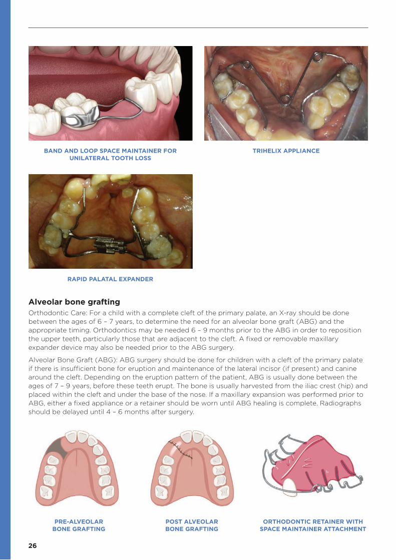
26
BAND AND LOOP SPACE MAINTAINER FOR UNILATERAL TOOTH LOSS
RAPID PALATAL EXPANDER
PRE-ALVEOLAR BONE GRAFTING
POST ALVEOLAR BONE GRAFTING
ORTHODONTIC RETAINER WITH SPACE MAINTAINER ATTACHMENT
TRIHELIX APPLIANCE
Alveolar bone grafting Orthodontic Care: For a child with a complete cleft of the primary palate, an X-ray should be done between the ages of 6 – 7 years, to determine the need for an alveolar bone graft (ABG) and the appropriate timing. Orthodontics may be needed 6 – 9 months prior to the ABG in order to reposition the upper teeth, particularly those that are adjacent to the cleft. A fixed or removable maxillary expander device may also be needed prior to the ABG surgery.
Alveolar Bone Graft (ABG): ABG surgery should be done for children with a cleft of the primary palate if there is insufficient bone for eruption and maintenance of the lateral incisor (if present) and canine around the cleft. Depending on the eruption pattern of the patient, ABG is usually done between the ages of 7 – 9 years, before these teeth erupt. The bone is usually harvested from the iliac crest (hip) and placed within the cleft and under the base of the nose. If a maxillary expansion was performed prior to ABG, either a fixed appliance or a retainer should be worn until ABG healing is complete. Radiographs should be delayed until 4 – 6 months after surgery.

27
PRINCIPLES OF MINIMALLY INVASIVE DENTISTRY
Introduction to minimally invasive dentistry Minimally invasive dentistry is a philosophy that integrates prevention, remineralization and minimal intervention for the placement and replacement of restorations. Minimally invasive dentistry means treatment using the least invasive surgical approach, with the removal of the minimal amount of healthy tissue.
When a lesion needs to be restored, removal of decay with maximum conservation of healthy tooth structure should be the priority. Since our “permanent” restorations seldom last for ever, we need to minimize the size of any restoration. This will prevent or limit the restoration cycle that ultimately leads to tooth fracture, endodontic treatment and crown, and (occasionally) root fracture and extraction of the tooth.
Minimally invasive dental procedures The most common procedures that can be called minimally invasive in children are the application of silver diamine fluoride (SDF), atraumatic restorative technique (ART) and the use of stainless-steel crowns for arresting multi-surface caries in primary molars.
Silver diamine fluoride (SDF) application Thirty-eight per cent SDF, if available, should be used when cavitation has occurred. It is most effective when applied semi-annually but annual applications may be sufficient. SDF is effective in arresting caries and preventing its progression to the pulp of the tooth. This can help increase the long-term longevity of the primary dentition.
Learning objectives: To ensure oral health professionals are trained to understand minimally invasive dentistry.
Pedo Planet - Children Dental Centers, (Chennai, New Delhi), India.
Centre for Early Childhood Caries Research (CECCRe), Sri Ramachandra Institute of Higher Education and Research, Chennai, India

28
HALL CROWNSPedo Planet - Children Dental Centers, (Chennai, New Delhi), India.
Centre for Early Childhood Caries Research (CECCRe), Sri Ramachandra Institute of Higher Education and Research, Chennai, India
Atraumatic restorative technique (ART) If restoration of primary teeth is required, ART is a minimally invasive technique where caries are removed by hand using an excavator, for example. This retains as much tooth substance as possible and aims to prevent pulp death. An adhesive restoration such as glass ionomer cement, preferably containing slow-release fluoride, should be used for the restoration.
Hall crownsThe Hall crown technique is the placement of a pre-formed stainless-steel crown over a carious primary tooth using a glass ionomer cement. This technique requires no preparation, local anaesthesia or caries removal. Due to this, it is especially useful in the treatment of children. It can be easily taught to an oral health professional (OHP) and is widely practised and accepted across the globe.
29
CASE 1
CASE 2
MANAGING RESTORATIVE CARE FOR COSMETIC IMPROVEMENTS
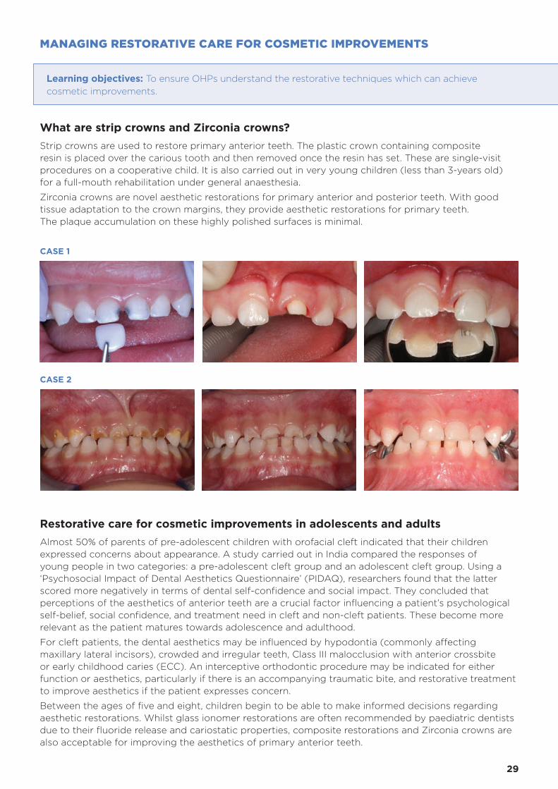
What are strip crowns and Zirconia crowns? Strip crowns are used to restore primary anterior teeth. The plastic crown containing composite resin is placed over the carious tooth and then removed once the resin has set. These are single-visit procedures on a cooperative child. It is also carried out in very young children (less than 3-years old) for a full-mouth rehabilitation under general anaesthesia.
Zirconia crowns are novel aesthetic restorations for primary anterior and posterior teeth. With good tissue adaptation to the crown margins, they provide aesthetic restorations for primary teeth. The plaque accumulation on these highly polished surfaces is minimal.
Restorative care for cosmetic improvements in adolescents and adults Almost 50% of parents of pre-adolescent children with orofacial cleft indicated that their children expressed concerns about appearance. A study carried out in India compared the responses of young people in two categories: a pre-adolescent cleft group and an adolescent cleft group. Using a ‘Psychosocial Impact of Dental Aesthetics Questionnaire’ (PIDAQ), researchers found that the latter scored more negatively in terms of dental self-confidence and social impact. They concluded that perceptions of the aesthetics of anterior teeth are a crucial factor influencing a patient’s psychological self-belief, social confidence, and treatment need in cleft and non-cleft patients. These become more relevant as the patient matures towards adolescence and adulthood.
For cleft patients, the dental aesthetics may be influenced by hypodontia (commonly affecting maxillary lateral incisors), crowded and irregular teeth, Class III malocclusion with anterior crossbite or early childhood caries (ECC). An interceptive orthodontic procedure may be indicated for either function or aesthetics, particularly if there is an accompanying traumatic bite, and restorative treatment to improve aesthetics if the patient expresses concern.
Between the ages of five and eight, children begin to be able to make informed decisions regarding aesthetic restorations. Whilst glass ionomer restorations are often recommended by paediatric dentists due to their fluoride release and cariostatic properties, composite restorations and Zirconia crowns are also acceptable for improving the aesthetics of primary anterior teeth.
Learning objectives: To ensure OHPs understand the restorative techniques which can achieve cosmetic improvements.

30
MONITORING AND MAINTAINING THE ORAL HEALTH OF PEOPLE WITH CLEFT
Specific interventions for patients with cleft Specific interventions for the age group of up to two years old:
• Work with the caregivers to understand the oral health needs of a patient with cleft.
• Demonstrate mouth cleansing after each feed and gentle tooth brushing as the primary dentition erupts.
• The use of wet cotton or gauze or disposable infant wipes after every meal or feed is of paramount importance in preventing early childhood caries.
• Giving a sip of water after every meal or feed can be beneficial in washing away food debris or remnants of milk.
• Explain the aetiology of dental disease and the caregiver’s role in prevention.
• An examination by a dentist or by a paediatric dentist, where available, is mandatory as soon as the first primary tooth erupts into the mouth.
• Provide instruction about the proper care and cleaning of the obturator and appliance to maintain good oral hygiene
• After lip repair surgery, which can take place in the first three months of life, caregivers should be informed about scar management. This can be carried out after the wound is completely healed and the sutures have been removed. Instruct parents or caregivers to massage from the columella end of the scar to the vermilion, with a downward motion for 8-10 minutes, three times a day.
• Explain to the caregivers the importance of discouraging thumb sucking and pacifier use. These harmful habits could have a negative impact on the baby’s face and oral development and growth, as well as problems with dental alignment and malocclusion.
• Early identification of white and brown spot lesions and treatment with minimally invasive dentistry is essential.
Learning objectives: Participants should understand how to follow the FDI/Smile Train Oral Health in Comprehensive Cleft Care Guidelines and maintain the oral health of patients at all ages

31
Specific interventions for patients with clefts aged between two and six years old:
• Work with caregivers to understand the oral health needs of a patient with cleft, demonstrating mouth cleansing after each feed and gentle toothbrushing as the primary dentition erupts. Explain the aetiology of dental disease and the caregiver’s role in prevention.
• Provide instruction about the proper care and cleaning of the obturator and appliance to maintain a good oral hygiene
• Scar management can be carried out when the surgical site has fully healed. Instruct parents or caregivers to massage from the columella end of the scar to the vermilion with a downward motion for 8-10 minutes three times a day.
• Explain to the caregivers the importance of discouraging thumb sucking and pacifier use. These harmful habits could have a negative impact on the baby’s face and oral development and growth, as well as problems with dental alignment and malocclusion.
• Early identification of white and brown spot lesions and treatment with minimally invasive dentistry is essential.
Specific interventions for patients with cleft aged 6-12 years old:
• In the mixed dentition, primary and permanent teeth are present in the mouth at the same time. Several dental interventions begin at this stage. As the bone structure is rapidly growing and permanent teeth are erupting, some patients might need interceptive orthodontics to avoid severe malocclusion and create better conditions for facial growth and oral function.
• Oral health care and routine dental examination are very important at this stage. A complete periodic oral examination with dental radiographs, dental prophylaxis and a fluoride treatment should be performed. Ongoing evaluation of dental hygiene and periodontal diseases should be provided. The risk of developing caries and gingival inflammation increases with the placement of orthodontic appliances. Fissure sealants should be placed as the permanent dentition erupts.
• Advise parents and caregivers to visit the dentist every six months for periodontal examination and monitoring of the permanent dentition eruption of their children.

32
Specific interventions for patients with clefts aged 12-18 years old:
• Good oral health care is critical during this stage. A complete oral examination with dental radiographs, dental prophylaxis and a fluoride treatment should be performed at regular intervals. Ongoing evaluation of dental hygiene and periodontal diseases should be provided.
• Specific oral hygiene instructions for patients with orthodontics should be delivered.
• Age-related advice tailored to the patient is essential, for example, reducing fizzy drinks and not snacking at school.
Specific interventions for patients with clefts aged above 18 years old:
• Good oral health care and age-related advice tailored to the patient is essential, for example, care of implants or prosthesis. Ongoing evaluation of dental hygiene and periodontal disease should be provided.
• Supply information and tools for smoking cessation and alcohol consumption reduction if required. Instruct patients about injury prevention and the use of fitted mouth guards during sports.

33
Care instructions for the obturator/applianceChildren in the 0-2 and 2-6 age groups will probably use a palatal maxillary orthopedic appliance, or obturator, to cover the palatine cleft and be able to eat and speak properly. These appliances must be cleaned twice a day, every day. In the first 48 hours after the appliance is installed, it must be removed and cleaned after each feed. Important points to remember are:
• After removing the maxillary oral appliance, clean it with a brush and cooled boiled water to remove any pellicle or detritus that has been formed to avoid microorganisms to grow in it. The area under the flattened nostril must be cleaned with a moist swab.
• Refer to the treating oral health professional if the appliance is producing any ulceration or bleeding. In this case, it needs to be adjusted. Advice the use of soft white paraffin in the lips and soft tissues when needed and at feeding time.
• Obturator appliances must be inserted carefully by the caregiver: slightly sidewards for unilateral cleft and straight for bilateral cleft.

34
DELIVERING ORAL HEALTH EDUCATION
Delivering oral health education It can be frustrating to provide patients and caregivers with multiple oral health messages at each appointment only to find that the adverse oral health behaviour has not changed. It is important to create an environment where the patient and caregiver feel supported and not judged, create an open dialogue and actively listen to the challenges they are facing with their oral health.
Health coaching/motivational interviewing
This approach to behaviour change is known as health coaching or motivational interviewing and is part of the 2015 Geneva Declaration on Person-centred Primary Health Care. It is an effective person-centred approach that facilitates, motivates and empowers individuals to become self-aware, and to identify barriers and facilitators to healthier behaviours. Individuals are encouraged to make effective decisions for changing their lifestyle or adopting changes in their environment, and to take sustainable actions on the way forward. It focuses on empowerment and active engagement of individuals in the decision-making process for their own health.
Follow the four steps to improving oral health behaviours through motivational interviewing:
1. Engaging: the first step is to build trust with the patient. Ask permission to discuss their oral health and to ask questions and let them know what you will be discussing. This could include any concerns they may have with their oral health.
2. Focusing: this is a way to guide the patient toward the behaviour change. Help them focus on what they want to change. This could be discussing sugar in their diet or their toothbrushing technique. For example, after noticing white spot lesions around the cervical margins, discuss the findings with the patient and ask them, “Shall we discuss ways we can prevent further tooth decay?” This will bring attention to the fact that they, the patient, are making that decision.
3. Evoking: this promotes the patients’ own reasons to change. Ask them what they know about tooth decay. What is their perception of tooth decay? Offer them more information on what you see clinically. Let them come to the conclusion that a change needs to be made. Let them be the one to state that change is needed, and then plan to make that change with them.
4. Planning: finally, ask the patient what it is they can do to make that change and offer assistance in helping them make that change. Help them identify a time in their routine to implement a new toothbrushing behaviour or how to replace sweet snacks with healthy alternatives.
Culturally appropriate health coaching
There are myths about teeth that all oral health professionals have heard: “the baby took the minerals from my teeth while I was pregnant” is a common one. It is easy to be dismissive of these, but it is important to remember that these myths may have strong familial or cultural beliefs behind them. Try to present the facts as sensitively as possible and encourage a positive behaviour change.
Learning objectives: Participants should understand how to provide oral health education and use a motivational interviewing style.

35
DAY 3
SCAR MANAGEMENT IN PEOPLE WITH CLEFT LIP AND PALATE
Scars in patients with cleft lip and palate The healing of cleft lip and palate repair results in fibrotic outcomes for both the lip and the palate. Scarring in this situation can have an array of aesthetic and functional consequences. Hypertrophic scarring can be common following cleft lip repair.
Scarring from cleft lip repair can cause lip asymmetry as the scar contracts leading to a shortened lip and nasal deformity on the affected side. Such scarring may require further surgical revision to restore normal aesthetics and function, which causes psychological stress, risk associated with additional surgeries and anaesthesia exposure, and significantly increased cost of treatment.
Factors influencing scarring
• The depth of injury, degree of tissue destruction, and introduction of pathogens: for scarring to occur, injury must involve the dermis, and pathologic forms of wound healing are more likely to occur if injury involves the bottom one-third of the dermis or is associated with infection.
• The location of the wound affecting variations in mechanical forces across the skin at different anatomical sites, such as: on the face, tension lines arise from interactions between the skin and the underlying muscles of facial expression; wounds running across a tension line experience greater perpendicular force and must respond with greater collagen deposition to hold the skin together, resulting in a larger scar.
• Patient demographics: it has been observed that darker-skinned and younger individuals are at higher risk of pathological healing, with hypertrophic scars and keloids, although the causative mechanisms explaining these risks are not well-understood.
Keloids and hypertrophic scarsIn the skin, pathological scars such as hypertrophic scars and keloids can be unsightly, itchy, and painful. By definition, both scar types rise above skin level and are result of wounds with excessive fibrosis. While hypertrophic scars do not extend beyond the initial site of injury, keloids typically project beyond the original wound margins.
Clinical differentiation between hypertrophic scars and keloids can be problematic. A single injury may produce regions of both normal and abnormal scarring that regress or progress over time. Incorrect identification of scar type may result in inappropriate management of pathologic scar formation, and, occasionally, contribute to inappropriate decision-making related to elective or cosmetic surgery. Unfortunately, these surgeries represent a second injury that may heal with repeated pathological scarring in some individuals.
Learning objectives: Participants will understand the impact scarring has on patients with cleft, what factors influence scarring and the daily management of a scar following surgery.

36
KELOID SCARRING HYPERTROPHIC SCARRING NORMAL SCARRING
Postoperative care and scar management The first three months after surgery is the time when remodelling is at its peak during a normal wound healing process. While most properly designed and executed operative incisions will continue to heal without significant protest, those incisions destined for hypertrophic response will begin to reveal themselves during this period. Therefore, a monthly clinic examination of the scar is reasonable. Patients should be encouraged to continue dressing the wound with skin tape (triple-layer, one over the other). Additionally, each patient should be counselled to return to the clinic earlier than the appointed clinic visit if the wound is beginning to look “beefy” or “upset”, e.g if it starts to rise above skin level or itches considerably. During this period, a scar can undergo hypertrophic response, either mild or severe.
Current therapies for scarringMany different therapeutic approaches have been developed to minimize the appearance and functional impact of scars. Therapies delivered at the time of wounding include dressings, tapes, and silicone sheets designed to reduce tension on the wound and suture lines. Also, if there is an increase of tissue above the normal skin level, compression therapy is recommended. This decreases the vascularity of the scar and helps to control the hypertrophy.
It is of fundamental importance not to forget that in addition to the hypertrophy of the scar, we must take care of the degree of retraction. It is suggested the scar should be regularly massaged from the columella to the vermillion border three to five times per day. The frequency should decrease as the scar gains flexibility.

37
QUALITY OF LIFE AND CLEFTS
Definition of quality of life:The World Health Organization defines quality of life as an individual’s perception of their position in life in the context of the culture and value systems in which they live and in relation to their goals, expectations, standards and concerns. It is a broad ranging concept affected in a complex way by the person’s physical health, psychological state, personal beliefs, social relationships and their relationship to salient features of their environment.
What makes it challenging to measure is that, although the term ‘quality of life’ is widely understood, individuals and groups can define it differently. Although health is one of the important domains of overall quality of life, there are other domains as well—for instance, jobs, housing, schools, the neighbourhood. Aspects of culture, values, and spirituality are also key domains of overall quality of life that add to the complexity of its measurement.
Definition of healthHealth is defined by WHO as a state of complete physical, mental and social well-being, not merely the absence of disease or infirmity. Better health is central to human happiness and well-being, while poor health has detrimental effects on both the individual and at societal level.
Since healthy populations live longer, are more productive and save more, good health also makes an important contribution to economic progress. Many factors influence health status and a country’s ability to provide a good quality health service for its people.
There are five main aspects of personal health: physical, emotional, social, spiritual, and intellectual.
The Sustainable Development GoalsThe Sustainable Development Goals are the United Nations blueprint for a better and more sustainable future for all. They address the global challenges we face, including poverty, inequality, climate change, environmental degradation, peace and justice. One of their goals is to ensure healthy lives and promote well-being for all, at all ages.
The work of UNICEF, the United Nations children’s fund, is structured around five overarching areas of well-being for every child, which are grounded in the 2030 Agenda for Sustainable Development. These five areas are that:
• every child survives and thrives;
• every child learns;
• every child is protected from violence and exploitation;
• every child lives in a safe and clean environment;
• every child has a fair chance in life.
This human rights-based approach pursues a vision of realizing the rights of every child, especially the most disadvantaged, and responds to the call to “leave no child behind”, so that the rights of every child, everywhere, will be met.
Oral healthFDI World Dental Federation defines oral health as:
Oral health is multi-faceted and includes the ability to speak, smile, smell, taste, touch, chew, swallow and convey a range of emotions through facial expressions with confidence and without pain, discomfort and disease of the craniofacial complex.
Learning objectives: Participants will understand the impact of cleft on quality of life as well as economic factors.

38
Oral diseases are the most common chronic disease and are important public health problems due to their prevalence, their impact on individuals and society, and the expense of their treatment.
The Global Burden of Oral Disease Study 2013 estimated that oral diseases affect 3.5 billion people worldwide, with untreated dental caries being among the most prevalent non-communicable disease (NCD). A follow-on study examined the global cost. Most oral diseases and conditions share modifiable risk factors with the four most signficant NCDs, including cardiovascular disease, cancer, chronic respiratory disease and diabetes. These risk factors include tobacco use, alcohol consumption and unhealthy diets high in free sugars. All are increasing at a global level.
Costs of the surgical treatment of cleft A study by Galloway et al., (2017) estimated the comparative cost of cleft treatment incorporating surgical intervention, speech and language therapy (SLT), orthodontics and orthognathic surgery. In high-income countries, where state provision or state health insurance is the most common source of funding, the average total direct cost of cleft treatment is $10,000-$13,000 whilst in low-income settings, where patient and charity organisations fund treatment, the cost is $3,000-5,000. Further research and more complete data are needed to study cost differences so patients and their families have an understanding of the lifelong financial implications of cleft where costs are not covered by the state or insurance.
Risk factorsDifferential in oral health status is multi-factorial, ranging from social, environmental, biological, behavioural and cultural factors to economic and political factors. Limited access to oral health care services, complicated oral health care systems, a lack of oral-health-information material, and oral health literacy also play a role.
These differences arise as a consequence of factors that are largely out of the control of population groups, such as access to dental services and differential exposure to unhealthy aspects of social environments. A common factor underlying these inequalities is social hierarchy. Socioeconomic position influences exposure and response to virtually all environmental, behavioural and psychosocial risk factors. The steeper the social hierarchy, the greater the magnitude of inequalities in health.
The main priority for oral health interventions should be collaborative, enabling policies underpinned by research that addresses the main determinants of oral diseases and the shared, modifiable risk factors outlined above.
A study among adults aged 18-64 reported that social security and health insurance, low literacy levels, dental self-care, or its absence, and other behavioural aspects, such as high tobacco consumption, were major risk factors for periodontal and other oral diseases among groups of equivalent socioeconomic status.
Public health and orofacial cleftsOrofacial clefts are a major public health challenge. Children with clefts rarely escape dental complications. The surgical correction of their clefts, in addition to the medical concerns common among children with cleft, are the major focus of their care. As a result, they tend to have more decayed and missing teeth and poorer oral health compared to their peers.
These differences can be attributed to:
• dry mouth caused by mouth-breathing habits;
• less natural cleaning of the teeth due to the morphology;
• variable diet or feeding habits;
• dental anomalies;
• increased consumption of sweetened medications;
• delayed oral clearance time for foods.

39
All the above contribute to more carious bacteria being present in the oral cavity of cleft children. Parents and caregivers are often so concerned with other aspects of their child’s health care, such as surgery, nutrition, mental health, and speech development, that they pay little attention to basic preventive dental care. All these factors put the child at a greater risk of developing ECC.
As they develop, the higher prevalence of poor oral hygiene in children with cleft may be associated with:
(a) the presence of residual scar tissue as a result of the multiple surgical procedures carried out at the cleft region, which in turn impairs tooth cleaning;
(b) the lack of interest in oral hygiene due to many other health problems such as otitis media, difficulty in speech;
(c) the anxiety that children often have when they brush their teeth in the cleft area.
Other barriers to oral health care in these children will include low literacy level of parents, behaviour induced by fear and anxiety, socio-economic status, competing priorities, poor knowledge of available medical and dental services, patient-dentist relationships and socio-cultural beliefs and myths.
All these factors highlight the importance of individualized preventive oral health programmes in cleft patients.
Challenges associated with the effects of cleftsCleft patients have multi-system and complex anatomical, physiological, pathological and psycho-social problems. These include aesthetics, feeding, speech and language delays, dental anomalies, ear infections, psycho-social issues, such as stigmatization and reduced quality of life.
40
IMPLEMENTING THE FDI/SMILE TRAIN SAFETY AND QUALITY PROTOCOL
Why is quality important in oral healthcare? • By maintaining the quality of healthcare provided and adhering to the recommended health
screenings, the cleft care team can improve patient health and achieve better health outcomes, e.g. fewer missed school hours due to dental pain.
• By improving the efficiency of managerial and clinical processes, organizations reduce the costs associated with mistakes, wasted materials and redundancy.
• Proactive processes that recognize and solve problems before they occur ensures that healthcare is reliable and predictable.
A culture of improvement frequently develops in an organization that is committed to quality, because errors are reported and addressed.
• A commitment to quality shines a positive light on an organization, which may result in increased partnership and funding opportunities.
The quality approach The quality approach is defined by WHO as :
an approach which should make it possible to guarantee each patient the combination of diagnostic and therapeutic procedures which will ensure the best possible health outcome for them, in accordance with the current state of medical science.
This means that all healthcare providers should implement recurring, reproducible and even “measurable” processes that guarantee the quality and safety of their interventions: this is quality assurance.
Quality assurance (QA) vs. quality improvement (QI) Quality assurance (QA) involves the development of a set of standards, and the process of a comparison of current standards with the recommended standards.
If standards are met, services are thought to be of adequate quality. If gaps are identified, plans to correct these are developed to address the problem.
Quality improvement (QI) consists of systematic and continuous actions that lead to measurable improvement in health care services and the health status of targeted patient groups.
How do we define quality in oral healthcare? One of the most commonly used definitions for quality in healthcare is by the Institute of Medicine (IOM) and consists of six domains: patient safety, timeliness, patient-centredness, equitability, efficiency and effectiveness.
Learning objectives: Understand:
• How to use the FDI/ST Dental Procedures Safety and Quality Protocol• The importance of quality assurance• Strategies for implementing quality assurance • How to carry out periodic assessment of quality of care

41
Patient safety In order to be considered safe, oral healthcare should• Avoid, mitigate, or minimize adverse events.
• Advocate a blame-free culture to facilitate quality improvement.
• Learn from safety incidents to improve the quality of care.
• Enact minimum safety standards.
Timeliness In order to be considered timely, oral healthcare should• Avoid unnecessary delays in access and utilization of care.
• Implement care co-ordination between healthcare providers and institutions.
• Prioritize prevention; avoid early use of restorative and other treatments.
Patient-centeredness In order to be considered patient-centered, oral healthcare should• Be respectful of and responsive to individual patient preferences,
needs, values, fears, concerns, and/or cultures.
• Follow a shared decision-making model when making clinical decisions. To gain a patient’s trust, the oral healthcare professional should communicate with and listen to the patient, then inform, educate and guide the patient to ensure that patient values shape all clinical decisions.
Equitability In order to be considered equitable, oral healthcare should• Not vary in quality and availability due gender, ethnicity, cultural
background, religion and belief, geographic location, and/or socioeconomic status.
• Address inequities in oral health service design, planning, and commissioning.
• Incorporate equitability in the design of policy and clinical practice guidelines.
Efficiency In order to be considered efficient, oral healthcare should • Encourage prevention
• Focus on patients’ oral health needs as the central basis for resource and workforce planning.
• Form an integral part of medicine and discourage the dental-medical divide.
Effectiveness In order to be considered effective, oral healthcare should• Be informed by the most recent available scientific evidence and
guidelines.
• Be provided to patients who will benefit from the care
• Aspire to minimize harm

42
Why do we need a quality approach? A quality approach in healthcare is imperative and reflected in diverse legislation:
• Legal and regulatory context: quality in healthcare is governed by a set of laws and regulations in force in each country.
• Administrative regulations: all clinicians should be appropriately trained and registered to practice in the chosen country, and stay within the “scope of practice” defined by legislation or regulation. There are many other administrative regulations, i.e. the obligation to take out civil liability insurance or various other types of insurance.
• Health regulations and good practice recommendations: specific measures for patient safety, defined by law and regulations under the control of the country’s health authorities i.e. regulations on ionising radiation.
• Ethical regulations: unlike business, dental offices and health centres have a responsibility for patient well-being. This is underpinned by a code of ethics which specifies what can and cannot be done. An official body, most often the medical or dental council, is responsible for ensuring that these ethical rules of confidentiality and patient safety are respected.
• Labour legislation: the rules of labour legislation set out in national legislation, would apply to staff members, so that practitioners need to be familiar with them.
Implementing quality improvement Implementing new procedures to comply with quality in healthcare standards, such as the FDI/ST Systems Level Organization, must be tested and validated, usually through a four-phase cycle of planning, doing, studying, and acting - the PDSA cycle:
Establish quality objectives and processes required to deliver the desired results.
React and improve. Take the necessary corrective measures to reduce gaps and make sure that what has been
achieved will remain stable. Each turn of the wheel takes the project higher
until the target is finally reached.
Implement the plan, carry out the objectives from
the previous step.
Check that the project is moving towards the objectives,
study the results, measure effectiveness.

43
Steps for implementing quality improvement
Steps Questions & recommendations
Identify and assess the problem
Identify specific objectives
What problem do you want to address?
What outcome measure will show the problem was resolved?
Produce a written description of the roles and responsibilities of each member of the dental team to reach the objective
Who is best positioned to be the champion and the leader of the project?
Who has the clinical knowledge and leadership skills to serve as a project leader?
Which staff members will participate in the core collaborative and work proactively to improve quality?
Education and training of staff to implement the QI measure
Train all staff in the new method adopted to solve the problem or improve the system
Establish a test group who will implement the QI measure
The test group carries out an assessment of the new system with all members of the dental team.
Implementation of QI measure
Implementing changes in small manageable steps to allow for continual appraisal and review.
Reducing the risk of failure and negative staff responses
Verification and outcome assessment Review of the first implementation cycle to review the process and make any necessary adjustments
Repetition of the quality improvement process and identification of new objectives if necessary.
The improvement process must have adequate time to succeed. It is important to review in the longer term to ensure the new habit and process has become a part of the daily routine.
Monitoring and evaluation Quality improvement is an ongoing process, and it is important to create a culture in your clinic where staff can make suggestions openly to improve patient care, procedures or systems. The team leader should use the FDI/ST Dental Procedures Safety and Quality Protocol as a basis for ensuring minimum standards of safety, care and hygiene and continue to develop this resource to continuously improve the standards you provide.
Monitoring and evaluation should be carried out routinely, as new quality improvement measures are introduced. One person should be assigned this responsibility, usually the clinic manager.