Embed Size (px)
Citation preview
SUMMARY
Patients suffering from a cleft palate and/or cleftlip present evident anatomical defects in bothformations. However, these developmentaldisorders are often accompanied by importantdisturbances in other anatomical structures ofthe mouth and adjacent tissues that may affectnormal dental occlusion and, consequently, thebasic functions of mastication and phonation.The objectives of the present work were to des-cribe and discuss the above structural modifica-tions, presenting several clinical cases in whichthe anatomical defects are described togetherwith the functional implications. The anomaliesfound include important variations in the num-ber of teeth, the position, disposition and rela-tion of the alveolar processes, and the impair-ment of dental occlusion. In the cases studied,these anatomical defects have elicited prominentalterations in the normal functions of masticationand phonation, and also in facial aesthetics. Pho-tographs of mouths, dental casts, and X-rays areused to illustrate our report.
Key words: Cleft palate – Cleft lip – Oral cavity– Dental occlusion – Mastication
INTRODUCTION
During the fifth and sixth weeks of prenatal life,the primary palate is formed below the primitiveposterior nares. From this structure, the upper
lip, the anterior portion of the maxillary alveolarprocesses, and the rostralmost portion of thepalate will develop later. While the nasal septumis developing, each mass of maxillary mesodermoriginates yet another medially directed exten-sion: the palatal processes (lateral palatal shel-ves). These processes extend medially belowand behind the primitive posterior nares on alevel with the primitive palate. With furthergrowth, the free edges fuse: first with the poste-rior margin of the primitive palate, and then pro-gressively with each other at the mid-line to formthe secondary definitive palate. Thus, the oraland nasal cavities become separated and thehard and soft palates are formed (Sperber, 2001).Successful fusion of the three embryonic com-ponents of the palate involves a complicatedsynchronization of shelf movements with growthand withdrawal of the tongue and with growthof the mandible and head.
Cleft palate and cleft lip represent multifacto-rial developmental anomalies involving bothgenetic predisposition (Bender, 2000; Aldred,2001; Vieira and Orioli, 2001) and environmentalagents (Presscott et al., 2001).
Clefting of the palate and/or lip may be cau-sed by failure at any of the stages of palatoge-nesis: defective growth or delayed elevation ofthe palatal shelves, defective shelf fusion, failureof medial-edge epithelial cell death, possiblepostfusion rupture, etc. (Berkovitz et al., 1995).
Patients with a cleft lip and/or cleft palatepresent evident anomalies in both structures.However, the development of these defects isoften associated with anatomical deficiencies in
Eur J Anat, 8 (3): 137-141 (2004)
137
Oral anatomical defects associated with cleftpalate and cleft lip
E. Jiménez-Castellanos2, A. Carmona1, C.J. Catalina-Herrera1
and J. Jiménez-Castellanos1
1- Departamento de Anatomía y Embriología Humana2- Departamento de Estomatología. Facultad de Medicina, Universidad de Sevilla, 41009 Sevilla, Spain
Correspondence to:Prof. J. Jiménez-Castellanos. Departamento de Anatomía y Embriología Humana, Facultad deMedicina, Avda. Sánchez Pizjuán 4, 41009 Sevilla, Spain. Tel.: 34 94552865; Fax: 34 94381662. E-mail: [email protected]
Submitted: July 12, 2004Accepted: September 25, 2004
other oral and perioral structures, frequentlyinvolving normal dental occlusion and hence themain masticatory and phonatory functions.
Here we used data from clinical cases to focuson the description of different anatomical anoma-lies linked to abnormal palatal development.
MATERIAL AND METHODS
Five cases of cleft palate and/or cleft lip were usedin this study. Photographs of mouths, dental castsand X-rays are used to illustrate our findings.
RESULTS
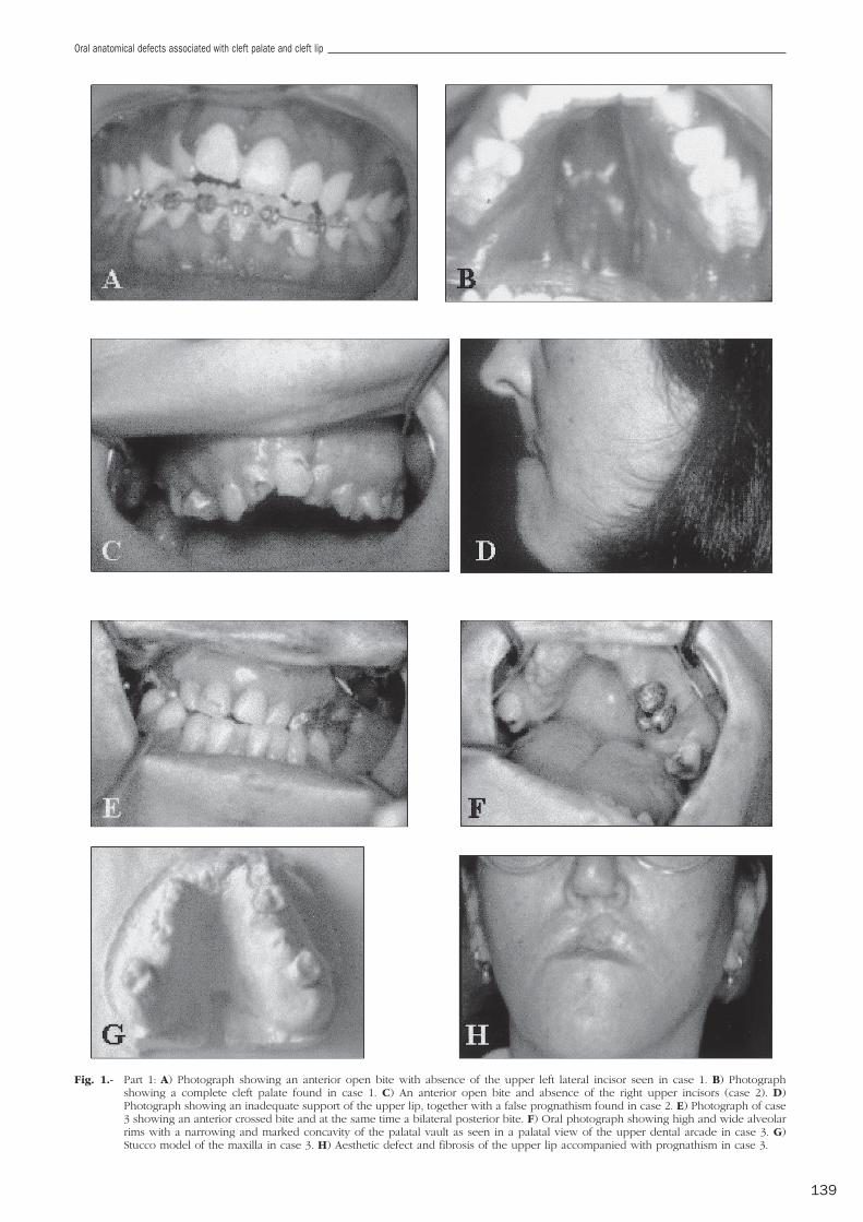
Case 1.- Male aged 19, presenting a completecleft palate without previous surgical treatment(Fig. 1, B). Corresponding to the location of thepalatal defect, the left upper lateral incisortooth was absent. A versioned palatolingualocclusion and an anterior open-bite had beenpartially corrected with orthodontic treatment(Fig. 1, A).
Case 2.- Female aged 25, showing cleft lipand cleft palate with previous surgical treatment.In relation to the palatal anomaly, the rightupper incisor was absent (Fig. 1, C). As a conse-quence of the premaxillary bony defect, a narro-wing of the maxillary bone and an inadequatesupport exerted by soft tissues, especially the lip,were observed, together with a mandibular pro-trusion (Fig. 1, D).
Case 3.- Female aged 46, showing cleft lipand a premaxillary defect. A maxillary compres-sion with bilaterally cross-bite was also seen(Fig. 1, E). Alveolar borders were thick and highand were associated with defects in supportingtissues (upper lip) (Fig. 1, F, G). A mandibularprotrusion was also evident (Fig. 1, H).
Case 4.- Male aged 19, presenting clefting ofthe palate and lip with several prior surgicalcorrections. Both left upper incisors were absent(Fig. 1, I), with important malocclusion mainlydue to the right upper dental hemiarcade (Fig. 1,J). Maxillary compression, bilateral cross-bite,defective support from soft tissues and a protru-sive mandibular position were also detected(Fig. 1, K).
Case 5.- Male aged 29, with cleft palate andlip and previous surgical treatment (Fig. 1, L).Both upper lateral incisors were absent (ortho-pantomographic study performed at age 5) (Fig.1, M). There was an important malocclusion withmaxillary compression followed by a prominentovergrowth of alveolar margins in height andwidth (Fig. 1, N). Other clinical signs were bila-teral cross-bite and mandibular protrusion (Fig.1, O).
DISCUSSION
There is disagreement in the literature about theassociation of cleft palate/lip and hypodontia.Thus, some authors have pointed to a prevalen-ce of hypodontia linked to clefting in 77% ofcases; six times higher than in the normal popu-lation (Roth and Hirschfelder, 1991; Jiroutovaand Mullerova, 1994; Shapira et al., 1999). Incontrast, other authors have considered thathypodontia is due to dental loss as a conse-quence of the surgical operations performed onthese patients. These authors argue that the pre-valence of hypodontia in the postcanine regionis similar to that found in the normal population(Lekkas et al., 2000; Budai et al., 2001; Wangpi-chit et al., 2001). Although the prevalence ofhypodontia in postcanine regions might beexpected to be similar to that of the normalpopulation, in zones where the palatal defect islocated it is frequent to find, in conjunction withthe anterior palatal malformation, an associatedagenesis of one or more teeth of the maxillarydental arcade, as occurred in our cases 1, 2, and3. Most authors agree about the existence ofconsiderable variations in the size, position, anddisposition of the alveolar processes; in particu-lar, in terms of a reduced width and depth of thedental arcades (Dericke et al., 1994). Conse-quently, such patients, in addition to presentingimportant speech problems (Lohmander-Agers-kov et al., 1995; Laitinen et al., 1998; Nandukar,2002), respond poorly to intensive phonatoryarticulation therapies (Van Demark and Hardin,1986; Chapman, 1993).
Once the phonatory function had begun,these patients used as a sounding board notonly their mouths but also their nasal cavities.Therefore, bearing in mind the considerableadded variations of the alveolar processes seenin our cases 3, 4, and 5, we agree with theabove authors as regards the poor response ofthese patients to phonatory articulation thera-pies, even after the anatomical defect has beensolved using prothesic obturators. The patientsstill maintain a nasal tone of voice with markeddifficulties to emitting certain palatolingual pho-nemes.
The disturbances of dental occlusion, even inpreviously treated patients, were present in morethan 2/3 of this palatal malformation. The altera-tions most frequently found were posteriorcross-bite, open-bite, and lingualized occlusion,together with a false prognathism (Da Silva Filhoet al., 1998; Tomioka, 1998; Kruk-Jeromin et al.,1999; Johnson et al., 2000; Lambrecht et al.,2000; Prasad et al., 2000; Morris et al., 2000;Braumann et al., 2001). In many cases, theseanomalies gave rise to severe deficiencies inmasticatory function.
E. Jiménez-Castellanos, A. Carmona, C.J. Catalina-Herrera and J. Jiménez-Castellanos
138
Oral anatomical defects associated with cleft palate and cleft lip
139
Fig. 1.- Part 1: A) Photograph showing an anterior open bite with absence of the upper left lateral incisor seen in case 1. B) Photographshowing a complete cleft palate found in case 1. C) An anterior open bite and absence of the right upper incisors (case 2). D)Photograph showing an inadequate support of the upper lip, together with a false prognathism found in case 2. E) Photograph of case3 showing an anterior crossed bite and at the same time a bilateral posterior bite. F) Oral photograph showing high and wide alveolarrims with a narrowing and marked concavity of the palatal vault as seen in a palatal view of the upper dental arcade in case 3. G)Stucco model of the maxilla in case 3. H) Aesthetic defect and fibrosis of the upper lip accompanied with prognathism in case 3.
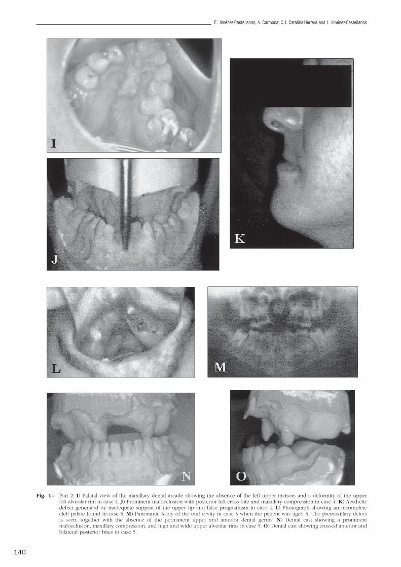
Fig. 1.- Part 2: I) Palatal view of the maxillary dental arcade showing the absence of the left upper incisors and a deformity of the upperleft alveolar rim in case 4. J) Prominent malocclusion with posterior left cross-bite and maxillary compression in case 4. K) Aestheticdefect generated by inadequate support of the upper lip and false prognathism in case 4. L) Photograph showing an incompletecleft palate found in case 5. M) Panoramic X-ray of the oral cavity in case 5 when the patient was aged 5. The premaxillary defectis seen, together with the absence of the permanent upper and anterior dental germs. N) Dental cast showing a prominentmalocclusion, maxillary compression, and high and wide upper alveolar rims in case 5. O) Dental cast showing crossed anterior andbilateral posterior bites in case 5.
E. Jiménez-Castellanos, A. Carmona, C.J. Catalina-Herrera and J. Jiménez-Castellanos
140

All the cases reported here are good repre-sentatives of disturbances of dental occlusion.Thus, they included an anterior open-bite (cases1 and 2), anterior cross- bite (cases 3 to 5), lin-gual occlusion (cases 1, 3, and 5) and false prog-nathism (cases 2, 3, and 4). The most extremesituation was seen in our case 5, where toothloss was accompanied by important malforma-tions of the alveolar processes, with an absoluteincongruence between the maxilla and mandibleand the consequent impairment of masticatoryfunction.
Also noteworthy in this issue is the existenceof significant aesthetic effects, caused primarilyby the weak support exerted by the upper lip asa consequence of the anterior bony and dentaldefects, as seen in our cases 2 and 4. This dis-turbance was also due to false prognathism(cases 2, 3 and 4) coupled to fibrosis and a poorelasticity of the upper lip, in many instances cau-sed by repeated surgical interventions (case 3).
In conclusion, in addition to the developmen-tal anomalies located in both structures, patientssuffering from cleft palate/lip have importantvariations in the number of teeth, the position,disposition and relation of the alveolar processes,and an impairment due to dental occlusion. Allthese anatomical defects led to prominent altera-tions in the normal functions of mastication andphonation and in facial aesthetics.
REFERENCES
ALDRED MA (2001). Cleft lip and palate: new genetic clue.Trends Mol Med, 71: 539-540.
BRAUMANN B, ROSENHAYN SE, BOURAUEL C and JAGER A (2001).Two- or three- dimensional cast analysis in patients withcleft lip and palate. J Orofac Orthod, 62: 451-465.
BENDER DL (2000). Genetics of cleft lip and palate. J PediatrSurg, 15: 242-249.
BERKOVITZ BKB, HOLLAND GR and MOXHAM BJ (1995). Atlasen color y texto de Anatomía oral, histología y embrio-logía, 2nd edition. Mosby/Doyma, Madrid, pp 236-238.
BUDAI M, KOCSIS SG, KORAI E, SAGI I and MARI A (2001).Caries, gingivitis and dental abnormalities in patientswith cleft lip and palate. Fogorv Sz, 94: 197-199.
CHAPMAN KL (1993). Phonologic processes in children withcleft palate. Cleft Palate Cran J, 30: 64-72.
DA SILVA FILHO OG, DE CASTRO MACHADO FM, DE ANDRADE
AC, DE SOUZA FREITAS JA and BISHARA SE (1998). Upperdental arch morphology of adult unoperated completebilateral cleft lip and palate. Am J Orthod Dentofac, 114:154-161.
DERICKE A, KUIJPERS-JAGTMAN AM, LEKKAS C, HARDJOWASITO Wand LATIEF B (1994). Dental arch dimensions in unope-rated adult cleft-palate patients: an analysis of 37 cases.J Craniofac Genet Dev Biol, 14: 69-74.
JIROUTOVA O and MULLEROVA Z (1994). The occurrence ofhypodontia in patients with cleft lip and/or palate. ActaChir Plast, 36: 53-56.
JOHNSON N, WILLIAMS AC, SINGER S, SOUTHALL P, ATACK N andSANDY JR (2000). Dentoalveolar relations in childrenborn with a unilateral cleft lip and palate (UCLP) inWestern Australia. Cleft Palate Cran J, 37: 12-16 .
KRUK-JEROMIN J, ANTOSZEWSKI B and KOT M (1999). Asses-ment of long-term results in cleft lip and palate. MedWieku Rozwoj, 3: 393-405.
LAITINEN J, RANTA R, PULKKINEN J and HAAPANEN ML (1998). Theassociation between dental arch dimensions and occu-rrence of Finnish dental consonant misarticulations in cleftlip/palate children. Acta Odontol Scand, 56: 308-312.
LAMBRECHT JT, KREUSCH T and SCHULZ L (2000). Position,shape and dimension of the maxilla in unoperated cleftlip and palate patients: review of the literature. ClinAnat, 13: 121-123.
LEKKAS C, LATIEF BS, TERRAHE SP and KUIJPERS-JAGTMAN AM(2000). The adult unoperated clefp patient: absence ofmaxillary teeth outside the cleft area. Cleft Palate CranJ, 37: 17-20.
LOHMANDER-AGERSKOV A, SODERPALM E, FRIEDE H, PERSSON ECand LILJA J (1995). Pre-speech in children with cleft lipand palate or cleft palate only: phonetic analysis relatedto morphologic and functional factors. Cleft Palate CranJ, 32: 353-359.
MORRIS DO, ROBERTS-HARRY D and MARS M (2000). Dentalarch relationships in Yorkshire children with unilateralcleft lip and palate. Cleft Palate Cran J, 37: 453-462.
NANDUKAR A (2002). Nasalance measures in Marathi conso-nant-vowel-consonant syllables with pressure conso-nants produced by children with and without cleft lipand palate. Cleft Palate Cran J, 39: 59-65.
PRASAD CN, MARSH JL, LONGRE J, GALIC M, HUEBENER DV andBRESINA SJ (2000). Quantitative 3D maxillary arch eva-luation of two different infant managements for unilate-ral cleft lip and palate. Cleft Palate Cran J, 37: 562-570.
PRESSCOTT NJ, WINTER RM and MALCOM S (2001). Nonsyndro-mic cleft lip and palate: complex genetics and environ-mental effects. Ann Hum Genet, 65: 505-515.
ROTH P and HIRSCHFELDER U (1991). Frequency of tooth age-nesis in CLP patients with eruption of all four thirdmolars. Dtsch Zahnarztl Z, 46: 734-736.
SHAPIRA Y, LUBIT E and KUFTINEC MM (1999). Congenitallymissing second premolars in cleft lip and cleft palatechildren. Dentofacial Orthop, 115: 396-400.
SPERBER GH (2001). Craniofacial development. BC DeckerInc, Hamilton, pp 114-122.
TOMIOKA T (1998). Mandibular movements in dentulouscleft lip and palate patients with lingualized occlusion.Kokubyo Gakkai Zasshi, 65: 213-232.
VAN DEMARK DR and HARDIN MA (1986). Effectiveness ofintensive articulation therapy for children with cleftpalate. Cleft Palate Cran J, 23: 215-224.
VIEIRA AR and ORIOLI IM (2001). Candidate genes fornonsyndromic cleft lip and palate. ASDC J Dent Child,68: 272-279.
WANGPICHIT K, HUNTINGTON NL and KAPALA JT (2001). Com-parison of three nonradiographic methods of mixeddentition analysis in cleft lip and palate patients. PediatrDent, 23: 476-480.
Oral anatomical defects associated with cleft palate and cleft lip
141