Embed Size (px)
Citation preview
Review Article
Opportunities and Challenges in Exploring Indian Non-mulberry Silk forBiomedical ApplicationsROCKTOTPAL KONWARH, BIBHAS K BHUNIA and BIMAN B MANDAL *
Biomaterial and Tissue Engineering Laboratory, Department of Biosciences & Bioengineering, IndianInstitute of Technology Guwahati, Guwahati 781 039, Assam, India
(Received on 07 June 2016; Revised on 08 August 2016; Accepted on 10 November 2016)
Owing to innate desirable features like biocompatibility, mechanical robustness, tunable biodegradability and amenabilityto multiple formatting, silk (christened as the ‘queen of textile’) has carved a unique niche in the realm of regenerativemedicine. Silkworms, being the major source of silk are generally classified as mulberry and non-mulberry types dependingon their feeding habit. Over the years, numerous patents and manuscripts on mulberry based silk for various biomedicalapplications have been published. In sharp contrast to this, the (immense) potential of the non-mulberry silk forbiotechnological applications has been realised quite late. In this article, we have presented the prospects and the recentendeavors to exploit non-mulberry silk (fibroin and sericin) extracted from Antheraea mylitta (tasar), Antheraea assamensis(muga), Philosamia ricini (eri) etc. for fabrication of various formats of biomaterials in applications such as tissueengineering, drug delivery, in vitro tumour modelling, antimicrobial sutures etc. The focus of this article is to highlight theprospective avenues of exploring non-mulberry silk in biomedical domain, as reflected through some of the recent selectresearch works.
Keywords: Non-mulberry Silk; Tissue Engineering; Biomaterials; Scaffolds; Drug Delivery
*Author for Correspondence: E-mail: [email protected]
Proc Indian Natn Sci Acad 83 No. 1 March 2017 pp. 85-101 Printed in India. DOI: 10.16943/ptinsa/2017/41288
Introduction
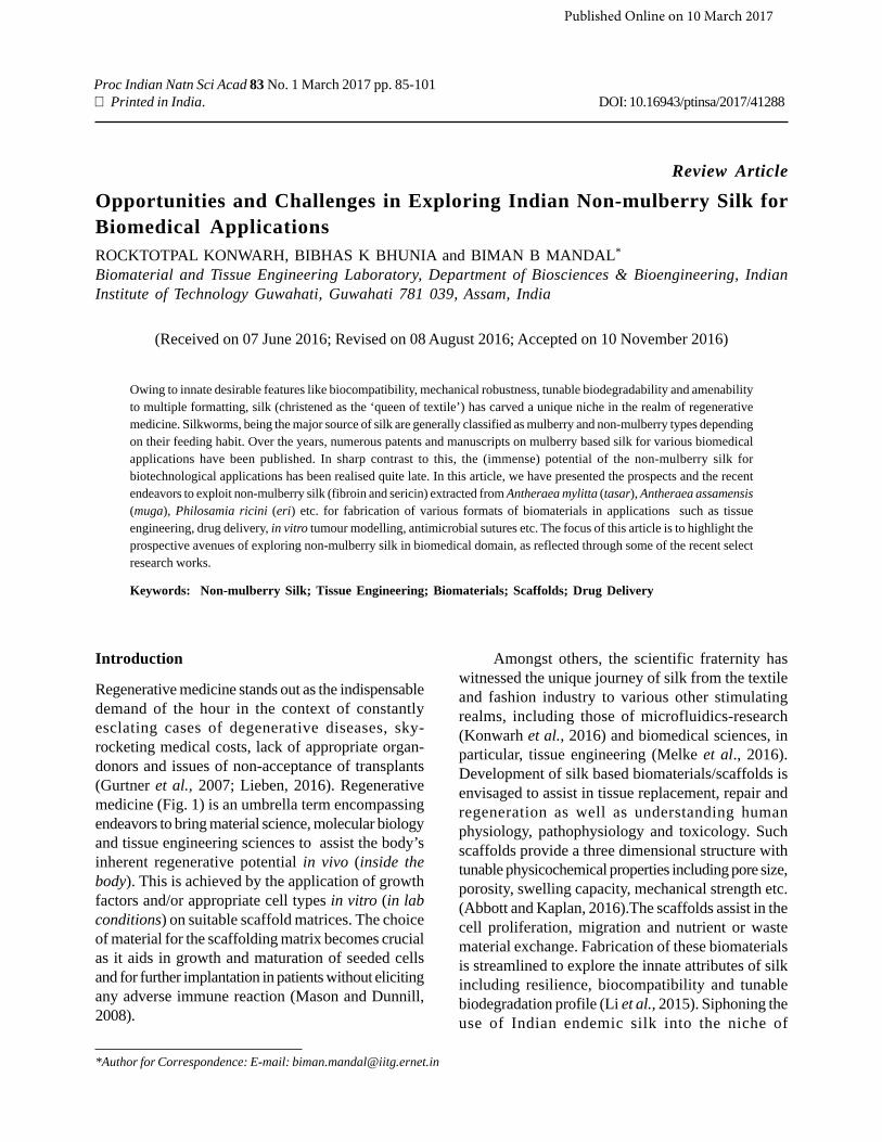
Regenerative medicine stands out as the indispensabledemand of the hour in the context of constantlyesclating cases of degenerative diseases, sky-rocketing medical costs, lack of appropriate organ-donors and issues of non-acceptance of transplants(Gurtner et al., 2007; Lieben, 2016). Regenerativemedicine (Fig. 1) is an umbrella term encompassingendeavors to bring material science, molecular biologyand tissue engineering sciences to assist the body’sinherent regenerative potential in vivo (inside thebody). This is achieved by the application of growthfactors and/or appropriate cell types in vitro (in labconditions) on suitable scaffold matrices. The choiceof material for the scaffolding matrix becomes crucialas it aids in growth and maturation of seeded cellsand for further implantation in patients without elicitingany adverse immune reaction (Mason and Dunnill,2008).
Amongst others, the scientific fraternity haswitnessed the unique journey of silk from the textileand fashion industry to various other stimulatingrealms, including those of microfluidics-research(Konwarh et al., 2016) and biomedical sciences, inparticular, tissue engineering (Melke et al., 2016).Development of silk based biomaterials/scaffolds isenvisaged to assist in tissue replacement, repair andregeneration as well as understanding humanphysiology, pathophysiology and toxicology. Suchscaffolds provide a three dimensional structure withtunable physicochemical properties including pore size,porosity, swelling capacity, mechanical strength etc.(Abbott and Kaplan, 2016).The scaffolds assist in thecell proliferation, migration and nutrient or wastematerial exchange. Fabrication of these biomaterialsis streamlined to explore the innate attributes of silkincluding resilience, biocompatibility and tunablebiodegradation profile (Li et al., 2015). Siphoning theuse of Indian endemic silk into the niche of
Published Online on 10 March 2017
86 Rocktotpal Konwarh et al.
regenerative medicine is expected to open up avenuesto address some of the pressing biomedical issues.
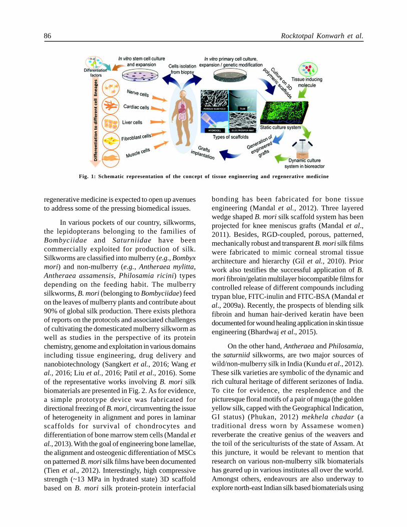
In various pockets of our country, silkworms,the lepidopterans belonging to the families ofBombyciidae and Saturniidae have beencommercially exploited for production of silk.Silkworms are classified into mulberry (e.g., Bombyxmori) and non-mulberry (e.g., Antheraea mylitta,Antheraea assamensis, Philosamia ricini) typesdepending on the feeding habit. The mulberrysilkworms, B. mori (belonging to Bombyciidae) feedon the leaves of mulberry plants and contribute about90% of global silk production. There exists plethoraof reports on the protocols and associated challengesof cultivating the domesticated mulberry silkworm aswell as studies in the perspective of its proteinchemistry, genome and exploitation in various domainsincluding tissue engineering, drug delivery andnanobiotechnology (Sangkert et al., 2016; Wang etal., 2016; Liu et al., 2016; Patil et al., 2016). Someof the representative works involving B. mori silkbiomaterials are presented in Fig. 2. As for evidence,a simple prototype device was fabricated fordirectional freezing of B. mori, circumventing the issueof heterogeneity in alignment and pores in laminarscaffolds for survival of chondrocytes anddifferentiation of bone marrow stem cells (Mandal etal., 2013). With the goal of engineering bone lamellae,the alignment and osteogenic differentiation of MSCson patterned B. mori silk films have been documented(Tien et al., 2012). Interestingly, high compressivestrength (~13 MPa in hydrated state) 3D scaffoldbased on B. mori silk protein-protein interfacial
bonding has been fabricated for bone tissueengineering (Mandal et al., 2012). Three layeredwedge shaped B. mori silk scaffold system has beenprojected for knee meniscus grafts (Mandal et al.,2011). Besides, RGD-coupled, porous, patterned,mechanically robust and transparent B. mori silk filmswere fabricated to mimic corneal stromal tissuearchitecture and hierarchy (Gil et al., 2010). Priorwork also testifies the successful application of B.mori fibroin/gelatin multilayer biocompatible films forcontrolled release of different compounds includingtrypan blue, FITC-inulin and FITC-BSA (Mandal etal., 2009a). Recently, the prospects of blending silkfibroin and human hair-derived keratin have beendocumented for wound healing application in skin tissueengineering (Bhardwaj et al., 2015).
On the other hand, Antheraea and Philosamia,the saturniid silkworms, are two major sources ofwild/non-mulberry silk in India (Kundu et al., 2012).These silk varieties are symbolic of the dynamic andrich cultural heritage of different serizones of India.To cite for evidence, the resplendence and thepicturesque floral motifs of a pair of muga (the goldenyellow silk, capped with the Geographical Indication,GI status) (Phukan, 2012) mekhela chadar (atraditional dress worn by Assamese women)reverberate the creative genius of the weavers andthe toil of the sericulturists of the state of Assam. Atthis juncture, it would be relevant to mention thatresearch on various non-mulberry silk biomaterialshas geared up in various institutes all over the world.Amongst others, endeavours are also underway toexplore north-east Indian silk based biomaterials using
Fig. 1: Schematic representation of the concept of tissue engineering and regenerative medicine
Indian Non-mulberry Silk for Biomedical Applications 87
innovative approaches like electrospinning and othersfor regeneration of a plethora of tissues. Thesebiomaterials may not only be used for tissue
engineering but also as 3D disease tissue models anddelivery of cells and drugs.
Fig. 2: [A] Silk scaffolds for functional meniscus tissue engineering; (a-c) scanning electron micrograph of fabricated threelayers, (a) top layer, (b) middle layer and (c) bottom layer, (d-f) Confocal images of hMSCs on individual scaffold layersin chondrogenic medium after 14 days, (d) top layer, (e) middle layer and (f) bottom layer, (g-i) saffranin O staining forsGAG secreted in scaffolds after 28 days (g) top layer, (h) middle layer and (i) top layer. Reproduced with permissionfr om (Mandal et al., 2011) ©2011 Elsevier. [B] Multi-layer ed silk films for corneal tissue engineering; (a, d) stacked filmswith cells for corneal construct and H&E staining after 1 week, (b, c) immunostaining for collagen type I of (b) withoutRGD pattern film and (c) RGD modified pattern film, (e, f) immunostaining for decorin of (e) pattern film without RGDand (f) RGD modified pattern film. Reproduced with permission from (Gil et al., 2010) ©2010 Elsevier. [C] High str engthsilk scaffold for bone tissue engineering; (a) fiber reinforced high strength scaffolds, (b-d) scanning electron microscopyimages of different length fiber reinforced scaffolds, (b) small fiber, (c) middle length fiber and (d) large fiber, (e)harvesting of implanted scaffold, (f-h) histological analysis for in vivo responses of transplanted scaffolds after 4 weeks,(f) small fiber (g) middle length fiber and (h) large fiber. Figure adopted from (Mandal et al., 2012)
88 Rocktotpal Konwarh et al.
Therefore, we have tried to compile the recentscientific endeavors on the exploration of Indian non-mulberry silk in the biomedical domain in the presentarticle. We start off with a short note on the non-mulberry silk worms and their life cycle. Then, asection is dedicated to the discussion of the majorproteins and signature-sequences, relevant tobiomedical engineering while keeping the prime focuson the plethora of biomedical applications and theassociated intricacies along with possible solutions in
exploring non-mulberry silk.
A note on non-mulberry silk worms
Diversity
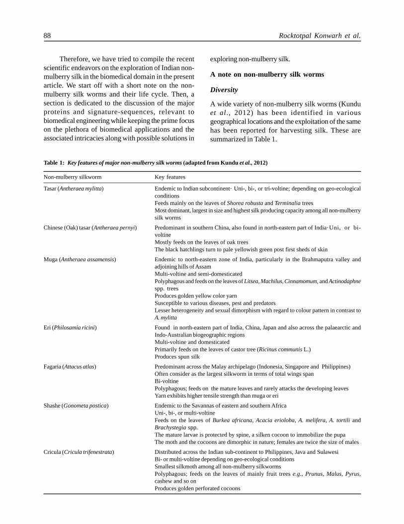
A wide variety of non-mulberry silk worms (Kunduet al., 2012) has been identified in variousgeographical locations and the exploitation of the samehas been reported for harvesting silk. These aresummarized in Table 1.
Table 1: Key features of major non-mulberry silk worms (adapted from Kundu et al., 2012)
Non-mulberry silkworm Key features
Tasar (Antheraea mylitta) Endemic to Indian subcontinent·Uni-, bi-, or tri-voltine; depending on geo-ecologicalconditionsFeeds mainly on the leaves of Shorea robusta and Terminalia treesMost dominant, largest in size and highest silk producing capacity among all non-mulberrysilk worms
Chinese (Oak) tasar (Antheraea pernyi) Predominant in southern China, also found in north-eastern part of India·Uni, or bi-voltineMostly feeds on the leaves of oak treesThe black hatchlings turn to pale yellowish green post first sheds of skin
Muga (Antheraea assamensis) Endemic to north-eastern zone of India, particularly in the Brahmaputra valley andadjoining hills of AssamMulti-voltine and semi-domesticatedPolyphagous and feeds on the leaves of Litsea, Machilus, Cinnamomum, and Actinodaphnespp. treesProduces golden yellow color yarnSusceptible to various diseases, pest and predatorsLesser heterogeneity and sexual dimorphism with regard to colour pattern in contrast toA. mylitta
Eri (Philosamia ricini) Found in north-eastern part of India, China, Japan and also across the palaearctic andIndo-Australian biogeographic regionsMulti-voltine and domesticatedPrimarily feeds on the leaves of castor tree (Ricinus communis L.)Produces spun silk
Fagaria (Attacus atlas) Predominant across the Malay archipelago (Indonesia, Singapore and Philippines)Often consider as the largest silkworm in terms of total wings spanBi-voltinePolyphagous; feeds on the mature leaves and rarely attacks the developing leavesYarn exhibits higher tensile strength than muga or eri
Shashe (Gonometa postica) Endemic to the Savannas of eastern and southern AfricaUni-, bi-, or multi-voltineFeeds on the leaves of Burkea africana, Acacia erioloba, A. melifera, A. tortili andBrachystegia spp.The mature larvae is protected by spine, a silken cocoon to immobilize the pupaThe moth and the cocoons are dimorphic in nature; females are twice the size of males
Cricula (Cricula trifenestrata) Distributed across the Indian sub-continent to Philippines, Java and SulawesiBi- or multi-voltine depending on geo-ecological conditionsSmallest silkmoth among all non-mulberry silkwormsPolyphagous; feeds on the leaves of mainly fruit trees e.g., Prunus, Malus, Pyrus,cashew and so onProduces golden perforated cocoons
Indian Non-mulberry Silk for Biomedical Applications 89
Life Cycle of Non-mulberry Silk Worm
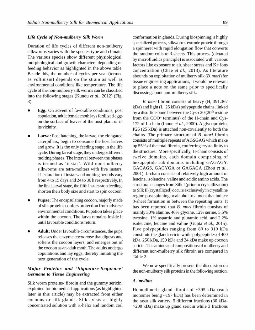
Duration of life cycles of different non-mulberrysilkworms varies with the species-type and climate.The various species show different physiological,morphological and growth characters depending onfeeding behavior as highlighted in the above table.Beside this, the number of cycles per year (termedas voltinism) depends on the strain as well asenvironmental conditions like temperature. The lifecycle of the non-mulberry silk worms can be classifiedinto the following stages (Kundu et al., 2012) (Fig.3).
l Egg: On advent of favorable conditions, postcopulation, adult female moth lays fertilized eggson the surface of leaves of the host plant or inits vicinity.
l Larva: Post hatching, the larvae, the elongatedcaterpillars, begin to consume the host leavesand grow. It is the only feeding stage in the lifecycle. During larval stage, they undergo differentmolting phases. The interval between the phasesis termed as ‘instar’. Wild non-mulberrysilkworms are tetra-molters with five instars.The duration of instars and molting periods varyfrom 4 to 15 days and 24 to 36 h respectively. Inthe final larval stage, the fifth instars stop feeding,shorten their body size and start to spin cocoon.
l Pupae: The encapsulating cocoon, majorly madeof silk proteins confers protection from adverseenvironmental conditions. Pupation takes placewithin the cocoon. The larva remains inside ituntil favorable conditions return.
l Adult: Under favorable circumstances, the pupareleases the enzyme cocoonase that digests andsoftens the cocoon layers, and emerges out ofthe cocoon as an adult moth. The adults undergocopulations and lay eggs, thereby initiating thenext generation of the cycle
Major Proteins and ‘Signature-Sequence’Germane to Tissue Engineering
Silk worm proteins- fibroin and the gummy sericin,exploited for biomedical applications (as highlightedlater in this article) may be extracted from eithercocoons or silk glands. Silk exists as highlyconcentrated solution with α-helix and random coil
conformation in glands. During biospinning, a highlyspecialized process, silkworms extrude protein througha spinneret with rapid elongation flow that convertsthe random coils to β-sheets. This process (dictatedby microfluidics principle) is associated with variousfactors like exposure to air, shear stress and K+ ionsconcentration (Chae et al., 2013). As literatureabounds on exploitation of mulberry silk (B. mori) fortissue engineering applications, it would be relevantto place a note on the same prior to specificallydiscussing about non-mulberry silk.
B. mori fibroin consists of heavy (H, 391.367kDa) and light (L, 25 kDa) polypeptide chains, linkedby a disulfide bond between the Cys-c20 (20th residuefrom the COO– terminus) of the H-chain and Cys-172 of L-chain (Inoue et al., 2000). A glycoprotein,P25 (25 kDa) is attached non-covalently to both thechains. The primary structure of B. mori fibroinconsists of multiple repeats of AGSGAG which makeup 55% of the total fibroin, conferring crystallinity tothe structure. More specifically, H-chain consists oftwelve domains, each domain comprising ofhexapeptide sub-domains including GAGAGY,GAGAGS, GAGYGA or GAGAGA (Zhou et al.,2001). L-chain consists of relatively high amount ofleucine, isoleucine, valine and acidic amino acids. Thestructural changes from Silk I (prior to crystallization)to Silk II (crystallized) occurs exclusively in crystallineregion post spinning or alcohol treatment that induceβ-sheet formation in between the repeating units. Ithas been reported that B. mori fibroin consists ofmainly 30% alanine, 46% glycine, 12% serine, 5.5%tyrosine, 1% aspartic and glutamic acid, and 2.2%isoleucine, leucine and valine (Gupta et al., 2015).Five polypeptides ranging from 80 to 310 kDaconstitute the gland sericin while polypeptides of 400kDa, 250 kDa, 150 kDa and 24 kDa make up cocoonsericin. The amino acid compositions of mulberry anddifferent non-mulberry silk fibroin are compared inTable 2.
We now specifically present the discussion onthe non-mulberry silk proteins in the following section.
A. mylitta
Homodimeric gland fibroin of ~395 kDa (eachmonomer being ~197 kDa) has been determined inthe tasar silk variety. 5 different fractions (30 kDa->200 kDa) make up gland sericin while 3 fractions
90 Rocktotpal Konwarh et al.
(70 kDa, 200 kDa and >200 kDa) were found toconstitute the cocoon sericin (Kundu et al., 2012).O-linked glycosylation is observed in A. mylitta silkfibroin (Datta et al., 2001). The O-linkage conformsto a core 1 mucin-type oligosaccharide structure whichconsists of at least eight monosaccharide residueslinked to the protein backbone by N-acetylgalactosamine moiety.
P. ricini
P. ricini fibroin protein is constituted of 97 kDa (withcompositional abundance of glutamic acid) and 45 kDapolypeptides connected through a disulfide bond while
Fig. 3: Typical life cycle/different stages of non-mulberry Indian tr opical tasar silkworm, Antheraea mylitta as an example.Figure adapted from (Kundu et al., 2012) ©2012 Wiley Periodicals, Inc.
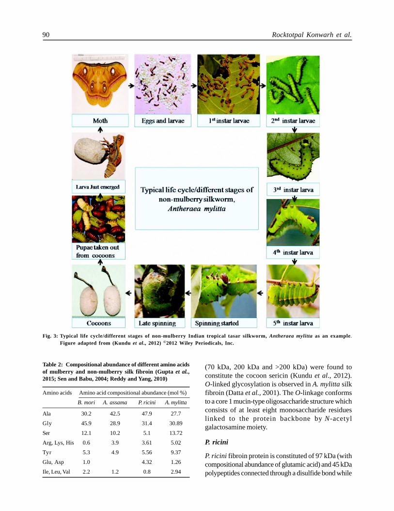
Table 2: Compositional abundance of different amino acidsof mulberry and non-mulberry silk fibroin (Gupta et al.,2015; Sen and Babu, 2004; Reddy and Yang, 2010)
Amino acids Amino acid compositional abundance (mol %)
B. mori A. assama P. ricini A. mylitta
Ala 30.2 42.5 47.9 27.7
Gly 45.9 28.9 31.4 30.89
Ser 12.1 10.2 5.1 13.72
Arg, Lys, His 0.6 3.9 3.61 5.02
Tyr 5.3 4.9 5.56 9.37
Glu, Asp 1.0 4.32 1.26
Ile, Leu, Val 2.2 1.2 0.8 2.94
Indian Non-mulberry Silk for Biomedical Applications 91
the cocoon- sericin comprises of a 66 kDa molecule(Ahmed et al., 2004). The basic repeating unit of P.ricini silk fibroin (SF) is (alanine)12-13 which is similarto spider (major ampullate) silk that possesses(alanine)5-6 as repeating unit.
A. assamensis
Two fractions of 220 kDa and 20 kDa have beenreported for A. assamensis fibroin while a singlefraction of 66 kDa constitute the sericin (Kundu etal., 2012).The primary structure of A. assamensisSF comprises of 42.5% alanine, 28.9% glycine, 10.2%serine and 5.5% tyrosine (Gupta et al., 2015). Thesecondary structure of the muga silk fibroin possessesthree main motifs: A-motif, consisting of alanineresidues; G-motif, primarily composed of glycineresidues; and R-motif that contains arginine residues.The A-motif contains poly-alanine stretches of(alanine)5-15 and is responsible for the crystallinity ofthe protein. One of the unique features of polyalaninestretches of A. assamensis SF is the absence of anyinterspersing amino acid unlike their counterparts with3-4% serine residues in other members of thesaturniid family.
Various amino acid ratios of different non-mulberry SF have been presented in Table 3. In arecent endeavor to correlate the physical propertiesand sequence in silkworm fibroin (Malay et al., 2016),strictly-defined structural transitions of the non-mulberry silk fibroin in the yielding and strain hardeningevents (tensile deformation and thermal assessments)were worthwhile to note in comparison to that of B.mori. It was predicted that modular repeats of thesaturniid sequences tend to produce structures thatrespond in a concerted fashion in contrast toheterogeneous structures of the mulberry counterpart.
The presence of intrinsic cell binding RGDsequences within non-mulberry silk fibroin (NMSF)
confers special niche to the resilient, biocompatibleand biodegradable biomaterials based on the latter inthe domain of tissue engineering (Bellis, 2011). Wehave previously shown desirable cell response in termsof good cytoskeleton organization, actin development,cell spreading, and strong binding to substratumusing A. mylitta fibroin protein films having RGDsequences in comparison to B. mori counterpart(Mandal et al., 2010). Furthermore, mechanicalrobustness of the NMSF reported in terms of its highertensile strength than mulberry SF has been bracketedtogether with the lesser heterogeneity and orderlyarrangement of non-polyalanine repeats in highernumber, tighter poly-alanine β-crystals that are devoidof non-alanine residues and lesser amount of polarresidues in the protein main chain (Datta et al., 2001).
Dissolution and Extraction of Non-mulberry SilkFibroin
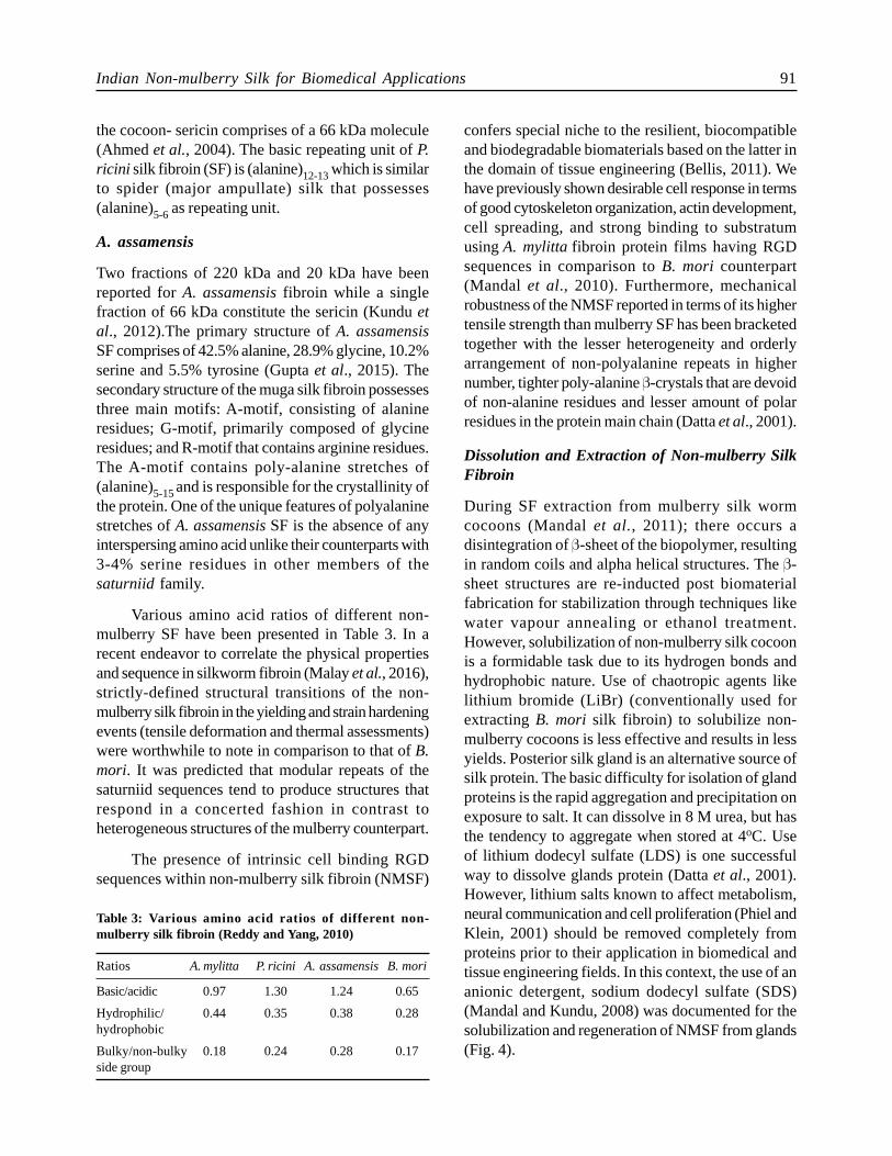
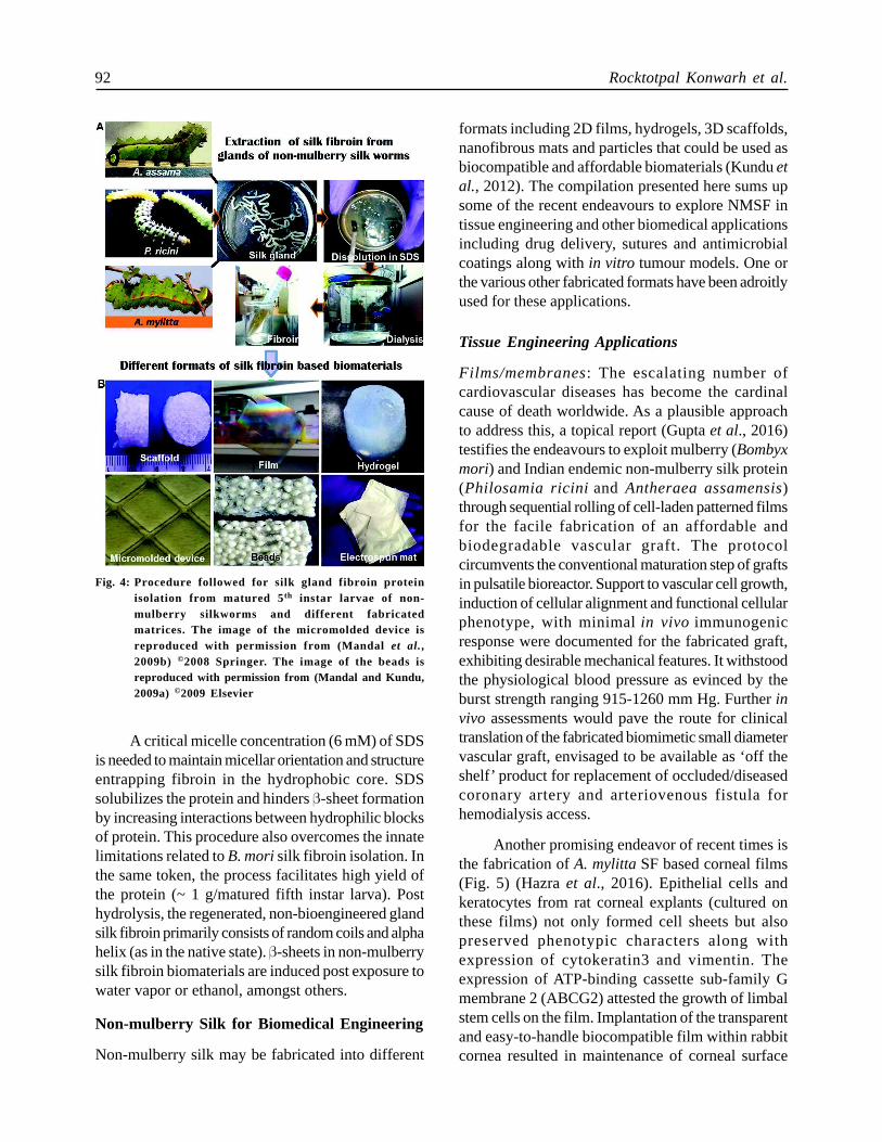
During SF extraction from mulberry silk wormcocoons (Mandal et al., 2011); there occurs adisintegration of β-sheet of the biopolymer, resultingin random coils and alpha helical structures. The β-sheet structures are re-inducted post biomaterialfabrication for stabilization through techniques likewater vapour annealing or ethanol treatment.However, solubilization of non-mulberry silk cocoonis a formidable task due to its hydrogen bonds andhydrophobic nature. Use of chaotropic agents likelithium bromide (LiBr) (conventionally used forextracting B. mori silk fibroin) to solubilize non-mulberry cocoons is less effective and results in lessyields. Posterior silk gland is an alternative source ofsilk protein. The basic difficulty for isolation of glandproteins is the rapid aggregation and precipitation onexposure to salt. It can dissolve in 8 M urea, but hasthe tendency to aggregate when stored at 4oC. Useof lithium dodecyl sulfate (LDS) is one successfulway to dissolve glands protein (Datta et al., 2001).However, lithium salts known to affect metabolism,neural communication and cell proliferation (Phiel andKlein, 2001) should be removed completely fromproteins prior to their application in biomedical andtissue engineering fields. In this context, the use of ananionic detergent, sodium dodecyl sulfate (SDS)(Mandal and Kundu, 2008) was documented for thesolubilization and regeneration of NMSF from glands(Fig. 4).
Table 3: Various amino acid ratios of different non-mulberr y silk fibr oin (Reddy and Yang, 2010)
Ratios A. mylitta P. ricini A. assamensisB. mori
Basic/acidic 0.97 1.30 1.24 0.65
Hydrophilic/ 0.44 0.35 0.38 0.28hydrophobic
Bulky/non-bulky 0.18 0.24 0.28 0.17side group
92 Rocktotpal Konwarh et al.
A critical micelle concentration (6 mM) of SDSis needed to maintain micellar orientation and structureentrapping fibroin in the hydrophobic core. SDSsolubilizes the protein and hinders β-sheet formationby increasing interactions between hydrophilic blocksof protein. This procedure also overcomes the innatelimitations related to B. mori silk fibroin isolation. Inthe same token, the process facilitates high yield ofthe protein (~ 1 g/matured fifth instar larva). Posthydrolysis, the regenerated, non-bioengineered glandsilk fibroin primarily consists of random coils and alphahelix (as in the native state). β-sheets in non-mulberrysilk fibroin biomaterials are induced post exposure towater vapor or ethanol, amongst others.
Non-mulberry Silk for Biomedical Engineering
Non-mulberry silk may be fabricated into different
formats including 2D films, hydrogels, 3D scaffolds,nanofibrous mats and particles that could be used asbiocompatible and affordable biomaterials (Kundu etal., 2012). The compilation presented here sums upsome of the recent endeavours to explore NMSF intissue engineering and other biomedical applicationsincluding drug delivery, sutures and antimicrobialcoatings along with in vitro tumour models. One orthe various other fabricated formats have been adroitlyused for these applications.
Tissue Engineering Applications
Films/membranes: The escalating number ofcardiovascular diseases has become the cardinalcause of death worldwide. As a plausible approachto address this, a topical report (Gupta et al., 2016)testifies the endeavours to exploit mulberry (Bombyxmori) and Indian endemic non-mulberry silk protein(Philosamia ricini and Antheraea assamensis)through sequential rolling of cell-laden patterned filmsfor the facile fabrication of an affordable andbiodegradable vascular graft. The protocolcircumvents the conventional maturation step of graftsin pulsatile bioreactor. Support to vascular cell growth,induction of cellular alignment and functional cellularphenotype, with minimal in vivo immunogenicresponse were documented for the fabricated graft,exhibiting desirable mechanical features. It withstoodthe physiological blood pressure as evinced by theburst strength ranging 915-1260 mm Hg. Further invivo assessments would pave the route for clinicaltranslation of the fabricated biomimetic small diametervascular graft, envisaged to be available as ‘off theshelf’ product for replacement of occluded/diseasedcoronary artery and arteriovenous fistula forhemodialysis access.
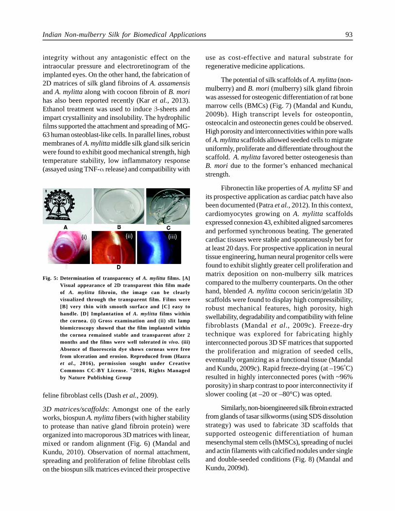
Another promising endeavor of recent times isthe fabrication of A. mylitta SF based corneal films(Fig. 5) (Hazra et al., 2016). Epithelial cells andkeratocytes from rat corneal explants (cultured onthese films) not only formed cell sheets but alsopreserved phenotypic characters along withexpression of cytokeratin3 and vimentin. Theexpression of ATP-binding cassette sub-family Gmembrane 2 (ABCG2) attested the growth of limbalstem cells on the film. Implantation of the transparentand easy-to-handle biocompatible film within rabbitcornea resulted in maintenance of corneal surface
Fig. 4: Procedure followed for silk gland fibroin proteinisolation from matured 5th instar larvae of non-mulberry silkworms and different fabricatedmatrices. The image of the micromolded device isreproduced with permission from (Mandal et al.,2009b) ©2008 Springer. The image of the beads isreproduced with permission from (Mandal and Kundu,2009a) ©2009 Elsevier
Indian Non-mulberry Silk for Biomedical Applications 93
integrity without any antagonistic effect on theintraocular pressure and electroretinogram of theimplanted eyes. On the other hand, the fabrication of2D matrices of silk gland fibroins of A. assamensisand A. mylitta along with cocoon fibroin of B. morihas also been reported recently (Kar et al., 2013).Ethanol treatment was used to induce β-sheets andimpart crystallinity and insolubility. The hydrophilicfilms supported the attachment and spreading of MG-63 human osteoblast-like cells. In parallel lines, robustmembranes of A. mylitta middle silk gland silk sericinwere found to exhibit good mechanical strength, hightemperature stability, low inflammatory response(assayed using TNF-α release) and compatibility with
use as cost-effective and natural substrate forregenerative medicine applications.
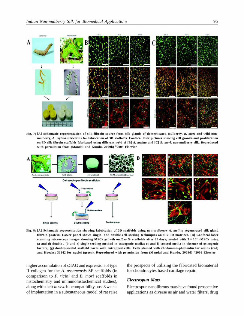
The potential of silk scaffolds of A. mylitta (non-mulberry) and B. mori (mulberry) silk gland fibroinwas assessed for osteogenic differentiation of rat bonemarrow cells (BMCs) (Fig. 7) (Mandal and Kundu,2009b). High transcript levels for osteopontin,osteocalcin and osteonectin genes could be observed.High porosity and interconnectivities within pore wallsof A. mylitta scaffolds allowed seeded cells to migrateuniformly, proliferate and differentiate throughout thescaffold. A. mylitta favored better osteogenesis thanB. mori due to the former’s enhanced mechanicalstrength.
Fibronectin like properties of A. mylitta SF andits prospective application as cardiac patch have alsobeen documented (Patra et al., 2012). In this context,cardiomyocytes growing on A. mylitta scaffoldsexpressed connexion 43, exhibited aligned sarcomeresand performed synchronous beating. The generatedcardiac tissues were stable and spontaneously bet forat least 20 days. For prospective application in neuraltissue engineering, human neural progenitor cells werefound to exhibit slightly greater cell proliferation andmatrix deposition on non-mulberry silk matricescompared to the mulberry counterparts. On the otherhand, blended A. mylitta cocoon sericin/gelatin 3Dscaffolds were found to display high compressibility,robust mechanical features, high porosity, highswellability, degradability and compatibility with felinefibroblasts (Mandal et al., 2009c). Freeze-drytechnique was explored for fabricating highlyinterconnected porous 3D SF matrices that supportedthe proliferation and migration of seeded cells,eventually organizing as a functional tissue (Mandaland Kundu, 2009c). Rapid freeze-drying (at –196°C)resulted in highly interconnected pores (with ~96%porosity) in sharp contrast to poor interconnectivity ifslower cooling (at –20 or –80°C) was opted.
Similarly, non-bioengineered silk fibroin extractedfrom glands of tasar silkworms (using SDS dissolutionstrategy) was used to fabricate 3D scaffolds thatsupported osteogenic differentiation of humanmesenchymal stem cells (hMSCs), spreading of nucleiand actin filaments with calcified nodules under singleand double-seeded conditions (Fig. 8) (Mandal andKundu, 2009d).
Fig. 5: Determination of transparency of A. mylitta films. [A]Visual appearance of 2D transparent thin film madeof A. mylitta fibroin, the image can be clearlyvisualized through the transparent film. Films were[B] very thin with smooth surface and [C] easy tohandle. [D] Implantation of A. mylitta films withinthe cornea. (i) Gross examination and (ii) slit lampbiomicroscopy showed that the film implanted withinthe cornea remained stable and transparent after 2months and the films were well tolerated in vivo. (iii)Absence of fluorescein dye shows corneas were freefrom ulceration and erosion. Reproduced from (Hazraet al., 2016), permission sought under CreativeCommons CC-BY License. ©2016, Rights Managedby Nature Publishing Group
feline fibroblast cells (Dash et al., 2009).
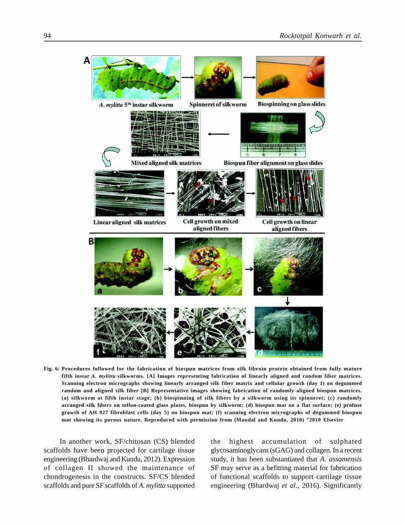
3D matrices/scaffolds: Amongst one of the earlyworks, biospun A. mylitta fibers (with higher stabilityto protease than native gland fibroin protein) wereorganized into macroporous 3D matrices with linear,mixed or random alignment (Fig. 6) (Mandal andKundu, 2010). Observation of normal attachment,spreading and proliferation of feline fibroblast cellson the biospun silk matrices evinced their prospective
94 Rocktotpal Konwarh et al.
In another work, SF/chitosan (CS) blendedscaffolds have been projected for cartilage tissueengineering (Bhardwaj and Kundu, 2012). Expressionof collagen II showed the maintenance ofchondrogenesis in the constructs. SF/CS blendedscaffolds and pure SF scaffolds of A. mylitta supported
the highest accumulation of sulphatedglycosaminoglycans (sGAG) and collagen. In a recentstudy, it has been substantiated that A. assamensisSF may serve as a befitting material for fabricationof functional scaffolds to support cartilage tissueengineering (Bhardwaj et al., 2016). Significantly
Fig. 6: Procedures followed for the fabrication of biospun matrices from silk fibroin protein obtained from fully maturefifth instar A. mylitta silkworms. [A] Images representing fabrication of linearly aligned and random fiber matrices.Scanning electron micrographs showing linearly arranged silk fiber matrix and cellular growth (day 1) on degummedrandom and aligned silk fiber [B] Representative images showing fabrication of randomly aligned biospun matrices.(a) silkworm at fifth instar stage; (b) biospinning of silk fibers by a silkworm using its spinneret; (c) randomlyarranged silk fibers on teflon-coated glass plates, biospun by silkworm; (d) biospun mat on a flat surface; (e) profusegrowth of AH 927 fibroblast cells (day 5) on biospun mat; (f) scanning electron micrographs of degummed biospunmat showing its porous nature. Reproduced with permission from (Mandal and Kundu, 2010) ©2010 Elsevier
Indian Non-mulberry Silk for Biomedical Applications 95
higher accumulation of sGAG and expression of typeII collagen for the A. assamensis SF scaffolds (incomparison to P. ricini and B. mori scaffolds inhistochemistry and immunohistochemical studies),along with their in vivo biocompatibility post 8 weeksof implantation in a subcutaneous model of rat raise
the prospects of utilizing the fabricated biomaterialfor chondrocytes based cartilage repair.
Electrospun Mats
Electrospun nanofibrous mats have found prospectiveapplications as diverse as air and water filters, drug
Fig. 7: [A] Schematic representation of silk fibroin source from silk glands of domesticated mulberry, B. mori and wild non-mulberr y, A. mylitta silkworms for fabrication of 3D scaffolds. Confocal laser pictures showing cell growth and proliferationon 3D silk fibroin scaffolds fabricated using different wt% of [B] A. mylitta and [C] B. mori, non-mulberry silk. Reproducedwith permission from (Mandal and Kundu, 2009b) ©2009 Elsevier
Fig. 8: [A] Schematic representation showing fabrication of 3D scaffolds using non-mulberry A. mylitta regenerated silk glandfibroin protein. Lower panel shows single- and double-cell-seeding techniques on silk 3D matrices. [B] Confocal laserscanning microscope images showing MSCs growth on 2 wt% scaffolds after 28 days; seeded with 3 × 10⁵ hMSCs using(a and d) double-, (b and e) single-seeding method in osteogenic media; (c and f) control media in absence of osteogenicfactors; (g) double-seeded scaffold pores with entrapped cells. Cells stained with rhodamine–phalloidin for actins (red)and Hoechst 33342 for nuclei (green). Reproduced with permission from (Mandal and Kundu, 2009d) ©2009 Elsevier
96 Rocktotpal Konwarh et al.
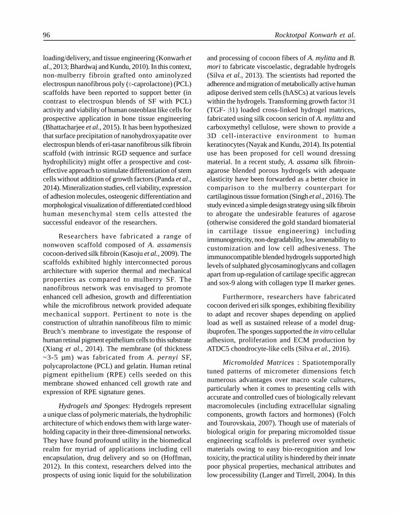
loading/delivery, and tissue engineering (Konwarh etal., 2013; Bhardwaj and Kundu, 2010). In this context,non-mulberry fibroin grafted onto aminolyzedelectrospun nanofibrous poly (ε-caprolactone) (PCL)scaffolds have been reported to support better (incontrast to electrospun blends of SF with PCL)activity and viability of human osteoblast like cells forprospective application in bone tissue engineering(Bhattacharjee et al., 2015). It has been hypothesizedthat surface precipitation of nanohydroxyapatite overelectrospun blends of eri-tasar nanofibrous silk fibroinscaffold (with intrinsic RGD sequence and surfacehydrophilicity) might offer a prospective and cost-effective approach to stimulate differentiation of stemcells without addition of growth factors (Panda et al.,2014). Mineralization studies, cell viability, expressionof adhesion molecules, osteogenic differentiation andmorphological visualization of differentiated cord bloodhuman mesenchymal stem cells attested thesuccessful endeavor of the researchers.
Researchers have fabricated a range ofnonwoven scaffold composed of A. assamensiscocoon-derived silk fibroin (Kasoju et al., 2009). Thescaffolds exhibited highly interconnected porousarchitecture with superior thermal and mechanicalproperties as compared to mulberry SF. Thenanofibrous network was envisaged to promoteenhanced cell adhesion, growth and differentiationwhile the microfibrous network provided adequatemechanical support. Pertinent to note is theconstruction of ultrathin nanofibrous film to mimicBruch’s membrane to investigate the response ofhuman retinal pigment epithelium cells to this substrate(Xiang et al., 2014). The membrane (of thickness~3-5 µm) was fabricated from A. pernyi SF,polycaprolactone (PCL) and gelatin. Human retinalpigment epithelium (RPE) cells seeded on thismembrane showed enhanced cell growth rate andexpression of RPE signature genes.
Hydrogels and Sponges: Hydrogels representa unique class of polymeric materials, the hydrophilicarchitecture of which endows them with large water-holding capacity in their three-dimensional networks.They have found profound utility in the biomedicalrealm for myriad of applications including cellencapsulation, drug delivery and so on (Hoffman,2012). In this context, researchers delved into theprospects of using ionic liquid for the solubilization
and processing of cocoon fibers of A. mylitta and B.mori to fabricate viscoelastic, degradable hydrogels(Silva et al., 2013). The scientists had reported theadherence and migration of metabolically active humanadipose derived stem cells (hASCs) at various levelswithin the hydrogels. Transforming growth factor β1(TGF- β1) loaded cross-linked hydrogel matrices,fabricated using silk cocoon sericin of A. mylitta andcarboxymethyl cellulose, were shown to provide a3D cell-interactive environment to humankeratinocytes (Nayak and Kundu, 2014). Its potentialuse has been proposed for cell wound dressingmaterial. In a recent study, A. assama silk fibroin-agarose blended porous hydrogels with adequateelasticity have been forwarded as a better choice incomparison to the mulberry counterpart forcartilaginous tissue formation (Singh et al., 2016). Thestudy evinced a simple design strategy using silk fibrointo abrogate the undesirable features of agarose(otherwise considered the gold standard biomaterialin cartilage tissue engineering) includingimmunogenicity, non-degradability, low amenability tocustomization and low cell adhesiveness. Theimmunocompatible blended hydrogels supported highlevels of sulphated glycosaminoglycans and collagenapart from up-regulation of cartilage specific aggrecanand sox-9 along with collagen type II marker genes.
Furthermore, researchers have fabricatedcocoon derived eri silk sponges, exhibiting flexibilityto adapt and recover shapes depending on appliedload as well as sustained release of a model drug-ibuprofen. The sponges supported the in vitro cellularadhesion, proliferation and ECM production byATDC5 chondrocyte-like cells (Silva et al., 2016).
Micromolded Matrices : Spatiotemporallytuned patterns of micrometer dimensions fetchnumerous advantages over macro scale cultures,particularly when it comes to presenting cells withaccurate and controlled cues of biologically relevantmacromolecules (including extracellular signalingcomponents, growth factors and hormones) (Folchand Tourovskaia, 2007). Though use of materials ofbiological origin for preparing micromolded tissueengineering scaffolds is preferred over syntheticmaterials owing to easy bio-recognition and lowtoxicity, the practical utility is hindered by their innatepoor physical properties, mechanical attributes andlow processibility (Langer and Tirrell, 2004). In this
Indian Non-mulberry Silk for Biomedical Applications 97
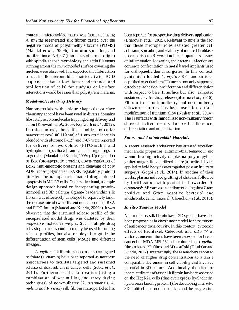
context, a micromolded matrix was fabricated usingA. mylitta regenerated silk fibroin casted over thenegative molds of polydimethylsiloxane (PDMS)(Mandal et al., 2009b). Uniform spreading andproliferation of AH927 (fibroblasts of murine origin)with spindle shaped morphology and actin filamentsrunning across the micromolded surface covering thenucleus were observed. It is expected that fabricationof such silk micromolded matrices (with RGDsequences that allow better adherence andproliferation of cells) for studying cell-surfaceinteractions would be easier than polystyrene material.
Model-molecule/drug Delivery
Nanomaterials with unique shape-size-surfacechemistry accord have been used in diverse domainslike catalysis, biomolecular trapping, drug delivery andso on (Konwarh et al., 2009; Konwarh et al., 2012).In this context, the self-assembled micellarnanostructures (100-110 nm) of A. mylitta silk sericinblended with pluronic F-127 and F-87 were reportedfor delivery of hydrophilic (FITC-inulin) andhydrophobic (paclitaxel, anticancer drug) drugs totarget sites (Mandal and Kundu, 2009e). Up-regulationof Bax (pro-apoptotic protein), down-regulation ofBcl-2 (anti-apoptotic protein) and cleavage of polyADP ribose polymerase (PARP, regulatory protein)attested the nanoparticle loaded drug-inducedapoptosis in MCF-7 cells. On the other hand, a simpledesign approach based on incorporating protein-immobilized 3D calcium alginate beads within silkfibroin was effectively employed to separately tailorthe release rate of two different model proteins- BSAand FITC-Inulin (Mandal and Kundu, 2009a). It wasobserved that the sustained release profile of theencapsulated model drugs was dictated by theirrespective molecular weight. Such multiple drugreleasing matrices could not only be used for tuningrelease profiles, but also employed to guide thedifferentiation of stem cells (MSCs) into differentlineages.
A. mylitta silk fibroin nanoparticles conjugatedto folate (a vitamin) have been reported as nontoxicnanocarriers to facilitate targeted and sustainedrelease of doxorubicin in cancer cells (Subia et al.,2014). Furthermore, the fabrication (using acombination of wet-milling and spray dryingtechniques) of non-mulberry (A. assamensis, A.mylitta and P. ricini) silk fibroin microparticles has
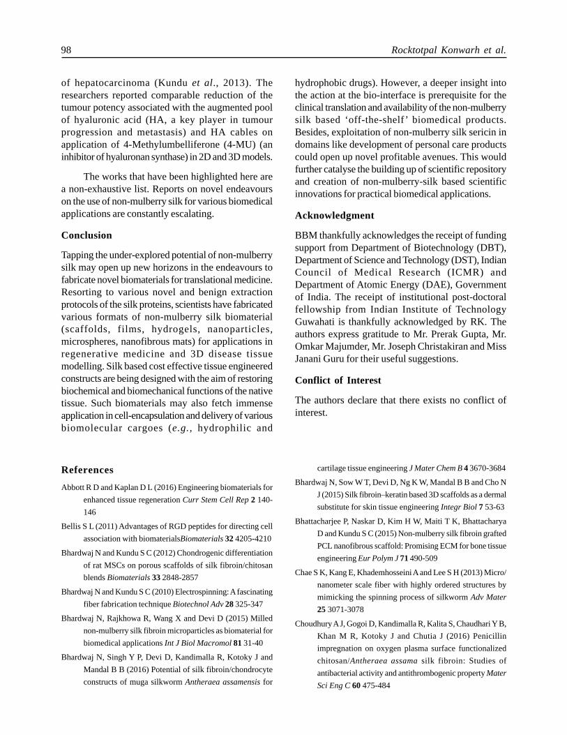
been reported for prospective drug delivery application(Bhardwaj et al., 2015). Relevant to note is the factthat these microparticles assisted greater celladhesion, spreading and viability of mouse fibroblastsin comparison to B. mori fibroin microparticles. Issuesof inflammation, loosening and bacterial infection arecommon confrontation in metal based implants usedfor orthopaedic/dental surgeries. In this context,gentamicin loaded A. mylitta SF nanoparticlesdeposited over titanium (Ti) surface not only supportedosteoblast adhesion, proliferation and differentiationwith respect to bare Ti surface but also exhibitedsustained in vitro drug release (Sharma et al., 2016).Fibroin from both mulberry and non-mulberrysilkworm sources has been used for surfacemodification of titanium alloy (Naskar et al., 2014).The Ti surfaces with immobilized non-mulberry fibroinshowed better results for cell adherence,differentiation and mineralization.
Suture and Antimicrobial Materials
A recent research endeavour has attested excellentmechanical properties, antimicrobial behaviour andwound healing activity of plasma polypropylenegrafted muga silk as sterilized suture (a medical deviceapplied to hold body tissues together post an injury orsurgery) (Gogoi et al., 2014). In another of theirworks, plasma induced grafting of chitosan followedby fortification with penicillin forwarded A.assamensis SF yarn as an antibacterial (against Grampositive and Gram negative bacteria) andantithrombogenic material (Choudhury et al., 2016).
In vitro Tumour Model
Non-mulberry silk fibroin based 3D systems have alsobeen proposed as in vitro tumor model for assessmentof anticancer drug activity. In this context, cytotoxiceffects of Paclitaxel, Celecoxib and ZD6474 atvarious concentrations have been assessed for breastcancer line MDA-MB-231 cells cultured on A. mylittafibroin based 2D films and 3D scaffold (Talukdar andKundu, 2012). Interestingly, the researchers reportedthe need of higher drug concentrations to attain acomparable decrement in cell viability and invasivepotential in 3D culture. Additionally, the effect ofinnate attributes of tasar silk fibroin has been assessedon the HepR21 cells (that overexpress hyaladherin,hyaluronan-binding protein 1) for developing an in vitro3D multicellular model to understand the progression
98 Rocktotpal Konwarh et al.
of hepatocarcinoma (Kundu et al., 2013). Theresearchers reported comparable reduction of thetumour potency associated with the augmented poolof hyaluronic acid (HA, a key player in tumourprogression and metastasis) and HA cables onapplication of 4-Methylumbelliferone (4-MU) (aninhibitor of hyaluronan synthase) in 2D and 3D models.
The works that have been highlighted here area non-exhaustive list. Reports on novel endeavourson the use of non-mulberry silk for various biomedicalapplications are constantly escalating.
Conclusion
Tapping the under-explored potential of non-mulberrysilk may open up new horizons in the endeavours tofabricate novel biomaterials for translational medicine.Resorting to various novel and benign extractionprotocols of the silk proteins, scientists have fabricatedvarious formats of non-mulberry silk biomaterial(scaffolds, films, hydrogels, nanoparticles,microspheres, nanofibrous mats) for applications inregenerative medicine and 3D disease tissuemodelling. Silk based cost effective tissue engineeredconstructs are being designed with the aim of restoringbiochemical and biomechanical functions of the nativetissue. Such biomaterials may also fetch immenseapplication in cell-encapsulation and delivery of variousbiomolecular cargoes (e.g., hydrophilic and
hydrophobic drugs). However, a deeper insight intothe action at the bio-interface is prerequisite for theclinical translation and availability of the non-mulberrysilk based ‘off-the-shelf’ biomedical products.Besides, exploitation of non-mulberry silk sericin indomains like development of personal care productscould open up novel profitable avenues. This wouldfurther catalyse the building up of scientific repositoryand creation of non-mulberry-silk based scientificinnovations for practical biomedical applications.
Acknowledgment
BBM thankfully acknowledges the receipt of fundingsupport from Department of Biotechnology (DBT),Department of Science and Technology (DST), IndianCouncil of Medical Research (ICMR) andDepartment of Atomic Energy (DAE), Governmentof India. The receipt of institutional post-doctoralfellowship from Indian Institute of TechnologyGuwahati is thankfully acknowledged by RK. Theauthors express gratitude to Mr. Prerak Gupta, Mr.Omkar Majumder, Mr. Joseph Christakiran and MissJanani Guru for their useful suggestions.
Conflict of Interest
The authors declare that there exists no conflict ofinterest.
References
Abbott R D and Kaplan D L (2016) Engineering biomaterials for
enhanced tissue regeneration Curr Stem Cell Rep 2 140-
146
Bellis S L (2011) Advantages of RGD peptides for directing cell
association with biomaterialsBiomaterials 32 4205-4210
Bhardwaj N and Kundu S C (2012) Chondrogenic differentiation
of rat MSCs on porous scaffolds of silk fibroin/chitosan
blends Biomaterials 33 2848-2857
Bhardwaj N and Kundu S C (2010) Electrospinning: A fascinating
fiber fabrication technique Biotechnol Adv 28 325-347
Bhardwaj N, Rajkhowa R, Wang X and Devi D (2015) Milled
non-mulberry silk fibroin microparticles as biomaterial for
biomedical applications Int J Biol Macromol 81 31-40
Bhardwaj N, Singh Y P, Devi D, Kandimalla R, Kotoky J and
Mandal B B (2016) Potential of silk fibroin/chondrocyte
constructs of muga silkworm Antheraea assamensis for
cartilage tissue engineering J Mater Chem B 4 3670-3684
Bhardwaj N, Sow W T, Devi D, Ng K W, Mandal B B and Cho N
J (2015) Silk fibroin–keratin based 3D scaffolds as a dermal
substitute for skin tissue engineering Integr Biol 7 53-63
Bhattacharjee P, Naskar D, Kim H W, Maiti T K, Bhattacharya
D and Kundu S C (2015) Non-mulberry silk fibroin grafted
PCL nanofibrous scaffold: Promising ECM for bone tissue
engineering Eur Polym J 71 490-509
Chae S K, Kang E, Khademhosseini A and Lee S H (2013) Micro/
nanometer scale fiber with highly ordered structures by
mimicking the spinning process of silkworm Adv Mater
25 3071-3078
Choudhury A J, Gogoi D, Kandimalla R, Kalita S, Chaudhari Y B,
Khan M R, Kotoky J and Chutia J (2016) Penicillin
impregnation on oxygen plasma surface functionalized
chitosan/Antheraea assama silk fibroin: Studies of
antibacterial activity and antithrombogenic property Mater
Sci Eng C 60 475-484
Indian Non-mulberry Silk for Biomedical Applications 99
Dash B C, Mandal B B and Kundu S C (2009) Silk gland sericin
protein membranes: Fabrication and characterization for
potential biotechnological applications J Biotechnol 144
321-329
Datta A, Ghosh A K and Kundu S C (2001) Purification and
characterization of fibroin from the tropical saturniid
silkworm, Antheraea mylittaInsect Biochem Mol Biol 31
1013-1018
Folch A and Tourovskaia A (2007) Therapeutic Micro/Nano
Technology, Springer
Gil E S, Mandal B B, Park S H, Marchant J K, Omenetto F G and
Kaplan D L (2010) Helicoidal multi-lamellar features of
RGD-functionalized silk biomaterials for corneal tissue
engineering Biomaterials 31 8953-8963
Gogoi D, Choudhury A J, Chutia J, Pal A R, Khan M, Choudhury
M, Pathak P, Das G and Patil D S (2014) Development of
advanced antimicrobial and sterilized plasma
polypropylene grafted muga (Antheraea assama) silk as
suture biomaterial Biopolymers 101 355-365
Gupta A, Mita K, Arunkumar K P and Nagaraju J (2015)
Molecular architecture of silk fibroin of Indian golden
silkmoth, Antheraea assamaSci Rep 5 12706
Gupta P, Kumar M, Bhardwaj N, Kumar J P, Krishnamurthy C
S, Nandi S K and Mandal B B (2016) Mimicking form and
function of native small diameter vascular conduits using
mulberry and non-mulberry patterned silk films ACS Appl
Mater Inter 8 15874-15888
Gurtner G C, Callaghan M J and Longaker M T (2007) Progress
and potential for regenerative medicine Annu Rev Med 58
299-312
Hazra S, Nandi S, Naskar D, Guha R, Chowdhury S, Pradhan N,
Kundu S C and Konar A (2016) Non-mulberry silk fibroin
biomaterial for corneal regeneration Sci Rep 6 21840
Hoffman A S (2012) Hydrogels for biomedical applications Adv
Drug Deliv Rev 64 18-23
Inoue S, Tanaka K, Arisaka F, Kimura S, Ohtomo K and Mizuno
S (2000) Silk fibroin of Bombyx mori is secreted, assembling
a high molecular mass elementary unit consisting of H-
chain, L-chain, and p25, with a 6: 6: 1 molar ratio J Biol
Chem 275 40517-40528
Kar S, Talukdar S, Pal S, Nayak S, Paranjape P and Kundu S
(2013) Silk gland fibroin from indian muga silkworm
Antheraea assama as potential biomaterial Tissue Eng
Regen Med 10 200-210
Kasoju N, Bhonde R R and Bora U (2009) Fabrication of a novel
micro–nano fibrous nonwoven scaffold with Antheraea
assama silk fibroin for use in tissue engineering Mater Lett
63 2466-2469
Konwarh R, Gupta P and Mandal B B (2016) Silk-microfluidics
for advanced biotechnological applications: A progressive
review. Biotechnol Adv 34 845-858
Konwarh R, Karak N, Misra M (2013) Electrospun cellulose
acetate nanofibers: The present status and gamut of
biotechnological applications Biotechnol Adv 31 421-437
Konwarh R, Karak N, Rai S K and Mukherjee A K (2009)
Polymer-assisted iron oxide magnetic nanoparticle
immobilized keratinase Nanotechnol 20 225107
Konwarh R, Pramanik S, Devi K S P, Saikia N, Boruah R, Maiti
T K, Deka R C and Karak N (2012) Lycopene coupled
‘trifoliate’ polyaniline nanofibers as multi-functional
biomaterial J Mater Chem 22 15062-15070
Kundu B, Saha P, Datta K and Kundu S C (2013) A silk fibroin
based hepatocarcinoma model and the assessment of the
drug response in hyaluronan-binding protein 1
overexpressed HepG2 cells Biomaterials 34 9462-9474
Kundu S, Kundu B, Talukdar S, Bano S, Nayak S, Kundu J,
Mandal B B, Bhardwaj N, Botlagunta M and Dash B C
(2012) Non-mulberry silk biopolymers Biopolymers 97
455-467
Langer R and Tirrell D A (2004) Designing materials for biology
and medicine Nature 428 487-492
Li G, Li Y, Chen G, He J, Han Y, Wang X and Kaplan D L (2015)
Silk-based biomaterials in biomedical textiles and fiber-
based implants Adv Healthc Mater 4 1134-1151
Lieben L (2016) Regenerative medicine: The future of 3D printing
of human tissues is taking shape Nat Rev Rheumatol 12
191
Liu W, Wang Y, Yao J, Shao Z and Chen X (2016) Tamoxifen-
loaded silk fibroin electrospun fibers Mater Lett 178 31-34
Malay A D, Kenjiro Y, Watanabe H, Sato R, Ifuku N, Masunaga
H, Hikima T, Guan J, Mandal B B, Damrongsakkul S and
Numata K (2016) Relationships between physical
properties and sequence in silkworm silks Sci Rep 6 27573
Mandal B B and Kundu S C (2008) A novel method for dissolution
and stabilization of non mulberry silk gland protein fibroin
using anionic surfactant sodium dodecyl sulfate Biotechnol
Bioeng 99 1482-1489
Mandal B B and Kundu S C (2010) Biospinning by silkworms:
Silk fiber matrices for tissue engineering applications Acta
Biomater 6 360-371
Mandal B B and Kundu S C (2009a) Calcium alginate beads
embedded in silk fibroin as 3D dual drug releasing scaffolds
Biomaterials 30 5170-5177
Mandal B B and Kundu S C (2009b) Osteogenic and adipogenic
differentiation of rat bone marrow cells on non-mulberry

100 Rocktotpal Konwarh et al.
and mulberry silk gland fibroin 3D scaffolds Biomaterials
30 5019-5030
Mandal B B and Kundu S C (2009c) Cell proliferation and migration
in silk fibroin 3D scaffolds Biomaterials 30 2956-2965
Mandal B B and Kundu S C (2009d) Non-mulberry silk gland
fibroin protein 3D scaffold for enhanced differentiation of
human mesenchymal stem cells into osteocytes Acta
Biomater 5 2579-2590
Mandal B B and Kundu S C (2009e) Self-assembled silk sericin/
poloxamer nanoparticles as nanocarriers of hydrophobic
and hydrophilic drugs for targeted delivery Nanotechnol
20 355101
Mandal B B, Das S, Choudhury K and Kundu S C (2010)
Implication of silk film RGD availability and surface
roughness on cytoskeletal organization and proliferation
of primary rat bone marrow cells Tissue Eng Part A 16
2391-2403
Mandal B B, Mann J K and Kundu S C (2009a) Silk fibroin/
gelatin multilayered films as a model system for controlled
drug release Eur J Pharm Sci 37 160-171
Mandal B B, Das T and Kundu S C (2009b) Non-bioengineered
silk gland fibroin micromolded matrices to study cell-surface
interactions Biomed Microdevices 11 467-476
Mandal B B, Priya A S and Kundu S C (2009c) Novel silk sericin/
gelatin 3D scaffolds and 2D films: Fabrication and
characterization for potential tissue engineering
applications Acta Biomater 5 3007-3020
Mandal B B, Gil E S, Panilaitis B and Kaplan D L (2013) Laminar
silk scaffolds for aligned tissue fabrication Macromol Biosci
13 48-58
Mandal B B, Grinberg A, Seok Gil E, Panilaitis B and Kaplan D
L (2012) High-strength silk protein scaffolds for bone
repair. Proc Nat Acad Sci USA 109 7699-7704
Mandal B B, Park S H, Gil E S and Kaplan D L (2011)
Multilayered silk scaffolds for meniscus tissue engineering
Biomaterials 32 639-651
Mason C and Dunnill P (2008) A brief definition of regenerative
medicine Regen Med 3 1-5
Melke J, Midha S, Ghosh S, Ito K and Hofmann S (2016) Silk
fibroin as biomaterial for bone tissue engineering Acta
Biomater 31 1-16
Naskar D, Nayak S, Dey T and Kundu S C (2014) Non-mulberry
silk fibroin influence osteogenesis and osteoblast-
macrophage cross talk on titanium based surface Sci Rep 4
4745
Nayak S and Kundu S (2014) Sericin–carboxymethyl cellulose
porous matrices as cellular wound dressing material J
Biomed Mater Res Part A 102 1928-1940
Panda N, Bissoyi A, Pramanik K and Biswas A (2014) Directing
osteogenesis of stem cells with hydroxyapatite precipitated
electrospun eri–tasar silk fibroin nanofibrous scaffold J
Biomater Sci, Polym Ed 25 1440-1457
Patil R, Naika H R, Rayar S, Balashanmugam N, Uppar V and
Bhattacharyya A (2016) Green synthesis of gold
nanoparticles: Its effect on cocoon and silk traits of
mulberry silkworm (Bombyx mori L.) Part Sci Technol
DOI: 10.1080/02726351.2016.1154121
Patra C, Talukdar S, Novoyatleva T, Velagala S R, Mühlfeld C,
Kundu B, Kundu S C and Engel FB (2012) Silk protein
fibroin from Antheraea mylitta for cardiac tissue engineering
Biomaterials 33 2673-2680
Phiel C J and Klein P S (2001) Molecular targets of lithium action
Annu Rev Pharmacol Toxicol 41 789-813
Phukan R D (2012) Muga silk industry of Assam in historical
perspectives GJHSS 12 5-8
Reddy N and Yang Y (2010) Structure and properties of ultrafine
silk fibers produced by Theriodopteryx ephemeraeformis
J Mater Sci 45 6617-6622
Sangkert S, Kamonmattayakul S, Chai W L and Meesane J (2016)
A biofunctional-modified silk fibroin scaffold with mimic
reconstructed extracellular matrix of decellularized pulp/
collagen/fibronectin for bone tissue engineering in alveolar
bone resorption Mater Lett 166 30-34
Sen K and Babu K (2004) Studies on Indian silk. I.
Macrocharacterization and analysis of amino acid
composition J Appl Pol Sci 92 1080-1097
Sharma S, Bano S, Ghosh A S, Mandal M, Kim H W, Dey T and
Kundu S C (2016) Silk fibroin nanoparticles support in
vitro sustained antibiotic release and osteogenesis on
titanium surface Nanomed Nanotechnol Biol Med 12 1193-
1204
Silva S S, Oliveira N M, Oliveira M B, Da Costa D P S, Naskar D,
Mano J F, Kundu S C and Reis R L (2016) Fabrication and
characterization of eri silk fibers-based sponges for
biomedical application Acta Biomater 32 178-189
Silva S S, Popa E G, Gomes M E, Oliveira M B, Nayak S, Subia
B, Mano J F, Kundu S C and Reis R L (2013) Silk hydrogels
from non-mulberry and mulberry silkworm cocoons
processed with ionic liquids Acta Biomater 9 8972-8982
Singh Y P, Bhardwaj N and Mandal B B (2016) Potential of
agarose/silk fibroin blended hydrogel for in vitro cartilage
tissue engineering ACS Appl Mater Interfaces 8 21236-
21249
Subia B, Chandra S, Talukdar S and Kundu S C (2014) Folate
Indian Non-mulberry Silk for Biomedical Applications 101
conjugated silk fibroin nanocarriers for targeted drug
delivery Integr Biol 6 203-214
Talukdar S and Kundu S C (2012) A non mulberry silk fibroin
protein based 3D in vitro tumor model for evaluation of
anticancer drug activity Adv Funct Mater 22 4778-4788
Tien L W, Gil E S, Park S H, Mandal B B and Kaplan D L (2012)
Patterned silk film scaffolds for aligned lamellar bone tissue
engineering Macromol Biosci 12 1671-1679
Wang Y, Kong Y, Zhao Y, Feng Q, Wu Y, Tang X, Gu X and Yang
Y (2016) Electrospun, reinforcing network-containing, silk
fibroin-based nerve guidance conduits for peripheral nerve
repair J Biomater Tissue Eng 6 53-60
Xiang P, Wu K C, Zhu Y, Xiang L, Li C, Chen D L, Chen F, Xu G,
Wang A and Li M (2014) A novel Bruch’s membrane-
mimetic electrospun substrate scaffold for human retinal
pigment epithelium cells Biomaterials 35 9777-9788
Zhou C Z, Confalonieri F, Jacquet M, Perasso R, Li Z G and
Janin J (2001) Silk fibroin: Structural implications of a
remarkable amino acid sequence Proteins: Struct Funct
Bioinf 44 119-122.