Embed Size (px)
Citation preview

Ophthalmology Corneal Abrasion
History - Usually clear history of very recent trauma - Foreign Body Sensation - Pain +++ - Lacrimation - Photophobia Fig. 1 Corneal Abrasion Examination - Abrasion stains yellow / green with flourescein - Only use Fluoroscein sodium 2% if suspecting aqueous leak - Absence of corneal opacity or hypopyon Management - Chloramphenicol Ointment QDS for 3-5 days, if not allergic. - Consider Cyclopentolate 1% (lasts 24hrs) to alleviate the ciliary spasm that accompanies any eye pain. - Small abrasions do not need follow up. - Refer larger abrasions to eye clinic. - No evidence to suggest eye patch of benefit so do not need to supply, even after instillation of topical local anaesthetic.

Corneal Foreign Body History - Always clear - !!Beware of high velocity FB, eg. Hammering, Grinding, Strimming. Examination - Always check for evidence of penetrating injury of high velocity FB (May be more than one). Management Fig. 2 Corneal Foreign Body - Proxymetacaine 0.5% many times until the eye is anaesthetized. - Remove FB with a green needle / cotton bud under slit-lamp microscope. - Try to distract patient by asking them to look at something, eg. Your ear. - Chloramphenicol Ointment QDS for 3-5 days - Refer eye clinic if residual rust stain (fig. 3) or large abrasion post removal - Rust ring comes off much more easily after 24 hours.
Fig. 3 Corneal Rust stain

- Viral / Bacterial / Allergic History - Gritty sensation in affected eye - Mucopurulent / watery discharge - ± Lashes stuck together on waking Examination - Conjunctival Hyperaemia - Remember to check for corneal
involvement Fig. 5 Bacterial conjunctivitis - ± Chemosis & Conjunctival papilla in
allergic conjunctivitis
Management 1) Infective Conjunctivitis - Chloramphenicol ointment QDS for
3-5days - Take a swab if eyes are sticky. - GP follow up if not settling. - If the bacteria are sensitive to the
antibiotic, there should be marked improvement in 24-36 hours.
Fig. 6 Chemosis
** If cornea involved (Kerato-conjunctivitis), refer eye clinic following day.
2) Allergic Conjunctivitis - Antihistamine drops and tablets
( Sodium Cromoglycate 2% drop QDS ± Piriton 4mg QDS)
- Refer eye clinic if ? Vernal conjunctivitis with conjunctival papillae and especially if there is central corneal plaque.
Fig. 7 Conjunctival papilla in Vernal
Conjunctivitis

Welding Flash / Arc Eye History - Welding without protective goggles /
Sun bed. - symptoms similar but more severe than
corneal abrasion Management - Proxymetacaine 0.5% - Chloramphenicol ointment QDS 3-5
days - Adequate analgesia eg. Cocodamol - Reassure it will get better in 24 hours. - Tell patient pain will recur when
topical anaesthetics wears off.
Eyelid laceration * Do not suture if through tarsal plate * Refer ophthalmologist for closure * Beware puncture of globe as well
especially if history suggest a stab injury.
Fig. 8 Laceration involving lid margin
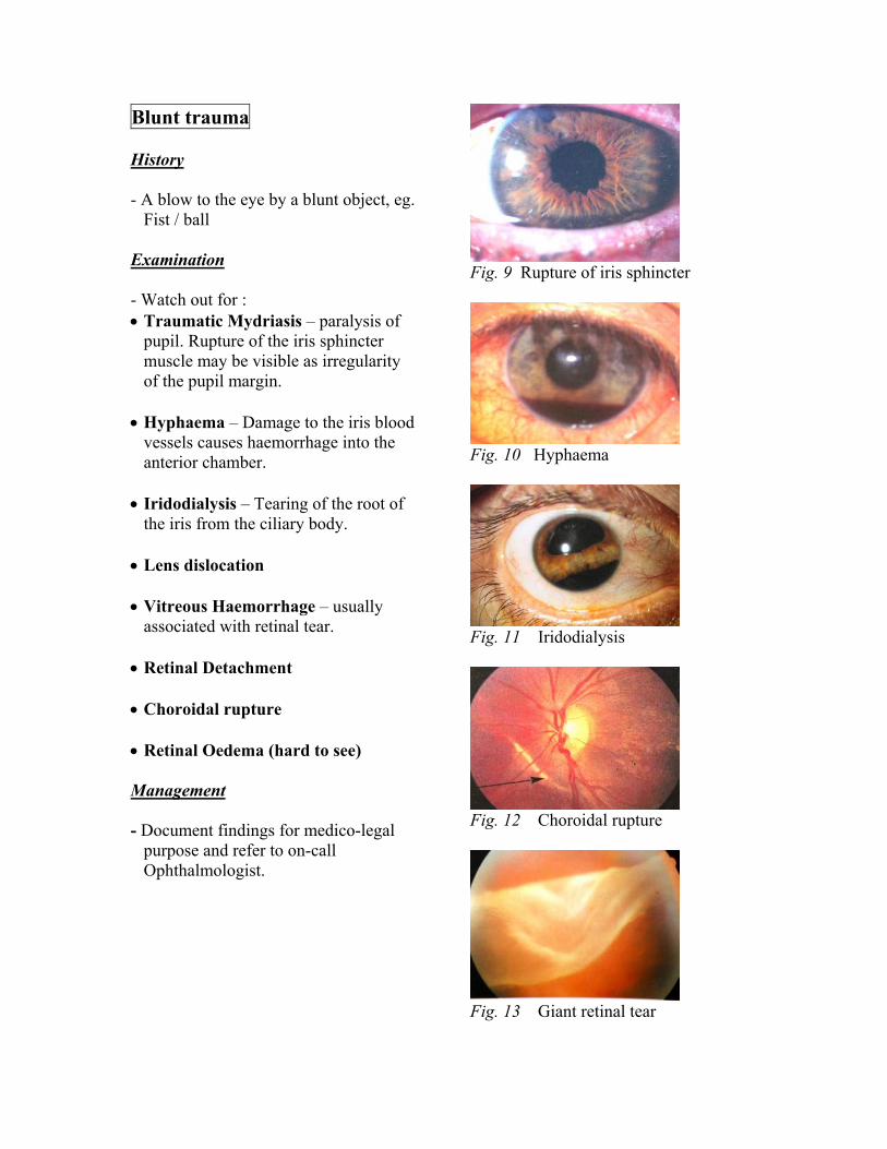
Blunt trauma
History - A blow to the eye by a blunt object, eg.
Fist / ball Examination
Fig. 9 Rupture of iris sphincter - Watch out for :
• Traumatic Mydriasis – paralysis of pupil. Rupture of the iris sphincter muscle may be visible as irregularity of the pupil margin.
• Hyphaema – Damage to the iris blood
vessels causes haemorrhage into the anterior chamber. Fig. 10 Hyphaema
• Iridodialysis – Tearing of the root of the iris from the ciliary body.
• Lens dislocation • Vitreous Haemorrhage – usually
associated with retinal tear. Fig. 11 Iridodialysis • Retinal Detachment
• Choroidal rupture • Retinal Oedema (hard to see) Management Fig. 12 Choroidal rupture - Document findings for medico-legal
purpose and refer to on-call Ophthalmologist.
Fig. 13 Giant retinal tear

Blow-out fracture
History - A blow to the eye by a blunt object, eg.
Ball, fist. Examination
Fig.14 mechanism of orbital floor # - usually orbital floor fracture with herniation of soft tissues into the maxillary sinus.
- Enophthalmos (Sunken eye) - Restriction of eye movement,
especially upward gaze, giving rise to diplopia.
- Loss of sensation over the regions supplied by the infraorbital nerve
Fig. 15 Right floor blow-out fracture. Defective elevation of the right eye. - Feel for air in subcutaneous tissue
( surgical emphysema ). - Check for nosebleed Management - warn patient to avoid blowing nose. - If Facial Xray shows signs of orbital
floor fracture, refer Maxillofacial SHO on-call.
- If vision is reduced, ask on-call
ophthalmologist’s advice.
- Refer eye clinic following day.

Penetrating trauma ±
Intraocular FB (IOFB) History - High index of suspicion from
mechanism of injury, esp. Grinding, Strimming, Hammering.
Fig. 16 Corneal laceration with iris prolapse - May follow previous cataract surgery
(weakened eye wall).
Examination - Soft eye. - Look for - reduced vision - Corneoscleral laceration
- Iris prolapse / distortion of pupil.
- Watch out for lid stabs that may hide a
stab injury to the eye itself Fig. 17 Scleral laceration
Management - Avoid pressure on the globe - Protective shield - Urgent referral - Not all FBs are radio-opaque, however
X-ray orbit if IOFB suspected - If IOFB suspected discuss with
ophthalmologist Fig. 18 Metal IOFB lying on retina

Chemical Burns
History - Do not waste time with a detailed
history and examination, especially if due to alkali
- Alkali burns can cause long term
corneal and conjunctival scarring, including untreatable blindness Fig.19 Cornea opacified with alkali burn
Examination - Use Proxymetacaine 0.5% instillation until comfortable to allow examination
- Ensure the eye is not penetrated - Ensure no bits of solid alkali in the eye Management - Irrigate copiously with sterile water / saline for 30 minutes within seconds!!
- Use litmus paper to ensure that pH is no longer alkali
- Refer to on-call ophthalmologist urgently.
* Similar management for all chemical burns, but alkali cause the most damage as it penetrates the cornea and causes intra-ocular damage as well as surface damage
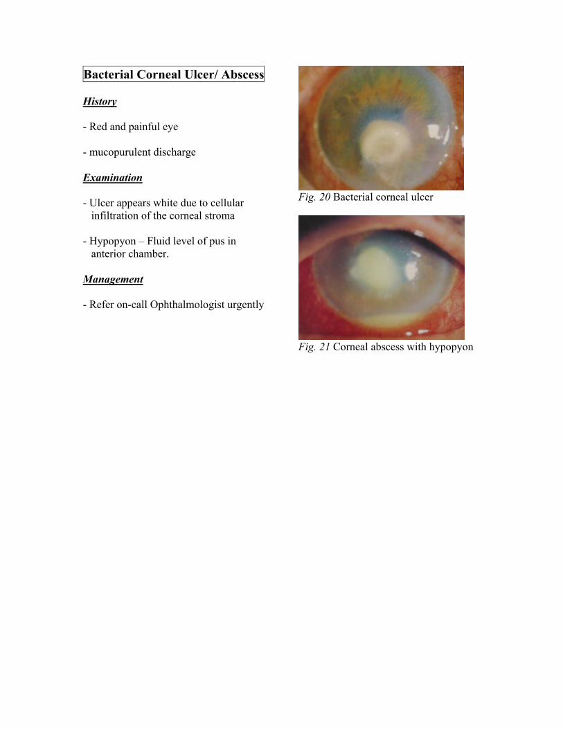
Bacterial Corneal Ulcer/ Abscess
History - Red and painful eye - mucopurulent discharge Examination Fig. 20 Bacterial corneal ulcer - Ulcer appears white due to cellular
infiltration of the corneal stroma
- Hypopyon – Fluid level of pus in
anterior chamber. Management - Refer on-call Ophthalmologist urgently Fig. 21 Corneal abscess with hypopyon
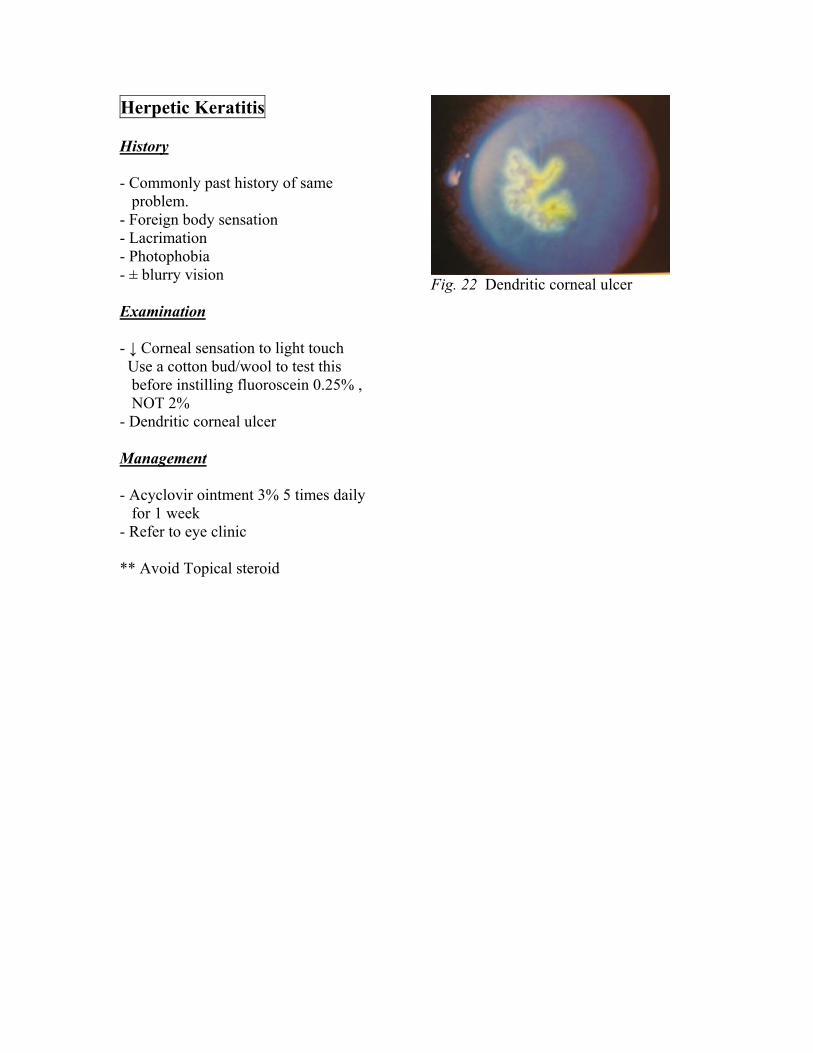
Herpetic Keratitis
History - Commonly past history of same
problem. - Foreign body sensation - Lacrimation - Photophobia - ± blurry vision Fig. 22 Dendritic corneal ulcer Examination - ↓ Corneal sensation to light touch Use a cotton bud/wool to test this
before instilling fluoroscein 0.25% , NOT 2%
- Dendritic corneal ulcer Management - Acyclovir ointment 3% 5 times daily
for 1 week - Refer to eye clinic ** Avoid Topical steroid

Shingles
History - Vesicular rash on face/scalp - swelling around the eye - If eye involved, photophobia, blurry
vision. Fig. 23 Shingles Examination - Vesicular rash with crust in CN V1 dermatome distribution (seldom V2)
- Look up for rash hidden on scalp covered by hair.
- Oedema++ both upper and lower eyelids.
* If rash on tip of the nose, likely eye involvement.
Management - Oral Acyclovir 5 times daily. - Refer Eye clinic following day if suspected eye involvement.

Acute Anterior Uveitis (Iritis / Iridocyclitis) (Iritis / Iridocyclitis)
HistoryHistory - Red eye - Discomfort/pain worse on reading close up (ie. Accommodation) Fig. 24 Keratic Precipitates - Photophobia - ± Blurred vision - Slight watering Examination - Unequal pupils (distorted due to previous adhesions – posterior synaechiae)
Fig. 25 Posterior Synaechie - Redness most marked in the circumcorneal region - ± Keratic precipitates – aggregates of cells on the posterior corneal surface - ± Hypopyon Management - Refer ophthalmologist on call especially if severe.

Orbital Cellulitis
History - Recent sinusitis - Painful, swollen eye - Malaise, fever Fig. 26 Hypopyon Examination
* Try to differentiate between a) Preseptal infection with normal
globe motility and b) Orbital involvement with proptosis
and limited motility of the globe. - Proptosed eye - Both lids and conjunctiva are inflamed and swollen
Fig. 27 Orbital Cellulitis
- Eye movements limited and painful - Pyrexia Management - Swab if pus visible - Refer on-call ophthalmologist - Needs IV antibiotics ** Children with pre-septal cellulitis ( inflammation to lids anterior to the orbital septum) often after trivial trauma to eyelids, usually responds well to oral Augmentin, but are at risk of developing meningitis.

Canalicular obstruction /
Acute Dacrocystitis History - Epiphora- watering - Swollen, painful affected site of face Fig. 28 Canalicular obstruction due to
streptothrix infection Examination - Tender over swelling
Management - If suspect, refer eye clinic - If severe, refer on-call Ophthalmologist - Augmentin 375mg TDS Fig. 29 Acute Dacrocystitis

Acute Glaucoma
- Painful loss of vision - Common in Long-sighted patients,
oriental races higher risk. History
- Severe pain in eye - Unilateral headache - Blurred vision - Nausea ± Vomiting Fig. 30 Acute glaucoma - Rainbow haloes around white light - Previous transient attacks of blurred
vision in the evenings * May have been to eye clinic and had
mydriatic eye drops earlier that day.
Examination - Conjunctival hyperaemia - Cornea steamy and oedematous - Pupil – oval, fixed and dilated - AC both eyes shallow (Eclipse test) - The eye is rock hard and acutely tender - Measure IOP – usually ↑ to 40-80mmHg
Management - Urgent referral to on-call ophthalmologist
- Pilocarpine 2% drop in affected and unaffected eye to prevent pupil dilation

Other caveats ** Do not put topical steroid in the eye.
This is for the ophthalmologist to do.
** Refer urgently any eye, which has
been subjected to : 1) Alkali/ Acid Burns (Irrigate copiously
first!!) 2) Suspected orbital cellulitis 3) Suspected acute glaucoma/Iritis 4) Sudden visual loss (such as shown
below) Fig. 31 Central retinal artery occlusion 5) Anything else you are bothered about. Seek their advice!!!
Sudden Loss of Vision (without preceding ocular upset)
1) Central retinal artery occlusion
(Fig.31) 2) Central retinal vein occlusion (Fig.32) 3) Massive Vitreous haemorrhage
(Fig.33) Fig. 32 Central retinal vein occlusion 4) Ischaemic optic neuropathy (Fig.34)
5) Retinal detachment (Fig. 13) Fig. 34 Ischaemic optic neuropathy Last reviewed July 2003 Dr Chee Thum, Dr I Young