Embed Size (px)
Citation preview
How to Spot and Manage Iris Cysts
Outer Retinal Tubulation: What It Is and What It Means
PEARLS
Central Retinal Vein Occlusion
J A N U A R Y 2 0 1 8
EyeNet®
Global OphthalmologyWhat Today’s Volunteers Do Differently

Join Your Colleagues in ChicagoLike Chicago, ophthalmology is a global community of innovators in art and science. Join us at AAO 2018, where you can contemplate the finer points of cross-linking, intraocular knot-tying and lenticule extraction — or Monet, van der Rohe and Chagall.
AAO 2018 October 27 – 30
Subspecialty Day October 26 – 27
AAOE Program October 27 – 30
aao.org/2018
Where All of Ophthalmology Meets®
In conjunction with the Pan-American Association of Ophthalmology
Call for AbstractsInstruction Courses and New Skills Labs December 14, 2017 – January 9, 2018
Papers/Posters and Videos March 8, 2018 – April 10, 2018

ZIOPTAN is licensed by Santen Pharmaceutical Co., Ltd.
©2017 Akorn, Inc. All rights reserved. P481(a) Rev 4/17
Cosopt is a registered trademark of Merck Sharp & Dohme Corp and is used under license. ZIOPTAN is a registered trademark of Merck Sharp & Dohme Corp and is used under license.
100% PRESERVATIVE-FREE
ORDER FREE SAMPLES
www.Zioptan.com www.CosoptPF.com
Go to either website and select “Request Sample”

Designed and Manufactured by NIDEK - Represented by Marco 800-874-5274 • marco.com
AND DESIRED OUTCOMES
The TRS-5100 rapidly completes all refractions and the split prism allows immediate patient comparison and verification of old vs. new prescriptions – without flipping through lenses and asking “which is better, 1 or 2?”
The TRS-5100 takes accuracy to a new level and provides the ultimate refraction information that we’ve been seeking. Now, we have fewer remakes with more satisfied patients who enjoy shorter refraction exam times.
The TRS-5100 is highly valued. It is extremely accurate and I’m very confident in the quality of refraction. It is such a timesaver, has cut down on remakes, reduced our frustrations and increased our bottom line.
The TRS provides our practice with a diagnostic tool that both improves clinical outcomes while increasing patient flow and overall efficiency. The investment that pays for itself. Wait times are reduced, we see more patients, and we’re performing more surgery than ever.
Mitchell A. Jackson, MDLake Villa, Illinois
Larry Patterson, MDCrossville, TN
Charles Collins, MDMiddleton, RI
Faisal Haq, MDPlano, Texas
The OPD-Scan III provides integrated wavefront aberrometry and corneal assessment with topography, pupillometry, autorefraction and keratometry. In just 10 seconds, per eye, over 20 diagnostic metrics are integrated.
TRS-5100Product/Model name:REFRACTOR RT-5100
Join MARCO January 18-20, 2018 CATARACT SURGERY: Telling It Like It Is!
Ritz Carlton • Amelia Island, FL
634 TRS Testiml-MD-EN.indd 1 12/6/17 11:13 AM

E Y E N E T M A G A Z I N E • 5
EyeNet® Magazine (ISSN 1097-2986) is published monthly by the American Academy of Ophthal mology, 655 Beach St., San Francisco, CA 94109-1336, as a membership service. Subscription is included in U.S. members’ annual dues. International Mem ber, IMIT, $135 per year. Nonmember in U.S., $150 per year. Nonmember outside U.S., $210 per year. Periodicals Postage Paid at San Francisco, CA, and at additional mailing offices. POSTMASTER: Send address changes to EyeNet, P.O. Box 7424, San Francisco, CA 94120-7424. American Academy of Ophthalmic Executives®, EyeSmart®, EyeWiki®, IRIS® Registry, and ONE® Network are trade-marks of the American Academy of Ophthalmology®. All other trademarks are the property of their respective owners.
CONTENTSJANUARY 2018VOLUME 22 • NUMBER 1
FEATURE
40-45 Volunteering AbroadMedical missions aren’t what they used to be. Here’s how to make a long-lasting, sustainable contribution to eye care in communities around the world.
CLINICAL INSIGHTS
17-19 News in ReviewNeuro-ophthalmology Remyelination in multiple sclerosis.
Cataract Retinal detachment after cataract surgery.
Glaucoma Gene editing with CRISPR.
Oncology Coming to consensus on retino-blastoma screening.
21-25 Journal HighlightsKey findings from Ophthalmology, Ophthal-mology Retina, AJO, JAMA Ophthalmology, and more.
27-32 Clinical Update Anterior Segment Iris cysts: what you need to know about identification and management.
Retina Outer retinal tubulation: a hallmark of neurodegeneration.
33-35 Ophthalmic Pearls CRVO Review the risk factors, diagnosis, and management of central retinal vein occlusion.
40
17 27
31 33

6 • J A N U A R Y 2 0 1 8
xxCOPYRIGHT © 2018, American Academy of Ophthalmology, Inc.® All rights reserved. No part of this publication may be reproduced with-out written permission from the publisher. Letters to the editor and other unsolicited material are assumed intended for publication and are subject to editorial review, acceptance, and editing. Disclaimer. The ideas and opinions expressed in EyeNet are those of the authors,
and do not necessarily reflect any position of the editors, the publisher, or the American Academy of Ophthalmology. Because this publication provides information on the latest developments in ophthalmology, articles may include information on drug or device applications that are not considered community standard, or that reflect indications not included in ap proved FDA labeling. Such ideas are provided as information and education only so that practitioners may be aware of alternative methods of the practice of medicine. Information in this publication should not be considered endorsement, promotion, or in any other way encouragement for the use of any particular procedure, technique, device, or product. EyeNet, its editors, the publisher, or the Academy in no event will be liable for any injury and/or damages arising out of any decision made or action taken or not taken in reliance on information contained herein.
EyeNet®
CLINICAL INSIGHTS
37-39 Morning Rounds A Case of Aches and Pains and Blurry Vision A 28-year-old woman had flulike symptoms followed by red, painful, photophobic eyes and decreased vision. What’s your diagnosis?
IN PRACTICE
51-52 Savvy CoderAudit! How Should Staff Respond? You need a written protocol. Here’s what it should address.
53-55 Practice PerfectSocial Media and Low Vision Help your patients to avoid depression.
From PQRS to MIPS To enjoy continued success with the IRIS Registry, meet the Jan. 15 deadline and avoid these mistakes.
FROM THE AAO
57-60 Academy Notebook State societies honored. l D.C. Report: Major Quality Program changes and stable ophthal-mology payments. l And more.
VIEWPOINTS
9 Letters How to shift perspective. l More on low vision.
10 Opinion Can you practice part time?
12 Current Perspective Sexual harassment and ophthalmology.
14 President’s Statement The value of education and giving back.
MYSTERY IMAGE
62 BlinkWhat do you see?
COVER PHOTOGRAPH
Marc Safran, MD
37
57
62

When the world demands the best, it looks to Wills Eye. With the nation’s highest clinical volume of annually referred patients, we’ve always been at the forefront of eye disease treatment. Our expertise and advanced capabilities are what defined the field in 1832, and continue to redefine it today.
840 Walnut Street Philadelphia, PA 19107
willseye.org1-877-289-4557
A doctor doesn’t give a hospital its reputation.A team of doctors do.

8 • J A N U A R Y 2 0 1 8
David W. Parke II, MDEditor-in-Chief
Ruth D. Williams, MDChief Medical Editor
Dale E. Fajardo, EdD, MBAPublisher
Patty AmesExecutive Editor
Carey S. BallardArt Director /
Production Manager
Chris McDonagh, Jean ShawSenior Editors
Catherine MorrisAssociate Editor / Content Manager
Lori Baker-Schena, MBA, EdD; Leslie Burling-Phillips;
Peggy Denny; Miriam Karmel; Mike Mott; Linda Roach;
Lynda Seminara; Annie Stuart Contributing Writers
Mark Mrvica, Kelly MillerM.J. Mrvica Associates, Inc.
2 West Taunton Ave., Berlin, NJ 08009
856-768-9360 [email protected]
Advertising Sales
655 Beach St.San Francisco, CA 94109
866-561-8558, 415-561-8500 aao.org
Governmental Affairs Division20 F Street NW, Suite 400
Washington, DC 20001 202-737-6662
ARTICLE REVIEW PROCESS. Articles involving single-source medical and tech-nical news are sent to quoted sources for verification of accuracy prior to publication. Quotes and other information in multisource articles are subject to confirmation by their respective sources. The chief medical editor and the executive editor review all news and feature articles and have sole discretion as to the acceptance and rejection of material and final authority as to revisions deemed necessary for publication.
DISCLOSURE KEY. Financial interests are indicated by the following abbrevia-tions:C = Consultant/AdvisorE = EmployeeL = Speakers bureauO = Equity ownerP = Patents/Royalty S = Grant supportFor definitions of each category, see aao.org/eyenet/disclosures.
MAGAZINE
®EDITORIAL BOARD
CATARACTKevin M. Miller, MD,Section Editor
William R. Barlow, MDKenneth L. Cohen, MDKendall E. Donaldson, MDJason J. Jones, MDBoris Malyugin, MD, PhDCathleen M. McCabe, MDRandall J. Olson, MDMarie Jose Tassignon, MD
COMPREHENSIVE OPHTHALMOLOGYPreston H. Blomquist, MD,Section Editor
Sherleen Huang Chen, MDApril Y. Maa, MDLinda M. Tsai, MD
CORNEA AND EXTERNAL DISEASEChristopher J. Rapuano, MD,Section Editor
Kathryn A. Colby, MD, PhDHelena Prior Filipe, MDBennie H. Jeng, MDStephen D. McLeod, MDSonal S. Tuli, MD
GLAUCOMASanjay G. Asrani, MD, Section Editor
Iqbal K. Ahmed, MDLama Al-Aswad, MD, MPHAhmad A. Aref, MDAnne Louise Coleman, MD, PhDSteven J. Gedde, MDCatherine Green, MBChB
Steven L. Mansberger, MD, MPHRonit Nesher, MDRichard K. Parrish II, MDSarwat Salim, MD, FACS
LOW VISIONLylas G. Mogk, MDJohn D. Shepherd, MD
NEURO-OPHTHALMOLOGYLeah Levi, MD, Section Editor
Kimberly Cockerham, MD, FACSHelen V. Danesh-Meyer, MBCHB, MDPrem S. Subramanian, MD, PhD
OCULOPLASTICSEvan H. Black, MD, Section Editor
Elizabeth A. Bradley, MDFemida Kherani, MDDon O. Kikkawa, MD
OPHTHALMIC ONCOLOGYZélia M. Corrêa, MD, PhD, Section Editor
Dan S. Gombos, MDTatyana Milman, MD
OPHTHALMIC PATHOLOGYDeepak Paul Edward, MDDavid J. Wilson, MD
OPHTHALMIC PHOTOGRAPHYJason S. CalhounMichael P. Kelly, FOPS
PEDIATRIC OPHTHALMOLOGYDavid A. Plager, MD, Section Editor
Michael F. Chiang, MDJane C. Edmond, MDFrank Joseph Martin, MDFederico G. Velez, MD
REFRACTIVE SURGERYGeorge O. Waring IV, MD, Section Editor
Damien Gatinel, MDSoosan Jacob, FRCS A. John Kanellopoulos, MDJ. Bradley Randleman, MDKarolinne M. Rocha, MDMarcony R. Santhiago, MD
RETINA/VITREOUSJulia A. Haller, MD, Section Editor
Neil M. Bressler, MDKimberly A. Drenser, MD, PhDSharon Fekrat, MDMitchell Goff, MDLawrence S. Halperin, MDGregg T. Kokame, MDAndreas K. Lauer, MDPrithvi Mruthyunjaya, MD, MHSKyoko Ohno-Matsui, MDAndrew P. Schachat, MD Ingrid U. Scott, MD, MPHGaurav K. Shah, MD
UVEITISGary N. Holland, MD, Section Editor
Muge R. Kesen, MD H. Nida Sen, MDSteven Yeh, MD
ACADEMY BOARDPRESIDENTKeith D. Carter, MD, FACS
PRESIDENT-ELECTGeorge A. Williams, MD
PAST PRESIDENTCynthia A. Bradford, MD
CEODavid W. Parke II, MD
SR. SECRETARY FOR ADVOCACYDaniel J. Briceland, MD
SECRETARY FOR ANNUAL MEETINGMaria M. Aaron, MD
SR. SECRETARY FOR CLINICAL EDUCATIONLouis B. Cantor, MD
SR. SECRETARY FOR OPHTHALMIC PRACTICERobert E. Wiggins Jr., MD, MHA
CHAIR, THE COUNCILLynn K. Gordon, MD, PhD
VICE CHAIR, THE COUNCILSarwat Salim, MD, FACS
OPHTHALMOLOGY EDITORStephen D. McLeod, MD
CHAIR OF THE FOUNDATION ADVISORY BOARDChristie L. Morse, MD
PUBLIC TRUSTEESPaul B. Ginsburg, PhD
TRUSTEES-AT-LARGEMichael F. Chiang, MD William S. Clifford, MDSanjay D. Goel, MDCynthia Mattox, MD, FACSWilliam F. Mieler, MDAndrew M. Prince, MD
INTERNATIONAL TRUSTEESKgaogelo Edward Legodi, MDLihteh Wu, MD
Learn more about the Board at aao.org/bot.

E Y E N E T M A G A Z I N E • 9
How to Shift Your Perspective
In light of Dr. Williams’ column titled “Solo Practice in Ophthalmology: Resist-ing the Tides?” (Opinion, October), I want to encour-age physicians to shape their futures despite the obstacles created by the following: Medicaid cost sharing with the federal government, uncertainty around the fate
of individual insurance markets, looming MACRA and MIPS regulations, and more.
What happens when we take the reins. I invite you to imagine what is possible (and importantly, under our own control) when we shift our perception of health care reform and value-based care from externally imposed burdens to internally driven improvements. By focusing on your indi-vidual practice, you can solve your own unique challenges. There is plenty to do to improve your practice for intrinsic reasons, and fortunately many of these changes can also help you survive the transition from fee-for-service to value-based payments—for example, inefficiencies are a threat like never before.
The 3 elements of improvement. The foundation for phy-sician-directed practice improvements rests upon 3 pillars: autonomy, mastery, and purpose. Much has been written lately about these and “physician engagement with work”—and, yes, it is possible to use these concepts to find joy in our work. For example, when you problem-solve to eliminate inefficiencies in your practice, you can find pleasure in this exercise of autonomy. As your practice improves due to the solutions you found, you experience the element of mastery. Finally, a higher-performing practice allows the individu-al and organization to more effectively fulfill its purpose of helping others. Conversely, actions that do not feature these 3 principles lead to frustration and increase the risk of burnout.
Choose the best path for you. Both solo and group prac-tice have their pros and cons; the choice boils down to how strongly you value autonomy. For some ophthalmologists, achieving mastery is found by working alone; for others, mastery can be facilitated through the advantages of a group practice.
At a minimum, we must continually evolve our clinical and surgical skills as well as basic business skills. At some point, however, we realize the importance of community and
advocacy to achieve a higher purpose (the pillar of fulfill-ment): service to others. The Academy, state and subspecialty societies, and their advocacy groups exist to defend our professional autonomy, support our individual and collective efforts to achieve professional mastery, create vital bonds with like-minded professionals, and ultimately fulfill our purpose as healers.
Alan E. Kimura, MD, MPHDenver
More on Low Vision
I read with interest “Low Vision Drivers: The Ophthalmol-ogist’s Role and Responsibility” (Clinical Update, October), which quite thoroughly discusses the benefit that bioptic vision aids may offer to many individuals who would otherwise be unable to acquire a driver’s license, with the independence that this certification offers.
This remarkable visual/driving aid was brought to my attention in 1996. At that time, I evaluated a then 8-year- old boy who was found to have Stargardt disease, with his acuity eventually dropping to 20/200 in both eyes. With subsequent evaluation by C.J. Reed, OD, COMS, at the Judith A. Read Low Vision Services in Akron, Ohio, and fit with the then-available Ocutech VES II/6 × magnification, he has been able to continue successfully through college and obtain ongoing driving privileges.
A debt of gratitude needs to be given to William Fein-bloom, OD, PhD, the “father of low vision care” in the United States, who introduced the concept of bioptic driving1 in this country. In 1932, at age 28, he used an astronomer’s telescope as a model to design a small 3 × power telescope that was small enough to be mounted in a spectacle frame, restoring one individual’s functional vision.2
Later, in 1958, he introduced the concept of a bioptic tele-scopic system, which combined a prescription eyewear lens with a small mounted Galilean telescopic system. His system allowed the patient to change view from the telescope to the general prescription.
The website referenced below, with information from Richard L. Windsor, OD, is a most valuable resource, describ-ing a number of up-to-date options that ophthalmologists/optometrists/low vision specialists may find useful, adding to EyeNet’s informative article.
Stuart M. Terman, MDCleveland
1 www.biopticdrivingusa.com
2 Feinbloom W. The training and after care of the partially blind patient.
Journal of the American Optometric Association. 1958:29:724.
Letters
Unintended Consequences New Cancer Drugs, New Ocular Side Effects
Bioptic Lenses for Driving?Counseling Low Vision Patients
OPINION
The Enduring Appeal of Solo Practice
O C T O B E R 2 0 1 7
EyeNet®
International Report
The Lowdown on High-Tech IOLs

10 • J A N U A R Y 2 0 1 8
Ruth D. Williams, MDChief Medical
Editor, EyeNet
During the open microphone session at an Academy Young Ophthalmologist symposium, an ophthalmol-ogist newly in practice, who was also a new father,
asked, “How do I manage the tension between the demands of my job and the responsibility I feel to my son?”
I wanted to hug this young person. Finally, the well-known challenge of managing a young career and a young family has become a nongendered issue. In response to this challenge, some young ophthalmologists—both men and women—now ask about the possibility of practicing part time. But while they might wish to decrease work hours to accommodate other priorities, the concept of a part-time ophthalmologist is a flawed one.
There is no such thing as a part-time ophthalmologist. Let me explain. A surprising number of physicians report
working part time. In a compensation survey of nearly 20,000 physicians across 26 specialties, 22% of the women and 10% of the men report that they work fewer than 40 hours per week.1 In ophthalmology, 24% of female and 15% of male ophthalmologists report working fewer than 40 hours each week. This trend might be increasing as dual professional career families become commonplace. Furthermore, we’re told that today’s young physicians tend to protect family and leisure time more than their older peers have and might be bolder about requesting a day off or protecting their weekends.
Yet no survey can capture the commitment that is required to be a physician. An ophthalmologist who chooses to work fewer hours for a period of time is still a completely commit-ted physician. She might shorten her workday or compress the work week into fewer days, but she brings her training, her expertise, her experience, her compassion, and her wisdom to work when seeing patients. The “part-time” physician often maintains a regular call schedule and full malpractice cover-age, and he is committed to continuous learning. The “part-time” ophthalmologist learns new techniques, innovates, and attends educational meetings. The “part-time” ophthalmolo-gist is available for patients, emergencies, and advice to other physicians. In other words, the “part-time” ophthalmologist has a 100% commitment to the practice of ophthalmology.
Much has been written about Baby Boomers, for whom work is a moral imperative, and Gen Xers and Millennials,
who reportedly want more work-life balance. This is an over-simplification of reality. For example, of the 15% of male ophthalmologists who work part time, I wonder how many are older physicians who immensely enjoy practice but now choose to work fewer hours and enjoy other activities. We value these physicians for their experience and their wisdom, and they ground us in a tradition of providing quality care, teaching colleagues, and continuing to learn. Likewise, we value the young ophthalmologists who might provide superb ophthalmic care and work fewer hours than their older peers did at the same career stage. And we recognize that many physicians who limit work hours early in their career increase their time commitment in later decades.
Traditionally, full-time work has been measured by how many hours a person works in the office. While this may be necessary for determining benefits, hours logged is hardly a meaningful measure of the value of a colleague. There are many metrics for valu-ating work, including RVUs, productivity, papers written, leadership roles, and teaching responsibilities. In my own practice, I’ve been impressed by the availability of my young colleagues to discuss a patient or provide advice at odd hours. Once, a colleague discussed a complex case with me, and at the end of the phone call I discovered that he was on a ski slope.
Some ophthalmologists who prac-tice part time feel diminished by the choice and don’t like the label. Let’s acknowledge the 100% commitment to patient care and stop counting the hours. An ophthalmologist is “all in,” even if it’s not all the time.
There is no such thing as a part-time ophthalmologist.
1 Grisham S. Medscape physician compensation report 2017. April 5, 2017.
www.medscape.com. Accessed Nov. 16, 2017.
Opinion
RUTH D. WILLIAMS, MD
Can You Practice Part Time?


12 • J A N U A R Y 2 0 1 8
David W. Parke II, MD Academy CEO
Current Perspective
DAVID W. PARKE II, MD
Sexual Harassment and Ophthalmology
Sexual harassment allegations know no workplace boundaries. While most recent public cases involve the media and entertainment industries or govern-
ment officials, other cases have touched nearly every type of organization and profession—including ophthalmology. This should not be surprising because sexual harassment frequently is driven by power differentials between the harasser and the victim, leading to feelings of vulnerability. Medicine is replete with such power relationships—between physician and staff; between professor and trainee; between senior and junior colleagues; and between physician and patient.
Harassment allegations run the gamut between single verbal episodes to patterns of frank sexual assault. The U.S. Equal Employment Opportunity Commission defines workplace sexual harassment in part as “unwelcome sexual advances or conduct of a sexual nature which … creates an intimidating, hostile, or offensive work environment.” How many times in our professional lives have we been a witness (or a party) to a crude joke, comments of a highly personal or sexual nature—or worse? How often is such behavior ra-tionalized by statements like, “I’ve always been a big hugger” or “I’ve always been like that and she has never complained.” (Although I use “she,” sexual harassment may be male to female, female to male, female to female, or male to male.)
As many gray zones as have been recently illuminated (for example, “when is a hug appropriate?” and “what discussion subjects cross the line?”), the #MeToo movement has created a valuable teaching moment, forcing all of us—men and women—to rethink appropriateness and inclusiveness.
Implicit in the trust and respect we receive as physicians is our responsibility to create a safe and respectful professional environment. This includes zero tolerance for inappropriate behavior that might be interpreted as constituting harassment.
What is the Academy’s role? Every Academy member agrees to abide by the Code of Ethics and its Principles. The Princi-ples describe “model standards of exemplary professional conduct for all Fellows or Members of the Academy.” Principle 2 states in part, “Ophthalmological services must be provided with compassion, respect for human dignity, honesty, and integrity.” This applies to all involved in the care process—colleagues, staff, patients, and families.
In the 30-plus years of the Academy’s Code of Ethics, there has never been an ethics challenge involving alleged sexual harassment or sexual misconduct involving patients, colleagues, or staff. However, surveys of female physicians in multiple specialties suggest that a large percentage of women ophthalmologists have personally experienced what they per ceived to be sexual harassment—verbal and/or physical—from their physician colleagues. One study of 1,066 physicians revealed that 30% of women said they had directly experi-enced sexual harassment in their careers, versus 4% of men.1
Members and Fellows also deserve to under-stand how the Academy addresses this issue, the seriousness with which it is taken, the organizational culture we attempt to engender, and the processes we have in place to protect our staff, our volun-teers, and our profession itself.
Every Academy staff mem-ber, without exception, must complete a sexual harassment training course every 2 years and acknowledge in writing familiarity with the Academy’s relevant policies. These policies acknowledge both organizational responsibility for compliance as well as individual responsibilities and report-ing obligations for every employee. Any alleged incident is taken seriously, investigated thoroughly, and (if validated) is accompanied by disciplinary action—possibly including dismissal. Regardless of the investigation outcome, retaliation against an accuser is forbidden not only by Academy policy but also under the law.
We owe it to ourselves, our patients, all members of our professional team, and to ophthalmology itself to always exhibit exemplary behavior and to make all involved feel welcome, comfortable, and respected. The Academy as an organization similarly makes that pledge.
1 Jagsi R et al. JAMA. 2016;315(19):2120-2121.

OMIC’s dividend returns averaged 20.8% the past 5 years vs. 6.6% for other carriers that have declared dividends.
OMIC consistently outperforms multispecialty malpractice insurance companies in almost all � nancial results, benchmarks, and ratios used to evaluate performance in the industry.
OMIC’s coverage year loss and loss expense ratio was the best among our peer companies in 2016, and has averaged 45% lower than competitors’ for years 2012 to 2016.
Policyholders’ surplus surpassed $200 Million and OMIC’s premium to surplus ratio of 17.7% distiguishes us as one of the most � scally sound insurance carriers in America.
Robert Wiggins, MDChair, Finance Committee and Treasurer
www.omic.com/request-a-quote
$80 MillionCumulative declared dividends 1987-2017
A Risk Retention Group
800.562.6642

14 • J A N U A R Y 2 0 1 8
President’s Statement
KEITH D. CARTER, MD
The Value of Education, and the Satisfaction of Giving Back
When I got the phone call from Dr. Parke inviting me to serve as Academy President-Elect in 2017 and Academy President in 2018, I was thrilled
and humbled. I am very honored to serve as your president. A member for 30 years, I value the Academy, and I have worked on a great many projects and assumed many roles, from writing for Focal Points to serving on the Ethics Com-mittee.
For my presidential term, I have several goals. One is tech-nical: to influence improvement of the ability of ophthalmic equipment to communicate with the electronic medical record. We see equipment with great promise at our meetings, but, often, it can’t “talk” to the other systems that we use to care for patients. If we can promote a common language, as radiology has done, it would be a great accomplishment.
The second goal stems from a core value of mine. One of the mainstays of my career has been educating physicians in training, both at the University of Iowa and through the Academy. This is because I would not be where I am without very good teachers and mentors in my life. My first mentor was the physician who delivered me! He wrote letters of recommendation in my support for my applications to pharmacy school and medical school, and he influenced me to choose academics. I would never have seen numerous trainees mature into successful physicians or had my gratify-ing experiences at the Academy if it weren’t for the guidance, support, and encouragement of my mentors.
For this reason, I am excited about participating in a new collaborative program with the Association of University Professors of Ophthalmology (AUPO) to attract underrepre-sented minorities to the profession. We haven’t made signifi-cant gains in 30 years in recruiting professionals who reflect the populations that we serve. Part of the challenge is gaining the attention of these young students and securing mentors to guide them through the process. Then, the student needs to be prepared to be a competitive applicant. When compet-itive students with diverse experiences are in the classroom, this enhances everybody’s education.
Enhancing diversity within ophthalmology is part of giving back to the generations that follow us. Many of the younger generation are less color- or gender-based in their
thinking, but efforts still must be made to change the profes-sional landscape.
Giving back is vital—and a pleasure. As I progressed in my own positions, I discovered the joy that comes from see-ing a student develop into a well-trained doctor, especially in terms of surgery. Another great pleasure about working with young people is that they constantly challenge you and keep you thinking. They bring excellent ideas—and we need to listen to them. Some of the biggest achievements we’ve seen come from physicians in training. For example, EyeRounds, our department’s online education forum, was originally presented by a resident who saw the vision of the Internet while we were all worrying about the book chapters we’d write. Now, EyeRounds is one of the most- visited ophthalmology websites around the world.
While much practical training and education is now online or increasingly done in simula-tion labs, the art of medicine can never be fully taught that way. Taking care of patients is passed on from doctor to doctor in real-life settings. Explaining how to alleviate a patient’s fears or how to deliver bad news cannot easily be taught from a book. It’s our role to be there for young ophthalmologists and share our experiences. Many practices recog-nize this and are asking to have trainees come to their offices for real-world exposure. That may be a bigger part of future training.
I am excited to step into this new role and hope to be seen as a president who values education and diversity and continually promotes the profession, but I can’t be successful without you. The Academy is great because of its many volunteers. If you have interests in education, diversity, technology, or other areas, let us know. Your involvement is welcome and necessary. Let’s get started!
Keith D. Carter, MD Academy 2018 President

For additional safety information, see accompanying Brief Summaryof Safety Information and Full Prescribing Information on Xiidra-ECP.com.
Marks designated ® and ™ are owned by Shire or an affi liated company. ©2017 Shire US Inc. Lexington, MA 02421 S28784 01/17
Proven to treat the signs of inferior corneal staining in 12 weeks and symptoms of eye dryness in 12, 6, and as little as 2.
Xiidra helped provide symptom relief from eye dryness in some patients at week 2—and a measurable reduction in signs of inferior corneal staining in just 12 weeks. Consider Xiidra to help your Dry Eye patients fi nd the relief they’ve been waiting for.
Check it out at Xiidra-ECP.com
Four randomized, double-masked, 12-week trials evaluated the effi cacy and safety of Xiidra versus vehicle as assessed by improvement in the signs (measured by Inferior Corneal Staining Score) and symptoms (measured by Eye Dryness Score) of Dry Eye Disease (N=2133).
Indication Xiidra® (lifi tegrast ophthalmic solution) 5% is indicated for the treatment of signs and symptoms of dry eye disease (DED).
Important Safety InformationIn clinical trials, the most common adverse reactions reported in 5-25% of patients were instillation site irritation, dysgeusia and reduced visual acuity. Other adverse reactions reported in 1% to 5% of the patients were blurred vision, conjunctival hyperemia, eye irritation, headache, increased lacrimation, eye discharge, eye discomfort, eye pruritus and sinusitis.
To avoid the potential for eye injury or contamination of the solution, patients should not touch the tip of the single-use container to their eye or to any surface.
Contact lenses should be removed prior to the administration of Xiidra and may be reinserted 15 minutes following administration.
Safety and effi cacy in pediatric patients below the age of 17 years have not been established.
When artifi cial tears aren’t enough, consider prescribing Xiidra for symptomatic Dry Eye patients.
MAKE XIIDRA YOUR FIIRST CHOICE

BRIEF SUMMARY:Consult the Full Prescribing Information for complete product information.
INDICATIONS AND USAGEXiidra® (lifitegrast ophthalmic solution) 5% is indicated for the treatment of the signs and symptoms of dry eye disease (DED).
DOSAGE AND ADMINISTRATIONInstill one drop of Xiidra twice daily (approximately 12 hours apart) into each eye using a single use container. Discard the single use container immediately after using in each eye. Contact lenses should be removed prior to the administration of Xiidra and may be reinserted 15 minutes following administration.
ADVERSE REACTIONSClinical Trials ExperienceBecause clinical studies are conducted under widely varying conditions, adverse reaction rates observed in clinical studies of a drug cannot be directly compared to rates in the clinical trials of another drug and may not reflect the rates observed in practice. In five clinical studies of dry eye disease conducted with lifitegrast ophthalmic solution, 1401 patients received at least 1 dose of lifitegrast (1287 of which received lifitegrast 5%). The majority of patients (84%) had ≤3 months of treatment exposure. 170 patients were exposed to lifitegrast for approximately 12 months. The majority of the treated patients were female (77%). The most common adverse reactions reported in 5-25 % of patients were instillation site irritation, dysgeusia and reduced visual acuity. Other adverse reactions reported in 1% to 5% of the patients were blurred vision, conjunctival hyperemia, eye irritation, headache, increased lacrimation, eye discharge, eye discomfort, eye pruritus and sinusitis.
USE IN SPECIFIC POPULATIONSPregnancyThere are no available data on Xiidra use in pregnant women to inform any drug associated risks. Intravenous (IV) administration of lifitegrast to pregnant rats, from pre-mating through gestation day 17, did not produce teratogenicity at clinically relevant systemic exposures. Intravenous administration of lifitegrast to pregnant rabbits during organogenesis produced an increased incidence of omphalocele at the lowest dose tested, 3 mg/kg/day (400-fold the human plasma exposure at the recommended human ophthalmic dose [RHOD], based on the area under the curve [AUC] level). Since human systemic exposure to lifitegrast following ocular administration of Xiidra at the RHOD is low, the applicability of animal findings to the risk of Xiidra use in humans during pregnancy is unclear.
Animal Data Lifitegrast administered daily by intravenous (IV) injection to rats, from pre-mating through gestation day 17, caused an increase in mean preimplantation loss and an increased incidence of several minor skeletal anomalies at 30 mg /kg /day, representing 5,400-fold the human plasma exposure at the RHOD of Xiidra, based on AUC. No teratogenicity was observed in the rat at 10 mg /kg /day (460-fold the human plasma exposure at the RHOD, based on AUC ). In the rabbit, an increased incidence of omphalocele was observed at the lowest dose tested, 3 mg /kg /day (400-fold the human plasma exposure at the RHOD, based on AUC), when administered by IV injection daily from gestation days 7 through 19. A fetal No Observed Adverse Effect Level (NOAEL) was not identified in the rabbit.
Lactation There are no data on the presence of lifitegrast in human milk, the effects on the breastfed infant, or the effects on milk production. However, systemic exposure to lifitegrast from ocular administration is low. The developmental and health benefits of breastfeeding should be considered, along with the mother’s clinical need for Xiidra and any potential adverse effects on the breastfed child from Xiidra.
Pediatric Use Safety and efficacy in pediatric patients below the age of 17 years have not been established.
Geriatric Use No overall differences in safety or effectiveness have been observed between elderly and younger adult patients.
NONCLINICAL TOXICOLOGYCarcinogenesis, Mutagenesis, Impairment of Fertility Carcinogenesis: Animal studies have not been conducted to determine the carcinogenic potential of lifitegrast. Mutagenesis: Lifitegrast was not mutagenic in the in vitro Ames assay. Lifitegrast was not clastogenic in the in vivo mouse micronucleus assay. In an in vitro chromosomal aberration assay using mammalian cells (Chinese hamster ovary cells), lifitegrast was positive at the highest concentration tested, without metabolic activation. Impairment of fertility: Lifitegrast administered at intravenous (IV) doses of up to 30 mg/kg/day (5400-fold the human plasma exposure at the recommended human ophthalmic dose (RHOD) of lifitegrast ophthalmic solution, 5%) had no effect on fertility and reproductive performance in male and female treated rats.
Manufactured for: Shire US Inc., 300 Shire Way, Lexington, MA 02421.
For more information, go to www.Xiidra.com or call 1-800-828-2088.
Marks designated ® and ™ are owned by Shire or an affiliated company.
©2016 Shire US Inc. US Patents: 8367701; 9353088; 7314938; 7745460; 7790743; 7928122; 9216174; 8168655; 8084047; 8592450; 9085553; 8927574; 9447077; 9353088 and pending patent applications. Last Modified: 12/2016 S26218
Rx Only

E Y E N E T M A G A Z I N E • 17
News in ReviewCOMMENTARY AND PERSPECT IVE
NEURO-OPHTHALMOLOGY
Using the Visual System to Treat Multiple Sclerosis
MULTIPLE SCLEROSIS (MS) IS A DEgenerative inflammatory disease of the central nervous system (CNS) involving destruction of myelin and progressive neuroaxonal loss. Treatments capable of remyelination are a major unmet need for patients with the disease. However, researchers at the University of Califor-nia, San Francisco (UCSF), may have taken a step toward filling that need using the visual system and the over-the-counter antihistamine clemastine fumarate (Claritin).1
The study. For this double-blind randomized trial, known as ReBUILD, investigators included 50 patients with relapsing MS and chronic demyelin-ating optic neuropathy. Patients were randomized into 2 groups: The first received oral clemastine fumarate twice daily for 3 months and then placebo for 2 months, while the second received the placebo for 3 months and the active treatment for 2 months.
The primary outcome measure was shortening of P100 latency delay on full-field visual-evoked potentials. “Visual sensory dysfunction is the first symptom in up to 40% of patients with MS, and injury to the optic nerve is extraordinarily common,” said Ari J. Green, MD, at the UCSF Multiple Sclerosis Center. “It made sense for us to choose the visual pathway for
investigation, espe-cially because of the precision of clinical tests available for assessment.”
Possible remyelination? Patients in both groups experi-enced a reduction in latency delay while on the antihistamine treatment, demon-strating that the drug has a possible remyelinating effect, even after prolonged damage to the CNS.
To Dr. Green, this represents what he termed “a major breakthrough” for drug-induced repair in a chronic neurodegenerative condition. “To our knowledge, this is the first time that a drug has reversed the deficits caused by MS. We aren’t saying it’s a cure, but this is a step toward that direction.”
Importance to ophthalmologists. For Dr. Green, the ReBUILD study demonstrates the value of ophthal-mology in treating MS, as there’s also preliminary evidence from the trial suggesting that drug-induced remy-elination might extend to improved low-contrast letter acuity in patients with MS. But that might only be the beginning.
“We’ve been taught in the past that the retina and optic nerve are incapa-ble of self-repair; however, we need to
develop a more nuanced view acknowl-edging that there is some capacity for regeneration,” he said. Thus, he said, as clinicians wait for other promising treatments, including stem cell therapy, to bear fruit, “we should harness the eye’s own natural regenerative abilities, utilizing the processes that biology already provides us and manipulating them via specific medications.”
—Mike Mott
1 Green AJ et al. Lancet. 2017;390(10111):2481-
2489.
Relevant financial disclosures—Dr. Green: NIH:
S; National Multiple Sclerosis Society: S; Rachleff
Family: S; UCSF: S.
OLIGODENDROCYTES. Clemastine fumarate can stimulate differentiation of oligodendrocytes (shown here). From the lab of Jonah R. Chan, PhD.
Co
urt
esy
of
Ari
J. G
reen
, MD

18 • J A N U A R Y 2 0 1 8
Th
e A
mer
ican
So
ciet
y o
f R
etin
a S
pec
ialis
ts. F
or
full
cred
it, s
ee t
his
art
icle
at
aao
.org
/eye
net
.
CATARACT
Assessing Retinal Redetachment Risk After Cataract Surgery IN PATIENTS WHO HAVE HAD SCLERAL buckling surgery to repair a retinal detachment (RD), the risk of a rede-tachment remains low for up to 10 years after cataract surgery, a popula-tion-based Swedish study has found.
“One could easily think that these patients would have a significant in-creased risk of redetachment, especially considering that they have had 1 RD already. This study found, in contrast to that, a low risk of redetachment, 2.1%,” said Sara Forsell, MD, coauthor of the study.1
Study details. The researchers stud-
ied records on all patients who under-went surgery for primary repair of an RD at Norrlands University Hospital in Umeå, Sweden, during a 10-year period (N = 537).
The records showed that 145 of
these patients subsequently had phaco emulsification surgery. Up to 10 years after the primary scleral buckling surgery, the cumulative rate of redetachment was 1%. The cumulative rate rose to 5% in the 10 years after cat-aract surgery, the researchers said. (In eyes with no prior detachment, the incidence of RD after cataract surgery is estimated to range between 0.6% and 1.7% in the first postoperative year, the re-searchers noted.)
Three redetachments (2.1%) occurred in the study cohort, taking place 2.4 years, 3.9 years, and 6.9 years after the cataract surgery. In all 3 cases, the retinas were successfully reattached with vitrectomy, and the final best-corrected visual acuity was
REASSURANCE. A previous history of retinal de-tachment and scleral buckling surgery should not necessarily serve as a contraindication for cataract surgery, the study results indicate.
GLAUCOMA
Gene Editing for POAG Proves Successful in MiceRESEARCHERS HAVE DEMONSTRATED THE FEASIbility of directly targeting and editing a gene mutation in the trabecular meshwork to treat the leading genetic cause of primary open-angle glaucoma (POAG).1 This novel approach delivers a one-two punch that both rescues cell function and prevents further glaucoma-tous damage—and it has implications for persons with mutations in the myocilin gene (MYOC), which have been reported in some 4% of POAG patients, most notably juveniles.
CRISPR to the rescue. “We found that reduction of myocilin gene and protein lowered intraocular pressure [IOP] and prevented vision loss in a mouse model of myocilin glaucoma,” said cell biologist Gulab Zode, PhD, at North Texas Eye Research Institute in Fort Worth, who conducted the study in collaboration with Val C. Shef-field, MD, PhD, at the University of Iowa in Iowa City.
The rescue mission deployed CRISPR-Cas9 technol-ogy, a biological cut-and-paste method that homes in on a gene defect, makes a double cut in the DNA, and then deletes, replaces, or repairs the damaged gene. For this study, the researchers used CRISPR (which stands for Clustered Regularly Interspaced Short Pal-indromic Repeats) to delete the MYOC gene in mice as well as in cultured trabecular meshwork (TM) cells and human donor eyes.
Building on earlier studies. Previously, the research-ers found that mutant myocilin is not secreted into the aqueous humor. Instead, it accumulates in the endo-plasmic reticulum (ER) of TM cells. ER stress leads to TM damage, resulting in increased outflow resistance and IOP elevation, Dr. Zode said. “We also found that normal myocilin is not required for regulation of IOP. Therefore, deleting the gene completely works in this case.”
Proof of concept. In the murine portion of this study, the researchers injected the MYOC gene intra-vitreally with the virus Ad5-crMYOC to halt expression of the mutant gene. In young asymptomatic mice, gene deletion prevented IOP elevation compared with controls. In older mice with MYOC ocular hypertension, treatment lowered pressure.
Treatment also significantly increased outflow facility, demonstrating that disruption of mutant MYOC also improves TM cell function in vivo.
Beyond mice. Although the study also demonstrated feasibility of human genome editing in cultured human eyes, additional research is needed before the treatment can be taken to the clinic, Dr. Zode said. “We hope that it translates in humans, but the main purpose was to demonstrate that genome editing is possible in vivo—and, especially, that human donor eyes can be used to study genome editing.” —Miriam Karmel
1 Jain A et al. PNAS. 2017;114(42):11199-11204.
Relevant financial disclosures—Dr. Zode: NEI: S.

E Y E N E T M A G A Z I N E • 19
See the financial disclosure key, page 8. For full disclosures, including category descriptions, view this News in Review at aao.org/eyenet.
20/70, 20/25, and 20/30, the researchers reported.
Reassuring cataract surgeons. The study’s results should reassure cataract surgeons who are considering surgery in patients with a history of RD and scleral buckling, Dr. Forsell said.
“Now that we know that the risk of redetachment is low, I have changed my view when counseling these patients
[and am] more prone to do the cataract surgery earlier,” she said. “It is also of value to know that there is no need for extended postoperative care and that the risk of redetachment is not related to the [length of] time after cataract surgery.”
Instead, pseudophakic patients who had a previous RD that was repaired with a scleral buckle should be advised
to seek prompt medical attention if they experience symptoms of a rede-tachment, even if several years have passed since the cataract procedure, Dr. Forsell said. —Linda Roach
1 Forsell S, Mönestam E. Ophthalmol Retina.
2018;2(1):5-10.
Relevant financial disclosures—Dr. Forsell: None.
ONCOLOGY
First U.S. Guidelines for Retinoblastoma ScreeningA PANEL OF OPHTHALMIC ONCOLOgists, pathologists, and geneticists has published the first set of U.S. screen-ing guidelines for children at risk for retinoblastoma—the most common eye tumor affecting children.1
The goal. The team from the Ameri-can Association of Ophthalmic Oncolo-gists and Pathologists met over the course of 2 years to identify the key problems and clinical discrepancies in approaching “at-risk” patients—that is, children with a family history of retino-blastoma in a parent, sibling, or first- or second-degree relative. The published consensus report is a consolidation of how to proceed in different scenarios to initially identify and stratify disease risk and then follow up with these patients.
“The ultimate goal is that all children at risk for retinoblastoma are diagnosed
as early as possible and followed up appropriately to treat tumors when they are very small and manageable with local therapies,” said coauthor Pa-tricia Chévez-Barrios, MD, at Houston Methodist. “The treatment itself will vary depending on tumor size and loca-tion and other features in the eye, and it’s at the discretion of the treating team to decide which approach is indicated once the diagnosis is made.”
The recommendations. Highlights of the report include the following:• All children with a family history of retinoblastoma should receive counsel-ing and testing to clarify disease risk. • The frequency of dilated fundus examination should be stratified on the basis of age and risk. Newborns at high risk, for example, require more fre-quent examination, every 2 to 4 weeks during their first 2 months of life. Newborns at intermediate or low risk should undergo monthly examination.• Exam frequency declines as the child grows older, but screening for all at-risk patients should occur up to age 7. For
asymptomatic children, no further screening is recommended after this time unless they are known to carry an RB1 mutation. These individuals should be followed indefinitely, every 1 to 2 years.• All decisions regarding examination method should be discussed with the child’s family. Anesthesia is strongly rec-ommended for any child unable to par-ticipate in a thorough in-office exam.• Examiners should also be aware that tumor location can be age-specific. Newborns may present with tumors in the posterior pole; however, in children who are older at the time of disease development, the tumor may present peripherally. Multispecialty support. The report has been endorsed by the Academy’s Quality of Care Secretariat as well as several medical organizations.
—Mike Mott
1 Skalet AH et al. Ophthalmology. Published
online Oct. 18, 2017.
Relevant financial disclosures—Dr. Chévez-
Barrios: None.
Management Guidelines for Childhood Screening for Retinoblastoma Families
Eye examination schedule based upon age of unaffected child% riskRisk Category
Birth to 8 weeks*
> 8 to 12 weeks
> 3 to 12 months
> 12 to 24 months
> 24 to 36 months
> 36 to 48 months
> 48 to 60 months
5-7 years
High Risk > 7.5
Intermediate Risk 1 - 7.5
Low Risk < 1
General Population 0.007
Every 2-4weeks
Every 2 months
Every 2 months
Every 3 months
Every 4 months
Every 3 months
Every 3 months
Every 4 months
Every 4-6 months
Every 6 months
Every 6 months
Every 6 months
Every 6 months
Annually
Monthly
Monthly
Monthly
Screening with pediatrician
Nonsedated office examination preferred by most centers
Examination under anesthesia preferred by most centers
@ 2
018
Am
eric
an A
cad
emy
of
Op
hth
alm
olo
gy

Staff Your Practice Through the #1 Job Site in Ophthalmology
Access the most qualified and talented pool of physicians and ophthalmic staff professionals—or find a new position that’s right for you—on the Academy’s Ophthalmology Job Center.
Find the Right Candidate Today.
aao.org/jobcenter
Ophthalmology Job Center

E Y E N E T M A G A Z I N E • 21
Journal HighlightsN E W F I N D I N G S F R O M T H E P E E R - R E V I E W E D L I T E R AT U R E
OphthalmologySelected by Stephen D. McLeod, MD
Treat-and-Extend for Wet AMD Garners More SupportJanuary 2018
Monthly injections of ranibizumab can improve best-corrected visual acuity (BCVA) outcomes in patients with neo-vascular age-related macular degen-eration (AMD), but the frequency of dosing can be inconvenient. Silva et al. compared monthly and treat-and-extend (T&E) protocols in patients with wet AMD and concluded that T&E was statistically noninferior and clinically comparable to monthly treat-ment for improving visual acuity.
This 12-month phase 3 trial was conducted at 90 centers in 18 countries. The main objective was to demonstrate noninferiority of ranibizumab T&E, as measured by change in BCVA from baseline to study endpoint.
Secondary outcome measures were safety, treatment exposure, and changes in retinal central subfield thickness (CSFT).
Patients ≥ 50 years of age (mean age, 75.2 years; 55.4% women; 91.8% white) with newly diagnosed wet AMD were assigned randomly to receive ranibizumab 0.5 mg either according to a T&E regimen (n = 323) or monthly (n = 327). Demographics and baseline ocular characteristics were similar for the study groups.
Approximately 90% of each group completed the study. At 12 months, the least-squares mean BCVA change from
baseline reflected improvement of 6.2 letters with T&E and 8.1 letters with the monthly regimen (p < .001 for non-inferiority). Both groups had rapid gains in BCVA, primarily during the first 6 months, which continued throughout the study. Mean changes in CSFT were sim-ilar: 169.2 µm in the T&E group and 173.3 µm in the monthly group.
The mean number of ranibizumab injections was lower in the T&E group (8.7, vs. 11.1 for those treated month-ly), as was the mean number of post-baseline visits (8.9 and 11.2, respective-ly). Types and rates of adverse events were similar.
The authors concluded that the T&E approach is not inferior to the monthly regimen. Advantages of T&E include treatment individualization, fewer in-jections, less-frequent visits, and lower costs.
Using Art Observation to Improve Medical Students’ Ophthalmology SkillsJanuary 2018
Although observation and description are crucial for practicing ophthal-mology and other medical specialties, medical education does not include specific training in these areas. Gurwin
et al. studied the effect of formal training in visual arts on the observation skills of medical students and found that just 6 sessions markedly improved the students’ skills.
This study included 36 first-year medical students who were assigned randomly (1:1) to receive either art education at the Philadelphia Museum of Art or a free membership to the museum. During a 3-month period,
the training group participated in 6 customized 1.5-hour sessions. The art educators used the “Artful Thinking” approach, which emphasizes introspec-tion and observation before interpre-tation.
Before and after the 3-month period, all participants underwent testing, which entailed writing descriptions of works of art, retinal pathology images, and external photographs that depicted eye diseases.
Reviewers graded each description according to an a priori rubric for the type of image presented. Descriptions of works of art were graded by museum educators, while those of retinal and external eye images were graded by 2 ophthalmologists and a fourth-year medical student.
The assessments showed that overall observational skills improved signifi-cantly in the training group, and results were similar for each image category. In a follow-up questionnaire, the students trained in art observation stated that they were applying their new knowl-
Volume 125 | Number 1 | January 2018Elsevier | ISSN 0161-6420
Vo
lume 125 | N
umb
er 1 | pp
. XX
X–X
XX
January 2018
OP
HTH
ALM
OLO
GY
INSERT A
DVERT
OPHTHA_v125_i1_COVER.indd 1 21-09-2017 17:01:47

22 • J A N U A R Y 2 0 1 8
edge in clinically meaningful ways.The authors concluded that art
observation training can improve the observational skills of medical students. Such training may be vital for specialties in which diagnosis and treatment are based mainly on direct observation, such as ophthalmology, dermatology, and radiology.
Additional research is warranted to document the durability of this effect and determine the impact on clinical care, the authors noted. (Also see related commentary by David Epstein and Malcom Gladwell in the same issue.)
Predicting Vision-Related Disability for Patients With GlaucomaJanuary 2018
The results of visual field assessments and self-reported questionnaires can help physicians assess the overall degree of vision-related disability in patients with glaucoma. However, translating the findings from these tools into clini-cal practice can be challenging. To help classify and analyze changes that occur with glaucoma, Abe et al. developed a novel methodology, which demonstrat-ed that the risk of disability is associated with disease severity at baseline and the rate of deterioration over time. In addition, their method also may help determine how aggressive the treatment must be to slow visual decline and avoid disability.
For this prospective observational study, vision-related quality of life (QoL) was assessed at baseline and the end of follow-up using portions of the 25-item National Eye Institute Visual Function Questionnaire (NEI VFQ-25). A latent transition analysis (LTA) model was used to characterize NEI VFQ-25 re-sults and to evaluate the probability of disability occurrence during follow-up. Standard automated perimetry (SAP) was conducted at 6-month intervals, and mean sensitivity (MS) of the inte-grated binocular visual field was used to determine rates of change. Predictors of future disability that were investigat-ed included baseline glaucoma severity and rate of visual field loss.
At baseline, 67 (28%) of 236 patients
with glaucoma were categorized as disabled and 169 (72%) as nondisabled based on NEI VFQ-25 results. Accord-ing to the LTA model, nondisabled participants had a 14.2% likelihood of transitioning to the disabled state during follow-up (mean, 4.3 years). Binocular MS data showed that visual field loss occurred nearly 4 times faster in patients who became disabled. With adjustments for age, baseline visual acuity, and follow-up duration, each 1-dB lower baseline binocular MS was associated with 34% higher odds of fu-ture disability. Each 0.5-dB/year faster rate of loss of binocular MS increased the risk of developing disability more than 3.5 times.
—Summaries by Lynda Seminara
Ophthalmology RetinaSelected by Andrew P. Schachat, MD
Artificial Intelligence Predicts Visual Outcomes in Neovascular AMDJanuary 2018
Schmidt-Erfurth et al. set out to eval-uate the ability of machine learning to predict functional outcomes in patients treated with ranibizumab for neovas-cular age-related macular degeneration (AMD). They found that, according to their artificial intelligence (AI) algorithms, best-corrected visual acuity (BCVA) at month 3 was the strongest predictive factor of functional outcomes at the 1-year mark. In addition, they found
that currently used morphological fea-tures were of limited value in predict-ing BCVA outcome.
For this post hoc analysis of a clinical trial database, the researchers evaluated data from 614 patients who partici-pated in the HARBOR trial. (During HARBOR, patients received intravitreal injections of ranibizumab monthly or on a pro re nata basis for 12 months; in addition, they were evaluated monthly via spectral-domain optical coherence tomography [SD-OCT] imaging.) The researchers used AI algorithms to first correlate OCT parameters observed at baseline to the corresponding visual function at months 1, 2, and 3 and then to predict the patients’ final BCVA at 1 year.
They found that the correlation between predicted and final 12-month BCVA scores was loose at baseline—but by month 3, individual BCVA levels reached a solid predictive power for month 12.
However, fluid-based morphological features proved to be largely irrelevant for predicting therapeutic response, the researchers said.
The latter finding implies that classic exudative features—such as fluid with-in and underneath the retina—may be of limited value in explaining visual function in wet AMD and in providing individual patients with a visual prog-nosis, the authors said, and they added that this should prompt researchers to search for additional markers, such as a disruption of the external limiting membrane. —Summary by Jean Shaw ©
20
18 A
mer
ican
Jo
urn
al o
f O
ph
thal
mo
log
y
MACHINE LEARNING. This diagram of the computational image analysis image pipe-line illustrates the steps in data assess ment. IRF = intraretinal fluid; SRF = subretinal fluid; PED = pigment epithelial detachment.
SD-OCT volume scans Motion artefact removal
Layer segmentationClinical dataVisual acuity
Treatment assignment
Fluid segmentation
Modellingdatabase
MachinelearningIRF
SRF
PED

E Y E N E T M A G A Z I N E • 23
American Journal of OphthalmologySelected by Richard K. Parrish II, MD
Do Normal Eyes Follow the ISNT Rule? January 2018
Neuroretinal rim loss and thinning of the retinal nerve fiber layer (RNFL) are hallmark features of glaucoma. As a result, eyes that deviate from the ISNT rule may need close monitoring for glaucoma—but research findings on the utility of this rule for establishing glaucoma are conflicting. Poon et al. sought to determine the percentage of normal eyes that follow the ISNT rule and found that, contrary to traditional teaching, the rule applies to less than 45% of rim assessments and RNFL measurements.
The authors’ cross-sectional study included 110 normal eyes (110 partic-ipants). Neuroretinal rim assessments were made from disc photographs, and measurements of RNFL thickness were obtained from spectral-domain optical coherence tomography. The main out-comes were the percentages of eyes that obeyed the ISNT rule and its variants.
The researchers found that the ISNT rule was valid for only 37% of rim assessments and 43.8% of RNFL measurements.
For both types of assessments, variance of the nasal sector from the expected ISNT pattern was a major rea-son for deviation. Nasal rims were wid-er than inferior rims in 11% of subjects and wider than superior rims in 29%. Nasal rims were narrower than tempo-ral rims in 15%. RNFL thickness was greater in the nasal quadrant than the temporal quadrant in 43%. Exclusion of the nasal quadrant from the ISNT rule significantly increased validity of the ISNT variants: 71% and 76% of disc photographs followed the IST rule and the IS rule, respectively. For RNFL thickness, 71% and 72% coincided with IST and IS rules, respectively.
As a result of these findings, the au-thors advocate use of IST and IS rules for distinguishing glaucomatous from nonglaucomatous eyes.
Corneal Changes in Pregnancy Linked to Fluctuating Thyroid Hormone LevelsJanuary 2018
Tabibian et al. documented corneal changes that occur during pregnancy and evaluated their association with simultaneous hormonal changes. They found that the changes they observed correlated with fluctuating thyroid hormone levels rather than altered estradiol levels.
This prospective single-center ob-servational study involved 24 pregnant women (48 eyes). Biomechanical and topographic properties of the cornea were measured with the Ocular Response Analyzer (ORA) and a Scheimpflug imaging system at 4 time points: once during each trimester and 1 month after delivery. During the same 4 visits, the blood plasma level of estradiol (E2) was determined, as were thyroid hormone levels (TSH, T3t, T4t). One-way multivariate analysis of covariance was used to detect interactions between hormonal plasma levels and changes in corneal biomechanical/topographic parameters.
Biomechanical and topographic data for the 4 time points were comparable. Although the level of E2 did not affect corneal parameters, TSH levels affected the maximal keratometry and vertical keratometry readings as well as the in-dex of height asymmetry (these results remained unchanged after excluding
patients with hypothyroidism from the analysis). Moreover, differences in corneal biomechanical and topographic parameters were found in relation to T3t and T4t as well as the T3t/T4t ratio.
Further research is needed to deter-mine the potential role of thyroid diseases in the development and progression of corneal disorders, the authors said.
—Summaries by Lynda Seminara
JAMA OphthalmologySelected by Neil M. Bressler, MD, and Deputy Editors
Prevalence and Features of CPR-Type Diplopia in Epiretinal MembraneDecember 2017
Veverka et al. sought to determine the prevalence of central-peripheral rivalry (CPR)–type diplopia among patients with epiretinal membrane (ERM) and to describe the common clinical fea-tures. They found that CPR-type diplo-pia is not uncommon in patients with ERM and is linked to greater severity of metamorphopsia.
This study included 31 adults with ERM treated at clinics specializing in retinal disease in addition to a retro-spective cohort of 25 adults with ERM treated at strabismus clinics. Diplopia was established by patient history and responses to questionnaires. CPR type
was defined as diplopia associated with evidence of retinal misregistration in the absence of other causes of diplopia. Visual acuity (VA) and ocular alignment findings were document-ed. Metamorphopsia was assessed qualitatively and quantitatively. Aniseikonia was determined by sub-jective description and the Awaya new aniseikonia test. Testing for retinal misregis-tration also was performed. Clinical findings of patients with and without CPR-type diplopia were compared to detect differentiating factors.©
20
18 A
mer
ican
Jo
urn
al o
f O
ph
thal
mo
log
y
EXCEPTION TO THE ISNT RULE. In this example, the RNFL thickness (bottom left) in the nasal quadrant is thinner than the temporal quadrant. This violates the ISNT rule, which holds that the inferior (I) rim is the widest, followed in turn by the superior (S), nasal (N), and temporal (T) rims.

24 • J A N U A R Y 2 0 1 8
Among the group of 31 patients, the prevalence of any type of diplopia was 23% (n = 7) and that of CPR-type diplopia was 16% (n = 5). In the entire series of 56 patients, 12 (21%) had CPR-type diplopia, and 37 (66%) had no diplopia. The other 7 had another type of diplopia and were excluded from subsequent analyses.
Relative to patients who did not have CPR-type diplopia, those with the disorder had better VA in their worse eye (mean difference of –0.23; p = .003) and more severe quantitative meta-morphopsia (mean M-score difference of 0.6; p = .01). Rates of aniseikonia misregistration were similar for those with and without the disorder.
Although results indicate that pa-tients with CPR-type diplopia generally have better worse-eye acuity and more metamorphopsia than those without the disorder, individual variability is considerable. Coexistence of retinal misregistration and metamorphopsia appears necessary for the development of CPR-type diplopia, but many patients without this diplopia may exhibit those features.
Generational Differences in AMD IncidenceDecember 2017
Cruickshanks et al. set out to deter-mine whether the 5-year risk of AMD is changing as longevity increases and found that the risk has declined over time.
For their assessment, the authors obtained longitudinal data from 2 Beaver Dam eye studies in which the 5-year incidence of AMD was mea-sured. A total of 4,819 participants (baseline mean age, 54 years) were at risk for AMD based on findings from the fundus images obtained at base-line. Fundus images were graded for AMD using the Wisconsin age-related maculopathy grading system, and AMD incidence was determined from 5-year follow-up results.
AMD was identified by the presence of pure geographic atrophy, exudative macular degeneration, any type of drusen with pigmentary abnormali-ties, or soft indistinct drusen without
pigmentary abnormalities.The 5-year incidence of AMD,
adjusted for age and sex, was 8.8% for those born from 1901-1924, 3% for those born from 1925-1945, 1% for those born 1946-1964, and 0.3% for those born 1965-1984.
Each generation was > 60% less likely to experience AMD than the pre-ceding generation, and this association remained significant after adjusting for age, sex, smoking status, education level, amount of exercise, selected lipid levels, and high-sensitivity C-reactive protein levels, and use/nonuse of non-steroidal anti-inflammatory drugs, statins, and multivitamins.
Although the 5-year risk of AMD declined throughout the 20th century, factors responsible for the decline were not apparent from this study. However, the results do suggest that modifiable factors contribute to the etiology of AMD and that the current epidemic of AMD among the oldest generation may diminish with time. Prospective epidemiologic studies are warranted to confirm the findings. (Also see related commentary in the same issue by Raphael R. Goldacre, MSc, and Tiaran D.L. Keenan, PhD.)
Does Cornea Preservation Time Affect DSAEK Success?December 2017
Although donor corneas can be pre-served in FDA-approved solutions for up to 14 days, many surgeons will not use cornea tissue that has been preserved for more than 7 days. To examine the effect of preservation time on graft success, Rosenwasser et al. compared 3-year outcomes of Descem-et stripping automated endothelial keratoplasty (DSAEK) among corneas preserved for varying periods. They found that preservation time of < 12 days was linked to better success rates.
This double-masked randomized trial was conducted at 40 U.S. clinical sites (70 surgeons) from April 2012 to June 2017.
Eligible patients scheduled to under-go DSAEK for Fuchs endothelial corne-al dystrophy (94.4% of participants) or pseudophakic or aphakic corneal
edema received donor corneas pre-served for ≤ 7 days (675 eyes) or 8-14 days (655 eyes). The median participant age was 70 years (range, 42-90 years), and 60.2% were female. Demographics of the study groups were similar.
The 3-year cumulative probability of graft success was 95.3% for donor cor-neas preserved for ≤ 7 days and 92.1% for those preserved 8-14 days. The up-per limit of the 1-sided 95% confidence interval of this difference was 5.4%, which surpassed the non inferiority limit of 4% and was attributed to more primary donor failures in the group with longer preservation time (condi-tional probability of failure after the first month: 3.1% vs. 2.4%).
A secondary analysis showed that the likelihood of graft success decreased as preservation time increased. The success rate was lower for a period of 12-14 days (89.3%) than for ≤ 4 days (96.5%), 5-7 days (94.9%), or 8-11 days (93.8%).
The comparable success rates attained for corneas that had been preserved for up to 11 days should reassure surgeons. The high 3-year success rates with DSAEK for Fuchs dystrophy, regard-less of preservation time, suggest that corneas that have been preserved for a longer time period can be used when necessary.
—Summaries by Lynda Seminara
Other JournalsSelected by Deepak P. Edward, MD
Spironolactone Effective for Acute Central Serous Chorio-retinopathyBritish Journal of OphthalmologyPublished online Oct. 31, 2017
Previous studies have shown the prom-ise of mineralocorticoid-receptor an-tagonists in the treatment of chronic or recurrent central serous chorioretinop-athy (CSC). Building on this premise, Sun et al. studied the efficacy of oral spironolactone among patients with acute CSC and found that, compared with observation alone, the treatment was much more effective and resulted in fast absorption of subretinal fluid (SRF).

E Y E N E T M A G A Z I N E • 25
For this prospective trial, the authors evaluated 30 patients (30 eyes) with acute CSC. The patients were assigned randomly either to the treatment group (spironolactone 40 mg orally, twice daily for 2 months; n = 18) or to the control group (observation alone; n = 12). Main outcome measures were the proportion of eyes with complete resolution of SRF by 2 months and the changes in central macular thickness (CMT), SRF height, best-corrected visual acuity (BCVA), and subfoveal choroidal thickness (SFCT) during the same period.
By 2 months, complete resolution of SRF had occurred in 10 of the 18 treat-ed eyes and in only 1 of the 12 control eyes. Both groups experienced a signif-icant decline in mean CMT and mean SRF height (p < .05), with significant between-group differences apparent at 2 months (p < .05 and p < .05, respec-tively). Mean BCVA improved in both groups by 2 months (p < .05). In the treatment group, mean SFCT decreased significantly, from 502.50 ± 87.38 µm at baseline to 427.44 ± 74.37 µm at 2 months (p < .01), whereas the change from baseline in the control group was not significant. Spironolactone did not produce any adverse effects in this study, perhaps because of the short duration of treatment.
Due to the multifactorial nature of CSC, the mineralocorticoid receptor may play a role in some patients but not others. Findings of this study may help to guide early intervention for acute CSC. In addition, the authors suggested that patients with CSC be given a medication guide to treatment of the disease.
Retinal Scanning May Help Detect Alzheimer Disease in Living PatientsJCI Insight2017;2(16):e93621
Retinal examination may be a nonin-vasive method of detecting Alzheimer disease (AD). The retinas of deceased patients with AD exhibit myriad retinal pathologies, including the hallmark amyloid-β (Aβ) protein. Koronyo et al., in a proof-of-concept study, demon-
strated that such evidence also exists in the retinas of living patients. According to the retinal amyloid index (RAI) developed by the investigators, index scores were more than twice as high for patients with AD than in cognitively normal controls.
The authors first examined the burden, distribution, cellular layer, and structure of retinal Aβ plaques in do-nor tissue (eyes and brain) of patients with definitive AD (n = 23) and cog-nitively normal controls (n = 14). An amyloid probe curcumin formulation was derived from histologic findings, and a protocol for retinal amyloid imaging was established and applied to living patients (10 with AD, 6 healthy controls).
Histologic examination showed that patients with AD had classic and neu-ritic-like Aβ deposits, with increased retinal Aβ42 plaques (4.7-fold; p = .0063) and neuronal loss (p = .0023) relative to matched controls. The retinal Aβ plaque presentation mirrored brain pathology, particularly in the primary visual cortex. Retinal deposits often were associated with blood vessels and occurred in hot-spot peripheral regions of the superior quadrant and inner-most layer of the retina. Transmission electron microscopy showed the assem-blage of retinal Aβ into protofibrils and fibrils.
The authors then demonstrated the ability to image retinal amyloid deposits with solid-lipid curcumin and a mod-ified scanning laser ophthalmoscope in living patients. A fully automated calculation of the RAI, a quantitative measure of increased curcumin fluo-rescence, was devised. Analysis of RAI scores showed that scores for patients with AD were 2.1 times higher than those of controls.
The geometric distribution and increased burden of retinal amyloid pathology in AD, coupled with the fea-sibility to noninvasively detect retinal amyloid deposits in living patients, may lead to a practical approach for large-scale diagnosis and monitoring of AD. Such imaging technology may prove to be sensitive and inexpensive for screen-ing people at risk for AD.
—Summaries by Lynda Seminara
EyeNet Gives You the Full Picture Get Extra Insights at aao.org/eyenet
Enrich your EyeNet® Magazine reading experience—go online each month for material that supplements the print issue.
View Web Extras, which can include text, photos, videos, graphs and slideshows that provide further detail and insight.
Comment on articles to voice your opinion or discuss research and techniques with your peers.
Explore the archives for more articles that can enhance your patient care.
Visit Us Onlineaao.org/eyenet
Write to Us [email protected]

Exclusively for Academy Members
Ophthalmologists Business SummitMarch 24-25, 2018 | Dallas, TX
The Academy’s first physician-oriented business summit will address the key financial and operational challenges your practice is facing right now.
Gain valuable insights and actionable strategies that positively impact the revenue and growth of your practice.
Register today: aao.org/business-summit
American Academy of Ophthalmic Executives®
E Y E N E T M A G A Z I N E • 27
ANTERIOR SEGMENT
CLINICAL UPDATE
Identifying and Managing Iris Cysts
Primary iris cysts originate in the iris pigment epithelium or iris stroma, and secondary iris cysts
are stimulated by outside factors. Most of these cysts are quite rare, but some can cause visual problems, requiring treatment. In addition, differential diagnosis is crucial to rule out more serious problems, mainly malignancies.1
Types of Iris CystsAlthough iris cysts are relatively rare, the following are more commonly seen.
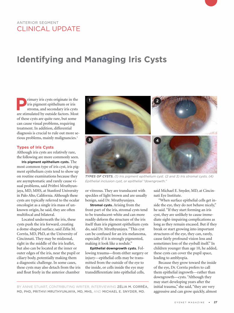
Iris pigment epithelium cysts. The most common type of iris cyst, iris pigment epithelium cysts tend to show up on routine examinations because they are asymptomatic and rarely cause visual problems, said Prithvi Mruthyunjaya, MD, MHS, at Stanford University in Palo Alto, California. Although these cysts are typically referred to the ocular oncologist as a single iris mass of unknown origin, he said, they are often multifocal and bilateral.
Located underneath the iris, these cysts push the iris forward, creating a domeshaped surface, said Zélia M. Corrêa, MD, PhD, at the University of Cincinnati. They may be midzonal, right in the middle of the iris leaflet, but also can be located at the inner or outer edges of the iris, near the pupil or ciliary body, potentially making them a diagnostic challenge. In some cases, these cysts may also detach from the iris and float freely in the anterior chamber
or vitreous. They are translucent with speckles of light brown and are usually benign, said Dr. Mruthyunjaya.
Stromal cysts. Arising from the front part of the iris, stromal cysts tend to be translucentwhite and can more readily deform the struc ture of the iris itself than iris pigment epithelium cysts do, said Dr. Mruth yunjaya. “This cyst can be confused for an iris melanoma, especially if it is strongly pigmented, making it look like a nodule.”
Epithelial downgrowth cysts. Following trauma—from either surgery or injury—epithelial cells may be transmitted from the outside of the eye to the inside, or cells inside the eye may transdifferentiate into epithelial cells,
said Michael E. Snyder, MD, at Cincinnati Eye Institute.
“When surface epithelial cells get inside the eye, they do not behave nicely,” he said. “If they start forming an iris cyst, they are unlikely to cause immediate sightimpairing complications as long as they remain encased. But if they break or start growing into important structures of the eye, they can, rarely, cause fairly profound vision loss and sometimes loss of the eyeball itself.” In children younger than age 10, he added, these cysts can cover the pupil space, leading to amblyopia.
Because they grow toward the inside of the eye, Dr. Corrêa prefers to call them epithelial ingrowth—rather than downgrowth—cysts. “Although they may start developing years after the initial trauma,” she said, “they are very aggressive and can grow quickly, almost
BY ANNIE STUART, CONTRIBUTING WRITER, INTERVIEWING ZÉLIA M. CORRÊA, MD, PHD, PRITHVI MRUTHYUNJAYA, MD, MHS, AND MICHAEL E. SNYDER, MD.
TYPES OF CYSTS. (1) Iris pigment epithelium cyst. (2 and 3) Iris stromal cysts. (4) Epithelial inclusion cyst, or epithelial “downgrowth.”
1
3
2
4
28 • J A N U A R Y 2 0 1 8
like a tumor, acquiring a surprising size.” Another interesting feature of these cysts, said Dr. Mruthyunjaya, is the proteinaceous reflectivity of the cystic fluid—they are not completely clear.
Confirming a Cyst Diagnosis“I want to be able to properly identify the location and origins of a cyst,” said Dr. Mruthyunjaya. “That will involve a thorough history; complete examination, including a gonioscopy and examination of the anterior segment; and imaging. Sometimes looking at both eyes also provides clues about the etiology of the cyst.”
In the case of a suspected downgrowth cyst, he added, confirming etiology might also involve looking for surgical wounds or previous operative notes.
Don’t overlook history. “Curious and peculiar stories have underscored the importance of historytaking for me,” said Dr. Corrêa. She recounted the story of a civil engineer who felt “a slight snag” in his eye while overseeing the paving of a road. A tiny piece of gravel had lodged in his iris, but it didn’t raise red flags until months later when his vision became blurry from a developing cyst. Complicating matters, said Dr. Corrêa, the cyst looked like a tumor because the gravel was dark in color.
Dr. Snyder mentioned another instance in which historytaking is critical: Ask patients if they have traveled to other parts of the world where parasites are endemic; they may have experienced a parasitic infection. “Parasites that travel to the eye, such as Cys-ticercus cellulosae, can cause cysts that can be quite dangerous. If you inadvertently open the cyst and expose the parasite to the eye, it can die, causing a significant inflammatory response.”
Other questions to ask. Dr. Corrêa counsels colleagues to take time to talk with the patient and ask questions like these: How long have you been aware of the cyst? How long has it been since your last ophthalmology visit? What kind of exam did you receive? Did you have your pupils dilated?
If a cyst appears suddenly, said Dr. Mruthyunjaya, it’s worth asking whether the patient is on prostaglandin analogs, which can affect cyst size. Alert
the doctor in charge of monitoring the eyedrops, especially if the cyst has become quite large.
Imaging. To rule out other problems and diagnose an iris cyst, ophthalmologists may use either anterior segment OCT or highresolution ultrasound biomicroscopy (UBM), said Dr. Corrêa. “As long as you scan the whole cyst and have a good enough image of the lesion, you’re in great shape,” she said, adding that each diagnostic modality has its advantages.
Advantages of OCT. Anterior segment OCT may identify small cystic structures, giving early confirmation of diagnosis. However, it does have limitations. For example, in patients with dark irides, the OCT signal may become attenuated, said Dr. Mruthyunjaya. Also, the pigmentation or size of the cyst may prevent ideal resolution, he said. “But I’ve been impressed with the ability to penetrate even small cysts,” he said, adding that repeatedly scanning the same area also provides a useful way to follow up on a cyst’s growth over time. OCT also has the advantage of no contact with the patient’s eye, said Dr. Corrêa, which can be especially helpful for younger patients.
Although Dr. Mruthyunjaya finds
OCT helpful in imaging peripheral cysts, Dr. Corrêa warned that it may not be possible to see the full extent of these cysts with OCT, especially if they are large.
Advantages of ultrasound. Highresolution ultrasound of the anterior segment provides a very good view, allow ing you to visualize thin walls and hollow cavities, indicating a fluidfilled cyst, versus the solid nature of a malignancy, said Dr. Snyder. In addition to looking at the quadrant of the suspected cyst, Dr. Mruthyunjaya’s initial evaluation involves a scanning protocol around the peripheral part of the iris and ciliary body—as well as the other eye. “It’s striking how often you will find tiny cysts in multiple locations that weren’t detected clinically.”
Dr. Corrêa also uses UBM for surgical planning. “It’s easier to turn the probe around and get a feel for the extent of the cyst and consistency of the tissue.” Those who lack experience with UBM should consider referring this out, she said.
What’s next? If you see a lesion with a solid component, you might confirm this with magnetic resonance imaging, said Dr. Corrêa, and even biopsy for confirmation of malignancy.
When Cancer Is a Concern
“Although it is easier to assume that small lumps or bumps in the iris are benign,” said Dr. Corrêa, “in the back of your mind, you need to consider other more serious differential diagnoses. Always remember that tumors can be occasionally associated with cysts or have a cystic component.”
Most worrisome. In her practice, Dr. Corrêa has seen a number of iridocil-iary melanomas with a cystic component located right at the root of the iris. “I’ve also seen patients with medulloepithelioma, a rare type of neuroectoder-mal tumor that can be aggressive and have a prominent cystic component, especially ones in the ciliary body.” Most worrisome, she said, are tumors in the transition between the iris and ciliary body. They are usually hidden, harder to diagnose, and trickier to treat. “You must rely on imaging such as high-resolu-tion ultrasound because it’s hard to visualize them directly or indirectly,” she said.
Other serious signs. “Any time you see an iris lesion associated with prom-inent episcleral blood vessels, unexplained acute increase in intraocular pres-sure, or iris heterochromia,” she said, “a neoplasm should be in the differential diagnosis.” Also, patients with localized graying or darkening of the sclera should be cautiously evaluated to rule out a melanocytic tumor, she said. Using transillumination, it is possible to identify a melanocytic lesion, which almost always will cast a dark shadow.

E Y E N E T M A G A Z I N E • 29
When Treatment Is NeededIn most cases, Dr. Corrêa simply observes iris pigment epithelial and stromal cysts.
Conditions dictate choices. However, said Dr. Corrêa, treatment may be needed if there are other problems such as increasing eye pressure or multiple cysts occluding the angle. Consider other conditions as well, said Dr. Mruthyunjaya: Is there an incomplete wound involved in epithelial downgrowth that you need to address? What is the size of the cyst and its velocity of growth? Is it intermittently leaking and causing inflammation? Is it rubbing against ocular structures such as the lens or the cornea, causing secondary problems?
Treat it like a tumor? Dr. Snyder said it is most effective to treat epithelial downgrowth cysts as though they are tumors, as they cause unrestricted growth of cells where they don’t belong. “Sometimes, treatment’s one and done,” said Dr. Mruthyunjaya, “but sometimes it requires multiple attempts at controlling the cyst. In other cases, reduction is good enough. Regardless, you need to watch them closely.”
What to consider beforehand. You can use a variety of options to treat epithelial downgrowth cysts. “But whatever you do with these cysts,” said Dr. Corrêa, “you must remove every little bit of abnormal epithelium from the anterior chamber, or the cyst may recur, proliferate rapidly, and cause a lot of damage inside the eye.” Before any procedure, ensure that imaging has revealed the full extent, size, and location of the cyst, she said, so you can approach it the right way.
Excision. To cause less collateral damage, Dr. Mruthyunjaya prefers excising these cysts when they are small. Dr. Snyder recommends a partial lamellar iridocorneal trabeculectomy—removing the internal eye wall and the areas the cyst is touching—to avoid rupturing or breaking the wall of the cyst. “If this procedure creates a serious iris defect,” he said, “it often causes glare or light sensitivity. If it is not possible to surgically close the area that’s been removed, an artificial iris may be needed.”
Drainage and injection. Dr. Corrêa also likes to treat ingrowth cysts by using a needle to drain the cyst, inject alcohol inside it, and then deflate the cyst. “In case there is any sign of residual epithelium on the surface of the iris, I use an endolaser probe to treat the area and make sure any epithelial cells are destroyed.”
To minimize the chances of leaving cells behind, Dr. Mruthyunjaya recommended avoiding going through the open aqueous humor and anterior chamber. “Try to go through the back or more peripheral part of the cyst. Consequently, if there’s anything that’s being released when you pull in and out of the eye, it’s coming right out of the eye with your needle.”
Dr. Mruthyunjaya also uses fluorescein eye stain to make sure the alcohol is contained within the cyst and not accidentally instilled into the anterior chamber, where it can be very toxic. Filling the eye with Healon viscoelastic also acts as a diffusion barrier for the alcohol, he said, but the viscoelastic must be thoroughly removed after the procedure.
Cryotherapy. Following drainage and injection, Dr. Mruthyunjaya then uses cryotherapy at the edge of the track by the limbus to sterilize any cells that may be remaining. Sometimes it is possible to freeze an entire cyst if it is tiny and located peripherally, right at the edge of the cornea at the limbus, added Dr. Snyder.
1 Shields JA, Shields CL. Asia Pac J Ophthalmol.
2017;6(1):6469.
Dr. Corrêa is professor of ophthalmology at the
University of Cincinnati in Ohio and director of
the Ocular Oncology Program. Relevant financial
disclosures: None.
Dr. Mruthyunjaya is associate professor of oph
thalmology at Stanford University and director of
ocular oncology at the Byers Eye Institute in Palo
Alto, Calif. Relevant financial disclosures: None.
Dr. Snyder is chair of clinical research and on
the board of directors at Cincinnati Eye Institute
and is associate professor of ophthalmology–af
filiated, at the University of Cincinnati in Ohio.
Relevant financial disclosures: HumanOptics: C.
See the disclosure key, page 8. For full disclo-
sures, view this article at aao.org/eyenet.
Coming in the next
FeatureMIGS Moves Forward An overview of the latest developments in microinvasive surgery and devices, and how they are expanding treatment options for glaucoma patients.
Clinical UpdateCornea Acceptance of DMEK is growing. An over-view of the procedure’s pros and cons, plus new research.
Oculoplastics What’s involved in facial recon-struction? Three experts discuss this challenging procedure.
PearlsSubmacular Hemorrhage How to treat this uncommon but serious complication.
Practice PerfectBenchmarking Know the “vital signs” of your practice.
BlinkTake a guess at the next issue’s mystery image.
For Your ConvenienceThese stories also will be available online at aao.org/eyenet.
FOR ADVERTISING INFORMATION
Mark Mrvica or Kelly MillerM. J. Mrvica Associates [email protected]
®

Ace Your MOC Exams with the Ultimate Ophthalmic Study GuidePracticing Ophthalmologists Learning System, 2017-2019 is an innovative program that will help you successfully prepare for maintenance of certification exams with more than 4,000 self-assessment questions covering 1,400 topics. It’s also perfect for lifelong learning and self-assessment.
Order todayaao.org/learningsystem

E Y E N E T M A G A Z I N E • 31
RETINA
CLINICAL UPDATE
Outer Retinal Tubulation: Sign of Neurodegeneration
Fo
r fu
ll cr
edit
, see
th
is a
rtic
le a
t aa
o.o
rg/e
yen
et.
If you use high-resolution imaging to guide treatment of retinal diseases, the most important thing to know
about outer retinal tubulation (ORT) structures is that they commonly appear in cross-sectional images as features that look somewhat like intraretinal cysts—but aren’t.
The tubular structures in ORT- affected eyes appear on high-resolu-tion, spectral-domain ocular coherence tomography (SD-OCT) B-scans as well-defined circular or ovoid areas of hyporeflectivity, surrounded by a hyperreflective band, said K. Bailey Freund, MD, who coauthored the first clinical description of ORT.1
“If you did not know that these lesions existed, particularly if you were using the older time-domain OCT devices, you could easily confuse them with cysts or subretinal fluid,” said Dr. Freund, who practices in New York City. “And if you were using a treatment regimen that was guided by presence or absence of fluid, you could end up giving intravitreal injections to an eye that really didn’t need them.”
Many Roads Lead to ORTDespite this narrow clinical utility, ORT has garnered increasing attention over the last several years because of the realization that tubulations in the outer nuclear layer represent a common neurodegenerative pathway in a variety of retinal diseases. These include neo-
vascular age-related macular degener-ation (AMD); geographic atrophy (GA) secondary to AMD and other disorders; inherited retinal diseases, particularly choroideremia; and mitochondrial diseases.2
Prognostic value. In all of these conditions, the presence of ORT structures indicates disorganized outer retinal layers, irreversible photoreceptor damage, and a worse visual prognosis, Dr. Freund said. “Most people would consider ORT a sign that, at least in that one particular area of the macula, you’re not going to be able to recover
visual function,” he said.A better understanding of the ORT
process eventually might yield clues about how to stop photoreceptors from dying, said Glenn J. Jaffe, MD, at Duke Eye Center in Durham, North Carolina. In treatment trials, a biomarker like ORT might prove valuable for exclud-ing prospective participants unlikely to regain vision, he said.
“The hope would be that we could help preserve people’s vision if we can prevent the degeneration of the photo-receptors,” Dr. Jaffe said. “Whether it’s with a neurotrophic agent or some oth-er type of treatment, we are becoming more aware that we need to be able to prevent the loss of the photoreceptors in these diseases.”
BY LINDA ROACH, CONTRIBUTING WRITER, INTERVIEWING CHRISTINE A. CURCIO, PHD, K. BAILEY FREUND, MD, AND GLENN J. JAFFE, MD.
ORT ON OCT. (1A) Representative SD-OCT B-scan of 3 ORT cross-sections (yellow arrowheads) in an 81-year-old woman with neovascular AMD. Two closed ORT structures are evident on the left, and a branching tubulation can be seen on the right. A hyporeflective wedge (pink arrowhead) and Bruch membrane (cyan arrow-head) are also evident. (1B) High-resolution histology section of degenerate cones in ORT, at 1.5 mm from the fovea, in a different 81-year-old woman with neovas-cular AMD. Cone lipofuscin (green arrowheads), mitochondria in outer fiber (red arrowheads), Müller cell body (white arrowhead), and ORT cross-sections (yellow arrowheads) are indicated. BLamD = basal laminar deposit; Ch = choroid; fv.s = fibrovascular scar; INL and ONL = inner and outer nuclear layer; M = lipid-containing macrophage; RPE = entombed and melanotic retinal pigment epithelium.
1A 1B
32 • J A N U A R Y 2 0 1 8
Clues from CATT. Dr. Jaffe said he became interested in ORT because of what he observed during the Compari-son Age-related Macular Degeneration Treatment Trials (CATT).3 “We were the OCT reading center for CATT, and while we were looking at the images I started to notice that ORT [structures] were becoming more frequent as the study went on. So we looked at the percentages and found that by 5 years, about 1 in 5 patients had this character-istic appearance,” Dr. Jaffe said.
Genesis of TubulationThe tubules characteristic of ORT are the retina’s heroic yet last-ditch attempt to protect localized areas of macular cone photoreceptors from the failure of the underlying retinal pigment epithe-lium (RPE), said Christine A. Curcio, PhD, at the University of Alabama at Birmingham.
“What ORT lesions have in com-mon with each other is that the RPE is dying or is gone. These are responses of retina cells to the extreme stress of this detachment,” Dr. Curcio said.
In 1996, her research group pub-lished the first histological descriptions of photoreceptors surviving in myste-rious interconnected tubes in the mac-ulas of eyes with neovascular AMD.4 It wasn’t until 2009 that Dr. Freund and his colleagues, using SD-OCT images, published the first clinically oriented paper about “a peculiar outer retinal morphologic change occurring in a variety of advanced degenerative retinal disorders.”1
Since those publications, Drs. Curcio and Freund have worked together to correlate high-resolution histopathol-ogy in donor eyes with eye-tracked OCT images of living eyes, in order to understand the ORT process.2,5
Gliosis implicated. Their overall conclusion: Tubulation is a protective gliotic response—ultimately futile—triggered by activated Müller cells, Dr. Curcio said. “The Müller cells are trying to take care of the remaining photore-ceptor cells. They are protecting them from the failure of the RPE and all the problems in the RPE/Bruch membrane complex.”
Dr. Freund agreed. “It’s the Müller
cells that are driving this process, and in the very late stage of tubulation you only have Müller cells and no surviving cones,” he said.
Rolling up the photoreceptors. As described recently by Dolz-Marco et al.,2 ORT occurs when the external limiting membrane (ELM), which is normally a thin horizontal reflec-tive line made by Müller cells and photoreceptors, begins “circling the wagons.” The ELM descends toward Bruch membrane and gradually begins scrolling the at-risk photoreceptors into a tubular structure. Depending on the stage of development, the outer band can appear in cross-section as flat, J-shaped, or partially (or fully) curved back on itself.
Both the inner and outer segments of the scrolled photoreceptors initially point radially into the lumen delineated by the ELM,2 and these can be seen clinically as a reflective fringe around the hyporeflective lumen (dark on the OCT).
As ORT progresses, the lumen be-comes uniformly hyporeflective as the outer segments degenerate and the in-ner segments are pulled back across the ELM. Remnants of the numerous mito-chondria in the inner segments migrate into the cell body of the degenerating cells, accounting for the reflectivity of the outer band of ORT.
In their study of 38 eyes with pre-existing ORT, Dolz-Marco et al. found that the mean time for new tubules to form was 14.9 months.2 It is unknown how long the end-stage ORT lesions persist in the retina, but in clinical experience they commonly are stable through at least 3 years of follow-up.6
Serpentine patterns on OCT. En face OCT scans of ORT-affected maculas reveal the tubules snaking in various patterns across the retina. “A branching or pseudodendritic pattern is observed mainly in association with neovas-cularization, whereas a singular tube may line the border of GA. Interest-ingly, analysis of ORT over time has shown fluctuations in ORT volume in cross-sectional SD-OCT scans, even while the ORT footprint seen with en face imaging remains constant,” Dolz-Marco et al. wrote.2
What about GA? In a subgroup of affected patients in the Geographic Atrophy Treatment Evaluation (GATE) trial, ORT was present in 65% of eyes in the atrophic region and in 26% of eyes in the junctional zone.7 But there is disagreement over what this means, Dr. Jaffe said.
“It’s a little bit unclear” as to how well the presence of ORT predicts GA progression, he said. “In one of the publications that we’ve done, we found that it was associated with more rapid progression. But at least one other group [led by SriniVas Sadda, MD, at the University of Southern California] has reported not seeing that.”8
Attention to the different stages of ORT, which were not appreciated at the time of these prior studies, might help solve this discrepancy in future studies, Dr. Freund’s group suggested.2
1 Zweifel SA et al. Arch Ophthalmol. 2009;127(12):
1596-1602.
2 Dolz-Marco R et al. Ophthalmology. 2017;124(9):
1353-1367.
3 Lee JY et al. Ophthalmology. 2014;121(12):2423-
2431.
4 Curcio CA et al. Invest Ophthalmol Vis Sci. 1996;
37(7):1236-1249.
5 Litts KM et al. Invest Ophthalmol Vis Sci. 2016;
57(6):2647-2656.
6 Jung JJ, Freund KB. Arch Ophthalmol. 2012;
130(12):1618-1619.
7 Moussa K et al. Retina. 2013;33(8):1590-1599.
8 Hariri A et al. Ophthalmology. 2015;122(2):407-
413.
Dr. Curcio is professor of ophthalmology and
director of the AMD Histopathology Lab at the
University of Alabama at Birmingham. Relevant
financial disclosures: Heidelberg Engineering: S.
Dr. Freund practices at Vitreous Retina Macula
Consultants of New York and is clinical professor
of ophthalmology at the New York Universi-
ty School of Medicine in New York City. Relevant
financial disclosures: Heidelberg Engineering: C;
Optovue: C; Spark Therapeutics: C.
Dr. Jaffe is the Robert Machemer Professor of
Ophthalmology and chief of the Vitreoretinal
Division at Duke University and director of the
Duke Reading Center at the Duke Eye Center
in Durham, N.C. Relevant financial disclosures:
Heidelberg Engineering: C.
See the disclosure key, page 8. For full disclo-
sures, view this article at aao.org/eyenet.

E Y E N E T M A G A Z I N E • 33
Kas
i San
dh
anam
an
d J
ose
ph
Ho
, Sin
gap
ore
Nat
ion
al E
ye C
entr
e
Diagnosis and Management of Central Retinal Vein Occlusion
RETINA
OPHTHALMIC PEARLS
Retinal vein occlusion (RVO) has a prevalence of 0.5%, making it the second most-common
retinal vascular disorder after diabetic retinopathy.1 RVO is classified accord-ing to the anatomic level of the occlu-sion, with 3 major distinct entities: • Central retinal vein occlusion (CRVO): occlusion of the central reti-nal vein at the level of, or posterior to, the lamina cribrosa (Fig. 1) • Hemiretinal vein occlusion (HRVO): occlusion at the disc, involving either the superior or inferior hemiretina • Branch retinal vein occlusion (BRVO): occlusion of a tributary vein, typically at the site of an arteriovenous crossing; thought to be caused by com-pression from an overlying atheroscle-rotic arteriole
This article will focus on diagnosis and management of the first entity, CRVO.
Risk Factors Systemic disorders. Systemic risk factors for CRVO include increasing age, diabetes mellitus, and hypertension. In selected cases, hypercoagulable states, including hyperhomocysteinemia and factor V Leiden mutation, or local vessel factors such as vasculitis are also associated with increased risk of CRVO. The literature also contains case reports of many other systemic conditions possi-bly associated with the development of CRVO.
Ocular conditions. Open-angle glau-coma is a major ocular risk factor for CRVO.
In addition, individuals with CRVO in 1 eye are at higher risk of developing CRVO in the fellow eye.2 In the Central Vein Occlusion Study (CVOS), 4% of patients presented with bilateral CRVO at study enrollment, and a further 5% had evidence of previous CRVO in the fellow eye at baseline. In the remaining subjects, 1.4% developed CRVO in the fellow eye during 3 years of follow-up.
Other ocular risk factors include retrobulbar external compression of the central retinal vein, as occurs in thyroid orbitopathy, or compression by intra-orbital space-occupying lesions.
Clinical PresentationPatients with CRVO typically present with a history of unilateral acute, pain-less visual loss. Visual impairment may be severe, ranging from better than 20/40 to worse than 20/200. A relative afferent pupillary defect may be present in the affected eye.
Fundus findings. Dilated fundus examination reveals unilateral disc swelling with peripapillary intraretinal hemorrhages, dilated tortuous veins, and intraretinal dot, blot, and flame hemorrhages in all quadrants, result-ing in the classic “blood and thunder” fundus appearance (Fig. 1). The macula may be edematous.
In less severe cases, disc swelling
may be absent. In subacute or late presentations in which disc swelling has resolved (with or without collateral vessel formation), the flame-shaped hemorrhages clear first, leaving deeper dot/blot hemorrhages that may be difficult to distinguish from a severe microangiopathic retinopathy such as diabetic retinopathy (Fig. 2). Fluores-cein angiography (FA) may help to confirm the diagnosis of CRVO.
Other key aspects. As part of the examination, the clinician should note the intraocular pressure and cup-to-disc ratio, which may suggest concurrent glaucoma, as well as any sequelae, such as rubeosis iridis. Undilated gonioscopic examination is important to rule out neovascularization of the angles.
Types of CRVOClinically, CRVO may be divided into 2 major subtypes: ischemic and nonisch-emic.
BY SIEHYEAN KIEW, MD, AND DANIEL S.W. TING, MD, PHD. EDITED BY SHA-RON FEKRAT, MD, AND INGRID U. SCOTT, MD, MPH.
ACUTE CRVO. Classic “blood and thun-der” fundus appearance of a patient presenting acutely with central retinal vein occlusion of the right eye.
1
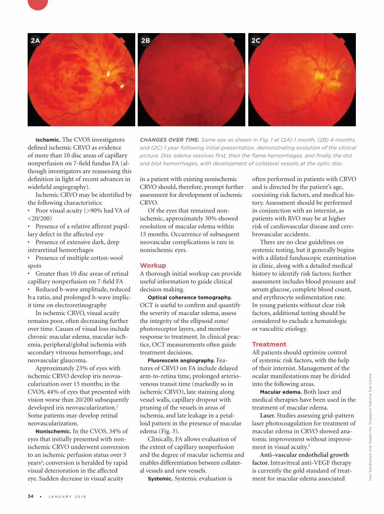
34 • J A N U A R Y 2 0 1 8
Kas
i San
dh
anam
an
d J
ose
ph
Ho
, Sin
gap
ore
Nat
ion
al E
ye C
entr
e
Ischemic. The CVOS investigators defined ischemic CRVO as evidence of more than 10 disc areas of capillary no nperfusion on 7-field fundus FA (al-though investigators are reassessing this definition in light of recent advances in widefield angiography).
Ischemic CRVO may be identified by the following characteristics:• Poor visual acuity (>90% had VA of <20/200)• Presence of a relative afferent pupil-lary defect in the affected eye• Presence of extensive dark, deep intraretinal hemorrhages• Presence of multiple cotton-wool spots• Greater than 10 disc areas of retinal capillary nonperfusion on 7-field FA • Reduced b-wave amplitude, reduced b:a ratio, and prolonged b-wave implic-it time on electroretinography
In ischemic CRVO, visual acuity remains poor, often decreasing further over time. Causes of visual loss include chronic macular edema, macular isch-emia, peripheral/global ischemia with secondary vitreous hemorrhage, and neovascular glaucoma.
Approximately 23% of eyes with ischemic CRVO develop iris neovas-cularization over 15 months; in the CVOS, 44% of eyes that presented with vision worse than 20/200 subsequently developed iris neovascularization.2 Some patients may develop retinal neovascularization.
Nonischemic. In the CVOS, 34% of eyes that initially presented with non-ischemic CRVO underwent conversion to an ischemic perfusion status over 3 years2; conversion is heralded by rapid visual deterioration in the affected eye. Sudden decrease in visual acuity
in a patient with existing nonischemic CRVO should, therefore, prompt further assessment for development of ischemic CRVO.
Of the eyes that remained non-ischemic, approximately 30% showed resolution of macular edema within 15 months. Occurrence of subsequent neovascular complications is rare in nonischemic eyes.
WorkupA thorough initial workup can provide useful information to guide clinical decision making.
Optical coherence tomography. OCT is useful to confirm and quantify the severity of macular edema, assess the integrity of the ellipsoid zone/ photoreceptor layers, and monitor response to treatment. In clinical prac-tice, OCT measurements often guide treatment decisions.
Fluorescein angiography. Fea-tures of CRVO on FA include delayed arm-to-retina time, prolonged arterio-venous transit time (markedly so in ischemic CRVO), late staining along vessel walls, capillary dropout with pruning of the vessels in areas of ischemia, and late leakage in a petal-loid pattern in the presence of macular edema (Fig. 3).
Clinically, FA allows evaluation of the extent of capillary nonperfusion and the degree of macular ischemia and enables differentiation between collater-al vessels and new vessels.
Systemic. Systemic evaluation is
often performed in patients with CRVO and is directed by the patient’s age, coexisting risk factors, and medical his-tory. Assessment should be performed in conjunction with an internist, as patients with RVO may be at higher risk of cardiovascular disease and cere-brovascular accidents.
There are no clear guidelines on systemic testing, but it generally begins with a dilated funduscopic examination in clinic, along with a detailed medical history to identify risk factors; further assessment includes blood pressure and serum glucose, complete blood count, and erythrocyte sedimentation rate. In young patients without clear risk factors, additional testing should be considered to exclude a hematologic or vasculitic etiology.
Treatment All patients should optimize control of systemic risk factors, with the help of their internist. Management of the ocular manifestations may be divided into the following areas.
Macular edema. Both laser and medical therapies have been used in the treatment of macular edema.
Laser. Studies assessing grid-pattern laser photocoagulation for treatment of macular edema in CRVO showed ana-tomic improvement without improve-ment in visual acuity.2
Anti–vascular endothelial growth factor. Intravitreal anti-VEGF therapy is currently the gold standard of treat-ment for macular edema associated
CHANGES OVER TIME. Same eye as shown in Fig. 1 at (2A) 1 month, (2B) 4 months, and (2C) 1 year following initial presentation, demonstrating evolution of the clinical picture. Disc edema resolves first, then the flame hemorrhages, and finally the dot and blot hemorrhages, with development of collateral vessels at the optic disc.
2A 2B 2C

E Y E N E T M A G A Z I N E • 35
Kas
i San
dh
anam
an
d J
ose
ph
Ho
, Sin
gap
ore
Nat
ion
al E
ye C
entr
e
with CRVO. There is increasing evi-dence that anti-VEGF therapy results in lower risk of visual loss, higher rates of visual gains, greater reduction in central retinal thickness, and reduced risk of progression to iris neovascular-ization.
For example, the CRUISE study reported that intravitreal ranibizumab significantly improved best-corrected visual acuity (BCVA) at 6 and 12 months compared with sham injec-tions. In the open-label extension HORIZON trial, the eyes initially treated with sham and subsequently treated with ranibizumab showed im-provement in BCVA but did not catch up to the visual outcomes attained by the group that received ranibizumab at enrollment. This finding suggests that delaying treatment for macular edema has adverse effects on visual outcomes.
Aflibercept, a VEGF-trap mole-cule, has also been shown to improve BCVA compared with sham and laser treatment in the COPERNICUS and GALILEO trials.
More recently, SCORE2, a random-ized noninferiority trial including eyes with CRVO or HRVO, demonstrated that bevacizumab was noninferior to aflibercept in terms of visual acuity gain at month 6 compared to baseline
(mean improve-ment of 18.6 vs. 18.9 ETDRS letters, respec-tively).3
Corticoste-roids. Cortico-steroids reduce retinal capillary permeability and inhibit the expression and metabolic path-way of VEGF. The SCORE-CRVO trial demonstrat-ed that intravitre-al triamcinolone acetonide was superior to obser-vation for visual loss associated with CRVO-relat-ed macular ede-
ma. The GENEVA trial evaluated the use of a sustained-release intravitreal dexamethasone implant (Ozurdex) and demonstrated improvements in visual acuity and macular thickness compared with both sham and laser-treated groups.
More recently, the Clinical Efficacy and Safety of Ranibizumab Versus Dexamethasone for Central Retinal Vein Occlusion (COMRADE C) trial compared intravitreal ranibizumab 0.5 mg (monthly for at least 3 months, followed by as-needed dosing) to a single injection of Ozurdex. This trial reported similar efficacy between ranibizumab and Ozurdex but found a higher incidence of adverse effects in the group receiving Ozurdex.
Retinal ischemia. Current evidence recommends regular monitoring of patients with ischemic CRVO for development of iris or angle neovas-cularization, for which panretinal laser photocoagulation (PRP) remains the mainstay of treatment.
There is currently no evidence to recommend prophylactic treatment prior to the development of new ves-sels. However, in circumstances where regular follow-up is impractical and the degree of ischemia is severe (high risk of progression to neovascularization), prophylactic PRP may be appropriate.
Anti-VEGF agents are antiangiogen-ic and may be useful adjuncts to PRP in the management of patients with CRVO and associated anterior segment neovascularization, particularly when the view of the fundus is not sufficient-ly clear to permit adequate PRP.
Venous outflow. A number of alter-native therapies focused on improving retinal blood flow have been described. These include the use of antiplatelet agents (e.g., ticlopidine),4 hemodilu-tion,5 and thrombolytic agents de-livered systemically, intravitreally, or directly into a retinal vein during pars plana vitrectomy.
Techniques to alleviate a possible compartment syndrome, with optic nerve sheath decompression through an orbital approach or radial optic neurotomy via a pars plana approach, have been tried. However, these are no longer used because of their limited benefit and significant risks.
Creation of a laser chorioretinal venous anastomosis (L-CRA) to by-pass the occluded central retinal vein has been reported to be beneficial in nonischemic CRVO, with improvement in visual acuity and reduced rates of ischemic progression,6 but less so in eyes with the ischemic type of disease. The failure of anastomosis was most likely due to endothelial cell damage secondary to ischemia.7
1 Klein R et al. Arch Ophthalmol. 2008;126(4):
513-518.
2 Central Vein Occlusion Study Group. Arch Oph-
thalmol. 1997;115(4):486-491. [Erratum in Arch
Ophthalmol. 1997;115(10):1275.]
3 Scott IU et al. JAMA. 2017;317(20):2072-2087.
4 Yamamoto T et al. Am J Ophthalmol. 2004;138(5):
809-817.
5 Wolf S et al. Graefes Arch Clin Exp Ophthalmol.
1994;232(1):33-39.
6 McAllister IL et al. Ophthalmology. 2010;117(5):
954-965.
7 Kwok AK et al. Br J Ophthalmol. 2003;87(8):
1043-1044.
Dr. Kiew is an ophthalmology resident at the
Singapore National Eye Centre. Dr. Ting is an
associate consultant at the Singapore National
Eye Centre and assistant professor at Duke-Na-
tional University Singapore. Relevant financial
disclosures: None.
FLUORESCEIN FINDINGS. FA at 4 time points shows (3A) masking from intraretinal hemorrhages, (3B) delayed arte-riovenous transit time, (3C) leakage at the swollen optic disc, and (3D) late staining of the vessel walls.
3A
3C
3B
3D

EyeNet and JRS ad: 8.125x10.875”
Enhance your clinical knowledge and connect with leading experts in refractive and anterior segment surgery with your ISRS membership.
Join now or learn more at www.isrs.org.

E Y E N E T M A G A Z I N E • 37
WHAT’S YOUR DIAGNOSIS?
MORNING ROUNDS
Sw
etan
gi B
hal
eeya
, MD
The Case of Aches and Pains and Blurry Vision
Becky Brown*, a 28-year-old Caucasian woman, was doing well until about 2 weeks prior
to presentation, when she felt as if she had the flu, complete with headaches and joint pain. A few days later, she noted that her eyes were red, painful, and highly sensitive to light. Her vision was also becoming blurred. Very wor-ried about her symptoms, Ms. Brown sought medical help.
We Get a LookPrior to evaluation by our vitreoretinal service, Ms. Brown had been treated by a primary care provider with oral methylprednisolone. By the time she came to our clinic, her arthralgia had resolved with steroid treatment, but her headaches and blurred vision persisted. She did not have any other significant ocular or medical history.
Exam findings. On presentation, Ms. Brown’s visual acuity was 20/40 in the right eye and 20/80 in the left eye. Intraocular pressure was 13 mm Hg in each eye. Her pupils were equal, round, and reactive, with no afferent pupillary defect. She had full ocular motility bilaterally.
We observed conjunctival injection in both eyes. Slit-lamp exam showed fine, diffuse keratic precipitates, as well as 2+ cells and fibrin in the anterior chamber. Trace anterior vitreous cells were also noted.
Fundus lesions. Dilated fundus
exam revealed optic disc hyperemia with obscured borders. Multiple creamy, yellow-white placoid lesions were visible in the posterior pole adjacent to the superior and inferior arcades, with extension into the central
macula. The lesions appeared to be at the level of deep retina and the retinal pigment epithelium (Fig. 1). The peripheral retina appeared normal.
Imaging. To augment the exam findings, we obtained the following imaging studies.
Optical coherence tomography. OCT demonstrated multiple hyper-reflective discrete areas at the level of
BY ALEC CHALEFF, MD, MBA, HERSHEL PATEL, MD, MS, AND SWETANGI BHALEEYA, MD. EDITED BY STEVEN J. GEDDE, MD.
AT PRESENTATION. Fundus photos of the patient’s right (1A) and left (1B) eyes. OCT images of central macula in the right (2A) and left (2B) eyes.
1A
2A
2B
1B

38 • J A N U A R Y 2 0 1 8
Sw
etan
gi B
hal
eeya
, MD
the outer nuclear layer and ellipsoid band, with attenuation of the normal retinal architecture (Fig. 2; see also online Figs. 2A-2D).
Fundus autofluorescence. FAF showed diffuse areas of hyperfluores-cence corresponding to the lesions in the posterior pole and some mottled areas of hypofluorescence (see online Fig. 3).
Fluorescein angiography. FA revealed hypofluorescence or blockage corresponding to areas of the lesions in the early phase, followed by hyper-fluorescence in the late phase consistent with staining (Fig. 3A; see also online Fig. 4).
Indocyanine green angiography. ICGA disclosed confluent areas of hypofluorescence in the posterior pole and midperiphery in both the early and late stages. On ICGA, the lesions were found to be more numerous than what was apparent on examination or FA, especially in the midperiphery (Fig. 3B; see also online Fig. 4).
Making the DiagnosisBased on the history, exam, and im-aging findings, our working diagnosis was acute posterior multifocal placoid pigment epitheliopathy (APMPPE). The creamy, yellow-white placoid reti-nal lesions we noted in Ms. Brown are characteristic funduscopic findings1; similarly, the early-phase blockage or hypofluorescence followed by diffuse late staining in the same areas are typi-cal of APMPPE.2,3
However, her fundus appearance also raised the possibility of serpiginous choroiditis or serpiginous-like choroi-dopathy associated with tuberculosis. Thus, we ordered a Quantiferon-TB Gold test, which was negative, ruling out the alternative diagnosis.
DiscussionAPMPPE, one of the white dot syn-dromes, was first described by Gass in 1968.4 It is an acute-onset bilateral disease with characteristic findings of multiple yellow-white patches of cho-rioretinal inflammation in the posterior pole. The mean age of onset is 27 years, with 85% of cases occurring in patients 16 to 40 years of age.1 APMPPE is
considered to be rare, and the true inci-dence and prevalence are unknown.1
Pathogenesis. Although the etiology of APMPPE is unclear, up to one-third of patients experience a viral prodrome before the onset of ocular symptoms. The pathogenesis is thought to be obstruction of choriocapillary lobules, possibly as an inflammatory reaction to a viral infection. Alternatively, APMPPE may be a primary disease of the retinal pigment epithelium itself.2
Systemic symptoms and complica-tions. APMPPE is a vasculitis that is often accompanied by a viral pro-drome, and systemic manifestations may include headaches, flulike symp-toms, erythema nodosum, arthralgias, and myalgias.1
Neurologic symptoms such as head-aches, transient aphasia, paresthesias, and muscular weakness may be associ-ated with cerebral vasculitis and require further evaluation.1 Thomas et al. reviewed 18 patients diagnosed with APMPPE; of these, 11 patients (61%) had neurologic symptoms, with the most common being headache (9 pa-tients).5 Stroke due to cerebral vasculi-tis was the most severe complication.5
Although there is no sexual predilec-tion for APMPPE per se, the majority of cerebral vasculitis cases occur in men.6
What About Our Patient’s Headaches?Ms. Brown’s symptoms of headaches along with ocular findings consistent with APMPPE were concerning for cerebral vasculitis, and we recommend-
ed urgent neurology consultation. She underwent extensive testing with neuroimaging, including conventional and computed tomography angiogra-phy, which did not demonstrate any evidence of cerebral vasculitis.
Further studies. Examination of her cerebrospinal fluid (CSF) showed nonspecific lymphocytic pleocytosis with normal protein and glucose levels. Polymerase chain reaction studies of the CSF found no evidence of herpes simplex virus, varicella-zoster virus, or cytomegalovirus infection.
Direct antigen testing of the CSF was also performed, and the results were negative for Haemophilus influ-enzae, Streptococcus pneumoniae, and Neisseria meningitidis.
In addition, the following laboratory studies were ordered as part of her neurology evaluation: rapid plasma reagin (RPR) and fluorescent trepone-mal antibody absorption (FTA-ABS) tests for syphilis, Quantiferon-TB Gold, and Lyme disease antibody testing. All were negative.
TreatmentAPMPPE is typically a self-limited condition. No particular therapy has proved to be beneficial, although some patients with severe ocular inflamma-tion or signs and symptoms of cerebral vasculitis are treated with systemic corticosteroids.
The chorioretinal lesions may begin to resolve in 1 to 2 weeks even without treatment.4 Visual prognosis is usually good, and patients can expect improve-ment in visual acuity in the long term;
3A 3B
FA VS. ICGA. Left eye at presentation as seen on early-phase FA (3A) and ICGA (3B).

E Y E N E T M A G A Z I N E • 39
however, visual recovery is often in-complete.3
Our patient’s course. Ms. Brown was started on topical prednisolone acetate 6 times per day and atropine twice a day in both eyes for the anterior chamber inflammation.
Treatment with systemic corticoste-roids was deferred until she completed evaluation for cerebral vasculitis, which was negative. We decided to initiate treatment with systemic steroids because of Ms. Brown’s persistent ocular in-flammation and lack of visual improve-ment. She was treated with a course of 60 mg of oral prednisone daily for 10 days, which was then tapered weekly over 4 weeks.
Follow-upAfter 1 month of treatment, Ms. Brown’s visual acuity had improved to 20/20 in the right eye and 20/40 in the left eye. Fundus exam showed pigmentary changes in area of the yellow-white placoid lesions (see online Fig. 5), which were better seen by FAF. The lesions, as seen on OCT, improved with restoration of near-normal retinal architecture (see online Fig. 6).
*Patient name is fictitious.
1 Kooragayala LM. http://emedicine.medscape.
com/article/1225531-overview. Updated May 3,
2017. Accessed November 21, 2017.
2 American Academy of Ophthalmology. Basic
and Clinical Science Course: Section 12, Retina and
Vitreous. 2013-2014: pp 185-187.
3 Fiore T et al. Retina. 2009;29(7):994-1001.
4 Gass JD. Arch Ophthalmol. 1968;80(2):177-185.
5 Thomas B et al. J Ophthalmic Inflamm Infect.
2012;2(3):125-131.
6 Case D et al. J Stroke Cerebrovasc Dis. 2012;2(3):
125-131.
Dr. Chaleff is a first-year transitional resident, Dr.
Patel is a second-year ophthalmology resident,
and Dr. Bhaleeya is a professor of ophthalmolo-
gy; all are at the University of South Florida Eye
Institute, Morsani College of Medicine, in Tampa.
Relevant financial disclosures: None.
MORE ONLINE. See this article at aao.org/eyenet for additional
OCT, FAF, FA, and ICGA images before and after treatment.
The Academy’s newest scientific publication, Ophthalmology® Retina, focuses on advances in medical drug treatment, surgery and technology for retina-related disesases and conditions.
Subscribe at aao.org/store.
High-quality research from the world’s leading ophthalmic journal

40 • J A N U A R Y 2 0 1 8

E Y E N E T M A G A Z I N E • 41
Global Ophthalmology
Volunteer ophthalmologists are ramping up the fight against eye disease by building capacity in communities
around the world. Here’s how you can help.
By Mike Mott, Contributing Writer
VOLUNTEERING ABROAD IS UNDERGOING A SEA CHANGE. GONE are the days of simply flying overseas to an under-resourced community, performing hundreds of cataract surgeries, and returning home to your
practice. Most communities now have their own health systems in place with local physicians who can perform procedures at a lower cost than in the past.
Rethinking the RationaleInstead of “delivering care,” the buzzword in medical volunteerism is now “build-ing capacity.”
Magnify your impact. “Given the cataract backlog in the world today, we are more than 100 years from eradicating this cause of blindness if we simply rely on delivery of care,” said Jeff H. Pettey, MD, at the University of Utah in Salt Lake City. “Yes, performing cataract surgeries will impact individuals, but it pales in comparison to the overall impact you’ll have if you build capacity in an individu-al, a practice, and a country.”
Build local partnerships. How is this done? By partnering with individuals and groups to improve education and training. By procuring equipment and connect-ing communities to local resources. By bringing local physicians to international meetings and uniting them with their ophthalmic societies. “It’s about substitut-ing single-episode surgical care with impact that functions 365 days a year,” said Dr. Pettey. “We as volunteers transfer our skills to a community and empower a population because it’s the members of that community who are best able to care for one another.”
And the more boots on the ground the better. Here’s how you can play a role in the leading edge of global ophthalmology.
Why They Do ItVolunteering is a uniquely individual experience, as each community has distinct needs with its own rewards and challenges. At the same time, each volunteer brings his or her own motivations.
STRABISMUS. Dr. Safran, shown with a young boy who had strabismus surgery that was provided via Healing the Children in Guayaquil, Ecuador.C
ou
rtes
y o
f M
arc
Saf
ran
, MD

42 • J A N U A R Y 2 0 1 8
Marc Safran, MD, who practices in Liverpool, N.Y., has volunteered in Bolivia, Ecuador, India, and Viet-nam. Doing work overseas allows him to reconnect with the core of why he originally went into medicine. “It’s a throwback to providing medical care to needy people in a free and unencumbered way,” said Dr.
Safran, “without elec-tronic health records, MIPS, MACRA, and all of the bureaucratic hassles we now face at home.”
For Mitchell V. Brinks, MD, MPH, the motivation to volunteer overseas in areas like Cambo-dia, Guatemala, and Myanmar stems from the power of human connection. “It’s a pretty special feeling to know you have friends and a sense of home in another part of the world,” said Dr. Brinks, at Oregon Health & Science University’s Casey Eye Institute in Portland. “Cataract surgery, for example, can open the door to
do so much good in so little time. And to have that access to the inside of a community? That’s pretty rare. But we as ophthalmologists have the key—it’s sitting there right in our hands.”
Become a Better ClinicianVolunteering might mean different things to different ophthalmologists, but all agree on one thing: Whether you are a resident, a young ophthalmologist new to practice, or a seasoned vet, the skills you learn doing mission work will have a significant impact on your practice at home.
Sharpen your skills. “There might not be a better way to hone your skills as an ophthalmologist,” said Dr. Pettey. “You’ll be faced with the most complicated cases and forced to make important decisions in incredibly difficult environments. Afterwards, little will faze you. You return more focused and more capable of making difficult decisions on the fly. When done right, you return a better surgeon.”
You’ll also see the natural progression of diseases in a way that’s just not part of most ophthalmologists’ day-to-day experience in the United States. During his travels, Dr. Safran has witnessed child after child with advanced strabismus, which could have been
easily corrected if eyeglasses were available. “Seeing the natural path of uncorrected disease gives you a genuine appreciation of its true nature and severity,” he said. “Whether it’s amblyopia, congenital cataract, or ptosis, I’ve gained a better appreciation for biology in a way that’s not possible in the normal U.S. clinic, where we have more early interventions [available].”
Deepen connections with patients. Not only can volunteering abroad make you a better surgeon, but it also will teach you how to build better physician-pa-tient relationships. “By having the opportunity to work with people from different cultures and backgrounds, I’m building a skill set that translates to a lot of differ-ent scenarios domestically,” said resident Travis Redd, MD, MPH, at the Casey Eye Institute.
Andreas K. Lauer, MD, also at the Casey Eye Insti-tute, agreed. “In my experience, it’s been really helpful to learn more about other cultures firsthand so that when I return home, I can interact more meaningfully with patients from diverse backgrounds. If I can tell a patient, ‘Hey, I recently visited your country,’ there’s automatically a better connection.”
Gain greater perspective. In addition to giving you a broader and deeper perspective on another culture, working abroad can also give you a new perspective on your own practice. “Volunteering gives you a window like no other into how commercialized and money -oriented medicine has become here in the United States,” said Dr. Brinks. “Many times, we as physicians buy into the earnings and the patient volume as our metrics of success. We can take it too seriously. Getting out of the clinic and setting foot in another world can help you build another model of professional satis-faction. It can make ophthalmology a truly enjoyable, long-term endeavor.”
For Dr. Redd, his mission work as a resident has made him rethink the role of public health in the prac-tice of ophthalmology. “Going overseas to places like Uganda and Ethiopia reminds me to think about med-icine in a different way when I return home. It’s more than just, how am I going to treat someone? It’s, how are my patients getting to the exam room? What hoops do they have to jump through? How can I better recruit my patients and better serve my local community?”
Getting StartedThere are several ways to approach volunteering abroad, depending on your career stage.
Residents. For ophthalmologists still in training, many U.S. residency programs have integrated overseas experiences into their educational program. And some of them will help subsidize a resident’s travel expenses and other costs. For example, the International Oph-thalmology Program at the Casey Eye Institute allows ophthalmology residents to travel internationally to areas like Fiji to learn low-cost alternative surgical skills, such as manual small-incision cataract surgery, for
BUILDING CAPACITY. Today’s No. 1 goal is to help build programs that will continue long after the volunteer returns home. Friendships— as the one between Dr. Brinks (left) and Benjamin D. Siatu’u, MD, who practices in Pago Pago, American Samoa—are a bonus.
Mit
chel
l V. B
rin
ks, M
D, M
PH

E Y E N E T M A G A Z I N E • 43
treating patients in underserved communities.Fellows. A small but growing number of institutions
around the United States have also created global oph-thalmology fellowships to help young ophthalmologists put their training at home into practice abroad. These include the following:• Dean McGee Eye Institute’s Global Eye Care Fellow-ship (University of Oklahoma) • Emory Eye Center’s Global Ophthalmology Fellow-ship (Emory University) • Moran Eye Center’s Moran International Ophthal-mology Fellowship (University of Utah)• Truhlsen Eye Institute’s Prevention of Global Blind-ness Fellowship (University of Nebraska) • Wills Eye Center for Academic Global Ophthalmolo-gy Fellowship (Wills Eye Hospital)
Midcareer and older physicians. More established ophthalmologists may want to consider volunteer-ing through a charitable or nonprofit organization. Groups like Orbis International (www.orbis.org), the Seva Foundation (www.seva.org), and the Himalayan Cataract Project (www.cureblindness.org), for exam-ple, serve close to 100 countries and train thousands of medical professionals a year in how to build self-sus-taining programs for preserving and restoring vision. For such a trip, most ophthalmologists can expect to take a hit on the income they’d normally bring in from their own practices, but some of these organizations might be able to offset a portion of the costs, including travel and lodging.
Developing a PlanRegardless of the route you take to get involved, prospective volunteers will want to heed the following expert advice when exploring their opportunities.
Start at home. After a decade of local volunteering, Linda M. Lawrence, MD, who practices in Salina, Kan-sas, began collaborating with more than 30 nonprofit organizations everywhere from Nigeria to Vietnam. Her advice for prospective global ophthalmologists? Don’t just jump on a plane. Start by doing work around your local community.
She said, “When a new volunteer asks me how they can get involved in international work, I ask them, ‘What are you doing at home?’ Home is your training ground. Home is where you learn about people and different health care systems. It’s where you learn how to network and really how to volunteer.”
EyeCare America. You can do mission work with-out ever leaving your office, thanks to the Academy’s
TEAM EFFORT. Patients, their family members, and members of the team from Healing the Children. The group of 5 ophthalmologists and 20 staff performed 125 general strabismus cases during a week-long mission in Guayaquil, Ecuador.
Before You Go
Once the path is clear and you’ve decided on your journey, you’ll want to make sure you follow a stan-dard to-do list:• Get your passport ready. The minute you decide you are going somewhere, check the official country website and figure out what you need (e.g., passport and visa) to enter the country. Plan ahead; the mate-rials can take months to acquire.• Make sure you’re licensed. You’ll head out over-seas with the best of intentions. But if you aren’t legally approved by the host country’s government to perform surgery in your host country, you’ll be heading back home sooner than expected. • Get your vaccines. This is essential. Some coun-tries may even require proof of immunization prior to entry.• Purchase evacuation insurance. You don’t want to
become acutely ill and stranded in a foreign country that lacks proper resources. In addition to purchas-ing health insurance when traveling abroad, you should obtain evacuation insurance. This service will provide a jet or helicopter to extricate you from the direst of circumstances.• Register with the appropriate U.S. consulate. You shouldn’t expect any danger when you are working overseas, but you’ll want to make sure the U.S. State Department knows where you are in case of ca-tastrophe, natural disaster, or political unrest.• Learn the language. Doing your homework about the local language and cultural makeup will be tre-mendously helpful down the road. You don’t need to be fluent, but learning simple terms like “thank you” and “good evening” will ingratiate you with hosts and patients.M
itch
ell V
. Bri
nks
, MD
, MP
H
Co
urt
esy
of
Mar
c S
afra
n, M
D

44 • J A N U A R Y 2 0 1 8
EyeCare America program (aao.org/eyecare-america/volunteer- ophthalmologists). The program helps medically underserved seniors and those who are at increased risk for glaucoma receive eye care through dedicat-ed volunteer ophthalmologists. Once an ophthalmologist enrolls as a volunteer, eligible patients are matched online to the nearest volunteer by zip code.
Find a mentor. Dr. Pettey’s experience echoes that of Dr. Lawrence. Before taking his experience overseas, he started volunteering locally in a homeless clinic in Salt Lake City. And he is adamant about the importance of mentors. Under the guidance of Drs. Randy Olson, Alan Crandall, and Geoff Tabin at the Moran Eye Center, he learned how to translate the skills he was developing at home into the ability to provide high-quality care abroad.
“Good mentors really are the doorway to involve-ment,” Dr. Pettey said. “As a young ophthalmologist, you don’t have a clinical skill set that translates to the developing world. The cataracts you’ll encounter, for example, are the most difficult cases you’ve seen. It takes mentors who can build that clinical capacity in you before you can really get to work in a different country.”
Even for the most seasoned physician, a good mentor can prevent you from becoming a burden, added Dr. Brinks. “You’re not sleeping well, you have jet lag, you might have a bug, everything is difficult,” he
said. “Unless you have someone alongside you who’s gone through it before, you’ll have to relearn their same mistakes on patients who are in dire need. So even for the smartest ophthalmologist out there who has the best of inten-tions, there’s just too much to learn on the ground to think you can do it by yourself the first try.”
Go with a group that has ex-perience. Your first impressions of your mission site will be vital, so find an experienced organization
to join. “Inspiration is your fuel when you’re volunteer-ing abroad,” said Dr. Brinks. “And it’s delicate. You don’t want a traumatic experience to destroy it. So, research the location and go with a group that reflects your own values, because once you go overseas, the regulations and standards of care that you are accustomed to are all gone. The only things you have to guide you are the values and principles of the people you surround yourself with.”
Consider long-term impact. Finally, be sure the organization has a plan for long-term follow-up care, Dr. Redd added. “Many new volunteers think they’ll just pop into an outreach camp, work for a few days, and then return home. That might sound appealing, but you can do more harm than good in the long run if the organization has no plan for follow-up care and has no foundation built up for referral networks to help pa-tients after you leave.” You have a duty to these patients to volunteer with a responsible organization, he said.
ZIKA OUTREACH. Dr. Lawrence with a mother and infant in Brazil, both of whom were affected by the Zika virus.
The Academy’s GO Guide
“Familiarizing yourself with global ophthalmology is one of the best ways to get started,” said Dr. Pettey. “And to really prep my residents, I send them to the Academy’s Global Ophthalmology (GO) Guide.”
The guide provides members-in-training with a listing of fellowships and observerships available as well as online CME courses for all physicians who want to work overseas. Topics include the following:• “Starting in Global Ophthalmology: Mentorship, Funding, and the Work-Home Balance” • “Assessing Outcomes in Global Health Programs”• “So You Want to Work Overseas?”
You can use the GO Guide to find region-based treatment and management information on a variety of disease topics; multimedia clips pertaining to global ophthalmology; and an Academy pamphlet, “Before You Go,” in which veteran physicians discuss how to make lasting changes around the world.
Prospective volunteers will also want to check out the Academy’s “Advisory Opinion on Ethical Issues in Global Ophthalmology.” It covers the challenges involved in international ophthalmic care, including how ophthalmologists can maintain professionalism and high clinical standards while practicing abroad.
For more information about the GO Guide, visit aao.org/global-ophthalmology-guide. To read the Academy’s Advisory Opinion as well as other ethics- related material, visit aao.org/clinical-education/redmond-ethics-center and click on “Global Oph-thalmology.”
Lin
da
M. L
awre
nce
, MD

E Y E N E T M A G A Z I N E • 45
On the GroundNo matter how long you prepare for your work overseas, there are certain things that only experience can teach when you touch down on the ground.
Don’t be the hero. “As a resident or new volunteer in a foreign country, keep in mind that you are first a guest,” said Dr. Lauer. “So be respectful of the host because the host has a lot of responsibility. There’s a cost to them to ensure that the visit achieves the aim. If you go over there thinking that you’re going to run the show, you won’t be welcomed back.”
It’s also important to remember that your surgical skills or station in life should never dictate how you interact with the people you meet on the ground. “Fifteen years ago, physicians in the developing world didn’t have great access to the internet,” Dr. Safran said. “Times have changed, and now an ophthalmologist from the furthest reaches of the globe might very likely be a better surgeon than you. So, treat every person you meet—physicians, nurses, patients, community mem-bers—as a fellow human citizen.”
Don’t undercut the local economy. “This is some-thing very important that I’ve learned from decades of volunteering abroad,” said Dr. Lawrence. “Where I work in Peru, for example, I used to bring donated [glasses] frames to a local school. But a mother of one of the students is an optician in the community. Why would I want to compete with her and take away her livelihood? In the end, you want to transfer your skills and promote the local economy and infrastructure.”
Understand your limits. Perhaps the most important caveat is to set realistic expectations of yourself, the experts said. For a resident, this might mean that you simply go as a student rather than a practitioner. But even the most experienced ophthalmologist will still want to be cognizant of his or her comfort zone. Clinics
overseas might be saving up the most difficult cases for someone like you. But if it involves something that you wouldn’t normally do or involves compromising on your own safety or the safety of your patients, you should know when to say “no” and defer to someone else.
“Most of the patients you’ll see are going to be blind, and they will need a lot of care,” said Dr. Brinks. Arriving at your destination “with an undefined desire to help isn’t a sophisticated strategy. People try to work themselves to death. They’ll do things they’re not ready to do, and then there’s nobody to help fix the problem afterwards. So be safe, be calm, and be humble.”
An Unmet NeedAccording to the World Health Organization, almost 90% of the world’s visually impaired live in low-income countries. And in these areas, treatable cataracts are the leading cause of blindness.1 It’s therefore no surprise that mission work to provide cataract surgery is the leading intervention for ophthalmologists. But there are also millions of others suffering from other diseases, such as glaucoma, who require ophthalmology’s atten-tion. And this is where new recruits play a vital role.
“Manual small-incision cataract surgery is definitely an amazing type of intervention to prevent blindness,” said Dr. Lauer. “It has the ‘wow’ factor. But as we get more sophisticated about doing international missions, building capacity, and investing in public health, we need to expand our focus and find new volunteers to tackle these other conditions. It’s going to be a chal-lenge, but if we can help these patients collectively, we really will be making the world a better place.”
1 World Health Organization. Global Data on Visual Impairments
2010. www.who.int/blindness/GLOBALDATAFINALforweb.pdf.
Accessed Oct. 25, 2017.
Meet the Experts
Mitchell V. Brinks, MD, MPH Assistant professor of ophthalmology and codirec-tor of the International Ophthalmology Program at Oregon Health & Science Uni-
versity’s Casey Eye Institute in Portland. Relevant financial disclosures: None.
Andreas K. Lauer, MD Professor of oph-thalmology, vice-chair for education, and chief of the Retina-Vitreous Division at
Oregon Health & Science University’s Ca-sey Eye Institute. Relevant financial disclo-
sures: None.Linda M. Lawrence, MD In private prac-tice in Salina, Kansas. Relevant financial disclosures: None.
Jeff H. Pettey, MD Associate professor of ophthalmology and visual sciences, residen-cy program director, and medical director of global outreach at the University of Utah in Salt Lake City. Relevant financial disclosures: None.Marc Safran, MD In practice at the Clay Eye Center in Liverpool, N.Y., and associate fac-ulty professor at Upstate Medical Center in Syracuse, N.Y. Relevant financial disclosures: None.Travis Redd, MD, MPH Second-year ophthal-mology resident at Oregon Health & Science University’s Casey Eye Institute. Relevant financial disclosures: None.
For full disclosures, view this article at aao.org/eyenet.
Lin
da
M. L
awre
nce
, MD

PREPARED BY FCB
Job #: 10751079Releasing as: PDFx1A Production: Maria Abreu x3124
Colors: 4/1 (4C Process/Black) AD: Trent Orvis x2492
Client: Genentech Flat Size: 16.25”w X 10.875”h AE: Katie Majewski x7927
Product: Actemra Bleed: 16.5”w X 11.125”h Producer: Karisa Williams x3721
Client Code: ACT/071217/0075 Trim: 16.25”w X 10.875”h QC: L.Powell
Date: August 14, 2017 5:19 PM Safety: 15.75”w X 10.375”h Digital Artist: VA,
Proof: M1
Add’l Size Info: PI Page Dimensions - Trim 8.125”w X 10.875”h; Live 7.875”w X 10.625”h; Bleed 8.375”w X 11.125”h
Fonts: Helvetica Neue LT Std, Avenir M1 Spellcheck: Jacob Albrecht
FR Spellcheck:
Path: PrePress:Genentech:10751079:10751079_GCA_NOW_JA_EYE_M1
4C GCA 2017 Journal Ad: Eyenet
WHEN IS THE TIME TO START ACTEMRA?
INDICATIONACTEMRA is indicated for the treatment of giant cell arteritis (GCA) in adult patients.IMPORTANT SAFETY INFORMATIONBOXED WARNING: RISK OF SERIOUS INFECTIONS Patients treated with ACTEMRA are at increased risk for developing serious infections that may lead to hospitalization or death, including tuberculosis (TB), bacterial, invasive fungal, viral, or other opportunistic infections. If a serious infection develops, interrupt ACTEMRA until the infection is controlled.Reported infections include: • Active tuberculosis, which may present with pulmonary or
extrapulmonary disease. Patients should be tested for latent tuberculosis before ACTEMRA use and during therapy. Treatment for latent infection should be initiated prior to ACTEMRA use.
• Invasive fungal infections, including candidiasis, aspergillosis, and pneumocystis. Patients with invasive fungal infections may present with disseminated, rather than localized, disease.
• Bacterial, viral and other infections due to opportunistic pathogens.
The risks and bene� ts of treatment with ACTEMRA should be carefully considered prior to initiating therapy in patients with chronic or recurrent infection.Patients should be closely monitored for the development of signs and symptoms of infection during and after treatment with ACTEMRA, including the possible development of tuberculosis in patients who tested negative for latent tuberculosis infection prior to initiating therapy. CONTRAINDICATION ACTEMRA is contraindicated in patients with known hypersensitivity to ACTEMRA. WARNINGS AND PRECAUTIONS Gastrointestinal Perforations : Events of gastrointestinal (GI) perforation have been reported in clinical trials, primarily as complications of diverticulitis in RA patients. Use ACTEMRA with caution in patients who may be at increased risk for GI perforation. Promptly evaluate patients presenting with new-onset abdominal symptoms for early identi� cation of GI perforation. Laboratory Parameters : Laboratory monitoring is recommended due to potential consequences of treatment-related laboratory abnormalities in
neutrophils, platelets, lipids, and liver function tests. Dosage modi� cations may be required.Neutropenia : Treatment with ACTEMRA was associated with a higher incidence of neutropenia. It is not recommended to initiate ACTEMRA treatment in patients with a low neutrophil count i.e., absolute neutrophil count (ANC) less than 2000 per mm3. In patients who develop an ANC less than 500 per mm3 treatment is not recommended.Thrombocytopenia: Treatment with ACTEMRA was associated with a reduction in platelet counts. It is not recommended to initiate ACTEMRA in patients with a platelet count below 100,000 per mm3. In patients who develop a platelet count less than 50,000 per mm3, treatment is not recommended.Elevated Liver Enzymes : Treatment with ACTEMRA was associated with a higher incidence of transaminase elevations. These elevations did not result in apparent permanent or clinically evident hepatic injury in clinical trials. Increased frequency and magnitude of these elevations was observed when potentially hepatotoxic drugs (e.g., methotrexate) were used in combination with ACTEMRA.– It is not recommended to initiate ACTEMRA treatment in patients with
elevated transaminases ALT or AST >1.5x ULN. In patients who develop elevated ALT or AST >5x ULN, treatment is not recommended.
Lipid Abnormalities: Treatment with ACTEMRA was associated with increases in lipid parameters such as total cholesterol, triglycerides, LDL cholesterols, and/or HDL cholesterol.Immunosuppression : The impact of treatment with ACTEMRA on the development of malignancies is not known, but malignancies were observed in clinical studies with ACTEMRA. ACTEMRA is an immunosuppressant, and treatment with immunosuppressants may result in an increased risk of malignancies.Hypersensitivity Reactions : Hypersensitivity reactions, including anaphylaxis, have been reported in association with ACTEMRA. For ACTEMRA subcutaneous injection, advise patients to seek immediate medical attention if they experience any symptoms of a hypersensitivity reaction. If anaphylaxis or other hypersensitivity reaction occurs, stop administration of ACTEMRA immediately and discontinue ACTEMRA permanently. Do not administer ACTEMRA to patients with known hypersensitivity to ACTEMRA. Demyelinating Disorders : The impact of treatment with ACTEMRA on demyelinating disorders is not known, but multiple sclerosis and chronic in� ammatory demyelinating polyneuropathy were reported rarely in clinical studies. Monitor patients for signs and symptoms of demyelinating disorders. Prescribers should exercise caution in considering the use
SUPERIOR EFFICACY AND STEROID-SPARING SUSTAINED REMISSION1
NOW APPROVED FOR GIANT CELL ARTERITIS (GCA) IN ADULT PATIENTS1
PBO + 26 weeks prednisone taper
(%)
PBO + 52 weeks prednisone taper
(%)
TCZ 162mg SC QW + 26 weeks
prednisone taper(%)
TCZ 162 mg SC Q2W+ 26 weeks
prednisone taper(%)
Headache 32.0 23.5 27.0 20.4
Nasopharyngitis 18.0 25.5 29.0 24.5
Peripheral Edema 16.0 11.8 16.0 24.5
Dizziness 12.0 15.7 6.0 20.4
Sustained Remission at 52 Weeks: ITT Population2
of ACTEMRA in patients with preexisting or recent-onset demyelinating disorders.Active Hepatic Disease and Hepatic Impairment : Treatment with ACTEMRA is not recommended in patients with active hepatic disease or hepatic impairment. Vaccinations : Avoid use of live vaccines concurrently with ACTEMRA. No data are available on the secondary transmission of infection from persons receiving live vaccines to patients receiving ACTEMRA or on the effectiveness of vaccination in patients receiving ACTEMRA. Patients should be brought up to date on all recommended vaccinations prior to initiation of ACTEMRA therapy.ADVERSE REACTIONSGIANT CELL ARTERITIS (GCA) In the Phase III clinical trial, the most common adverse events (>20% of patients treated with ACTEMRA-SC) during the 52-week study were:
The overall safety pro� le observed in the ACTEMRA treatment groups was generally consistent with the known safety pro� le of ACTEMRA. There was an overall higher incidence of infections in GCA patients relative to RA patients.Infections : The rate of infections was 200.2 per 100 patient-years in the ACTEMRA SC weekly group and 160.2 per 100 patient-years in the ACTEMRA SC every other week group, as compared to 156.0 per 100 patient-years in the placebo + 26 week prednisone taper and 210.2 per 100 patient-years in the placebo + 52 week taper groups. The rate of serious infections was 9.7 per 100 patient-years in the ACTEMRA SC weekly group and 4.4 per 100 patient-years in the ACTEMRA SC every other week group, as compared to 4.2 per 100 patient-years in the placebo + 26 week prednisone taper and 12.5 per 100 patient-years in the placebo + 52 week prednisone taper groups.
The most common types of infections across all treatment groups were nasopharyngitis, upper respiratory tract infection, bronchitis, and urinary tract infection.Injection-Site Reactions : The frequency of injection-site reactions was 6% (6/100) in the ACTEMRA SC weekly group, and 14% (7/49) in the ACTEMRA SC every other week group, as compared to 10% (5/50) in the placebo + 26 week prednisone taper and 2% (1/51) in the placebo + 52 week taper groups. No injection site reaction was reported as a serious adverse event or required treatment discontinuation.DRUG INTERACTIONS In GCA patients, no effect of concomitant corticosteroid on ACTEMRA exposure was observed.Cytochrome P450s in the liver are down-regulated by infection and in� ammation stimuli including cytokines such as IL-6. Exercise caution when coadministering ACTEMRA with CYP3A4 substrate drugs where decrease in effectiveness is undesirable, e.g., oral contraceptives, lovastatin, atorvastatin, etc.USE IN PREGNANCYThere is a pregnancy exposure registry that monitors pregnancy outcomes in women exposed to ACTEMRA during pregnancy. Physicians are encouraged to register patients and pregnant women are encouraged to register themselves by calling 1-877-311-8972. The limited available data with ACTEMRA in pregnant women are not suf� cient to determine whether there is a drug-associated risk for major birth defects and miscarriage.
You may report side effects to the FDA at (800) FDA- 1088 or www.fda.gov/medwatch. You may also report side effects to Genentech at (888) 835- 2555.
Please see following brief summary of Prescribing Information, including Boxed WARNING, for additional important safety information.
References: 1. ACTEMRA [package insert]. South San Francisco, CA: Genentech, Inc. 2. Data on � le, GiACTA CSR, Genentech, Inc. South San Francisco, CA.
Q2W=every-other-week dose; QW=every-week dose. *Prednisone.
© 2017 Genentech USA, Inc. All rights reserved. ACT/071217/0075 08/17
Most patients in the ACTEMRA arms were steroid free from Week 26 through Week 522
The most commonly reported adverse reactions were nasopharyngitis, headache, and peripheral edema2
Primary Endpoint Key Secondary Endpoint
Sust
aine
d re
mis
sion
(% o
f pat
ient
s)
Primary Endpoint: Sustained Remission Achieved in 4x More Patients
14%
56% 53.1%
17.6%
56% 53.1%
0
10
20
30
40
50
60
70
80
90
100
placebo+ 52w
steroid tapern=51
ACTEMRAQW + 26w
steroid tapern=100
ACTEMRAQ2W + 26wsteroid taper
n=49
placebo+ 26w
steroid tapern=50
ACTEMRA QW + 26w
steroid tapern=100
ACTEMRAQ2W + 26wsteroid taper
n=49
p<0.0001
p<0.0001 p=0.0002
p<0.0001
In the ACTEMRA + Steroid* Taper Arms, More Patients Experienced Sustained Remission at 52 Weeks vs Placebo + Steroid Taper Arms2
GiACTA was a randomized, double-blind, multicenter study in patients with active GCA. Patients (N=251) were randomized to one of four treatment arms. Two SC doses of ACTEMRA (162 mg QW and 162 mg Q2W) were compared to two different placebo control groups (pre-specified prednisone-taper regimen over 26 weeks and 52 weeks) randomized 2:1:1:1.
S:15.75”
S:10.375”
T:16.25”
T:10.875”
B:16.5”
B:11.125”
F:8.125”
FS:7.375”
F:8.125”
FS:7.375”
10751079_GCA_NOW_JA_EYE_M1.indd 1 8/14/17 5:19 PM

PREPARED BY FCB
Job #: 10751079Releasing as: PDFx1A Production: Maria Abreu x3124
Colors: 4/1 (4C Process/Black) AD: Trent Orvis x2492
Client: Genentech Flat Size: 16.25”w X 10.875”h AE: Katie Majewski x7927
Product: Actemra Bleed: 16.5”w X 11.125”h Producer: Karisa Williams x3721
Client Code: ACT/071217/0075 Trim: 16.25”w X 10.875”h QC: L.Powell
Date: August 14, 2017 5:19 PM Safety: 15.75”w X 10.375”h Digital Artist: VA,
Proof: M1
Add’l Size Info: PI Page Dimensions - Trim 8.125”w X 10.875”h; Live 7.875”w X 10.625”h; Bleed 8.375”w X 11.125”h
Fonts: Helvetica Neue LT Std, Avenir M1 Spellcheck: Jacob Albrecht
FR Spellcheck:
Path: PrePress:Genentech:10751079:10751079_GCA_NOW_JA_EYE_M1
4C GCA 2017 Journal Ad: Eyenet
WHEN IS THE TIME TO START ACTEMRA?
INDICATIONACTEMRA is indicated for the treatment of giant cell arteritis (GCA) in adult patients.IMPORTANT SAFETY INFORMATIONBOXED WARNING: RISK OF SERIOUS INFECTIONS Patients treated with ACTEMRA are at increased risk for developing serious infections that may lead to hospitalization or death, including tuberculosis (TB), bacterial, invasive fungal, viral, or other opportunistic infections. If a serious infection develops, interrupt ACTEMRA until the infection is controlled.Reported infections include: • Active tuberculosis, which may present with pulmonary or
extrapulmonary disease. Patients should be tested for latent tuberculosis before ACTEMRA use and during therapy. Treatment for latent infection should be initiated prior to ACTEMRA use.
• Invasive fungal infections, including candidiasis, aspergillosis, and pneumocystis. Patients with invasive fungal infections may present with disseminated, rather than localized, disease.
• Bacterial, viral and other infections due to opportunistic pathogens.
The risks and bene� ts of treatment with ACTEMRA should be carefully considered prior to initiating therapy in patients with chronic or recurrent infection.Patients should be closely monitored for the development of signs and symptoms of infection during and after treatment with ACTEMRA, including the possible development of tuberculosis in patients who tested negative for latent tuberculosis infection prior to initiating therapy. CONTRAINDICATION ACTEMRA is contraindicated in patients with known hypersensitivity to ACTEMRA. WARNINGS AND PRECAUTIONS Gastrointestinal Perforations : Events of gastrointestinal (GI) perforation have been reported in clinical trials, primarily as complications of diverticulitis in RA patients. Use ACTEMRA with caution in patients who may be at increased risk for GI perforation. Promptly evaluate patients presenting with new-onset abdominal symptoms for early identi� cation of GI perforation. Laboratory Parameters : Laboratory monitoring is recommended due to potential consequences of treatment-related laboratory abnormalities in
neutrophils, platelets, lipids, and liver function tests. Dosage modi� cations may be required.Neutropenia : Treatment with ACTEMRA was associated with a higher incidence of neutropenia. It is not recommended to initiate ACTEMRA treatment in patients with a low neutrophil count i.e., absolute neutrophil count (ANC) less than 2000 per mm3. In patients who develop an ANC less than 500 per mm3 treatment is not recommended.Thrombocytopenia: Treatment with ACTEMRA was associated with a reduction in platelet counts. It is not recommended to initiate ACTEMRA in patients with a platelet count below 100,000 per mm3. In patients who develop a platelet count less than 50,000 per mm3, treatment is not recommended.Elevated Liver Enzymes : Treatment with ACTEMRA was associated with a higher incidence of transaminase elevations. These elevations did not result in apparent permanent or clinically evident hepatic injury in clinical trials. Increased frequency and magnitude of these elevations was observed when potentially hepatotoxic drugs (e.g., methotrexate) were used in combination with ACTEMRA.– It is not recommended to initiate ACTEMRA treatment in patients with
elevated transaminases ALT or AST >1.5x ULN. In patients who develop elevated ALT or AST >5x ULN, treatment is not recommended.
Lipid Abnormalities: Treatment with ACTEMRA was associated with increases in lipid parameters such as total cholesterol, triglycerides, LDL cholesterols, and/or HDL cholesterol.Immunosuppression : The impact of treatment with ACTEMRA on the development of malignancies is not known, but malignancies were observed in clinical studies with ACTEMRA. ACTEMRA is an immunosuppressant, and treatment with immunosuppressants may result in an increased risk of malignancies.Hypersensitivity Reactions : Hypersensitivity reactions, including anaphylaxis, have been reported in association with ACTEMRA. For ACTEMRA subcutaneous injection, advise patients to seek immediate medical attention if they experience any symptoms of a hypersensitivity reaction. If anaphylaxis or other hypersensitivity reaction occurs, stop administration of ACTEMRA immediately and discontinue ACTEMRA permanently. Do not administer ACTEMRA to patients with known hypersensitivity to ACTEMRA. Demyelinating Disorders : The impact of treatment with ACTEMRA on demyelinating disorders is not known, but multiple sclerosis and chronic in� ammatory demyelinating polyneuropathy were reported rarely in clinical studies. Monitor patients for signs and symptoms of demyelinating disorders. Prescribers should exercise caution in considering the use
SUPERIOR EFFICACY AND STEROID-SPARING SUSTAINED REMISSION1
NOW APPROVED FOR GIANT CELL ARTERITIS (GCA) IN ADULT PATIENTS1
PBO + 26 weeks prednisone taper
(%)
PBO + 52 weeks prednisone taper
(%)
TCZ 162mg SC QW + 26 weeks
prednisone taper(%)
TCZ 162 mg SC Q2W+ 26 weeks
prednisone taper(%)
Headache 32.0 23.5 27.0 20.4
Nasopharyngitis 18.0 25.5 29.0 24.5
Peripheral Edema 16.0 11.8 16.0 24.5
Dizziness 12.0 15.7 6.0 20.4
Sustained Remission at 52 Weeks: ITT Population2
of ACTEMRA in patients with preexisting or recent-onset demyelinating disorders.Active Hepatic Disease and Hepatic Impairment : Treatment with ACTEMRA is not recommended in patients with active hepatic disease or hepatic impairment. Vaccinations : Avoid use of live vaccines concurrently with ACTEMRA. No data are available on the secondary transmission of infection from persons receiving live vaccines to patients receiving ACTEMRA or on the effectiveness of vaccination in patients receiving ACTEMRA. Patients should be brought up to date on all recommended vaccinations prior to initiation of ACTEMRA therapy.ADVERSE REACTIONSGIANT CELL ARTERITIS (GCA) In the Phase III clinical trial, the most common adverse events (>20% of patients treated with ACTEMRA-SC) during the 52-week study were:
The overall safety pro� le observed in the ACTEMRA treatment groups was generally consistent with the known safety pro� le of ACTEMRA. There was an overall higher incidence of infections in GCA patients relative to RA patients.Infections : The rate of infections was 200.2 per 100 patient-years in the ACTEMRA SC weekly group and 160.2 per 100 patient-years in the ACTEMRA SC every other week group, as compared to 156.0 per 100 patient-years in the placebo + 26 week prednisone taper and 210.2 per 100 patient-years in the placebo + 52 week taper groups. The rate of serious infections was 9.7 per 100 patient-years in the ACTEMRA SC weekly group and 4.4 per 100 patient-years in the ACTEMRA SC every other week group, as compared to 4.2 per 100 patient-years in the placebo + 26 week prednisone taper and 12.5 per 100 patient-years in the placebo + 52 week prednisone taper groups.
The most common types of infections across all treatment groups were nasopharyngitis, upper respiratory tract infection, bronchitis, and urinary tract infection.Injection-Site Reactions : The frequency of injection-site reactions was 6% (6/100) in the ACTEMRA SC weekly group, and 14% (7/49) in the ACTEMRA SC every other week group, as compared to 10% (5/50) in the placebo + 26 week prednisone taper and 2% (1/51) in the placebo + 52 week taper groups. No injection site reaction was reported as a serious adverse event or required treatment discontinuation.DRUG INTERACTIONS In GCA patients, no effect of concomitant corticosteroid on ACTEMRA exposure was observed.Cytochrome P450s in the liver are down-regulated by infection and in� ammation stimuli including cytokines such as IL-6. Exercise caution when coadministering ACTEMRA with CYP3A4 substrate drugs where decrease in effectiveness is undesirable, e.g., oral contraceptives, lovastatin, atorvastatin, etc.USE IN PREGNANCYThere is a pregnancy exposure registry that monitors pregnancy outcomes in women exposed to ACTEMRA during pregnancy. Physicians are encouraged to register patients and pregnant women are encouraged to register themselves by calling 1-877-311-8972. The limited available data with ACTEMRA in pregnant women are not suf� cient to determine whether there is a drug-associated risk for major birth defects and miscarriage.
You may report side effects to the FDA at (800) FDA- 1088 or www.fda.gov/medwatch. You may also report side effects to Genentech at (888) 835- 2555.
Please see following brief summary of Prescribing Information, including Boxed WARNING, for additional important safety information.
References: 1. ACTEMRA [package insert]. South San Francisco, CA: Genentech, Inc. 2. Data on � le, GiACTA CSR, Genentech, Inc. South San Francisco, CA.
Q2W=every-other-week dose; QW=every-week dose. *Prednisone.
© 2017 Genentech USA, Inc. All rights reserved. ACT/071217/0075 08/17
Most patients in the ACTEMRA arms were steroid free from Week 26 through Week 522
The most commonly reported adverse reactions were nasopharyngitis, headache, and peripheral edema2
Primary Endpoint Key Secondary Endpoint
Sust
aine
d re
mis
sion
(% o
f pat
ient
s)Primary Endpoint: Sustained Remission Achieved in 4x More Patients
14%
56% 53.1%
17.6%
56% 53.1%
0
10
20
30
40
50
60
70
80
90
100
placebo+ 52w
steroid tapern=51
ACTEMRAQW + 26w
steroid tapern=100
ACTEMRAQ2W + 26wsteroid taper
n=49
placebo+ 26w
steroid tapern=50
ACTEMRA QW + 26w
steroid tapern=100
ACTEMRAQ2W + 26wsteroid taper
n=49
p<0.0001
p<0.0001 p=0.0002
p<0.0001
In the ACTEMRA + Steroid* Taper Arms, More Patients Experienced Sustained Remission at 52 Weeks vs Placebo + Steroid Taper Arms2
GiACTA was a randomized, double-blind, multicenter study in patients with active GCA. Patients (N=251) were randomized to one of four treatment arms. Two SC doses of ACTEMRA (162 mg QW and 162 mg Q2W) were compared to two different placebo control groups (pre-specified prednisone-taper regimen over 26 weeks and 52 weeks) randomized 2:1:1:1.
S:15.75”S:10.375”
T:16.25”T:10.875”
B:16.5”B:11.125”
F:8.125”
FS:7.375”
F:8.125”
FS:7.375”
10751079_GCA_NOW_JA_EYE_M1.indd 1 8/14/17 5:19 PM

ACTEMRA® (tocilizumab)Injection, for intravenous use Injection, for subcutaneous use This is a brief summary. Before prescribing, please refer to the full Prescribing Information.
WARNING: RISK OF SERIOUS INFECTIONSPatients treated with ACTEMRA are at increased risk for developing serious infections that may lead to hospitalization or death [see Warnings and Precautions (5.1), Adverse Reactions (6.1)] . Most patients who developed these infections were taking concomitant immunosuppressants such as methotrexate or corticosteroids. If a serious infection develops, interrupt ACTEMRA until the infection is controlled.Reported infections include:• Active tuberculosis, which may present with pulmonary or extrapulmonary disease.
Patients should be tested for latent tuberculosis before ACTEMRA use and during therapy. Treatment for latent infection should be initiated prior to ACTEMRA use.
• Invasive fungal infections, including candidiasis, aspergillosis, and pneumocystis. Patients with invasive fungal infections may present with disseminated, rather than localized, disease.
• Bacterial, viral and other infections due to opportunistic pathogens.The risks and benefits of treatment with ACTEMRA should be carefully considered prior to initiating therapy in patients with chronic or recurrent infection. Patients should be closely monitored for the development of signs and symptoms of infection during and after treatment with ACTEMRA, including the possible development of tuberculosis in patients who tested negative for latent tuberculosis infection prior to initiating therapy [see Warnings and Precautions (5.1)].
1 INDICATIONS AND USAGE1.1 Rheumatoid Arthritis (RA)ACTEMRA® (tocilizumab) is indicated for the treatment of adult patients with moderately to severely active rheumatoid arthritis (RA) who have had an inadequate response to one or more Disease-Modifying Anti-Rheumatic Drugs (DMARDs).
1.2 Giant Cell Arteritis (GCA)ACTEMRA® (tocilizumab) is indicated for the treatment of giant cell arteritis (GCA) in adult patients.
4 CONTRAINDICATIONSACTEMRA is contraindicated in patients with known hypersensitivity to ACTEMRA [see Warnings and Precautions (5.5)].
5 WARNINGS AND PRECAUTIONS5.1 Serious InfectionsSerious and sometimes fatal infections due to bacterial, mycobacterial, invasive fungal, viral, protozoal, or other opportunistic pathogens have been reported in patients receiving immunosuppressive agents including ACTEMRA. The most common serious infections included pneumonia, urinary tract infection, cellulitis, herpes zoster, gastroenteritis, diverticulitis, sepsis and bacterial arthritis [see Adverse Reactions (6.1)]. Among opportunistic infections, tuberculosis, cryptococcus, aspergillosis, candidiasis, and pneumocystosis were reported with ACTEMRA. Other serious infections, not reported in clinical studies, may also occur (e.g., histoplasmosis, coccidioidomycosis, listeriosis). Patients have presented with disseminated rather than localized disease, and were often taking concomitant immunosuppressants such as methotrexate or corticosteroids which in addition to rheumatoid arthritis may predispose them to infections.Do not administer ACTEMRA in patients with an active infection, including localized infections. The risks and benefits of treatment should be considered prior to initiating ACTEMRA in patients:• with chronic or recurrent infection;• who have been exposed to tuberculosis;• with a history of serious or an
opportunistic infection;
• who have resided or traveled in areas of endemic tuberculosis or endemic mycoses; or
• with underlying conditions that may predispose them to infection.
Closely monitor patients for the development of signs and symptoms of infection during and after treatment with ACTEMRA, as signs and symptoms of acute inflammation may be lessened due to suppression of the acute phase reactants [see Dosage and Administration (2.5), Adverse Reactions (6.1), and Patient Counseling Information (17)]. Hold ACTEMRA if a patient develops a serious infection, an opportunistic infection, or sepsis. A patient who develops a new infection during treatment with ACTEMRA should undergo a prompt and complete diagnostic workup appropriate for an immunocompromised patient, initiate appropriate antimicrobial therapy, and closely monitor the patient.Tuberculosis Evaluate patients for tuberculosis risk factors and test for latent infection prior to initiating ACTEMRA.Consider anti-tuberculosis therapy prior to initiation of ACTEMRA in patients with a past history of latent or active tuberculosis in whom an adequate course of treatment cannot be confirmed, and for patients with a negative test for latent tuberculosis but having risk factors for tuberculosis infection. Consultation with a physician with expertise in the treatment of tuberculosis is recommended to aid in the decision whether initiating anti-tuberculosis therapy is appropriate for an individual patient.Closely monitor patients for the development of signs and symptoms of tuberculosis including patients who tested negative for latent tuberculosis infection prior to initiating therapy.It is recommended that patients be screened for latent tuberculosis infection prior to starting ACTEMRA. The incidence of tuberculosis in worldwide clinical development programs is 0.1%. Patients with latent tuberculosis should be treated with standard antimycobacterial therapy before initiating ACTEMRA.Viral Reactivation Viral reactivation has been reported with immunosuppressive biologic therapies and cases of herpes zoster exacerbation were observed in clinical studies with ACTEMRA. No cases of Hepatitis B reactivation were observed in the trials; however patients who screened positive for hepatitis were excluded.5.2 Gastrointestinal Perforations Events of gastrointestinal perforation have been reported in clinical trials, primarily as complications of diverticulitis in patients treated with ACTEMRA. Use ACTEMRA with caution in patients who may be at increased risk for gastrointestinal perforation. Promptly evaluate patients presenting with new onset abdominal symptoms for early identification of gastrointestinal perforation [see Adverse Reactions (6.1)].5.3 Laboratory ParametersApproved Adult IndicationsNeutropenia Treatment with ACTEMRA was associated with a higher incidence of neutropenia. Infections have been uncommonly reported in association with treatment-related neutropenia in long-term extension studies and postmarketing clinical experience.– It is not recommended to initiate ACTEMRA treatment in patients with a low neutrophil count,
i.e., absolute neutrophil count (ANC) less than 2000 per mm3. In patients who develop an absolute neutrophil count less than 500 per mm3 treatment is not recommended.
– Monitor neutrophils 4 to 8 weeks after start of therapy and every 3 months thereafter [see Clinical Pharmacology (12.2)]. For recommended modifications based on ANC results, [see Dosage and Administration (2.8)].
Thrombocytopenia Treatment with ACTEMRA was associated with a reduction in platelet counts.
Treatment-related reduction in platelets was not associated with serious bleeding events in clinical trials [see Adverse Reactions (6.1, 6.2)].– It is not recommended to initiate ACTEMRA treatment in patients with a platelet count below
100,000 per mm3. In patients who develop a platelet count less than 50,000 per mm3 treatment is not recommended.
– Monitor platelets 4 to 8 weeks after start of therapy and every 3 months thereafter. For recommended modifications based on platelet counts see [Dosage and Administration (2.8)].
Elevated Liver Enzymes Treatment with ACTEMRA was associated with a higher incidence of transaminase elevations. These elevations did not result in apparent permanent or clinically evident hepatic injury in clinical trials [see Adverse Reactions (6.1, 6.2)]. Increased frequency and magnitude of these elevations was observed when potentially hepatotoxic drugs (e.g., MTX) were used in combination with ACTEMRA.In one case, a patient who had received ACTEMRA 8 mg per kg monotherapy without elevations in transaminases experienced elevation in AST to above 10x ULN and elevation in ALT to above 16x ULN when MTX was initiated in combination with ACTEMRA. Transaminases normalized when both treatments were held, but elevations recurred when MTX and ACTEMRA were restarted at lower doses. Elevations resolved when MTX and ACTEMRA were discontinued.– It is not recommended to initiate ACTEMRA treatment in patients with elevated transaminases
ALT or AST greater than 1.5x ULN. In patients who develop elevated ALT or AST greater than 5x ULN treatment is not recommended.
– Monitor ALT and AST levels 4 to 8 weeks after start of therapy and every 3 months thereafter. When clinically indicated, other liver function tests such as bilirubin should be considered. For recommended modifications based on transaminases see [Dosage and Administration (2.8)].
Lipid Abnormalities Treatment with ACTEMRA was associated with increases in lipid parameters such as total cholesterol, triglycerides, LDL cholesterol, and/or HDL cholesterol [see Adverse Reactions (6.1, 6.2)].– Assess lipid parameters approximately 4 to 8 weeks following initiation of ACTEMRA therapy,
then at approximately 24 week intervals.– Manage patients according to clinical guidelines [e.g., National Cholesterol Educational
Program (NCEP)] for the management of hyperlipidemia. 5.4 Immunosuppression The impact of treatment with ACTEMRA on the development of malignancies is not known but malignancies were observed in clinical studies [see Adverse Reactions (6.1)]. ACTEMRA is an immunosuppressant, and treatment with immunosuppressants may result in an increased risk of malignancies.5.5 Hypersensitivity Reactions, Including Anaphylaxis Hypersensitivity reactions, including anaphylaxis, have been reported in association with ACTEMRA [see Adverse Reactions (6)] and anaphylactic events with a fatal outcome have been reported with intravenous infusion of ACTEMRA. Anaphylaxis and other hypersensitivity reactions that required treatment discontinuation were reported in 0.1% (3 out of 2644) of patients in the 6-month controlled trials of intravenous ACTEMRA, 0.2% (8 out of 4009) of patients in the intravenous all-exposure RA population, 0.7% (8 out of 1068) in the subcutaneous 6-month controlled RA trials, and in 0.7% (10 out of 1465) of patients in the subcutaneous all-exposure population. Reactions that required treatment discontinuation included generalized erythema, rash, and uticaria. Injection site reactions were categorized separately [see Adverse Reactions (6) ].In the postmarketing setting, events of hypersensitivity reactions, including anaphylaxis and death have occurred in patients treated with a range of doses of intravenous ACTEMRA, with or without concomitant therapies. Events have occurred in patients who received premedication. Hypersensitivity, including anaphylaxis events, have occurred both with and without previous hypersensitivity reactions and as early as the first infusion of ACTEMRA [see Adverse Reactions (6.5)]. ACTEMRA for intravenous use should only be infused by a healthcare professional with appropriate medical support to manage anaphylaxis. For ACTEMRA subcutaneous injection, advise patients to seek immediate medical attention if they experience any symptoms of a hypersensitivity reaction. If anaphylaxis or other hypersensitivity reaction occurs, stop administration of ACTEMRA immediately and discontinue ACTEMRA permanently. Do not administer ACTEMRA to patients with known hypersensitivity to ACTEMRA [see Contraindications (4) and Adverse Reactions (6)]. 5.6 Demyelinating DisordersThe impact of treatment with ACTEMRA on demyelinating disorders is not known, but multiple sclerosis and chronic inflammatory demyelinating polyneuropathy were reported rarely in RA clinical studies. Monitor patients for signs and symptoms potentially indicative of demyelinating disorders. Prescribers should exercise caution in considering the use of ACTEMRA in patients with preexisting or recent onset demyelinating disorders. 5.7 Active Hepatic Disease and Hepatic Impairment Treatment with ACTEMRA is not recommended in patients with active hepatic disease or hepatic impairment [see Adverse Reactions (6.1), Use in Specific Populations (8.6)].5.8 Vaccinations Avoid use of live vaccines concurrently with ACTEMRA as clinical safety has not been established. No data are available on the secondary transmission of infection from persons receiving live vaccines to patients receiving ACTEMRA. No data are available on the effectiveness of vaccination in patients receiving ACTEMRA. Because IL-6 inhibition may interfere with the normal immune response to new antigens, it is recommended that all patients, particularly pediatric or elderly patients, if possible, be brought up to date with all immunizations in agreement with current immunization guidelines prior to initiating ACTEMRA therapy. The interval between live vaccinations and initiation of ACTEMRA therapy should be in accordance with current vaccination guidelines regarding immunosuppressive agents.
6 ADVERSE REACTIONSBecause clinical studies are conducted under widely varying conditions, adverse reaction rates observed in the clinical studies of a drug cannot be directly compared to rates in the clinical studies of another drug and may not predict the rates observed in a broader patient population in clinical practice.6.1 Clinical Trials Experience in Rheumatoid Arthritis Patients Treated with Intravenous ACTEMRA (ACTEMRA-IV)The ACTEMRA-IV data in rheumatoid arthritis (RA) includes 5 double-blind, controlled, multicenter studies. In these studies, patients received doses of ACTEMRA-IV 8 mg per kg monotherapy (288 patients), ACTEMRA-IV 8 mg per kg in combination with DMARDs (including methotrexate) (1582 patients), or ACTEMRA-IV 4 mg per kg in combination with methotrexate (774 patients).The all exposure population includes all patients in registration studies who received at least one dose of ACTEMRA-IV. Of the 4009 patients in this population, 3577 received treatment for at least 6 months, 3309 for at least one year; 2954 received treatment for at least 2 years and 2189 for 3 years. All patients in these studies had moderately to severely active rheumatoid arthritis. The study population had a mean age of 52 years, 82% were female and 74% were Caucasian. The most common serious adverse reactions were serious infections [see Warnings and Precautions (5.1)]. The most commonly reported adverse reactions in controlled studies up to 24 weeks (occurring in at least 5% of patients treated with ACTEMRA-IV monotherapy or in combination with
PREPARED BY FCB
Job #: 10751079Releasing as: PDFx1A Production: Maria Abreu x3124
Colors: 4/1 (4C Process/Black) AD: Trent Orvis x2492
Client: Genentech Flat Size: 16.25”w X 10.875”h AE: Katie Majewski x7927
Product: Actemra Bleed: 16.5”w X 11.125”h Producer: Karisa Williams x3721
Client Code: ACT/071217/0075 Trim: 16.25”w X 10.875”h QC: L.Powell
Date: August 14, 2017 5:19 PM Safety: 15.75”w X 10.375”h Digital Artist: VA,
Proof: M1
Add’l Size Info: PI Page Dimensions - Trim 8.125”w X 10.875”h; Live 7.875”w X 10.625”h; Bleed 8.375”w X 11.125”h
Fonts: Helvetica Neue LT Std, Avenir M1 Spellcheck: Jacob Albrecht
FR Spellcheck:
Path: PrePress:Genentech:10751079:10751079_GCA_NOW_JA_EYE_M1
4C GCA 2017 Journal Ad: Eyenet
S:7.875”S:10.625”
T:8.125”T:10.875”
B:8.375”B:11.125”
10751079_GCA_NOW_JA_EYE_M1.indd 2 8/14/17 5:19 PM

DMARDs) were upper respiratory tract infections, nasopharyngitis, headache, hypertension and increased ALT.The proportion of patients who discontinued treatment due to any adverse reactions during the double-blind, placebo-controlled studies was 5% for patients taking ACTEMRA-IV and 3% for placebo-treated patients. The most common adverse reactions that required discontinuation of ACTEMRA-IV were increased hepatic transaminase values (per protocol requirement) and serious infections.Overall Infections In the 24 week, controlled clinical studies, the rate of infections in the ACTEMRA-IV monotherapy group was 119 events per 100 patient-years and was similar in the methotrexate monotherapy group. The rate of infections in the 4 mg per kg and 8 mg per kg ACTEMRA-IV plus DMARD group was 133 and 127 events per 100 patient-years, respectively, compared to 112 events per 100 patient-years in the placebo plus DMARD group. The most commonly reported infections (5% to 8% of patients) were upper respiratory tract infections and nasopharyngitis.The overall rate of infections with ACTEMRA-IV in the all exposure population remained consistent with rates in the controlled periods of the studies.Serious Infections In the 24 week, controlled clinical studies, the rate of serious infections in the ACTEMRA-IV monotherapy group was 3.6 per 100 patient-years compared to 1.5 per 100 patient-years in the methotrexate group. The rate of serious infections in the 4 mg per kg and 8 mg per kg ACTEMRA-IV plus DMARD group was 4.4 and 5.3 events per 100 patient-years, respectively, compared to 3.9 events per 100 patient-years in the placebo plus DMARD group.In the all-exposure population, the overall rate of serious infections remained consistent with rates in the controlled periods of the studies. The most common serious infections included pneumonia, urinary tract infection, cellulitis, herpes zoster, gastroenteritis, diverticulitis, sepsis and bacterial arthritis. Cases of opportunistic infections have been reported [see Warnings and Precautions (5.1)].Gastrointestinal Perforations During the 24 week, controlled clinical trials, the overall rate of gastrointestinal perforation was 0.26 events per 100 patient-years with ACTEMRA-IV therapy.In the all-exposure population, the overall rate of gastrointestinal perforation remained consistent with rates in the controlled periods of the studies. Reports of gastrointestinal perforation were primarily reported as complications of diverticulitis including generalized purulent peritonitis, lower GI perforation, fistula and abscess. Most patients who developed gastrointestinal perforations were taking concomitant nonsteroidal anti-inflammatory medications (NSAIDs), corticosteroids, or methotrexate [see Warnings and Precautions (5.2)]. The relative contribution of these concomitant medications versus ACTEMRA-IV to the development of GI perforations is not known.Infusion Reactions In the 24 week, controlled clinical studies, adverse events associated with the infusion (occurring during or within 24 hours of the start of infusion) were reported in 8% and 7% of patients in the 4 mg per kg and 8 mg per kg ACTEMRA-IV plus DMARD group, respectively, compared to 5% of patients in the placebo plus DMARD group. The most frequently reported event on the 4 mg per kg and 8 mg per kg dose during the infusion was hypertension (1% for both doses), while the most frequently reported event occurring within 24 hours of finishing an infusion were headache (1% for both doses) and skin reactions (1% for both doses), including rash, pruritus and urticaria. These events were not treatment limiting.Anaphylaxis Hypersensitivity reactions requiring treatment discontinuation, including anaphylaxis, associated with ACTEMRA-IV were reported in 0.1% (3 out of 2644) in the 24 week, controlled trials and in 0.2% (8 out of 4009) in the all-exposure population. These reactions were generally observed during the second to fourth infusion of ACTEMRA-IV. Appropriate medical treatment should be available for immediate use in the event of a serious hypersensitivity reaction [see Warnings and Precautions (5.5)].Laboratory AbnormalitiesNeutropenia In the 24 week, controlled clinical studies, decreases in neutrophil counts below 1000 per mm3 occurred in 1.8% and 3.4% of patients in the 4 mg per kg and 8 mg per kg ACTEMRA-IV plus DMARD group, respectively, compared to 0.1% of patients in the placebo plus DMARD group. Approximately half of the instances of ANC below 1000 per mm3 occurred within 8 weeks of starting therapy. Decreases in neutrophil counts below 500 per mm3 occurred in 0.4% and 0.3% of patients in the 4 mg per kg and 8 mg per kg ACTEMRA-IV plus DMARD, respectively, compared to 0.1% of patients in the placebo plus DMARD group. There was no clear relationship between decreases in neutrophils below 1000 per mm3 and the occurrence of serious infections. In the all-exposure population, the pattern and incidence of decreases in neutrophil counts remained consistent with what was seen in the 24 week controlled clinical studies [see Warnings and Precautions (5.3)].Thrombocytopenia In the 24 week, controlled clinical studies, decreases in platelet counts below 100,000 per mm3 occurred in 1.3% and 1.7% of patients on 4 mg per kg and 8 mg per kg ACTEMRA-IV plus DMARD, respectively, compared to 0.5% of patients on placebo plus DMARD, without associated bleeding events.In the all-exposure population, the pattern and incidence of decreases in platelet counts remained consistent with what was seen in the 24 week controlled clinical studies [see Warnings and Precautions (5.3)].Elevated Liver Enzymes Liver enzyme abnormalities are summarized in Table 1. In patients experiencing liver enzyme elevation, modification of treatment regimen, such as reduction in the dose of concomitant DMARD, interruption of ACTEMRA-IV, or reduction in ACTEMRA-IV dose, resulted in decrease or normalization of liver enzymes [see Dosage and Administration (2.6)]. These elevations were not associated with clinically relevant increases in direct bilirubin, nor were they associated with clinical evidence of hepatitis or hepatic insufficiency [see Warnings and Precautions (5.3)].Table 1 Incidence of Liver Enzyme Abnormalities in the 24 Week Controlled Period of Studies I to V*
ACTEMRA8 mg per
kgMONO-
THERAPY
N = 288 (%)
Metho- trexate
N = 284 (%)
ACTEMRA4 mg per
kg +DMARDs
N = 774 (%)
ACTEMRA8 mg per
kg +DMARDs
N = 1582 (%)
Placebo +DMARDs
N = 1170 (%)
AST (U/L)> ULN to 3x ULN 22 26 34 41 17
> 3x ULN to 5x ULN 0.3 2 1 2 0.3
> 5x ULN 0.7 0.4 0.1 0.2 <0.1
ALT (U/L)
> ULN to 3x ULN 36 33 45 48 23
> 3x ULN to 5x ULN 1 4 5 5 1
> 5x ULN 0.7 1 1.3 1.5 0.3
ULN = Upper Limit of Normal
* For a description of these studies, see Section 14, Clinical Studies.In the all-exposure population, the elevations in ALT and AST remained consistent with what was seen in the 24 week, controlled clinical trials.Lipids Elevations in lipid parameters (total cholesterol, LDL, HDL, triglycerides) were first assessed at 6 weeks following initiation of ACTEMRA-IV in the controlled 24 week clinical trials. Increases were observed at this time point and remained stable thereafter. Increases in triglycerides to levels above 500 mg per dL were rarely observed. Changes in other lipid parameters from baseline to week 24 were evaluated and are summarized below:– Mean LDL increased by 13 mg per dL in the ACTEMRA 4 mg per kg+DMARD arm, 20 mg
per dL in the ACTEMRA 8 mg per kg+DMARD, and 25 mg per dL in ACTEMRA 8 mg per kg monotherapy.
– Mean HDL increased by 3 mg per dL in the ACTEMRA 4 mg per kg+DMARD arm, 5 mg per dL in the ACTEMRA 8 mg per kg+DMARD, and 4 mg per dL in ACTEMRA 8 mg per kg monotherapy.
– Mean LDL/HDL ratio increased by an average of 0.14 in the ACTEMRA 4 mg per kg+DMARD arm, 0.15 in the ACTEMRA 8 mg per kg+DMARD, and 0.26 in ACTEMRA 8 mg per kg monotherapy.
– ApoB/ApoA1 ratios were essentially unchanged in ACTEMRA-treated patients. Elevated lipids responded to lipid lowering agents.In the all-exposure population, the elevations in lipid parameters remained consistent with what was seen in the 24 week, controlled clinical trials.Immunogenicity In the 24 week, controlled clinical studies, a total of 2876 patients have been tested for anti-tocilizumab antibodies. Forty-six patients (2%) developed positive anti-tocilizumab antibodies, of whom 5 had an associated, medically significant, hypersensitivity reaction leading to withdrawal. Thirty patients (1%) developed neutralizing antibodies.The data reflect the percentage of patients whose test results were positive for antibodies to tocilizumab in specific assays. The observed incidence of antibody positivity in an assay is highly dependent on several factors, including assay sensitivity and specificity, assay methodology, sample handling, timing of sample collection, concomitant medication, and underlying disease. For these reasons, comparison of the incidence of antibodies to tocilizumab with the incidence of antibodies to other products may be misleading.Malignancies During the 24 week, controlled period of the studies, 15 malignancies were diagnosed in patients receiving ACTEMRA-IV, compared to 8 malignancies in patients in the control groups. Exposure-adjusted incidence was similar in the ACTEMRA-IV groups (1.32 events per 100 patient-years) and in the placebo plus DMARD group (1.37 events per 100 patient-years).In the all-exposure population, the rate of malignancies remained consistent with the rate observed in the 24 week, controlled period [see Warnings and Precautions (5.4)]. Other Adverse Reactions Adverse reactions occurring in 2% or more of patients on 4 or 8 mg per kg ACTEMRA-IV plus DMARD and at least 1% greater than that observed in patients on placebo plus DMARD are summarized in Table 2.
Table 2 Adverse Reactions Occurring in at Least 2% or More of Patients on 4 or 8 mg per kg ACTEMRA plus DMARD and at Least 1% Greater Than That Observed in Patients on Placebo plus DMARD
24 Week Phase 3 Controlled Study Population
Preferred Term
ACTEMRA8 mg per
kgMONO-
THERAPY
N = 288 (%)
Metho- trexate
N = 284 (%)
ACTEMRA4 mg per
kg +DMARDs
N = 774 (%)
ACTEMRA8 mg per
kg +DMARDs
N = 1582 (%)
Placebo +DMARDs
N = 1170 (%)
Upper Respiratory Tract Infection
7 5 6 8 6
Nasopharyngitis 7 6 4 6 4Headache 7 2 6 5 3Hypertension 6 2 4 4 3ALT increased 6 4 3 3 1Dizziness 3 1 2 3 2Bronchitis 3 2 4 3 3Rash 2 1 4 3 1Mouth Ulceration 2 2 1 2 1Abdominal Pain Upper
2 2 3 3 2
Gastritis 1 2 1 2 1Transaminase increased
1 5 2 2 1
Other infrequent and medically relevant adverse reactions occurring at an incidence less than 2% in rheumatoid arthritis patients treated with ACTEMRA-IV in controlled trials were:Infections and Infestations: oral herpes simplexGastrointestinal disorders: stomatitis, gastric ulcerInvestigations: weight increased, total bilirubin increasedBlood and lymphatic system disorders: leukopeniaGeneral disorders and administration site conditions: edema peripheralRespiratory, thoracic, and mediastinal disorders: dyspnea, coughEye disorders: conjunctivitisRenal disorders: nephrolithiasisEndocrine disorders: hypothyroidism6.2 Clinical Trials Experience in Rheumatoid Arthritis Patients Treated with Subcutaneous ACTEMRA (ACTEMRA-SC)The ACTEMRA-SC data in rheumatoid arthritis (RA) includes 2 double-blind, controlled, multicenter studies. Study SC-I was a non-inferiority study that compared the efficacy and safety of tocilizumab 162 mg administered every week subcutaneously (SC) and 8 mg/kg intravenously (IV) every four weeks in 1262 adult subjects with rheumatoid arthritis. Study SC-II was a placebo controlled superiority study that evaluated the safety and efficacy of tocilizumab 162 mg administered every other week SC or placebo in 656 patients. All patients in both studies received background non-biologic DMARDs.The safety observed for ACTEMRA administered subcutaneously was consistent with the known safety profile of intravenous ACTEMRA, with the exception of injection site reactions, which were more common with ACTEMRA-SC compared with placebo SC injections (IV arm).Injection Site Reactions In the 6-month control period, in SC-I, the frequency of injection site reactions was 10.1% (64/631) and 2.4% (15/631) for the weekly ACTEMRA-SC and placebo SC (IV-arm) groups, respectively. In SC-II, the frequency of injection site reactions was 7.1% (31/437) and 4.1% (9/218) for the every other week SC ACTEMRA and placebo groups, respectively. These injection site reactions (including erythema, pruritus, pain and hematoma) were mild to moderate in severity. The majority resolved without any treatment and none necessitated drug discontinuation.Immunogenicity In the 6-month control period in SC-I, 0.8% (5/625) in the ACTEMRA-SC arm and 0.8% (5/627) in the IV arm developed anti-tocilizumab antibodies; of these, all developed
PREPARED BY FCB
Job #: 10751079Releasing as: PDFx1A Production: Maria Abreu x3124
Colors: 4/1 (4C Process/Black) AD: Trent Orvis x2492
Client: Genentech Flat Size: 16.25”w X 10.875”h AE: Katie Majewski x7927
Product: Actemra Bleed: 16.5”w X 11.125”h Producer: Karisa Williams x3721
Client Code: ACT/071217/0075 Trim: 16.25”w X 10.875”h QC: L.Powell
Date: August 14, 2017 5:19 PM Safety: 15.75”w X 10.375”h Digital Artist: VA,
Proof: M1
Add’l Size Info: PI Page Dimensions - Trim 8.125”w X 10.875”h; Live 7.875”w X 10.625”h; Bleed 8.375”w X 11.125”h
Fonts: Helvetica Neue LT Std, Avenir M1 Spellcheck: Jacob Albrecht
FR Spellcheck:
Path: PrePress:Genentech:10751079:10751079_GCA_NOW_JA_EYE_M1
4C GCA 2017 Journal Ad: Eyenet
S:7.875”S:10.625”
T:8.125”T:10.875”
B:8.375”B:11.125”
10751079_GCA_NOW_JA_EYE_M1.indd 3 8/14/17 5:19 PM

neutralizing antibodies. In SC-II, 1.6% (7/434) in the ACTEMRA-SC arm compared with 1.4% (3/217) in the placebo arm developed anti- tocilizumab antibodies; of these, 1.4% (6/434) in the ACTEMRA-SC arm and 0.5% (1/217) in the placebo arm also developed neutralizing antibodies.A total of 1454 (>99%) patients who received ACTEMRA-SC in the all exposure group have been tested for anti-tocilizumab antibodies. Thirteen patients (0.9%) developed anti-tocilizumab antibodies, and, of these, 12 patients (0.8%) developed neutralizing antibodies.The rate is consistent with previous intravenous experience. No correlation of antibody development to adverse events or loss of clinical response was observed.Laboratory AbnormalitiesNeutropenia During routine laboratory monitoring in the 6-month controlled clinical trials, a decrease in neutrophil count below 1 × 10⁹/L occurred in 2.9% and 3.7% of patients receiving ACTEMRA-SC weekly and every other week, respectively.There was no clear relationship between decreases in neutrophils below 1 x 10⁹/L and the occurrence of serious infections.Thrombocytopenia During routine laboratory monitoring in the ACTEMRA-SC 6-month controlled clinical trials, none of the patients had a decrease in platelet count to ≤50,000/mm3.Elevated Liver Enzymes During routine laboratory monitoring in the 6-month controlled clinical trials, elevation in ALT or AST ≥3 x ULN occurred in 6.5% and 1.4% of patients, respectively, receiving ACTEMRA-SC weekly and 3.4% and 0.7% receiving ACTEMRA SC every other week.Lipid Parameters Elevations During routine laboratory monitoring in the ACTEMRA-SC 6-month clinical trials, 19% of patients dosed weekly and 19.6% of patients dosed every other week and 10.2% of patients on placebo experienced sustained elevations in total cholesterol > 6.2 mmol/l (240 mg/dL), with 9%, 10.4% and 5.1% experiencing a sustained increase in LDL to 4.1 mmol/l (160 mg/dL) receiving ACTEMRA-SC weekly, every other week and placebo, respectively.6.3 Clinical Trials Experience in Giant Cell Arteritis Patients Treated with Subcutaneous ACTEMRA (ACTEMRA-SC)The safety of subcutaneous ACTEMRA (tocilizumab) has been studied in one Phase III study (WA28119) with 251 GCA patients. The total patient years duration in the ACTEMRA GCA all exposure population was 138.5 patient years during the 12-month double blind, placebo-controlled phase of the study. The overall safety profile observed in the ACTEMRA treatment groups was generally consistent with the known safety profile of ACTEMRA. There was an overall higher incidence of infections in GCA patients relative to RA patients. The rate of infection/serious infection events was 200.2/9.7 events per 100 patient years in the ACTEMRA weekly group and 160.2/4.4 events per 100 patient years in the ACTEMRA every other week group as compared to 156.0/4.2 events per 100 patient years in the placebo + 26 week prednisone taper and 210.2/12.5 events per 100 patient years in the placebo + 52 week taper groups.
7 DRUG INTERACTIONS7.1 Concomitant Drugs for Treatment of Adult IndicationsIn RA patients, population pharmacokinetic analyses did not detect any effect of methotrexate (MTX), non-steroidal anti-inflammatory drugs or corticosteroids on tocilizumab clearance. Concomitant administration of a single intravenous dose of 10 mg/kg ACTEMRA with 10-25 mg MTX once weekly had no clinically significant effect on MTX exposure. ACTEMRA has not been studied in combination with biological DMARDs such as TNF antagonists [see Dosage and Administration (2.1)].In GCA patients, no effect of concomitant corticosteroid on tocilizumab exposure was observed.7.2 Interactions with CYP450 SubstratesCytochrome P450s in the liver are down-regulated by infection and inflammation stimuli including cytokines such as IL-6. Inhibition of IL-6 signaling in RA patients treated with tocilizumab may restore CYP450 activities to higher levels than those in the absence of tocilizumab leading to increased metabolism of drugs that are CYP450 substrates. In vitro studies showed that tocilizumab has the potential to affect expression of multiple CYP enzymes including CYP1A2, CYP2B6, CYP2C9, CYP2C19, CYP2D6 and CYP3A4. Its effect on CYP2C8 or transporters is unknown. In vivo studies with omeprazole, metabolized by CYP2C19 and CYP3A4, and simvastatin, metabolized by CYP3A4, showed up to a 28% and 57% decrease in exposure one week following a single dose of ACTEMRA, respectively. The effect of tocilizumab on CYP enzymes may be clinically relevant for CYP450 substrates with narrow therapeutic index, where the dose is individually adjusted. Upon initiation or discontinuation of ACTEMRA, in patients being treated with these types of medicinal products, perform therapeutic monitoring of effect (e.g., warfarin) or drug concentration (e.g., cyclosporine or theophylline) and the individual dose of the medicinal product adjusted as needed. Exercise caution when coadministering ACTEMRA with CYP3A4 substrate drugs where decrease in effectiveness is undesirable, e.g., oral contraceptives, lovastatin, atorvastatin, etc. The effect of tocilizumab on CYP450 enzyme activity may persist for several weeks after stopping therapy [see Clinical Pharmacology (12.3)]. 7.3 Live VaccinesAvoid use of live vaccines concurrently with ACTEMRA [see Warnings and Precautions (5.8)].
8 USE IN SPECIFIC POPULATIONS8.1 PregnancyPregnancy Exposure Registry There is a pregnancy exposure registry that monitors pregnancy outcomes in women exposed to ACTEMRA during pregnancy. Physicians are encouraged to register patients and pregnant women are encouraged to register themselves by calling 1-877-311-8972.Risk Summary The limited available data with ACTEMRA in pregnant women are not sufficient to determine whether there is a drug-associated risk for major birth defects and miscarriage. Monoclonal antibodies, such as tocilizumab, are actively transported across the placenta during the third trimester of pregnancy and may affect immune response in the in utero exposed infant [see Clinical Considerations]. In animal reproduction studies, intravenous administration of tocilizumab to Cynomolgus monkeys during organogenesis caused abortion/embryo-fetal death at doses 1.25 times and higher than the maximum recommended human dose by the intravenous route of 8 mg per kg every 2 to 4 weeks. The literature in animals suggests that inhibition of IL-6 signaling may interfere with cervical ripening and dilatation and myometrial contractile activity leading to potential delays of parturition [see Data]. Based on the animal data, there may be a potential risk to the fetus.The estimated background risk of major birth defects and miscarriage for the indicated population is unknown. All pregnancies have a background risk of birth defect, loss or other adverse outcomes. In the U.S. general population, the estimated background risk of major birth defects and miscarriage in clinically recognized pregnancies is 2 to 4% and 15 to 20%, respectively.Clinical ConsiderationsFetal/Neonatal adverse reactions Monoclonal antibodies are increasingly transported across the placenta as pregnancy progresses, with the largest amount transferred during the third trimester. Risks and benefits should be considered prior to administering live or live-attenuated vaccines to infants exposed to ACTEMRA in utero [see Warnings and Precautions (5.8)].
DataAnimal Data An embryo-fetal developmental toxicity study was performed in which pregnant Cynomolgus monkeys were treated intravenously with tocilizumab at daily doses of 2, 10, or 50 mg/ kg during organogenesis from gestation day (GD) 20-50. Although there was no evidence for a teratogenic/dysmorphogenic effect at any dose, tocilizumab produced an increase in the incidence of abortion/embryo-fetal death at doses 1.25 times and higher the MRHD by the intravenous route at maternal intravenous doses of 10 and 50 mg/ kg. Testing of a murine analogue of tocilizumab in mice did not yield any evidence of harm to offspring during the pre- and postnatal development phase when dosed at 50 mg/kg intravenously with treatment every three days from implantation (GD 6) until post-partum day 21 (weaning). There was no evidence for any functional impairment of the development and behavior, learning ability, immune competence and fertility of the offspring. Parturition is associated with significant increases of IL-6 in the cervix and myometrium. The literature suggests that inhibition of IL-6 signaling may interfere with cervical ripening and dilatation and myometrial contractile activity leading to potential delays of parturition. For mice deficient in IL-6 (ll6-/- null mice), parturition was delayed relative to wild-type (ll6+/+) mice. Administration of recombinant IL-6 to ll6-/- null mice restored the normal timing of delivery.8.2 LactationRisk Summary No information is available on the presence of tocilizumab in human milk, the effects of the drug on the breastfed infant, or the effects of the drug on milk production. Maternal immunoglobulin G (IgG) is present in human milk. If tocilizumab is transferred into human milk, the effects of local exposure in the gastrointestinal tract and potential limited systemic exposure in the infant to tocilizumab are unknown. The lack of clinical data during lactation precludes clear determination of the risk of ACTEMRA to an infant during lactation; therefore the developmental and health benefits of breastfeeding should be considered along with the mother’s clinical need for ACTEMRA and the potential adverse effects on the breastfed child from tocilizumab or from the underlying maternal condition.8.5 Geriatric UseOf the 2644 patients who received ACTEMRA in Studies I to V [see Clinical Studies (14)], a total of 435 rheumatoid arthritis patients were 65 years of age and older, including 50 patients 75 years and older. Of the 1069 patients who received ACTEMRA-SC in studies SC-I and SC-II there were 295 patients 65 years of age and older, including 41 patients 75 years and older. The frequency of serious infection among ACTEMRA treated subjects 65 years of age and older was higher than those under the age of 65. As there is a higher incidence of infections in the elderly population in general, caution should be used when treating the elderly.8.6 Hepatic ImpairmentThe safety and efficacy of ACTEMRA have not been studied in patients with hepatic impairment, including patients with positive HBV and HCV serology [see Warnings and Precautions (5.7)].8.7 Renal ImpairmentNo dose adjustment is required in patients with mild or moderate renal impairment. ACTEMRA has not been studied in patients with severe renal impairment [see Clinical Pharmacology (12.3)].
10 OVERDOSAGEThere are limited data available on overdoses with ACTEMRA. One case of accidental overdose was reported with intravenous ACTEMRA in which a patient with multiple myeloma received a dose of 40 mg per kg. No adverse drug reactions were observed. No serious adverse drug reactions were observed in healthy volunteers who received single doses of up to 28 mg per kg, although all 5 patients at the highest dose of 28 mg per kg developed dose-limiting neutropenia. In case of an overdose, it is recommended that the patient be monitored for signs and symptoms of adverse reactions. Patients who develop adverse reactions should receive appropriate symptomatic treatment.
17 PATIENT COUNSELING INFORMATIONSee FDA-approved patient labeling (Medication Guide)Patient CounselingAdvise patients of the potential benefits and risks of ACTEMRA. Physicians should instruct their patients to read the Medication Guide before starting ACTEMRA therapy.• Infections:Inform patients that ACTEMRA may lower their resistance to infections. Instruct the patient of the importance of contacting their doctor immediately when symptoms suggesting infection appear in order to assure rapid evaluation and appropriate treatment.• Gastrointestinal Perforation:Inform patients that some patients who have been treated with ACTEMRA have had serious side effects in the stomach and intestines. Instruct the patient of the importance of contacting their doctor immediately when symptoms of severe, persistent abdominal pain appear to assure rapid evaluation and appropriate treatment.• Hypersensitivity and Serious Allergic Reactions:Assess patient suitability for home use for SC injection. Inform patients that some patients who have been treated with ACTEMRA have developed serious allergic reactions, including anaphylaxis. Advise patients to seek immediate medical attention if they experience any symptom of serious allergic reactions.Instruction on Injection Technique Perform the first injection under the supervision of a qualified healthcare professional. If a patient or caregiver is to administer subcutaneous ACTEMRA, instruct him/her in injection techniques and assess his/her ability to inject subcutaneously to ensure proper administration of subcutaneous ACTEMRA and the suitability for home use [see Patient Instructions for Use]. Prior to use, remove the prefilled syringe from the refrigerator and allow to sit at room temperature outside of the carton for 30 minutes, out of the reach of children. Do not warm ACTEMRA in any other way. Advise patients to consult their healthcare provider if the full dose is not received. A puncture-resistant container for disposal of needles and syringes should be used and should be kept out of the reach of children. Instruct patients or caregivers in the technique as well as proper syringe and needle disposal, and caution against reuse of these items.Pregnancy Exposure Registry Inform patients that there is a pregnancy registry to monitor fetal outcomes of pregnant women exposed to ACTEMRA [see Use in Specific Populations (8.1)]. Pregnancy Inform female patients of reproductive potential that ACTEMRA may cause fetal harm and to inform their prescriber of a known or suspected pregnancy [see Use in Specific Populations (8.1)].
ACTEMRA® (tocilizumab)Genentech USA, Inc., A Member of the Roche Group South San Francisco, California 94080-4990Copyright © 2017 Genentech USA, Inc. All rights reserved. ACT/060717/0072
PREPARED BY FCB
Job #: 10751079Releasing as: PDFx1A Production: Maria Abreu x3124
Colors: 4/1 (4C Process/Black) AD: Trent Orvis x2492
Client: Genentech Flat Size: 16.25”w X 10.875”h AE: Katie Majewski x7927
Product: Actemra Bleed: 16.5”w X 11.125”h Producer: Karisa Williams x3721
Client Code: ACT/071217/0075 Trim: 16.25”w X 10.875”h QC: L.Powell
Date: August 14, 2017 5:19 PM Safety: 15.75”w X 10.375”h Digital Artist: VA,
Proof: M1
Add’l Size Info: PI Page Dimensions - Trim 8.125”w X 10.875”h; Live 7.875”w X 10.625”h; Bleed 8.375”w X 11.125”h
Fonts: Helvetica Neue LT Std, Avenir M1 Spellcheck: Jacob Albrecht
FR Spellcheck:
Path: PrePress:Genentech:10751079:10751079_GCA_NOW_JA_EYE_M1
4C GCA 2017 Journal Ad: Eyenet
S:7.875”S:10.625”
T:8.125”T:10.875”
B:8.375”B:11.125”
10751079_GCA_NOW_JA_EYE_M1.indd 4 8/14/17 5:19 PM

E Y E N E T M A G A Z I N E • 51
CODING & REIMBURSEMENT
SAVVY CODER
Put on Your Audit Armor, Part 1
It’s not a matter of if but when a third-party payer sends you a request for records. And when that day ar-
rives, having a written protocol in place will help to ease your angst. Here’s how to get started.
Be Audit Ready: CreateWritten ProtocolUse the protocol below as a starting point, and customize it to fit your practice.
You receive a request for records. What do you do next?
1. Do not toss the envelope. It shows the postmark date. The letter inside may be dated much earlier than the date when you received the request.
2. Determine the type of audit or investigation. The government has assigned auditing duties to several types of organization, each with its own type of audit. These include the following:• CERT: Comprehensive Error Rate Testing• OIG: Office of Inspector General investigation• RA and RAC: Recovery Audit (RA) and Recovery Audit Contractor (RAC) • SMRC: Supplemental Medical Re-view Contractor• TPE: Targeted Probe and Educate• ZPIC: Zone Program Integrity Contractor
The mechanics of the audit may vary, depending on which type of audit is performed.
3. Identify the due date for sending records. Respond within the time limits provided, or immediately request an extension. Make sure you document written confirmation of new due date.
4. Look for the common theme. Does the auditor seem to be zeroing in on a particular level of E&M or Eye visit code? A consistent modifier? A particular testing service? A high-
volume surgery? Is it a single date of service versus a series of encounters?
5. Note the date of service request-ed. Make sure that you are gathering documentation for the correct date of service.
6. Check the records for signatures. For paper charts, is the physician signa-ture present and identifiable? If signatures are illegible, immediately prepare a signature log. Include the names of all who document in the medical record and identify their title (i.e., MD, DO, OD, technician, scribe, receptionist,
Two Real-Life Scenarios
Scenario 1: The dutiful but ill-informed staff member. A staff member opens the mail and finds an audit request for 40 charts. In an effort to be helpful, he compiles what he perceives to be the appropriate documentation. He submits it without telling the physician.
The audit results are not favorable, and a substantial refund is requested. Now the employee needs to plan for an appeal or a refund and must tell the physician. If the refund is not made within the allotted time frame, future pay-ments will be withheld until the recoupment is paid in full. This can have some awkward repercussions. Suppose, for instance, the physician’s partner submits the next few claims to this payer. It will be the partner who is impacted when the payer withholds payment for those claims. Patients may also be affected, as the practice won’t be able to post a payment to a patient’s account if actual funds aren’t available for deposit.
Scenario 2: Kicking the can down the road. A request for 30 records is re-ceived. Rather than putting her best foot forward and making sure all the doc-umentation that the auditor requested is submitted, the practice administrator determines that the practice will just send in records and hope for the best, knowing that the practice can always appeal. While appealing denied claims is always an option, it is a costly one. Best practice is to review the request and carefully compile the documentation. When you send the documentation, include a cover letter that can provide further explanation and can help set the tone for the audit.
BY SUE VICCHRILLI, COT, OCS, ACADEMY DIRECTOR OF CODING AND REIMBURSEMENT.

52 • J A N U A R Y 2 0 1 8
etc.). If the signature is missing from the medical documentation, it is acceptable for the author of the medical record entry to add a signed attestation that he or she had entered the original information into the record.
If you use electronic health records (EHRs), provide documentation that the physician signature is “secure” and nobody else has or uses the physician password. Most audits also request EHR signature protocol. Without an identifiable, secure physician signature, the auditors do not have to complete the actual audit—they can just deny payment.
7. Provide a list of abbreviations used. Don’t assume that the auditor will understand the abbreviations used in your records.
8. Check whether LCD(s) apply. If the payer has a Local Coverage Deter-mination (LCD) on a test or surgery performed, quote chapter and verse from that LCD. Be sure to use the LCD that was in place at the time the test or surgery took place.
9. Make sure tests are fully docu-mented. If testing services are part of the audit, make sure there is a written order that identifies by name what test and which eye(s). Furthermore, the medical necessity for the test should be obvious in the medical record; the phy-sician should provide the interpretation and report as soon as possible; and if the delegated test falls under direct su-pervision, make sure the payer is aware that one of the practice’s physicians was on site during the test.
10. Self-audit. If the auditor is looking at E&M or Eye visit codes, audit internally before submitting the documentation, so you can gauge your worst-case scenario. Next, you can estimate how much money the auditor might seek to recoup and plan accord-ingly.
11. Make sure that a physician re-views the documentation. Physicians should have the opportunity to review all records before that documentation is sent to the auditor.
With paper charts, if the handwrit-ing is not readily legible, physicians should take the time to dictate (not embellish) the notes. Include the actual
chart note plus the dictation. After all, only that which can be read can be audited.
With EHR, check whether all fields are populated. For example, if doc-umentation shows only those body systems that have a problem and not those that are normal, you won’t receive credit for reviewing 10 or more systems. Work with the vendor to make sure all fields show when the record is printed.
12. Remember that the Academy is here for you. Email [email protected].
Codequest 2018
Strengthen your audit armor. Stay up to date with shifting regulations by attending Codequest 2018, a half-day event that will be tailored to the region where you practice. New for Codequest’s 2018 program. Audit-proof your documentation with payer-specific checklists for the top surgical procedures for each subspe-cialty. Learn about new CPT, HCPCS, and ICD-10 codes as well as new CCI edits. Get tips on coding for telemedi-cine, blepharoplasty, and much more. When is Codequest coming to your state? Codequest will have vis-ited 15 states by the end of April and several more by the end of the year. Here are the first 16 Codequests:• Columbia, S.C. (Friday, Jan. 12)• Nashville, Tenn. (Saturday, Jan. 13)• Southern California (Friday, Jan. 19)• Little Rock, Ark. (Thursday, Jan. 25)• Northern California (Friday, Jan. 26)• Lubbock, Texas (Friday, Feb. 2)• San Marcos, Texas (Saturday, Feb. 3)• Greensboro, N.C. (Saturday, Feb. 3)• St. Paul, Minn. (Saturday, Feb. 17)• Salt Lake City, Utah (Saturday, March 3)• Manhattan, N.Y. (Thursday, March 15)• Long Island, N.Y. (Friday, March 16)• Detroit, Mich. (Saturday, March 17)• Rochester, N.Y. (Thursday, March 22)• Albany, N.Y. (Friday, March 23)• Dallas, Texas (Saturday, March 24) For the full schedule, plus informa-tion on educational credits (including CME for physicians), visit aao.org/codequest.
Ace Your MOC Exams with the Ultimate Ophthalmic Study Guide
Practicing Ophthalmologists Learning System, 2017-2019 is an innovative program that will help you successfully prepare for maintenance of certification exams with more than 4,000 self-assessment questions covering 1,400 topics. It’s also perfect for lifelong learning and self-assessment.
Order todayaao.org/learningsystem

E Y E N E T M A G A Z I N E • 53
Help Low Vision Patients Avoid Depression:Get Them Started on Social Media
LOW VISION
PRACTICE PERFECT
We all derive support from family and friends. But af-ter patients develop visual
impairment, that network of support can start to unravel. This is because new impediments—such as the inabil-ity to recognize an acquaintance’s face or the inability to drive—can prevent patients from maintaining social ties.
One potential remedy: online social networks. “I believe that this oppor-tunity for social interaction can bring hope to our patients,” said Rahul N. Khurana, MD, a retina specialist in Mountain View, California.
Feeling Isolated and MiserableRecognize the risk of depression. “As ophthalmologists, we often give all of our attention to disease management and overlook the importance of man-aging the impairment caused by the disease. But it is essential to recognize that individuals with low vision are up to 3 times more likely to develop de-pression than are those without a visual impairment,” said low vision specialist John D. Shepherd, MD. “Furthermore, the degree of depression directly cor-relates with the level of disability. This essentially means that the more difficult it is for our patients to participate in their favorite activities, such as so-cializing, the greater the likelihood of depression,” said Dr. Shepherd, at the Weigel Williamson Center for Visual Rehabilitation in Omaha, Nebraska.
Look for cues and ask questions. Because patients are prone to report the physical symptoms that they are expe-riencing rather than identifying tasks that they are unable to accomplish, it may be necessary to delve a little deeper to determine the level of impact that vision loss may be having on an indi-vidual’s activity participation.
Vision loss is on the rise. A study published in JAMA Ophthalmology found that the number of new cases of low vision and blindness is projected to double during the next 30 years.1 According to the authors, however, this estimate may be low due to the limited sample size of particular populations (e.g., certain racial/ethnic groups). In any event, the number of your patients at risk for depression is set to rise.
Urge Patients to Go OnlineKeeping patients engaged is thought to reduce feelings of isolation and depression that frequently accompany vision loss. And for many people, social media platforms—Facebook, Insta-gram, and Twitter, for example—are a good option for maintaining social ties. “These and other platforms have made tremendous strides to make the inter-net, and specifically social media, more accessible to individuals with low vision and vision loss,” said Dr. Khurana, who has observed patients socially withdraw and become depressed once they lose vision. “It is important for us to inform them about the various options that can help them connect or remain con-nected with others,” he said.
Support networks are important. Although there is currently no empiri-cal evidence to suggest that starting to use social media will help prevent or reduce depression, it is likely that this
BY LESLIE BURLING-PHILLIPS, CONTRIBUTING WRITER, INTERVIEWING RAHUL N. KHURANA, MD, JOHN D. SHEPHERD, MD, AND JEFF WIELAND.
Depression—Who Is at Risk?
“Individuals who might be having significant trouble participating in favorite activities include those who have a chronic eye condition, visual acuity of less than 20/40, or a central or paracentral scotoma,” said Dr. Shepherd.
Ask a few basic questions. “When you encounter any of those scenarios, you should simply ask your patient if he/she is having difficulty participating in activities—particularly reading or using the computer,” said Dr. Shepherd. “If your patient responds with a lengthy list of deficits, there is a much greater likelihood for depression.”
Provide solutions. “Many of our patients with vision loss do not realize that participating in social media is possible,” said Dr. Khurana. “It is therefore im-portant for us to raise awareness and educate them about the available social media accessibility options.”

54 • J A N U A R Y 2 0 1 8
social interaction could minimize the sense of loneliness that often accompa-nies visual impairment.
Patients can exchange tips with their peers. Patients can connect with others who are experiencing the challenges of low vision and blindness; ideas and suggestions can be exchanged.
The ToolsUsage patterns are similar. More than 100 million people use the zoom feature in a desktop browser, and 1 in 5 increase the text size on their mo-bile device for a more readable online experience, according to Jeff Wieland, director of accessibility at Facebook. His department is responsible for edu-cating Facebook’s product teams about accessibility so that they can build products for people with disabilities. “We have found that people with vision loss and blindness use social media in the same ways as everyone else,” said Mr. Wieland. “The only difference is the mechanism employed for interacting with Facebook. For instance, someone who is blind will use a screen reader to access Facebook and, therefore, navi-gation patterns change. This individual is likely to navigate by page headings or landmarks and, in some cases, from user-interface element to user-interface
element to gain context,” he explained. Facebook utilizes shortcut keys
that enable individuals who only use their keyboards for online navigation to “Friend,” “Like,” “Comment,” and “Share,” just as sighted users can. This is particularly relevant to those with low vision because research indicates that patients with a strong desire for social interaction use online social groups to make one-to-one connections with other users by friending.2
Font size and screen readers. All computer operating systems have capa-bility features that enable the end-user to increase the font size or change the text and background colors for im-proved readability. When this is not sufficient, screen readers enable even those who have extremely low vision or are completely blind to access the inter-net and social media. This technology converts text to speech and reads aloud what is displayed on the screen.
What about images? Now that every smartphone includes a camera, photographs often play a starring role in social media posts. And the text that accompanies these image-based posts might not make much sense without the context that the image provides.
Automated Alt Text technology can help unlock the meaning of
image-based Facebook posts. “Auto-matic Alt Text uses Facebook’s propri-etary object recognition service that currently detects approximately 120 distinct objects and concepts within the hundreds of millions of photo-graphs that are uploaded and shared on Facebook daily,” said Mr. Wieland. “The service runs instantaneously at the time of an upload and is supported in 20 languages.” The result is a brief but descriptive narrative of what is depict-ed in the photograph. Now, when the screen reader encounters a photograph on Facebook, rather than simply stating “image,” Automatic Alt Text enables screen readers to read aloud a descrip-tion of the photo (e.g., “Image may contain: Two people, smiling, beach”).
Refer Patients When NecessaryPatients who struggle with using the computer, navigating the internet, and other day-to-day activities due to their visual impairments should be referred to a low vision specialist.
“The goal of the low vision specialist is first to identify all of the functional difficulties that an individual is having and then to minimize the disability through the use of optical devices, accessibility options, skill training, environmental adaptations, and coun-seling,” said Dr. Shepherd. For those in areas without a low vision specialist, state and local services for the blind and visually impaired are a good start-ing point for helpful information.
1 Chan T et al. JAMA Ophthalmol. Published
online Nov. 2, 2017.
2 Chung JE. J Health Commun. 2014;19(6):639-
659.
Dr. Khurana is a retina specialist at Northern
California Retina Vitreous Associates, which has
6 locations in the San Francisco area. Relevant
financial disclosures: None.
Dr. Shepherd is the director of the Weigel
Williamson Center for Visual Rehabilitation at
the University of Nebraska Medical Center in
Omaha. Relevant financial disclosures: None.
Mr. Wieland is head of accessibility engineering
and operations at Facebook. Relevant financial
disclosures: Facebook: E.
See the disclosure key, page 8. For full disclo-
sures, view this article at aao.org/eyenet.
Additional Resources
American Foundation for the Blind has a social media overview: www.afb.org/info/living-with-vision-loss/using-technology/using-social-media-with- a-visual-impairment-or-blindness-facebook-twitter-and-linkedin/123.
Facebook offers some basic resources to get started: • The Facebook Accessibility Help Center: www.facebook.com/help/ • The Facebook Accessibility Page for news and updates: www.facebook.com/accessibility
More than 100 iOS apps designed for blind and low vision users can be found at www.applevis.com/apps/ios-apps-for-blind-and-vision-impaired.
The Academy initiative in vision rehabilitation will help you to refer low vision patients or provide them with vision rehabilitation. To learn more, visit aao.org/low-vision-and-vision-rehab. Related reading. Visit aao.org/eyenet and click “Archive” for the following articles: Low Vision Drivers: The Ophthalmologist’s Role and Responsibility (Clinical Update, October 2017); Boost Website Accessibility for Those With No Vision and Low Vision (Practice Perfect, October 2017); A Guide to Vision Aid Apps for Apple and Android Smartphones (Practice Perfect, April 2016); Make Your Office Safer for Patients With Low Vision (Practice Perfect, Novem-ber 2014).

E Y E N E T M A G A Z I N E • 55
From PQRS to MIPS, the IRIS Registry Is a Winning Tool for Quality Reporting
QUALITY REPORTING
PRACTICE PERFECT
The IRIS Registry (aao.org/iris- registry) helped ophthalmology succeed at Physician Quality
Reporting System (PQRS) and is now the specialty’s tool of choice for the Merit-Based Incentive Payment System (MIPS). Indeed, CMS chief Seema Verma, MPH, has said that although quality reporting is too burdensome, “one bright spot” is the IRIS Registry.
The IRIS Registry has 2 reporting options. Use a web portal to manually report up to 3 MIPS performance cate-gories; if you integrate your electronic health record (EHR) system with the IRIS Registry, an automated process extracts data for quality reporting.
PQRS—Widespread Success, Plus Some Lessons LearnedIn fall of 2017, practices learned wheth-er they had successfully reported PQRS for the 2016 performance year.
What was at stake. What happens to those who failed to successfully report PQRS measures for the 2016 reporting period? In 2018, their Medicare Part B service payments will be adjusted downward. This penalty will be 2% plus an additional value-based mod-ifier penalty of 1% or 2% for smaller (no more than 10 eligible clinicians) and larger practices, respectively. Based on an average Medicare Fee Sched-ule of $270,036 for all PQRS-eligible ophthal mologists, this would translate into a penalty adjustment of $8,101 to
$10,801 per ophthalmologist in 2018. Most ophthalmologists reported
PQRS via IRIS Registry–EHR integra-tion. For the 2016 performance year, the IRIS Registry sent CMS 11,612 files for eligible clinicians and group prac-tices. Of these, 9,177 files were from practices that had integrated their EHR system with the IRIS Registry.
Closing in on 100% success for IRIS Registry–EHR integrated practices. Through Dec. 1, 2017, the IRIS Registry had not been notified of any participat-ing practice’s fully completed EHR submission receiving the penalty adjust-ment due to unsuccess ful reporting of quality measures. How ever, there were a handful of cases with incorrect combinations of the 2 identifiers: the National Provider Identifier (NPI), which is used to identify individual clinicians, and the Tax Identification Number (TIN), which is used to iden-tify the practice. These were corrected upon appeal to CMS.
Most manual reporters were suc-cessful. Practices could manually re-port PQRS measures through the IRIS Registry web portal and were respon-sible for their own data entry. While it seems that most of these manual reporters were successful, several prac-tices did receive penalty notification letters for the following reasons:• incorrect TIN, NPI, or TIN/NPI combination• not reporting for a physician in
the practice (in some cases the physi-cian had joined the practice partway through the year; there also were cases in which the physician worked in the practice part time or only occasionally)• not reporting for a TIN that is used just now and again• not reporting and instead relying on an Accountable Care Organization that failed PQRS
MIPS: Beat the Jan. 15 DeadlineFirst, see if you’re exempt from MIPS (https://qpp.cms.gov/participation- lookup). Next, if you signed up to use the IRIS Registry for 2017 MIPS reporting, make sure you meet the Jan. 15 deadline for (1) providing the TIN/NPI combinations, (2) submitting Data Release Consent Forms, (3) attes ting to improvement activities and advancing care information measures, and, if re-porting quality measures manually via the web portal, (4) entering quality mea-sure data. (For consent form instruc-tions, see aao.org/consent-form.)
If your practice is one in which eli-gible clinicians are reporting MIPS as individuals, ensure that the correct TIN/NPI combination(s) is entered for each one. Also be sure that every eligible clinician expected to remain in the practice in 2019 is included and signs a Data Release Consent Form. If your practice is group reporting, ensure that all the applicable TINs used in Medicare billing are correct in their Data Release Consent Forms.
For more on 2017 MIPS reporting, see EyeNet’s MIPS Manual at aao.org/mips-manual-2017.
BY FLORA LUM, MD, VICE PRESIDENT OF THE ACADEMY QUALITY AND DATA SCIENCE DIVISION.

Solve the challenge of informing your patients about their conditions and treatment options. The American Academy of Ophthalmology makes educating patients easy with the most complete suite of patient education tools available.
Brochures
Print-on-Demand Handouts
Video Education
Booklets
PATIENT EDUCATION TOOLS
Turn Your Patients into Informed Partners
Order Todayaao.org/store 866.561.8558

E Y E N E T M A G A Z I N E • 57
Academy NotebookN E W S ● T I P S ● R E S O U R C E S
WHAT’S HAPPENING
State Societies Honored On Nov. 13 during AAO 2017, the Academy’s Secretariat for State Affairs recognized 2 societies with its 2017 Star Award. The Star Award program provides special recognition to state ophthalmology societies for outstand-ing efforts on programs or projects they have implemented in the previous year. The winning societies are as follows.
North Carolina Society of Eye Phy-sicians and Surgeons (NCSEPS)—for its Ocular Melanoma Cluster Response project, which focused on investigating and increasing patient awareness about an increased rate of diagnosis of ocular melanoma among younger people in the Huntersville, North Carolina area. With other health care organizations, NCSEPS encouraged citizens to get eye examinations to screen for ocular melanoma and other eye diseases.
Virginia Society of Eye Physicians and Surgeons (VSEPS)—for its Pro-tecting Patients’ Access to Emerging Technologies effort, which worked to pass legislation that protects physicians’ right to evaluate and adopt new tech-nologies that may improve patient care.
To date, the Secretariat for State Affairs has recognized 64 state ophthal-mology society programs and projects with the Star Award. State ophthalmol-
ogy societies may apply for this award by responding to the Secretariat for State Affairs’ annual organizational survey of state societies.
State Societies’ Outstanding Executive Directors Each year, the Academy Secretariat for State Affairs publicly acknowledges state ophthalmology society executive directors for their outstanding con-tributions to their state societies and for their partnership and collabora-tion with the Academy on its national efforts. During AAO 2017 in New Orleans, the Secretariat recognized executive staff of 2 state ophthalmology societies for their work.
2017 Outstanding Executive Direc-tor: Organizational Development—Debra Alderman, Executive Director, Washington Academy of Eye Physicians and Surgeons (www.waeps.org).
2017 Outstanding Executive Direc-tor: Political Action—Alan Skipper, Executive Director, North Carolina
Society of Eye Physicians and Surgeons (www.nceyemed.org).
The Academy Secretary for State Affairs, Kurt F. Heitman, MD, applaud-ed the dedication and professionalism of all executive directors on behalf of state societies and ophthalmologists across the country. “State society exec-utive directors are crucial members of ophthalmology’s team, and we in State Affairs value their expertise and their commitment to preserving quality eye care in their states.”
TAKE NOTICE
Nominate a Colleague for the Laureate AwardThe Academy is accepting nominations through Jan. 31 for the 2018 Laureate Recognition Award. This award honors an outstanding ophthalmologist whose scientific contribution to the field has shaped modern ophthalmology.
To submit a nomination, visit aao.org/about/awards/laureate#nominations.
STAR AWARDS. From left to right: Alan L. Wagner, MD, FACS, (VSEPS Past Pres-ident), Michael R. Keverline, MD (VSEPS Membership Chair), Anthony J. Viti, MD (VSEPS President-Elect and Academy Councilor for Virginia), Geoffrey G. Cooper, MD, FACS (VSEPS Past President), and Kurt F. Heitman, MD (Academy Secretary for State Affairs).

58 • J A N U A R Y 2 0 1 8
Don’t Miss the Jan. 15 Deadline for MIPSIf you are using the IRIS Registry to report the Merit-Based Incentive Pay-ment System (MIPS), Jan. 15 is a key date on 2 counts.
Finish manually entering your MIPS information by Jan. 15. This dead line applies if you are using the IRIS Regis-try web portal to manually report qual-ity measures, advancing care information (ACI) measures, or improvement activ-ities. If you successfully integrated your electronic health record (EHR) system with the IRIS Registry, your MIPS quality data is automatically extracted from your EHRs, but you can only report ACI measures and improvement activities manually.
Submit a signed data-release con-sent form for each provider by Jan. 15. The IRIS Registry won’t submit a provider’s MIPS data to Centers for Medicare & Medicaid Services (CMS) unless it has received the signed con-sent form by Jan 15. You must submit a new consent form each year and can do so via the IRIS Registry dashboard. For instructions, visit aao.org/consent- form.
To learn more about the IRIS Regis-try and MIPS, visit aao.org/iris-registry and aao.org/medicare.
International Blindness Prevention AwardEstablished in 1992, the International Blindness Pre-vention Award is presented at the Academy’s annual meeting to honor individuals who have made significant contributions to the prevention of blindness or resto-ration of sight. The deadline for 2019 nominations is Feb. 20.
To submit a nomination, visit aao.org/about/awards/blindness-prevention.
Seeking Outstanding OphthalmologistsWould you like to nominate a colleague for next year’s Outstanding Human-itarian Service Award? The Academy must receive your nomination by March 16, 2018. This award recognizes Academy fellows and members for outstanding contributions to human-
itarian efforts, such as participation in charitable activities, care of the indigent, and community service. It ac-knowledges those who have performed above and beyond the normal duties of an ophthalmologist.
To obtain a nomination form, please contact Member Services by phone, 866-561-8558 (toll-free) or 415-561-8581; by fax, 415-561-8575; or by email, [email protected]. You can also complete a nomination form at aao.org/about/awards/humanitarian.
Follow @AAOjournal for the Latest Academy Journal ArticlesStay up-to-date on research from Ophthalmology and Ophthalmology Retina via the @AAOjournal Twitter handle. New content is posted every day, including new articles in press, fascinating “Pictures and Perspectives,” thought-provoking editorials, and new issue alerts.
MEMBERS AT LARGE
ACS ElectionSarwat Salim, MD, has been elected to the Board of Governors of the American
College of Surgeons (ACS), the largest organization
of surgeons in the world with over 80,000 members. Dr. Salim said, “I am honored to represent ophthal-
mology and look for-ward to working with ACS leadership in raising the standards
of surgical practice across the boardthrough education, quality, advocacy, and health policy.”
LDP in Sri LankaMadhuwanthi Dissanayake, MBBS, MD, a graduate of the Asia Pacific Academy of Ophthalmology’s (APAO) Leadership Development Program (LDP) and now president of the College of Ophthalmologists of Sri Lanka (COSL), presented LDP faculty and Academy past president Michael W. Brennan, MD, with “A Tribute” booklet. It highlights Dr. Dissanayake’s
collection of poems in honor of teachers who were instrumental in steering her career, including a poem for the faculty at the APAO LDP. Dr. Dissanayake wrote that the APAO LDP was “a unique experience during a period of 2 years expanding into 3 countries. The jour-ney started in Japan, was followed by the APAO LDP Masterclass in Vietnam, and then wound up in China.” The Academy’s 20th LDP class meets in San Francisco Jan. 12-14. The Academy continues to collaborate with its coun-terpart LDPs from the supranational societies, including APAO, European Society of Ophthalmology, Pan-Ameri-can Association of Ophthalmology, and the African Council of Ophthal-mology.
OWL AwardsOphthalmic World Leaders (OWL) is a U.S. not-for-profit organization dedi-cated to driving ophthalmic innovation and patient care by advancing diversity in leadership. On Nov. 12 during OWL’s signature event at AAO 2017, OWL’s president, Heather Ready, presented Cynthia Matossian, MD, FACS, with the Visionary Award. The OWL Awards are given to those who best exemplify OWL’s core values and vision.
Dr. Salim
LEADERSHIP DEVELOPMENT ACROSS THE WORLD. Drs. Dissanayake and Brennan during the Asia Pacific Academy of Ophthalmology’s (APAO) Leadership Development Program (LDP) master class session held Sept. 29 through Oct. 1 in Sri Lanka.

E Y E N E T M A G A Z I N E • 59
Who’s in the NewsBrian Boxer Wachler, MD, was featured on Great Day Washington Morning Show (WUSA9) with hosts Chris Leary, Markette Sheppard, and Meaghan Mooney to discuss his new book, Per-ceptual Intelligence: The Secret Behind Perception Revealed. Via concrete exam-ples and case studies, the book explains why senses do not always match reality and describes how we can influence the world through perceptions.
For more information, visit www.perceptualintelligence.com.
ACADEMY STORE
Jan. 10 Webinar on New Cat-aract Surgery TechnologiesJoin experts Sonia H. Yoo, MD, Eric D. Donnenfeld, MD, Douglas D. Koch, MD, Rachel A. Lieberman, MD, and Bruna V. Ventura, MD, on Jan. 10 for a live, interactive webinar that will expand your repertoire of techniques for achieving improved refractive outcomes. New Technology in Cataract Surgery and Multifocal Implants will deepen your understanding of both manual and femtosecond laser-assisted techniques for limbal-relaxing inci-sions and multifocal toric intraocular lenses (IOLs). Presenters will also share pearls in patient selection and surgical planning.
Visit aao.org/store to sign up for the webinear or purchase the recording.
Help Your Patients Remem-ber What You Tell Them Nearly half of American adults find it challenging to comprehend basic medi-cal information, and many immediately forget the information and instruc-tions provided by physicians. To save time, improve recall, and mitigate your risk, give your patients the Academy’s easy-to-understand, ophthalmologist- reviewed brochures and handouts.
To order brochures, visit aao.org/patientbrochures.
To subscribe to handouts, visit aao.org/patienthandouts.
Now Shipping: Ophthalmol-ogy’s Leading Coding Tools The Academy’s complete suite of 2018
coding products are now available for shipping. These updated coding tools developed by coding experts ensure you’re coding correctly so you can max-imize your reimbursements and avoid audit triggers. Save 10% when you buy 4 or more. Visit aao.org/codingproducts.
Jan. 5 Webinar on 2018 Coding UpdatesMake sure you’re up to date by attend-ing the 2018 Coding Update on Jan. 5. The 60-minute webinar will spotlight
the most significant coding and reim-bursement changes impacting ophthal-mology. If you can’t attend, you can purchase a recording.
To register, visit store.aao.org/ webinar-2018-ophthalmology-coding- updates.html.
Codequest 2018 Is Coming to a City Near YouGet expert instruction from the rec-ognized leader of ophthalmic coding programs at the Academy’s Codequest 2018 course, a 4-hour course presented
D.C. REPORT
Major Quality Program Changes and Stable Payments The 2018 Medicare fee schedule will provide some relief for ophthal-mologists. The policy, unveiled by the Centers for Medicare & Medic-aid Services (CMS) in November, adopts much of what it proposed in the summer of 2017, including several major Academy wins. High-lights of the CMS policy include the following:• All proposed, retroactive changes to the Physician Quality Report-ing System (PQRS) have been finalized. This reduced the number of required quality measures physicians must report on. This change stemmed from the Academy spearheading a months-long regulatory relief campaign, and it is a significant win for ophthalmology.• All proposed, retroactive changes to meaningful use were finalized. • All changes to the value-based modifier, including a 50% cut in value-based modifier penalties, were finalized. • There was a zero net change, overall, for ophthalmology during 2017—CMS generally spared us from major reductions.• All revised work values proposed by the Academy and the AMA/Specialty Society RVS Update Committee were accepted.
Changes to legacy quality programs stick. CMS will adopt changes to legacy quality programs, including PQRS, meaningful use, and the value-based modifier. In doing so, the agency reduces the require-ments on which you’ll be scored. It will not, however, reopen PQRS submissions for 2016. The Academy devoted many hours to convincing CMS that these were necessary changes to the existing policy, as they provide significant relief for affected physicians.
Slight uptick in physicians’ conversion factor. CMS increased the physician conversion factor to 35.99. This is an increase from 2017’s 35.8887.
CMS limits what the public will see on Physician Compare. The Physician Compare website will not share your value-based modifier results. This is significant because the public lacks the necessary con-text to understand this program.
No overall reduction in payments. There are some reductions to the low-volume services. These stem from the time for these procedures, which has changed significantly over the years. Overall, though, pay-ments for ophthalmic services will remain stable for 2018.

60 • J A N U A R Y 2 0 1 8
with the ophthalmic state societies. Stay up-to-date on changing regula-tions, get strategies for maintaining compliance with federal and commer-cial payers’ rules, and learn the latest tactics to maximize your reimburse-ments.
For a list of 2018’s first 16 Code-quest events, see page 52. To learn more and sign up, visit aao.org/codequest.
FOR THE RECORD
Nominations for the Academy BoardBy Cynthia A. Bradford, MDAs past president of the Academy, it is my privilege to serve as chairman of the Academy’s Nominating Committee in 2018. This committee represents a vari-ety of interests within the Academy and is charged with identifying appropriate candidates for the open positions on the 2019 Board of Trustees.
We are interested in identifying leaders in our profession with experi-ence in confronting the critical issues facing organized medicine and who reflect the strength and diversity of our members. The Academy’s leaders should be knowledgeable, experienced, and prepared to devote the time and energy required by a large organization in these challenging times. This work is both demanding and rewarding for those interested in helping to assure the Academy’s success and responsiveness to members. With these character-istics in mind, I ask you to assist the committee by suggesting appropriate candidates for the following positions in 2019:
President-elect (to serve as presi-dent in 2020). Nominees should have leadership experience within the Acad-emy as well as demonstrated leadership qualities in clinical practice, in their own ophthalmic communities, and in other medical or ophthalmological organizations.
Senior Secretary for Clinical Education (3-year term). This senior secretary coordinates the programs and activities of the Academy’s education division including curriculum develop-ment, online education, lifelong learn-
ing and assessment, and educational publications.
Trustee-at-large (4-year term). This individual should be an Academy Fellow who demonstrates strong leadership potential and would be able to rep-resent and articulate the needs and concerns of the membership to the Academy board.
International trustee-at-large (3-year term). This individual should be an Academy International Fellow or Member who practices exclusively outside of the United States. He or she should have a strong affinity for the Academy and broad experience and understanding of his or her region. This individual should be able to represent and articulate to the Academy board the perspective of international mem-bers.
Public trustee (a renewable 3-year appointment; an advisor to and mem-ber of the Board of Trustees). The by-laws allow the board to appoint up to 3 public trustees. We currently are served by Paul B. Ginsburg, PhD. Public trust-ees provide insight on how ophthal-mology can better work with the rest of medicine, the public, government, and industry. The nominating committee will be pleased to receive suggestions for individuals, who may include physi-cians from other medical specialties or leaders in industry, government, public policy, or advocacy.
Thank you for your interest and
participation in this process. Member-ship participation is vital, not only for the Academy but also for our collective goals of being able to provide appropri-ate, accessible, and affordable eye care to the public. On behalf of the Nom-inating Committee, I look forward to receiving your suggestions as we seek to identify our profession’s future leaders.
Send your confidential suggestions by Jan. 31 to Cynthia A. Bradford, MD; Nominating Committee Chair, American Academy of Ophthalmolo-gy, P.O. Box 7424, San Francisco, CA 94120-7424. Suggestions can also be e-mailed to [email protected] or faxed to 415-561-8526.
For more information, go to aao.org/ about/governance/board-nominations.
Election ResultsOn Nov. 13, voting opened for 5 po-sitions on the 2018 Board of Trustees. The results are as follows:President-Elect: George A. Williams, MDSenior Secretary for Advocacy: Daniel J. Briceland, MDTrustee-at-Large: William S. Clifford, MDChair, the Council: Lynn K. Gordon, MD, PhDVice Chair, the Council: Sarwat Salim, MD, FACS
For more information about the elections, visit aao.org/about/gover nance/elections.
ABOUT THE NOMINATING COMMITTEEThe Academy nominating process has been carefully crafted to be inclu-sive, fair, and efficient. This process encourages a broad base of nomina-tions from the entire Academy membership. The Nominating Committee composition is delineated by the bylaws, and it considers a number of factors when screening potential candidates. These include integrity, ophthalmology leadership ability, special expertise, past committee and leadership experience and performance, and knowledge and interest in the multitude of issues currently facing ophthalmology. In addition to nominations from the current year, the committee reviews prior-year nominations to ensure a wide range of potential candidates for each position. Following months of confidential deliberations, the committee presents final recommendations to the Board of Trustees for approval. This single-candidate method avoids the loss of valuable future leaders, as there are no public “losers” in the election. Often, those considered but not selected for an open position one year become the nominee of choice in a future year.

Help Make a Positive Impact on Ophthalmology’s FutureMid-Year Forum is one of the Academy’s most significant yearly meetings, bringing the ophthalmology community together to drive change and shape our profession’s future.
• Meet with federal lawmakers during Congressional Advocacy Day.
• Directly advocate for your profession and patients.
• Learn about changes that impact how you practice.
• Develop key strategies for successfully implementing new programs into your patient-care approach.
• Hear from expert panels on the future of our profession.
• Play a key role in driving the highest quality of care for your patients.
Registration Opens in January 2018.aao.org/myf #myf2018
Save the Date
Mid-Year Forum 2018Politics. Policy. Practice Management. April 18 – 21 Washington, DC

62 • J A N U A R Y 2 0 1 8
MYSTERY IMAGE
BLINK
WHAT IS THIS MONTH’S MYSTERY CONDITION? Visit aao.org/eyenet to make your diagnosis in the comments.
LAST MONTH’S BLINK
Traumatic Aniridia
Pat
rick
A. C
oad
y, M
D, M
BA
, Wes
t H
aven
Vet
eran
s A
ffai
rs E
ye C
linic
, W
est
Hav
en, C
on
n.
A 77-year-old woman, 2 weeks after under-going uncomplicated laser-assisted phaco -emulsification of her right eye, presented
2 hours after falling onto the right side of her face. Her vision was hand motion with 2+ corneal folds, total hyphema, and IOP of 40 mm Hg. There was no evidence of corneal or scleral laceration. Upon emergent anterior chamber washout (1A; note residual blood), absence of the iris was observed (1B). No vitreous hemorrhage or retinal detach-ment was evident. The recently im planted IOL was centered and intact within the capsular bag. The patient was placed on topical glaucoma med ications, bed rest, and a short course of oral prednisone. Over 1 month, her corneal edema and hyphema cleared (1C). A year later, with total aniridia and a centered, intact IOL (1D), she denies complaints of glare and has maintained 20/30 uncorrected VA.
There are few reports of isolated aniridia in pseudophakic eyes after nonperforating blunt trauma, without dehiscence/extension of the cataract incision.1-6 Small self-sealing modern cataract incisions appear to be protective against expulsion of intraocular contents, which has led to several theories regarding the mechanism of traumatic aniridia.2-6 Acute rise in IOP from blunt trauma may allow the corneal incision to act as a “release valve,” promoting rapid progression of an iridodialysis to complete avulsion and subsequent expulsion due to the high-velocity injury.3,4 In our case, there was total iris loss, but the remainder of the intraocular contents remained stable and
intact. Thus, it is possible that the IOL may have acted to absorb the impact and block disruption of surrounding tissue.5 An additional theory is that the traumatized iris may have remained with-in the eye, only to undergo rapid phagocytosis by macrophages and trabecular meshwork cells.3
1 Gencer B et al. Turk J Ophthalmol. 2014;44:80-82.
2 Kim KH, Kim WS. Arq Bras Oftalmol. 2016;79(1):44-45.
3 Parmeggiani F et al. J Ultrasound Med. 2007;26:1795-1797.
4 Muzaffar W, O’Duffy D. J Cataract Refract Surg. 2006;32(2):
361-362.
5 Khemka S et al. The Internet Journal of Ophthalmology and
Visual Science. 2005;4(1):1-4.
6 Mikhail M et al. Clin Ophthalmol. 2012;6:237-241.
WRITTEN BY ALANNA S. NATTIS, DO, LINDENHURST
EYE PHYSICIANS AND SURGEONS, BABYLON, N.Y.;
HENRY D. PERRY, MD, NASSAU UNIVERSITY MEDICAL
CENTER, EAST MEADOW, N.Y.; ERIC D. DONNENFELD,
MD, NEW YORK UNIVERSITY, NEW YORK, N.Y.; ERIC D.
ROSENBERG, DO, NEW YORK MEDICAL COLLEGE,
VALHALLA, N.Y.
1A
1C
1B
1D

Make Your Voice Heard on Key Issues That Impact Patient CareRenew your Academy membership to support sound healthcare policy.
Amplifying your voice in Washington, D.C. — The Academy leads ophthalmology’s efforts to prevent new administrative burdens and minimize the effect of penalties on your practice. No other ophthalmic organization has our deep relationships with leaders who shape guidelines for Medicare, the Department of Veterans Affairs and the F.D.A.
Protecting surgery by medically and surgically trained physicians — The Academy is actively fighting in every U.S. state and territory to ensure that eye surgery is the exclusive responsibility of surgeons.
Providing expert insight into the policies that affect your practice and patients — The Academy’s experts provide leading analysis to help you understand how your practice is affected when federal agencies change and evolve the nation’s myriad health policies.
Activate your benefits and renew your valuable membership today. aao.org/benefits
Rep. Dave Loebsack (D-Iowa), left, met with Academy Advocacy Ambassador Philip I. Niles, MD, MBA, during Mid-Year Forum 2016’s Congressional Advocacy Day. This in-person advocacy allows attendees to directly interface with federal lawmakers on behalf of ophthalmology’s patients, discussing topics such as fair Medicare physician reimbursements, relief from administrative burdens, and preserving access to sight-saving compounded drugs.

Combines Azithromycin with DuraSite®
Now included in the RxDirect® Service• Eye patients save significantly on their prescription and it can be delivered to their home for free Visit azasite.com to learn more
AzaSite® is a registered trademark of Insite Vision Incorporated and is used under license.©2017 Akorn, Inc. All rights reserved. P807 REV 10/17
ONLY
C
M
Y
CM
MY
CY
CMY
K
azasite new master ad.pdf 1 12/12/17 11:08 AM