Embed Size (px)
Citation preview
Vol. 2, No. 4 October-December 2014
pp 251-260
Revista Mexicana de Ortodoncia
CASE REPORT
www.medigraphic.org.mx
INTRODUCTION
Open bite results from an obvious lack of contact between the upper and lower teeth, in the incisor area or in the posterior segments of the arches.1
Vertical dimension control has been a very important factor in open bite treatment and molar intrusion. There are several methods to solve this problem depending on the etiology of the malocclusion2 whether it is due to genetic factors, unfavorable growth patterns, thumb-sucking, mouth-breathing and atypical swallowing habits, among others. For the correction of less severe problems, there are functional orthopedic appliances such as the high-pull headgear, posterior bite-planes and appliances to correct tongue thrust .2-4
The more severe cases usually end up being corrected with combined surgical-orthodontic treatment.
The need to provide absolute anchorage in orthodontics has caused the development and evolution of mini-implants, perfect alternative treatment for anterior open bite correction by molar intrusion. Mini-implants
are pyramidal, self-drilling screws, with a slightly tapered profi le which come in different heights, diameters and lengths. They are biocompatible, do not suffer expansion, and are small in order to be placed in any area of the mouth. Mini-implants must with stand orthodontic loads (up to 300 g) in all planes of the space and can be placed and removed with ease under local anesthesia upon completion of the biomechanical therapy.5-7
Recently, some case reports of molar intrusion have been published as a method for correcting open bites through titanium plates which are invasive, expensive and require an operating room for their placement.
Open bite correction through molar intrusion with mini-implants
Corrección de mordida abierta mediante intrusión de molares con mini-implantes
Adriana García Argumedo,* Patricia Shirley Castro Prado,* Enrique Grageda Núñez§
* Student of the Orthodontics Specialty.§ Professor of the Orthodontics Specialty.
Postgraduate Studies and Research Division, School of Dentistry, National Autonomous University of Mexico.
This article can be read in its full version in the following page:http://www.medigraphic.com/ortodoncia
RESUMEN
La mordida abierta anterior es una maloclusión donde uno o más dientes no establecen contacto con sus antagonistas, se presenta en la zona de los incisivos y puede extenderse hasta los molares. La intrusión molar es uno de los mecanismos principales para tra-tarla, los medios que se han utilizado para este fi n han sido poco efi caces, pues se basan en estructuras dentales dando como re-sultado la pérdida del anclaje. En contraparte, los mini-implantes proporcionan una fácil colocación, remoción y bajo costo para tratar la mordida abierta anterior y son una herramienta más para obte-ner un anclaje sin la colaboración del paciente. Este artículo explica cómo se logró el cierre de mordida abierta anterior, por medio de mini-implantes en maxila (zona vestibular y palatina con un botón de acrílico con ganchos) y mandíbula (zona vestibular). Se pretende explicar que los mini-implantes son efi cientes para el tratamiento de la intrusión molar, porque ofrecen más opciones para la corrección de las maloclusiones sin depender tanto de los pacientes.
Key words: Mini-implants, mini screws, open bite, molar intrusion, skeletal anchorage.Palabras clave: Mordida abierta, mini-implante, anclaje máximo, intrusión de molares.
ABSTRACT
Anterior open bite is a malocclusion in which one or more teeth do not make contact with its antagonists. The malocclusion occurs in the incisors zone and can spread even to posterior teeth. Molar intrusion is one of the main treatment mechanisms, but the methods used to achieve it have been ineffective, mainly because they depend on dental structures resulting in anchorage loss. On the other hand, mini-implants are easy to place, remove and a low-cost alternative to treat anterior open bite. They are an efficient tool to provide anchorage without patient cooperation. This article explains how closure of an anterior open bite was achieved using mini-implants in the maxilla (buccal and palate area with an acrylic button with hooks) and mandible (buccal area). It aims to explain that mini-implants are effi cient in causing molar intrusion because they provide more options to correct malocclusions without patient’s cooperation.
www.medigraphic.org.mx
García AA et al. Open bite correction through molar intrusion with mini-implants252
www.medigraphic.org.mx
In the 2008 Sakai et al. reported a case of open bite corrected with molars intrusion through mini-implants.8
The great diversity in mini-implant designs that are available nowadays has facilitated the construction of appliances that can be applied on them. Björn Ludwig suggested placing a lingual button to counteract the force applied to the mini-implants on the buccal thus obtaining a vertical force vector and avoiding buccal crown torque on the molars.9
Orthognathic surgery was, until recently, the only alternative to treat severe bite open bites but now there are mini-implants, which have revolutionized orthodontic treatments into more conservative ones, without putting the patient’s life at risk.
CASE REPORT
Diagnosis
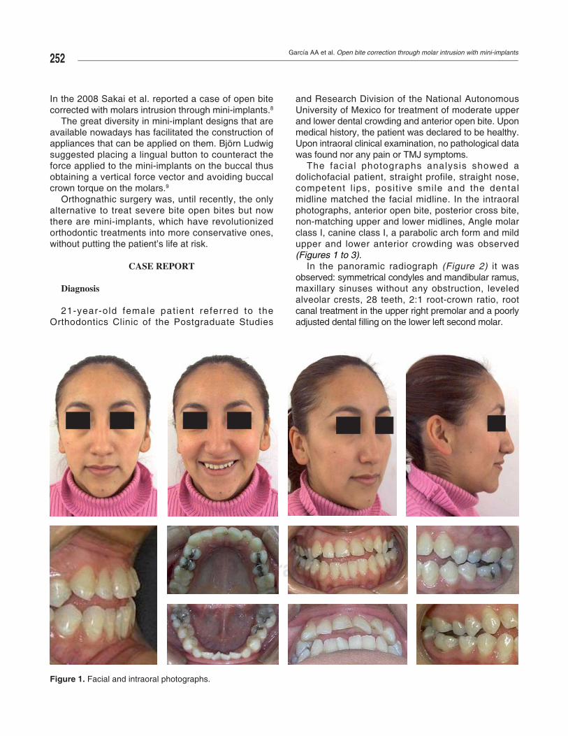
21-year-old female pat ient referred to the Orthodontics Clinic of the Postgraduate Studies
and Research Division of the National Autonomous University of Mexico for treatment of moderate upper and lower dental crowding and anterior open bite. Upon medical history, the patient was declared to be healthy. Upon intraoral clinical examination, no pathological data was found nor any pain or TMJ symptoms.
The facial photographs analysis showed a dolichofacial patient, straight profile, straight nose, competent l ips, posit ive smile and the dental midline matched the facial midline. In the intraoral photographs, anterior open bite, posterior cross bite, non-matching upper and lower midlines, Angle molar class I, canine class I, a parabolic arch form and mild upper and lower anterior crowding was observed (Figures 1 to 3).
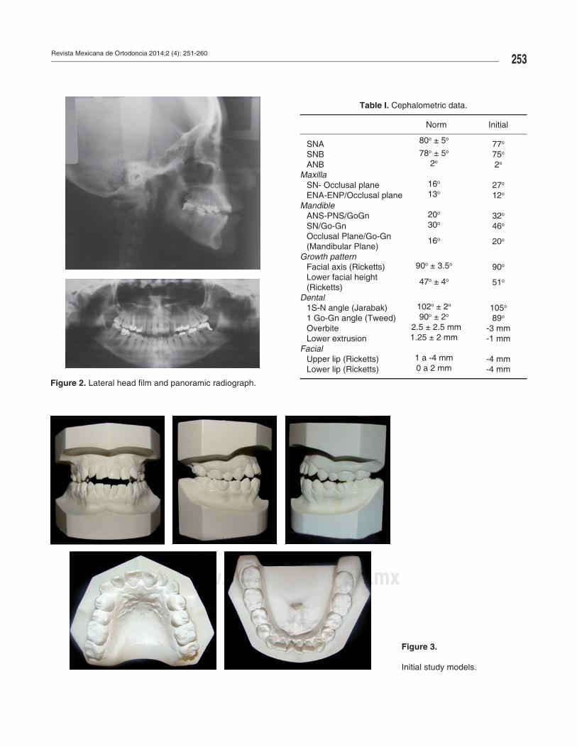
In the panoramic radiograph (Figure 2) it was observed: symmetrical condyles and mandibular ramus, maxillary sinuses without any obstruction, leveled alveolar crests, 28 teeth, 2:1 root-crown ratio, root canal treatment in the upper right premolar and a poorly adjusted dental fi lling on the lower left second molar.
wwwwwwwwwwwwwwwwwwwwwwwwwwwwwwwwwwwwwwwwwwwwwwwwwwwwwwwwwwwwwwwwwwwwwwwwwwwwwwwwwwwwwwwwwwwwwwwwwwwwwwwwwwwwwwwwwwwwwwwwwwwwwwwwwwwwwwwwwwwwwwwwwwwwwwwwwwwwwwwwwwwwwwwwwwwwwwwwwwwwwwwwwwwwwwwwwwwwwwwwwwwwwwwwwwwwwwwwwwwwwwwwwwwwwwwwwwwwwwwwwwwwwwwwwwwwwwwwwwwwwwwwwwwwwwwwwwwwwwwwwwwwwwwwwwwwwwwwwwwwwwwwwwwwwwwwwwwwwwwwwwwwwwwwwwwwwwwwwwwwwwwwwwwwwwwwwwwwwwwwwwwwwwwwwwwwwwwwwwwwwwwwwwwwwwwwwwwwwwwwwwwwwwwwwwwwwwwwwwwwwwwwwwwwwwwwwwwwwwwwwwwwwwwwwwwwwwwwwwwwwwwwwwwwwwwwwwwwwwwwwwwwwwwwwwwwwwwwwwwwwwwwwwwwwwwwwwwwwwwwwwwwwwwwwwwwwwwwwwwwwwwwwwwwwwwwwwwwwwwwwwwwwwwwwwwwwwwwwwwwwwwwwwwwwwwwwwwwwwwwwwwwwwwwwwwwwwwwwwwwwwwwwwwwwwwwwwwwwwwwwwwwwwwwwwwwwwwwwwwwwwwwwwwwwwwwwwwwwwwwwwwwwwwwwwwwww............mmmmmmmmmmmmmmmmmmmmmmmmmmmmmmmmmmmmmmmmmmmmmmmmmeeeeeeeeeeeeeeeeeeeeeeeeeeeeeeeeeeeeeeeeeeeeeeeeeeeeeeeddddddddddddddddddddddddddddddddddddddddddddddddddddddddddddddddddddddddddddddddddddddddddddddddddddddddddddddddddddddddddddddddddddddddddddddddddddddddddddddddddddddddddddddddddddddddddddddddddddddddddddddddddddddddddddddddddddddddddddddddddddddddddddddddddddddddddddddddddddddddddddddddddddddddddddddiiiiiiiiiiiiiiiiiiiiiiiiiiiiiiiiiiiiiiiiiiiiiiiiiiiiiiiiiiiiiiiiiiiiiiiiiiiiiiiiiiiiiiiiiiiiiiiiiiiiiiiiiiiiiiiiiiiiiiiiiiiiiiiiiiiiiiiiiiiiiiiiiiiiiiiiiiiiiiiiiiiiiiiiiiiiiiiiiiiggggggggggggggggggggggggggggggggggggggggggggggggggggggggggggggggggggggggggggggggggggggggggggggggggggggggggggggggggggggggggggggggggggggggggggggggggggggggggggggggggggggggggggggggggggggggggggggggggggggggggggggggggggggggggggggggggggggggggggggggggggggggggggggggggggggggggggggggggggggggggggggggggggggggggggggrrrrrrrrrrrrrrrrrrrrrrrrrrrrrrrrrrrrrrrrrrrrrrrrrrrrrrrrrrrrrrrrrrrrrrrrrrrrrrrrrrrrrrrrrrrrrrrrrrrrrrrrrrrrrrrrrrrraaaaaaaaaaaaaaaaaaaaaaaaaaaaaaaaaaaaaaaaaaaaaaaaaaaaaaaaaaaaaaaapppppppppppppppppppppppppppppppppppppppppppppppppppppppppppppppppppppppppppppppppppppppppppppppppppppppppppppppppppppppppppppppppppppppppppppppppppppppppppppppppppppppppppppppppppppppppppppppppppppppppppppppppppppphhhhhhhhhhhhhhhhhhhhhhhhhhhhhhhhhhhhhhhhhhhhhhhhhhhhhhhhhhhhhhhhhhhhhhhhhhhhhhhhhhhhhhhhhhhhhhhhhhhhhhhhhhhhhhhhhhhhhhhhhhhhhhhhhhhhhhhhhhhhhhhhhhhhhhhhhhhhhhhhhhhhhhhhhhhhhhhhhhhhhhhhhhhhhhhhhhhhhhhhhhhhhhhhhhhhhhhhhhhhhhhhhhhhhhhhhhhhhhhhhhhhhhhhhhhhhhhhhhhhhhiiiiiiiiiiiiiiiiiiiiiiiiiiiiiiiiiiiiiiiiiiiiiiiiiiiiiiiiiiiiiiiiiiiiiiiiiiiiiiiiiiiiiiiiiiiiiicccccccccccccccccccccccccccccccccccccccccccccccccccccccccccccccccccccccccccccccccccccccccccccccccccccccccccccccccccccccccccccccccccccccc......................................................................oooooooooooooooooooooooooooooooooooooooooooooooooooooooooooooooooooooooooooooooooooooooooooooooooooooooooooooooooooooooooooooooooooooooooooooooooooooooooooorrrrrrrrrrrrrrrrrrrrrrrrrrrrrrrrrrrrrrrrrrrrrrrrrrrrrrrrrrrrrrrrrgggggggggggggggggggggggggggggggggggggggggggggggggggggggggggggggggggggggggggggggggggggggggggggggggggggggggggggggggggggggggggggggggggggggggggggggggggggggggg............................................................................................mmmmmmmmmmmmmmmmmmmmmmmmmmmmmmmmmmmmmmmmmmmmmmmmmmmmmmmmmmmmmmmmmmmmmmmmmmmmmmmmmmmmmmmmmmmmmmmmmmmmmmmmmmmmmmmmmmmmmmmmmmmmmmmmmmmmmmmmmmmmmmmmmmmmmmmmxxxxxxxxxxxxxxxxxxxxxxxxxxxxxxxxxxxxxxxxxxxxxxxxxxxxxxxxxxxxxxxxxxxxxxxxxxxxxxxxxxxxxxxxxxxxxxxxxxxxxxxxxxxxxxxxxxxxxxxxxxxxxxxxxxxxxxxxxxxxxxxxxxxx
Figure 1. Facial and intraoral photographs.
Revista Mexicana de Ortodoncia 2014;2 (4): 251-260253
www.medigraphic.org.mx
Figure 2. Lateral head fi lm and panoramic radiograph.
wwwwwwwwwwwwwwwwwwwwwwwwwwwwwwwwwwwwwwwwwwwwwwwwwwwwwwwwwwwwwwwwwwwwwwwwwwwwwwwwwwwwwwwwwwwwwwwwwwwwwwwwwwwwwwwwwwwwwwwwwwwwwwwwwwwwwwwwwwwwwwwwwwwwwwwwwwwwwwwwwwwwwwwwwwwwwwwwwwwwwwwwwwwwwwwwwwwwwwwwwwwwwwwwwwwwwwwwwwwwwwwwwwwwwwwwwwwwwwwwwwwwwwwwwww...........mmmmmmmmmmmmmmmmmmmmmmmmmmmmmmmmmmmmmmmmmmmmmmmmmmmmmmmmmmmmmmmmmmmmmmmmmmmmmmmmmmmmmmmmmmmmmmmmmmmmmmmmmmmmmmmmmmmmmmmmmmmmmmmmmmmmmmmmmmmmmmmmmmmmmmmmmmmmmmmmmmmmmmmmmmmmmmmmmmmmmmmmmmmmmmmmmmmmmmmmmmmmmmmmmmmmmmmmmmmmmmmmmmmmmmmmmmmmmmmmmmmmmmmmmmmmmmmmmmmmmeeeeeeeeeeeeeeeeeeeeeeeeeeeeeeeeeeeeeeeeeeeeeeeeeeeeeeeeeeeeeeeeeeeeeeeeeeeeeeeeeeeeeeeddddddddddddddddddddddddddddddddddddddddddddddddddddddddddddddddddddddddddddddddddddddddddddddddddddddddddddddddddddddddddddddddddddddiiiiiiiiiiiiiiiiiiiiiggggggggggggggggggrrrrrrrrrrrraaaaaaaaaaaaaaappppppppppppppppppppppphhhhhhhhhhhhhhhhhhhhhhhiiiiiiiiiiiiiiiiiccccccccccccccccccccccccccccccccccccccccccccccccc...........................ooooooooooooooooooooooooooooooooooooooooooooooooooooooooooooooooorrrrrrrrrrrrrrrrrrrrrrrrrrrrrrrrrrrrrrrrrrrrrrrrrrrrrrrrrrrrrrrrrrrrrrrrrrrrrrrrrrrrrrrrrrrggggggggggggggggggggggggggggggggggggggggggggggggggggggggggggggggggggggggggggggggggggggggggggggggggggggggggggggggggggggggggggggggggggggggggggggggggggggggggggggggggggggggggggggggggggggggggggggggggggggggggggggggggggggggggggggggggggggggggggggggggggggggggggggggggggggggggggggggggggggggggg...........
Figure 3.
Initial study models.
Table I. Cephalometric data.
Norm Initial
SNA 80o ± 5o77o
SNB 78o ± 5o 75o
ANB 2o 2o
MaxillaSN- Occlusal plane 16o 27o
ENA-ENP/Occlusal plane 13o 12o
MandibleANS-PNS/GoGn 20o 32o
SN/Go-Gn 30o 46o
Occlusal Plane/Go-Gn (Mandibular Plane)
16o 20o
Growth patternFacial axis (Ricketts) 90o ± 3.5o 90o
Lower facial height(Ricketts)
47o ± 4o 51o
Dental1S-N angle (Jarabak) 102o ± 2o 105o
1 Go-Gn angle (Tweed) 90o ± 2o 89o
Overbite 2.5 ± 2.5 mm -3 mmLower extrusion 1.25 ± 2 mm -1 mm
FacialUpper lip (Ricketts) 1 a -4 mm -4 mmLower lip (Ricketts) 0 a 2 mm -4 mm
García AA et al. Open bite correction through molar intrusion with mini-implants254
www.medigraphic.org.mx
The cephalometric data showed that the patient was skeletal class I with open bite, vertical growth pattern and incisors within their basal bones. The data suggested by Acuña et al.10,11 and Argüelles et al. for open bites were considered (Table I).
Treatment goals
According to the diagnosis, it was decided to perform upper and lower molar intrusion to correct the open bite and obtain an adequate overbite and overjet, avoiding at the same time, the extrusion of the teeth adjacent to the molars and facial vertical changes.
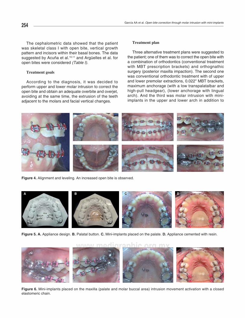
Treatment plan
Three alternative treatment plans were suggested to the patient; one of them was to correct the open bite with a combination of orthodontics (conventional treatment with MBT prescription brackets) and orthognathic surgery (posterior maxilla impaction). The second one was conventional orthodontic treatment with of upper and lower premolar extractions, 0.022" MBT brackets, maximum anchorage (with a low transpalatalbar and high-pull headgear), (lower anchorage with lingual arch). And the third was molar intrusion with mini-implants in the upper and lower arch in addition to
A B C D
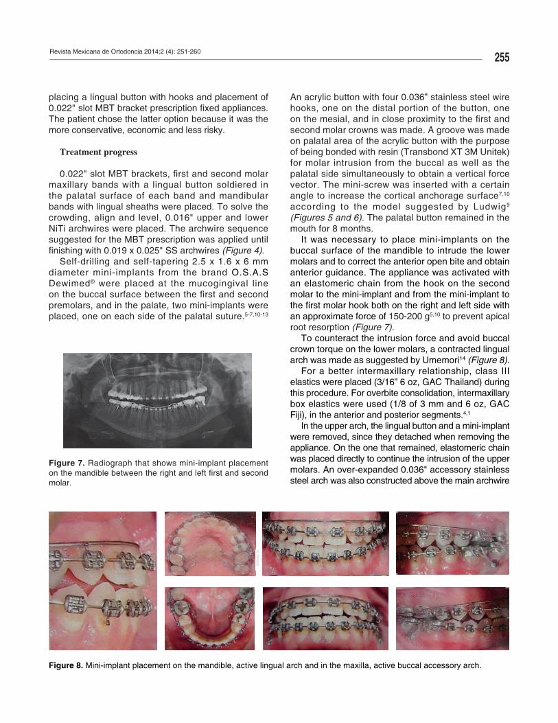
Figure 5. A. Appliance design. B. Palatal button. C. Mini-implants placed on the palate. D. Appliance cemented with resin.
wwwwwwwwwwwwwwwwwwwwwwwwwwwwwwwwwwwwwwwwwwwwwwwwwwwwwwwwwwwwwwwwwwwwwwwwwwwwwwwwwwwwwwwwwwwwwwwwwwwwwwwwwwwwwwwwwwwwwwwwwwwwwwwwwwwwwwwwwwwwwwwwwwwwwwwwwwwwwwwwwwwwwwwwwwwwwwwwwwwwwwwwwwwwwwwwwwwwwwwwwwwwwwwwwwwwwwwwwwwwwwwwwwwwwwwwwwwwwwwwwwwwwwwwwwwwwwww.................mmmmmmmmmmmmmmmmmmmmmmmmmmmmmmmmmmmmmmmmmeeeeeeeeeeeeeeeeeeeeeeeeeeeeeeeeeeeeeeddddddddddddddddddddddddddddddddddddddddddddiiiiiiiiiiiiiiiiiiiiiiiiiiiiiiggggggggggggggggggggggggggggggggggggggggggggggggggggggggggggggggggggggggggggggggggggggggggggggggggggggggggggggggrrrrrrrrrraaaaaaaaaaaaaaaaaaaaaaaaaaaaaappppppppppppppppppppppppppppppppppppppppppppppppppppppppppppppppppppppppppppppppppphhhhhhhhhhhhhhhhhhhhhiiiiiiiiiicccccccccccccccccccccccccc...........................................ooooooooooooooooooooooooooooooooooooooooooooooooooooooooooooooooooooooooooooooooooooooooooooooooooooooooooooooooooooooooooooooooooooooooooooooooooooooooooorrrrrrrrrrrrrrrrrrrrrrrrrrrrrrrrrrrrrrrrrrrrrrrrrrrrrrrrrrrrrrrrrrrrrrrrrrrrrrrrrrrrrrrrrrrrrrrrrrrrrrrrrrrrrggggggggggggggggggggggggggggggggggggggggggggggggggggggggggggggggggggggggggggggggggggggggggggggggggggggggggggggggggggggggggggggggggggggggggggggggggggggggggggggggggggggggggggggggggggggggggggggggggggggggggggggggggggggggggggggggggggggggggggggggggggggggggggggggggg..............mmmmmmmmmmmmmmmmmmmmmmmmmmmmmmmmmxxxxxxxxxxxxxxxxxxxxxxxxxxxxxxxxxxxxxxxxxxxxxxxxxxxxxxxxxxxxxxxxxxxxxxxxxxxxxxxxxxxxxxxxxxxxxxxxxxxxxxxxxxxxxxxxxxxx
Figure 6. Mini-implants placed on the maxilla (palate and molar buccal area) intrusion movement activation with a closed elastomeric chain.
Figure 4. Alignment and leveling. An increased open bite is observed.
Revista Mexicana de Ortodoncia 2014;2 (4): 251-260255
www.medigraphic.org.mx
Figure 7. Radiograph that shows mini-implant placement on the mandible between the right and left fi rst and second molar.
placing a lingual button with hooks and placement of 0.022" slot MBT bracket prescription fi xed appliances. The patient chose the latter option because it was the more conservative, economic and less risky.
Treatment progress
0.022" slot MBT brackets, fi rst and second molar maxillary bands with a lingual button soldiered in the palatal surface of each band and mandibular bands with lingual sheaths were placed. To solve the crowding, align and level, 0.016" upper and lower NiTi archwires were placed. The archwire sequence suggested for the MBT prescription was applied until fi nishing with 0.019 x 0.025" SS archwires (Figure 4).
Self-drilling and self-tapering 2.5 x 1.6 x 6 mm diameter mini-implants from the brand O.S.A.S Dewimed® were placed at the mucogingival line
on the buccal surface between the fi rst and second premolars, and in the palate, two mini-implants were placed, one on each side of the palatal suture.5-7,10-13
An acrylic button with four 0.036” stainless steel wire hooks, one on the distal portion of the button, one on the mesial, and in close proximity to the fi rst and second molar crowns was made. A groove was made on palatal area of the acrylic button with the purpose of being bonded with resin (Transbond XT 3M Unitek) for molar intrusion from the buccal as well as the palatal side simultaneously to obtain a vertical force vector. The mini-screw was inserted with a certain angle to increase the cortical anchorage surface7,10 according to the model suggested by Ludwig9 (Figures 5 and 6). The palatal button remained in the mouth for 8 months.
It was necessary to place mini-implants on the buccal surface of the mandible to intrude the lower molars and to correct the anterior open bite and obtain anterior guidance. The appliance was activated with an elastomeric chain from the hook on the second molar to the mini-implant and from the mini-implant to the fi rst molar hook both on the right and left side with an approximate force of 150-200 g5,10 to prevent apical root resorption (Figure 7).
To counteract the intrusion force and avoid buccal crown torque on the lower molars, a contracted lingual arch was made as suggested by Umemori14 (Figure 8).
For a better intermaxillary relationship, class III elastics were placed (3/16” 6 oz, GAC Thailand) during this procedure. For overbite consolidation, intermaxillary box elastics were used (1/8 of 3 mm and 6 oz, GAC Fiji), in the anterior and posterior segments.4,1
In the upper arch, the lingual button and a mini-implant were removed, since they detached when removing the appliance. On the one that remained, elastomeric chain was placed directly to continue the intrusion of the upper molars. An over-expanded 0.036" accessory stainless steel arch was also constructed above the main archwire
wwwwwwwwwwwwwwwwwwwwwwwwwwwwwwwwwwwwwwwwwwwwwwwwwwwwwwwwwwwwwwwwwwwwwwwwwwwwwwwwwwwwwwwwwwwwwwwwwwwwwwwwwwwwwwwwwwwwwwwwwwwwwwwwwwwwwwwwwwwwwwwwwwwwwwwwwwwwwwwwwwwwwwwwwwwwwwwwwwwwwwwwwwwwwwwwwwwwwwwwwwwwwwwwwwwwwwwwwwwwwwwwwwwwwwwwwwwwwwwwwwwwwwwwwwwwwwwwwwwwwwwwwwwwwwwwwwwwwwwwwwwwwwwwwwwwwwwwwwwwwwwwwwwwwwwwwwwwwwwwwwwwwwwwwwwwwwwwwwwwwwwwwwwwwwwwwwwwwwwwwwwwwwwwwwwwwwwwwwwwwwwwwwwwwwwwwwwwwwwwwwwwwwwwwwwwwwwwwwwwwwwwwwwwwwwwwwwwwwwwwwwwwwwwwwwwwwwwwwwwwwwwwwwwwwwwwwwwwwwwwwwwwwwwwwwwwwwwwww...............mmmmmmmmmmmmmmmmmmmmmmmmmmmmmmmmmmmmmmmmmmmmmmmmmmmmmmmmmmmmmmmmmmmmmmmmmmmmmmmmmmmmmmmmmmmmmmmmmmmmmmmmmmmmmmmmmmmmmmmmmmmmmmmmmmmmmmmmmmmmmmmmmmmmmmmmmmmmmmmmmmmmmmmmmmmmmmmmmmmmmmmmmmmmmmmmmmmmmmmmmmmeeeeeeeeeeeeeeeeeeeeeeeeeeeeeeeeeeeeeeeeeeeeeeeeeeeeeeeeeeeeeeeeeeeeeeeeeeeeeeeeeeeeeeeeeeeeeeeeeeeeeeeeeeeeeeeeeeeeeeeeeeeeeeeeeeeeeeeeeeeeeeeeeeeeeeeeeeeeeeedddddddddddddddddddddddddddddddddddddddddddddddddddddddddddddddddddddddddddddddddddddddddddddddddddddddddddddddddddddddddddddddddddddddddddddddddddddddddddddddddddddddddddddddddddddddddddddddddddddddddddddddddddddddddddddddddddddddddddddddddddddddddddddddddddddddddddddddddddddddddddddddddddddddddddddddddddddddddddddddddddddddddddddddddddddddddddddddddddddddddddddddddddddddddddddddddddddddddddddddddddddddddddddddddddddddddddddddddddddddddddddddddddd ggggggggggggggggggggggggggggggggggggggggggggggggggggggggggggggggggggggggggggggggggggggggggggggggggggggrrrrrrrrrrrrrrrrrrrrrrrrrrrrrrrrrrrrrrrrrrrrrrrrrrrrrrrrrrrrrrrrrrrrrrrrrrrrrrrrrrrrrrrrrrrrrrrrrrrrrrrrrrrrrrrrrrrrrrrrrrrrrrrrrrrrrrrrrrrrrrrrraaaaaaaaaaaaaaaaaaaaaaaaaaaaaaaaaaaaaaaaaaaaaaaaaaaaaaaaaaaaaaaaaaaaaaaaaaaaaaaaaaaaaaaaaaaaaaaaaaaaaaaaaaaaaaaaaaaaaaaaaaaaaaaaaaaaaaaaaaaaaaaaaaaaaaaaaaaaaaaaaaaaaaaaaaaaaaaaaaaaaaaaaaaaaaaaaaaaaaaaaaaaaaaaaaaaaaaaaaaaaaaaaaaaaaaaaaaaaaaaaaaaaaaaaaaaaaaaaaaaaaaaaaaaaaaaaaaaaaaaaaaaaaaaaaaaaaaaaaaaaaaaaaaaaaaaaaaaaaaaaaaaaaaaaaaaaaaaaaaaaaaaaaaaaaaaaaaaaaaaaaaaaaaaaaaaaaaaaaaaaaaaaaaaaaaaaaaaaaaaaaaaaaaaaaaaaaaaaaaaaaaaaaaaaaaaaaaaaaaaaaaaaaaaaaaaaaaaaaaaaaaaaaaaaaaaaaaaaaaaaaaaaaaaaaaaaaaaaaaaaaaaaaaaaaaaaaaaaaaaaaaaaaaaaaaaaaaaaaaaaaaaaaaaaaaaaaaaaaaaaaaaaaaaaaaaaaaaaaapppppppppppppppppppppppppppppppppppppppppppppppppppppppppppppppppppppppppppppppppppppppppppppppppppppppppppppppppppppppppppppppppppppppppppppppppppppppppppppppppppppppppppppppppppppppppppppppppppppppppppppppppppppppppppppppppppppppppppppppppppppppppppppppppppppppppppppppppppppppppppppppppppppppppppppppppppppppppppppppppppppppppppppppppppppppppppppppppppppppppppppppppppppppppppppppppppppppppppppppppppppppppppppppppppppppppppppppppppppppppppppppppppppppppppppppppppppppppppppppppppppppppppppppppppppppppppppppppppppppphhhhhhhhhhhhhhhhhhhhhhhhhhhhhhhhhhhhhhhhhhhhhhhhhhhhhhhhhhhhhhhhhhhhhhhhhhhhhhhhhhhhhhhhhhhhhhhhhhhhhhhhhhhhhhhhhhhhhhhhhhhhhhhhhhhhhhhhhhhhhhhhhhhhhhhhhhhhhhhhhhhhhhhhhhhhhhhhhhhhhhhhhhhhhhhhhhhhhhhhhhhhhhhhhhhhhhhhhhhhhhhhhhhhhhhhhhhhhhhhhhhhhhhhhhhhhhhhhhhhhhhhhhhhhhhhhhhhhhhhhhhhhhhhhhhhhhhhhhhhhhhhhhhhhhhhhhhhhhhhhhhhhhhhhhhhhhhhhhhhhhhhhhhhhhhhhhhhhhhhhhhhhhhhhhhhhhhhhhhhhhhhhhhhhhhhhhhhhhhhhhhhhhhhhhhhhhhhhhhhhhhhhhhhhhhhhhhhhhhhhhhhhhhhhhhhhhhhhhhhhhhhhhhhhhhhhhhhhhhhhhhhhhhhhhhhhhhhhhhhhhhhhhhhhhhhhhhhhhhhhhhhhhhhhhhhhhhhiiiiiiiiiiiiiiiiiiiiiiiiiiiiiiiiiiiiiiiiiiiiiiiiiiiiiiiiiiiiiiiiiiiiiiiiiiiiiiiiiiiiiiiiiiiiiiiiiiiiiiiiiiiiiiiiiiiiiiiiiiiiiiiiiiiiiiiiiiiiiiiiiiiiiiiiiiiiiiiiiiiiiiiiiiiiiiiiiiiiiiiiiiiiiiiiiiiiiiiiiiiiiiiiiiiiiiiiiiiiiiiiiiiiiiiiiiiiiiiiiiiiiiiiiiiiiiiiiiiiiiiiiiiiiiiiiiiiiicccccccccccccccccccccccccccccccccccccccccccccccccccccccccccccccccccccccccccccccccccccccccccccccccccccccccccccccccccccccccccccccccccccccccccccccccccccccccccccccccccccccccccccccccccccccccccccccccccccccccccccccccccccccccccccccccccccccccccccccccccccccccccccccccccccccccccccccccccccccccccccccccccccccccccccccccccccccccccccccccccccccccccccccccccccccccccccccccccccccccccccccccccccccccccccccccccccccccccccccccccccccccccccccccccccccccccccccccccccccccccccccccccccccccccccccccccccccccccccccccccccccc..................................................ooooooooooooooooooooooooooooooooooooooooooooooooooooooooooooooooooooooooooooooooooooooooooooooooooooooooooooooooooooooooooooooooooooooooooooooooooooooooooooooooooooooooooooooooooooooooooooooooooooooooooooooooooooooooooooooooooooooooooooooooooooooooooooooooooooooooooooooooooooooooooooooooooooooooooooooooooooooooooooooooooooooooooooooooooooooooooooooooooooooooooooooooooooooooooooooooooooooooooooooooooooooooooooooooooooooooooooooooooooooooooooooooooooooooooooooooooooooooooooooooooooooooooooooooooooooooooooooooooooooooooooooooooooooooooooooooooooooooooooooooooooooooooooooooooooooooooooooooooooooooooooooooooooooooooooooooooooooooooooooooooooooooooooooooorrrrrrrrrrrrrrrrrrrrrrrrrrrrrrrrrrrrrrrrrrrrrrrrrrrrrrrrrrrrrrrrrrrrrrrrrrrrrrrrrrrrrrrrrrrrrrrrrrrrrrrrrrrrrrrrrrrrrrrrrrrrrrrrrrrrrrrrrrrrrrrrrrrrrrrrrrrrrrrrrrrrrrrrrrrrrrrrrrrrrrrrrrrrrrrrrrrrrrrrrrrrrrrrrrrrrrrrrrrrrrrrrrrrrrrrrrrrrrrrrrrrrrrrrrrrrrrrrrrrrrrrrrrrrrrrrrrrrrrrrrggggggggggggggggggggggggggggggggggggggggggggggggggggggggggggggggggggggggggggggggggggggggggggggggggggggggggggggggggggggggggggggggggggggggggggggggggggggggggggggggggggggggggggggggggggggggggggggggggggggggggggggggggggggggggggggggggggggggggggggggggggggggggggggggggggggggggggggggggggggggggggggggggggggggggggggggggggggggggggggggggggggggggggggggggggggggggggggggggggggggggggggggggggggggggggggggggggggggggggggggggggggggggggggggggggggggggggggggggggggggggggggggggggggggggggggggggggggggggggggggggggggggggggggggggggggggggggggggggggggggggggggggggggggggggggggg............mmmmmmmmmmmmmmmmmmmmmmmmmmmmmmmmmmmmmmmmmmmmmmmmmmmmmmmmmmmmmmmmmmmmmmmmmmmmmmmmmmmmmmmmmmmmmmmmmmmmmmmmmmmmmmmmmmmmmmmmmmmmmmmmmmmmmmmmmmmmmmmmmmmmmmmmmmmmmmmmmmmmmmmmmmmmmmmmmmmmmmmmmmmmmmmmmmmmmmmmmmmmmmmmmmmmmmmmmmmmmmmmmmmmmmmmmmmmmmmmmmmmmmmmmmmmmmmmmmmmmmmmmmmmmmmmmmmmmmmmmmmmmmmmmmmmmmmmmmmmmmmmmmmmmmmmmmmmmmmmmmmmmmmmmmmmmmmmmmmmmmmmmmmmmmmmmmmmmmmmmmmxxxxxxxxxxxxxxxxxxxxxxxxxxxxxxxxxxxxxxxxxxxxxxxxxxxxxxxxxxxxxxxxxxxxxxxxxxxxxxxxxxxxxxxxxxxxxxxxxxxxxxxxxxxx
Figure 8. Mini-implant placement on the mandible, active lingual arch and in the maxilla, active buccal accessory arch.
García AA et al. Open bite correction through molar intrusion with mini-implants256
www.medigraphic.org.mx
to prevent the molars moving lingually. Additionally, stripping was performed from dental organ 35 to 45 to obtain overjet. After thirteen months (eight on the maxilla and fi ve in the mandible) the mini implants were removed, ties and intermaxillary box elastics were placed in each of the anterior teeth for overbite consolidation (Figure 9).
The system of force that was used to get the vertical and horizontal overbite, focused mainly on the intrusion of the maxillary and mandibular molars with the help of the mini-implants, the button palatino and lingual arch, the arches used in this moment of force (0.019 x 0.025" of NiTi), which contributed to the slight extrusion in anterior teeth and premolar teeth as a result of the alignment and leveling of the same, producing as a whole the closure of the previous bite.
This system, in addition to providing a molar intrusion mechanics, worked as skeletal anchorage, so while the anterior segment suffered mild extrusion, the molars were maintained in their position, isolating the reciprocal force toward them.
RESULTS
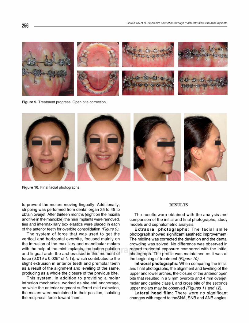
The results were obtained with the analysis and comparison of the initial and fi nal photographs, study models and cephalometric analysis.
Extraoral photographs : The facia l smi le photograph showed signifi cant aesthetic improvement. The midline was corrected the deviation and the dental crowding was solved. No difference was observed in regard to dental exposure compared with the initial photograph. The profi le was maintained as it was at the beginning of treatment (Figure 10).
Intraoral photographs: When comparing the initial and fi nal photographs, the alignment and leveling of the upper and lower arches, the closure of the anterior open bite that resulted in a 3 mm overbite and 4 mm overjet, molar and canine class I, and cross bite of the seconds upper molars may be observed (Figures 11 and 12).
Lateral head film: There were no significant changes with regard to theSNA, SNB and ANB angles.
Figure 10. Final facial photographs.
Figure 9. Treatment progress. Open bite correction.
Revista Mexicana de Ortodoncia 2014;2 (4): 251-260257
www.medigraphic.org.mx
Este documento es elaborado por MedigraphicEEEEEEEEEEEEEEsEsEsEsEsEsssEEEEsEsEsEEsEsEsEEEsssEEEsEEEEssEEsEEsEEEEEEsEEEEEEEEssEEEEEsEEEEEEEEsEEEEssEsEEEEEsEsEsEEEEsEsEsEEEEEEsEsEsEsEsEsEsEEEEsEsEEEssEEsEEssEsEsEssssssstteteeeetteeteteeeeeeteeteteteteteeeeeeeetetetttteeeteeettteeeteeeeeet dddddddddddddddddddddddddddddddddddddddddddddddddddddddddddddddddddddddddooccccccccccococcccccccococccoccccccocccccoocccoccooccococccccccooooocccocoococccoooococccooocccocccccccccoooooccooooocccoooooooooooooocooooo uuuumummmummmmmmmmumummmmmmmmmumummmmmmuuuuuummmmumummummmmmumumuuumummummmumuuuuummmmmuuuummuuuuumumuuuummummmmumumumumummuuumumummmumuuumummmummumumuummuuummmmuumummumummmeeeeeenenenenenenenennnnnnnnnenenenenennnnneennneeenneneennneneenneneneenenennnennnennnneeennenneeeenennenenenenenenneneeenennnneeeneeeentotototottototototootootototototottoottootttooooottotoooootooooototototototottotototttoototottttotottotoottototototottototottottotoooo eeeeeeeeeeeeeeeeeeeeeeeeeeeeeeeeeeeeeeeeeeeeeeeeeeeeeeeeeeeeeeeeeeeeeesssssssssssssssssssssssssssssssssssssssssssssssssssssssssss ssssss s sssssss eeeeeelelelelellleleleleleeeleleelleleeeeelllelellllleleeeeeeeeeeeee abababbbbbbbbbbbabbbbbbbbbbabbbbababaabaababbbaabababbaabbaabaaaaaabaaaabaababaabbbababbbabbbbbbbbbbbbbbbbbbbbbororooooororororrrrrorroroororrroorrrorrorroorrorooorrororoooooooro aaaaaaaaaaaaaaaaaaaaaaaa ooooooo ooooooo oooooooooooo oooooooo oo oooooooooo o ppppppppppppppopopopopppppppopoopppppppppopppopopppppoppppppppoppppppppoppppoppoppppppppppppppppppppppooppppppopppppoopopooppppopopppppopoooppppopopppppppppppppp rrr rr rr r rr rrr rrrrrrrrr rrrrrrr rrrrrrrrrrrrr rrrrr MMMMMMMMeeMeMeMeMeMeMeMMMeMeMMMMMeMMMMeMeMeMeMMeMeMMeMeMMMMMMeeMMeMeMeMMMMeMeeMMeeeeMeeeeeeeMeMeeMeeeeeeMeeMMMMMMeMeeeeeMeMeMMMMeMMMMeeeeMeeeMMMMeMeMeMMeMeMMeMeMeeeMMeMMeeMMeMMMMMeMMMMMMMMMMMeMMMMeeMMeedddididdddddddidididdddddidddididdddiddddddiddidddiddididdididdiddididdidddidddididdididdididddididiidddddididdiddddddiddddddddddiiiidddddddddddiiddddidddiidddddiddididdiddiidddddid gggrgrgrgrgrrgrrrrrrrgrrgrgrgrgrggrrgrrggrgrggrgrggrgrggrgrgrgrgrgrgrrgrgggggggggggggggggggggggggggggg aaaaaaaapppppaapapaaapaapaapaaapapaapaaaapapapappapapapaaaaaaaaaaaaaaaaaaaaaaaaaaaaaaaaaaapaaaaaaaaaaaaaaaaaaapphihihihihihihihhhhhhhihihihihhhhhihihihhhihhiihihihihiiihihihihihhihiiiihiiiiiiiihiiiiiiiiiiiiihhiihihhiiiiiiiiiihiiiihhiihihhhhiiiihhhhhihihhihiihhihihhhhhhhhhiiccccccccccccccccccccccccccccccccccccccccccccccccccccccccccccccccccccccccccccccccccccccccccccccccccccccccccccccccccccccccccccccccccccccccccc
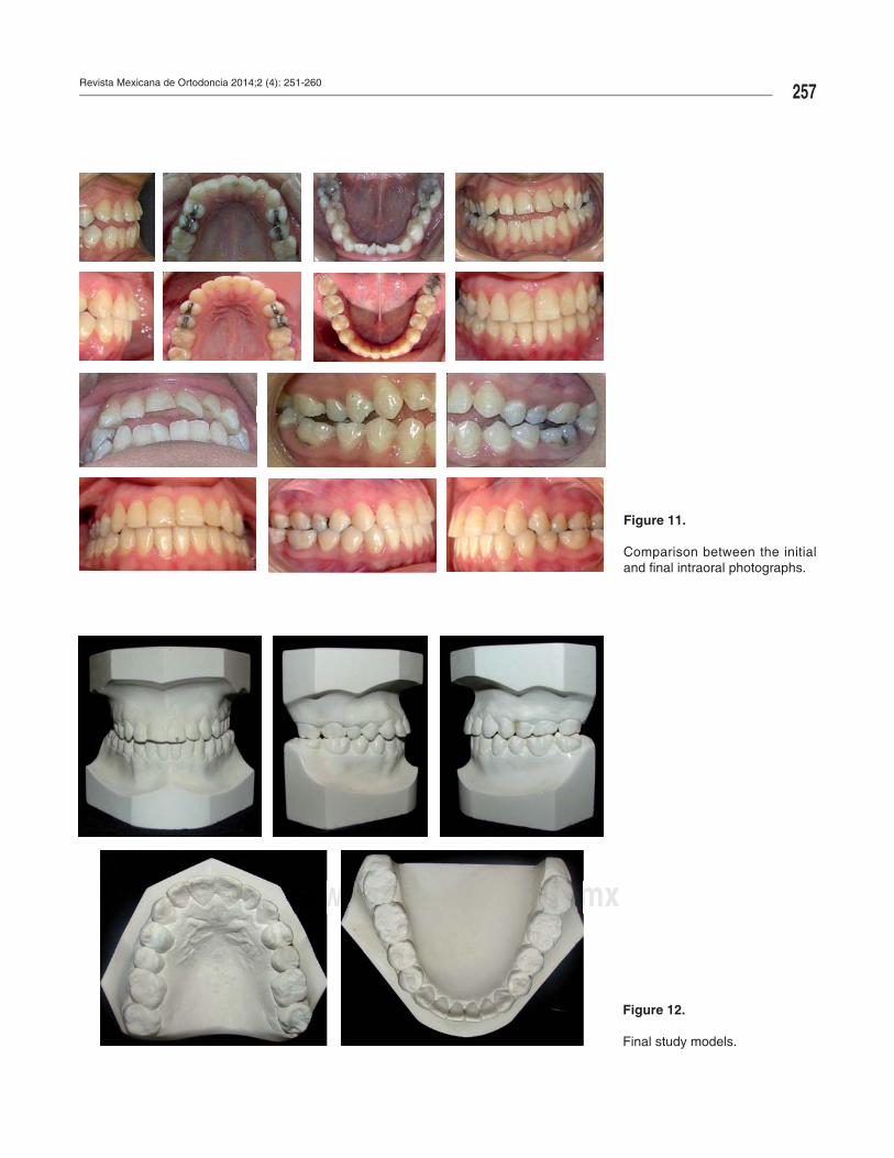
Figure 11.
Comparison between the initial and fi nal intraoral photographs.
wwwwwwwwwwwwwwwwwwwwwwwwwwwwwwwwwwwwwwwwwwwwwwwwwwwwwwwwwwwwwwwwwwwwwwwwwwwwwwwwwwwwwwwwwwwwwwwwwwwwwwwwwwwwwwwwwwwwwwwwwwwwwwwwwwwwwwwwwwwwwwwwwwwwwwwwwwwwwwwwwwwwwwwwwwwwwwwwwwwwwwwwwwwwwwwwwwwwwwwwwwwwwwwwwwwwwwwwwwwwwwwwwwwwwwwwwwwwwwwwwwwwwwwwwwwwwwwwwwwww........................................................mmmmmmmmmmmmmmmmmmmmmmmmmmmmmmmmmmmmmmmmmmmmmmmmmmmmmmmmmmmmmmmmmmmmmmmmmmmmmmmmmmmmmmmmmmmmmmmmmmmmmmmmmmmmmmmmmmmmmmmmmmmmmmmmmmmmmmmmmmmmmmmmmmmmmmmmmmmmmmmeeeeeeeeeeeeeeeeeeeeeeeeeeeeeeeeeeeeeeeeeeeeeeeeeeeeeeeeeeeeeeeeeeeeeeeeeeeeeeeeeeeeeeeeeeeeeeeeeeeeeeeeeeeeeeeeeeeeddddddddddddddddddddddddddddddddddddddddddddddddddddddddddddddddddddddddddddddddddddddddddddddddddddddddddddddddddddddddddddddddddddddddddddddddddddddddddddddddddddddddddiiiiiiiiigggggggggggggggggrrrrrrrrrraaaaaaaaaaaaappppppppppppppphhhhhhhhhhhhiiiiiiicccccccccccccccccc......ooooooooooooooooooooooooooooooooooooooooooooooooooooooooooooooooooooooorrrrrrrrrrrrrrrrrrrrrrrrrrrrrrrrrrrrrrgggggggggggggggggggggggggggggggggggggggggggggggggggggg.................................................................mmmmmmmmmmmmmmmmmmmmmmmmmmmmmmmmm
Figure 12.
Final study models.
García AA et al. Open bite correction through molar intrusion with mini-implants258
www.medigraphic.org.mx
The angles that were considered for determining a change in the open bite with respect to the maxilla were: SN-occlusal plane that decreased 2o as well as ANS-PNS/occlusal plane which decreased 1o. This is favorable for the open bite correction. To assess the changes that occurred in the mandible, the SN/ Go-Gn Angle which decreased 2o and the occlusal plane/Go Gn angle that decreased by 1o were considered (Figure 13).
To determine the closure of the open bite the Ricketts lower facial height which decreased 2o and the Ricketts facial axis which increased 1o, were considered. It can be assumed that the mandible had a slight counter-clockwise rotation, thanks to the molar intrusion which contributed to the open bite correction.
The incisor inclination was determined by the 1 S-N angle which increased 1o. This caused a dentoalveolar modification of the upper incisors and the 1 Go-Gn angle decreased 1o. This was interpreted as retroclination of the lower incisor that combination with the upper incisor resulted in an overjet.
The overbite increased by +5 mm and the lower incisor extrusion was +1 mm, which contributed to obtaining an appropriate overbite. In conclusion, the main goal of our problem list was satisfactorily achieved.
No changes were noted in the Ricketts facial line determined by the upper lip of and the lower lip which decreased 1 mm (Table II).
DISCUSSION
The effectiveness, simplicity of placement and removal, stability and low cost has been some of the advantages that mini-implants provide for bone anchorage. They provide a solution to several problems of anchorage loss caused by the reciprocal force that is exerted to perform movements such as retraction of the anterior segment, molar distalization, extrusion and intrusion.
The literature reports several methods for molar intrusion. One of them is used in growing patients and consists in myofunctional appliances that must be used before the eruption of the second molar with the disadvantage that relapse may occur and that the treatment’s success depends on the patient’s growth.4
Iscan and Sarisoy14 studied the effects of passive posterior bite planes for the early treatment of open bite in growing patients through molar intrusion produced by masticatory muscle pressure and upward
Figure 13. Final lateral head fi lm and panoramic radiograph.
Table II. Final cephalometric data.
Norm Initial Final
SNA 80o ± 5o 77o 77°SNB 78o ± 5o 75o 75°ANB 2o 2o 2°
MaxillaSN-Occlusal plane 16° 27° 25°*ANS-PNS/occlusal Plane
13° 12° 11°
MandibleANS-PNS/GoGn 20° 32o 31°*SN/Go-Gn 30o 46o 44°*Oclusal Plane/Go-Gn 16o 20o 19°
Growth pattern 90o ± 3.5o 90o 91°Facial axis (Ricketts) 47o ± 4o 51o 49°Lower face height (Ricketts)
Dental 102o ± 2o 105o 106°1S-N Angle (Jarabak) 90o ± 2o 89o 88°1 Go-Gn Angle (Tweed)
2.5 ± 2.5 mm -3 mm 2 mm**
Overbite 1.25 ± 2 mm -1 mm 0 mm*Lower incisor extrusion
1 a -4 mm -4 mm -4 mm
Facial 0 a 2 mm -4 mm -5 mmUpper lip (Ricketts)Lower lip (Ricketts)
Revista Mexicana de Ortodoncia 2014;2 (4): 251-260259
www.medigraphic.org.mx
and forward mandibular rotation. Satisfactory results were obtained. The disadvantage of this treatment is that many times this malocclusion is not timely diagnosed. Regularly, the patient arrives at the clinic with the problem when growth has fi nished, the patient hereby presented was such a case.
Sakai Y. et al.,8 mentioned a method for open bite closure through the use of headgears, intermaxillary elastics or multiloops, which resulted in extrusion of the anterior teeth compromising function, stability and aesthetics if they are not well managed by the specialist.
Another commonly used procedure for open bite closure with upper crowding is the first or second molar extraction or fi rst or second premolar extraction, where the open bite closes with the aid of extrusion of the anterior segment and incisor retroclination, since the center of rotation is at the apex. This procedure is ideal for patients with severe crowding, and not the treatment of choice for the patient presented in this case report.1
Orthognathic surgery for correction of the anterior open bite causes a major impact in the patient. Facial changes, post-surgical pain and complications, hemorrhage, infection, tooth vitality loss, long post-treatment time and costs must be explained to the patient. In sight of this, other treatment options must be offered to the patient as Lin J.C.Y. et al.15 suggest. In the case report hereby presented, it was explained to the patient all of the abovementioned and she refused «such a radical» surgery.
One of the advantages of mini-implants is that they do not require patient’s cooperation besides from the fact that reciprocal forces do not exist among the teeth that are wished to be moved and the anchorage teeth. Chang Y. et al. mentioned that it is one of the most ideal force systems for molar intrusion without side effects.16
Sakai Y.8 reported a clinical case that was satisfactorily resolved with the intrusion of upper and lower molars through mini-implants, one of the most conservative options for the solution of the problem posed by the patient in this article.
Chun et al. mentioned that the importance applying forces of molar intrusion is that they must be applied simultaneously, both towards the buccal and towards the lingual aspect of the molar.17
De Cleerk and Timmerman18 suggested that in order to prevent the molar intrusion force applied from the buccal from having a tipping effect on the crowns, an attachment in the palatal area must be placed at the same time. Jong-Suk Lee et al.13 considered the palatal area to be the most stable zone as it is formed
from dense cortical bone. They mentioned that it is the best area for mini-implant’s placement and that it creates an opposite force moment towards that direction, which will help the intrusion movement. The use of buccal-palatal mechanics introduced by Park H.S. et al.19 to prevent rotation and inclinations was used to correct the case presented here.
Taking into considerat ion al l the previous recommendations, a device was made according to the design by Lee et al13 and also, an attachment to perform the intrusion with vertical vectors. Some modifications were needed in the case hereby presented. An acrylic palatal button with four hooks bonded to two mini-screws placed at each side of the palatal suture was made to serve the same function as the one designed by Lee et al. Modifi cation such as described by Björn Ludwig9 were performed resulting in an applied intrusion force close to the center of resistance.
Mini-implants were also placed in the mandible for intrusion of the mandibular molars. A lingual arch with torque was placed to compensate for the tipping caused by intrusionforces, as recommended by Mikako Umemori et al.10
The combination of molar intrusion with mini-implants and alignment and leveling with the NiTi arches caused a slight extrusion of the anterior segment and produced an appropriate vertical overbite. Maku, Kawai, Koseki et al 21 reported a similar case where multiloop arches were used, which, unlike the case presented in this article shows to be a more complicated mechanic due to the loop realization.
Closure of the anterior open bite was achieved with the combination of molarintrusion in upper and lower molars in addition to a slight extrusion of the incisors obtained by means of intermaxillaryelastics as suggested by Quiros and Nanda for the consolidation of the overbite and overjet.4,1
Open bite is one of the most diffi cult malocclusions to treat, so a more accurate diagnosis should be performed since the success or failure of treatment depends on it. The present case was analyzed with published open bitearticles14,22-24 which suggest specifi c angles to diagnose it. Molar intrusion cases reported by Lee J,13 Umemori M,20 Sakai Y,8 Park H,19 showed no major changes in the cephalometric data but instead notorious clinical changes. First of all, open bite closure, the consolidation of molar and canine class I, an appropriate overbite and overjet, a more pleasant facial appearance, more relaxed facial muscles, in some cases anadequate labial competence and preservation of the vertical dimension. All these results are similar to those obtained in the patient presented in this article.
García AA et al. Open bite correction through molar intrusion with mini-implants260
www.medigraphic.org.mx
Despite all the care that was taken in relation to the force vector’s direction for molar intrusion, the patient presented relapse of the upper second molars cross bite. If this method of molar intrusion is to be used, the placement of a transpalatal arch with torque is suggested after removing the buccal mini- implants in order to avoid the molars returning to their position. Another option is to over-expand the maxillary buccal archwire until treatment is completed. Quiros4 reported a relapse in dolichofacial patients with open bite combined with posterior cross bite. He recommended overcorrection to avoid relapse.
In terms of intrusion stability with mini-implants no long-term data were found in the literature, due to the fact that the implementation of this treatment mechanics is recent. Lin J. et al.15 reported relapses of 30% in the case of dental intrusion of the incisors but not in molars.
Xun, Zeng and Wang23 assessed the effectiveness of mini-implants to correct the open bite in 12 patients between 14 and 27 years of age. All the cases were successfully resolved thus concluding that mini-implants are easy to place and remove, are minimally invasive and require little cooperation from the patient.
CONCLUSIONS
Open bite can be corrected with the use of mini implants for molar intrusion, finally providing a less invasive option compared to orthognathic surgery and the risks that the latter involves. Additionally, it is an option for patients who do not have the economic means to afford an orthodontic-surgical treatment.
The use of the mini- implants as a skeletal anchorage method creates a wider outlook in orthodontic biomechanics because as mentioned above, not only they can be used for molar intrusion, but also to perform movements that require a high magnitude of force without compromising the adjacent teeth with secondary movements.
The success or failure of the anchorage and biomechanics of orthodontic treatment will depend mainly on the specialist, making patient cooperation less important.
REFERENCES
1. Ravindra N. Biomecánicas y estética. Estrategias en ortodoncia clínica. Colombia: Editorial actualidades médico odontológicas Latinoamérica, C.A., AMOLCA; 2007. pp. 157-176.
2. García C. Mordida abierta anterior, revisión de la literatura. Revista Esomatológica. 2004; 12 (2): 4-19.
3. Rodríguez E, Casasa R, Natera M. 1001 Tips en ortodoncia y sus secretos. Colombia: Editorial actualidades médico odontológicas Latinoamérica, C.A., AMOLCA; 2007. pp. 159-184.
4. Quirós O. Uso de bloque de intrusión posterior, en el tratamiento de las mordidas abiertas anteriores. Revista Latinoamericana de Ortodoncia y Odontopediatría. 2003: 1-3.
5. Arismendi J, Ocampo Z, González F, Morales M. Mini-implantes como anclaje en ortodoncia. Rev Fac Odontol Univ Anioq. 2006; 18 (1): 82-94.
6. Ismail S, Johal A. The role of implants in orthodontics. Journal of Orthodontics. 2002; 29 pp 239-245.
7. Kanomi R. Mini implant for orthodontic anchorahe. J Clin Orthod. 1997; 31: 763-767.
8. Sakai Y, Kuroda S, Murshid SA, Takano-Yamamoto T. Skeletal class III severe open bite treatment using implant anchorage. Angle Orthodontist. 2008; 78 (1): 151-166.
9. Ludwig B, Baumgaertel S, Bohm B. Mini-implants in orthodontics: Innovative anchorage concepts. Editorial Quintessence Publishing (IL); 2008.
10. Ángeles L, Peralta A, Vázquez M, Cruz L. Uso de mini-impalntes ortodóncicos para intrusión de molares superiores en pacientes de la Unidad de Especialidades Odontológicas. Rev Sanid Milit Mex. 2006; 60 (5): 334-340.
11. Park YC, Lee SY, Kim DH, Jee SH. Intrusion of posterior teeth sing miniscrew implants. Am J Orthod and Dentofacial Orthop. 2003; 123 (6): 690-694.
12. Jane CC, Wu CB, Wu HY, Kok SH, Frank HF, Chen YJ. Intrusion of the overupted upper left first and second molars by mini-implants with partial-fi xed orthodontic appliances. A case report. Angle Orthodontic. 2003; 74 (4): 550-557.
13. Lee JS, Kim DH, Park YC, Kyung SH, Kim TK. The effi cient use of the midpalatal miniscrew implants. Angle Orthodontist. 2004; 74 (5): 711-715.
14. Iscan H, Sarisoy L. Comparison of the effects of passive posterior bite-blocks whit different construccion bites on the craniofacial and dentoalveolar strucutures. Am J Orthod and Orthop. 2007; 112 (2): 171-178.
15. Lin JCY, Yeh CL, Liou EJW, Bowman SJ. Treatment of skeletal origin gummysmiles with miniscrew anchorage. J Clin Orthod. 2008; 42 (5): 285-296.
16. Chang Y, Lee H, Chun Y. Miniscrew anchorage for molar intrusion. JCO. 2004; 37 (6): 325-330.
17. Chun YS, Woo YJ, Row J, Jung EJ. Maxillary molar intrusion with the molar intrusion arch. J Clin Orthod. 2000; 34 (2): 90-93.
18. De Clerck H, Timmerman H, Cornelis M. Biomechanics of skeletal anchorage. Part 3 Intrusion. Journal Clin Orthod. 2008; 43 (5): 270-278.
19. Park HS. Intrusión molar con anclaje de microimplantes (MIA Micro-implant Anchorage). Ortodoncia Clínica. 2003; 6 (1): 31-36.
20. Umemori M, Sugawara J, Mitani H, Nagasaka H, KawamuraH. Skeletal anchorage system for open bite correction. Am J Orthod and Orthop. 1999; 115 (2); 166-174.
21. Kaku M, Kawai A, Koseki H, Abedini S, Kawazoe A, Sasamoto T et al. Correction of severe open bite using miniscrew anchorage. Australian Dental Journal. 2009; 54: 374-380.
22. Acuña G, Ballesteros M, Oropeza G. Descripción cefalométrica del patrón facial en mordida abierta esqueletal. Revista Odontológica Mexicana. 2013; 17 (1): 15-19.
23. Xun Ch, Zeng X, Wang X. Microscrew anchorage in skeletal anterior open-bite treatment. Angle Orthodontist. 2007; 77 (1): 47-56.
24. Argüelles A, Oropeza G, Ibarra J. Características radiográfi cas de la mordida abierta esqueletal. Revista Odontológica Mexicana. 2007; 11 (1): 20-23.
Mailing address:Adriana García ArgumedoE-mail: [email protected]