Embed Size (px)
Citation preview
REVIEW Open Access
Effect of molar intrusion with temporaryanchorage devices in patients with anterioropen bite: a systematic reviewAhmad Saleem Alsafadi1*, Mohannad M. Alabdullah1, Humam Saltaji2 , Anas Abdo3 and Mohamed Youssef1
Abstract
Objective: The objective of the study is to assess the effect of molar intrusion with temporary anchoragedevices on the vertical facial morphology and mandibular rotation during open bite treatment in thepermanent dentition.
Methods: We performed a systematic review of the published data in seven electronic databases up toSeptember 2015. We considered studies for inclusion if they were examining the effects of posterior teethintrusion on the vertical facial morphology with open bite malocclusion in the permanent dentition. Studyselection, risk of bias assessment, and data-extraction were performed in duplicate. Meta-analysis was notpossible due to dissimilarity and heterogeneity among the included studies.
Results: Out of the 42 articles that met the initial eligibility criteria, 12 studies were finally selected. Low levelof scientific evidence was identified after risk of bias assessment of the included studies with no relevantrandomized controlled trial performed. Out of the 12 selected studies, five studies used miniplates and sevenstudies used miniscrews. Mandibular counterclockwise rotation was found to be between 2.3° and 3.9° in sixstudies (as sassed by mandibular plane angle, between MeGo or GoGn and SN or FH plane) while it was lessthan 2° in the remaining studies.
Conclusions: Current weak evidence suggests that molar intrusion with temporary anchorage devices maycause mandibular counterclockwise autorotation. Future well-conducted and clearly reported multicenterrandomized controlled trials that include a non-treatment control group are needed to make robustrecommendations regarding the amount of mandibular rotation during open bite treatments.
Keywords: Systematic review, Skeletal open bite, Molar intrusion
ReviewIntroductionOpen bite malocclusion is considered one of the mostdifficult orthodontic problems to correct because it ap-pears as a result of the interaction of numerous etio-logical factors (genetic, dental, skeletal, functional, softtissue, and habit) that contribute to its development[1]. An open bite can occur unilaterally or bilaterally inthe buccal segments; it is particularly seen in the anter-ior teeth. Generally, different features have been found
to be associated with the skeletal anterior open bite dis-tinguishing it from other types of malocclusion includ-ing increased lower face height, short posterior faceheight, [2] increased gonial and mandibular plane an-gles, [3] and increased maxillary molar dentoalveolarheight [4]. Several reports have found correlations be-tween orofacial muscle activity and vertical facial morph-ology [5–8]. These studies showed positive relationshipsbetween anterior open bite and weak musculature.Various therapeutic approaches have been proposed for
the treatment of an anterior open bite. These approachesvary depending on the causative factors and involvemyotherapy, preventive treatment, functional therapy,orthognathic surgery, and orthodontic treatment using
* Correspondence: [email protected] of Orthodontics, Faculty of Dentistry, Damascus University,Damascus, SyriaFull list of author information is available at the end of the article
© 2016 Alsafadi et al. Open Access This article is distributed under the terms of the Creative Commons Attribution 4.0International License (http://creativecommons.org/licenses/by/4.0/), which permits unrestricted use, distribution, andreproduction in any medium, provided you give appropriate credit to the original author(s) and the source, provide a link tothe Creative Commons license, and indicate if changes were made.
Alsafadi et al. Progress in Orthodontics (2016) 17:9 DOI 10.1186/s40510-016-0122-4
anterior teeth extrusion or posterior teeth intrusion [9].Among the non-surgical orthodontic treatment methodsare the temporary anchorage devices (TADs) includingminiplates [10] and miniscrew or micro-screw implants[9, 11]. Extrusion of anterior teeth is another alternativeapproach for open bite management, but it must take intoconsideration the smile esthetic [12]. Extrusion, however,is a less stable treatment than intrusion. The intrusionof posterior teeth with temporary anchorage deviceswas suggested to lead to decreased lower facial heightby a counterclockwise rotation of the mandible; thismay resemble the orthognathic surgery outcomes forany open bite patients [10].Molar intrusion is recommended in open bite patients
who usually have increased molar height [13]. Whilemany reports indicated that increased molar height isone of the common findings in individuals with skeletalopen bite [14], others do not support those findings [15,16]. In order to evaluate the results of molar intrusion inthe treatment of open bite malocclusion, it is necessaryto recognize the effect of the posterior teeth intrusionon the mandibular rotation and facial morphology.Many reports evaluated the effect of open bite treat-
ment during mixed dentition stage [17] and [18]. A re-cent systematic review examined open bite treatmentmodalities in children found no consistent findings re-garding the most effective treatment modality in grow-ing patients with open bite malocclusion [19]. However,no comprehensive review was conducted to examinethe effects of posterior teeth intrusion on vertical facialmorphology in non-growing patients. Therefore, thegoal of the current report is to systematically reviewthe effect of molar intrusion with temporary anchoragedevices on the vertical facial morphology and mandibu-lar rotation during open bite treatment in the perman-ent dentition stage.
Material and methodsThis systematic review was reported according to theprinciples of the PRISMA statement for reporting system-atic reviews of the health sciences [20].
Search strategyComprehensive electronic searches up to September30, 2015, were conducted in the following databases:PubMed, Embase, Cochrane Database of Systematic Re-views, Cochrane Central Register of Controlled Trials,Ovid, Scopus, and Web of science. The literaturesearches used the following Medical Subject Headings(MeSH) terms: “molar intrusion,” “posterior teeth in-trusion,” and “anterior open bite” which were crossedwith the following terms “mandibular autorotation” and“facial morphology.” In addition, the following journalswere searched individually to find out any missing
articles: The Angle Orthodontists, American Journal ofOrthodontics and Dentofacial Orthopedics, EuropeanJournal of Orthodontics, Korean journal of orthodontics,and Journal of Orofacial Orthopedics. No restrictionswere applied regarding date of publication, language, orstatus during database searches. The search strategy forPubMed can be found in Table 1.
Focused questionWhat is the effect of molar intrusion with temporaryanchorage devices on the vertical facial morphologyand mandibular rotation in the permanent dentitionstage during open bite treatment?
Selection criteria (PICO question: population, intervention,comparison, outcome)The following eligibility criteria were used to determineeligible reports for this systematic review:
Population: Adolescent and adult patients with anterioropen bite malocclusion. Studies examining patientswith craniofacial anomalies or syndromes, cleft lip and/or palate, surgically assisted treatment, and patients inthe mixed dentition stage were excluded from thereview. Only human studies were included withoutconsideration of gender.Intervention: Patients undergoing orthodontictreatment for open bite correction by posterior teethintrusion in the upper and/or lower arch by usingtemporary anchorage devices.Comparison: A temporary anchorage devices techniquefor posterior segment intrusion vs. control or anotherequivalent intervention, or before and after treatment.Outcomes: Angular and linear measurements used toassess the vertical changes of the mandible: overbiteand maxillary and mandibular plane angle (MMA);mandibular plane angle (MPA) between Go-Gn or Me-Go and reference plane (FH or SN); lower anteriorfacial height (LAFH), the distance between anteriornasal spine ANS and point Me; Jarabak ratio, the ratiobetween posterior facial height (PFH) and anterior fa-cial height (AFH); Y-axis angle, the angle between Sell-Nasion (SN) and Sell-Gnathion (S-Gn); the distance be-tween lower first molar (L1) and mandibular plane; andthe distance between upper first molar U6 and the ref-erence plane, either palatal plane or horizontal plane.Study design: Randomized and non-randomizedcontrolled clinical trials, clinical trials (prospective
Table 1 Search strategy in PubMed
#1 molar intrusion OR posterior teeth intrusion OR anterior open bite
#2 facial morphology OR mandibular autorotation
#3 #1 AND #2
Alsafadi et al. Progress in Orthodontics (2016) 17:9 Page 2 of 13
and retrospective), and case series studies. Excludedarticles included case reports with ≤8 subjects, animalstudies, review articles, abstracts, and discussions.
Study selectionThe titles and abstracts of all articles obtained throughthe electronic searches were screened independently bythree reviewers (ASA, MA, and AA). Since it cannot relyon abstracts to get enough information about the resultsof posterior teeth intrusion on the facial structure, noattempt at this stage was made to identify studies thatdid not mention the effect of molar intrusion on facialmorphology and mandibular rotation. After obtaining asufficient number of abstracts, full articles were retrievedfor the final selection process. The reference list of thearticles that have been retrieved was checked out foradditional studies that may have been missed in the initialsearches. A consensus was reached among the assessorsabout the articles that met the eligibility criteria.
Data collection and analysisData was extracted in duplicate by two reviewers (ASAand MA) on the following items: year of publication, studydesign, materials, method measurements, age, sample size,treatment period, force applied, amount of reduction ofopen bite, mandibular rotation obtained, improvement offacial morphology gained, side effects, and author’s con-clusions, among others.
Risk of bias in individual studiesThe risk of bias of included trials was assessed usingthe methodological index for non-randomized trials(MINORS) tool (Table 2) [21]. Two reviewers (ASAand MA) performed the evaluations, and in cases ofdisagreement, consensus was reached after discussion.Methodological quality was done for each article with-out blinding to the authors.
Data synthesisWe planned to perform a meta-analysis if both qualityand quantity of the information retrieved from the fi-nally selected studies justified a meaningful statisticalcombination.
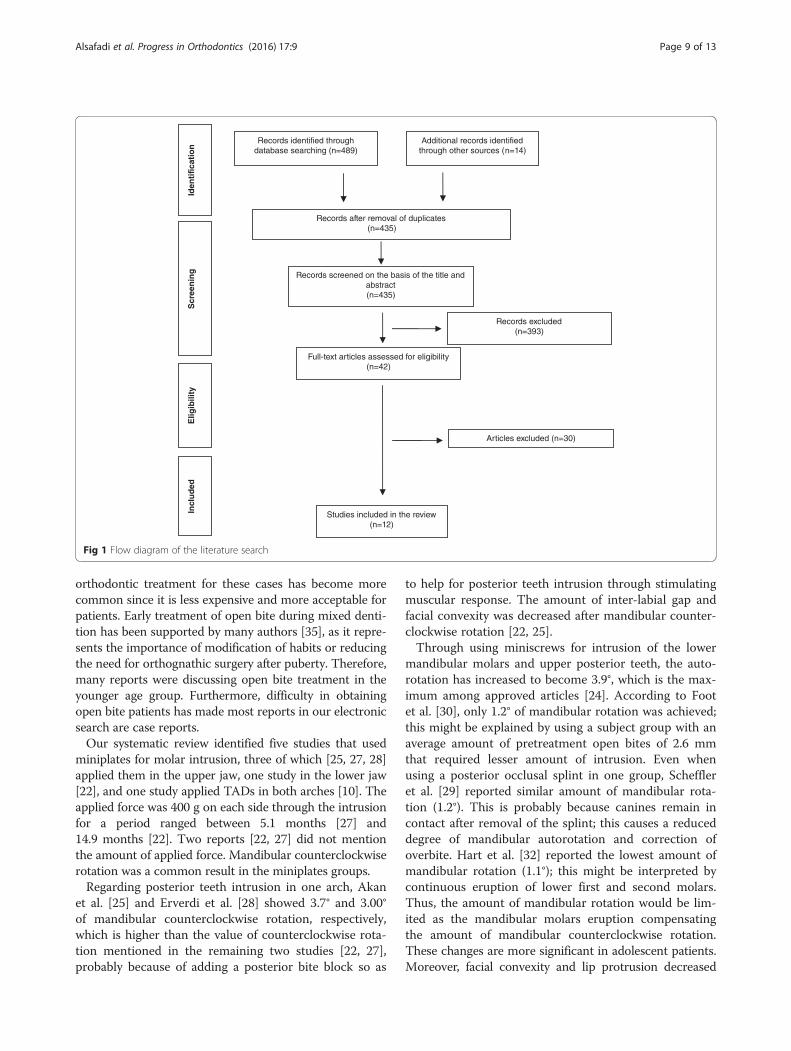
ResultsAmong 503 articles retrieved as a result of the initialsearching process, 393 articles were excluded accordingto the information provided in their titles and abstracts,while 68 articles were excluded as they were duplicatearticles. Consequently, 42 studies remained for the eligi-bility process, and eventually, only 12 studies fulfilledthe inclusion criteria [10, 22–32]. One article by Hart etal. [32] was identified after the date of our search. Table 3shows the study design and characteristics of the final
selected articles. After searching manually within refer-ences of the approved articles, it was found that all re-lated studies were included in the initial electronicsearch process. Figure 1 shows the scheme of article selec-tion and the number of articles accepted and excluded.The complete list of excluded articles and reasons for ex-clusion are available upon request.Different measurements have been used to determine
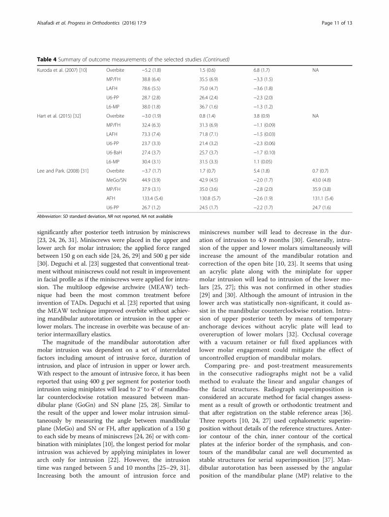
the outcomes of the skeletal changes resulting from pos-terior teeth intrusion during open bite treatment. Table 4shows linear and angular measurements mentioned inthe selected reports, which indicate the amount of man-dibular autorotation and the consequent improvementin facial esthetics.All studies included in our methodological scoring
process have low-moderate quality as presenting in Table 2.Randomization, sample size evaluation, and blinding werenot mentioned in any studies. Three clinical trials withcontrol groups were found in this review [10, 22, 23]. Thecriteria used to assess the amount of molar intrusion andmandibular rotation with its effect on facial morphologywere stated properly by nine articles [10, 22, 23, 25–27,29–32], while follow-up length stability was examined infive reports [22, 23, 28, 29, 31].Out of the 12 selected studies, five studies [10, 22, 25,
27, 28] used miniplates and seven studies [23, 24, 26,29–32] used miniscrews. The amount of mandibularrotation was more than 2° in six studies [10, 23–26,28]. The maximum amount of mandibular counter-clockwise rotation was found to be 3.9° [24], while thelowest amount was 1.1° as reported by Hart et al. [32]by using miniscrews in the upper arch only. In the Kur-oda et al. [10] study, 3.3° mandibular counterclockwiserotation was achieved using temporary anchorage de-vices compared with the orthognathic surgery. Detailedreport of outcome measurements and characteristicsfor each study are presented in Tables 3 and 4.Both dissimilarity and heterogeneity was found in the
outcome measures, after analyzing data in the relatedstudies. As a result, meta-analysis was not possible for thissystematic review.
DiscussionMolar intrusion is one of the valid approaches used foropen bite treatment. While true molar intrusion wasquantified clinically in a previous review [33], no sys-tematic review was conducted to examine the effect ofmolar intrusion on the facial morphology and mandibu-lar autorotation in the permanent dentition.Despite progress in orthodontic treatment techniques,
open bite treatment still represents a challenge for ortho-dontists. While orthognathic surgery is considered thegold standard for skeletal open bite treatment to achievethe optimal esthetic and occlusal result [34], non-surgical
Alsafadi et al. Progress in Orthodontics (2016) 17:9 Page 3 of 13
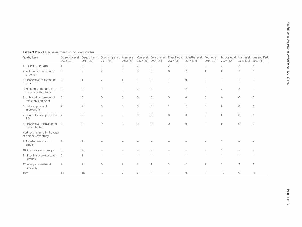
Table 2 Risk of bias assessment of included studies
Quality item Sugawara et al.2002 [22]
Deguchi et al.2011 [23]
Buschang et al.2011 [24]
Akan et al.2013 [25]
Xun et al.2007 [26]
Erverdi et al.2004 [27]
Erverdi et al.2007 [28]
Scheffler et al.2014 [29]
Foot et al.2014 [30]
kuroda et al.2007 [10]
Hart et al.2015 [32]
Lee and Park2008. [31]
1. A clear stated aim 1 2 1 2 2 2 2 1 2 2 2 2
2. Inclusion of consecutivepatients
0 2 2 0 0 0 0 2 1 0 2 0
3. Prospective collection ofdata
0 1 2 1 1 0 1 0 2 1 1 1
4. Endpoints appropriate tothe aim of the study
2 2 1 2 2 2 1 2 2 2 2 1
5. Unbiased assessment ofthe study end point
0 0 0 0 0 0 0 0 0 0 0 0
6. Follow-up periodappropriate
2 2 0 0 0 0 1 2 0 0 0 2
7. Loss to follow-up less than5 %
2 2 0 0 0 0 0 0 0 0 0 2
8. Prospective calculation ofthe study size
0 0 0 0 0 0 0 0 0 0 0 0
Additional criteria in the caseof comparative study
9. An adequate controlgroup
2 2 – – – – – – – 2 – –
10. Contemporary groups 0 2 – – – – – – – 2 – –
11. Baseline equivalence ofgroups
0 1 – – – – – – – 1 – –
12. Adequate statisticalanalyses
2 2 0 2 2 1 2 2 2 2 2 2
Total 11 18 6 7 7 5 7 9 9 12 9 10
Alsafadiet
al.Progressin
Orthodontics
(2016) 17:9 Page
4of
13
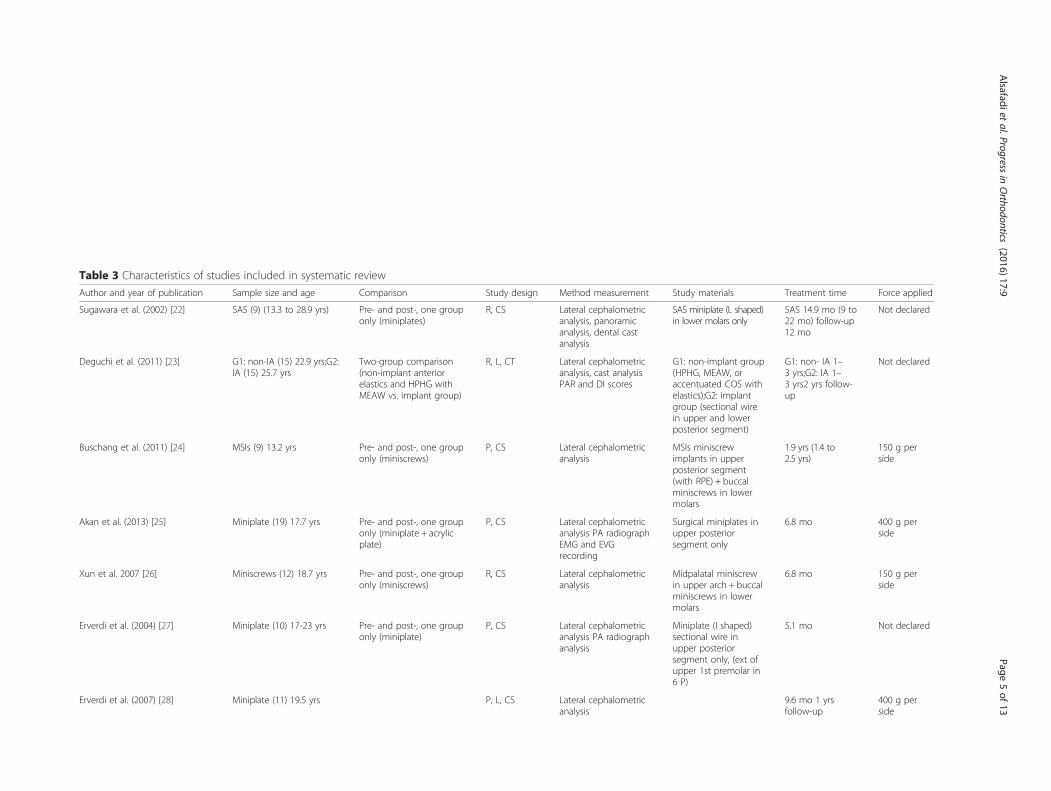
Table 3 Characteristics of studies included in systematic review
Author and year of publication Sample size and age Comparison Study design Method measurement Study materials Treatment time Force applied
Sugawara et al. (2002) [22] SAS (9) (13.3 to 28.9 yrs) Pre- and post-, one grouponly (miniplates)
R, CS Lateral cephalometricanalysis, panoramicanalysis, dental castanalysis
SAS miniplate (L shaped)in lower molars only
SAS 14.9 mo (9 to22 mo) follow-up12 mo
Not declared
Deguchi et al. (2011) [23] G1: non-IA (15) 22.9 yrs;G2:IA (15) 25.7 yrs
Two-group comparison(non-implant anteriorelastics and HPHG withMEAW vs. implant group)
R, L, CT Lateral cephalometricanalysis, cast analysisPAR and DI scores
G1: non-implant group(HPHG, MEAW, oraccentuated COS withelastics);G2: implantgroup (sectional wirein upper and lowerposterior segment)
G1: non- IA 1–3 yrs;G2: IA 1–3 yrs2 yrs follow-up
Not declared
Buschang et al. (2011) [24] MSIs (9) 13.2 yrs Pre- and post-, one grouponly (miniscrews)
P, CS Lateral cephalometricanalysis
MSIs miniscrewimplants in upperposterior segment(with RPE) + buccalminiscrews in lowermolars
1.9 yrs (1.4 to2.5 yrs)
150 g perside
Akan et al. (2013) [25] Miniplate (19) 17.7 yrs Pre- and post-, one grouponly (miniplate + acrylicplate)
P, CS Lateral cephalometricanalysis PA radiographEMG and EVGrecording
Surgical miniplates inupper posteriorsegment only
6.8 mo 400 g perside
Xun et al. 2007 [26] Miniscrews (12) 18.7 yrs Pre- and post-, one grouponly (miniscrews)
R, CS Lateral cephalometricanalysis
Midpalatal miniscrewin upper arch + buccalminiscrews in lowermolars
6.8 mo 150 g perside
Erverdi et al. (2004) [27] Miniplate (10) 17-23 yrs Pre- and post-, one grouponly (miniplate)
P, CS Lateral cephalometricanalysis PA radiographanalysis
Miniplate (I shaped)sectional wire inupper posteriorsegment only, (ext ofupper 1st premolar in6 P)
5.1 mo Not declared
Erverdi et al. (2007) [28] Miniplate (11) 19.5 yrs P, L, CS Lateral cephalometricanalysis
9.6 mo 1 yrsfollow-up
400 g perside
Alsafadiet
al.Progressin
Orthodontics
(2016) 17:9 Page
5of
13
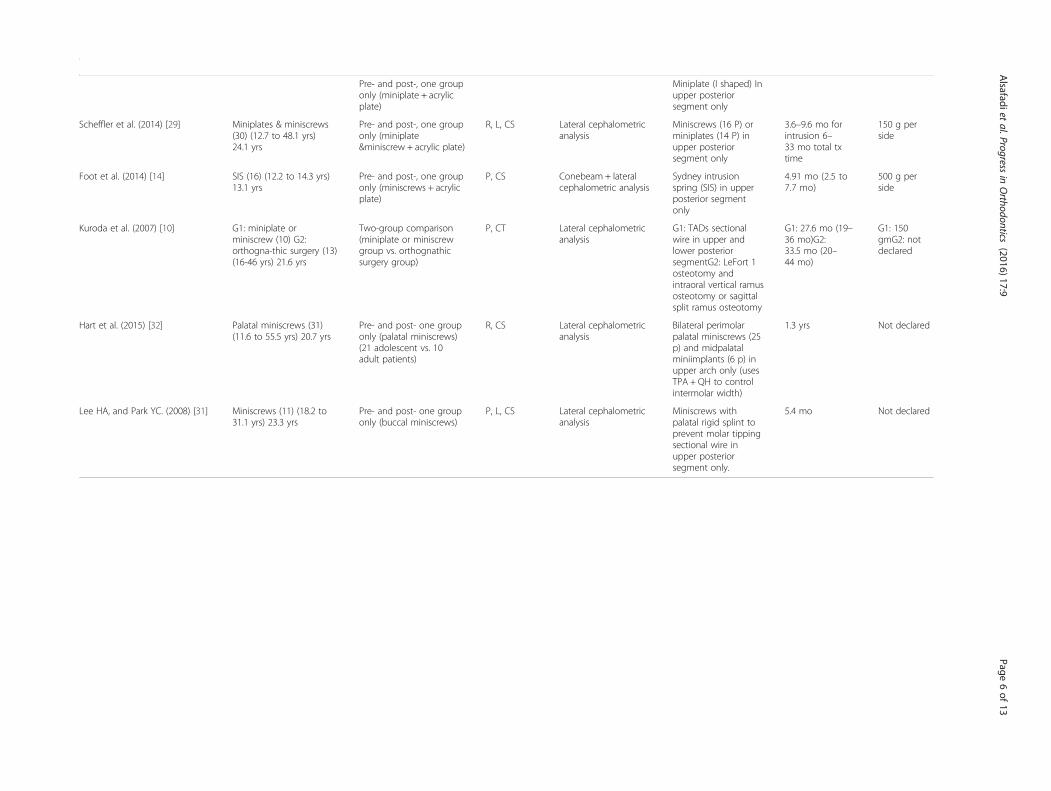
Table 3 Characteristics of studies included in systematic review (Continued)
Pre- and post-, one grouponly (miniplate + acrylicplate)
Miniplate (I shaped) Inupper posteriorsegment only
Scheffler et al. (2014) [29] Miniplates & miniscrews(30) (12.7 to 48.1 yrs)24.1 yrs
Pre- and post-, one grouponly (miniplate&miniscrew + acrylic plate)
R, L, CS Lateral cephalometricanalysis
Miniscrews (16 P) orminiplates (14 P) inupper posteriorsegment only
3.6–9.6 mo forintrusion 6–33 mo total txtime
150 g perside
Foot et al. (2014) [14] SIS (16) (12.2 to 14.3 yrs)13.1 yrs
Pre- and post-, one grouponly (miniscrews + acrylicplate)
P, CS Conebeam + lateralcephalometric analysis
Sydney intrusionspring (SIS) in upperposterior segmentonly
4.91 mo (2.5 to7.7 mo)
500 g perside
Kuroda et al. (2007) [10] G1: miniplate orminiscrew (10) G2:orthogna-thic surgery (13)(16-46 yrs) 21.6 yrs
Two-group comparison(miniplate or miniscrewgroup vs. orthognathicsurgery group)
P, CT Lateral cephalometricanalysis
G1: TADs sectionalwire in upper andlower posteriorsegmentG2: LeFort 1osteotomy andintraoral vertical ramusosteotomy or sagittalsplit ramus osteotomy
G1: 27.6 mo (19–36 mo)G2:33.5 mo (20–44 mo)
G1: 150gmG2: notdeclared
Hart et al. (2015) [32] Palatal miniscrews (31)(11.6 to 55.5 yrs) 20.7 yrs
Pre- and post- one grouponly (palatal miniscrews)(21 adolescent vs. 10adult patients)
R, CS Lateral cephalometricanalysis
Bilateral perimolarpalatal miniscrews (25p) and midpalatalminiimplants (6 p) inupper arch only (usesTPA + QH to controlintermolar width)
1.3 yrs Not declared
Lee HA, and Park YC. (2008) [31] Miniscrews (11) (18.2 to31.1 yrs) 23.3 yrs
Pre- and post- one grouponly (buccal miniscrews)
P, L, CS Lateral cephalometricanalysis
Miniscrews withpalatal rigid splint toprevent molar tippingsectional wire inupper posteriorsegment only.
5.4 mo Not declared
Alsafadiet
al.Progressin
Orthodontics
(2016) 17:9 Page
6of
13
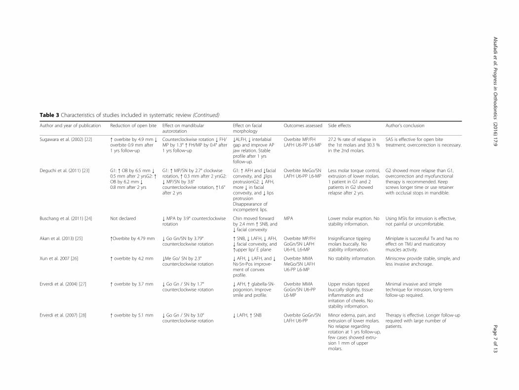
Table 3 Characteristics of studies included in systematic review (Continued)
Author and year of publication Reduction of open bite Effect on mandibularautorotation
Effect on facialmorphology
Outcomes assessed Side effects Author’s conclusion
Sugawara et al. (2002) [22] ↑ overbite by 4.9 mm ↓overbite 0.9 mm after1 yrs follow-up
Counterclockwise rotation ↓ FH/MP by 1.3° ↑ FH/MP by 0.4° after1 yrs follow-up
↓ALFH, ↓ interlabialgap and improve APjaw relation. Stableprofile after 1 yrsfollow-up.
Overbite MP/FHLAFH U6-PP L6-MP
27.2 % rate of relapse inthe 1st molars and 30.3 %in the 2nd molars.
SAS is effective for open bitetreatment; overcorrection is necessary.
Deguchi et al. (2011) [23] G1: ↑ OB by 6.5 mm ↓0.5 mm after 2 yrsG2: ↑OB by 6.2 mm ↓0.8 mm after 2 yrs
G1: ↑ MP/SN by 2.7° clockwiserotation, ↑ 0.3 mm after 2 yrsG2:↓ MP/SN by 3.6°counterclockwise rotation, ↑1.6°after 2 yrs
G1: ↑ AFH and ↓facialconvexity, and ↓lipsprotrusionG2: ↓ AFH,more ↓ in facialconvexity, and ↓ lipsprotrusionDisappearance ofincompetent lips.
Overbite MeGo/SNLAFH U6-PP L6-MP
Less molar torque control,extrusion of lower molars.1 patient in G1 and 2patients in G2 showedrelapse after 2 yrs.
G2 showed more relapse than G1,overcorrection and myofunctionaltherapy is recommended. Keepscrews longer time or use retainerwith occlusal stops in mandible.
Buschang et al. (2011) [24] Not declared ↓ MPA by 3.9° counterclockwiserotation
Chin moved forwardby 2.4 mm ↑ SNB, and↓ facial convexity
MPA Lower molar eruption. Nostability information.
Using MSIs for intrusion is effective,not painful or uncomfortable.
Akan et al. (2013) [25] ↑Overbite by 4.79 mm ↓ Go Gn/SN by 3.79°counterclockwise rotation
↑ SNB, ↓ LAFH, ↓ AFH,↓ facial convexity, and↑upper lip/ E plane
Overbite MP/FHGoGn/SN LAFHU6-HL L6-MP
Insignificance tippingmolars buccally. Nostability information.
Miniplate is successful Tx and has noeffect on TMJ and masticatorymuscles activity.
Xun et al. 2007 [26] ↑ overbite by 4.2 mm ↓Me Go/ SN by 2.3°counterclockwise rotation
↓ AFH, ↓ LAFH, and ↓Ns-Sn-Pos improve-ment of convexprofile.
Overbite MMAMeGo/SN LAFHU6-PP L6-MP
No stability information. Miniscrew provide stable, simple, andless invasive anchorage.
Erverdi et al. (2004) [27] ↑ overbite by 3.7 mm ↓ Go Gn / SN by 1.7°counterclockwise rotation
↓ AFH, ↑ glabella-SN-pogonion. Improvesmile and profile.
Overbite MMAGoGn/SN U6-PPL6-MP
Upper molars tippedbuccally slightly, tissueinflammation andirritation of cheeks. Nostability information.
Minimal invasive and simpletechnique for intrusion, long-termfollow-up required.
Erverdi et al. (2007) [28] ↑ overbite by 5.1 mm ↓ Go Gn / SN by 3.0°counterclockwise rotation
↓ LAFH, ↑ SNB Overbite GoGn/SNLAFH U6-PP
Minor edema, pain, andextrusion of lower molars.No relapse regardingrotation at 1 yrs follow-up,few cases showed extru-sion 1 mm of uppermolars.
Therapy is effective. Longer follow-uprequired with large number ofpatients.
Alsafadiet
al.Progressin
Orthodontics
(2016) 17:9 Page
7of
13
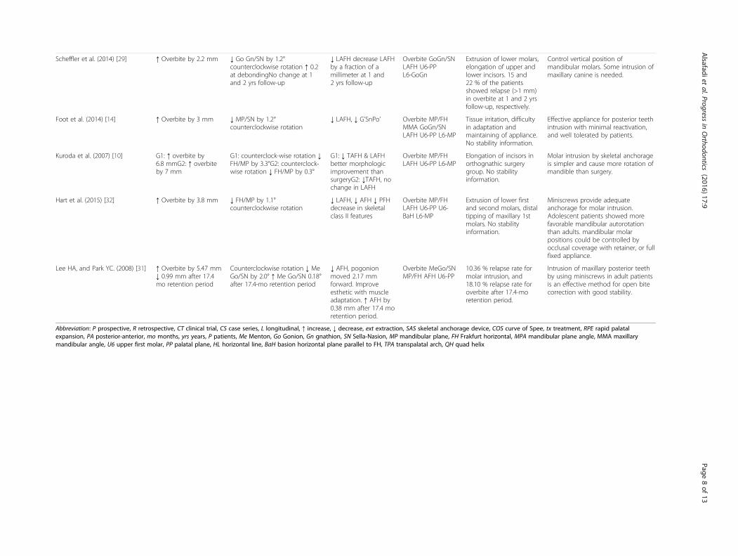
Table 3 Characteristics of studies included in systematic review (Continued)
Scheffler et al. (2014) [29] ↑ Overbite by 2.2 mm ↓ Go Gn/SN by 1.2°counterclockwise rotation ↑ 0.2at debondingNo change at 1and 2 yrs follow-up
↓ LAFH decrease LAFHby a fraction of amillimeter at 1 and2 yrs follow-up
Overbite GoGn/SNLAFH U6-PPL6-GoGn
Extrusion of lower molars,elongation of upper andlower incisors. 15 and22 % of the patientsshowed relapse (>1 mm)in overbite at 1 and 2 yrsfollow-up, respectively.
Control vertical position ofmandibular molars. Some intrusion ofmaxillary canine is needed.
Foot et al. (2014) [14] ↑ Overbite by 3 mm ↓ MP/SN by 1.2°counterclockwise rotation
↓ LAFH, ↓ G’SnPo’ Overbite MP/FHMMA GoGn/SNLAFH U6-PP L6-MP
Tissue irritation, difficultyin adaptation andmaintaining of appliance.No stability information.
Effective appliance for posterior teethintrusion with minimal reactivation,and well tolerated by patients.
Kuroda et al. (2007) [10] G1: ↑ overbite by6.8 mmG2: ↑ overbiteby 7 mm
G1: counterclock-wise rotation ↓FH/MP by 3.3°G2: counterclock-wise rotation ↓ FH/MP by 0.3°
G1: ↓ TAFH & LAFHbetter morphologicimprovement thansurgeryG2: ↓TAFH, nochange in LAFH
Overbite MP/FHLAFH U6-PP L6-MP
Elongation of incisors inorthognathic surgerygroup. No stabilityinformation.
Molar intrusion by skeletal anchorageis simpler and cause more rotation ofmandible than surgery.
Hart et al. (2015) [32] ↑ Overbite by 3.8 mm ↓ FH/MP by 1.1°counterclockwise rotation
↓ LAFH, ↓ AFH ↓ PFHdecrease in skeletalclass II features
Overbite MP/FHLAFH U6-PP U6-BaH L6-MP
Extrusion of lower firstand second molars, distaltipping of maxillary 1stmolars. No stabilityinformation.
Miniscrews provide adequateanchorage for molar intrusion.Adolescent patients showed morefavorable mandibular autorotationthan adults. mandibular molarpositions could be controlled byocclusal coverage with retainer, or fullfixed appliance.
Lee HA, and Park YC. (2008) [31] ↑ Overbite by 5.47 mm↓ 0.99 mm after 17.4mo retention period
Counterclockwise rotation ↓ MeGo/SN by 2.0° ↑ Me Go/SN 0.18°after 17.4-mo retention period
↓ AFH, pogonionmoved 2.17 mmforward. Improveesthetic with muscleadaptation. ↑ AFH by0.38 mm after 17.4 moretention period.
Overbite MeGo/SNMP/FH AFH U6-PP
10.36 % relapse rate formolar intrusion, and18.10 % relapse rate foroverbite after 17.4-moretention period.
Intrusion of maxillary posterior teethby using miniscrews in adult patientsis an effective method for open bitecorrection with good stability.
Abbreviation: P prospective, R retrospective, CT clinical trial, CS case series, L longitudinal, ↑ increase, ↓ decrease, ext extraction, SAS skeletal anchorage device, COS curve of Spee, tx treatment, RPE rapid palatalexpansion, PA posterior-anterior, mo months, yrs years, P patients, Me Menton, Go Gonion, Gn gnathion, SN Sella-Nasion, MP mandibular plane, FH Frakfurt horizontal, MPA mandibular plane angle, MMA maxillarymandibular angle, U6 upper first molar, PP palatal plane, HL horizontal line, BaH basion horizontal plane parallel to FH, TPA transpalatal arch, QH quad helix
Alsafadiet
al.Progressin
Orthodontics
(2016) 17:9 Page
8of
13
orthodontic treatment for these cases has become morecommon since it is less expensive and more acceptable forpatients. Early treatment of open bite during mixed denti-tion has been supported by many authors [35], as it repre-sents the importance of modification of habits or reducingthe need for orthognathic surgery after puberty. Therefore,many reports were discussing open bite treatment in theyounger age group. Furthermore, difficulty in obtainingopen bite patients has made most reports in our electronicsearch are case reports.Our systematic review identified five studies that used
miniplates for molar intrusion, three of which [25, 27, 28]applied them in the upper jaw, one study in the lower jaw[22], and one study applied TADs in both arches [10]. Theapplied force was 400 g on each side through the intrusionfor a period ranged between 5.1 months [27] and14.9 months [22]. Two reports [22, 27] did not mentionthe amount of applied force. Mandibular counterclockwiserotation was a common result in the miniplates groups.Regarding posterior teeth intrusion in one arch, Akan
et al. [25] and Erverdi et al. [28] showed 3.7° and 3.00°of mandibular counterclockwise rotation, respectively,which is higher than the value of counterclockwise rota-tion mentioned in the remaining two studies [22, 27],probably because of adding a posterior bite block so as
to help for posterior teeth intrusion through stimulatingmuscular response. The amount of inter-labial gap andfacial convexity was decreased after mandibular counter-clockwise rotation [22, 25].Through using miniscrews for intrusion of the lower
mandibular molars and upper posterior teeth, the auto-rotation has increased to become 3.9°, which is the max-imum among approved articles [24]. According to Footet al. [30], only 1.2° of mandibular rotation was achieved;this might be explained by using a subject group with anaverage amount of pretreatment open bites of 2.6 mmthat required lesser amount of intrusion. Even whenusing a posterior occlusal splint in one group, Scheffleret al. [29] reported similar amount of mandibular rota-tion (1.2°). This is probably because canines remain incontact after removal of the splint; this causes a reduceddegree of mandibular autorotation and correction ofoverbite. Hart et al. [32] reported the lowest amount ofmandibular rotation (1.1°); this might be interpreted bycontinuous eruption of lower first and second molars.Thus, the amount of mandibular rotation would be lim-ited as the mandibular molars eruption compensatingthe amount of mandibular counterclockwise rotation.These changes are more significant in adolescent patients.Moreover, facial convexity and lip protrusion decreased
Fig 1 Flow diagram of the literature search
Alsafadi et al. Progress in Orthodontics (2016) 17:9 Page 9 of 13
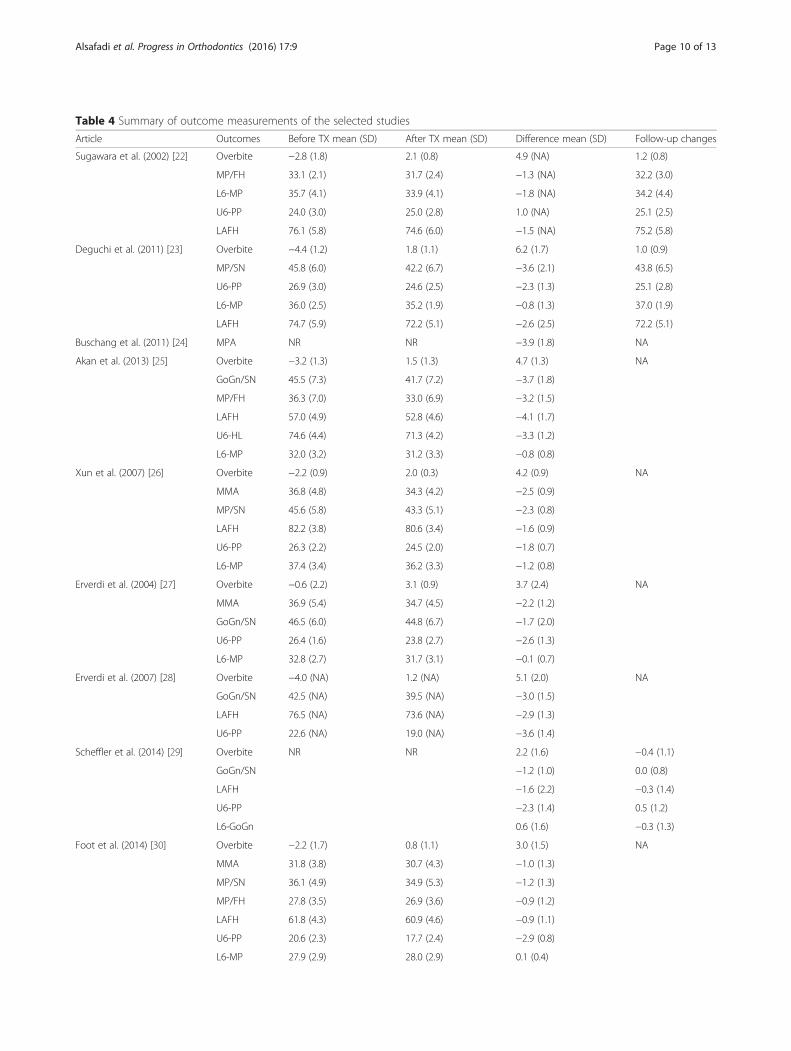
Table 4 Summary of outcome measurements of the selected studies
Article Outcomes Before TX mean (SD) After TX mean (SD) Difference mean (SD) Follow-up changes
Sugawara et al. (2002) [22] Overbite −2.8 (1.8) 2.1 (0.8) 4.9 (NA) 1.2 (0.8)
MP/FH 33.1 (2.1) 31.7 (2.4) −1.3 (NA) 32.2 (3.0)
L6-MP 35.7 (4.1) 33.9 (4.1) −1.8 (NA) 34.2 (4.4)
U6-PP 24.0 (3.0) 25.0 (2.8) 1.0 (NA) 25.1 (2.5)
LAFH 76.1 (5.8) 74.6 (6.0) −1.5 (NA) 75.2 (5.8)
Deguchi et al. (2011) [23] Overbite −4.4 (1.2) 1.8 (1.1) 6.2 (1.7) 1.0 (0.9)
MP/SN 45.8 (6.0) 42.2 (6.7) −3.6 (2.1) 43.8 (6.5)
U6-PP 26.9 (3.0) 24.6 (2.5) −2.3 (1.3) 25.1 (2.8)
L6-MP 36.0 (2.5) 35.2 (1.9) −0.8 (1.3) 37.0 (1.9)
LAFH 74.7 (5.9) 72.2 (5.1) −2.6 (2.5) 72.2 (5.1)
Buschang et al. (2011) [24] MPA NR NR −3.9 (1.8) NA
Akan et al. (2013) [25] Overbite −3.2 (1.3) 1.5 (1.3) 4.7 (1.3) NA
GoGn/SN 45.5 (7.3) 41.7 (7.2) −3.7 (1.8)
MP/FH 36.3 (7.0) 33.0 (6.9) −3.2 (1.5)
LAFH 57.0 (4.9) 52.8 (4.6) −4.1 (1.7)
U6-HL 74.6 (4.4) 71.3 (4.2) −3.3 (1.2)
L6-MP 32.0 (3.2) 31.2 (3.3) −0.8 (0.8)
Xun et al. (2007) [26] Overbite −2.2 (0.9) 2.0 (0.3) 4.2 (0.9) NA
MMA 36.8 (4.8) 34.3 (4.2) −2.5 (0.9)
MP/SN 45.6 (5.8) 43.3 (5.1) −2.3 (0.8)
LAFH 82.2 (3.8) 80.6 (3.4) −1.6 (0.9)
U6-PP 26.3 (2.2) 24.5 (2.0) −1.8 (0.7)
L6-MP 37.4 (3.4) 36.2 (3.3) −1.2 (0.8)
Erverdi et al. (2004) [27] Overbite −0.6 (2.2) 3.1 (0.9) 3.7 (2.4) NA
MMA 36.9 (5.4) 34.7 (4.5) −2.2 (1.2)
GoGn/SN 46.5 (6.0) 44.8 (6.7) −1.7 (2.0)
U6-PP 26.4 (1.6) 23.8 (2.7) −2.6 (1.3)
L6-MP 32.8 (2.7) 31.7 (3.1) −0.1 (0.7)
Erverdi et al. (2007) [28] Overbite −4.0 (NA) 1.2 (NA) 5.1 (2.0) NA
GoGn/SN 42.5 (NA) 39.5 (NA) −3.0 (1.5)
LAFH 76.5 (NA) 73.6 (NA) −2.9 (1.3)
U6-PP 22.6 (NA) 19.0 (NA) −3.6 (1.4)
Scheffler et al. (2014) [29] Overbite NR NR 2.2 (1.6) −0.4 (1.1)
GoGn/SN −1.2 (1.0) 0.0 (0.8)
LAFH −1.6 (2.2) −0.3 (1.4)
U6-PP −2.3 (1.4) 0.5 (1.2)
L6-GoGn 0.6 (1.6) −0.3 (1.3)
Foot et al. (2014) [30] Overbite −2.2 (1.7) 0.8 (1.1) 3.0 (1.5) NA
MMA 31.8 (3.8) 30.7 (4.3) −1.0 (1.3)
MP/SN 36.1 (4.9) 34.9 (5.3) −1.2 (1.3)
MP/FH 27.8 (3.5) 26.9 (3.6) −0.9 (1.2)
LAFH 61.8 (4.3) 60.9 (4.6) −0.9 (1.1)
U6-PP 20.6 (2.3) 17.7 (2.4) −2.9 (0.8)
L6-MP 27.9 (2.9) 28.0 (2.9) 0.1 (0.4)
Alsafadi et al. Progress in Orthodontics (2016) 17:9 Page 10 of 13
significantly after posterior teeth intrusion by miniscrews[23, 24, 26, 31]. Miniscrews were placed in the upper andlower arch for molar intrusion; the applied force rangedbetween 150 g on each side [24, 26, 29] and 500 g per side[30]. Deguchi et al. [23] suggested that conventional treat-ment without miniscrews could not result in improvementin facial profile as if the miniscrews were applied for intru-sion. The multiloop edgewise archwire (MEAW) tech-nique had been the most common treatment beforeinvention of TADs. Deguchi et al. [23] reported that usingthe MEAW technique improved overbite without achiev-ing mandibular autorotation or intrusion in the upper orlower molars. The increase in overbite was because of an-terior intermaxillary elastics.The magnitude of the mandibular autorotation after
molar intrusion was dependent on a set of interrelatedfactors including amount of intrusive force, duration ofintrusion, and place of intrusion in upper or lower arch.With respect to the amount of intrusive force, it has beenreported that using 400 g per segment for posterior toothintrusion using miniplates will lead to 2° to 4° of mandibu-lar counterclockwise rotation measured between man-dibular plane (GoGn) and SN plane [25, 28]. Similar tothe result of the upper and lower molar intrusion simul-taneously by measuring the angle between mandibularplane (MeGo) and SN or FH, after application of a 150 gto each side by means of miniscrews [24, 26] or with com-bination with miniplates [10], the longest period for molarintrusion was achieved by applying miniplates in lowerarch only for intrusion [22]. However, the intrusiontime was ranged between 5 and 10 months [25–29, 31].Increasing both the amount of intrusion force and
miniscrews number will lead to decrease in the dur-ation of intrusion to 4.9 months [30]. Generally, intru-sion of the upper and lower molars simultaneously willincrease the amount of the mandibular rotation andcorrection of the open bite [10, 23]. It seems that usingan acrylic plate along with the miniplate for uppermolar intrusion will lead to intrusion of the lower mo-lars [25, 27]; this was not confirmed in other studies[29] and [30]. Although the amount of intrusion in thelower arch was statistically non-significant, it could as-sist in the mandibular counterclockwise rotation. Intru-sion of upper posterior teeth by means of temporaryanchorage devices without acrylic plate will lead toovereruption of lower molars [32]. Occlusal coveragewith a vacuum retainer or full fixed appliances withlower molar engagement could mitigate the effect ofuncontrolled eruption of mandibular molars.Comparing pre- and post-treatment measurements
in the consecutive radiographs might not be a validmethod to evaluate the linear and angular changes ofthe facial structures. Radiograph superimposition isconsidered an accurate method for facial changes assess-ment as a result of growth or orthodontic treatment andthat after registration on the stable reference areas [36].Three reports [10, 24, 27] used cephalometric superim-position without details of the reference structures. Anter-ior contour of the chin, inner contour of the corticalplates at the inferior border of the symphasis, and con-tours of the mandibular canal are well documented asstable structures for serial superimposition [37]. Man-dibular autorotation has been assessed by the angularposition of the mandibular plane (MP) relative to the
Table 4 Summary of outcome measurements of the selected studies (Continued)
Kuroda et al. (2007) [10] Overbite −5.2 (1.8) 1.5 (0.6) 6.8 (1.7) NA
MP/FH 38.8 (6.4) 35.5 (6.9) −3.3 (1.5)
LAFH 78.6 (5.5) 75.0 (4.7) −3.6 (1.8)
U6-PP 28.7 (2.8) 26.4 (2.4) −2.3 (2.0)
L6-MP 38.0 (1.8) 36.7 (1.6) −1.3 (1.2)
Hart et al. (2015) [32] Overbite −3.0 (1.9) 0.8 (1.4) 3.8 (0.9) NA
MP/FH 32.4 (6.3) 31.3 (6.9) −1.1 (0.09)
LAFH 73.3 (7.4) 71.8 (7.1) −1.5 (0.03)
U6-PP 23.7 (3.3) 21.4 (3.2) −2.3 (0.06)
U6-BaH 27.4 (3.7) 25.7 (3.7) −1.7 (0.10)
L6-MP 30.4 (3.1) 31.5 (3.3) 1.1 (0.05)
Lee and Park. (2008) [31] Overbite −3.7 (1.7) 1.7 (0.7) 5.4 (1.8) 0.7 (0.7)
MeGo/SN 44.9 (3.9) 42.9 (4.5) −2.0 (1.7) 43.0 (4.8)
MP/FH 37.9 (3.1) 35.0 (3.6) −2.8 (2.0) 35.9 (3.8)
AFH 133.4 (5.4) 130.8 (5.7) −2.6 (1.9) 131.1 (5.4)
U6-PP 26.7 (1.2) 24.5 (1.7) −2.2 (1.7) 24.7 (1.6)
Abbreviation: SD standard deviation, NR not reported, NA not available
Alsafadi et al. Progress in Orthodontics (2016) 17:9 Page 11 of 13
FH plane [10, 22, 31, 32] or to the cranial base (SN)[23, 25–30]. However, none of the studies measured theJarabak ratio and Y-axis angle. Others used MMA toevaluate maxillary or mandibular rotation [26, 27, 30].Improvement of facial esthetics by decreasing LAFH
was mentioned in most studies except those conductedby Buschang et al. [24] and Erverdi et al. [27]. Soft tissuechanges must be quantified carefully in order to assessproper changes in them. The use of 3D is a promisingmethod to evaluate soft tissue changes during open bitetreatment provided that they are compared with thecontrol group and obtain an accurate measurement.
LimitationsThere are no randomized clinical trials performed fo-cused on the open bite treatment using temporary an-chorage devices. Presence of randomization is animportant issue to consider when determining the besttreatment modality for posterior teeth intrusion. It isclinically important to investigate the amount of man-dibular rotation during open bite treatment by means ofminiscrews and/or miniplates in comparison with othertherapeutic treatment options (such as MEAW, premo-lars extraction, high-pull headgear, and orthognathic sur-gery), as well as evaluation of the long-term stability ofposterior teeth intrusion by different techniques. Thedrawbacks in most of the articles such as absence of un-treated control groups, short follow-up period, smallsample size, and presence of confounding factors shouldbe avoided in future studies so as to reach a more accur-ate conclusion concerning open bite treatment.
ConclusionsCurrent available evidence suggests that that posteriorteeth intrusion in the permanent dentition stage usingTADs might cause mandibular counterclockwise rota-tion and improve facial esthetics. Miniscrews showed2.3° to 3.9° of mandibular counterclockwise rotation(as sassed by mandibular plane angle, between MeGoor GoGn and SNFH plane) when an intrusive force ap-plied to both upper and lower molars, which is almostsimilar to what was observed after application of thehigh intrusive force in the upper posterior segmentonly by means of miniplate and acrylic bite block. Ab-sence of a standardized method of intrusion, outcomemeasurements, and differences in the protocolsfollowed for molar intrusion (in one arch or botharches) have led to concluding weak clinical evidence.Future well-conducted and clearly reported multicen-ter randomized controlled trials with a non-treatmentcontrol group are needed to provide the best scientificevidence relating to the effect of molar intrusion onthe mandibular rotation and facial esthetic during openbite treatment.
Competing interestsThe authors declare that they have no competing interests.
Authors’ contributionsASA, MA, and AA collected and analyzed the data; ASA, MA, and AAassessed the risk of bias of included studies; ASA, MA, and AA drafted themanuscript and integrated the critical feedback from the other authors.All of the authors were involved in interpretation of the data. All of theauthors provided feedback on the revisions to the manuscript. All authorsread and approved the final manuscript.
Author details1Department of Orthodontics, Faculty of Dentistry, Damascus University,Damascus, Syria. 2Orthodontic Graduate Clinic, Faculty of Medicine andDentistry, University of Alberta, Edmonton, Canada. 3Faculty of Dentistry,Damascus University, Damascus, Syria.
Received: 10 January 2016 Accepted: 23 February 2016
References1. Baek MS, Choi YJ, Yu HS, Lee KJ, Kwak J, Park YC. Long-term stability of
anterior open-bite treatment by intrusion of maxillary posterior teeth.Am J Orthod Dentofacial Orthop. 2010;138(4):396 e1–9.
2. Lopez-Gavito G, Wallen TR, Little RM, Joondeph DR. Anterior open-bitemalocclusion: a longitudinal 10-year postretention evaluation oforthodontically treated patients. Am J Orthod Dentofacial Orthop.1985;87(3):175–86.
3. Kim YH. Anterior openbite and its treatment with multiloop edgewisearchwire. Angle Orthod. 1987;57(4):290–321.
4. Schudy FF. Vertical growth versus anteroposterior growth as related tofunction and treatment. Angle Orthod. 1964;34(2):75–93.
5. Lowe AA. Correlations between orofacial muscle activity and craniofacialmorphology in a sample of control and anterior open-bite subjects. Am JOrthod Dentofacial Orthop. 1980;78(1):89–98.
6. Alabdullah M, Saltaji H, Abou-Hamed H, Youssef M. Association betweenfacial growth pattern and facial muscle activity: a prospective cross-sectionalstudy. Int Orthod. 2015;13(2):181–94.
7. Alabdullah MM, Saltaji H, Abou-Hamed H, Youssef M. The relationshipbetween molar bite force and incisor inclination: a prospective cross-sectional study. Int Orthod. 2014;12(4):494–504.
8. Saltaji H, Flores-Mir C, Major PW, Youssef M. The relationship betweenvertical facial morphology and overjet in untreated class II subjects. AngleOrthod. 2012;82(3):432–40. doi:10.2319/050711-322.1.
9. Park HS, Kwon OW, Sung JH. Nonextraction treatment of an open bite withmicroscrew implant anchorage. Am J Orthod Dentofacial Orthop. 2006;130(3):391–402.
10. Kuroda S, Sakai Y, Tamamura N, Deguchi T, Takano-Yamamoto T. Treatmentof severe anterior open bite with skeletal anchorage in adults: comparisonwith orthognathic surgery outcomes. Am J Orthod Dentofacial Orthop.2007;132(5):599–605.
11. Hwang DH, Park KH, Kwon YD, Kim SJ. Treatment of class II open bitecomplicated by an ankylosed maxillary central incisor. Angle Orthod. 2011;81(4):726–35.
12. Pikdoken L, Erkan M, Usumez S. Gingival response to mandibular incisorextrusion. Am J Orthod Dentofacial Orthop. 2009;135(4):432 e1–6.
13. Kucera J, Marek I, Tycova H, Baccetti T. Molar height and dentoalveolarcompensation in adult subjects with skeletal open bite. Angle Orthod. 2011;81(4):564–9.
14. Uribe F, Nanda R. Management of open bite malocclusion. In: Nanda R,editor. Biomechanics and esthetic strategies in clinical orthodontics. StLouis: Elsevier; 2005. p. 156–76.
15. Bjork A. Prediction of mandibular growth rotation. Am J Orthod DentofacialOrthop. 1969;55(6):585–99.
16. Nahoum HI. Vertical proportions: a guide for prognosis and treatment inanterior open-bite. Am J Orthod Dentofacial Orthop. 1977;72(2):128–46.
17. Lentini-Oliveira Débora A, Carvalho Fernando R, Rodrigues Clarissa G, Ye Q,Hu R, Minami-Sugaya H, et al. Orthodontic and orthopaedic treatment foranterior open bite in children. Cochrane Database Syst Rev. 2014. doi:10.1002/14651858.CD005515.pub3.
Alsafadi et al. Progress in Orthodontics (2016) 17:9 Page 12 of 13
18. Cozza P, Mucedero M, Baccetti T, Franchi L. Early orthodontic treatment ofskeletal open-bite malocclusion: a systematic review. Angle Orthod. 2005;75(5):707–13.
19. Feres MF, Abreu LG, Insabralde NM, Almeida MR, Flores-Mir C. Effectivenessof the open bite treatment in growing children and adolescents. Asystematic review. Eur J Orthod. 2015 Jul 1. [Epub ahead of print].
20. Liberati A, Altman DG, Tetzlaff J, Mulrow C, Gøtzsche PC, Ioannidis JPA, et al.The PRISMA statement for reporting systematic reviews and meta-analysesof studies that evaluate health care interventions: explanation andelaboration. J Clin Epidemiol. 2009;62(10):e1–34.
21. Slim K, Nini E, Forestier D, Kwiatkowski F, Panis Y, Chipponi J.Methodological index for non-randomized studies (minors): developmentand validation of a new instrument. ANZ J Surg. 2003;73(9):712–6.
22. Sugawara J, Baik UB, Umemori M, Takahashi I, Nagasaka H, Kawamura H, etal. Treatment and posttreatment dentoalveolar changes following intrusionof mandibular molars with application of a skeletal anchorage system (SAS)for open bite correction. Int J Adult Orthodon Orthognath Surg. 2002;17(4):243–53.
23. Deguchi T, Kurosaka H, Oikawa H, Kuroda S, Takahashi I, Yamashiro T, et al.Comparison of orthodontic treatment outcomes in adults with skeletal openbite between conventional edgewise treatment and implant-anchoredorthodontics. Am J Orthod Dentofacial Orthop. 2011;139(4 Suppl):S60–8.
24. Buschang PH, Carrillo R, Rossouw PE. Orthopedic correction of growinghyperdivergent, retrognathic patients with miniscrew implants. J OralMaxillofac Surg. 2011;69(3):754–62.
25. Akan S, Kocadereli I, Aktas A, Tasar F. Effects of maxillary molar intrusionwith zygomatic anchorage on the stomatognathic system in anterior openbite patients. Eur J Orthod. 2013;35(1):93–102.
26. Xun C, Zeng X, Wang X. Microscrew anchorage in skeletal anterior open-bite treatment. Angle Orthod. 2007;77(1):47–56.
27. Erverdi N, Keles A, Nanda R. The use of skeletal anchorage in open bitetreatment: a cephalometric evaluation. Angle Orthod. 2004;74(3):381–90.
28. Erverdi N, Usumez S, Solak A, Koldas T. Noncompliance open-bitetreatment with zygomatic anchorage. Angle Orthod. 2007;77(6):986–90.doi:10.2319/101206-422.1.
29. Scheffler NR, Proffit WR, Phillips C. Outcomes and stability in patients withanterior open bite and long anterior face height treated with temporaryanchorage devices and a maxillary intrusion splint. Am J Orthod DentofacialOrthop. 2014;146(5):594–602.
30. Foot R, Dalci O, Gonzales C, Tarraf NE, Darendeliler MA. The short-termskeleto-dental effects of a new spring for the intrusion of maxillary posteriorteeth in open bite patients. Prog Orthod. 2014;15:56.
31. Lee HA, Park YC. Treatment and posttreatment changes following intrusionof maxillary posterior teeth with miniscrew implants for open bitecorrection. Korean J Orthod. 2008;38(1):31–40.
32. Hart TR, Cousley RR, Fishman LS, Tallents RH. Dentoskeletal changesfollowing mini-implant molar intrusion in anterior open bite patients.Angle Orthod. 2015;85(6):941–8.
33. Ng J, Major PW, Flores-Mir C. True molar intrusion attained duringorthodontic treatment: a systematic review. Am J Orthod DentofacialOrthop. 2006;130(6):709–14.
34. Reichert I, Figel P, Winchester L. Orthodontic treatment of anterior openbite: a review article—is surgery always necessary? J Oral Maxillofac Surg.2014;18(3):271–7.
35. Huang GJ, Justus R, Kennedy DB, Kokich VG. Stability of anterior openbitetreated with crib therapy. Angle Orthod. 1990;60(1):17–24.
36. Bishara SE, Athanasiou AE. Cephalometric methods for assessment ofdentofacial changes. In: Athanasiou AE, editor. Othodontic Cephalometry.1st ed. St Louis: Mosby-Wolfe; 1995. p. 105–24.
37. Bjork A, Skieller V. Superimposition of profile radiographs by the structuralmethod. In: normal and abnormal growth of the mandible. Eur J Orthod.1983;5:40–6.
Submit your manuscript to a journal and benefi t from:
7 Convenient online submission
7 Rigorous peer review
7 Immediate publication on acceptance
7 Open access: articles freely available online
7 High visibility within the fi eld
7 Retaining the copyright to your article
Submit your next manuscript at 7 springeropen.com
Alsafadi et al. Progress in Orthodontics (2016) 17:9 Page 13 of 13