Embed Size (px)
Citation preview
Okazaki Fragment Metabolism
Lata Balakrishnan1 and Robert A. Bambara2
1Department of Biochemistry and Biophysics, University of Rochester Medical Center, Rochester,New York 14642
2Department of Microbiology and Immunology, University of Rochester Medical Center, Rochester,New York 14642
Correspondence: [email protected]
Cellular DNA replication requires efficient copying of the double-stranded chromosomalDNA. The leading strand is elongated continuously in the direction of fork opening, whereasthe lagging strand is made discontinuously in the opposite direction. The lagging strandneeds to be processed to form a functional DNA segment. Genetic analyses and reconstitu-tion experiments identified proteins and multiple pathways responsible for maturation of thelagging strand. In both prokaryotes and eukaryotes the lagging-strand fragments are initiatedby RNA primers, which are removed by a joining mechanism involving strand displacementof the primer into a flap, flap removal, and then ligation. Although the prokaryotic fragmentsare �1200 nucleotides long, the eukaryotic fragments are much shorter, with lengths deter-mined by nucleosome periodicity. The prokaryotic joining mechanism is simple and effi-cient. The eukaryotic maturation mechanism involves many enzymes, possibly three path-ways, and regulation that can shift from high efficiency to high fidelity.
Replication of cellular chromosomal DNA isinitiated by the multienzyme replisome ma-
chinery, which unwinds the DNA helix to createa replication fork. The antiparallel structure ofdouble-helical DNA and the 30 end extensionspecificity of all DNA polymerases confine themechanisms that can be used by the cell for DNAduplication. One copied strand, called leading,can conveniently be extended in a continuousmanner in the same direction that the helixmust open to allow exposure of templates forpolymerization. The other, or lagging strand,must be periodically extended away from theopening helix. This can only be accomplished
if the strand is made discontinuously (Kornbergand Baker 1992). The strand is synthesized inshort segments, named Okazaki fragments, af-ter their discoverer (Sakabe and Okazaki 1966;Okazaki et al. 1968) and the segments are thenjoined. This requirement has two fundamentalconsequences: (1) The lagging strand must haveevolved priming and fragment joining mecha-nisms involving many additional steps and reac-tions than needed for leading-strand extension.(2) Mechanisms of lagging-strand replicationmust have developed means of avoiding muta-genesis while handling the necessary strand ma-nipulations.
Editors: Stephen D. Bell, Marcel Mechali, and Melvin L. DePamphilis
Additional Perspectives on DNA Replication available at www.cshperspectives.org
Copyright # 2013 Cold Spring Harbor Laboratory Press; all rights reserved; doi: 10.1101/cshperspect.a010173
Cite this article as Cold Spring Harb Perspect Biol 2013;5:a010173
1
on June 20, 2018 - Published by Cold Spring Harbor Laboratory Press http://cshperspectives.cshlp.org/Downloaded from
PARALLELS BETWEEN PROKARYOTIC ANDEUKARYOTIC REPLICATION
Although replication of eukaryotic DNA ona chromatinized DNA template is a relativelymore complex process than replication in bacte-ria, involving more types of proteins and re-actions, the fundamental processes of DNA du-plication have striking parallels in all cells. Thereplisome machineries of both organisms areminimally composed of helicases, which un-wind the duplex strands, primase, which in-itiates synthesis and DNA polymerases, whichduplicate the parental strands of the DNA. Inbacteria, DNA replication proceeds within afork, wherein the lagging strand loops into a“trombonelike” structure allowing for the repli-cation enzymes to be continually recycled on theDNA for repeated synthesis and joining (Albertset al. 1982). Presumably, use of a similar mech-
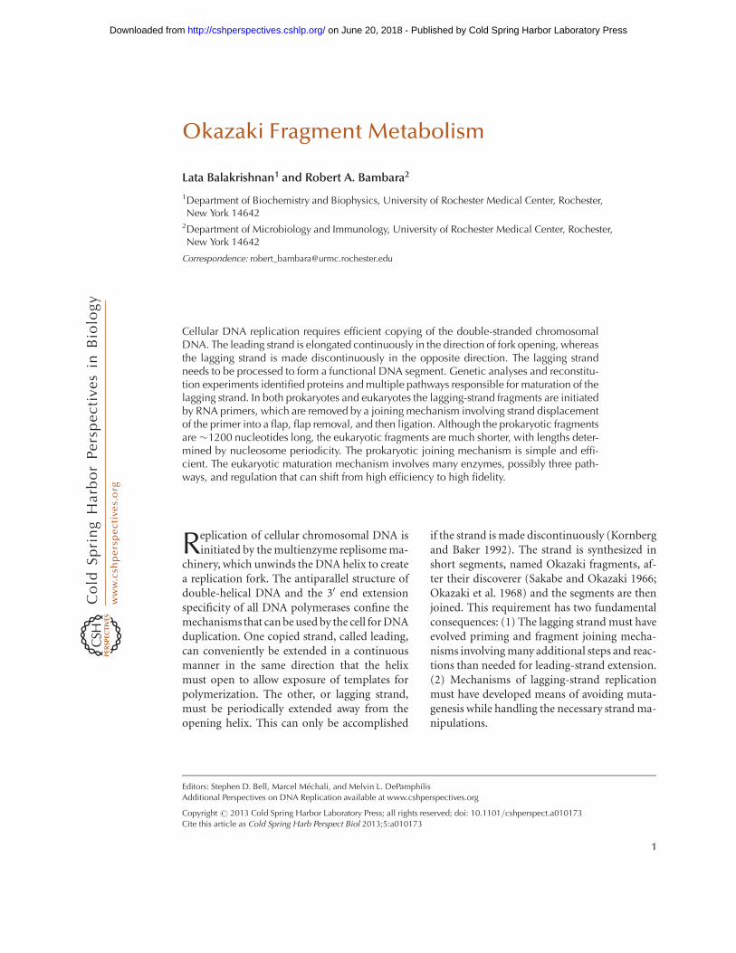
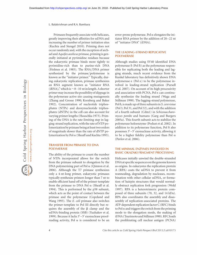
anism in eukaryotes allows coordination of syn-thesis between the leading and lagging strands(Fig. 1) (Pandey et al. 2009). Because DNA po-lymerases cannot incorporate dNTPs without aprimer terminated by a 30 hydroxyl, the leadingstrand and each Okazaki fragment are primedby RNA to initiate synthesis (Hubscher et al.2002). On the lagging strand the primer is ex-tended by the addition of dNMPs to form shortsegments of DNA. These segments need to befurther processed to form a fully functionalstrand of DNA. Specifically, the RNA primershave to be excised from the fragments. Removalof RNA primers is performed partly by a ribo-nuclease H (RNase H). There are two distinctclasses of RNase H enzymes in bacterial andeukaryotic systems (type 1: RNase HI [Escher-ichia coli] and RNase H1 [eukaryotes] and type2: RNase HII [E. coli] and RNase H2 [eukary-otes]) (Cerritelli and Crouch 2009). Although
DNA helicase
RPA
RFC Lagging strand
Leading strand
Pol α
Pol δ
Pol ε PCNA
Figure 1. Eukaryotic replisome. A DNA helicase initially unwinds the duplex DNA (red and blue strands) toseparate the DNA and form a replication fork. The single-stranded DNA (ssDNA) is coated by the single-strandbinding protein, replication protein A (RPA). On the leading strand, replication factor C (RFC) loads prolif-erating cell nuclear antigen (PCNA) and DNA polymerase 1 to continuously synthesize the leading strand. Thelagging strand is initially primed by DNA polymerase a (Pol a), which synthesizes a short RNA/DNA initiatorprimer (orange strand). RFC then displaces Pola from the lagging strand to initiate the switch from the primingmode to the elongation mode. The initiator primer is extended by PCNA/DNA polymerase d complex to formshort segments of DNA known as Okazaki fragments.
L. Balakrishnan and R.A. Bambara
2 Cite this article as Cold Spring Harb Perspect Biol 2013;5:a010173
on June 20, 2018 - Published by Cold Spring Harbor Laboratory Press http://cshperspectives.cshlp.org/Downloaded from
type 1 RNases H require a minimum of fourribonucleotides for hydrolysis, type 2 RNasesH can recognize a single ribonucleotide (Cerri-telli and Crouch 2009). In eukaryotes, the ini-tiator RNA primers are removed, apparentlyby two partly redundant processes (Kao andBambara 2003). The first is the action of cellularRNase H2, which can begin degrading the prim-er as soon as it is made, but presumably not sosoon that it interferes with initiation of DNAsynthesis (Turchi et al. 1994; Murante et al.1998). RNase H2 degrades between ribonucleo-tides of an RNA strand annealed to DNA. It can-not cleave between the 30-most ribonucleotideand the initial deoxynucleotide. A second nucle-ase is needed for this. The second nuclease oper-ates after strand displacement synthesis resultingfrom extension of the 30 end of the adjacent frag-ment. This synthesis raises what remains of theRNA primer into a single-stranded flap structurethat is removed by endonuclease action. The re-sulting nick is then ligated to make a continuousstrand. The absence of RNase HI in E. coli leadsto initiation of replication at sites other thanthe replication initiation site oriC (Hong andKogoma 1993). Genetic deletions of the RNaseH enzymes in Saccharomyces cerevisiae did notyield a distinct phenotype, leading to the sug-gestion that RNase H is not the primary path-way for RNA removal in those cells (Frank et al.1998; Qiu et al. 1999). Fundamental differencesamong organisms include structural variationsin the proteins involved, and length variationsin the fragments. There are also as many as threepathways in eukaryotes, which involve differentbut overlapping sets of proteins (Balakrishnanand Bambara 2011b). Most recently, there isevidence that flow through these pathways isregulated to optimize fidelity and rate of syn-thesis (Balakrishnan and Bambara 2011b).
KORNBERG SIXTH COMMANDMENT:“DEPEND ON VIRUSES TO OPENWINDOWS”
Most of our initial understanding of the processof DNA replication was obtained from studies invitro using cellular extracts to replicate SimianVirus 40 (SV40) DNA, a short double-stranded
circle (Waga et al. 1994; Bambara et al. 1997;Waga and Stillman 1998). Because SV40 en-codes only the minimal proteins required for aviral assembly, it utilizes the cellular machineryfrom the host cell for replication (Tooze 1981).Studies by Stillman and coworkers (Waga et al.1994; Waga and Stillman 1998) using SV40 as amodel system showed that T antigen (encodedby the early viral genes), along with host single-stranded DNA-binding protein, called replica-tion protein A (RPA), and topoisomerases, ini-tiates DNA replication by binding to the origin(ori) and unwinding the duplex DNA (Tsuri-moto et al. 1990). Reconstitution of SV40 DNAreplication using purified proteins helped inthe identification of specific enzymatic mecha-nisms used by the eukaryotic replication fork(Dean et al. 1992; Eki et al. 1992; Ishimi et al.1992). Individual steps of this process and theenzymes involved are described in the sectionsbelow. Although the SV40 replication system invitro provided valuable insights into eukaryoticreplication, the absence of Pol 1 as the leading-strand polymerase and the use of viral Tantigenas the helicase for SV40 replication made it dif-ficult to consider it as an absolutely analogoussystem to the actual more complex mammalianeukaryotic system.
PRIMING THE LAGGING STRAND—THEINITIAL STEP
Because DNA polymerases cannot initiate syn-thesis de novo, the replicative polymerase hasevolvedapartnershipwithprimasetoenablesyn-thesis on a DNA template. DnaG acts as the pri-mase in bacteria and in eukaryotes the primaseis part of DNA polymerase a (Pol a) (Hubscheret al. 2002; Kuchta and Stengel 2010). Pol a is afour-subunit complex, comprised of the poly-merase subunit (Pol I, p140), a regulatory sub-unit (Pol 12, p79), and two primase subunits.The Pri1 subunit (p48) acts as the fundamentalinitiator of replication by catalyzing the forma-tion of the short RNA primers, which are subse-quentlyelongated by Pol I (Hubscheret al. 2002).The second primase subunit, Pri2 (p58) acts as ascaffold, which holds together the primase andthe polymerase subunits.
Okazaki Fragment Metabolism
Cite this article as Cold Spring Harb Perspect Biol 2013;5:a010173 3
on June 20, 2018 - Published by Cold Spring Harbor Laboratory Press http://cshperspectives.cshlp.org/Downloaded from
Primases frequently associate with helicases,greatly improving their affinities for ssDNA andincreasing the number of primer initiation sites(Kuchta and Stengel 2010). Priming does notoccur randomly and, with the exception of arch-ael and Aquifex aeolicus primase, priming is gen-erally initiated at pyrimidine residues becausethe eukaryotic primase binds more tightly topyrimidine-rich than to purine-rich DNA(Holmes et al. 1985). The RNA/DNA primersynthesized by the primase/polymerase isknown as the “initiator primer.” Typically, dur-ing eukaryotic replication, primase synthesizesan RNA segment known as “initiator RNA(iRNA),” which is�8–10 nt in length. A shorterprimer may increase the possibilityof slippage inthe polymerase active site causing mutagenesis(Zhang and Grosse 1990; Kornberg and Baker1992). Concentration of nucleotide triphos-phates (NTPs) and deoxynucleotide triphos-phates (dNTPs) in the cell can also account forvarying primer lengths (Hauschka 1973). Prim-ing of the DNA is the rate-limiting step in lag-ging-strand replication, with the rate of NTP po-lymerization by primase being at least twoordersof magnitude slower than the rate of dNTP po-lymerization by Pola (Sheaff and Kuchta 1993).
TRANSFER FROM PRIMASE TO DNAPOLYMERASE
The ability of the primase to count the numberof NTPs incorporated allows for the switchfrom the primase subunit to elongation by theDNA polymerizing part of Pol a (Qimron et al.2006). Although the T7 primase synthesizesonly a 4-nt-long primer, eukaryotic primasestypically synthesize primers longer than 7 nt toenable efficient hand off of the primer-templatefrom the primase to DNA Pol a (Sheaff et al.1994). This is performed by the p58 subunit,which acts as the point of contact between theprimase and the polymerase (Copeland andWang 1993). The E. coli primase also switchesthe primer-template to Pol III directly but re-quires the assembly of the b clamp and thessDNA-binding protein (SSB) (Yuzhakov et al.1999). Because it lacks 30 –50 exonuclease proof-reading activity, Pol a is considered to be an
error-prone polymerase. Pola elongates the ini-tiator RNA primer by the addition of 20–22 ntof “initiator DNA” (iDNA).
THE LEADING-STRAND REPLICATIVEPOLYMERASE
Although studies using SV40 identified DNApolymerase d (Pol d) as the polymerase respon-sible for replicating both the leading and lag-ging strands, much recent evidence from theKunkel laboratory has definitively shown DNApolymerase 1 (Pol 1) to be the polymerase in-volved in leading-strand replication (Pursellet al. 2007). On account of its high processivityand association with PCNA, Pol 1 can continu-ally synthesize the leading strand (Waga andStillman 1998). The lagging-strand polymerase,Pold, is made up of three subunits in S. cerevisiae(Pol 3, Pol 31, and Pol 32), and with the additionof a fourth subunit (Cdm1) in Schizosaccharo-myces pombe and humans (Garg and Burgers2005a). This fourth subunit acts to stabilize thepolymerase holoenzyme (Podust et al. 2002). Inaddition to its polymerase function, Pol d alsopossesses 30–50 exonuclease activity, allowing itto be a higher fidelity polymerase than Pol a(Pavlov et al. 2006).
THE MINIMAL ENZYMES INVOLVED INBASIC OKAZAKI FRAGMENT PROCESSING
Helicases initially unwind the double-strandedDNAat specific sequenceson the genome knownas origins. In eukaryotes the replication proteinA (RPA) coats the ssDNA to prevent it fromreannealing, degradation by nucleases, recom-bination with other cellular ssDNA, or forma-tion of hairpin structures that would normal-ly obstruct replication fork progression (Wold1997). RPA is a heterotrimeric protein com-posed of three subunits (70, 32, and 14 kDa).RPA also coordinates the assembly and disas-sembly of replication-associated proteins. TheATP-dependent replication factor C (RFC) bindsto Pola and triggers the switch from the primingmode to the elongation mode, the making ofiDNA (Tsurimoto andStillman1990).RFCloadsthe proliferating cell nuclear antigen (PCNA)
L. Balakrishnan and R.A. Bambara
4 Cite this article as Cold Spring Harb Perspect Biol 2013;5:a010173
on June 20, 2018 - Published by Cold Spring Harbor Laboratory Press http://cshperspectives.cshlp.org/Downloaded from
along with Pol d to initiate the elongation on thelagging-strand DNA template (Tsurimoto andStillman 1990). PCNA, a functional homologof the prokaryotic b clamp, having a similarring-shaped structure, is opened up with thehelp of RFC to be loaded onto the double-stranded iDNA (Burgers 2009). PCNA binds tothe back of the Pol d and acts as a sliding clamp,increasing the processivityof the polymerase. Pold adds �100 nt of DNA in humans and �250 ntof DNA in S. cerevisiae to form short Okazakifragments, which need to be further matured toform afunctional strand of DNA. During a singleround of nuclear DNA replication in S. cerevisiae�100,000 Okazaki fragments are made and ma-tured (Garg and Burgers 2005b).
SIGNIFICANCE OF FRAGMENT SIZE
Despite the much larger DNA content of eu-karyotic compared with prokaryotic cells, Oka-zaki fragments are �1200 nt long in bacteria butonly about 200 nt long in eukaryotes (Ogawaand Okazaki 1980). This means that to preparefor every human cell division, .10 million frag-ments must be made and joined. The bacterialfragments might be larger because the ligationprocess is much slower than the time needed tosynthesize each fragment. Pol III holoenzymeelongates primers at 1200 nt/sec. This is consis-tent with the ability of bacteria to double in therange of a half-hour. Possibly the joining of thelagging strand could not keep up this pace ifthe fragments were shorter. Another possibilityis that the nucleosomal structure of DNA influ-ences the frequency of fragment priming. Is itcoincidence that the average fragment size issimilar to the length of DNA associated with amononucleosome? Recent evidence using high-resolution analysis has shown that Okazaki frag-ments are sized according to chromatin repeats(Smith and Whitehouse 2012).
PATHWAYS OF EUKARYOTIC OKAZAKIFRAGMENT PROCESSING
Short Flap Pathway
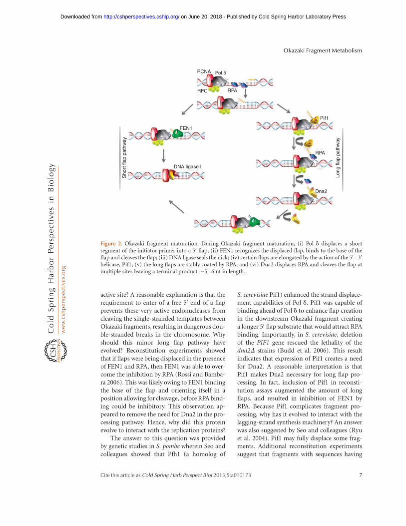
Because eukaryotic lagging-strand DNA isprimed at short intervals, Pol d frequently en-
counters the downstream primed Okazaki frag-ment and displaces the RNA/DNA initiatorprimer into a 50 flap structure. This strand dis-placing activity is very similar to that reportedfor bacterial Pol I. However, unlike Pol I, Pol ddoes not possess a nuclease activity to cleave thedisplaced flap. Flap endonuclease 1 (FEN1; orscRad27), a structure-specific 50-30 endonucle-ase, recognizes the displaced 50 flap and cleavesat the base creating a nicked substrate for liga-tion (Bambara et al. 1997; Lieber 1997). Thisis the predominant method of removing thePol a-synthesized initiator primer during thematuration process. Initial studies characteriz-ing the mechanism of FEN1 suggested a “track-ing” model for FEN1 in which the nucleasemoves from the 50 end of the flap to its basewhere it performs a specific cleavage (Bambaraet al. 1997). This mechanism was proposed be-cause studies in vitro showed that endonu-cleolytic cleavage by FEN1 is inhibited whenthe 50 end of the flap is blocked either with acomplementary primer or a biotin-conjugatedstreptavidin moiety (Murante et al. 1995). How-ever, recent work has shown that FEN1 initial-ly binds to the base of the flap causing a changein the substrate conformation that orients FEN1in a manner that allows for precise cleavage(Gloor et al. 2010; Tsutakawa et al. 2011). Thefree 50 end of the flap is then threaded past orthrough the helical arch and active site of FEN1permitting a single cleavage event (Gloor et al.2010).
DNA ligase I (cdc9 in S. cerevisiae) seals thenick generated by FEN1 to create a fully func-tional continuous double-stranded DNA (Bam-bara et al. 1997). PCNA interacts with bothFEN1 and DNA ligase I and stimulates the en-zymatic functions of both these proteins (Rossiet al. 2006). Interactions with PCNA are criticalfor the creation and proper ligation of the lag-ging-strand DNA. It has been previously sug-gested that PCNA serves to recruit the core en-zymes to the replication fork and functions tosequentially hand off the proteins to performtheir enzymatic tasks during the maturationprocess (Kao and Bambara 2003). Sequentialstrand displacement and cleavage by Pol d andFEN1, respectively, helps to remove the entire
Okazaki Fragment Metabolism
Cite this article as Cold Spring Harb Perspect Biol 2013;5:a010173 5
on June 20, 2018 - Published by Cold Spring Harbor Laboratory Press http://cshperspectives.cshlp.org/Downloaded from
initiator RNA before nick ligation. The wild-type Pol d shows reduced strand displacementactivity compared with its exonuclease mutants(Pol d-5DV and Pol d-01) (Garg and Burgers2005b). Apparently the exonuclease regulatesdisplacement (Garg and Burgers 2005b). It isstill not very clear how the wild-type enzymesenses how many nucleotides of the RNA/DNA primer it has displaced. However, analysisof lagging-strand replication using purifiedS. cerevisiae proteins has shown that the major-ity of the substrates Pol d exonuclease activityand FEN1 cleavage functions cooperate to createand cleave flaps shorter than 10 nt long (Ayya-gari et al. 2003; Rossi and Bambara 2006). Sev-eral displacement and cleavage reactions are re-quired to remove the initiator primer. A flapcreated and processed via this mechanism hasmatured by the “short flap pathway” (Fig. 2).
Long Flap Pathway
In some instances FEN1 transiently disengagesfrom the replication complex. This can causeenough of a delay in cleavage that flaps dis-placed by Pol d become long. When they reachlengths .22 nt, RPA can bind stably (Rossi andBambara 2006). RPA-bound flaps are refractoryto FEN1 cleavage, requiring the action of anoth-er nuclease for proper processing (Bae et al.2001). Budd and Campbell identified this al-ternate nuclease, Dna2, in a genetic screen inS. cerevisiae (Budd and Campbell 1995). Dna2nuclease/helicase is a multifunctional proteincontaining 50 –30 endonuclease and minor 30 –50 exonuclease activities, plus 50 –30 helicase andATPase functions (Kang et al. 2010). Campbelland colleagues showed that overexpression ofDna2 compensated for defects in FEN1 andoverexpression of FEN1 did the same for Dna2(Budd and Campbell 1997). More recent workshowed that the double mutant of 50 –30 nucle-ase-defective Dna2 (dna2-1) and 30 nuclease-deficient Pol d ( pol3-01), which has augmentedstrand displacement activity, is lethal (Buddet al. 2005). These results imply that Dna2works with FEN1 specifically to process longflaps. In addition, Dna2 was recently found tobe complexed with FEN1 in human cell extracts,
suggesting it as a physical and functional part-ner of FEN1 (Balakrishnan et al. 2010).
S. cerevisiae Dna2 can dissociate the RPAfrom a long flap (Bae et al. 2001; Stewart et al.2008). Using a mechanism very similar to thatof FEN1, Dna2 binds the flap base, and thenthreads the free 50 end of the flap (Stewartet al. 2010). However, this nuclease cleaves pe-riodically up to a terminal product flap �5–6 nt in length. This is too short to bind RPA, sothe short flap is readily available for cleavage byFEN1 and subsequent ligation. This method offlap processing is known as the “long flap path-way” (Fig. 2). Reconstitution experiments haveshown that although the majority of the dis-placed flaps are processed by the short flap path-way, a minority require the long flap pathway.Seo and colleagues originally proposed theDna2 pathway as the primary means of Okazakifragment processing (Bae et al. 2001). Theirproposal was likely influenced by genetic evi-dence in S. cerevisiae that Dna2 inactivation incells was lethal, whereas FEN1 mutants onlyshowed a slow-growing phenotype. However,these observations can be misleading. Exo1, a50 –30 exonuclease interacts with both FEN1 andDna2 and can specifically act as a backup forFEN1 nuclease activity. Although this is an in-efficient process, and probably not biologicallyrelevant, it explains why rad27D (FEN1) mu-tants are viable. Also, recent work from theCampbell laboratory showed that Dna2 inter-acts with Rad9, the damage checkpoint activa-tor, participating in the double-strand break re-pair response. The double deletion dna2D andrad9D rescued dna2D lethality suggesting thatRad9-dependent activation of the checkpointcontributed to the lethality in dna2D cells(Budd et al. 2011). In the absence of Dna2and Rad9, the damage response utilizes theExo1 pathway for repairing the damaged DNA(Balakrishnan and Bambara 2011a).
EVOLVED INTERACTIONS OF FEN1,Dna2, AND Pif1
Why did both FEN1 and Dna2 develop a mech-anism in which the nucleases bind to the base ofthe flap and thread the free 50 end through their
L. Balakrishnan and R.A. Bambara
6 Cite this article as Cold Spring Harb Perspect Biol 2013;5:a010173
on June 20, 2018 - Published by Cold Spring Harbor Laboratory Press http://cshperspectives.cshlp.org/Downloaded from
active site? A reasonable explanation is that therequirement to enter of a free 50 end of a flapprevents these very active endonucleases fromcleaving the single-stranded templates betweenOkazaki fragments, resulting in dangerous dou-ble-stranded breaks in the chromosome. Whyshould this minor long flap pathway haveevolved? Reconstitution experiments showedthat if flaps were being displaced in the presenceof FEN1 and RPA, then FEN1 was able to over-come the inhibition by RPA (Rossi and Bamba-ra 2006). This was likely owing to FEN1 bindingthe base of the flap and orienting itself in aposition allowing for cleavage, before RPA bind-ing could be inhibitory. This observation ap-peared to remove the need for Dna2 in the pro-cessing pathway. Hence, why did this proteinevolve to interact with the replication proteins?
The answer to this question was providedby genetic studies in S. pombe wherein Seo andcolleagues showed that Pfh1 (a homolog of
S. cerevisiae Pif1) enhanced the strand displace-ment capabilities of Pol d. Pif1 was capable ofbinding ahead of Pol d to enhance flap creationin the downstream Okazaki fragment creatinga longer 50 flap substrate that would attract RPAbinding. Importantly, in S. cerevisiae, deletionof the PIF1 gene rescued the lethality of thedna2D strains (Budd et al. 2006). This resultindicates that expression of Pif1 creates a needfor Dna2. A reasonable interpretation is thatPif1 makes Dna2 necessary for long flap pro-cessing. In fact, inclusion of Pif1 in reconsti-tution assays augmented the amount of longflaps, and resulted in inhibition of FEN1 byRPA. Because Pif1 complicates fragment pro-cessing, why has it evolved to interact with thelagging-strand synthesis machinery? An answerwas also suggested by Seo and colleagues (Ryuet al. 2004). Pif1 may fully displace some frag-ments. Additional reconstitution experimentssuggest that fragments with sequences having
PCNA Pol δ
RPA
Pif1
RPA
Dna2
DNA ligase I
FEN1
Long
flap
pat
hway
Sho
rt fl
ap p
athw
ay
RFC
Figure 2. Okazaki fragment maturation. During Okazaki fragment maturation, (i) Pol d displaces a shortsegment of the initiator primer into a 50 flap; (ii) FEN1 recognizes the displaced flap, binds to the base of theflap and cleaves the flap; (iii) DNA ligase seals the nick; (iv) certain flaps are elongated by the action of the 50 –30
helicase, Pif1; (v) the long flaps are stably coated by RPA; and (vi) Dna2 displaces RPA and cleaves the flap atmultiple sites leaving a terminal product �5–6 nt in length.
Okazaki Fragment Metabolism
Cite this article as Cold Spring Harb Perspect Biol 2013;5:a010173 7
on June 20, 2018 - Published by Cold Spring Harbor Laboratory Press http://cshperspectives.cshlp.org/Downloaded from
the potential to form 50 end region secondarystructure are difficult to process. When the flapis created, it folds in a way that prevents cleavageby either FEN1 or Dna2. However, Pif1 can bindbetween the structure and the flap base and fullydisplace the fragment. The upstream fragmentcan then extend through the structured region.This is effectively a third pathway of fragmentprocessing (Pike et al. 2010). Although the longflap pathway has evolved elegantly to processsuch flaps, evidence in vitro suggests that theshort flap pathway is more commonly used,with each protein from the long flap pathway(Dna2, Pif1, and RPA) stimulating the functionof FEN1, and so promoting the use of the shortflap pathway (Henry et al. 2010).
MECHANISTIC SIMILARITIES OF OKAZAKIFRAGMENT PROCESSING AND LONGPATCH BASE EXCISION REPAIR
Cells are constantly being exposed to endoge-nous and exogenous stresses that cause oxida-tive damage to DNA bases. Base excision repair(BER) is the most commonly utilized means ofdealing with these damaged bases. It proceedsvia two pathways. In the short patch pathway(SP-BER) a damaged base is recognized by aDNA glycosylase and removed. The abasic siteis cleaved by apurinic/apyrimidinic endonucle-ase 1 (APE1). The one-nucleotide gap is filledby DNA polymerase b (Pol b), which also re-moves the 50 deoxyribose phosphate (dRP) us-ing an intrinsic lyase activity. However, if thedRP is oxidized, reduced, or otherwise altered,the lyase function does not work. The Polb thendisplaces the damaged site into a 2–12 nt 50 flap(Balakrishnan et al. 2009). These flaps are di-rected down the long patch pathway (LP-BER)in which they are cleaved by FEN1 and sealed byDNA ligase I. It is not clear whether the longestflaps would bind RPA with sufficient avidity torequire Dna2; however, the recent report ofDna2 involvement in mitochondrial LP-BERsuggests that some do (Zheng et al. 2008). Theoverlap of proteins used for Okazaki fragmentprocessing and LP-BER suggests that the twoprocesses evolved from the same ancestral basicpathway. The overlap also suggests that regula-
tory mechanisms for one process will similarlyinfluence the other.
POSTTRANSLATIONAL REGULATION OFREPLICATION PROTEINS
Most of the proteins involved in eukaryoticDNA replication experience various posttransla-tional modifications, regulating their enzymat-ic functions, subcellular localizations, or par-ticipation in a specific pathway. Pol a/primasehas been reported to be phosphorylated both inhumans and S. cerevisiae. Phosphorylation ofPol a occurs late in S phase, thereby possiblycoordinating the S phase with the mitotic phase.Results obtained in vitro suggest that althoughthe polymerase activity of Pol a is not alteredby phosphorylation, the primase function isslightly stimulated without affecting the lengthof the initiator primer (Waga and Stillman1998). Recent evidence from the Lee laboratoryhas shown that phosphorylation of Pol d on thep68 subunit decreases the binding affinity ofthe polymerase to PCNA (Rahmeh et al. 2012).This in turn decreases the processivity of poly-merization. Pol dwas also found to be acetylatedon the catalytic subunit in a mass spectromet-ric analysis (Choudhary et al. 2009). PCNA ismodified by a diverse range of modificationssuch as acetylation, phosphorylation, and ubiq-uitination. Although acetylation improves thebinding affinity to PCNA to DNA polymerases(Naryzhny and Lee 2004), tyrosine phosphory-lation of PCNA controls the protein stabili-ty (Wang et al. 2006). Ubiquitination and su-moylation alter the pathways in which PCNAfunctions (Papouli et al. 2005). FEN1 is post-translationally modified by phosphorylation,methylation, and acetylation. Phosphorylationof FEN1 decreases its binding affinity to PCNA,whereas methylation prevents phosphorylationof FEN1 (Zheng and Shen 2011). Acetylationgreatly diminishes the cleavage function ofFEN1 (Hasan et al. 2001). Interestingly, a recentreport shows that phosphorylation of FEN1stimulates its sumoylation. This subsequentlyhelps in the ubiquitination of the protein lead-ing to degradation via the proteosome pathway,thereby regulating the levels of FEN1 in the cell
L. Balakrishnan and R.A. Bambara
8 Cite this article as Cold Spring Harb Perspect Biol 2013;5:a010173
on June 20, 2018 - Published by Cold Spring Harbor Laboratory Press http://cshperspectives.cshlp.org/Downloaded from
(Guo et al. 2012). Dna2 is also phosphorylatedand acetylated. Phosphorylation of Dna2 im-proves recruitment to sites of double-strandbreaks (DSBs) (Chen et al. 2011), whereas acet-ylation of Dna2 greatly alters its enzymatic ac-tivities (Balakrishnan et al. 2010). RPA is alsomodified by phosphorylation (on the 70 and32 kDa subunits) in response to DSBs, whichin turn allows the RPA to help in the recruit-ment of DSB response proteins (Wold 1997).Finally, RPA was found to be acetylated on the70 kDa subunit (Choudhary et al. 2009).
REGULATION OF PATHWAYS BYACETYLATION
Although most posttranslational modificationsalter the function of individual proteins, recentevidence of the regulation of proteins by acety-lation suggests a coherent hypothesis in whichacetylation of replication proteins regulates thechoice of a specific pathway for Okazaki frag-ment maturation. It has been known for sometime that FEN1 can be acetylated by the histoneacetyltransferase p300 (Hasan et al. 2001). Com-plete acetylation reduces the cleavage activityby about 90%. The reason why the cell wouldwant to down-regulate FEN1 activity to that de-gree was initially unclear, because when expres-sion of FEN1 was knocked out on one of thetwo chromosomes in diploid cells, the 50% re-duction in cleavage activity resulted in geno-mic damage (Kucherlapati et al. 2002). We nowknow that this phenomenon makes more sensewhen viewed in the context of regulation of mostlagging-strand replication proteins by acety-lation. Suggestions of a more global regulationmechanism came from analyses of the effects ofacetylation on the properties of other lagging-strand replication proteins.
The p300 acetylase also reacts with Dna2,with multifold stimulation of nuclease, helicase,and ATPase activities (Balakrishnan et al. 2010).Notably, alteration of the helicase activity allowsDna2 to drive the nuclease active site to the baseof the original flap, and on some flaps evenfarther. This results in a shifted cleavage distri-bution farther downstream. Moreover, Pol d isacetylated on the catalytic subunit. Preliminary
results show that acetylation of the polymerasegreatly augments its ability to perform stranddisplacement synthesis. The combined effect isthat flaps are created faster by the modified Pold. This would normally be a genome stabilityproblem because long flaps can form secondarystructures that inhibit processing, and can re-combine at ectopic sites. However, the increasedefficiency of Dna2 must prevent the flaps fromactually achieving great length. Instead, dis-placement will occur for a greater distance, andultimate ligationwill be delayed because the low-ered activity of FEN1 will not be able to rapid-ly create nicks. The overall consequence of themodification is that, without actually makinglong flap intermediates, a longer patch of thedownstream fragment would be removed andreplaced.
Why would this be desirable? In higher eu-karyotes, millions of 150- to 200-nt fragmentsare needed to make the lagging strand. If anaverage of 40 nucleotides are removed and re-placed from each, then �23% of the laggingstrand has to be made twice. This would onlybe desirable if it protects DNA that provides theorganism with a selective advantage. A reason-able interpretation is that lagging-strand repli-cation is selectively regulated for fidelity. Pol amakes the primer for each fragment, but thenadds an additional �20 nt of DNA. Because Pola lacks a proofreading exonuclease, those nucle-otides are added at lower fidelity than the laternucleotides added by the much more accuratePol d. As a consequence, each fragment can beviewed as having a gradient of potential errorsdecreasing from the 50 to the 30 end. Because ofthis gradient, replacement of a long patch of thePol a-synthesized nucleotides with extensionfrom an upstream primer by Pol dwould correcta very high proportion of replication errors inthe lagging strand.
Why isn’t this corrective effect constitutive?The p300 acetylase activates selected areas ofchromatin for gene expression. Its distributionto these areas suggests that it preferentially acet-ylates replication/repair proteins for synthesisof active genes. Possibly Pol a synthesis patchfull replacement has evolved into a regulatedprocess because the cell tries to protect actively
Okazaki Fragment Metabolism
Cite this article as Cold Spring Harb Perspect Biol 2013;5:a010173 9
on June 20, 2018 - Published by Cold Spring Harbor Laboratory Press http://cshperspectives.cshlp.org/Downloaded from
transcribed DNA but replicates most otherDNA with unacetylated proteins, in an efficientbut less accurate manner.
SUMMARY
Although Okazaki fragment processing is one ofthe fundamental processes of life, it can be op-timized in any particular organism for speed,fidelity, energy consumption, or some combi-nation. Speed and energy consumption wouldappear to be most important in bacteria becausethey are competing with other rapidly growingcells. Moreover, occasional lethal mutationsshould not affect the success of the population.The result is the evolution of a long fragmentmechanism. Higher eukaryotes appear to havedeveloped processing that is optimized for fidel-ity in active genes. This would appear best forsurvival through development, delay of cancers,and a long average life span.
ACKNOWLEDGMENTS
This work was supported by National Insti-tutes of Health grant GM098328 to L.B. andGM024441 to R.A.B. We especially thank Chris-topher Petrides and Athena Kantartzis for assis-tance with the figures.
REFERENCES
Alberts BM, Barry K, Bedinger P, Formosa T, Jongeneel CV,Kreuzer KN. 1982. Studies on DNA replication in the T4bacteriophage in vitro system. Cold Spring Harb SympQuant Biol 47: 655–668.
Ayyagari R, Gomes XV, Gordenin DA, Burgers PM. 2003.Okazaki fragment maturation in yeast. I. Distribution offunctions between FEN1 AND DNA2. J Biol Chem 278:1618–1625.
Bae SH, Bae KH, Kim JA, Seo YS. 2001. RPA governs endo-nuclease switching during processing of Okazaki frag-ments in eukaryotes. Nature 412: 456–461.
Balakrishnan L, Bambara RA. 2011a. The changing view ofDna2. Cell Cycle 10: 2620–2621.
Balakrishnan L, Bambara RA. 2011b. Eukaryotic laggingstrand DNA replication employs a multi-pathway mech-anism that protects genome integrity. J Biol Chem 286:6865–6870.
Balakrishnan L, Brandt PD, Lindsey-Boltz LA, Sancar A,Bambara RA. 2009. Long patch base excision repair pro-ceeds via coordinated stimulation of the multienzymeDNA repair complex. J Biol Chem 284: 15158–15172.
Balakrishnan L, Stewart J, Polaczek P, Campbell JL, BambaraRA. 2010. Acetylation of Dna2 endonuclease/helicaseand flap endonuclease 1 by p300 promotes DNA stabilityby creating long flap intermediates. J Biol Chem 285:4398–4404.
Bambara RA, Murante RS, Henricksen LA. 1997. Enzymesand reactions at the eukaryotic DNA replication fork.J Biol Chem 272: 4647–4650.
Budd ME, Campbell JL. 1995. A yeast gene required forDNA replication encodes a protein with homology toDNA helicases. Proc Natl Acad Sci 92: 7642–7646.
Budd ME, Campbell JL. 1997. A yeast replicative helicase,Dna2 helicase, interacts with yeast FEN-1 nuclease incarrying out its essential function. Mol Cell Biol 17:2136–2142.
Budd ME, Tong AH, Polaczek P, Peng X, Boone C,Campbell JL. 2005. A network of multi-tasking proteinsat the DNA replication fork preserves genome stability.PLoS Genet 1: e61.
Budd ME, Reis CC, Smith S, Myung K, Campbell JL. 2006.Evidence suggesting that Pif1 helicase functions in DNAreplication with the Dna2 helicase/nuclease and DNApolymerase delta. Mol Cell Biol 26: 2490–2500.
Budd ME, Antoshechkin IA, Reis C, Wold BJ, Campbell JL.2011. Inviability of a DNA2 deletion mutant is due to theDNA damage checkpoint. Cell Cycle 10: 1690–1698.
Burgers PM. 2009. Polymerase dynamics at the eukaryoticDNA replication fork. J Biol Chem 284: 4041–4045.
Cerritelli SM, Crouch RJ. 2009. Ribonuclease H: The en-zymes in eukaryotes. FEBS J 276: 1494–1505.
Chen X, Niu H, Chung WH, Zhu Z, Papusha A, Shim EY,Lee SE, Sung P, Ira G. 2011. Cell cycle regulation of DNAdouble-strand break end resection by Cdk1-dependentDna2 phosphorylation. Nat Struct Mol Biol 18: 1015–1019.
Choudhary C, Kumar C, Gnad F, Nielsen ML, Rehman M,Walther TC, Olsen JV, Mann M. 2009. Lysine acetylationtargets protein complexes and co-regulates major cellularfunctions. Science 325: 834–840.
Copeland WC, Wang TS. 1993. Enzymatic characterizationof the individual mammalian primase subunits reveals abiphasic mechanism for initiation of DNA replication.J Biol Chem 268: 26179–26189.
Dean FB, Borowiec JA, Eki T, Hurwitz J. 1992. The simianvirus 40 Tantigen double hexamer assembles around theDNA at the replication origin. J Biol Chem 267: 14129–14137.
Eki T, Matsumoto T, Murakami Y, Hurwitz J. 1992. Thereplication of DNA containing the simian virus 40 originby the monopolymerase and dipolymerase systems. J BiolChem 267: 7284–7294.
Frank G, Qiu J, Somsouk M, Weng Y, Somsouk L, Nolan JP,Shen B. 1998. Partial functional deficiency of E160D flapendonuclease-1 mutant in vitro and in vivo is due todefective cleavage of DNA substrates. J Biol Chem 273:33064–33072.
Garg P, Burgers PM. 2005a. DNA polymerases that propa-gate the eukaryotic DNA replication fork. Crit Rev Bio-chem Mol Biol 40: 115–128.
Garg P, Burgers PM. 2005b. How the cell deals with DNAnicks. Cell Cycle 4: 221–224.
L. Balakrishnan and R.A. Bambara
10 Cite this article as Cold Spring Harb Perspect Biol 2013;5:a010173
on June 20, 2018 - Published by Cold Spring Harbor Laboratory Press http://cshperspectives.cshlp.org/Downloaded from
Gloor JW, Balakrishnan L, Bambara RA. 2010. Flap endo-nuclease 1 mechanism analysis indicates flap base bind-ing prior to threading. J Biol Chem 285: 34922–34931.
Guo Z, Kanjanapangka J, Liu N, Liu S, Liu C, Wu Z, Wang Y,Loh T, Kowolik C, Jamsen J, et al. 2012. Sequential post-translational modifications program FEN1 degradationduring cell-cycle progression. Mol Cell 47: 444–456.
Hasan S, Stucki M, Hassa PO, Imhof R, Gehrig P,Hunziker P, Hubscher U, Hottiger MO. 2001. Regulationof human flap endonuclease-1 activity by acetylationthrough the transcriptional coactivator p300. Mol Cell7: 1221–1231.
Hauschka PV. 1973. Analysis of nucleotide pools in animalcells. Methods Cell Biol 7: 361–462.
Henry RA, Balakrishnan L, Ying-Lin ST, Campbell JL,Bambara RA. 2010. Components of the secondary path-way stimulate the primary pathway of eukaryotic Okazakifragment processing. J Biol Chem 285: 28496–28505.
Holmes AM, Cheriathundam E, Bollum FJ, Chang LM.1985. Initiation of DNA synthesis by the calf thymusDNA polymerase-primase complex. J Biol Chem 260:10840–10846.
Hong X, Kogoma T. 1993. Absence of a direct role for RNaseHI in initiation of DNA replication at the oriC site on theEscherichia coli chromosome. J Bacteriol 175: 6731–6734.
Hubscher U, Maga G, Spadari S. 2002. Eukaryotic DNApolymerases. Annu Rev Biochem 71: 133–163.
Ishimi Y, Sugasawa K, Hanaoka F, Eki T, Hurwitz J. 1992.Topoisomerase II plays an essential role as a swivelase inthe late stage of SV40 chromosome replication in vitro. JBiol Chem 267: 462–466.
Kang YH, Lee CH, Seo YS. 2010. Dna2 on the road to Oka-zaki fragment processing and genome stability in eukary-otes. Crit Rev Biochem Mol Biol 45: 71–96.
Kao HI, Bambara RA. 2003. The protein components andmechanism of eukaryotic Okazaki fragment maturation.Crit Rev Biochem Mol Biol 38: 433–452.
Kornberg A, Baker TA. 1992. DNA replication. W.H. Free-man, New York.
Kucherlapati M, Yang K, Kuraguchi M, Zhao J, Lia M,Heyer J, Kane MF, Fan K, Russell R, Brown AM, et al.2002. Haploinsufficiency of Flap endonuclease (Fen1)leads to rapid tumor progression. Proc Natl Acad Sci 99:9924–9929.
Kuchta RD, Stengel G. 2010. Mechanism and evolution ofDNA primases. Biochim Biophys Acta 1804: 1180–1189.
Lieber MR. 1997. The FEN-1 family of structure-specificnucleases in eukaryotic DNA replication, recombinationand repair. Bioessays 19: 233–240.
Murante RS, Rust L, Bambara RA. 1995. Calf 50 to 30 exo/endonuclease must slide from a 50 end of the substrate toperform structure-specific cleavage. J Biol Chem 270:30377–30383.
Murante RS, Henricksen LA, Bambara RA. 1998. Junctionribonuclease: An activity in Okazaki fragment process-ing. Proc Natl Acad Sci 95: 2244–2249.
Naryzhny SN, Lee H. 2004. The post-translational modifi-cations of proliferating cell nuclear antigen: Acetylation,not phosphorylation, plays an important role in the reg-ulation of its function. J Biol Chem 279: 20194–20199.
Ogawa T, Okazaki T. 1980. Discontinuous DNA replication.Annu Rev Biochem 49: 421–457.
Okazaki R, Okazaki T, Sakabe K, Sugimoto K, Sugino A.1968. Mechanism of DNA chain growth. I. Possible dis-continuity and unusual secondary structure of newly syn-thesized chains. Proc Natl Acad Sci 59: 598–605.
Pandey M, Syed S, Donmez I, Patel G, Ha T, Patel SS. 2009.Coordinating DNA replication by means of priming loopand differential synthesis rate. Nature 462: 940–943.
Papouli E, Chen S, Davies AA, Huttner D, Krejci L, Sung P,Ulrich HD. 2005. Crosstalk between SUMO and ubiqui-tin on PCNA is mediated by recruitment of the helicaseSrs2p. Mol Cell 19: 123–133.
Pavlov YI, Frahm C, Nick McElhinny SA, Niimi A,Suzuki M, Kunkel TA. 2006. Evidence that errors madeby DNA polymerase a are corrected by DNA polymerased. Curr Biol 16: 202–207.
Pike JE, Henry RA, Burgers PM, Campbell JL, Bambara RA.2010. An alternative pathway for Okazaki fragment pro-cessing: Resolution of fold-back flaps by Pif1 helicase. JBiol Chem 285: 41712–41723.
Podust VN, Chang LS, Ott R, Dianov GL, Fanning E. 2002.Reconstitution of human DNA polymerase d using re-combinant baculoviruses: The p12 subunit potentiatesDNA polymerizing activity of the four-subunit enzyme.J Biol Chem 277: 3894–3901.
Pursell ZF, Isoz I, Lundstrom EB, Johansson E, Kunkel TA.2007. Yeast DNA polymerase epsilon participates in lead-ing-strand DNA replication. Science 317: 127–130.
Qimron U, Lee SJ, Hamdan SM, Richardson CC. 2006.Primer initiation and extension by T7 DNA primase.EMBO J 25: 2199–2208.
Qiu J, Qian Y, Frank P, Wintersberger U, Shen B. 1999.Saccharomyces cerevisiae RNase H(35) functions inRNA primer removal during lagging-strand DNA syn-thesis, most efficiently in cooperation with Rad27 nucle-ase. Mol Cell Biol 19: 8361–8371.
Rahmeh AA, Zhou Y, Xie B, Li H, Lee EY, Lee MY. 2012.Phosphorylation of the p68 subunit of Pol d acts as amolecular switch to regulate its interaction with PCNA.Biochemistry 51: 416–424.
Rossi ML, Bambara RA. 2006. Reconstituted Okazaki frag-ment processing indicates two pathways of primer re-moval. J Biol Chem 281: 26051–26061.
Rossi ML, Purohit V, Brandt PD, Bambara RA. 2006. Lag-ging strand replication proteins in genome stability andDNA repair. Chem Rev 106: 453–473.
Ryu GH, Tanaka H, Kim DH, Kim JH, Bae SH, Kwon YN,Rhee JS, MacNeill SA, Seo YS. 2004. Genetic and bio-chemical analyses of Pfh1 DNA helicase function in fis-sion yeast. Nucleic Acids Res 32: 4205–4216.
Sakabe K, Okazaki R. 1966. A unique property of the repli-cating region of chromosomal DNA. Biochem BiophysActa 129: 651–654.
Sheaff RJ, Kuchta RD. 1993. Mechanism of calf thymus DNAprimase: Slow initiation, rapid polymerization, and in-telligent termination. Biochemistry 32: 3027–3037.
Sheaff RJ, Kuchta RD, Ilsley D. 1994. Calf thymus DNApolymerase a-primase: “Communication” and primer-template movement between the two active sites. Bio-chemistry 33: 2247–2254.
Okazaki Fragment Metabolism
Cite this article as Cold Spring Harb Perspect Biol 2013;5:a010173 11
on June 20, 2018 - Published by Cold Spring Harbor Laboratory Press http://cshperspectives.cshlp.org/Downloaded from
Smith D, Whitehouse I. 2012. Intrinsic coupling of laggingstrand synthesis to chromatin assembly. Nature 483:434–438.
Stewart JA, Miller AS, Campbell JL, Bambara RA. 2008.Dynamic removal of replication protein A by Dna2 facil-itates primer cleavage during Okazaki fragment process-ing in Saccharomyces cerevisiae. J Biol Chem 283: 31356–31365.
Stewart JA, Campbell JL, Bambara RA. 2010. Dna2 is astructure-specific nuclease, with affinity for 50-flap inter-mediates. Nucleic Acids Res 38: 920–930.
Tooze J. 1981. DNA tumor viruses, 2nd ed. Cold SpringHarbor Laboratory Press, Cold Spring Harbor, NY.
Tsurimoto T, Stillman B. 1990. Functions of replication fac-tor C and proliferating-cell nuclear antigen: Functionalsimilarity of DNA polymerase accessory proteins fromhuman cells and bacteriophage T4. Proc Natl Acad Sci87: 1023–1027.
Tsurimoto T, Melendy T, Stillman B. 1990. Sequential initi-ation of lagging and leading strand synthesis by two dif-ferent polymerase complexes at the SV40 DNA replica-tion origin. Nature 346: 534–539.
Tsutakawa SE, Classen S, Chapados BR, Arvai AS,Finger LD, Guenther G, Tomlinson CG, ThompsonP, Sarker AH, Shen B, et al. 2011. Human flap endonu-clease structures, DNA double-base flipping, and a uni-fied understanding of the FEN1 superfamily. Cell 145:198–211.
Turchi JJ, Huang L, Murante RS, Kim Y, Bambara RA. 1994.Enzymatic completion of mammalian lagging-strandDNA replication. Proc Natl Acad Sci 91: 9803–9807.
Waga S, Stillman B. 1998. The DNA replication fork ineukaryotic cells. Annu Rev Biochem 67: 721–751.
Waga S, Bauer G, Stillman B. 1994. Reconstitution of com-plete SV40 DNA replication with purified replication fac-tors. J Biol Chem 269: 10923–10934.
Wang SC, Nakajima Y, Yu YL, Xia W, Chen CT, YangCC, McIntush EW, Li LY, Hawke DH, Kobayashi R, etal. 2006. Tyrosine phosphorylation controls PCNA func-tion through protein stability. Nat Cell Biol 8: 1359–1368.
Wold MS. 1997. Replication protein A: A heterotrimeric,single-stranded DNA-binding protein required for eu-karyotic DNA metabolism. Annu Rev Biochem 66: 61–92.
Yuzhakov A, Kelman Z, O’Donnell M. 1999. Trading placeson DNA—A three-point switch underlies primer hand-off from primase to the replicative DNA polymerase. Cell96: 153–163.
Zhang SS, Grosse F. 1990. Accuracy of DNA primase. J MolecBiol 216: 475–479.
Zheng L, Shen B. 2011. Okazaki fragment maturation: Nu-cleases take centre stage. J Mol Cell Biol 3: 23–30.
Zheng L, Zhou M, Guo Z, Lu H, Qian L, Dai H, Qiu J,Yakubovskaya E, Bogenhagen DF, Demple B, et al. 2008.Human DNA2 is a mitochondrial nuclease/helicase forefficient processing of DNA replication and repair inter-mediates. Mol Cell 32: 325–336.
L. Balakrishnan and R.A. Bambara
12 Cite this article as Cold Spring Harb Perspect Biol 2013;5:a010173
on June 20, 2018 - Published by Cold Spring Harbor Laboratory Press http://cshperspectives.cshlp.org/Downloaded from
2013; doi: 10.1101/cshperspect.a010173Cold Spring Harb Perspect Biol Lata Balakrishnan and Robert A. Bambara Okazaki Fragment Metabolism
Subject Collection DNA Replication
Barr Viral DNA−Replication of EpsteinWolfgang Hammerschmidt and Bill Sugden
Endoreplication
DePamphilisNorman Zielke, Bruce A. Edgar and Melvin L.
Replication Proteins and Human Disease
Nicholas ColemanAndrew P. Jackson, Ronald A. Laskey and
Replication-Fork Dynamics
Antoine M. van Oijen, et al.Karl E. Duderstadt, Rodrigo Reyes-Lamothe,
Break-Induced DNA Replication
HaberRanjith P. Anand, Susan T. Lovett and James E.
ReplicationReplication Forks at Chromosomal Origins of Helicase Activation and Establishment of
Seiji Tanaka and Hiroyuki ArakiRegulating DNA Replication in Eukarya
Khalid Siddiqui, Kin Fan On and John F.X. DiffleyPoxvirus DNA Replication
Bernard MossArchaeology of Eukaryotic DNA Replication
Kira S. Makarova and Eugene V. Koonin HelicaseThe Minichromosome Maintenance Replicative
Stephen D. Bell and Michael R. BotchanTranslesion DNA Polymerases
Myron F. Goodman and Roger WoodgateDNA Replication Origins
Alan C. Leonard and Marcel Méchali
Cancer?Human Papillomavirus Infections: Warts or
Louise T. Chow and Thomas R. BrokerBacteria, Archaea, and EukaryaPrinciples and Concepts of DNA Replication in
StillmanMichael O'Donnell, Lance Langston and Bruce
Chromatin and DNA ReplicationDavid M. MacAlpine and Geneviève Almouzni
DNA Replication TimingNicholas Rhind and David M. Gilbert
http://cshperspectives.cshlp.org/cgi/collection/ For additional articles in this collection, see
Copyright © 2013 Cold Spring Harbor Laboratory Press; all rights reserved
on June 20, 2018 - Published by Cold Spring Harbor Laboratory Press http://cshperspectives.cshlp.org/Downloaded from