Embed Size (px)
Citation preview
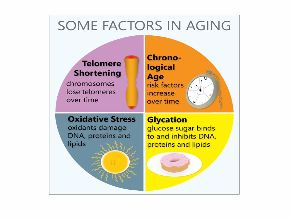
Maintaining the ends of a linear DNA
Mitesh Shrestha
• Maintaining the ends of a linear DNA: o Okazaki fragments, o Synthesis of telomeric DNA, o Senescence of telomere length.

Okazaki fragments
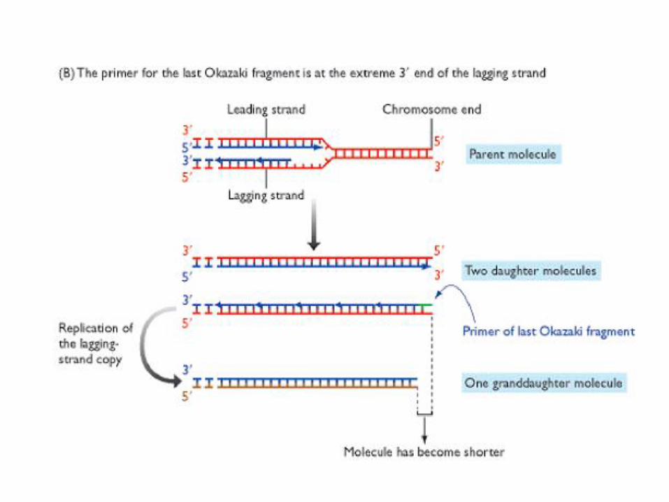
• The extreme 3’ end of the lagging strand might not be copied because the final Okazaki fragment cannot be primed, the natural position for the priming site being beyond the end of the template. The absence of this Okazaki fragment means that the lagging-strand copy is shorter than it should be If the copy remains this length then when it acts as a parental polynucleotide in the next round of replication the resulting daughter molecule will be shorter than its grandparent.


Okazaki Fragments
• If the primer for the last Okazaki fragment is placed at the extreme 3’ end of the lagging strand, then shortening will still occur, although to a lesser extent, because this terminal RNA primer cannot be converted into DNA by the standard processes for primer removal. This is because the methods for primer removal require extension of the 3’ end of an adjacent Okazaki fragment, which cannot occur at the very end of the molecule.

Telomeres • Telomeres are important because they mark the ends of chromosomes and therefore enable
the cell to distinguish a real end from an unnatural end caused by chromosome breakage – an essential requirement because the cell must repair the latter but not the former.
• Telomeric DNA is made up of hundreds of copies of a repeated motif, 5’-TTAGGG-3’ in humans, with a short extension of the 3’ terminus of the double-stranded DNA molecule.
• Two special proteins bind to the repeat sequences in human telomeres. These are called TRF1, which helps to regulate the length of the telomere, and TRF2, which maintains the single-strand extension.
• If TRF2 is inactivated then this extension is lost and the two polynucleotides fuse together in a covalent linkage. Other telomeric proteins are thought to form a linkage between the telomere and the periphery of the nucleus, the area in which the chromosome ends are localized. Still others mediate the enzymatic activity that maintains the length of each telomere during DNA replication.
• Telomeres are bound by a characteristic multiprotein complex known as shelterin . A main function of this complex is to prevent the access of DNA repair proteins to the telomeres. Otherwise, telomeres would be “repaired” as DNA breaks leading to chromosome fusions. Due to their restricted DNA repair, DNA damage at telomeres is notably persistent and highly efficient in inducing senescence and/or apoptosis

Telomeric end is protected by a “cap”. It consists of TRF1 and TRF2 (that bind to telomeric repeats) and proteins such as WRN that bind to TRF1 and TRF2.
The Telomeric Cap Structure


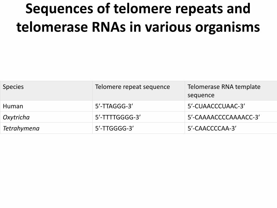
Sequences of telomere repeats and telomerase RNAs in various organisms
Species Telomere repeat sequence Telomerase RNA template sequence
Human 5′-TTAGGG-3′ 5′-CUAACCCUAAC-3′
Oxytricha 5′-TTTTGGGG-3′ 5′-CAAAACCCCAAAACC-3′
Tetrahymena 5′-TTGGGG-3′ 5′-CAACCCCAA-3′

Telo
mere
Len
gth
(h
um
an
s)
Number of Doublings
20
10
Cellular (Replicative) Senescence
Normal Somatic Cells
(Telomerase Negative)
Germ Cells (Telomerase Positive)
+ Telomerase
Telomere Length and Cell Division Potential

Dolly is aging too rapidly?….or was born 6 years old (telomers were
80% of normal sheep)
Dolly has developed pre-mature arthritis

A Japanese-American Werner patient as a teenager (left), and at age 48 (Case #1 Epstein et al,1966, Medicine 45:177). She had eight children, two of whom were also affected. At 48, she hadhair loss and greying, thin extremities, chronic ulcerations of the ankles, atrophy of the skin and herthe right eye had been enucleated several years earlier due to acute glaucoma resulting from bilat-eral cateract extraction at the age of 27. She lived longer than many Werner patients, dying at 57.
Werner Patient

Synthesis of Telomeric DNA • Most of the telomeric DNA is copied in the normal fashion during DNA replication
but this is not the only way in which it can be synthesized. • To compensate for the limitations of the replication process, telomeres can be
extended by an independent mechanism catalyzed by the enzyme telomerase. This is an unusual enzyme in that it consists of both protein and RNA.
• In the human enzyme the RNA component is 450 nucleotides in length and contains near its 5′ end the sequence 5′-CUAACCCUAAC-3′, whose central region is the reverse complement of the human telomere repeat sequence 5′-TTAGGG-3′. This enables telomerase to extend the telomeric DNA at the 3′ end of a polynucleotide in which the telomerase RNA is used as a template for each extension step, the DNA synthesis being carried out by the protein component of the enzyme, which is a reverse transcriptase .
• The correctness of this model is indicated by comparisons between telomere repeat sequences and the telomerase RNAs of other species : in all organisms that have been looked at, the telomerase RNA contains a sequence that enables it to make copies of the repeat motif present at the organism's telomeres.
• An interesting feature is that in all organisms the strand synthesized by telomerase has a preponderance of G nucleotides; it is therefore referred to as the G-rich strand.
Synthesis of Telomeric DNA
• Telomerase can only synthesize this G-rich strand. It is not clear how the other polynucleotide - the C-rich strand - is extended, but it is presumed that when the G-rich strand is long enough, the primase-DNA polymerase α complex attaches at its end and initiates synthesis of complementary DNA in the normal way.
• This requires the use of a new RNA primer, so the C-rich strand will still be shorter than the G-rich one, but the important point is that the overall length of the chromosomal DNA has not been reduced.

Telomerase is composed of both RNA
and protein

Human telomerase complex
hTERT (telomerase
reverse
transcriptase)
hTR (telomerase
RNA)
aka hTERC
Other telomerase-
interacting proteins:
RNA processing and
ribonucleoprotein assembly
(snoRNA-associated proteins)
Dyskerin, NHP2, NOP10, GAR1
Molecular chaperones (Hsp90, p23)
Localization (TCAB1)
Post-translational
modification
Recruitment of telomerase
to telomeres TPP1, Pot1
DNA replication machinery
Dokal I. And Vulliamy T. 2003. Blood Rev. 17, 217-225
Minimal telomerase components (RRL reconstitution) = hTR + hTERT


Synthesis of telomeric sequences 1) Recognition
2) Elongation
3) Translocation
DNA substrate binding to hTERT and RNA template
Addition of nucleotides
DNA substrate and enzyme repositioning
CAAUCCCAAUC
3’
5’
hTERT
hTR 5’- GGTTAGGGTTAGGGTTAG
3’- CCAAT
GGTTAG GGTTAG GGTTAG
4) Repeated translocation and elongation=repeat addition processivity
G A T T G G
TRAP assay
Direct primer extension assay

Synthesis of Telomeric DNA

Hallmarks of Aging

Hallmarks of Cancer

Shorting chromosomes leading to cancer and
aging
• Telomeres have been compared with the plastic tips on shoelaces, because they keep chromosome ends from fraying and sticking to each other, which would destroy or scramble an organism's genetic information.
• Telomere length in humans seems to decrease at a rate of 24.8–27.7 base pairs per year.
• Human liver tissues have been reported to lose 55 base pairs of telomeric DNA per year.

Shorting chromosomes leading to cancer and aging
• Telomere length is affected by a combination of factors including donor age , genetic, epigenetic make-up and environment , social and economic status, exercise, body weight, and smoking.
• Certain lifestyle factors such as smoking, obesity, lack of exercise, and consumption of unhealthy diet can increase the pace of telomere shortening, leading to illness and/or premature death.

Shorting chromosomes leading to cancer and aging
• Accelerated telomere shortening is associated with early onset of many age-associated health problems, including coronary heart disease, heart failure, diabetes, increased cancer risk , and osteoporosis.
• Shorter telomeres can also induce genomic instability by mediating interchromosomal fusion and may contribute to telomere stabilization and development of cancer

Shorting chromosomes leading to cancer and aging
• Telomeres shorten with age and progressive telomere shortening leads to senescence and/or apoptosis. Shorter telomeres have also been implicated in genomic instability and oncogenesis. Older people with shorter telomeres have three and eight times increased risk to die from heart and infectious diseases, respectively.
• Individuals with shorter telomeres seem to have a greater risk for development of lung, bladder, renal cell, gastrointestinal, and head and neck cancers.
The telomere theory of aging
• Potentially immortal cells (germ cells, cancer cells) maintain telomerase activity – Can divide indefinitely.
• Cells with a limited replicative lifespan. – Should have no telomerase activity.
– Progressively shortening telomeres.
– Cell division serves as a mitotic clock for replicative senescence.
• Provides a mechanistic explanation for the Hayflick limit.

Hayflick limit: The Hayflick
limit(or Hayflick
phenomenon) is the
number of times a normal
human cell population will
divide until cell
division stops.
One serial passage or
doubling of cells

The telomere hypothesis of aging
Telomere length is not related to life span (mice vs human; M musculus vs M spretus) Telomeres contribute to aging ONLY if senescent cells contribute to aging Telomerase protects against replicative senescence but not senescence induce by other causes

Telomere erosion is unlikely to be a primary tumor suppressor mechanism in
rodents
Mouse telomeres ~ 20 KB longer than human telomeres
Telomerase activity is not stringently repressed in the somatic tissues of mice
Replicative senescence is different in rodent and human cells
Replicative senescence occurs in rodent cells
with long telomeres
Rodent cells can spontaneously immortalize in
culture at detectable frequencies without the aid
of oncogenes (unlike human cells)
Mouse models: Differences in the biology of telomeres, telomerase
and replicative senescence in mice and humans

Shorting chromosomes leading to cancer and aging

Cell proliferation potential greater in long-lived species

Cellular senescence
• Once the telomere shrinks to a certain extent, the cell stops dividing.
– ~4kb in human cells triggers end to cell division.
• This leads to other changes called cellular senescence:
– Cell morphology changes.
– Gene expression changes.

Causes of cellular senescence
‘Culture stress’: inappropriate
substrata, serum, hyper-
physiological oxygen
Coppé, J.-P. et al. 2010. The senescence-associated secretory phenotype: the dark side of tumor
suppression. Annu. Rev. Pathol. Mech. Dis. 2010. 5, 99-118.
PTEN tumor suppressor loss

Telo
mere
Len
gth
Cell divisions
Cellular Senescence
Germline Cells
Stem cells
Deregulated
Cellular Growth
Checkpoint Escape
-p53, -pRb
Telomerase or ALT
Reactivation
Tumor Cells
Telomere Hypothesis of Cellular Aging
and Immortalization
TELOMERASE
ON
TELOMERASE
ON
TELOMERASE
OFF
Cellular Crisis

Shorting chromosomes leading to cancer and aging
• Without new synthesis of telomeres at chromosome ends the chromosomes shorten with progressive cell division, eventually triggering either replicative senescence or apoptosis when telomere length becomes critically short.
• The regulation of telomerase activity in human cells plays a significant role in the development of cancer.
• Telomerase is tightly repressed in the vast majority of normal human somatic cells but becomes activated during cellular immortalization and in cancers.
• While the mechanisms for telomerase activation in cancers have not been fully defined, they include telomerase catalytic subunit gene (hTERT) amplification and trans-activation of the hTERT promoter by the myc oncogene product.
• Ectopic expression of hTERT is sufficient to restore telomerase activity in cells that lack the enzyme and can immortalize many cell types.

Shorting chromosomes leading to cancer and aging
• The high level of telomerase in the majority of cancer cells—which is typically a couple of orders of magnitude higher in cancer cells than in the differentiated normal cells around it—usually does not lead to the cancer cells having long telomeres. Their telomeres are mostly maintained at a rather short length; the 40 kb telomeres in LOX melanoma cells are the exception rather than the rule.
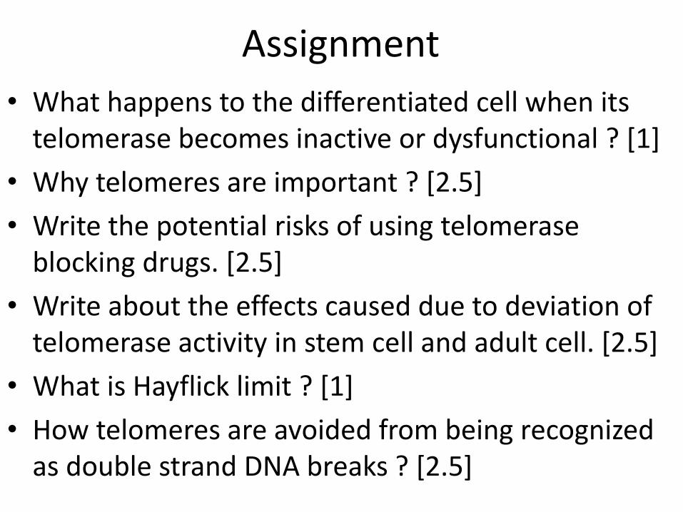
Assignment
• What happens to the differentiated cell when its telomerase becomes inactive or dysfunctional ? [1]
• Why telomeres are important ? [2.5]
• Write the potential risks of using telomerase blocking drugs. [2.5]
• Write about the effects caused due to deviation of telomerase activity in stem cell and adult cell. [2.5]
• What is Hayflick limit ? [1]
• How telomeres are avoided from being recognized as double strand DNA breaks ? [2.5]