Embed Size (px)
Citation preview
IOSR Journal of Dental and Medical Sciences (IOSR-JDMS)
e-ISSN: 2279-0853, p-ISSN: 2279-0861.Volume 13, Issue 9 Ver. VI (Sep. 2014), PP 31-40 www.iosrjournals.org
www.iosrjournals.org 31 | Page
Stem Cells in Periodontal Regeneration
Dr. Sivaram G. M.D.S., Dr.R.Shri Nandhini Devi,BDS
( Department of Periodontics, Ragas Dental College / Dr. M.G.R. University, India )
( Department of Periodontics, Ragas Dental College / Dr. M.G.R. University, India )
Abstract: Stem cells are undifferentiated cells capable of self - renewal and multilineage differentiation. The
use of cells as mesenchymal dental stem cells, which has immunosuppressant properties, high proliferation, and
capacity to differentiate into osteoblasts, odontoblasts, cementoblasts and other cells, suppose a good
perspective in clinical dentistry. Also, these cells need a scaffold that facilitate their integration, differentiation, matrix synthesis and promote multiple specific interactions between cells. To date, different human dental stem
cell progenitors have been identified and isolated - bone marrow stromal cells (BMMSC), human pulp tissue
(DPSC), exfoliated deciduous teeth (SHED), periodontal ligament (PDLSC), apical papilla (SCAP), dental
follicle (DFSC). Also included in this article are the potential applications of stem cell therapy in periodontics.
The tissue engineering triad of cells, signaling molecules, scaffolds plays a significant role in achieving
regeneration.
Key Words: Multilineage differentiation , self- renewal , tissue engineering triad, undifferentiated cells.
I. Introduction Regeneration of the lost periodontal structure has been a challenging task in the current scenario.
Modern dentistry has dealt with the replacement of missing teeth and surrounding structures with synthetic
materials by treating with the patient’s own tissues, autogenic grafts, or synthetic substitute (Crubezy E
[1],1998). Due to various drawbacks such as risk of failure, damage to the donor site and poor tissue
acceptability, diffentetial tissue response, the use of stem cells for periodontal regeneration is the current
emerging trend.
II. Role of Stem Cells In Periodontal Regeneration Periodontal regeneration aims at complete restoration of lost tissues and their original architecture and function by recapitulating the crucial wound healing events associated with their during development. This
entails the reformation of all components of the periodontium- periodontal ligament, gingival connective tissue,
cementum and alveolar bone (Shalini H [2].2013). With advances in stem cell biology and tissue engineering,
the concept of periodontal regeneration is made possible. The idea is to cultivate stem cells with odontogenic
induction cells through epithelial mesenchymal interactions, thereby programming the stem cells to adapt to
dental cell lineages and with the help of scaffold matrix to become a part of the tooth.
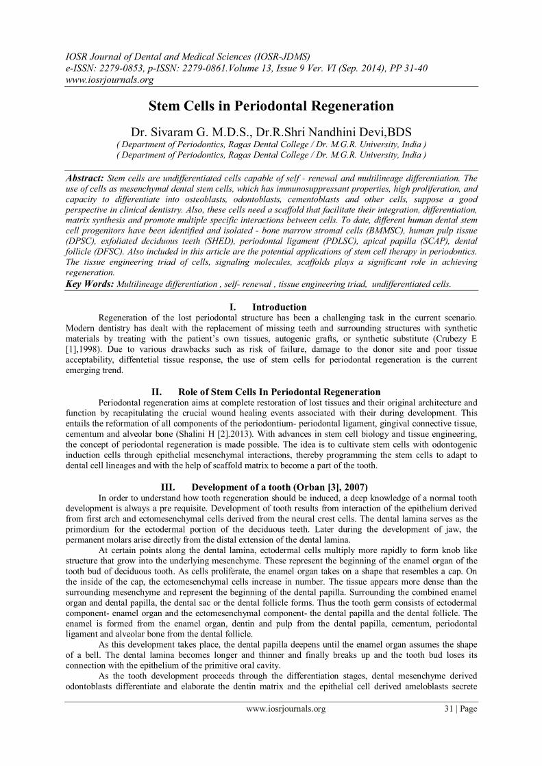
III. Development of a tooth (Orban [3], 2007) In order to understand how tooth regeneration should be induced, a deep knowledge of a normal tooth
development is always a pre requisite. Development of tooth results from interaction of the epithelium derived
from first arch and ectomesenchymal cells derived from the neural crest cells. The dental lamina serves as the
primordium for the ectodermal portion of the deciduous teeth. Later during the development of jaw, the
permanent molars arise directly from the distal extension of the dental lamina.
At certain points along the dental lamina, ectodermal cells multiply more rapidly to form knob like
structure that grow into the underlying mesenchyme. These represent the beginning of the enamel organ of the
tooth bud of deciduous tooth. As cells proliferate, the enamel organ takes on a shape that resembles a cap. On
the inside of the cap, the ectomesenchymal cells increase in number. The tissue appears more dense than the
surrounding mesenchyme and represent the beginning of the dental papilla. Surrounding the combined enamel
organ and dental papilla, the dental sac or the dental follicle forms. Thus the tooth germ consists of ectodermal
component- enamel organ and the ectomesenchymal component- the dental papilla and the dental follicle. The
enamel is formed from the enamel organ, dentin and pulp from the dental papilla, cementum, periodontal ligament and alveolar bone from the dental follicle.
As this development takes place, the dental papilla deepens until the enamel organ assumes the shape
of a bell. The dental lamina becomes longer and thinner and finally breaks up and the tooth bud loses its
connection with the epithelium of the primitive oral cavity.
As the tooth development proceeds through the differentiation stages, dental mesenchyme derived
odontoblasts differentiate and elaborate the dentin matrix and the epithelial cell derived ameloblasts secrete
Stem Cells in Periodontal Regeneration
www.iosrjournals.org 32 | Page
enamel matrix. After the tooth crown has formed, root structures develop from the rudimentary hertwig’s
epithelial root sheath forming dentin, cementum, periodontal ligament, alveolar bone. In naturally formed teeth,
the enamel exhibits no regenerative properties while the remaining tissues all of which are formed from neural crest derived ectomesenchyme exhibit regenerative capacity, partially related to the existence of stem cells
Stem Cells in Periodontal Regeneration
www.iosrjournals.org 33 | Page
IV. History of stem cells 19th century-The term stem cell first appeared in the literature.
1868 -The term ‘stem cell’ was proposed by Russian histologist Alexander Maksimov (Ramalho-Santos M [4],
2007).
1954- Birth of stem cell research took place when Leroy Stevens [5] identified teratoma like cells in testicles of
mice.
1976- The concept that stem cells may reside in the periodontal tissues was proposed by Melcher, who queried
whether the cementoblasts, osteoblasts and fibrioblasts were derived from a single population of ancestral cells
or stem cells.
1987- The studies of Mc Culloch et al provided the most compelling evidence that these cells are present within
the periodontal tissues.
2005 – National Institute of health announces discovery of DPSCs by Dr. Irina Kerkis 2006 - Dr.Irina Kerkis reported discovery of Immature Dental Pulp Stem Cells (IDPSC), a pluripotent sub-
population of DPSC using dental pulp organ culture.
2007 - DPSC 1st animal studies begin for bone regeneration.
2008 – DPSC 1st advanced animal study for bone grafting announced. Reconstruction of large size cranial bone
defects in rats.
2009 – Scientists from Italy announced the first-ever human clinical application using patients’ own dental stem
cells to repair large mandibular bone defects..(Aquino’d R [6], 2009)
2013 – A study showed that stem cells from teeth can create islet – like cells which produce insulin (a potential
therapy for type 1 diabetes.
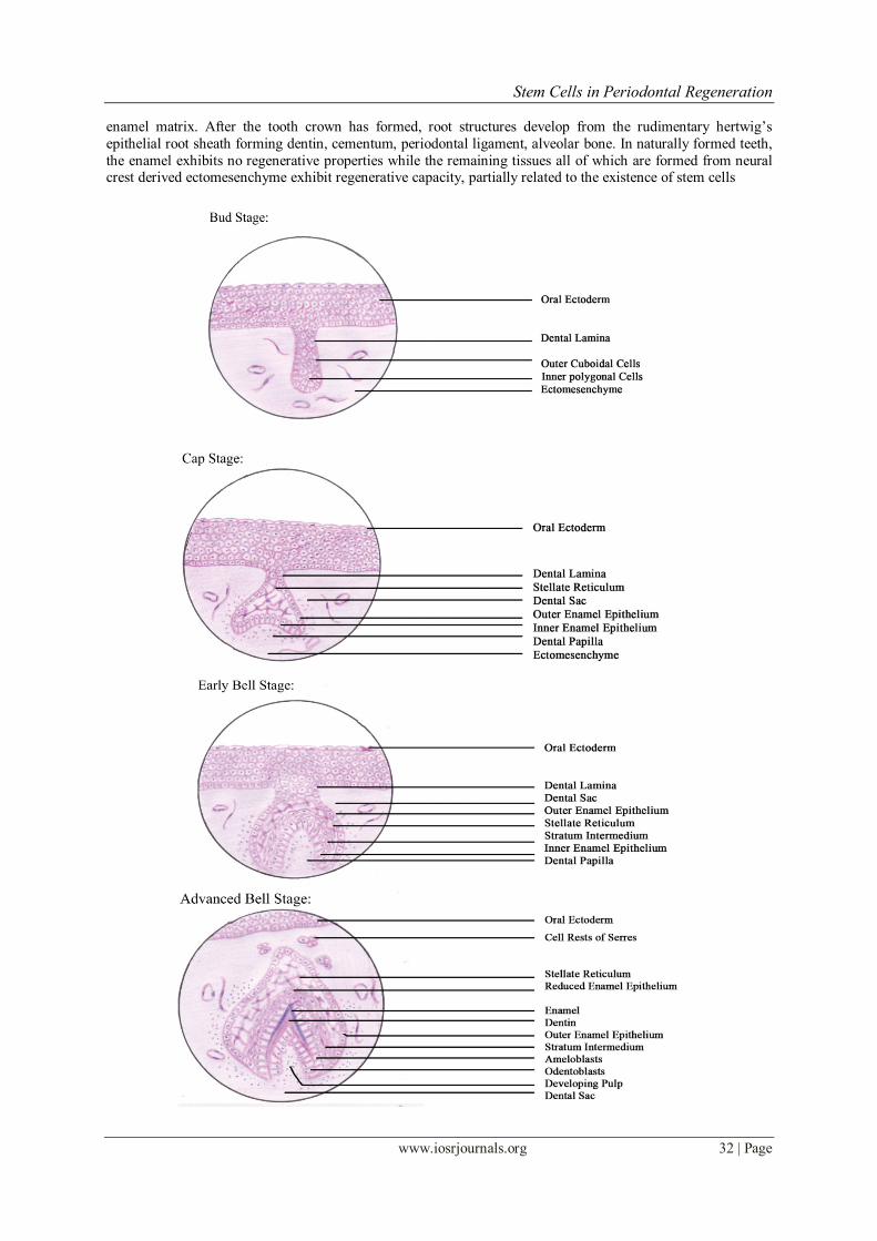
V. Stem cells and their characteristics Stem cells are unspecialized cells (Blau HM, Brazelton R, Weimann JM [7], 2001) with an
extraordinary ability to self -renew, capable of differentiating into one or more specialized cell types playing a
crucial role in hemostasis and tissue repair (Nadig RR [8], 2009).
A stem cell has the property of ‘self renewal’ I.e., when a stem cell is called into action, it undegoes
cell division. One daughter cell remains a stem cell, while the other becomes more committed forming a
particular cell type called ‘committed progenitor’ by a process called ‘asymmetric division’. A progenitor cell is
an intermediate cell type formed before it achieves a fully differentiated state. It is regarded as committed to
differentiating along a particular cellular developmental pathway.
5.1 Characteristics:(Rodriguez-Lozano FJ et al.[9], 2012)
• Totipotency- generates all types of cells including germ cells. They are derived from the first few
divisions of fertilized egg. These cells can divide into embryonic and extra embryonic cell types.
Stem Cells in Periodontal Regeneration
www.iosrjournals.org 34 | Page
• Pluripotency- can give rise to every cell of an organism except its extraembryonic tissues such as
placenta. This restricts pluripotent stem cells from developing into full organism. Eg., embryonic stem cells, induced pluripotent stem cells.
• Multipotency- these stem cells are adult stem cells which only generate specific lineages of cells. Eg.,
hematopoietic stem cells.
• Self renewal- divides without differentiation and creates everlasting supply.
• Plasticity- mesenchymal stem cells have plasticity and can undergo differentiation. The trigger for
plasticity are stress or tissue injury which upregulates the stem cells and releases chemoattractants and
growth factors.
• Clonogenicity- a stem cell is thought to be clonogenic when it can proliferate to form colony of cells.
VI. Types of stem cells Based on their origin, stem cells are categorized as embryonic stem cells (ESC’s) and somatic/ adult
stem cells (ASC’s).
6.1 Embryonic stem cells
6.1.1 Origin- Embryonic stem cells are derived from embryos that are 2-11 days old called blastocysts. They are
best grown from supernumerary embryos, obtained from within invitro fertilization centers. 6.1.2 Properties- They are totipotent cells capable of differentiating into any type of cells including germ cell.
6.1.3 Advantages- They are considered immortal as they propagated and maintained in an undifferentiated state
indefinitely. These stem cells have the highest potential to regenerate and repair diseased tissue and organ.
(Evans MJ[10], 1981and Keller GM[11], 1995)
6.1.4 Disadvantage- There is a belief that the process of extraction of stem cells from embryo destroys the
embryo itself, raising moral and ethical concerns. Also it is difficult to control the growth and differentiation of
embryonic stem cells posing risk of tumorogenicity and teratoma formation
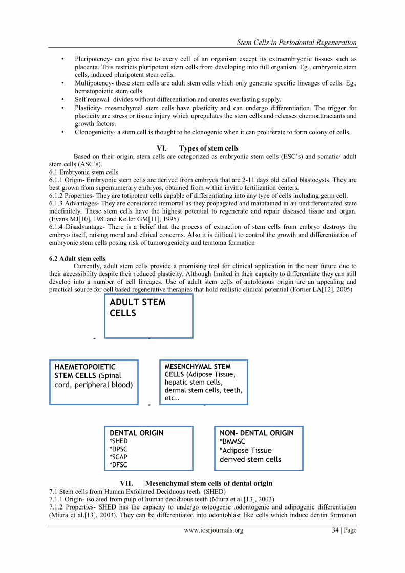
6.2 Adult stem cells
Currently, adult stem cells provide a promising tool for clinical application in the near future due to
their accessibility despite their reduced plasticity. Although limited in their capacity to differentiate they can still develop into a number of cell lineages. Use of adult stem cells of autologous origin are an appealing and
practical source for cell based regenerative therapies that hold realistic clinical potential (Fortier LA[12], 2005)
VII. Mesenchymal stem cells of dental origin 7.1 Stem cells from Human Exfoliated Deciduous teeth (SHED)
7.1.1 Origin- isolated from pulp of human deciduous teeth (Miura et al.[13], 2003)
7.1.2 Properties- SHED has the capacity to undergo osteogenic ,odontogenic and adipogenic differentiation
(Miura et al.[13], 2003). They can be differentiated into odontoblast like cells which induce dentin formation
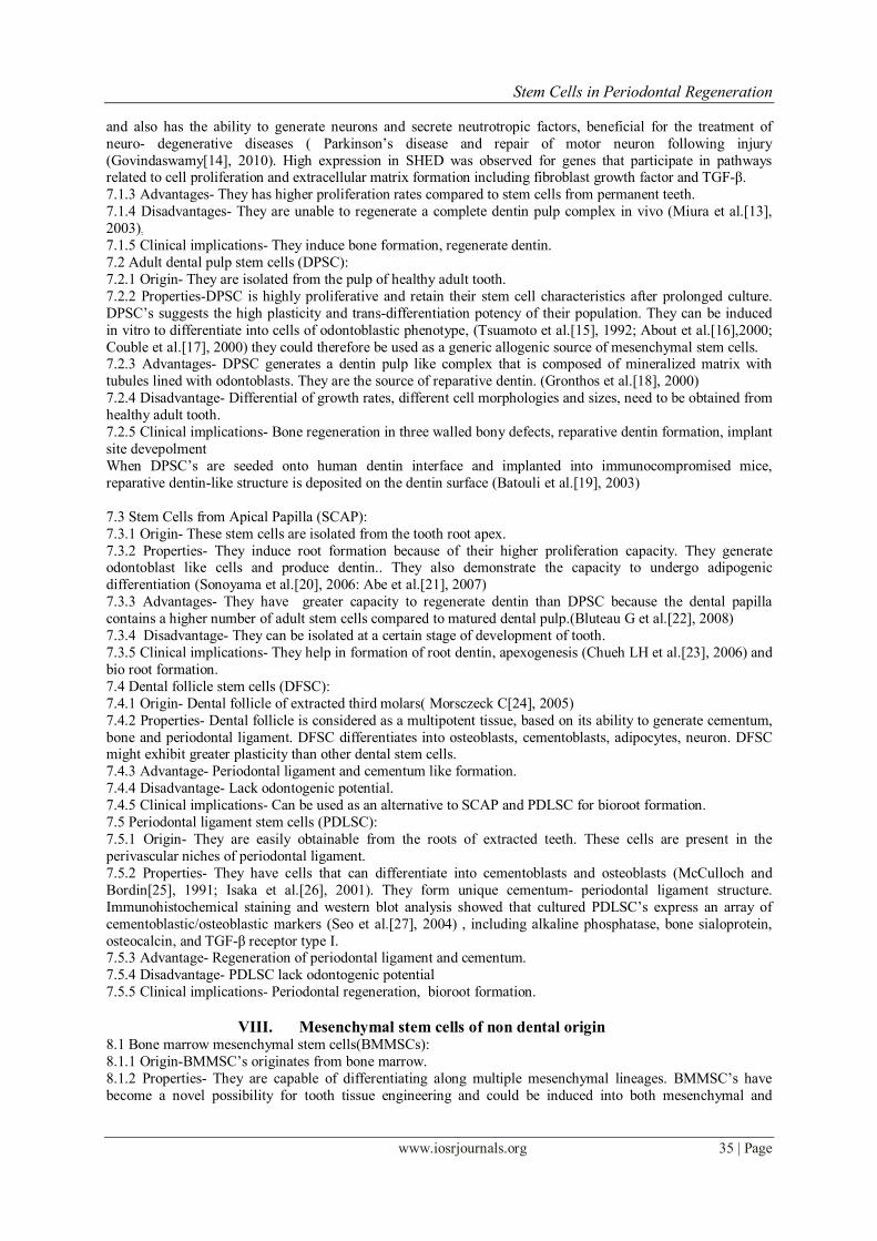
ADULT STEM
CELLS
HAEMETOPOIETIC STEM CELLS (Spinal
cord, peripheral blood)
MESENCHYMAL STEM CELLS (Adipose Tissue, hepatic stem cells, dermal stem cells, teeth, etc..
DENTAL ORIGIN *SHED *DPSC *SCAP *DFSC *PDLSC
NON- DENTAL ORIGIN *BMMSC *Adipose Tissue
derived stem cells
Stem Cells in Periodontal Regeneration
www.iosrjournals.org 35 | Page
and also has the ability to generate neurons and secrete neutrotropic factors, beneficial for the treatment of
neuro- degenerative diseases ( Parkinson’s disease and repair of motor neuron following injury
(Govindaswamy[14], 2010). High expression in SHED was observed for genes that participate in pathways related to cell proliferation and extracellular matrix formation including fibroblast growth factor and TGF-β.
7.1.3 Advantages- They has higher proliferation rates compared to stem cells from permanent teeth.
7.1.4 Disadvantages- They are unable to regenerate a complete dentin pulp complex in vivo (Miura et al.[13],
2003).
7.1.5 Clinical implications- They induce bone formation, regenerate dentin.
7.2 Adult dental pulp stem cells (DPSC):
7.2.1 Origin- They are isolated from the pulp of healthy adult tooth.
7.2.2 Properties-DPSC is highly proliferative and retain their stem cell characteristics after prolonged culture.
DPSC’s suggests the high plasticity and trans-differentiation potency of their population. They can be induced
in vitro to differentiate into cells of odontoblastic phenotype, (Tsuamoto et al.[15], 1992; About et al.[16],2000;
Couble et al.[17], 2000) they could therefore be used as a generic allogenic source of mesenchymal stem cells. 7.2.3 Advantages- DPSC generates a dentin pulp like complex that is composed of mineralized matrix with
tubules lined with odontoblasts. They are the source of reparative dentin. (Gronthos et al.[18], 2000)
7.2.4 Disadvantage- Differential of growth rates, different cell morphologies and sizes, need to be obtained from
healthy adult tooth.
7.2.5 Clinical implications- Bone regeneration in three walled bony defects, reparative dentin formation, implant
site devepolment
When DPSC’s are seeded onto human dentin interface and implanted into immunocompromised mice,
reparative dentin-like structure is deposited on the dentin surface (Batouli et al.[19], 2003)
7.3 Stem Cells from Apical Papilla (SCAP):
7.3.1 Origin- These stem cells are isolated from the tooth root apex.
7.3.2 Properties- They induce root formation because of their higher proliferation capacity. They generate odontoblast like cells and produce dentin.. They also demonstrate the capacity to undergo adipogenic
differentiation (Sonoyama et al.[20], 2006: Abe et al.[21], 2007)
7.3.3 Advantages- They have greater capacity to regenerate dentin than DPSC because the dental papilla
contains a higher number of adult stem cells compared to matured dental pulp.(Bluteau G et al.[22], 2008)
7.3.4 Disadvantage- They can be isolated at a certain stage of development of tooth.
7.3.5 Clinical implications- They help in formation of root dentin, apexogenesis (Chueh LH et al.[23], 2006) and
bio root formation.
7.4 Dental follicle stem cells (DFSC):
7.4.1 Origin- Dental follicle of extracted third molars( Morsczeck C[24], 2005)
7.4.2 Properties- Dental follicle is considered as a multipotent tissue, based on its ability to generate cementum,
bone and periodontal ligament. DFSC differentiates into osteoblasts, cementoblasts, adipocytes, neuron. DFSC might exhibit greater plasticity than other dental stem cells.
7.4.3 Advantage- Periodontal ligament and cementum like formation.
7.4.4 Disadvantage- Lack odontogenic potential.
7.4.5 Clinical implications- Can be used as an alternative to SCAP and PDLSC for bioroot formation.
7.5 Periodontal ligament stem cells (PDLSC):
7.5.1 Origin- They are easily obtainable from the roots of extracted teeth. These cells are present in the
perivascular niches of periodontal ligament.
7.5.2 Properties- They have cells that can differentiate into cementoblasts and osteoblasts (McCulloch and
Bordin[25], 1991; Isaka et al.[26], 2001). They form unique cementum- periodontal ligament structure.
Immunohistochemical staining and western blot analysis showed that cultured PDLSC’s express an array of
cementoblastic/osteoblastic markers (Seo et al.[27], 2004) , including alkaline phosphatase, bone sialoprotein,
osteocalcin, and TGF-β receptor type I. 7.5.3 Advantage- Regeneration of periodontal ligament and cementum.
7.5.4 Disadvantage- PDLSC lack odontogenic potential
7.5.5 Clinical implications- Periodontal regeneration, bioroot formation.
VIII. Mesenchymal stem cells of non dental origin 8.1 Bone marrow mesenchymal stem cells(BMMSCs):
8.1.1 Origin-BMMSC’s originates from bone marrow.
8.1.2 Properties- They are capable of differentiating along multiple mesenchymal lineages. BMMSC’s have
become a novel possibility for tooth tissue engineering and could be induced into both mesenchymal and
Stem Cells in Periodontal Regeneration
www.iosrjournals.org 36 | Page
epithelial cells in tooth engineering (Peng L et al.[28], 2009). BMMSC’s have been described as colony forming
unit- fibrioblasts (CFU-FS) invitro (Cohnheim[29], 1867; Friedenstein et al.[30], 1976).
8.1.3 Advantage- They have osteogenic, myogenic and neurogenic potential 8.1.4 Disadvantage-. Obtaining BMMSC’s causes pain and morbidity and low cell number upon harvest.
BMMSC’s have limited growth potential of 30-50 population doublings following ex vivo expansion (Bruder et
al.[31], 1999; Bianco et al.[32], 2001). The heterogenecity in colony morphology, growth and function makes it
difficult to identify the exact phenotypic fingerprint of a stromal stem cell (Bianco et al.[32], 2001).
8.2 MSC’s from other sources
MSC’s are derived from adipose tissue obtained by suction assisted lipectomy (Zuk et al.[33], 2001;
Mizuno et al.[34], 2002) and MSC like populations from umbilical cord have been isolated and characterized
(Mareschi et al.[35], 2001). They are morphologically and immunophenotypically similar, but not identical
(Kern et al.[36], 2006). UCB MSC’s form fewest colonies and show highest proliferative capacity, whereas
adipose tissue derived MSC’s form the greatest number of colonies. These tissues are versatile and possess great
potential for many clinical applications. Though these stem cells have various odontogenic properties, a proper interaction between these cells
and the host tissue is necessary for successful periodontal regeneration. This can be accomplished by by
ensuring a proper interaction between these cells, growth factors and a scaffold by the process of tissue
engineering.
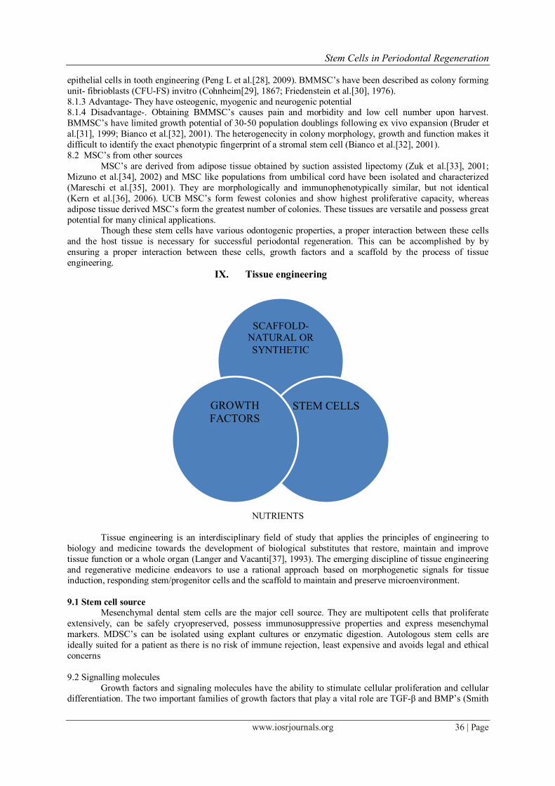
IX. Tissue engineering
NUTRIENTS
Tissue engineering is an interdisciplinary field of study that applies the principles of engineering to
biology and medicine towards the development of biological substitutes that restore, maintain and improve
tissue function or a whole organ (Langer and Vacanti[37], 1993). The emerging discipline of tissue engineering
and regenerative medicine endeavors to use a rational approach based on morphogenetic signals for tissue induction, responding stem/progenitor cells and the scaffold to maintain and preserve microenvironment.
9.1 Stem cell source
Mesenchymal dental stem cells are the major cell source. They are multipotent cells that proliferate
extensively, can be safely cryopreserved, possess immunosuppressive properties and express mesenchymal
markers. MDSC’s can be isolated using explant cultures or enzymatic digestion. Autologous stem cells are
ideally suited for a patient as there is no risk of immune rejection, least expensive and avoids legal and ethical
concerns
9.2 Signalling molecules
Growth factors and signaling molecules have the ability to stimulate cellular proliferation and cellular differentiation. The two important families of growth factors that play a vital role are TGF-β and BMP’s (Smith
SCAFFOLD- NATURAL OR
SYNTHETIC
STEM CELLS GROWTH
FACTORS
Stem Cells in Periodontal Regeneration
www.iosrjournals.org 37 | Page
AJ[38], 1998 and Smith AJ, Murray PE[39], 2001). BMP’s are used sequentially and repeated throughout
embryonic tooth development, initiation, morphogenesis, cytodifferentiation and matrix secretion.
9.3 Scaffolds
The scaffold should be biocompatible, non toxic and have optimal physical and mechanical properties.
They should be highly porous to facilitate cell seeding and diffusion of nutrients. The scaffolds provide
structural integrity and eventually breakdown leaving the newly formed tissue.
The materials used for scaffolds include both synthetic and natural. Natural materials include collagen,
alginate, agarose (Hutmacher DW[40], 2001), chitosan, glycosaminoglycans (Griffon DJ[41], 2005 and Guo
T[42], 2006). Synthetic materials like hydroxyapatite/ tricalcium phosphate, polymers like polylactic acid,
polyglycolic acid, polycaprolactone are found to be more conductive and show less contraction compared to
collagen (Taylor MS[43], 1994 and Ma PX[44], 2001).
X. Induced Pluripotent Stem cells (iPS) – a recent development Somatic cells cannot always be derived from the same patient for use in autologous transplantation.
Another concern is the limited replicative potential of these cells. To overcome these issues, increasing attention
has been paid to the potential use of pluripotent cells for cell based regenerative medicine. Human embryonic
stem cell represents the gold standard source of pluripotent cells. They can self renew indefinitely in culture
without losing their capacity to differentiate. Yamanake and Takahashi[45] (2006) reported the generation of
induced pluripotent stem cells delivered by retroviral transduction. Further advances allowed the generations of
virus free iPS cell lines that are functionally indistinguishable from true ES cells. The method for iPS cell
induction is called ‘ground breaking’ because somatic cells are converted into pluripotent cells through
introduction of 4 genes: Oct-4, Sox-2, C-Myc, K1f4.(Takahashi K, Tanabe K et al[46]., 2007) However, there are several limitations. Reprogramming process appears to be stochastic and
incomplete reprogramming can result in iPS cell lines with some ‘epigenetic memory’ of their tissue of origin.
Further, rapid cell division makes it more susceptible to replication- induced DNA mutation. Robust directed
differentiation of pluripotent cells into the desired functional effector cell type is prerequisite for successful cell
based therapy.
XI. MSC niche The stem cell niche concept was proved by Schofield[47] (1978) as a specialized microenvironment
needed for cells to retain their ‘stemness’. It is considered as a fixed compartment of a three dimensional structure containing elements that participate in the regulation of stem cell proliferation, control the fate of stem
cell progeny, and prevent the stem cells from exhaustion and death (Scadden[48], 2006; Jones and Wagers[49],
2008). The bone marrow microenvironment is the major site of MSC niche in the body. MSC’s are known to
reside into two different niches- endosteal and perivascular niches. The endosteal niche is thought to maintain
MSC quiescence over a long term, whereas the perivascular niche is to maintain MSC proliferation and mediate
circulation (Mitsiadis et al.[50], 2007). BMMSC’s and Adipose derived MSC harbor in the perivascular areas of
BM (Shi and Gronthos[51], 2003; Gronthos, Zannetino et al.[52], 2008).
XII. Potential applications: • Periodontal regeneration
• Implant site preparation
• Maxillary sinus lift procedure
• 3 walled bony defects
• Bio root formation
12.1 Periodontal Regeneration
Due to complex structure of periodontium, its complete regeneration has always remained a challenge.
MSC’s can regenerate new cementum, alveolar bone and periodontal ligament. Further periodontal ligament
cells cultured in vitro where successfully reimplanted into periodontal defects. PDLSC’s and DFSC’s have
become an alternative cell source for periodontal regenerate therapy.
Masako Miura and Songtao Shi[53] (2004) investigated the notion that human PDL contains stem cells
that could be used to regenerate periodontal tissue. They isolated PDLSCs by single colony selection and
various other analyses and transplanted them into immunocompromised mice to assess the capacity for tissue
regeneration and repair. Under defined culture conditions,PDLSCs differentiated into cementoblast like cells,
adipocytes and collagen forming cells.
12.2 Implant site preparation
Stem Cells in Periodontal Regeneration
www.iosrjournals.org 38 | Page
Stem cells can be used as an alternative for autogenous bone grafts. Use of stem cells prevents the damage to the
donor site which is caused due to harvesting of autogenous bone graft. SHED and DPSC has osteoinductive
capacity thus can differentiate into osteoblasts and can promote bone formation. A study was conducted by Yamada Y, Ueda M, Naiki T [54] (2004) to regenerate bone in a significant
osseous defect with minimal invasiveness and good plasticity. They used a combination of platelet rich plasma
as an autologous scaffold with MSCs to increase osteogenesis. At 8 weeks, newly formed bone areas was 67.3 ±
2.06 %
12.3 Maxillary Sinus Lift Procedure
Use of stem cells can reduce the morbidity (Mao JJ[55], 2008) introduced by a second surgical site
while maintaining equally good implant success rate. A study was conducted by Yadollah Soleymani et al.[56],
2008, to augment the maxillary sinus using human mesenchymal stem cells loaded into a tricalcium phosphate/
hydroxyapaite scaffold. All patients in the study had less than 3mm initial bone height in posterior maxilla.
MSCs were cultured from bone marrow aspirate for each patient. Three months after sinus elevation, the secondary bone height was measured. Mean bone regenerate was 41.34%.
12.4 Three Walled bony Defects
Autologous bone grafts are considered as the best option but it has limited donor sites Bone tissue
engineering endeavours to repair large bone losses using three dimentional scaffolds to deliver vital cells to the
defective sites. Stem cells are placed on a macroporous hydroxyapatite scaffolds and implanted at the site of
defect. Periodic review revealed abundant callus formation.
A recent report on a minipig model has shown that periodontal defects may be repaired using PDLSCs
(Liu et al.[57], 2008). This PDLSC mediated treatment resulted in regeneration of PDL and recovery of the
heights of alveolar bone
12.5 Bio root Formation SCAP and PDLSC are currently extensively studied for bio root engineering. Dentin structure
regeneration was observed but not enamel. Thus regeneration of whole tooth structure was not achieved in many
cases. Thus instead of attempting to form an entire tooth, Sonoyama et al (2006) demonstrated that by using
SCAP along with PDLSC’s they were able to generate a bioroot with periodontal ligament tissues. A mini swine
model was used and the autologous SCAP and PDLSCs were then loaded onto HA/TCP and gel foam
scaffolds,respectively and reimplanted into the sockets.three months later, the bioroot was formed in the porcine
jaw(Sonoyama et al., 2006). The bioroot structure comprised dentin randomly deposited by the SCAP. The
bioroot was encircled with periodontal ligament tissue and appeared to have normal relationship with
surrounding bone. However the presence of residual HA in the newly regenerated dentin formed a structure
different from that of normal dentin. This leads to a reduced mechanical strength of the bioroot, nearly two-
thirds of a natural tooth.
XIII. Conclusion Mesenchymal dental stem cells derived from teeth are easily accessible multipotent cells with the
capacity to differentiate into distinct cell types. This new source of stem cells could be of benefit in cellular
therapy and the eventual development of techniques for use in regenerative dentistry and degenerative diseases.
It is very important to analysis the current knowledge, barriers and challenges in the clinical use of stem cells
with an emphasis on application in dentistry. Although many challenges remain, stem-cell based tissue
engineering for periodontal regeneration will be one of the most successful treatment modalities in near future.
References [1]. Crubezy E, Murali P, Girard L, Bernadou JP. False teeth of the roman world. Nature 1998, 391-429.
[2]. Shalini H. Stem cells in periodontal regeneration. JournalofDentalandMedicalSciences , 12(2), 2013, 59-63.
[3]. Orban , Development and growth of teeth, Kumar.G.S. (Ed), Orban’s Oral Histology And Embryology, 12e , 2007, 22-43.
[4]. Ramalho-Santos M, Willenbring H. On the origin of the term ‘stem cell’, StemCell , 1, 2007, 35-38.
[5]. Stevens LC, Little CC. Spontaneous testicular teratomas in an inbred strain of mice. PNAS, 40(11), 1954, 1080-1087.
[6]. Aquino R, Papaccio J. Human mandibular bone defect repair by grafting of dental pulp stem cells and collagen sponge
biocomplexes. Europeancellsandmaterials, 18, 2009, 75-83.
[7]. Blau HM,Brazelton R,Weimann JM. The evolutioning concept of stem cell: entity or function? Cell , 105, 2001, 829-841.
[8]. Nadig RR Stem Cell Therapy-Hype or Hope. JConserveDent , 12(4), 2009, 131-138.
[9]. Rodriguez-Lozano FJ, Insausti CL, Iniesta F, Blanquer M, Ramirez MC, Meseguer L,Meseguer-Henarejos AB, Marin N, Martinez
S, Moraleda JM. Mesenchymal Dental Stem Cells In Regenerative Dentistry .MedOralPatolOralCirBucal, 17(6)9e, 2012, 1062-
1067.
[10]. Evans MJ, Kaufman MH. Establishment in culture of pluripotent cells from mouse embryos. Nature., 292, 1981, 154-156.
[11]. Keller GM. In vitro differentiation of embryonic stem cells. CurrOpinCellBiol, 7, 1995, 862-869.
[12]. Fortier I.A, Stem Cells, classifications, controversies and clinical Applications. VetSurg, 34, 2005 ,415-23.
Stem Cells in Periodontal Regeneration
www.iosrjournals.org 39 | Page
[13]. Miura M, Gronthos S, Zhao M, Lu B, Fisher LW, Robey PG, et al. SHED: stem cells from human exfoliated deciduous teeth.
ProcNatlAcadSciUSA , 100, 2003, 5807-5812.
[14]. Govindaswamy V,Abdullah AN, Ronald VS, Musa S, Ab Aziz ZA, Zain RB, et al. Inherant differential propensity of dental pulp
stem cells derived from human deciduous and permanent teeth. JEndod., 36, 2010, 1504-1515.
[15]. Tsuamoto Y, Fukutani S, Shin-Ike T, Kubota T, Sato S, Suzuki Y, et al. Mineralized nodule formation by cultures of human dental
pulp-derived fibroblasts. ArchOralBiol , 37, 1992, 1045-1055.
[16]. About I, Bottero MJ, de Denato P, Camps J, Franquin JC, Mitsiadis TA. Human dentin production in vitro. ExpCellRes , 258, 2000,
33-41.
[17]. Couble ML, Farges JC, Bleicher F, Perrat-Mabillon B, Boudeulle M, Magloire H. Odontoblast differentiation of human dental pulp
cells in explant cultures. CalcifTissueInt , 66, 2000, 129-138.
[18]. Gronthos S, Mankani M, Brahim J, Robey PG, Shi S. Postnatal human dental pulp stem cells (DPSCs) in vitro and in vivo. Proc
Natl Acad Sci USA , 97, 2000, 13625-13630.
[19]. Batouli ,Miura M, Brahim J,Tsutsui TW, Fisher LW,Gronthos S, et al. Comparison of stem-cell-mediated osteogenesis and
dentinogenesis. J Dent Res, 82, 2003, 976-981.
[20]. Sonoyama W, Liu Y, Fang D, Yamaza T, Seo BM, Zhang C, et al. Mesenchymal stem-cell mediated functional tooth regeneration
in Swine. PLoS , 1:e79, 2006.
[21]. Abe S, Yamaguchi S, Amagasa T. Multilineage cells from apical pulp of human tooth with immature apex. Oral Sci Int , 4, 2007,
45-58.
[22]. Bluteau G, Luder HU,De Bari C, Mitsiadis TA. Stem cells for tooth engineering. Cell Mater., 16, 2008, 1-9.
[23]. Chueh LH, Huang GT. Immature teeth with periradicular periodontitis or abcess undergoing apexogenesis. A paradigm shift. J
Endod, 32, 2006, 1205-1213.
[24]. Morsczeck C, Gotz W, Schierholz J, Zeilhofer F, Kuhn U, Mohl C, et al. Isolation of precursor cells (PCs) from human dental
follicle of wisdom teeth. Matrix Biol , 24, 2005, 155-165.
[25]. McCulloch CA, Bordin S. Role of fibroblast subpopulations in periodontal physiology and pathology. J Periodontal Res , 26(3 pt 1),
1991, 144-154.
[26]. Isaka J, Ohazama A, Kobayashi M, Nagashima C, Takiguchi T, Kawasaki H, et al. Participation of periodontal ligament cells with
regeneration of alveolar bone. J Periodontal Res, 72, 2001, 314-323.
[27]. Seo BM, Miura M, Gronthos S, Bartold PM, Batouli S, Brahim J, et al. Investigation of multipotent postnatal stem cells from
human periodontal ligament. Lancet , 364, 2004, 149-155.
[28]. Peng L, Ye L, Zhou XD. Mesenchymal stem cells and tooth engineering. Int J Oral Sci, 1, 2009, 6-12.
[29]. Cohnheim J. Ueber entzundung und eiterung. Virchows Arch Path Anat Physiol Klin Med , 40, 1867, 1-79.
[30]. Friendenstein AJ, Gorskaja JF, Kulagina NN. Fibroblast precursors in normal and irradiated mouse hematopoietic organs. Exp
Hematol, 4, 1976, 267-274.
[31]. Bruder SP, Jaiswal N, Haynesworth SE. Growth kinetics, self renewal and the osteogenic potential of purified human mesenchymal
stem cells during extensive subcultivation and following cryopreservation. J Cell Biochem , 64, 1997, 278-294.
[32]. Bianco P, Riminucci M, Grontos S, Robey PG. Bone marrow stromal stem cells: Nature, Biology, and Potential Applications. Stem
Cells, 19, 2001, 180-192.
[33]. Zuk PA, Zhu M, Mizuno H, Huang J, Futrell JW, Katz AJ, et al. Multilinieage cells from human adipose tissue: implications for
cell-based therapies. Tissue Eng, 7, 2001, 211-228.
[34]. Mizuno H, Zuk PA, Zhu M, Lorenz HP, Benhaim P, Hedrick MH. Myogenic differentiation by human processed lipoaspirate cells.
Plast Reconstr Surg , 109, 2002, 199-209.
[35]. Mareschi K, Biasin E, Piacibello W, Agiletta M, Madon E, Fagioli F. Isolation of human mesenchymal stem cells: bone marrow
versus umbilical cord blood. Haematologica, 86, 2001, 1099-1100.
[36]. Kern S, Eichler H, Stoeve J, Kluter H, Bieback K. Comparative analysis of mesenchymal stem cells from bone marrow, umbilical
cord blood, or adipose tissue. Stem Cells , 24, 2006;24:1294-1301.
[37]. Langer R, Vacanti JP. Tissue engineering. Science,260(5110) , 1993, 920-926.
[38]. Smith AJ, Mathews JB, Hall RC. Trannsforming growth factor-beta 1 in dentin matrix. Ligand activation and receptor expression.
Eur J Oral Sci., 106, 1998, 179-184.
[39]. Smith AJ, Murray PE, Sloan AJ, Mathews JB, Zhao S. Trans-dentinal stimulation pf tertiary dentinogenesis. Adv Dent Res, 15,
2001, 51-54.
[40]. Hutmacher DW, Goh JC, Tech SH, An Introduction to Biodegradable Materials for Tissue Engineering application. Ann Acad Med
Singapore, 30, 2001, 183-191.
[41]. Griffon DJ, Sedighi MR, Sendimir –Urkmez A, Stewart AA, Jamison R. Evaluation of Vaccum and Dynamic Cell Seeding of
Polyglycolic Acid and Chitosan Scaffolds for Cartilage Engineering, Am J Vet Res., 66, 2005, 599-605.
[42]. Guo T, Zhao J, Chang J, Ding Z, Hong H, Cheng J, et.al. Porous Chitosan Gelatin Scaffold containing Plasmid DNA encoding
Transforming growth factor-Beta1 for Chondrocytes Proliferation. Biomaterials., 8, 2006, 150-161.
[43]. Taylor MS, Daniels AU, Andriano KP, Heller J, Six Bioabsorbable Polymers; In vitro acute Toxicity of Accumulated Degradation
Products. J Appl Biomater, 5, 1994, 151-157.
[44]. Ma PX, Choi JW, Biodegradable Polymer Scaffolds with Well Defined Interconnected Spherical pore Network. Tissue Eng , 7,
2001, 23-33.
[45]. Takahashi K, Yamanake S.Induction of pluripotent stem cells from mouse embryonic adult fibroblast cultures by defined factors.
Cell , 126, 2006, 663.
[46]. Takahashi K, Tanabe K, Ohnuki M, Narita M, Ichisaka T, Tomoda K, et al. Induction of pluripotent stem cells from adult human
fibroblasts by defined factors. Cell , 131, 2007, 861-872.
[47]. Schofield R. The relationship between the spleen colony- forming cell and the haemopoietic stem cell. Blood Cells , 4, 1978, 7-25.
[48]. Scadden DT. The stem-cell niche as an entry of action. Nature , 441, 20061075-1079.
[49]. Jones DL, Wagers AJ. No place like home: anatomy and function of the stem cell niche. Nat Rev Mol Cell Biol , 9, 2008, 11-21.
[50]. Mitsiadis TA, Barrandon O, Rochat A, Barrandon Y, De Bari C. Stem cell niches in mammals. Exp Cell Res , 313, 2007, 3377-
3385.
[51]. Shi S, Gronthos S. Perivascular niche of postnatal mesenchymal stem cells in human bone marrow and dental pulp. J Bone Miner
res , 18, 2003696-704.
[52]. Gronthos S, Zannettino ACW.. A method to isolate and purify human bone marrow stromal stem cells. Prockop DJ, Bunnell BA,
Phinney DG (Ed), Mesenchymal stem cells: methods and protocols. editors. (Totowa, NJ: Humana Press 2008), 45-57.

Stem Cells in Periodontal Regeneration
www.iosrjournals.org 40 | Page
[53]. Miura M, Shi S. Human PDL contains stem cells that could be used to regenerate periodontal tissue. Lancet , 364(9429), 2004, 149-
155.
[54]. Yamada Y, Ueda M, Naiki T,Takahashi M, Hata K, and Nagasaka T. Tissue Engineering, 10 (5-6), 2004, 955-964.
[55]. Mao JJ. Stem cells and the future of dental care. N Y State Dent J, 74, 2008, 20-24.
[56]. Soleymani Y. Augmentation of maxillary sinus using human mesenchymal stem cells loaded into a tricalcium phosphate/
hydroxyapaite scaffold. Annals of allergy, asthma and immunology, 106(2), 2008, 203-209.
[57]. Liu et al; Mesenchymal Stem Cells Derived from Dental Tissues vs. Those from Other Sources: Their Biology and Role in
Regenerative MedicineJournal of Dental Research, 88, 2009792-806.