Embed Size (px)
Citation preview
442
but not for H haemoglobinophilus (4), H aphrophilus (4),Hparaphrophilus (2), H ducreyi (2), and H parahaemolyticus (1).Other bacteria often found in sputum did not react with 8BD9 in
situ-namely, Pseudomonas aeruginosa (38), Streptococcuspneumoniae (28), Branhamella catarrhalis (24), Escherichia coli (22),Klebsiella pneumonia (9), Proteus mirabilis (8), Neisseria spp (7),coliform rods (6), and Klebsiella oxytoca (5). 7/36 Staphylococcusaureus strains did stain with 8BD9 because of non-specific bindingof 8BD9, probably to protein A in the cell wall; preincubation withhuman serum obtained from healthy donors inhibited the staining.
839 sputum smears were stained and culture of washed sputumsamples and 8BD9 staining were in accord in 824 (98 2 %).169 werepositive and 655 were negative in both tests. In 2 sputum smears8BD9 stained bacteria were not found, while a few colonies ofH influenzae were cultured; in gram-stained smears of the samespecimens no bacteria were detected. In 13 of the 182 8BD9 positivespecimens rod-like bacteria stained with the monoclonal antibodywhile H influenzae could not be cultured. 5 of these 13 discrepancieswere caused by H haemolyticus (1) or H parainfluenzae (4) whichreacted with 8BD9. In 7 sputa where H influenzae was not isolated,even after reculturing, the patients had been treated withantibiotics. In 1 of these 7 sputa P aeruginosa was found, and in 3 theprimary culture was overgrown by P mirabilis. In 1 sputumB catarrhalis was found in abundance in culture,- and anyH influenzae might well have been missed.We conclude that the immunological staining technique with
monoclonal antibody 8BD9 is a fast and reliable diagnostic test forH influenzae in sputum, especially in patients with chronicbronchitis or cystic fibrosis where isolation of this bacterium may bedifficult.
Department of Medical Microbiology,Division of Bacteriology,University of Amsterdam,1105 AZ Amsterdam, The Netherlands
Laboratory for Medical Microbiology,Academic Medical Centre, Amsterdam
K. GROENEVELDL. VAN ALPHEN
N. J. GEELEN-VAN DEN BROEKP. P. EIJKH. C. ZANEN
R. J. VAN KETEL
1. May JR. In: Tavemer D, Trounce M, eds. The chemotherapy of chronic bronchitisand allied disorders. London. English Universities Press, 1972. 11-14.
2. Riley TV, Hoffman DC. Interference with Haemophilus influenzae growth by othermicroorganisms. FEMS Microbiol Lett 1986; 33: 55-58.
3. Poolman J, Zanen HC. Detection of antibody activity in human sera againstmeningococcal cell wall antigens using a gel-immuno-radio-assay (GIRA). FEMSMicrobiol Lett 1980; 7: 293-96.
4. Murphy TF, Nelson MB, Dudas KC, Mylotte JM, Apicella MA. Identification of aspecific epitope of Haemophilus influenzae on a 16,000-Dalton outer membraneprotein. J Infect Dis 1985, 152: 1300-07.
5. Van Alphen L, Riemens T, Poolman J, Zanen HC. Characteristics of major outermembrane proteins of Haemophilus influenzae. J Bacteriol 1983; 155: 878-85.
6. Van Alphen L, Riemens T, Poolman J, Hopman C, Zanen HC. Homogeneity of cellenvelope protein subtypes, lipopolysaccharide serotypes, and biorypes amongHaemophilus influenzae type b from patients with meningitis in The Netherlands.J Infect Dis 1983; 148: 75-81.
OCCULT BLOOD AND FAECAL LEUCOCYTE TESTSIN ACUTE INFECTIOUS DIARRHOEA IN CHILDREN
SiR,—To reduce the number of culture studies in children withacute diarrhoea a screening test for cases that are most probablybacterial in origin would be helpful. Faecal leucocytes are thought tobe a reliable predictor of bacterial diarrhoea’ but in 1983 Vog2lin etaF suggested that a modified guaiac test (’Haemoccult’) on stoolsamples could replace the sometimes troublesome search for
leucocytes. Haemoccult testing has been included in the initialdiagnostic approach to acute diarrhoea in children outlined byRadetsky.3 We have studied 337 consecutive cases of acutediarrhoea presenting to this paediatric department in 1984-86.Stool samples were examined for Salmonella, Campylobacterjejuni,Yersinia enterocolitica, rotavirus, ova, and parasites. The physicianon duty in the outpatient department looked for faecal leucocytesand occult blood (haemoccult). A smear of mucus or faeces wasstained with methylene-blue for 5 min, and a finding of more than 5leucocytes per x 1000 microscope field was regarded as positive.We identified 24 cases of Salmonella spp, 38 Cjejuni, 86 rotavirus,
3 Y enterocolitica, 3 Giardia lamblia, and 9 mixed infections
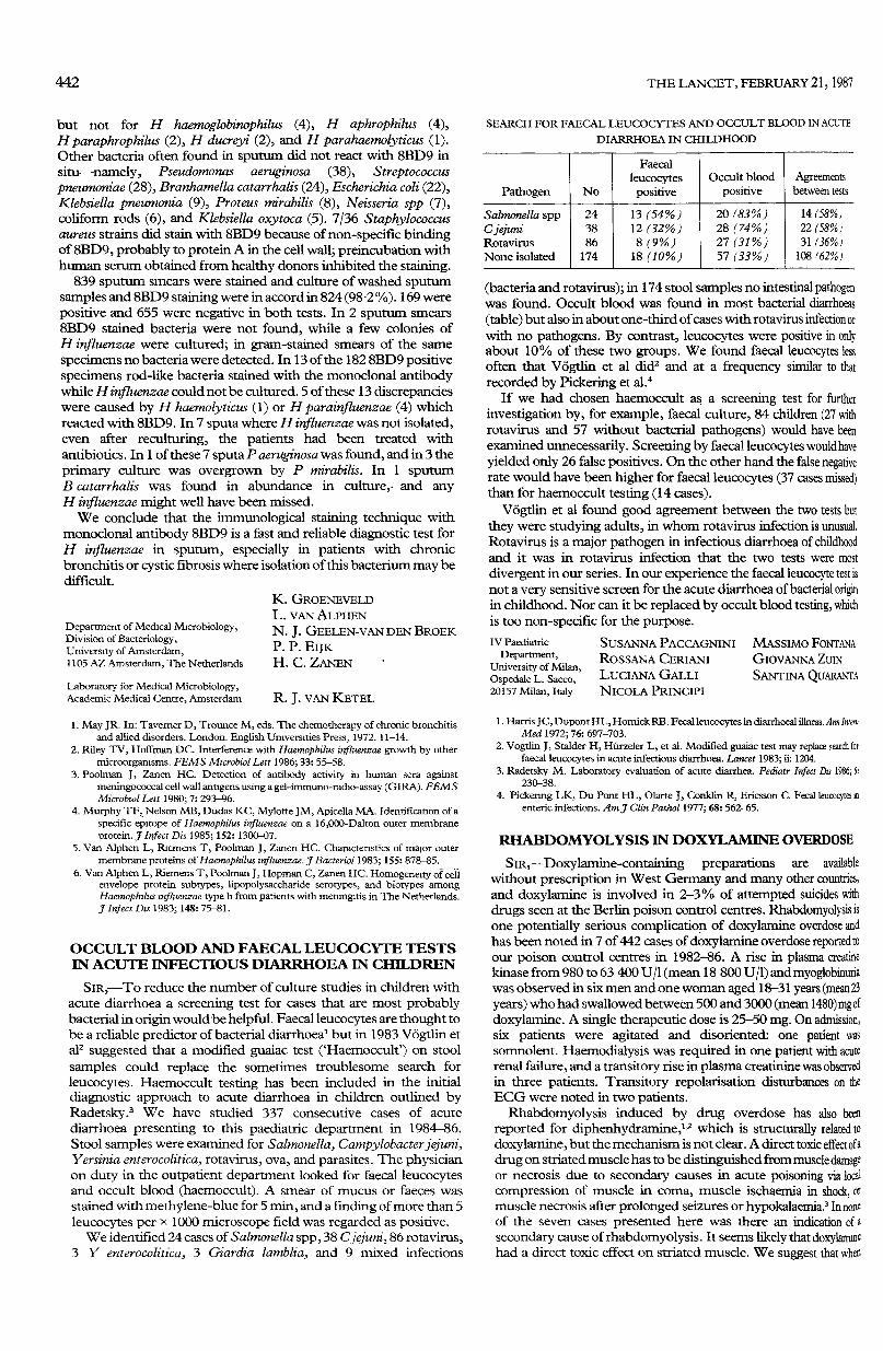
SEARCH FOR FAECAL LEUCOCYTES AND OCCULT BLOOD IN ACUTE
DIARRHOEA IN CHILDHOOD
(bacteria and rotavirus); in 174 stool samples no intestinal pathogenwas found. Occult blood was found in most bacterial diarrhoeas(table) but also in about one-third of cases with rotavirus infection orwith no pathogens. By contrast, leucocytes were positive in onlyabout 10% of these two groups. We found faecal leucocytes lessoften that Vögtlin et al did2 and at a frequency similar to thatrecorded by Pickering et al.4
If we had chosen haemoccult as a screening test for furtherinvestigation by, for example, faecal culture, 84 children (27 withrotavirus and 57 without bacterial pathogens) would have beenexamined unnecessarily. Screening by faecal leucocytes would haveyielded only 26 false positives. On the other hand the false negativerate would have been higher for faecal leucocytes (37 cases missed)than for haemoccult testing (14 cases).
Vögtlin et al found good agreement between the two tests butthey were studying adults, in whom rotavirus infection is unusual.Rotavirus is a major pathogen in infectious diarrhoea of childhoodand it was in rotavirus infection that the two tests were most
divergent in our series. In our experience the faecal leucocyte test isnot a very sensitive screen for the acute diarrhoea of bacterial originin childhood. Nor can it be replaced by occult blood testing, whichis too non-specific for the purpose.IV Paediatric
Department,University of Milan,Ospedale L. Sacco,20157 Milan, Italy
SUSANNA PACCAGNINIROSSANA CERIANILUCIANA GALLINICOLA PRINCIPI
MASSIMO FONTANAGIOVANNA ZUINSANTINA QUARANTA
1. Harris JC, Dupont HL, Hornick RB. Fecal leucocytes in diarrhoeal illness. Ann InternMed 1972; 76: 697-703.
2. Vogtlin J, Stalder H, Hürzeler L, et al. Modified guaiac test may replace search forfaecal leucocytes in acute infectious diarrhoea. Lancet 1983; ii: 1204.
3. Radetsky M. Laboratory evaluation of acute diarrhea. Pediatr Infect Dis 1986; 5:230-38.
4. Pickering LK, Du Pont HL, Olarte J, Conklin R, Ericsson C. Fecal leucocytes inenteric infections. Am J Clin Pathol 1977; 68: 562-65.
RHABDOMYOLYSIS IN DOXYLAMINE OVERDOSE
SIR,—Doxylamine-containing preparations are availablewithout prescription in West Germany and many other countries,and doxylamine is involved in 2-3% of attempted suicides withdrugs seen at the Berlin poison control centres. Rhabdomyolysis isone potentially serious complication of doxylamine overdose andhas been noted in 7 of 442 cases of doxylamine overdose reported toour poison control centres in 1982-86. A rise in plasma creatinekinase from 980 to 63 400 U/1 (mean 18 800 U/1) and myoglobinuriawas observed in six men and one woman aged 18-31 years (mean 23years) who had swallowed between 500 and 3000 (mean 1480) mg ofdoxylamine. A single therapeutic dose is 25-50 mg. On admission,six patients were agitated and disoriented: one patient was
somnolent. Haemodialysis was required in one patient with acuterenal failure, and a transitory rise in plasma creatinine was observedin three patients. Transitory repolarisation disturbances on theECG were noted in two patients.
Rhabdomyolysis induced by drug overdose has also been
reported for diphenhydramine,t,2 which is structurally related todoxylamine, but the mechanism is not clear. A direct toxic effect of adrug on striated muscle has to be distinguished from muscle damageor necrosis due to secondary causes in acute poisoning via localcompression of muscle in coma, muscle ischaemia in shock, ormuscle necrosis after prolonged seizures or hypokalaemia.3 In noneof the seven cases presented here was there an indication of asecondary cause of rhabdomyolysis. It seems likely that doxylamuzhad a direct toxic effect on striated muscle. We suggest that when