Embed Size (px)
Citation preview

J Neurosurg 78: 167-175, 1993
Occlusive hyperemia: a theory for the hemodynamic complications following resection of intracerebral arteriovenous malformations
NAYEF R. F. AL-RODHAN, M.D., PH.D., THORALF M. SUNDT, JR., M.D., DAVID G . PIEPGRAS, M.D., DOUGLAS A. NICHOLS, M.D., DANIEL R~~FENACHT, M.D., AND LORNA N. STEVENS, R.N.
Departments of Neurosurgery and Neuroradiology, Maj~o Clinlc and Mayo Graduate School of Medicine, Rochester. Minnesota
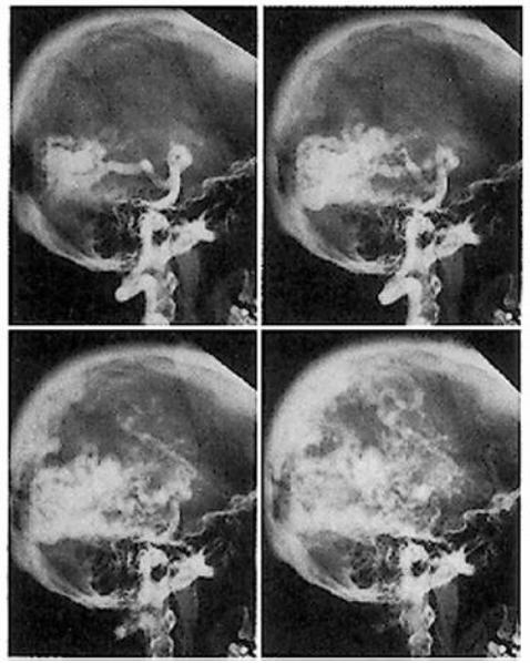
I/ An alternative theory is proposed to explain the brain edema and hemorrhage that may occur after resection of high-flow intracerebral arteriovenous malformations (AVM's). This theory, termed "occlusive hyperemia," is based on a retrospective analysis of operative dictations along with postoperative imaging studies (191 angiograms and 273 computerized tomography scans) in 295 cases of intracerebral AVM's operated on at the Mayo Clinic between 1970 and 1990. In this series, 34 cases (12%) of postoperative deterioration were documented, of which 15 were due to incomplete resection of the AVM. Of the remaining 19 cases, six had brain edema alone and 13 had hemorrhage with edema, despite complete excision of the AVM. In these 19 cases, the AVM's were greater than 6 cm in diameter in 10 patients, between 3 and 6 cm in six, and less than 3 cm in three. Obstruction of the venous drainage system was observed in 14 (74%) of the 19 cases. Ten of these 14 were due to obstruction of the primary venous drainage of the brain parenchyma immediately surrounding the lesions, while four were due to obstruction of other venous structures. In no case was a rapid circulation identified on postoperative angiograms. The flow pattern was slow or stagnant in former AVM feeders and their parenchymal branches. It is proposed that postoperative intracranial hemorrhage and/or brain edema in AVM patients may be due to: 1) obstruction of the venous outflow system of brain adjacent to the AVM, with subsequent passive hyperemia and engorgement: and 2) stagnant arterial flow in former AVM feeders and their parenchymal branches, with subsequent worsening of the existing hypoperfusion, ischemia, and hemorrhage or edema into these areas. Supportive hemodynamic evidence for this theory was derived from the literature.
KEY WORDS arterioven~~~malformation . cerebral hemodynamics autoregulation intracerebral hemorrhage cerebral venous occlusion . cerebral ischemia
D ESPITE the advances in microneurosurgical tech- niques to deal with large intracerebral arterio- venous malformations (AVM's), these lesions
continue to present a major challenge. One of the recognized complications that may follow the emboli- zation and/or surgical obliteration of large AVM's is the development of brain edema and/or hemorrhage in the normal brain parenchyma that surrounds the bed of the excised AVM, despite the complete obliteration of the AVM nidus.4.5.9.10,15,16,23.26,28.29,31,36,42,45 While there is widespread support for the concept that the disrup- tion of local hemodynamics is responsible for these complications, the precise underlying mechanism(s) re- main poorly understood. Cerebral hypoperfusion in- duced by high-flow AVM's (that is, steal) and the sub- sequent improvement in perfusion after the excision of
the AVM was first proposed by Olivecrona and Riives2' in 1948 and by NorlinZ4 in 1949, while the role of cerebral hyperemia in producing neurological disability was hinted at by GowersL4 as early as 1888.
A pioneering theory that has provided a plausible explanation for this phenomenon was proposed in 1978 by Spetzler and coworkers,36 and was termed "normal perfusion pressure breakthrough." The basic concept of the theory is that the parenchyma surrounding high- flow AVM's is chronically hypoperfused and has an impaired autoregulation mechanism that renders it vul- nerable to the restoration of normal perfusion after AVM excision. The combination of disturbed autoreg- ulation and normal perfusion pressure is thought to result in disruption of local capillary beds with subse- quent edema and/or hemorrhage. Based on this theory,
J. Neurosurg. / Volume 78 /February, 1993


![[PPT]HYPEREMIA & CONGESTION IIhkmu.online/wp-content/uploads/2016/11/HYPEREMIA... · Web viewMorphologic changes is seen most commonly in the lungs, liver, spleen andkidney CVC LUNG:](https://img.pdfslide.us/doc/110x75/5ab399547f8b9ac66c8e6af8/ppthyperemia-congestion-viewmorphologic-changes-is-seen-most-commonly-in-the-lungs.jpg)