Embed Size (px)
Citation preview
Object Activation from Features in the Semantic System
Michael A. Kraut, Sarah Kremen, Jessica B. Segal,Vincent Calhoun, Lauren R. Moo, and John Hart, Jr.
Abstract
& The human brain is thought to elicit an object representa-tion via co-activation of neural regions that encode variousobject features. The cortical regions and mechanisms involvedin this process have never been elucidated for the semanticsystem. We used functional magnetic resonance imaging(fMRI) to evaluate regions activated during a task designedto elicit object activation within the semantic system (e.g.,presenting the words ‘‘desert’’ and ‘‘humps’’ with the task to
determine if they combine to form an object, in this case a‘‘camel’’). There were signal changes in the thalamus for wordpairs that activated an object, but not for pairs that (a) failed toactivate an object, (b) were simply semantically associated, or(c) were members of the same category. These results suggestthat the thalamus has a critical role in coordinating the corticalactivity required for activating an object concept in thesemantic system. &
INTRODUCTION
Numerous models of semantic memory (Millikan, 1998;Schyns, Goldstone, & Thibaut, 1998; Lambert &Shanks, 1997; Estes, 1994; Hinton & Anderson, 1989;Fodor & Pylyshyn, 1988; Paivio, 1986; Collins & Loftus,1975; Smith, Shoben, & Rips, 1974) have suggested theexistence of specific subprocesses, including, but notlimited to, making semantic choices from among multi-ple possibilities, feature–object correlation, noun–verbassociation, and adjective–noun association (Ricci et al.,1999; Thompson-Schill, D’Esposito, Aguirre, & Farah,1997; Demb et al., 1995; Kapur et al., 1994; Saffran &Schwartz, 1994; Demonet et al., 1992; Petersen, Fox,Posner, Mintun, & Raichle, 1988; Petersen, Fox, Snyder,& Raichle, 1990; Posner, Petersen, Fox, & Raichle, 1988;Shallice, 1988; Saffran, 1982). One other related proc-ess that has been investigated via a variety of tasks issemantic association. In a typical semantic associationparadigm, two items are presented with the generalinstructions/question of, ‘‘Are these two stimuli related/associated with each other?’’ The terms ‘‘related’’ or‘‘associated’’ do not typically require a formal definitionor instruction to the subject; their meaning or intent isconveyed by an example. (‘‘For instance, salt andpepper are related.’’)
Within one theoretical framework, semantic associa-tions have been shown to be behaviorally and anatom-ically dissociable into two major subtypes: (1)compositional associations (two items are associatedbut retain their individuality in the relationship—salt
and pepper), and (2) noncompositional associations(the two items become fused semantically—‘‘computer’’and ‘‘virus’’ forming ‘‘computer virus’’) (Kounios, Smith,Yang, Bachman, & D’Esposito, 2001; Fodor & Pylyshyn,1988). Of interest, in the compositional associations, thetwo related items remain distinct both lexically andsemantically, while in the noncompositional associa-tions, the two stimuli fuse to form a third novel stimulus(as in the ‘‘computer virus’’ example) at the semantic,but not at the lexical, level. This concept of semanticfusion, where two entities combine to form a newrepresentation, can be applied to other semantic rela-tionships. One such application has been the proposalthat features that are common to an object could ‘‘fuse’’to activate a previously stored semantic object represen-tation (Gray, 1999; Eichenbaum & Bunsey, 1995; Singer& Gray, 1995; Damasio, 1989, 1990; Warrington & Mc-Carthy, 1987; Allport, 1985). While this semantic fusionprocess has been proposed in theoretical form, a para-digm addressing it during object activation in semanticmemory has not been formally developed.
To address the question of object activation withinthis framework, we have designed an analog of thesemantic fusion task (a noncompositional association)where the stimuli are two features that are common tothe target object. The particular features were chosensuch that, in combination, they are both sufficient andspecific to elicit the target object in semantic memory.The task instructions are to push a button if the twofeatures (e.g., ‘‘desert’’ and ‘‘humps’’) combine to makeyou think of an object (e.g., ‘‘camel’’). These taskinstructions are in contrast, yet analogous to, those ofother semantic association tasks (‘‘Are these items re-Johns Hopkins University
D 2002 Massachusetts Institute of Technology Journal of Cognitive Neuroscience 14:1, pp. 24–36
lated?’’), since the feature stimuli may not be directlyrelated/associated with each other on their own (e.g.,‘‘desert’’ and ‘‘dry’’ being typically associated as op-posed to ‘‘desert’’ and ‘‘humps’’). The other notabledifference is that the two feature stimuli do fuse toactivate/recall a third identity that differs from the inputstimuli at both a semantic and lexical level.
The cognitive operations purportedly engaged by thisobject activation task are similar to those described indetail for standard semantic association (Kounios et al.,2001; Smith et al., 1974) until the semantic search stage.In a typical semantic association task, the search be-tween related stimulus items likely ends when thesubject determines that relevant conjunction criteriafor association between the two items’ semantic fieldshas been met (Smith et al., 1974). In contrast, the objectactivation task differs in that the semantic search likelycan proceed via two routes: (1) the semantic fields ofpotential target objects are searched until both stimulusfeatures are detected in a single object’s field(s), thusending the search and activating that object; and/or (2)the two features, by their saliency to a specific object,the frequency of their co-occurrence, or idiosyncraticnature of the feature combination, focus the search to aspecific target object’s semantic field(s), followed byfeature detection and object activation as in (1). Ineither case, the end-result for the feature pairs that fuseis object activation.
The objective of this study is to assess the neuralsubstrates of the object activation process within seman-tic memory, acknowledging that activation of an objectin semantics can likely proceed along multiple pathways.Damasio (1989, 1990) proposed that the object activa-tion from feature process exists in semantic memory andconcluded that this is mediated by convergence zones orregions in the brain (e.g., prefrontal, anterior temporalpole), with no single convergence zone being sufficientto sustain object activation and recall. Our hypothesis isthat there is an identifiable network of brain regionsinvolved in object activation in semantic memory andthat we can identify these with functional magneticresonance imaging (fMRI) using the object activationand control experiments.
We studied 11 normal volunteers with fMRI during theperformance of three tasks: (1) object activation fromfeatures (referred to as the ‘‘object activation’’ experi-ment), (2) semantic association (referred to as the‘‘association’’ experiment), and (3) category activationfrom objects (referred to as the ‘‘category’’ experiment).Within each of the three experiments, the same wordswere used but paired differently to minimize stimulus-specific effects.
In the object activation experiment, word pairs forboth the trials in which the features did elicit an object(O+ pairs) and in those pairs in which the features didnot elicit an object (O� pairs) were admixed andvisually presented, with the instruction being to push
a button if these features combined to generate anobject not presented in the stimulus words themselves(e.g., ‘‘desert’’ and ‘‘humps’’ produce ‘‘camel’’, while‘‘bullets’’ and ‘‘milk’’ do not produce a third object).The cognitive operations associated with processingboth the O+ and the O� pairs are quite similar untilthe object representation is activated. Thus, the pri-mary comparison to best determine the brain region(s)associated with this object activation is the differencebetween the signal changes detected for the O+ pairsto those of the O� pairs.
The association and category experiments served assecondary controls in order to address the possibilitythat the activation observed in the O+ trials reflectssimply an element of semantic relatedness between thestimulus pairs or a general process invoked whenevertwo elements in the semantic system combine to acti-vate a third (or possibly higher level) entity. In theassociation task, participants were visually presentedword pairs and were required to push a button if theywere related (e.g., ‘‘hammer’’ and ‘‘nail’’ are related, A+,‘‘nail’’ and ‘‘coffee’’ are not, A�). The category taskrequired participants to view object word pairs and pusha button if they are members of the same category (e.g.,‘‘skunk’’ and ‘‘monkey’’ are of the same category, C+,but ‘‘monkey’’ and ‘‘ hammer ’’ are not, C�).
RESULTS
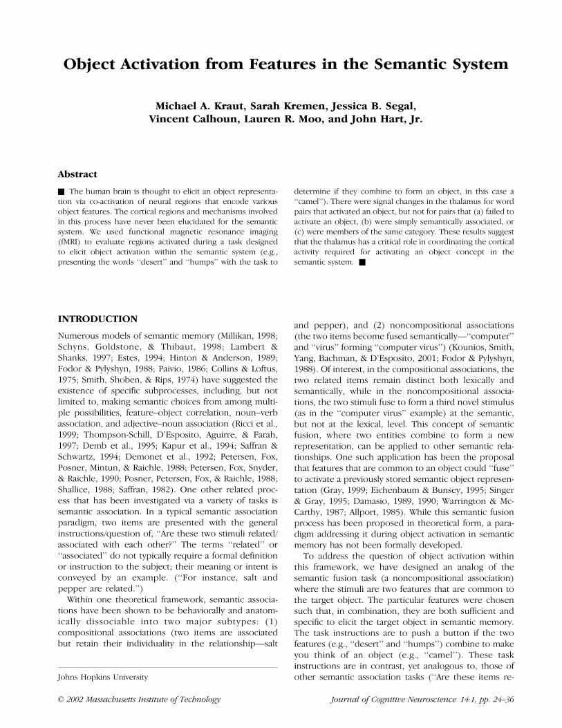
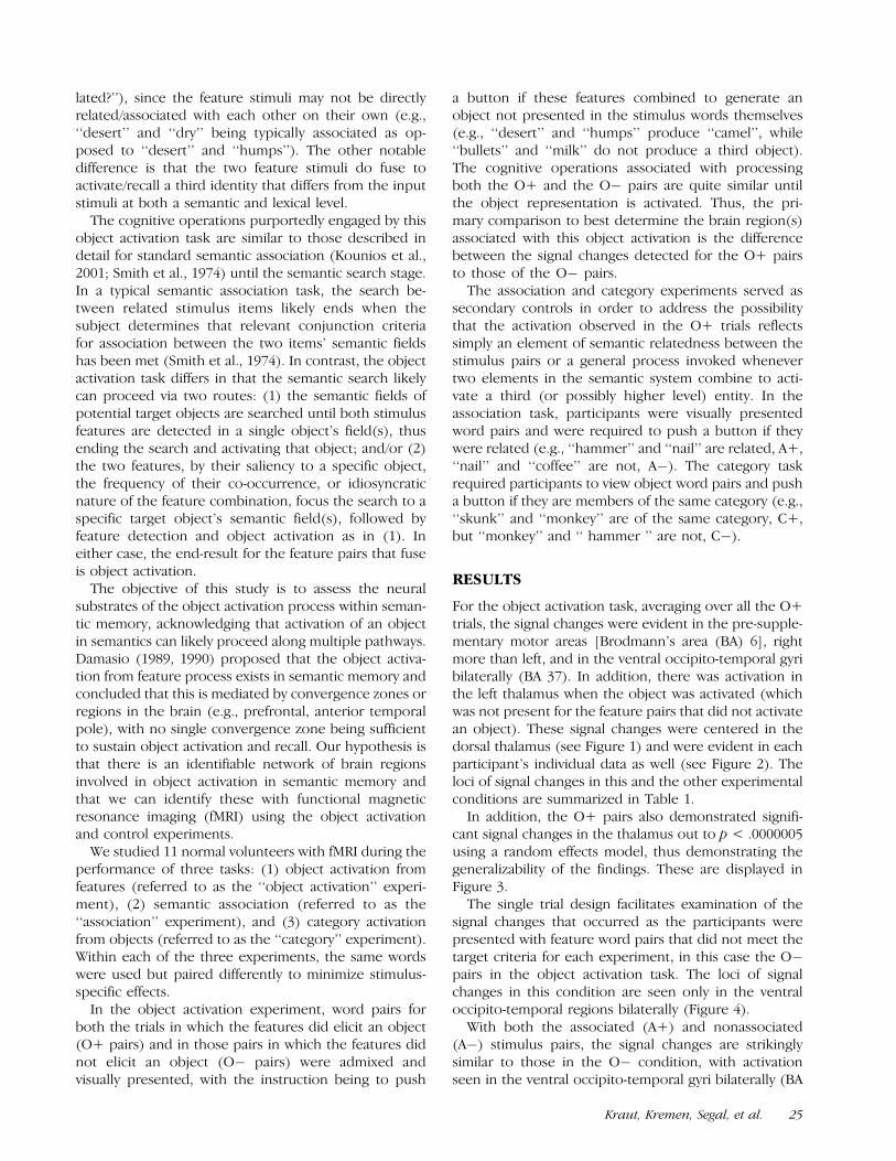
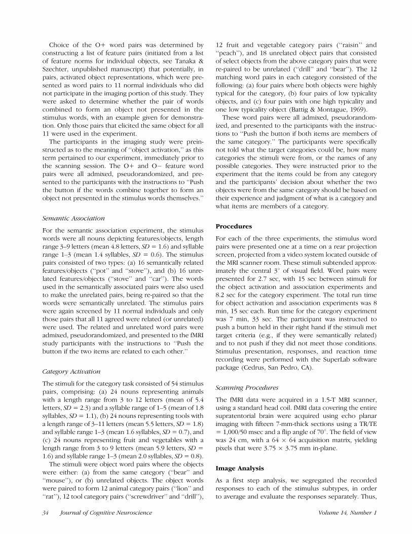
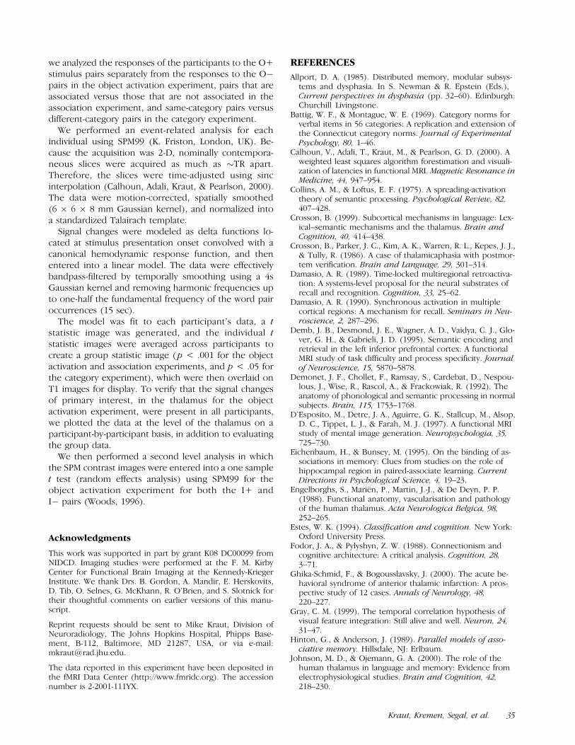
For the object activation task, averaging over all the O+trials, the signal changes were evident in the pre-supple-mentary motor areas [Brodmann’s area (BA) 6], rightmore than left, and in the ventral occipito-temporal gyribilaterally (BA 37). In addition, there was activation inthe left thalamus when the object was activated (whichwas not present for the feature pairs that did not activatean object). These signal changes were centered in thedorsal thalamus (see Figure 1) and were evident in eachparticipant’s individual data as well (see Figure 2). Theloci of signal changes in this and the other experimentalconditions are summarized in Table 1.
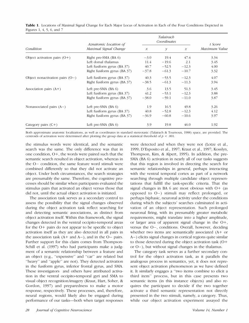
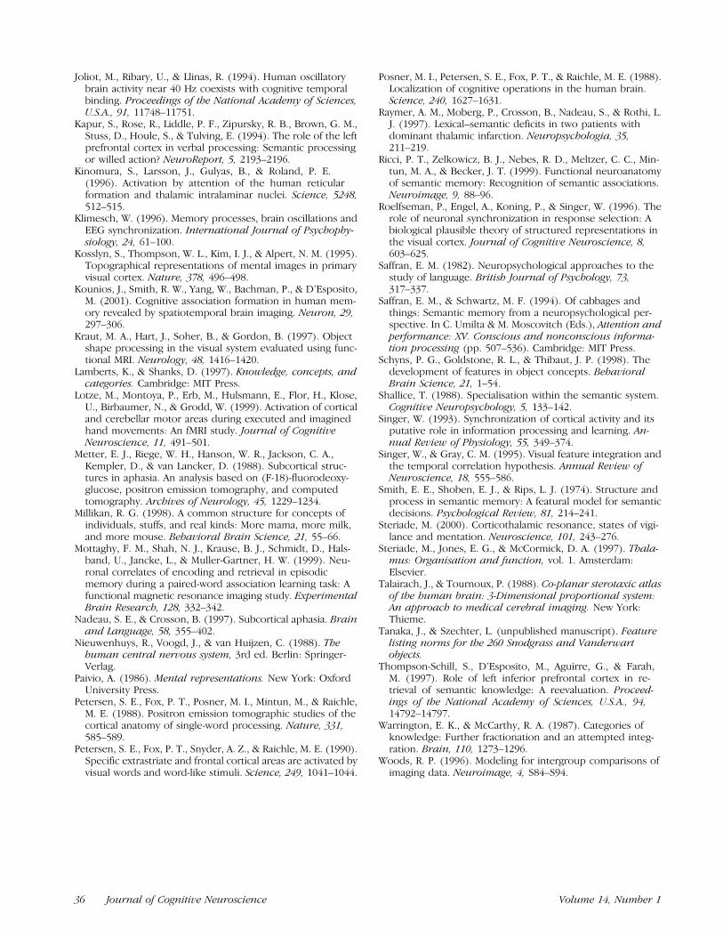
In addition, the O+ pairs also demonstrated signifi-cant signal changes in the thalamus out to p < .0000005using a random effects model, thus demonstrating thegeneralizability of the findings. These are displayed inFigure 3.
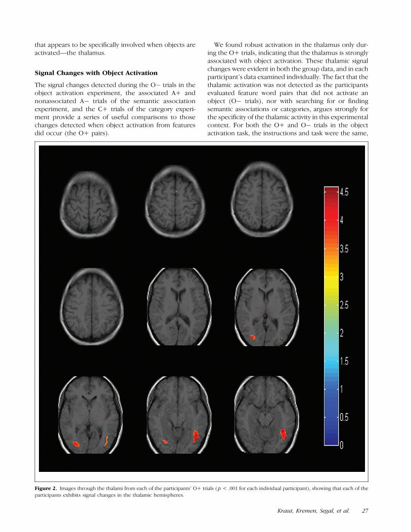
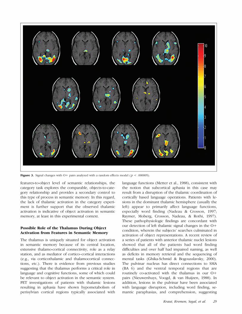
The single trial design facilitates examination of thesignal changes that occurred as the participants werepresented with feature word pairs that did not meet thetarget criteria for each experiment, in this case the O�pairs in the object activation task. The loci of signalchanges in this condition are seen only in the ventraloccipito-temporal regions bilaterally (Figure 4).
With both the associated (A+) and nonassociated(A�) stimulus pairs, the signal changes are strikinglysimilar to those in the O� condition, with activationseen in the ventral occipito-temporal gyri bilaterally (BA
Kraut, Kremen, Segal, et al. 25
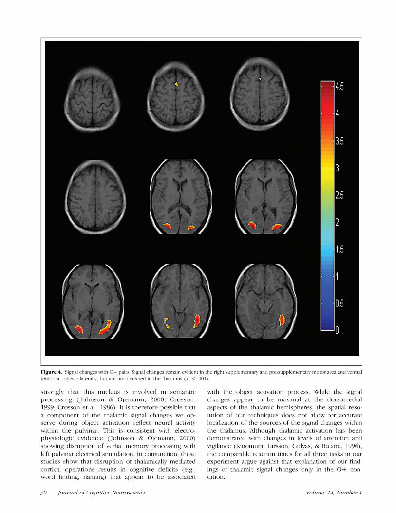
37), and minimally in the pre-supplementary motor area(BA 6). See Figures 5 and 6.
The signal changes in the categorization task, averag-ing over the object word pairs that were from the samecategory (C+), irrespective of the specific categories,reach a less robust level of statistical significance than dothe changes in the other conditions (Table 1); they arefocused in medial BA 6, slightly to the left of midline(Figure 7).
There was no significant difference in the participants’reaction times across these three tasks [F(2,7) = .86;p = .44].
DISCUSSION
These experiments delineate neural regions associatedwith object activation resulting from a semantic searchtask involving feature stimuli, and point to one region
Figure 1. Signal changes with object activation (O+ trials). The sections are angled perpendicular to the intercommisural line. The group data
images show the loci of fMRI signal changes ( p < .001) superimposed upon a normalized T1-weighted image gathered at the same location. Nineaxial-oblique sections are displayed, showing foci of signal changes at representative levels through the dorsal cerebral hemispheres, the thalamus,
and the ventral temporal lobes. The color bar at the right side of each figure reflects the range of t scores evident in the depicted data.
26 Journal of Cognitive Neuroscience Volume 14, Number 1
that appears to be specifically involved when objects areactivated—the thalamus.
Signal Changes with Object Activation
The signal changes detected during the O� trials in theobject activation experiment, the associated A+ andnonassociated A� trials of the semantic associationexperiment, and the C+ trials of the category experi-ment provide a series of useful comparisons to thosechanges detected when object activation from featuresdid occur (the O+ pairs).
We found robust activation in the thalamus only dur-ing the O+ trials, indicating that the thalamus is stronglyassociated with object activation. These thalamic signalchanges were evident in both the group data, and in eachparticipant’s data examined individually. The fact that thethalamic activation was not detected as the participantsevaluated feature word pairs that did not activate anobject (O� trials), nor with searching for or findingsemantic associations or categories, argues strongly forthe specificity of the thalamic activity in this experimentalcontext. For both the O+ and O� trials in the objectactivation task, the instructions and task were the same,
Figure 2. Images through the thalami from each of the participants’ O+ trials ( p < .001 for each individual participant), showing that each of the
participants exhibits signal changes in the thalamic hemispheres.
Kraut, Kremen, Segal, et al. 27
the stimulus words were identical, and the semanticsearch was the same. The only difference was that inone condition, O+, the words were paired such that thesemantic search resulted in object activation, whereas inthe O� condition, the same feature word stimuli werecombined differently so that they did not activate anobject. Under both circumstances, the search strategiesare presumably the same. Therefore, the cognitive pro-cesses should be similar when participants evaluated thestimulus pairs that activated an object versus those thatdid not, until the actual object activation is initiated.
The association task serves as a secondary control toassess the possibility that the signal changes observedduring the object activation task reflect searching forand detecting semantic associations, as distinct fromobject activation itself. Within this framework, the signalchanges detected in the ventral occipito-temporal areasfor the O+ pairs do not appear to be specific to objectactivation itself as they are also detected in all pairs inthe association task (A+ and A�), and in the O� pairs.Further support for this claim comes from Thompson-Schill et al. (1997) who had participants make a judg-ment of a semantic relationship between a feature andan object (e.g., ‘‘expensive’’ and ‘‘car’’ are related but‘‘heavy’’ and ‘‘apple’’ are not). They detected activationin the fusiform gyrus, inferior frontal gyrus, and SMA.These investigators and others have attributed activa-tion in the ventral occipito-temporal gyri and SMA tovisual object recognition/imagery (Kraut, Hart, Soher, &Gordon, 1997) and preparedness to make a motorresponse, respectively. These processes, and, therefore,neural regions, would likely also be engaged duringperformance of our tasks—both when target responses
were detected and when they were not (Lotze et al.,1999; D’Esposito et al., 1997; Kraut et al., 1997; Kosslyn,Thompson, Kim, & Alpert, 1995). In addition, the pre-SMA (BA 6) activation in nearly all of our tasks suggeststhat this region is involved in directing the search forsemantic relationships in general, perhaps interactingwith the ventral temporal cortex as part of a networksearching through multiple candidate object represen-tations that fulfill the task-specific criteria. That thesignal changes in BA 6 are most obvious with O+ (asopposed to O�) stimuli may reflect prolonged, orperhaps biphasic, neuronal activity under the conditionsduring which the subjects’ searches culminated in acti-vation of an object representation. Such prolongedneuronal firing, with its presumably greater metabolicrequirements, might translate into a higher amplitude,or larger area of apparent signal change in the O+,versus the O�, conditions. Overall, however, decidingwhether two items are semantically associated (A+ orA�) elicits signal changes in cortical regions quite similarto those detected during the object activation task (O+or O�), but without signal changes in the thalamus.
The category task serves as a further secondary con-trol for the object activation task, as it parallels theanalogous process in semantics, yet, it does not repre-sent object activation phenomenon as we have definedit. It similarly engages a ‘‘two items combine to elicit athird item’’ process, but in this case presents twosemantic items (in this instance objects) and also re-quires the participant to decide if the two togetheractivate a third semantic representation not directlypresented in the two stimuli, namely, a category. Thus,while our object activation experiment assayed the
Table 1. Locations of Maximal Signal Change for Each Major Locus of Activation in Each of the Four Conditions Depicted inFigures 1, 4, 5, 6, and 7
TalairachCoordinates
Condition Maximal Signal Change x y z Maximum Value
Object activation pairs (O+) Right pre-SMA (BA 6) �3.0 19.4 47.4 3.34Left dorsal thalamus 11.4 �19.6 2.1 3.45Left fusiform gyrus (BA 37) 40.7 �52.5 �12.3 4.00Right fusiform gyrus (BA 37) �37.8 �61.3 �10.7 3.32
Object nonactivation pairs (O�) Left fusiform gyrus (BA 37) 40.3 �53.5 �12.3 4.07Right fusiform gyrus (BA 37) �38.5 �61.3 �11.3 3.94
Association pairs (A+) Left pre-SMA (BA 6) 3.6 13.5 51.3 3.45Left fusiform gyrus (BA 37) 41.2 �53.1 �12.3 3.88Right fusiform gyrus (BA 37) �38.0 �59.1 �11.0 3.87
Nonassociated pairs (A�) Left pre-SMA (BA 6) 1.9 16.5 49.8 3.26Left fusiform gyrus (BA 37) 40.8 �52.8 �12.3 4.12Right fusiform gyrus (BA 37) �36.9 �60.8 �10.6 3.97
Category pairs (C+) Left pre-SMA (BA 6) 3.9 19.8 46.0 1.92
Both approximate anatomic localizations, as well as coordinates in standard stereotaxic (Talairach & Tournoux, 1988) space, are provided. Thecentroids of activation were determined after plotting the group data at a statistical threshold of p < .001.
Aznatomic Location of t Score
28 Journal of Cognitive Neuroscience Volume 14, Number 1
features-to-object level of semantic relationships, thecategory task explores the comparable, objects-to-cate-gory relationship and provides a secondary control tothis type of process in semantic memory. In this regard,the lack of thalamic activation in the category experi-ment is further support that the observed thalamicactivation is indicative of object activation in semanticmemory, at least in this experimental context.
Possible Role of the Thalamus During ObjectActivation from Features in Semantic Memory
The thalamus is uniquely situated for object activationin semantic memory because of its central location,extensive thalamo-cortical connectivity, role as a relaystation, and as mediator of cortico–cortical interactions(e.g., via corticothalamic and thalamocortical connec-tions, etc.). There is evidence from previous studiessuggesting that the thalamus performs a critical role inlanguage and cognitive functions, some of which couldbe relevant to object activation in the semantic system.PET investigations of patients with thalamic lesionsresulting in aphasia have shown hypometabolism ofperisylvian cortical regions typically associated with
language functions (Metter et al., 1988), consistent withthe notion that subcortical aphasia in this case mayresult from a disruption of the thalamic coordination ofcortically based language operations. Patients with le-sions in the dominant thalamic hemisphere (usually theleft) appear to primarily affect language functions,especially word finding (Nadeau & Crosson, 1997;Raymer, Moberg, Crosson, Nadeau, & Rothi, 1997).These pathophysiologic findings are concordant withour detection of left thalamic signal changes in the O+condition, wherein the subjects’ searches culminated inactivation of object representations. A recent review ofa series of patients with anterior thalamic nuclei lesionsshowed that all of the patients had word findingdifficulties and over half had impaired naming, as wellas deficits in memory retrieval and the sequencing ofmental tasks (Ghika-Schmid & Bogousslavsky, 2000).The pulvinar nucleus has direct connections to SMA(BA 6) and the ventral temporal regions that areroutinely co-activated with the thalamus in our O+pairs (Nieuwenhuys, Voogd, & van Huijzen, 1988). Inaddition, lesions in the pulvinar have been associatedwith language disruption, including word finding, se-mantic paraphasias, and comprehension, suggesting
Figure 3. Signal changes with O+ pairs analyzed with a random effects model ( p < .000005).
Kraut, Kremen, Segal, et al. 29
strongly that this nucleus is involved in semanticprocessing ( Johnson & Ojemann, 2000; Crosson,1999; Crosson et al., 1986). It is therefore possible thata component of the thalamic signal changes we ob-serve during object activation reflect neural activitywithin the pulvinar. This is consistent with electro-physiologic evidence ( Johnson & Ojemann, 2000)showing disruption of verbal memory processing withleft pulvinar electrical stimulation. In conjunction, thesestudies show that disruption of thalamically mediatedcortical operations results in cognitive deficits (e.g.,word finding, naming) that appear to be associated
with the object activation process. While the signalchanges appear to be maximal at the dorsomedialaspects of the thalamic hemispheres, the spatial reso-lution of our techniques does not allow for accuratelocalization of the sources of the signal changes withinthe thalamus. Although thalamic activation has beendemonstrated with changes in levels of attention andvigilance (Kinomura, Larsson, Gulyas, & Roland, 1996),the comparable reaction times for all three tasks in ourexperiment argue against that explanation of our find-ings of thalamic signal changes only in the O+ con-dition.
Figure 4. Signal changes with O� pairs. Signal changes remain evident in the right supplementary and pre-supplementary motor area and ventral
temporal lobes bilaterally, but are not detected in the thalamus ( p < .001).
30 Journal of Cognitive Neuroscience Volume 14, Number 1
While it is clear from our investigation that the thala-mus plays an active role during our object activation task,it is unclear how this is mediated. The presumed under-lying neural activity that evokes the observed signalchanges during the semantic object activation could beassociated with the functions of stimulus recognition andstimulus–stimulus association, both of which may benecessary in object activation and have been attributedto the dorsal medial nucleus of the thalamus (Mottaghyet al., 1999; Nadeau & Crosson, 1997; Steriade, Jones, &McCormick, 1997). Other plausible operations includecoordinating a search or detection circuit for correlatedfeatures and possible target objects, pointing to object
representations in the imagery or lexical system, ormediating shifts in selective attention.
Nevertheless, based upon previous investigations inother cognitive systems, we suggest that the process thatelicits object activation involves coordinating and syn-chronizing feature representations via an oscillatingrhythm (Roelfseman, Engel, Koning, & Singer, 1996;Singer, 1993). In humans, the thalamus has been asso-ciated with oscillatory rhythms in multiple frequencyranges (Klimesch, 1996; Joliot, Ribary, & Llinas, 1994).Thus, both the animal and human data are consistentwith the notion that the thalamus mediates objectactivation through the synchronization of oscillatory
Figure 5. Signal changes with semantic association (A+), gathered at the same locations as the data in Figure 1 ( p < .001).
Kraut, Kremen, Segal, et al. 31

neural activity in cortical regions encoding features(from multiple sensorimotor and lexical–semantic sys-tems) of that object (Steriade, 2000; Engelborghs, Mar-ien, Martin, & De Deyn, 1998).
Object activation, as we have defined it in our exper-imental context, is essential to activating a conceptualrepresentation in the semantic system, which, in turn, isbasic to word finding, naming, comprehension, andtranslating thought into language. Among patients withneurodegenerative disorders and focal brain lesions,word finding is one of the most prevalent cognitiveimpairments, but little is known about the mechanismsby which objects are identified and named. The patho-physiological disruption of these mechanisms similarlyremains unknown. The intermittent nature of wordfinding difficulties (e.g., being able to recall a name atone time, but not at another) may suggest transientdisruptions of synchronizing electrical rhythms; how-ever, further investigations will be necessary to delineate
what appears to be the thalamus’ critical role in seman-tic object activation.
METHODS
Research Participants
Eleven normal right-handed individuals, seven womenand four men, between the ages of 23 and 48 (meanage 31.45 years, SD = 8.57) participated in the objectactivation and association experiments. Ten of theseindividuals from the group above (seven women andthree men, mean age of 31.2 years [SD = 8.99])participated in the category experiment.
Stimuli
The stimuli for the three tasks consisted of visuallypresented word pairs printed in lower case, with oneword above the other.
Figure 6. Signal changes with the word pairs that were not semantically associated (A�) ( p < .001).
32 Journal of Cognitive Neuroscience Volume 14, Number 1

Object Activation
For the object activation experiment, the stimulus wordswere all features of objects, length range 3–7 letters (mean5.3 letters, SD = 1.2) and syllable range 1–3 (mean 1.3syllables, SD = 0.5). The features chosen were not specificto one particular perceptual or verbal domain. The targetobjects came from seven different categories. There weretwo types of word pairs: (a) 16 O+ pairs, where the twowords describing features of an object combine to elicit anobject that was not presented (e.g., the words ‘‘desert’’and ‘‘humps,’’ which produce the object ‘‘camel’’), and
( b) 16 word pairs that do not combine to activate anobject not presented, O� pairs, and are semanticallyunrelated (e.g., ‘‘humps’’ and ‘‘alarm’’).
To minimize stimulus-specific effects based on thewords themselves (vs. the relationship between thewords), the same feature words used in the O+ pairswere the stimuli in the O� pairs, being paired in thesecases with a semantically unrelated word (e.g., ‘‘humps’’and ‘‘alarm’’). Serial position in the word pairs (uppervs. lower positions on the screen) was counterbalancedbetween the O+ and O� word pairs.
Figure 7. Signal changes ( p < .05) with the object pairs from the category task where both objects were from the same category, irrespective of
the specific categories.
Kraut, Kremen, Segal, et al. 33
Choice of the O+ word pairs was determined byconstructing a list of feature pairs (initiated from a listof feature norms for individual objects, see Tanaka &Szechter, unpublished manuscript) that potentially, inpairs, activated object representations, which were pre-sented as word pairs to 11 normal individuals who didnot participate in the imaging portion of this study. Theywere asked to determine whether the pair of wordscombined to form an object not presented in thestimulus words, with an example given for demonstra-tion. Only those pairs that elicited the same object for all11 were used in the experiment.
The participants in the imaging study were prein-structed as to the meaning of ‘‘object activation,’’ as thisterm pertained to our experiment, immediately prior tothe scanning session. The O+ and O� feature wordpairs were all admixed, pseudorandomized, and pre-sented to the participants with the instructions to ‘‘Pushthe button if the words combine together to form anobject not presented in the stimulus words themselves.’’
Semantic Association
For the semantic association experiment, the stimuluswords were all nouns depicting features/objects, lengthrange 3–9 letters (mean 4.8 letters, SD = 1.6) and syllablerange 1–3 (mean 1.4 syllables, SD = 0.6). The stimuluspairs consisted of two types: (a) 16 semantically relatedfeatures/objects (‘‘pot’’ and ‘‘stove’’), and (b) 16 unre-lated features/objects (‘‘stove’’ and ‘‘car’’). The wordsused in the semantically associated pairs were also usedto make the unrelated pairs, being re-paired so that thewords were semantically unrelated. The stimulus pairswere again screened by 11 normal individuals and onlythose pairs that all 11 agreed were related (or unrelated)were used. The related and unrelated word pairs wereadmixed, pseudorandomized, and presented to the fMRIstudy participants with the instructions to ‘‘Push thebutton if the two items are related to each other.’’
Category Activation
The stimuli for the category task consisted of 54 stimuluspairs, comprising: (a) 24 nouns representing animalswith a length range from 3 to 12 letters (mean of 5.4letters, SD = 2.3) and a syllable range of 1–5 (mean of 1.8syllables, SD = 1.1), (b) 24 nouns representing tools witha length range of 3–11 letters (mean 5.5 letters, SD = 1.8)and syllable range 1–3 (mean 1.6 syllables, SD = 0.7), and(c) 24 nouns representing fruit and vegetables with alength range from 3 to 9 letters (mean 5.9 letters, SD =1.6) and syllable range 1–3 (mean 2.0 syllables, SD = 0.8).
The stimuli were object word pairs where the objectswere either: (a) from the same category (‘‘bear’’ and‘‘mouse’’), or (b) unrelated objects. The object wordswere paired to form 12 animal category pairs (‘‘lion’’ and‘‘rat’’), 12 tool category pairs (‘‘screwdriver’’ and ‘‘drill’’),
12 fruit and vegetable category pairs (‘‘raisin’’ and‘‘peach’’), and 18 unrelated object pairs that consistedof select objects from the above category pairs that werere-paired to be unrelated (‘‘drill’’ and ‘‘bear’’). The 12matching word pairs in each category consisted of thefollowing: (a) four pairs where both objects were highlytypical for the category, (b) four pairs of low typicalityobjects, and (c) four pairs with one high typicality andone low typicality object (Battig & Montague, 1969).
These word pairs were all admixed, pseudorandom-ized, and presented to the participants with the instruc-tions to ‘‘Push the button if both items are members ofthe same category.’’ The participants were specificallynot told what the target categories could be, how manycategories the stimuli were from, or the names of anypossible categories. They were instructed prior to theexperiment that the items could be from any categoryand the participants’ decision about whether the twoobjects were from the same category should be based ontheir experience and judgment of what is a category andwhat items are members of a category.
Procedures
For each of the three experiments, the stimulus wordpairs were presented one at a time on a rear projectionscreen, projected from a video system located outside ofthe MRI scanner room. These stimuli subtended approx-imately the central 38 of visual field. Word pairs werepresented for 2.7 sec, with 15 sec between stimuli forthe object activation and association experiments and8.2 sec for the category experiment. The total run timefor object activation and association experiments was 8min, 15 sec each. Run time for the category experimentwas 7 min, 33 sec. The participant was instructed topush a button held in their right hand if the stimuli mettarget criteria (e.g., if they were semantically related)and to not push if they did not meet those conditions.Stimulus presentation, responses, and reaction timerecording were performed with the SuperLab softwarepackage (Cedrus, San Pedro, CA).
Scanning Procedures
The fMRI data were acquired in a 1.5-T MRI scanner,using a standard head coil. fMRI data covering the entiresupratentorial brain were acquired using echo planarimaging with fifteen 7-mm-thick sections using a TR/TE= 1,000/50 msec and a flip angle of 708. The field of viewwas 24 cm, with a 64 � 64 acquisition matrix, yieldingpixels that were 3.75 � 3.75 mm in-plane.
Image Analysis
As a first step analysis, we segregated the recordedresponses to each of the stimulus subtypes, in orderto average and evaluate the responses separately. Thus,
34 Journal of Cognitive Neuroscience Volume 14, Number 1
we analyzed the responses of the participants to the O+stimulus pairs separately from the responses to the O�pairs in the object activation experiment, pairs that areassociated versus those that are not associated in theassociation experiment, and same-category pairs versusdifferent-category pairs in the category experiment.
We performed an event-related analysis for eachindividual using SPM99 (K. Friston, London, UK). Be-cause the acquisition was 2-D, nominally contempora-neous slices were acquired as much as �TR apart.Therefore, the slices were time-adjusted using sincinterpolation (Calhoun, Adali, Kraut, & Pearlson, 2000).The data were motion-corrected, spatially smoothed(6 � 6 � 8 mm Gaussian kernel), and normalized intoa standardized Talairach template.
Signal changes were modeled as delta functions lo-cated at stimulus presentation onset convolved with acanonical hemodynamic response function, and thenentered into a linear model. The data were effectivelybandpass-filtered by temporally smoothing using a 4sGaussian kernel and removing harmonic frequencies upto one-half the fundamental frequency of the word pairoccurrences (15 sec).
The model was fit to each participant’s data, a tstatistic image was generated, and the individual tstatistic images were averaged across participants tocreate a group statistic image ( p < .001 for the objectactivation and association experiments, and p < .05 forthe category experiment), which were then overlaid onT1 images for display. To verify that the signal changesof primary interest, in the thalamus for the objectactivation experiment, were present in all participants,we plotted the data at the level of the thalamus on aparticipant-by-participant basis, in addition to evaluatingthe group data.
We then performed a second level analysis in whichthe SPM contrast images were entered into a one samplet test (random effects analysis) using SPM99 for theobject activation experiment for both the I+ andI� pairs (Woods, 1996).
Acknowledgments
This work was supported in part by grant K08 DC00099 fromNIDCD. Imaging studies were performed at the F. M. KirbyCenter for Functional Brain Imaging at the Kennedy-KriegerInstitute. We thank Drs. B. Gordon, A. Mandir, E. Herskovits,D. Tib, O. Selnes, G. McKhann, R. O’Brien, and S. Slotnick fortheir thoughtful comments on earlier versions of this manu-script.
Reprint requests should be sent to Mike Kraut, Division ofNeuroradiology, The Johns Hopkins Hospital, Phipps Base-ment, B-112, Baltimore, MD 21287, USA, or via e-mail:[email protected].
The data reported in this experiment have been deposited inthe fMRI Data Center (http://www.fmridc.org). The accessionnumber is 2-2001-111YX.
REFERENCES
Allport, D. A. (1985). Distributed memory, modular subsys-tems and dysphasia. In S. Newman & R. Epstein (Eds.),Current perspectives in dysphasia (pp. 32–60). Edinburgh:Churchill Livingstone.
Battig, W. F., & Montague, W. E. (1969). Category norms forverbal items in 56 categories: A replication and extension ofthe Connecticut category norms. Journal of ExperimentalPsychology, 80, 1–46.
Calhoun, V., Adali, T., Kraut, M., & Pearlson, G. D. (2000). Aweighted least squares algorithm forestimation and visuali-zation of latencies in functional MRI. Magnetic Resonance inMedicine, 44, 947–954.
Collins, A. M., & Loftus, E. F. (1975). A spreading-activationtheory of semantic processing. Psychological Review, 82,407–428.
Crosson, B. (1999). Subcortical mechanisms in language: Lex-ical–semantic mechanisms and the thalamus. Brain andCognition, 40, 414–438.
Crosson, B., Parker, J. C., Kim, A. K., Warren, R. L., Kepes, J. J.,& Tully, R. (1986). A case of thalamicaphasia with postmor-tem verification. Brain and Language, 29, 301–314.
Damasio, A. R. (1989). Time-locked multiregional retroactiva-tion: A systems-level proposal for the neural substrates ofrecall and recognition. Cognition, 33, 25–62.
Damasio, A. R. (1990). Synchronous activation in multiplecortical regions: A mechanism for recall. Seminars in Neu-roscience, 2, 287–296.
Demb, J. B., Desmond, J. E., Wagner, A. D., Vaidya, C. J., Glo-ver, G. H., & Gabrieli, J. D. (1995). Semantic encoding andretrieval in the left inferior prefrontal cortex: A functionalMRI study of task difficulty and process specificity. Journalof Neuroscience, 15, 5870–5878.
Demonet, J. F., Chollet, F., Ramsay, S., Cardebat, D., Nespou-lous, J., Wise, R., Rascol, A., & Frackowiak, R. (1992). Theanatomy of phonological and semantic processing in normalsubjects. Brain, 115, 1753–1768.
D’Esposito, M., Detre, J. A., Aguirre, G. K., Stallcup, M., Alsop,D. C., Tippet, L. J., & Farah, M. J. (1997). A functional MRIstudy of mental image generation. Neuropsychologia, 35,725–730.
Eichenbaum, H., & Bunsey, M. (1995). On the binding of as-sociations in memory: Clues from studies on the role ofhippocampal region in paired-associate learning. CurrentDirections in Psychological Science, 4, 19–23.
Engelborghs, S., Marien, P., Martin, J.-J., & De Deyn, P. P.(1988). Functional anatomy, vascularisation and pathologyof the human thalamus. Acta Neurologica Belgica, 98,252–265.
Estes, W. K. (1994). Classification and cognition. New York:Oxford University Press.
Fodor, J. A., & Pylyshyn, Z. W. (1988). Connectionism andcognitive architecture: A critical analysis. Cognition, 28,3–71.
Ghika-Schmid, F., & Bogousslavsky, J. (2000). The acute be-havioral syndrome of anterior thalamic infarction: A pros-pective study of 12 cases. Annals of Neurology, 48,220–227.
Gray, C. M. (1999). The temporal correlation hypothesis ofvisual feature integration: Still alive and well. Neuron, 24,31–47.
Hinton, G., & Anderson, J. (1989). Parallel models of asso-ciative memory. Hillsdale, NJ: Erlbaum.
Johnson, M. D., & Ojemann, G. A. (2000). The role of thehuman thalamus in language and memory: Evidence fromelectrophysiological studies. Brain and Cognition, 42,218–230.
Kraut, Kremen, Segal, et al. 35
Joliot, M., Ribary, U., & Llinas, R. (1994). Human oscillatorybrain activity near 40 Hz coexists with cognitive temporalbinding. Proceedings of the National Academy of Sciences,U.S.A., 91, 11748–11751.
Kapur, S., Rose, R., Liddle, P. F., Zipursky, R. B., Brown, G. M.,Stuss, D., Houle, S., & Tulving, E. (1994). The role of the leftprefrontal cortex in verbal processing: Semantic processingor willed action? NeuroReport, 5, 2193–2196.
Kinomura, S., Larsson, J., Gulyas, B., & Roland, P. E.(1996). Activation by attention of the human reticularformation and thalamic intralaminar nuclei. Science, 5248,512–515.
Klimesch, W. (1996). Memory processes, brain oscillations andEEG synchronization. International Journal of Psychophy-siology, 24, 61–100.
Kosslyn, S., Thompson, W. L., Kim, I. J., & Alpert, N. M. (1995).Topographical representations of mental images in primaryvisual cortex. Nature, 378, 496–498.
Kounios, J., Smith, R. W., Yang, W., Bachman, P., & D’Esposito,M. (2001). Cognitive association formation in human mem-ory revealed by spatiotemporal brain imaging. Neuron, 29,297–306.
Kraut, M. A., Hart, J., Soher, B., & Gordon, B. (1997). Objectshape processing in the visual system evaluated using func-tional MRI. Neurology, 48, 1416–1420.
Lamberts, K., & Shanks, D. (1997). Knowledge, concepts, andcategories. Cambridge: MIT Press.
Lotze, M., Montoya, P., Erb, M., Hulsmann, E., Flor, H., Klose,U., Birbaumer, N., & Grodd, W. (1999). Activation of corticaland cerebellar motor areas during executed and imaginedhand movements: An fMRI study. Journal of CognitiveNeuroscience, 11, 491–501.
Metter, E. J., Riege, W. H., Hanson, W. R., Jackson, C. A.,Kempler, D., & van Lancker, D. (1988). Subcortical struc-tures in aphasia. An analysis based on (F-18)-fluorodeoxy-glucose, positron emission tomography, and computedtomography. Archives of Neurology, 45, 1229–1234.
Millikan, R. G. (1998). A common structure for concepts ofindividuals, stuffs, and real kinds: More mama, more milk,and more mouse. Behavioral Brain Science, 21, 55–66.
Mottaghy, F. M., Shah, N. J., Krause, B. J., Schmidt, D., Hals-band, U., Jancke, L., & Muller-Gartner, H. W. (1999). Neu-ronal correlates of encoding and retrieval in episodicmemory during a paired-word association learning task: Afunctional magnetic resonance imaging study. ExperimentalBrain Research, 128, 332–342.
Nadeau, S. E., & Crosson, B. (1997). Subcortical aphasia. Brainand Language, 58, 355–402.
Nieuwenhuys, R., Voogd, J., & van Huijzen, C. (1988). Thehuman central nervous system, 3rd ed. Berlin: Springer-Verlag.
Paivio, A. (1986). Mental representations. New York: OxfordUniversity Press.
Petersen, S. E., Fox, P. T., Posner, M. I., Mintun, M., & Raichle,M. E. (1988). Positron emission tomographic studies of thecortical anatomy of single-word processing. Nature, 331,585–589.
Petersen, S. E., Fox, P. T., Snyder, A. Z., & Raichle, M. E. (1990).Specific extrastriate and frontal cortical areas are activated byvisual words and word-like stimuli. Science, 249, 1041–1044.
Posner, M. I., Petersen, S. E., Fox, P. T., & Raichle, M. E. (1988).Localization of cognitive operations in the human brain.Science, 240, 1627–1631.
Raymer, A. M., Moberg, P., Crosson, B., Nadeau, S., & Rothi, L.J. (1997). Lexical–semantic deficits in two patients withdominant thalamic infarction. Neuropsychologia, 35,211–219.
Ricci, P. T., Zelkowicz, B. J., Nebes, R. D., Meltzer, C. C., Min-tun, M. A., & Becker, J. T. (1999). Functional neuroanatomyof semantic memory: Recognition of semantic associations.Neuroimage, 9, 88–96.
Roelfseman, P., Engel, A., Koning, P., & Singer, W. (1996). Therole of neuronal synchronization in response selection: Abiological plausible theory of structured representations inthe visual cortex. Journal of Cognitive Neuroscience, 8,603–625.
Saffran, E. M. (1982). Neuropsychological approaches to thestudy of language. British Journal of Psychology, 73,317–337.
Saffran, E. M., & Schwartz, M. F. (1994). Of cabbages andthings: Semantic memory from a neuropsychological per-spective. In C. Umilta & M. Moscovitch (Eds.), Attention andperformance: XV. Conscious and nonconscious informa-tion processing (pp. 507–536). Cambridge: MIT Press.
Schyns, P. G., Goldstone, R. L., & Thibaut, J. P. (1998). Thedevelopment of features in object concepts. BehavioralBrain Science, 21, 1–54.
Shallice, T. (1988). Specialisation within the semantic system.Cognitive Neuropsychology, 5, 133–142.
Singer, W. (1993). Synchronization of cortical activity and itsputative role in information processing and learning. An-nual Review of Physiology, 55, 349–374.
Singer, W., & Gray, C. M. (1995). Visual feature integration andthe temporal correlation hypothesis. Annual Review ofNeuroscience, 18, 555–586.
Smith, E. E., Shoben, E. J., & Rips, L. J. (1974). Structure andprocess in semantic memory: A featural model for semanticdecisions. Psychological Review, 81, 214–241.
Steriade, M. (2000). Corticothalamic resonance, states of vigi-lance and mentation. Neuroscience, 101, 243–276.
Steriade, M., Jones, E. G., & McCormick, D. A. (1997). Thala-mus: Organisation and function, vol. 1. Amsterdam:Elsevier.
Talairach, J., & Tournoux, P. (1988). Co-planar sterotaxic atlasof the human brain: 3-Dimensional proportional system:An approach to medical cerebral imaging. New York:Thieme.
Tanaka, J., & Szechter, L. (unpublished manuscript). Featurelisting norms for the 260 Snodgrass and Vanderwartobjects.
Thompson-Schill, S., D’Esposito, M., Aguirre, G., & Farah,M. (1997). Role of left inferior prefrontal cortex in re-trieval of semantic knowledge: A reevaluation. Proceed-ings of the National Academy of Sciences, U.S.A., 94,14792–14797.
Warrington, E. K., & McCarthy, R. A. (1987). Categories ofknowledge: Further fractionation and an attempted integ-ration. Brain, 110, 1273–1296.
Woods, R. P. (1996). Modeling for intergroup comparisons ofimaging data. Neuroimage, 4, S84–S94.
36 Journal of Cognitive Neuroscience Volume 14, Number 1