Embed Size (px)
Citation preview
REVIEW
Obesity – is it a genetic disorder?
R . J . F . L O O S & C . B O U C H A R DFrom the Human Genomics Laboratory, Pennington Biomedical Research Center, Baton Rouge, LA, USA
Abstract. Loos RJF, Bouchard C (Human Genomics
Laboratory, Pennington Biomedical Research Center,
Baton Rouge, LA, USA). Obesity – is it a genetic dis-
order? (Review). J Intern Med 2003; 254: 401–425.
Obesity is one of the most pressing problems in the
industrialized world. Twin, adoption and family
studies have shown that genetic factors play a sig-
nificant role in the pathogenesis of obesity. Rare
mutations in humans and model organisms have
provided insights into the pathways involved in
body weight regulation. Studies of candidate genes
indicate that some of the genes involved in pathways
regulating energy expenditure and food intake may
play a role in the predisposition to obesity. Amongst
these genes, sequence variations in the adrenergic
receptors, uncoupling proteins, peroxisome prolifer-
ator-activated receptor, and the leptin receptor
genes are of particular relevance. Results that have
been replicated in at least three genome-wide scans
suggest that key genes are located on chromosomes
2p, 3q, 5p, 6p, 7q, 10p, 11q, 17p and 20q. We
conclude that the currently available evidence sug-
gests four levels of genetic determination of obesity:
genetic obesity, strong genetic predisposition, slight
genetic predisposition, and genetically resistant. This
growing body of research may help in the develop-
ment of anti-obesity agents and perhaps genetic tests
to predict the risk for obesity.
Keywords: candidate genes, familial risk, genetic
testing, genetics, obesity, positional cloning.
Introduction
According to the National Center of Health Statistics
(2002) almost 65% of adults in the United States are
overweight [body mass index (BMI) ‡ 25] and 31%
of them are obese (BMI ‡ 30) [1]. In European
countries, the prevalence of obesity increased also
dramatically in the past 10 years. Current data from
individual national studies suggest that the preval-
ence of obesity in western European countries
ranges between 10 and 20% for men and between
10 and 25% for women. In central and eastern
European women, the prevalence is even higher,
ranging between 20 and 30% [2, 3]. Of particular
concern for the future is the alarming rise of obesity
in children and adolescents. The prevalence of
overweight children [BMI ‡ 95th percentile BMI
cut-off Centres for Disease Control and Prevention
(CDC) growth charts] in the United States has more
than doubled since 1976, currently exceeding 15%
[1]. In 1999, the prevalence of obesity in 15–24 year
olds in Europe was reported to be as high as 8% in
Ireland and 11% in Greece [4]. In France, the
number of obese children has increased fivefold
during the past decade [5]. No longer solely confined
to Western societies, obesity has increased world-
wide by more than 75% since 1980 [6]. Urbaniza-
tion, rapid shifts in technology and abundant
availability of low-cost, highly palatable foods have
altered the way people live. These changes are
fuelling the obesity epidemic [7–9]. Worldwide,
more than one billion adults are overweight or
Journal of Internal Medicine 2003; 254: 401–425
� 2003 Blackwell Publishing Ltd 401
obese, and there is no sign that the rapid increase in
obesity seen over the past two decades is abating.
The World Health Organization has declared over-
weight as one of the top 10 risk conditions for the
overall burden of disease in the world and one of the
top five in developed nations [10].
The major culprit of the recent obesity epidemic
appears to be a changing environment that pro-
motes excessive calorie intake and discourages
physical activity, conditions that are poorly com-
pensated for under the prevailing characteristics of
our genome [11, 12]. Indeed, our genome has
evolved under times of privation, when food was
only periodically available and the risk of famine
was ever present. In addition, large amounts of
physical effort were required to obtain food and to
fight or flight. As a consequence, modern human
species are populated with individuals who are likely
to be endowed with the ability to sustain biological
functions with efficiency, and to store excess energy
in adipose tissue and triglycerides in nonadipose
tissue as well [13]. Genes that predispose to obesity
may have provided survival advantage in times of
famine. Adults of present-day industrialized coun-
tries have a hunter–gatherer genome but live in a
sedentary, food-abundant society [14]. This mis-
match between our ancient biology and present-day
living circumstances can lead to energy imbalance
and eventually to obesity.
Although the so called ‘thrifty genotype hypothe-
sis’, put forward by James Neel in 1962 [15],
postulated that such an imbalance could be traced to
a genotype, the hypothesis needs to be revised to
accommodate more complex multifactorial systems
and the polygenic nature of the genetic predisposi-
tion to obesity. One evolutionary acquisition of
Homo sapiens has been the ability to consume and
store large amounts of dietary fats in adipose tissue.
In contrast, carbohydrate and protein storage is
limited and more tightly regulated [16]. Although
rapid globalization of the westernized way of life is
responsible for its increasing prevalence, obesity is a
typical common multifactorial disease that arises
through the joint actions of multiple genetic and
environmental factors.
In this paper, we review the role of genetic factors
in the development of obesity based on genetic
epidemiological studies, monogenic forms of obesity
and association and linkage studies for common
types of obesity. Furthermore, we attempt to assess
how such information can be used to predict the risk
of becoming obese, which may eventually be helpful
in making decisions about prevention and therapy.
Genetic predisposition and interactionswith the environment
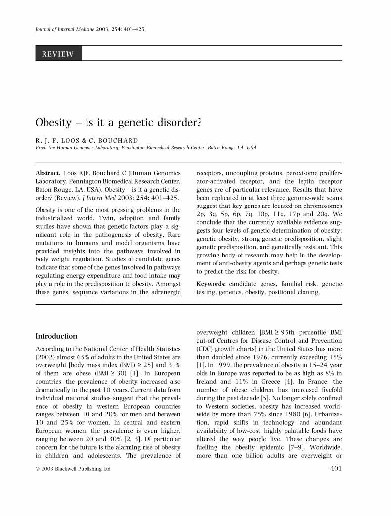
Obesity results from a chronic disruption of the
energy balance. The long-term relations amongst
energy intake, energy expenditure, nutrient parti-
tioning and adipogenesis determine the amount of
energy stored in the body. When energy intake
chronically exceeds energy expenditure and when
fuel partitioning favours lipid storage and carbohy-
drate oxidation, the resulting imbalance causes
expansion of fat cells and increased number of fat
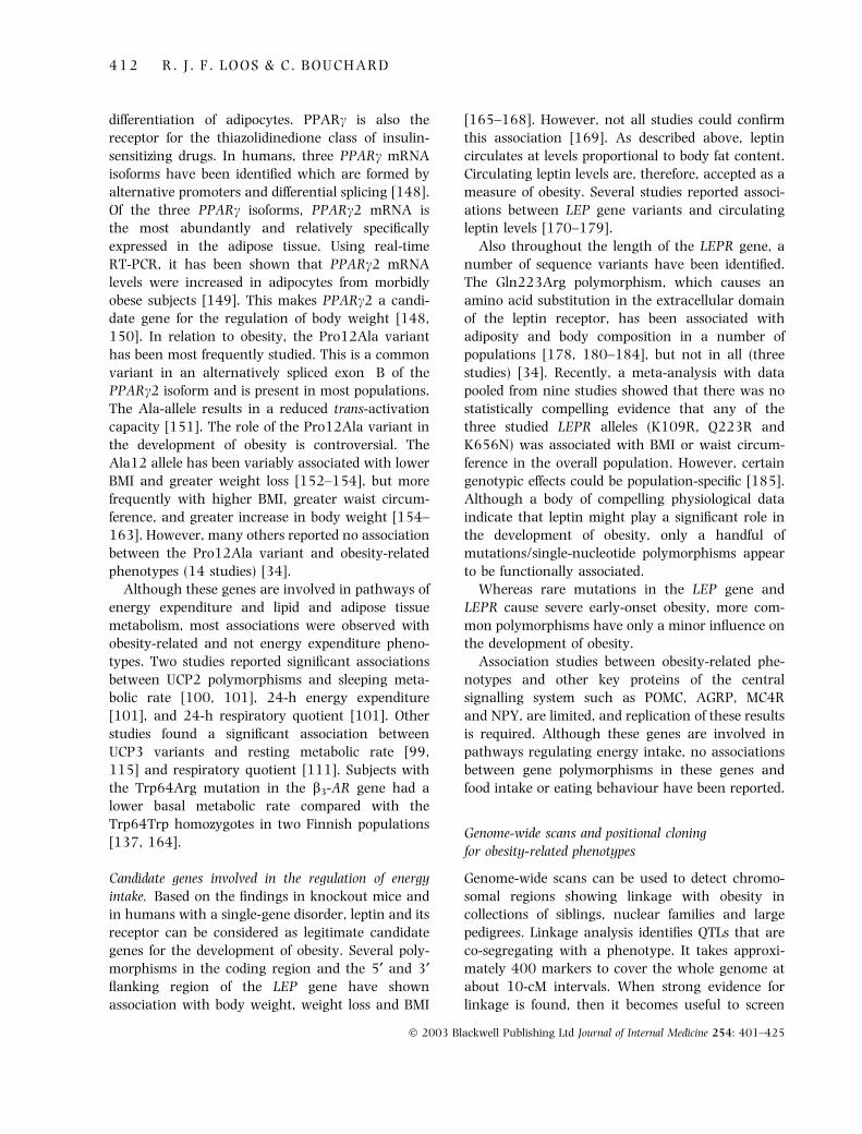
cells. Figure 1 describes a model integrating the
various classes of affectors and the paths leading to
obesity.
It is accepted that lifestyle and environmental
factors have an influence on the determinants
identified in Fig. 1. However, human obesity has
also important genetic correlates that interact with
relevant environmental factors. In other words, the
susceptibility to obesity is partly determined by
genetic factors, but an ‘obesogenic’ environment is
typically necessary for its phenotypic expression.
There is a synergistic relationship between genes
and environment: in the presence of a genetic
predisposition to obesity, the severity of the disease
is largely determined by lifestyle and environmental
conditions. When individuals living in a ‘restrictive’
environment evolve towards an ‘obesogenic’ envi-
ronment, such as that found in industrialized
countries, most are likely to gain weight. However,
those with a high genetic predisposition for obesity
Fig. 1 Diagram of the determinants of positive energy balance
and fat deposition with indication about the sites of action of a
genetic predisposition (from Ref. 233).
4 0 2 R . J . F . L O O S & C . B O U C H A R D
� 2003 Blackwell Publishing Ltd Journal of Internal Medicine 254: 401–425
will gain the most weight, whereas those resistant to
obesity will gain little if any weight.
An example of this gene–environment interaction
effect can be derived from a population apparently
predisposed to obesity and type 2 diabetes, such as
the Pima Indians. Pima Indians living in the
‘restrictive’ environment of the remote Mexican
Sierra Madre mountains have a much lower preval-
ence of obesity and type 2 diabetes mellitus than
those living in the ‘obesogenic’ environment of
Arizona, in the south western United States [17].
Another illustration of the gene–lifestyle interaction
phenomenon relevant to obesity can be found in
experimental studies conducted in pairs of identical
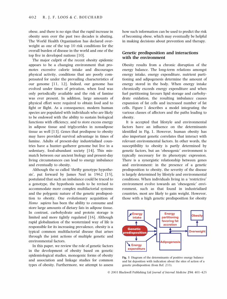
twins. The response to a positive [18] or a negative
[19] energy balance treatment was found to be
heterogeneous amongst twin pairs, but more homo-
geneous within members of the same pair. In a 100-
day overfeeding study [18], 12 male monozygotic
pairs ate a 1000 kcal per day surplus (6 days per
week) over the energy cost for weight maintenance.
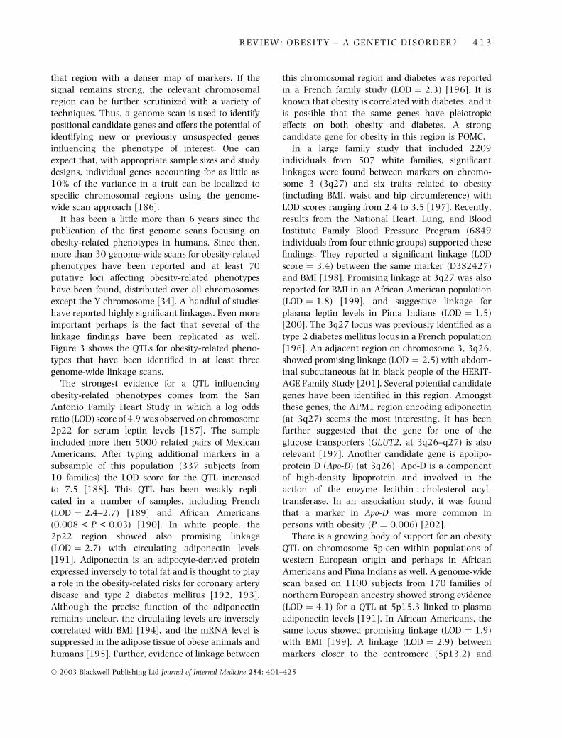
There was at least three times more variance in
response between pairs than within pairs for the
gains in body weight, sum of skinfolds, fat mass and
fat-free mass (Fig. 2). These data demonstrate that
some individuals are more likely to gain fat in an
obesogenic environment than others. Along the
same line, seven pairs of young adult male mono-
zygotic twins completed a negative energy balance
protocol during which they exercised on cycle
ergometers twice a day, nine of 10 days, over a
period of 93 days, whilst being kept on a constant
daily energy and nutrient intake [19]. The exercise
caused a mean total energy deficit above the energy
cost for body weight maintenance of 58 000 kcal.
Again, the data showed large interindividual differ-
ences in weight loss, but only small differences
within pairs (Fig. 2).
These experiments confirm that the magnitude of
a subject’s response to changes in lifestyle or
environmental conditions (from rather restrictive
to more obesogenic and vice versa) depends on a
genetic predisposition thought to be largely inher-
ited.
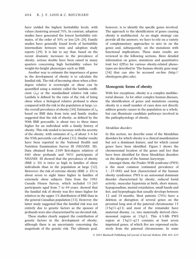
Evidence from genetic epidemiology
Genetic epidemiology has been helpful in defining
the magnitude of the genetic contribution to obesity
in a population perspective. The level of heritability
has been considered in a large number of twin,
adoption and family studies. Heritability is simply
the fraction of the population variation in a trait
(e.g. BMI) that can be explained by genetic trans-
mission. Several studies [20–22] have indicated that
genetic factors account for a substantial portion of
variation in human adiposity [18, 23]. However, the
reported heritability estimates range from as low as
5% to as high as 90%. Plausible explanations for this
wide range of heritability estimates include the way
the study was conducted and the kinds of relatives
upon which they are based. For instance, studies
conducted with monozygotic and dizygotic twins
[24], or monozygotic twins reared apart [20, 25]
Overfeeding Negative energy balance
Changes in body weight Changes in body weight
(kg) (kg)
Ch
ang
es in
bo
dy
wei
gh
t (k
g)
Ch
ang
es in
bo
dy
wei
gh
t (k
g)
r = 0.55
F = 3.4 (P < 0.02)
Twin A Twin A
r = 0.74F = 6.8 (P < 0.01)
Twin B Twin B
0
–2
–4
–6
–8
–8 –6 –4 –2 0
14
12
10
8
6
4
4 6 8 10 12
Fig. 2 Intrapair resemblance in the
response of identical twins to long-
term changes in energy balance.
Left, 12 pairs of identical twins
were submitted to an 84 000 kcal
energy intake surplus over
100 days. Right, seven pairs were
subjected to a negative energy bal-
ance protocol caused by exercise.
The energy deficit was 58 000 kcal
over 93 days. Reproduced from
Bouchard [18, 19].
R E V I E W : O B E S I T Y – A G E N E T I C D I S O R D E R ? 4 0 3
� 2003 Blackwell Publishing Ltd Journal of Internal Medicine 254: 401–425
have yielded the highest heritability levels, with
values clustering around 70%. In contrast, adoption
studies have generated the lowest heritability esti-
mates, of the order of 30% or less [26–28]. Family
studies have generally found levels of heritability
intermediate between twin and adoption study
reports [29]. It is fair to say that, based on the
recent dramatic increases in the prevalence of
obesity, serious doubts have been raised in many
quarters concerning high heritability values for
weight-for-height phenotypes such as BMI.
Another way to estimate the importance of genes
in the development of obesity is to calculate the
familial risk. The risk of becoming obese when a first-
degree relative is overweight or obese can be
quantified using a statistic called the lambda coeffi-
cient (kR) or the standardized relative risk ratio.
Lambda is defined by the ratio of the risk of being
obese when a biological relative proband is obese
compared with the risk in the population at large, i.e.
the overall prevalence of obesity [30]. Estimates of kR
based on BMI data from twin and family studies
suggested that the risk of obesity, as defined by the
90th BMI percentile, is about two to three times
higher for an individual with a family history of
obesity. This risk tended to increase with the severity
of the obesity, with estimates of kR of about 3–6 for
the 95th percentile cut-off point [31]. Similar results
have been reported in the National Health and
Nutrition Examination Survey III (NHANES III).
Data obtained from 2349 first-degree relatives of
840 obese probands and 5851 participants of
NHANES III showed that the prevalence of obesity
(BMI P 30) is twice as high in families of obese
individuals than in the population at large [32].
Moreover, the risk of extreme obesity (BMI P 45) is
about seven to eight times higher in families of
extremely obese subjects. Data from the 1981
Canada Fitness Survey, which included 15 245
participants aged from 7 to 69 years, showed that
the familial risk of obesity was five times higher for
relatives in the upper 1% distribution of BMI than in
the general Canadian population [33]. However, the
latter study suggested that the familial risk was not
entirely due to genetic factors as the spouses of
probands were also characterized by an elevated risk.
These studies clearly support the contribution of
genetic factors in the development of obesity,
although there is an uncertainty concerning the
magnitude of the genetic risk. The ultimate goal,
however, is to identify the specific genes involved.
The approach to the identification of genes causing
obesity is multifaceted. As no single strategy can
provide all the answers, we have to rely on a variety
of complementary approaches to zero in on the
genes and, subsequently, on the mutations with
functional implications. These main results are
reviewed in the following sections. More detailed
information on genes, mutations and quantitative
trait loci (QTLs) for various obesity-related pheno-
types are described in ‘The human obesity gene map’
[34] that can also be accessed on-line (http://
obesitygene.pbrc.edu).
Monogenic forms of obesity
With few exceptions, obesity is a complex multifac-
torial disease. As for other complex human diseases,
the identification of genes and mutations causing
obesity in a small number of cases does not directly
address genetic causes in the population as a whole,
but can illuminate candidate pathways involved in
the pathophysiology of obesity.
Mendelian disorders
In this section, we describe some of the Mendelian
disorders for which obesity is a clinical manifestation
but not a dominant feature, and for which causal
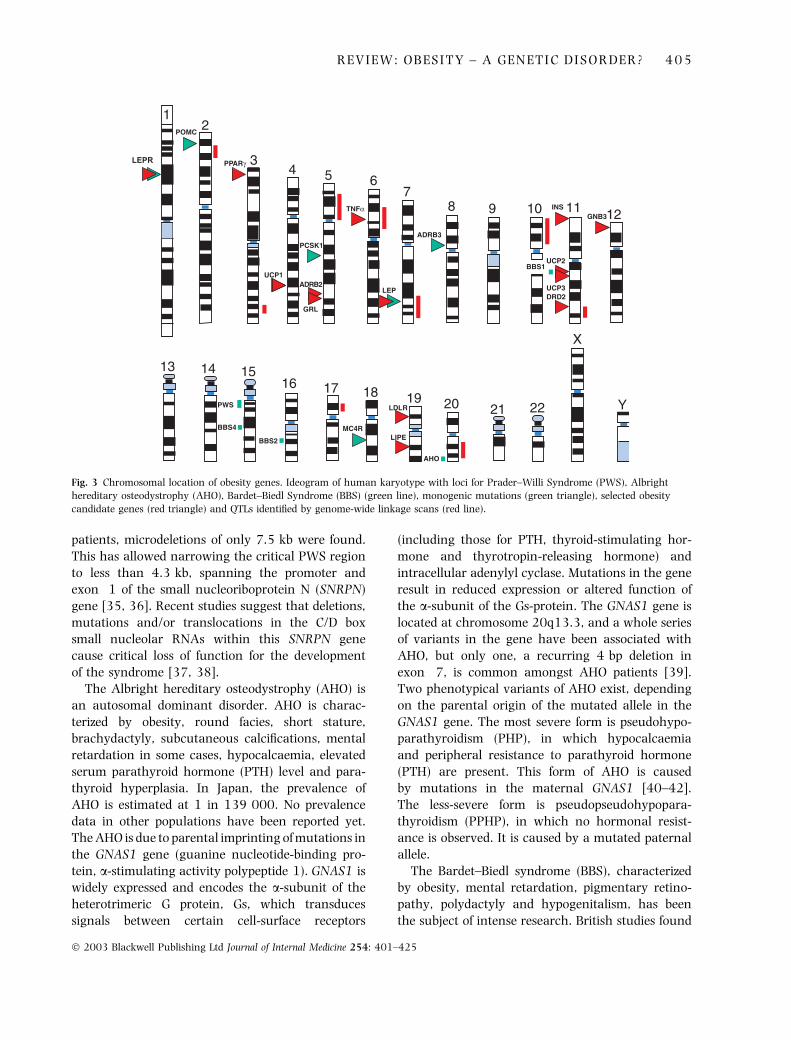
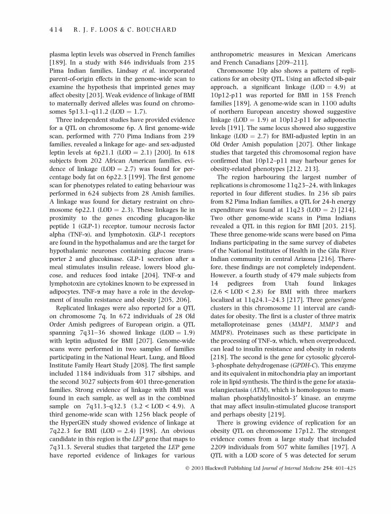
genes have been identified. Figure 3 shows the
chromosomal location of the genes and loci that
have been identified for these Mendelian disorders
on the ideogram of the human karyotype.
Amongst them, the Prader–Willi syndrome (PWS)
is the most common (estimated prevalence of
1 : 25 000) and best characterized of the human
obesity syndromes. PWS is an autosomal dominant
disorder characterized by obesity, reduced foetal
activity, muscular hypotonia at birth, short stature,
hypogonadism, mental retardation, small hands and
feet, and hyperphagia that usually develops between
12 and 18 months. Most patients (70%) have a
deletion or disruption of several genes on the
proximal long arm of the paternal chromosome 15
(15q11–q13) and most of the remainder have
maternal disomy, i.e. two maternally derived chro-
mosomal regions at 15q11. This 4.5-Mb PWS
region at 15q11–q13 contains at least seven
imprinted genes, of which five are expressed exclu-
sively from the paternal chromosome. In some
4 0 4 R . J . F . L O O S & C . B O U C H A R D
� 2003 Blackwell Publishing Ltd Journal of Internal Medicine 254: 401–425
patients, microdeletions of only 7.5 kb were found.
This has allowed narrowing the critical PWS region
to less than 4.3 kb, spanning the promoter and
exon 1 of the small nucleoriboprotein N (SNRPN)
gene [35, 36]. Recent studies suggest that deletions,
mutations and/or translocations in the C/D box
small nucleolar RNAs within this SNRPN gene
cause critical loss of function for the development
of the syndrome [37, 38].
The Albright hereditary osteodystrophy (AHO) is
an autosomal dominant disorder. AHO is charac-
terized by obesity, round facies, short stature,
brachydactyly, subcutaneous calcifications, mental
retardation in some cases, hypocalcaemia, elevated
serum parathyroid hormone (PTH) level and para-
thyroid hyperplasia. In Japan, the prevalence of
AHO is estimated at 1 in 139 000. No prevalence
data in other populations have been reported yet.
The AHO is due to parental imprinting of mutations in
the GNAS1 gene (guanine nucleotide-binding pro-
tein, a-stimulating activity polypeptide 1). GNAS1 is
widely expressed and encodes the a-subunit of the
heterotrimeric G protein, Gs, which transduces
signals between certain cell-surface receptors
(including those for PTH, thyroid-stimulating hor-
mone and thyrotropin-releasing hormone) and
intracellular adenylyl cyclase. Mutations in the gene
result in reduced expression or altered function of
the a-subunit of the Gs-protein. The GNAS1 gene is
located at chromosome 20q13.3, and a whole series
of variants in the gene have been associated with
AHO, but only one, a recurring 4 bp deletion in
exon 7, is common amongst AHO patients [39].
Two phenotypical variants of AHO exist, depending
on the parental origin of the mutated allele in the
GNAS1 gene. The most severe form is pseudohypo-
parathyroidism (PHP), in which hypocalcaemia
and peripheral resistance to parathyroid hormone
(PTH) are present. This form of AHO is caused
by mutations in the maternal GNAS1 [40–42].
The less-severe form is pseudopseudohypopara-
thyroidism (PPHP), in which no hormonal resist-
ance is observed. It is caused by a mutated paternal
allele.
The Bardet–Biedl syndrome (BBS), characterized
by obesity, mental retardation, pigmentary retino-
pathy, polydactyly and hypogenitalism, has been
the subject of intense research. British studies found
12
34 5 6
78 9 10 11 12
13 14 1516 17 18 19 20 21 22
X
Y
POMC
PPARγ
TNFα
ADRB3
INSGNB3
UCP2
UCP3DRD2
LEP
BBS1
PCSK1
ADRB2
GRL
UCP1
PWS
BBS4
BBS2
MC4R
LDLR
LIPE
AHO
LEPR
Fig. 3 Chromosomal location of obesity genes. Ideogram of human karyotype with loci for Prader–Willi Syndrome (PWS), Albright
hereditary osteodystrophy (AHO), Bardet–Biedl Syndrome (BBS) (green line), monogenic mutations (green triangle), selected obesity
candidate genes (red triangle) and QTLs identified by genome-wide linkage scans (red line).
R E V I E W : O B E S I T Y – A G E N E T I C D I S O R D E R ? 4 0 5
� 2003 Blackwell Publishing Ltd Journal of Internal Medicine 254: 401–425
a prevalence of 1 in 160 000, whereas in the
Middle East the occurrence is 1 in 13 500 due to
co-sanguinity. BBS is a genetically heterogeneous
disorder linked to at least seven loci [34]. Although
BBS was originally thought to be a recessive
disorder, the clinical manifestation of some BBS
forms (BBS2, BBS4 and BBS6) requires recessive
mutations at one of the loci plus an additional
mutation at a second locus [43]. The BBS1 locus
was narrowed down to a 1 cM region on 11q13
[44, 45]. By positional cloning, Mykytyn et al. [46]
identified the mutated gene (designated BBS1 gene),
on chromosome 11q34. Several mutations have
been reported, amongst which the methionine-
to-arginine conversion at codon 390 (Met390Arg)
accounts for approximately 80% of all BBS1 muta-
tions [46, 47]. The BBS2 gene maps on 16q21.
Numerous mutations have been found that segre-
gate with the syndrome. However, the function of
the gene has not been determined yet [43, 48].
Interestingly, Katsanis et al. reported that 40% of
the BBS2 patients had also a mutation in another
BBS gene [43]. Mykytyn et al. identified the gene
related to BBS4 (designated BBS4 gene) on chro-
mosome 15q22.3–q23 [49]. The predicted protein
shows strong homology to O-linked N-acetylgluco-
samine transferase (OGT) from several species. In
humans, OGT has been implicated in insulin resist-
ance and may play a role in diabetes [50]. However,
four mutations in the BBS4 gene contributed less
than 3% of affected families. Mutational data from
other BBS genes raised the possibility that BBS4
may participate in triallelic inheritance with BBS2
and BBS1 [51]. BBS6 is caused by mutations in the
MKKS (McKusick–Kaufman Syndrome) gene [52,
53] which encodes for a chaperonin protein that
plays a role in protein integrity [54].
Although causative mutations underlying the
above obesity syndromes have been identified, no
clear mechanistic link between the product of
mutant gene and disordered energy balance has
been defined [55]. Furthermore, attempts to link loci
involved in these obesity syndromes to obesity in
otherwise clinically normal subjects have led to
negative results. Reed et al. investigated the linkage
relations between 17 genetic markers spanning
chromosomal regions implicated in five different
obesity syndromes (PWS, BBS, Cohen, Borjeson and
Wilson–Turner) and the BMI in 44 families segre-
gating for morbid obesity (average BMI of the
proband was 49.8) [56]. Sib-pair linkage analyses
revealed no evidence of linkage between any of the
markers and obesity in these families. These results
suggest that the genetic loci contributing to obesity
in these families are not the same as those involved
in the most common form of obesity. However,
before excluding these chromosomal regions as
potential carriers of loci predisposing to common
obesity, other studies with genetic markers within
the syndrome causative genes must be performed.
Single-gene disorders
Several mutations in human genes with homology
to obesity-causing genes in mice or in genes
involved in the same metabolic pathways have been
identified. In such cases, obesity is the dominant
feature and is largely independent of environmental
factors. Although these cases are rare, they have led
to a better understanding of physiological regulatory
pathways of appetite and energy homeostasis. Find-
ing that mutations in a gene cause similar pheno-
types in rodents and humans underscores the
fundamental and highly conserved nature of the
pathway regulating energy balance. Figure 3 shows
the chromosomal location of these obesity genes on
the ideogram of the human karyotype.
Leptin and leptin receptor. Several mutations that
arose spontaneously in mice have been shown to
cause obesity. The first breakthrough resulted from
innovative parabiosis experiments performed with
obese ob/ob and db/db mice [57]. By surgical cross-
anastomosis between the circulatory systems of
obese and wild-type animals, an adiposity-sensing
pathway, regulated by a circulating hormone and
its cognate receptor, was predicted. This prediction
was confirmed by the cloning of the ob [58] and db
[59] genes and the characterization of their prod-
ucts, known as leptin and leptin receptor, respect-
ively.
Leptin is an endocrine hormone primarily secreted
by adipocytes. Leptin participates in many biological
pathways [60]. One of its key roles is that of
communicating to the brain information on long-
term energy stores. Leptin circulates at levels pro-
portional to body fat content and enters the central
nervous system (CNS) in proportion to its plasma
concentration. The primary site of leptin action is in
the hypothalamus, where its absence triggers a
4 0 6 R . J . F . L O O S & C . B O U C H A R D
� 2003 Blackwell Publishing Ltd Journal of Internal Medicine 254: 401–425
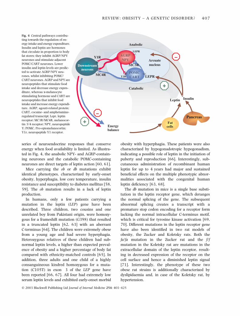
series of neuroendocrine responses that conserve
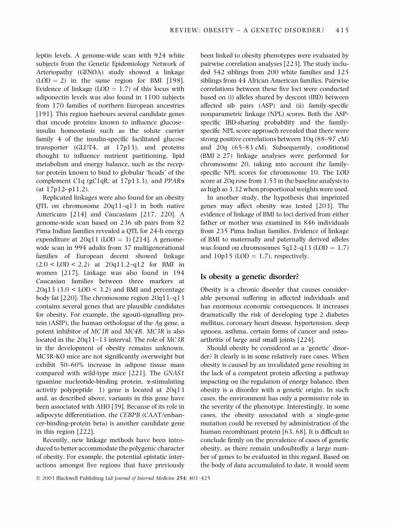
energy when food availability is limited. As illustra-
ted in Fig. 4, the anabolic NPY- and AGRP-contain-
ing neurones and the catabolic POMC-containing
neurones are direct targets of leptin action [60, 61].
Mice carrying the ob or db mutations exhibit
identical phenotypes, characterized by early-onset
obesity, hyperphagia, low core temperature, insulin
resistance and susceptibility to diabetes mellitus [58,
59]. The ob mutation results in a lack of leptin
production.
In humans, only a few patients carrying a
mutation in the leptin (LEP) gene have been
described. Three children, two cousins and one
unrelated boy from Pakistani origin, were homozy-
gous for a frameshift mutation (G398) that resulted
in a truncated leptin [62, 63] with an aberrant
C-terminus [64]. The children were extremely obese
from a young age and had severe hyperphagia.
Heterozygous relatives of these children had sub-
normal leptin levels, a higher than expected preval-
ence of obesity and a higher percentage of body fat
compared with ethnicity-matched controls [65]. In
addition, three adults and one child of a highly
consanguineous kindred homozygous for a muta-
tion (C105T) in exon 3 of the LEP gene have
been reported [66, 67]. All four had extremely low
serum leptin levels and exhibited early-onset morbid
obesity with hyperphagia. These patients were also
characterized by hypogonadotropic hypogonadism,
indicating a possible role of leptin in the initiation of
puberty and reproduction [66]. Interestingly, sub-
cutaneous administration of recombinant human
leptin for up to 4 years had major and sustained
beneficial effects on the multiple phenotypic abnor-
malities associated with the congenital human
leptin deficiency [63, 68].
The db mutation in mice is a single base substi-
tution in the leptin receptor gene, which deranges
the normal splicing of the gene. The subsequent
abnormal splicing creates a transcript with a
premature stop codon encoding for a receptor form
lacking the normal intracellular C-terminus motif,
which is critical for tyrosine kinase activation [69,
70]. Different mutations in the leptin receptor gene
have also been identified in two rat models of
obesity, the Zucker and Koletsky rats. Both the
fa/fa mutation in the Zucker rat and the f/f
mutation in the Koletsky rat are mutations in the
extracellular domain of the leptin receptor, result-
ing in decreased expression of the receptor on the
cell surface and hence a diminished leptin signal
[71]. Interestingly, the phenotype of these two
obese rat strains is additionally characterized by
dyslipidaemia and, in case of the Koletsky rat, by
hypertension.
Fig. 4 Central pathways contribu-
ting towards the regulation of en-
ergy intake and energy expenditure.
Insulin and leptin are hormones
that circulate in proportion to body
fat stores; they inhibit AGRP/NPY
neurones and stimulate adjacent
POMC/CART neurones. Lower
insulin and leptin levels are predic-
ted to activate AGRP/NPY neu-
rones, whilst inhibiting POMC/
CART neurones. AGRP and NPY are
neuropeptides that stimulate food
intake and decrease energy expen-
diture, whereas a-melanocyte
stimulating hormone and CART are
neuropeptides that inhibit food
intake and increase energy expendi-
ture. AGRP, agouti-related protein;
CART, cocaine- and amphetamine-
regulated transcript; Lepr, leptin
receptor; MC3R/MC4R, melanocor-
tin 3/4 receptor; NPY, neuropeptide
Y; POMC, Pro-opiomelanocortin;
Y1r, neuropeptide Y1 receptor.
R E V I E W : O B E S I T Y – A G E N E T I C D I S O R D E R ? 4 0 7
� 2003 Blackwell Publishing Ltd Journal of Internal Medicine 254: 401–425
In humans, only one family to date has been
identified with a leptin receptor (LEPR) mutation
[72]. In homozygotes for the mutation, a truncation
of the receptor before the transmembrane domain
abolishes leptin signalling leading to hyperphagia
and massive obesity soon after birth. The homozy-
gous patients did not spontaneously undergo pub-
erty. These symptoms are similar to those of
individuals with a LEP deficiency. A striking differ-
ence with LEP patients is that homozygous LEPR
patients show significant growth retardation and
hypothalamic hypothyroidism. This suggests that in
humans, even in the absence of circulating leptin,
an intact leptin receptor maintains its capacity to
stimulate certain hypothalamic-releasing factors.
This would explain why the loss of function of the
leptin receptor results in a more severe phenotype
compared with that caused solely by the lack of
leptin. The heterozygous LEPR family members were
overweight or moderately obese. Their leptin levels
were about half those of the homozygous LEPR
patients, indicating that the LEPR mutation may
have a pleiotropic effect.
Following the cloning of the ob and db genes,
considerable attention is now focused on decipher-
ing the neural pathways that mediate the neuroen-
docrine and metabolic responses to the action of
leptin (Fig. 4).
Pro-opiomelanocortin. Pro-opiomelanocortin (POMC)
is post-transcriptionally processed to produce a
number of hormones in the hypothalamic–pituitary–
adrenal axis, such a-melanocyte-stimulating hor-
mone (MSH), adrenocorticotropic hormone (ACTH)
and b-endorphin. These POMC-derived neuropeptides
are physiological agonists of the melanocortin-4
receptor (MC4R). Normally, the hypothalamic syn-
thesis of a-MSH is stimulated by increased leptin lev-
els, and the signal generated by a-MSH at MC4R
promotes energy expenditure and decreases food
intake (Fig. 4) [73].
Mutations in the POMC gene were found in two
patients from unrelated families [74]. One patient
was a compound homozygote for two mutations in
exon 3. A G-to-T transversion in the paternal allele
at nucleotide 7013 (G7013T) resulted in a prema-
ture truncation at codon 79. This truncation of
the protein predicted a complete absence of ACTH,
a-MSH and b-endorphin. In the maternal allele,
a 1 bp deletion (C7133D) caused a frameshift
mutation which was predicted to disrupt the struc-
ture of the receptor-binding core motif of ACTH and
a-MSH and introduced a premature termination at
codon 131. Compound heterozygosity was also
found in the brother of this patient. He died at
7 months of age due to hepatic failure following
severe cholestasis, which was caused by adrenal
insufficiency due to bilateral adrenal hypoplasia. A
second patient was homozygous for a C-to-A trans-
version at nt 3804 (C3804A) in exon 2, which
abolished POMC translation. Both patients had
developed early-onset obesity with hyperphagia
and exhibited a constellation of symptoms reflecting
the lack of neuropeptides derived from the POMC
gene. The lack of a-MSH was responsible for the
ensuing obesity (a result of the absence of the
melanocortin ligand for MC4R) as well as altered
pigmentation and red hair (from the absence of the
ligand for the MC1R). Additionally, the lack of
ACTH, an MC3R agent, led to adrenal deficiency.
In rodents, no naturally occurring mutations in
the POMC gene have been reported. However, the
role of POMC in the central melanocortin pathways
and energy homeostasis has been confirmed by
experiments with transgenic animals with a targeted
deletion of POMC gene [75].
The melanocortin-4 receptor. The importance of the
MC4R gene in the regulation of human body weight
became apparent in 1998 when the first mutations
in the human MC4R gene were described in
some human obese patients [76, 77]. Two groups
reported heterozygous frameshift mutations that
co-segregated in a dominant fashion with severe
early-onset obesity. Subsequently, more than 30
different mutations, including missense, nonsense
and frameshift mutations, in the MC4R gene
have been reported in French, English, German,
American, Italian and Spanish individuals [78–90].
The vast majority of subjects thus far described are
heterozygotes, with only six homozygotes [81, 89]
and one compound heterozygote reported [79].
MC4R is a seven-transmembrane G protein-cou-
pled receptor that activates the cyclic adenosine
monophosphate second-messenger system. Func-
tional studies showed that many of the missense
mutations, and all three frameshift mutations des-
cribed so far, lead to a complete or partial loss of
function of the MC4R gene [78, 80, 87, 89, 91, 92].
In a recent study, Farooqi et al. found that the
4 0 8 R . J . F . L O O S & C . B O U C H A R D
� 2003 Blackwell Publishing Ltd Journal of Internal Medicine 254: 401–425
signalling properties of mutant MC4R receptors, as
determined in cell cultures, correlated with the
severity of obesity [89]. The study included 500
probands with severe childhood obesity of whom 29
had mutations in the MC4R gene. Heterozygotes for
mutations that abolished MC4R signalling had a
higher average BMI than those for mutations
resulting in partial retention of receptor signalling.
Similarly, subjects who were homozygous for such
MC4R mutations had a higher average BMI
than heterozygous carriers of the mutations. These
findings indicate that the obesity resulting from
mutations in MC4R is associated with a co-dominant
mode of inheritance. In this regard, obesity caused by
MC4R mutations is similar to the more common
forms of obesity in comparison with the previously
described single-gene disorder.
The frequency of the MC4R mutations causing
obesity is controversial, with estimates ranging from
under 0.5% [80, 86, 87] to more than 4% [79, 81,
89, 90]. This wide range may be explained by the
difference in prevalence of such mutations in certain
ethnic groups, but it may also reflect variations in
age of onset and severity of obesity.
Carboxypeptidase E and prohormone convertase 1.
Impaired processing of POMC appears to be the
cause of obesity in the fat mouse. The fat mutation
is an inactivating mutation of the gene encoding
for carboxypeptidase E (Cpe) [93]. This enzyme is
required for the post-translational cleavage of C-ter-
minal amino acid residues from many prohormones
and proneuropeptides, such as pro-insulin, pro-
neuropeptide Y, pro-gonadotrophins and POMC. The
fat mutation encodes a nonfunctional Cpe, which
results in the secretion of incompletely processed
precursors that lack the biological activity of the
normal peptides. Consequently, the fat mouse
exhibits multiple endocrine disorders, including
hyperinsulinaemia, infertility and hypoadrenalism,
as well as late-onset obesity, the latter ensuing from
the lack of a-MSH [93].
Cases of human obesity syndromes caused by
mutations of the Cpe gene homologue have not yet
been identified. However, mutations have been
described in the prohormone convertase 1 gene
(PCSK1). Like Cpe, PCSK1 is involved in the post-
translational processing of prohormones and neuro-
peptides. In a woman who had extreme childhood
obesity, abnormal glucose homeostasis, and clinical
manifestations of defective prohormone processing,
Jackson et al. [94] identified compound heterozyg-
osity for two mutations in the PCSK1 gene. One
mutation was a Gly483Arg substitution, whilst the
second was an A-to-C transversion, close to a donor
splice site, which resulted in the skipping of exon 5
and the creation of a premature stop codon within
the catalytic domain of the enzyme. Three of her
four clinically unaffected children had the Gly483Arg
missense mutation, whilst the fourth child had the
splice site mutation.
In rodents, other naturally occurring single-gene
mutations causing obesity have been described,
including in the agouti (Ay), mahogany (mg), tubby
(tub), growth hormone (gh), cholecystokinin A
receptor (Cckar) and lipin1 (Lpin1) genes [34]. So
far, mutations in the human homologues of these
genes have not been associated with obesity in
humans.
It is notable that most of the aforementioned
naturally occurring mutations in rodent models of
obesity concern genes encoding for molecules that
mediate pivotal steps of the same principal regula-
tory pathway. This complex pathway interacts with
a variety of neural circuits of the CNS, implicating a
plethora of other neurotransmitters and neuropep-
tides in the homeostatic weight regulation system,
as illustrated in Fig. 4. The intricate functional
relationship between these pathways appears to
have several critical control points that are presently
being examined. So far, no naturally occurring
mutations that cause obesity due to reduced meta-
bolic rate have been reported.
Polygenic//common forms of obesity
Individuals affected with Mendelian obesity syn-
dromes or single-gene disorders represent only a
small fraction of the obese population and cannot
explain the magnitude of the obesity problem that
industrialized societies are facing today. Obesity is a
complex multifactorial phenotype; interindividual
variation in such phenotypes is thought to result
from the action of multiple genes and environmental
factors.
Human studies to identify the specific genes
involved in common obesity are currently domin-
ated by three types of strategies. A first approach is
the candidate gene approach that relies on the
current understanding of the pathophysiology of
R E V I E W : O B E S I T Y – A G E N E T I C D I S O R D E R ? 4 0 9
� 2003 Blackwell Publishing Ltd Journal of Internal Medicine 254: 401–425
obesity. The candidate genes are selected on the
basis of their perceived role or function in biochemi-
cal pathways related to the regulation of energy
balance or to adipose tissue biology. A second
approach is to perform genome-wide linkage scans
with a view to identifying chromosomal regions of
interest, the so-called QTLs, and eventually genes
within these QTLs. A third approach is based on
tissue-specific gene expression profile comparing
lean versus obese individuals and other informative
samples. The latter will not be reviewed here.
Candidate genes
Currently, positive associations with obesity pheno-
types have been reported for more than 70 genes
[34]. Some of these candidate genes pertain to body
mass, body fat, or fat distribution, whereas others
are retained for their potential contributions to the
regulation of energy intake, energy expenditure, or
nutrient partitioning.
Overall, the results of these association studies
have not been very consistent and when associa-
tions were found, they were relatively weak. Here
we focus on a few selected candidate genes for which
the positive findings have been replicated in inde-
pendent studies. Figure 3 also shows the chromoso-
mal location of these obesity genes on the ideogram
of the human karyotype. A comprehensive review of
all candidate gene studies is beyond the scope of the
present paper. However, the reader is referred to the
paper by Chagnon et al. for a more comprehensive
summary of these genes [34].
Candidate genes involved in the regulation of energy
expenditure. Although all single-gene disorders
causing obesity identified thus far result from
defective genes altering primarily food intake, most
association studies have focused on genes that are
involved in pathways of energy expenditure and
lipid and adipose tissue metabolism.
The mitochondrial uncoupling proteins (UCPs)
have been examined extensively for association with
energy expenditure phenotypes. UCP1 dissipates the
proton electrochemical gradient across the mitoch-
ondrial membrane, thereby uncoupling substrate
oxidation from conversion of adenosine diphosphate
to adenosine triphosphate, leading to generation of
heat. UCP1 plays an important thermogenic role in
brown adipose tissue. The lack of substantial brown
adipose tissue in adult humans suggests that UCP1
has a limited role in thermogenesis in man. A
handful of studies have reported a weak, although
significant, association between polymorphisms in
UCP1 and obesity-related phenotypes such as BMI or
changes in body weight and percentage body fat
[95–99], but no association has been reported by
others (six studies) [34] (to limit the list of refer-
ences, we refer to the paper by Chagnon et al. [34]
for the inventory of the negative studies).
Unlike UCP1, UCP2 is widely expressed, whereas
UCP3 is predominantly expressed in skeletal muscle,
a major tissue contributing to nonshivering therm-
ogenesis in humans. Since the mapping of the UCP2
and UCP3 genes, many researchers have reported
association between allelic variation in these genes
and resting energy expenditure, respiratory quo-
tient, BMI, waist-to-hip ratio and other obesity-
related phenotypes. The coding region of the UCP2
gene exhibits little variability and only one common
genetic variant, Ala55Val, has been identified.
Sleeping metabolic rate, 24-h energy expenditure
and 24-h respiratory quotient were significantly
associated with this UCP2 variant [100, 101].
However, no association was found with age of
onset of obesity [102]. In the 3¢ untranslated region
(3¢UTR), 158 bp downstream of the stop codon, a
prevalent 45 bp insertion polymorphism causes a
change in the stability of the transcript [103].
Significant associations were found between this
UCP2 variant and BMI, body weight, weight gain,
percentage body fat, skinfold thickness, 24-h energy
expenditure and sleeping metabolic rate in most
[100, 101, 103–109] but not all studies (two
studies) [34].
In the UCP3 gene and its promoter, at least 14
polymorphisms have been identified [110].
Although functional analyses showed that several
of these UCP3 polymorphisms resulted in a trun-
cated protein, an increased expression and a reduced
or completely absent activity of the protein, the
results of association studies are inconclusive. Pos-
itive associations were found with BMI, percentage
body fat, waist-to-hip ratio, change in skinfold
thickness, respiratory quotient and resting metabolic
rate [99, 100, 108, 109, 111–115]. However, many
other studies (n ¼ 7) reported no effect of these
UCP3 variants on obesity-related phenotypes [34].
The adrenergic system plays a key role in the
regulation of energy balance through the stimulation
4 1 0 R . J . F . L O O S & C . B O U C H A R D
� 2003 Blackwell Publishing Ltd Journal of Internal Medicine 254: 401–425
of thermogenesis and lipid mobilization. Lipolysis in
fat cells can be stimulated by catecholamines
(amongst other hormones) through b-adrenoceptors
(b-AR), and inhibited through a2-adrenoceptor
(a2-AR) [116].
The b2-adrenoceptor (b2-AR) is the most abun-
dant lipolytic adrenoceptor subtype and is downreg-
ulated in subcutaneous adipose tissue of obese
subjects. Polymorphisms in the b2-AR have been
associated with obesity-related phenotypes in several
ethnic groups [117–125], but these associations
could not be confirmed by others (seven studies)
[34]. The most commonly studied polymorphisms
are Gly16Arg and Gln27Glu. The Gly16 form of the
receptor shows enhanced downregulation in vitro,
whilst the Glu27 allele exhibits attenuated down-
regulation after exposure to b-agonists [126].
The cloning of the human b3-adrenoreceptor
(b3-AR) generated excitement because of its therm-
ogenic, anti-obesity, and antidiabetic properties in
animal models [127]. The human b3-AR gene is
expressed predominantly in infant peripheral brown
adipose tissue. In adults, it is expressed at low levels
in deep fat, such as perirenal and omental, but at
much lower levels in subcutaneous fat [128]. It is
also highly expressed in the gallbladder but to a
lower extent in the colon, suggesting a potential
overall role in the control of lipid metabolism and
triglyceride storage and mobilization in adipose
tissues [128, 129]. A Trp64Arg mutation located
in the first transmembrane domain of the receptor
was first weakly correlated with some obesity and
insulin resistance phenotypes in Pima Indians [130],
and in French [131] and Finnish [132] populations.
Significant associations were subsequently reported
for the same and other obesity-related phenotypes,
such as waist-to-hip ratio, waist circumference, fat
mass, abdominal visceral and subcutaneous fat and
basal metabolic rate [133–138]. In contrast, a large
number of studies failed to replicate these findings in
the same and in other populations [34].
Three meta-analyses of the association between
the Trp64Arg polymorphism and BMI have been
reported [139–141]. The meta-analysis of Allison
et al. pooled 23 studies including 7399 subjects
[139]. Despite the high statistical power, the differ-
ence between the Trp64Trp homozygotes and
Trp64Arg heterozygotes for mean BMI was not
significant (mean difference ¼ 0.19 kg m)2,
P ¼ 0.07). Fujisawa et al. included eight more
studies (31 studies, 9236 subjects) and found a
significantly higher BMI for the Arg-carriers com-
pared with the Trp64Trp homozygotes (mean dif-
ference ¼ 0.30 kg m)2, 95% CI: 0.13–0.47) [140].
In a more recent meta-analysis, data of 27 studies
that included only Japanese subjects (n ¼ 6582)
were pooled [141]. The mean BMI of Arg-carriers
was 0.26 kg m)2 (95% CI: 0.18–0.42, P < 0.01)
higher than in the Trp64Trp homozygotes.
Despite the fact that the functional correlates of
some of these AR polymorphisms (changes in
agonist-promoted downregulation, protein expres-
sion levels, lipolytic sensitivity, basal metabolic rate,
sympathetic nervous system activity) suggest that
they may be important in the aetiology of obesity,
the data indicate that their role, if any, in human
obesity is modest. The end result may depend on
population-specific characteristics such as ethnic
origin, diet, exercise and environmental factors, i.e.
gene–gene or gene–environment interactions.
In this respect, the interactions between polymor-
phisms in the UCP1 gene and the b3-AR have been
studied. The simultaneous presence of the )3826G
UCP1 variant and the Arg64b3-AR variant was
associated with a tendency to gain weight in four
populations [106, 142–144]. Both the a2-AR and
b2-AR genes showed significant interactions with
the glucocorticoid receptor gene in their influence
on total abdominal fat area [145]. Gene–gene
interactions have also been reported amongst the
ARs. Significant interactions have been observed
between the b3-AR and a2-AR genes for total and
subcutaneous body fat in the Quebec Family Study
[146]. After a 20-week endurance training pro-
gramme, the decrease in total and subcutaneous
body fat was more pronounced when subjects were
carriers of both the b2-AR Arg16 and b3-AR Arg64
polymorphisms [106, 144, 147]. In addition, gene–
environment interactions were reported for poly-
morphism in the b2-AR and physical activity. In a
large French cohort, highly significant associations
between body weight, BMI and waist and hip
circumferences, and the Gln27Glu polymorphism
were observed in sedentary subjects, but not in the
physically active [123].
Another candidate gene is peroxisome prolifera-
tor-activated receptor gamma (PPARc). PPARc is a
nuclear receptor, which upon activation with var-
ious natural and synthetic ligands, stimulates the
transcription of genes responsible for growth and
R E V I E W : O B E S I T Y – A G E N E T I C D I S O R D E R ? 4 1 1
� 2003 Blackwell Publishing Ltd Journal of Internal Medicine 254: 401–425
differentiation of adipocytes. PPARc is also the
receptor for the thiazolidinedione class of insulin-
sensitizing drugs. In humans, three PPARc mRNA
isoforms have been identified which are formed by
alternative promoters and differential splicing [148].
Of the three PPARc isoforms, PPARc2 mRNA is
the most abundantly and relatively specifically
expressed in the adipose tissue. Using real-time
RT-PCR, it has been shown that PPARc2 mRNA
levels were increased in adipocytes from morbidly
obese subjects [149]. This makes PPARc2 a candi-
date gene for the regulation of body weight [148,
150]. In relation to obesity, the Pro12Ala variant
has been most frequently studied. This is a common
variant in an alternatively spliced exon B of the
PPARc2 isoform and is present in most populations.
The Ala-allele results in a reduced trans-activation
capacity [151]. The role of the Pro12Ala variant in
the development of obesity is controversial. The
Ala12 allele has been variably associated with lower
BMI and greater weight loss [152–154], but more
frequently with higher BMI, greater waist circum-
ference, and greater increase in body weight [154–
163]. However, many others reported no association
between the Pro12Ala variant and obesity-related
phenotypes (14 studies) [34].
Although these genes are involved in pathways of
energy expenditure and lipid and adipose tissue
metabolism, most associations were observed with
obesity-related and not energy expenditure pheno-
types. Two studies reported significant associations
between UCP2 polymorphisms and sleeping meta-
bolic rate [100, 101], 24-h energy expenditure
[101], and 24-h respiratory quotient [101]. Other
studies found a significant association between
UCP3 variants and resting metabolic rate [99,
115] and respiratory quotient [111]. Subjects with
the Trp64Arg mutation in the b3-AR gene had a
lower basal metabolic rate compared with the
Trp64Trp homozygotes in two Finnish populations
[137, 164].
Candidate genes involved in the regulation of energy
intake. Based on the findings in knockout mice and
in humans with a single-gene disorder, leptin and its
receptor can be considered as legitimate candidate
genes for the development of obesity. Several poly-
morphisms in the coding region and the 5¢ and 3¢flanking region of the LEP gene have shown
association with body weight, weight loss and BMI
[165–168]. However, not all studies could confirm
this association [169]. As described above, leptin
circulates at levels proportional to body fat content.
Circulating leptin levels are, therefore, accepted as a
measure of obesity. Several studies reported associ-
ations between LEP gene variants and circulating
leptin levels [170–179].
Also throughout the length of the LEPR gene, a
number of sequence variants have been identified.
The Gln223Arg polymorphism, which causes an
amino acid substitution in the extracellular domain
of the leptin receptor, has been associated with
adiposity and body composition in a number of
populations [178, 180–184], but not in all (three
studies) [34]. Recently, a meta-analysis with data
pooled from nine studies showed that there was no
statistically compelling evidence that any of the
three studied LEPR alleles (K109R, Q223R and
K656N) was associated with BMI or waist circum-
ference in the overall population. However, certain
genotypic effects could be population-specific [185].
Although a body of compelling physiological data
indicate that leptin might play a significant role in
the development of obesity, only a handful of
mutations/single-nucleotide polymorphisms appear
to be functionally associated.
Whereas rare mutations in the LEP gene and
LEPR cause severe early-onset obesity, more com-
mon polymorphisms have only a minor influence on
the development of obesity.
Association studies between obesity-related phe-
notypes and other key proteins of the central
signalling system such as POMC, AGRP, MC4R
and NPY, are limited, and replication of these results
is required. Although these genes are involved in
pathways regulating energy intake, no associations
between gene polymorphisms in these genes and
food intake or eating behaviour have been reported.
Genome-wide scans and positional cloning
for obesity-related phenotypes
Genome-wide scans can be used to detect chromo-
somal regions showing linkage with obesity in
collections of siblings, nuclear families and large
pedigrees. Linkage analysis identifies QTLs that are
co-segregating with a phenotype. It takes approxi-
mately 400 markers to cover the whole genome at
about 10-cM intervals. When strong evidence for
linkage is found, then it becomes useful to screen
4 1 2 R . J . F . L O O S & C . B O U C H A R D
� 2003 Blackwell Publishing Ltd Journal of Internal Medicine 254: 401–425
that region with a denser map of markers. If the
signal remains strong, the relevant chromosomal
region can be further scrutinized with a variety of
techniques. Thus, a genome scan is used to identify
positional candidate genes and offers the potential of
identifying new or previously unsuspected genes
influencing the phenotype of interest. One can
expect that, with appropriate sample sizes and study
designs, individual genes accounting for as little as
10% of the variance in a trait can be localized to
specific chromosomal regions using the genome-
wide scan approach [186].
It has been a little more than 6 years since the
publication of the first genome scans focusing on
obesity-related phenotypes in humans. Since then,
more than 30 genome-wide scans for obesity-related
phenotypes have been reported and at least 70
putative loci affecting obesity-related phenotypes
have been found, distributed over all chromosomes
except the Y chromosome [34]. A handful of studies
have reported highly significant linkages. Even more
important perhaps is the fact that several of the
linkage findings have been replicated as well.
Figure 3 shows the QTLs for obesity-related pheno-
types that have been identified in at least three
genome-wide linkage scans.
The strongest evidence for a QTL influencing
obesity-related phenotypes comes from the San
Antonio Family Heart Study in which a log odds
ratio (LOD) score of 4.9 was observed on chromosome
2p22 for serum leptin levels [187]. The sample
included more then 5000 related pairs of Mexican
Americans. After typing additional markers in a
subsample of this population (337 subjects from
10 families) the LOD score for the QTL increased
to 7.5 [188]. This QTL has been weakly repli-
cated in a number of samples, including French
(LOD ¼ 2.4–2.7) [189] and African Americans
(0.008 < P < 0.03) [190]. In white people, the
2p22 region showed also promising linkage
(LOD ¼ 2.7) with circulating adiponectin levels
[191]. Adiponectin is an adipocyte-derived protein
expressed inversely to total fat and is thought to play
a role in the obesity-related risks for coronary artery
disease and type 2 diabetes mellitus [192, 193].
Although the precise function of the adiponectin
remains unclear, the circulating levels are inversely
correlated with BMI [194], and the mRNA level is
suppressed in the adipose tissue of obese animals and
humans [195]. Further, evidence of linkage between
this chromosomal region and diabetes was reported
in a French family study (LOD ¼ 2.3) [196]. It is
known that obesity is correlated with diabetes, and it
is possible that the same genes have pleiotropic
effects on both obesity and diabetes. A strong
candidate gene for obesity in this region is POMC.
In a large family study that included 2209
individuals from 507 white families, significant
linkages were found between markers on chromo-
some 3 (3q27) and six traits related to obesity
(including BMI, waist and hip circumference) with
LOD scores ranging from 2.4 to 3.5 [197]. Recently,
results from the National Heart, Lung, and Blood
Institute Family Blood Pressure Program (6849
individuals from four ethnic groups) supported these
findings. They reported a significant linkage (LOD
score ¼ 3.4) between the same marker (D3S2427)
and BMI [198]. Promising linkage at 3q27 was also
reported for BMI in an African American population
(LOD ¼ 1.8) [199], and suggestive linkage for
plasma leptin levels in Pima Indians (LOD ¼ 1.5)
[200]. The 3q27 locus was previously identified as a
type 2 diabetes mellitus locus in a French population
[196]. An adjacent region on chromosome 3, 3q26,
showed promising linkage (LOD ¼ 2.5) with abdom-
inal subcutaneous fat in black people of the HERIT-
AGE Family Study [201]. Several potential candidate
genes have been identified in this region. Amongst
these genes, the APM1 region encoding adiponectin
(at 3q27) seems the most interesting. It has been
further suggested that the gene for one of the
glucose transporters (GLUT2, at 3q26–q27) is also
relevant [197]. Another candidate gene is apolipo-
protein D (Apo-D) (at 3q26). Apo-D is a component
of high-density lipoprotein and involved in the
action of the enzyme lecithin : cholesterol acyl-
transferase. In an association study, it was found
that a marker in Apo-D was more common in
persons with obesity (P ¼ 0.006) [202].
There is a growing body of support for an obesity
QTL on chromosome 5p-cen within populations of
western European origin and perhaps in African
Americans and Pima Indians as well. A genome-wide
scan based on 1100 subjects from 170 families of
northern European ancestry showed strong evidence
(LOD ¼ 4.1) for a QTL at 5p15.3 linked to plasma
adiponectin levels [191]. In African Americans, the
same locus showed promising linkage (LOD ¼ 1.9)
with BMI [199]. A linkage (LOD ¼ 2.9) between
markers closer to the centromere (5p13.2) and
R E V I E W : O B E S I T Y – A G E N E T I C D I S O R D E R ? 4 1 3
� 2003 Blackwell Publishing Ltd Journal of Internal Medicine 254: 401–425
plasma leptin levels was observed in French families
[189]. In a study with 846 individuals from 235
Pima Indian families, Lindsay et al. incorporated
parent-of-origin effects in the genome-wide scan to
examine the hypothesis that imprinted genes may
affect obesity [203]. Weak evidence of linkage of BMI
to maternally derived alleles was found on chromo-
somes 5p13.1–q11.2 (LOD ¼ 1.7).
Three independent studies have provided evidence
for a QTL on chromosome 6p. A first genome-wide
scan, performed with 770 Pima Indians from 239
families, revealed a linkage for age- and sex-adjusted
leptin levels at 6p21.1 (LOD ¼ 2.1) [200]. In 618
subjects from 202 African American families, evi-
dence of linkage (LOD ¼ 2.7) was found for per-
centage body fat on 6p22.3 [199]. The first genome
scan for phenotypes related to eating behaviour was
performed in 624 subjects from 28 Amish families.
A linkage was found for dietary restraint on chro-
mosome 6p22.1 (LOD ¼ 2.3). These linkages lie in
proximity to the genes encoding glucagon-like
peptide 1 (GLP-1) receptor, tumour necrosis factor
alpha (TNF-a), and lymphotoxin. GLP-1 receptors
are found in the hypothalamus and are the target for
hypothalamic neurones containing glucose trans-
porter 2 and glucokinase. GLP-1 secretion after a
meal stimulates insulin release, lowers blood glu-
cose, and reduces food intake [204]. TNF-a and
lymphotoxin are cytokines known to be expressed in
adipocytes. TNF-a may have a role in the develop-
ment of insulin resistance and obesity [205, 206].
Replicated linkages were also reported for a QTL
on chromosome 7q. In 672 individuals of 28 Old
Order Amish pedigrees of European origin, a QTL
spanning 7q31–36 showed linkage (LOD ¼ 1.9)
with leptin adjusted for BMI [207]. Genome-wide
scans were performed in two samples of families
participating in the National Heart, Lung, and Blood
Institute Family Heart Study [208]. The first sample
included 1184 individuals from 317 sibships, and
the second 3027 subjects from 401 three-generation
families. Strong evidence of linkage with BMI was
found in each sample, as well as in the combined
sample on 7q31.3–q32.3 (3.2 < LOD < 4.9). A
third genome-wide scan with 1256 black people of
the HyperGEN study showed evidence of linkage at
7q22.3 for BMI (LOD ¼ 2.4) [198]. An obvious
candidate in this region is the LEP gene that maps to
7q31.3. Several studies that targeted the LEP gene
have reported evidence of linkages for various
anthropometric measures in Mexican Americans
and French Canadians [209–211].
Chromosome 10p also shows a pattern of repli-
cations for an obesity QTL. Using an affected sib-pair
approach, a significant linkage (LOD ¼ 4.9) at
10p12-p11 was reported for BMI in 158 French
families [189]. A genome-wide scan in 1100 adults
of northern European ancestry showed suggestive
linkage (LOD ¼ 1.9) at 10p12-p11 for adiponectin
levels [191]. The same locus showed also suggestive
linkage (LOD ¼ 2.7) for BMI-adjusted leptin in an
Old Order Amish population [207]. Other linkage
studies that targeted this chromosomal region have
confirmed that 10p12–p11 may harbour genes for
obesity-related phenotypes [212, 213].
The region harbouring the largest number of
replications is chromosome 11q23–24, with linkages
reported in four different studies. In 236 sib pairs
from 82 Pima Indian families, a QTL for 24-h energy
expenditure was found at 11q23 (LOD ¼ 2) [214].
Two other genome-wide scans in Pima Indians
revealed a QTL in this region for BMI [203, 215].
These three genome-wide scans were based on Pima
Indians participating in the same survey of diabetes
of the National Institutes of Health in the Gila River
Indian community in central Arizona [216]. There-
fore, these findings are not completely independent.
However, a fourth study of 479 male subjects from
14 pedigrees from Utah found linkages
(2.6 < LOD < 2.8) for BMI with three markers
localized at 11q24.1–24.3 [217]. Three genes/gene
clusters in this chromosome 11 interval are candi-
dates for obesity. The first is a cluster of three matrix
metalloproteinase genes (MMP1, MMP3 and
MMP8). Proteinases such as these participate in
the processing of TNF-a, which, when overproduced,
can lead to insulin resistance and obesity in rodents
[218]. The second is the gene for cytosolic glycerol-
3-phosphate dehydrogenase (GPDH-C). This enzyme
and its equivalent in mitochondria play an important
role in lipid synthesis. The third is the gene for ataxia-
telangiectasia (ATM), which is homologous to mam-
malian phosphatidylinositol-3¢ kinase, an enzyme
that may affect insulin-stimulated glucose transport
and perhaps obesity [219].
There is growing evidence of replication for an
obesity QTL on chromosome 17p12. The strongest
evidence comes from a large study that included
2209 individuals from 507 white families [197]. A
QTL with a LOD score of 5 was detected for serum
4 1 4 R . J . F . L O O S & C . B O U C H A R D
� 2003 Blackwell Publishing Ltd Journal of Internal Medicine 254: 401–425
leptin levels. A genome-wide scan with 924 white
subjects from the Genetic Epidemiology Network of
Arteriopathy (GENOA) study showed a linkage
(LOD ¼ 2) in the same region for BMI [198].
Evidence of linkage (LOD ¼ 1.7) of this locus with
adiponectin levels was also found in 1100 subjects
from 170 families of northern European ancestries
[191]. This region harbours several candidate genes
that encode proteins known to influence glucose–
insulin homeostasis such as the solute carrier
family 4 of the insulin-specific facilitated glucose
transporter (GLUT4, at 17p13), and proteins
thought to influence nutrient partitioning, lipid
metabolism and energy balance, such as the recep-
tor protein known to bind to globular ‘heads’ of the
complement C1q (gC1qR, at 17p13.3), and PPARa(at 17p12–p11.2).
Replicated linkages were also found for an obesity
QTL on chromosome 20q11–q13 in both native
Americans [214] and Caucasians [217, 220]. A
genome-wide scan based on 236 sib pairs from 82
Pima Indian families revealed a QTL for 24-h energy
expenditure at 20q11 (LOD ¼ 3) [214]. A genome-
wide scan in 994 adults from 37 multigenerational
families of European decent showed linkage
(2.0 < LOD < 2.2) at 20q11.2–q12 for BMI in
women [217]. Linkage was also found in 194
Caucasian families between three markers at
20q13 (3.0 < LOD < 3.2) and BMI and percentage
body fat [220]. The chromosome region 20q11–q13
contains several genes that are plausible candidates
for obesity. For example, the agouti-signalling pro-
tein (ASIP), the human orthologue of the Ay gene, a
potent inhibitor of MC3R and MC4R. MC3R is also
located in the 20q11–13 interval. The role of MC3R
in the development of obesity remains unknown.
MC3R-KO mice are not significantly overweight but
exhibit 50–60% increase in adipose tissue mass
compared with wild-type mice [221]. The GNAS1
(guanine nucleotide-binding protein, a-stimulating
activity polypeptide 1) gene is located at 20q13
and, as described above, variants in this gene have
been associated with AHO [39]. Because of its role in
adipocyte differentiation, the CEBPB (CAAT/enhan-
cer-binding-protein beta) is another candidate gene
in this region [222].
Recently, new linkage methods have been intro-
duced to better accommodate the polygenic character
of obesity. For example, the potential epistatic inter-
actions amongst five regions that have previously
been linked to obesity phenotypes were evaluated by
pairwise correlation analyses [223]. The study inclu-
ded 542 siblings from 200 white families and 125
siblings from 44 African American families. Pairwise
correlations between these five loci were conducted
based on (i) alleles shared by descent (IBD) between
affected sib pairs (ASP) and (ii) family-specific
nonparametric linkage (NPL) scores. Both the ASP-
specific IBD-sharing probability and the family-
specific NPL score approach revealed that there were
strong positive correlations between 10q (88–97 cM)
and 20q (65–83 cM). Subsequently, conditional
(BMI ‡ 27) linkage analyses were performed for
chromosome 20, taking into account the family-
specific NPL scores for chromosome 10. The LOD
score at 20q rose from 1.53 in the baseline analysis to
as high as 3.32 when proportional weights were used.
In another study, the hypothesis that imprinted
genes may affect obesity was tested [203]. The
evidence of linkage of BMI to loci derived from either
father or mother was examined in 846 individuals
from 235 Pima Indian families. Evidence of linkage
of BMI to maternally and paternally derived alleles
was found on chromosomes 5q12–q13 (LOD ¼ 1.7)
and 10p15 (LOD ¼ 1.7), respectively.
Is obesity a genetic disorder?
Obesity is a chronic disorder that causes consider-
able personal suffering in affected individuals and
has enormous economic consequences. It increases
dramatically the risk of developing type 2 diabetes
mellitus, coronary heart disease, hypertension, sleep
apnoea, asthma, certain forms of cancer and osteo-
arthritis of large and small joints [224].
Should obesity be considered as a ‘genetic’ disor-
der? It clearly is in some relatively rare cases. When
obesity is caused by an invalidated gene resulting in
the lack of a competent protein affecting a pathway
impacting on the regulation of energy balance, then
obesity is a disorder with a genetic origin. In such
cases, the environment has only a permissive role in
the severity of the phenotype. Interestingly, in some
cases, the obesity associated with a single-gene
mutation could be reversed by administration of the
human recombinant protein [63, 68]. It is difficult to
conclude firmly on the prevalence of cases of genetic
obesity, as there remain undoubtedly a large num-
ber of genes to be evaluated in this regard. Based on
the body of data accumulated to date, it would seem
R E V I E W : O B E S I T Y – A G E N E T I C D I S O R D E R ? 4 1 5
� 2003 Blackwell Publishing Ltd Journal of Internal Medicine 254: 401–425
that cases of genetic obesity could represent at least
5% of the obesity cases and a large percentage of the
severely obese.
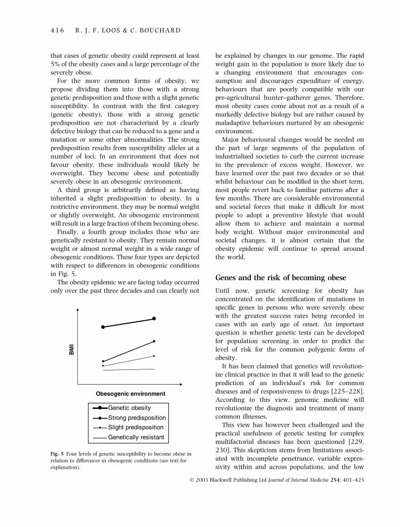
For the more common forms of obesity, we
propose dividing them into those with a strong
genetic predisposition and those with a slight genetic
susceptibility. In contrast with the first category
(genetic obesity), those with a strong genetic
predisposition are not characterized by a clearly
defective biology that can be reduced to a gene and a
mutation or some other abnormalities. The strong
predisposition results from susceptibility alleles at a
number of loci. In an environment that does not
favour obesity, these individuals would likely be
overweight. They become obese and potentially
severely obese in an obesogenic environment.
A third group is arbitrarily defined as having
inherited a slight predisposition to obesity. In a
restrictive environment, they may be normal weight
or slightly overweight. An obesogenic environment
will result in a large fraction of them becoming obese.
Finally, a fourth group includes those who are
genetically resistant to obesity. They remain normal
weight or almost normal weight in a wide range of
obesogenic conditions. These four types are depicted
with respect to differences in obesogenic conditions
in Fig. 5.
The obesity epidemic we are facing today occurred
only over the past three decades and can clearly not
be explained by changes in our genome. The rapid
weight gain in the population is more likely due to
a changing environment that encourages con-
sumption and discourages expenditure of energy,
behaviours that are poorly compatible with our
pre-agricultural hunter–gatherer genes. Therefore,
most obesity cases come about not as a result of a
markedly defective biology but are rather caused by
maladaptive behaviours nurtured by an obesogenic
environment.
Major behavioural changes would be needed on
the part of large segments of the population of
industrialized societies to curb the current increase
in the prevalence of excess weight. However, we
have learned over the past two decades or so that
whilst behaviour can be modified in the short term,
most people revert back to familiar patterns after a
few months. There are considerable environmental
and societal forces that make it difficult for most
people to adopt a preventive lifestyle that would
allow them to achieve and maintain a normal
body weight. Without major environmental and
societal changes, it is almost certain that the
obesity epidemic will continue to spread around
the world.
Genes and the risk of becoming obese
Until now, genetic screening for obesity has
concentrated on the identification of mutations in
specific genes in persons who were severely obese
with the greatest success rates being recorded in
cases with an early age of onset. An important
question is whether genetic tests can be developed
for population screening in order to predict the
level of risk for the common polygenic forms of
obesity.
It has been claimed that genetics will revolution-
ize clinical practice in that it will lead to the genetic
prediction of an individual’s risk for common
diseases and of responsiveness to drugs [225–228].
According to this view, genomic medicine will
revolutionize the diagnosis and treatment of many
common illnesses.
This view has however been challenged and the
practical usefulness of genetic testing for complex
multifactorial diseases has been questioned [229,
230]. This skepticism stems from limitations associ-
ated with incomplete penetrance, variable expres-
sivity within and across populations, and the low
Obesogenic environment
BM
I
Genetic obesity
Strong predisposition
Slight predisposition
Genetically resistant
Genetic obesity
Strong predisposition
Slight predisposition
Genetically resistant
Fig. 5 Four levels of genetic susceptibility to become obese in
relation to differences in obesogenic conditions (see text for
explanation).
4 1 6 R . J . F . L O O S & C . B O U C H A R D
� 2003 Blackwell Publishing Ltd Journal of Internal Medicine 254: 401–425
magnitude of risks typically associated with a
defective genotype in the population [229]. Despite
the fact that many ‘obesity genes’ have been
mapped, high-penetrant high-risk genotypes have
not been found yet for the most common forms of
obesity. Therefore, the contribution of an ‘obesity
gene’ to the development of common obesity will be
difficult to quantify and is likely to be low. For
example, it has been shown that mutations in the
MC4R gene are the most common mutations that
cause severe early-onset obesity. However, the
reported prevalence of these mutations varies by a
factor of 10, ranging from 0.5 to 5.8% [85–87, 92].
This observation suggests variable expressivity
across populations. Furthermore, the penetrance
of the obesity phenotype is incomplete, even in
subjects carrying MC4R mutations predicted to
result in a nonfunctional receptor [86, 92]. The
experience with the MC4R gene, one of the most
promising candidates for a genetic test, strongly
suggests that genetic screening may not be
as straightforward and productive as originally
anticipated. Moreover, gene–gene and gene–
environment interactions in the etiology of obesity
are still poorly understood and are not taken into
account at the moment.
Holtzman and Marteau [229] have argued that
the positive predictive value (i.e. the probability that
the disease will develop in a person with a positive
test result) of a genetic test for a complex disease will
likely be low. However, most studies have examined
the effect of only one gene at a time to estimate
a genetic risk [229–231]. A more productive
approach may be to rely on the use of multiple
genes [232]. Indeed, the prediction for common
diseases, such as obesity, may be improved by
considering multiple genes simultaneously [232].
For instance, in one study on the risk of venous
thrombosis, the concurrent use of a panel of three
genetic tests increased the positive predictive value
at least eightfold compared with only a single test.
This approach may improve the clinical validity of
predictive testing for common multifactorial dis-
eases, such as obesity. However, a major prerequis-
ite for this approach is knowledge about the risk
associated with each genotype under various envi-
ronmental exposures. Multiplex genetic testing
needs to be further evaluated for obesity. However,
we do not have sufficient data to justify widespread
screening for obesity at present.
Conflict of interest statement
Relations with organizations that may constitute
potential conflicts of interest for Claude Bouchard:
Louisiana State University & Pennington Biomedical
Research Center (salary of Executive Director);
Endowed George A. Bray Chair in Nutrition at
Pennington Biomedical Research Center; Almond
Board of California (consultant); Baylor Children’s
Nutrition Research Center of USDA (external advi-
sory board member); Boston Obesity & Nutrition
Research Center of NIH (external advisory board
member); Bristol Myers Squibb (consultant); Bristol
Myers Squibb (unrestricted grant); Institutes for
Pharmaceutical Discovery (scientific advisory board
member); Mars, Inc. (Nutritional Research Council
Member); Pennington Management of Clinical Trials
(member, Board of Directors); Sanofi-synthelabo
(member of advisory board for Rimonabant); The
Cooper Institute for Aerobic Research (scientific
advisory board member); Weight Watchers Interna-
tional (scientific advisory board member).
In addition, as Executive Director of the Penning-
ton Biomedical Research Center, I oversee research
undertaken with grants and contracts from phar-
maceutical companies and food companies. Finally,
I serve also as the institution’s official for grants
from federal agencies (NIH, USDA, US Army, etc.)
and nonprofit foundations (American Heart Associ-
ation, American Diabetes Association, American
Cancer Society, etc.).
Acknowledgements
Ruth Loos is supported by a postdoctoral fellowship
from the American Heart Association, Southeast
affiliate (no. 0325355B). Claude Bouchard is sup-
ported by the George A. Bray Chair in Nutrition, by
an unrestricted grant from Bristol-Meyers Squibb,
a grant from the National Institutes of Health
(HL-45670) and by the Pennington Biomedical
Research Center.
References
1 Pastor P, Makuc DM, Reuben C, Xia H. Chartbook on Trends in
the Health of Americans. Health, United States, 2002. Hyatts-
ville, Maryland: National Center for Health Statistics, 2002.
2 International Obesity Task Force. http://www.obesite.
chaire.ulaval.ca/iotf.htm, 2003.
R E V I E W : O B E S I T Y – A G E N E T I C D I S O R D E R ? 4 1 7
� 2003 Blackwell Publishing Ltd Journal of Internal Medicine 254: 401–425
3 Seidell JC. Prevalence and time trends of obesity in Europe. J
Endocrinol 2002; 25: 816–22.
4 Martinez JA, Kearney JM, Kafatos A, Paquet S, Martinez-
Gonzales MA. Variables independently associated with self-
reported obesity in the European Union. Public Health Nutr
1999; 2: 125–33.
5 Maillard G, Charles MA, Tibult N. Trends in the prevalence
of obesity in the French adult population between 1980 and
1991. Int J Obes Relat Metab Disord 1999; 23: 389–94.
6 Flegal KM, Caroll MD, Kuczmarski RJ, Johnson CL. Over-
weight and obesity in the United States: prevalence and
trends, 1960–1994. Int J Obes Relat Metab Disord 1998; 22:
39–47.
7 Popkin BM. The Nutrition Transition and Obesity in the
Developing World. J Nutr 2001; 131: 871S–873S.
8 Shetty PS. Obesity in children in developing societies: indi-
cator of economic progress or a prelude to a health disaster?
Indian Pediatr 1999; 36: 11–15.
9 Fall CHD. Non-industrialised countries and affluence. Br
Med Bull 2001; 60: 33–55.
10 World Health Organisation. Obesity: Preventing and Managing
the Global Epidemic. Geneva: World Health Organisation,
1998.
11 Hill JO, Peters JC. Environmental contributions to the obesity
epidemic. Science 1998; 280: 1371–4.
12 French SA, Story M, Jeffery RW. Environmental influences
on eating and physical activity. Ann Rev Public Health 2001;
22: 309–35.
13 Unger RH, Orci L. Lipotoxic diseases of nonadipose tissues in
obesity. Int J Obes Relat Metab Disord 2000; 24: S28–32.
14 Eaton SB, Konner M, Shotak M. Stone agers in the fast lane:
chronic degenerative diseases in evolutionary perspective.
Am J Med 1988; 84: 739–49.
15 Neel J. Diabetes mellitus: a ‘thrifty’ genotype rendered det-
rimental by ‘progress’? Am J Hum Genet 1962; 14: 353–62.
16 Stubbs RJ, Harbron CG, Murgatroyd PR, Prentice AM.
Covert manipulation of dietary fat and energy density: effect
on substrate flux and food intake in men eating ad libitum.
Am J Clin Nutr 1995; 62: 316–29.
17 Ravussin E, Valencia ME, Esparza J, Bennett PH, Schulz O.
Effects of a traditional lifestyle on obesity in Pima Indians.
Diabetes Care 1994; 17: 1067–74.
18 Bouchard C, Tremblay A, Despres JP et al. The response to
long-term overfeeding in identical twins. N Engl J Med 1990;
322: 1477–82.
19 Bouchard C, Tremblay A, Despres JP, Theriault G, Nadeau
A, Lupien PJ et al. The response to exercise with constant
energy intake in identical twins. Obes Res 1994; 2: 400–
10.
20 Allison DB, Kaprio J, Korkeila M, Koskenvuo M, Neale M,
Hayakawa K. The heritability of body mass index among an
international sample of monozygotic twins reared apart. Int
J Obes Relat Metab Disord 1996; 20: 501–6.
21 Comuzzie AG, Blangero J, Mahaney MC et al. Genetic and
environmental correlations among hormone levels and
measures of body fat accumulation and topography. J Clin
Endocrinol Metab 1996; 81: 597–600.
22 Price RA, Gottesman II. Body fat in identical twins reared
apart: roles for genes and environment. Behav Genet 1991;
21: 1–7.
23 Stunkard AJ, Sorensen TI, Hanis C et al. An adoption study
of human obesity. N Engl J Med 1986; 314: 193–8.
24 Maes HH, Neale MC, Eaves LJ. Genetic and environmental
factors in relative body weight and human obesity. Behav
Genet 1997; 27: 325–51.
25 Stunkard AJ, Harris JR, Pedersen NL, McClearn GE. The
body mass index of twins who have been reared apart.
N Engl J Med 1990; 322: 1483–7.
26 Sorensen TIA, Holst C, Stunkard AJ. Childhood body mass
index – genetic and familial environmental influences
assessed in a longitudinal adoption study. Int J Obes Relat
Metab Disord 1992; 16: 705–14.
27 Sorensen TIA, Holst C, Stunkard AJ, Skovgaard LT. Corre-
lations of body mass index of adult adoptees and their
biological and adoptive relatives. Int J Obes Relat Metab
Disord 1992; 16: 227–36.
28 Vogler GP, Sorensen TI, Stunkard AJ, Srinivasan MR, Rao
DC. Influences of genes and shared family environment on
adult body mass index assessed in an adoption study by a
comprehensive path model. Int J Obes Relat Metab Disord
1995; 19: 40–45.
29 Rice T, Perusse L, Bouchard C, Rao DC. Familial aggregation
of body mass index and subcutaneous fat measures in the
longitudinal Quebec Family Study. Genet Epidemiol 1999;
16: 316–34.
30 Risch N. Linkage strategies for genetically complex traits. III.
The effect of marker polymorphism on analysis of affected
relative pairs. Am J Hum Genet 1990; 46: 242–53.
31 Allison DB, Faith J, Nathan JS. Risch’s lambda values
for human obesity. Int J Obes Relat Metab Disord 1996; 20:
990–9.
32 Lee JH, Reed DR, Price RA. Familial risk ratios for extreme
obesity: implications for mapping human obesity genes. Int J
Obes Relat Metab Disord 1997; 21: 935–40.
33 Katzmarzyk PT, Perusse L, Rao DC, Bouchard C. Familial
risk of obesity and central adipose tissue distribution in the
general Canadian population. J Clin Endocrinol Metab 1999;
84: 2093–7.
34 Chagnon YC, Rankinen T, Snyder EE, Weisnagel SJ, Perusse
L, Bouchard C. The human obesity gene map: The 2002
update. Obes Res 2003; 11.
35 Ohta T, Gray TA, Rogan PK et al. Imprinting-mutation
mechanisms in Prader-Willi syndrome. Am J Hum Genet
1999; 64: 397–413.
36 Ming JE, Blagowidow N, Knoll JH et al. Submicroscopic
deletion in cousins with Prader-Willi syndrome causes a
grandmatrilineal inheritance pattern: effects of imprinting.
Am J Med Genet 2000; 92: 19–24.
37 Meguro M, Mitsuya K, Nomura N et al. Large-scale
evaluation of imprinting status in the Prader-Willi syn-
drome region: an imprinted direct repeat cluster resem-
bling small nucleolar RNA genes. Hum Mol Genet 2001;
10: 383–94.
38 Gallagher RC, Pils B, Albawi M, Francke U. Evidence for
the role of PWCR1/HBII-85 C/D box small nucleolar RNAs
in Prader-Willi syndrome. Am J Hum Genet 2002; 71: 669–
78.
39 Ishikawa Y, Tajima T, Nakae J et al. Two mutations of the
Gsalpha gene in two Japanese patients with sporadic pseu-
dohypoparathyroidism type Ia. J Hum Genet 2001; 46: 426–
30.
40 Ahmed SF, Dixon PH, Bonthron DT et al. GNAS1 mutational
analysis in pseudohypoparathyroidism. Clin Endocrinol (Oxf)
1998; 49: 525–31.
4 1 8 R . J . F . L O O S & C . B O U C H A R D
� 2003 Blackwell Publishing Ltd Journal of Internal Medicine 254: 401–425
41 Aldred MA, Trembath RC. Activating and inactivating
mutations in the human GNAS1 gene. Hum Mutat 2000;
16: 183–9.
42 Rickard SJ, Wilson LC. Analysis of GNAS1 and Overlapping
Transcripts Identifies the Parental Origin of Mutations in
Patients with Sporadic Albright Hereditary Osteodystrophy
and Reveals a Model System in Which to Observe the Effects
of Splicing Mutations on Translated and Untranslated Mes-
senger RNA. Am J Hum Genet 2003; 72: 961–74.
43 Katsanis N, Ansley SJ, Badano JL et al. Triallelic Inheritance
in Bardet-Biedl Syndrome, a Mendelian Recessive Disorder.
Science 2001; 293: 2256–9.
44 Katsanis N, Lewis RA, Stockton DW et al. Delineation of the
critical interval of Bardet-Biedl syndrome 1 (BBS1) to a small
region of 11q13, through linkage and haplotype analysis of
91 pedigrees. Am J Hum Genet 1999; 65: 1672–9.
45 Young TL, Woods MO, Parfrey PS, Green JS, Hefferton D,
Davidson WS. A founder effect in the newfoundland popu-
lation reduces the Bardet-Biedl syndrome I (BBS1) interval
to 1 cM. Am J Hum Genet 1999; 65: 1680–7.
46 Mykytyn K, Nishimura DY, Searby CC et al. Identification of
the gene (BBS1) most commonly involved in Bardet-Biedl
syndrome, a complex human obesity syndrome. Nat Genet
2002; 31: 435–8.
47 Mykytyn K, Nishimura DY, Searby CC et al. Evaluation of
Complex Inheritance Involving the Most Common Bardet-
Biedl Syndrome Locus (BBS1). Am J Hum Genet 2003; 72:
429–37.
48 Nishimura DY, Searby CC, Carmi R et al. Positional cloning of
a novel gene on chromosome 16q causing Bardet-Biedl syn-
drome (BBS2). Hum Mol Genet 2001; 10: 865–74.
49 Mykytyn K, Braun T, Carmi R et al. Identification of the gene
that, when mutated, causes the human obesity syndrome
BBS4. Nat Genet 2001; 28: 188–91.
50 Konrad RJ, Kudlow JE. The role of O-linked protein glyco-
sylation in beta-cell dysfunction. Int J Mol Med 2002; 10:
535–9.
51 Katsanis N, Eichers ER, Ansley SJ et al. BBS4 is a minor
contributor to Bardet-Biedl syndrome and may also partici-
pate in triallelic inheritance. Am J Hum Genet 2002; 71: 22–
29.
52 Katsanis N, Beales PL, Woods MO et al. Mutations in MKKS
cause obesity, retinal dystrophy and renal malformations
associated with Bardet-Biedl syndrome. Nat Genet 2000; 26:
67–70.
53 Slavotinek AM, Searby CC, Al-Gazali L et al. Mutation ana-
lysis of the MKKS gene in McKusick-Kaufman syndrome and
selected Bardet-Biedl syndrome patients. Hum Genet 2002;
110: 561–7.
54 Slavotinek AM, Stone EM, Mykytyn K et al. Mutations in
MKKS cause Bardet-Biedl syndrome. Nat Genet 2000; 26:
15–16.
55 Katsanis N, Lupski JR, Beales PL. Exploring the molecular
basis of Bardet-Biedl syndrome. Hum Mol Genet 2001; 10:
2293–9.
56 Reed DR, Ding Y, Xu W, Cather C, Price RA. Human
obesity does not segregate with the chromosomal regions
of Prader-Willi, Bardet-Biedl, Cohen, Borjeson or Wilson-
Turner syndromes. Int J Obes Relat Metab Disord 1995; 19:
599–603.
57 Coleman DL. Effects of parabiosis of obese with diabetes and
normal mice. Diabetologia 1973; 9: 294–8.
58 Zhang Y, Proenca R, Maffei M, Barone M, Leopold L,
Friedman JM. Positional cloning of the mouse obese gene
and its human homologue. Nature 1994; 372: 425–32.
59 Tartaglia LA, Dembski M, Weng X et al. Identification and
expression cloning of a leptin receptor, OB-R. Cell 1995; 83:
1263–71.
60 Margetic S, Gazzola C, Pegg GG, Hill RA. Leptin: a review of
its peripheral actions and interactions. Int J Obes Relat Metab
Disord 2002; 26: 1407–33.
61 Porte D, Jr, Baskin DG, Schwartz MW. Leptin and insulin
action in the central nervous system. Nutr Rev 2002; 60:
S20–29.
62 Montague CT, Farooqi IF, Whitehead JP et al. Congenital
leptin deficiency is associated with severe early-onset obesity
in humans. Nature 1995; 387: 903–8.
63 Farooqi IS, Matarese G, Lord GM et al. Beneficial effects of
leptin on obesity, T cell hyporesponsiveness, and neuroen-
docrine/metabolic dysfunction of human congenital leptin
deficiency. J Clin Invest 2002; 110: 1093–103.
64 Rau H, Reaves BJ, O’Rahilly S, Whitehead JP. Truncated
Human Leptin (Delta133) Associated with Extreme
Obesity Undergoes Proteasomal Degradation after Defect-
ive Intracellular Transport. Endocrinology 1999; 140:
1718–23.
65 Farooqi IS, Keogh JM, Kamath S et al. Partial leptin defici-
ency and human adiposity. Nature 2001; 414: 34–35.
66 Strobel A, Issad T, Camoin L, Ozata M, Strosberg AD. A
leptin missense mutation associated with hypogonadism
and morbid obesity. Nat Genet 1998; 18: 213–5.
67 Ozata M, Ozdemir IC, Licinio J. Human leptin deficiency
caused by a missense mutation: multiple endocrine defects,
decreased sympathetic tone, and immune system dysfunc-
tion indicate new targets for leptin action, greater central
than peripheral resistance to the effects of leptin, and
spontaneous correction of leptin-mediated defects. J Clin
Endocrinol Metab 1999; 84: 3686–95.
68 Farooqi IS, Jebb SA, Langmack G et al. Effects of recombin-
ant leptin therapy in a child with congenital leptin defici-
ency. N Engl J Med 1999; 341: 879–84.
69 Chen H, Charlat O, Tartaglia LA et al. Evidence that the
diabetes gene encodes the leptin receptor: identification of a
mutation in the leptin receptor gene in db/db mice. Cell
1996; 84: 491–5.
70 Lee GH, Proenca R, Montez JM et al. Abnormal splicing of the
leptin receptor in diabetic mice. Nature 1996; 379: 632–5.
71 Phillips MS, Liu Q, Hammond HA et al. Leptin receptor
missense mutation in the fatty Zucker rat. Nat Genet 1996;
13: 18–19.
72 Clement K, Vaisse C, Lahlou N et al. A mutation in the
human leptin receptor gene causes obesity and pituitary
dysfunction. Nature 1998; 392: 398–401.
73 Flier SJ, Maratos-Flier E. Obesity and the hypothala-
mus: novel peptides for new pathways. Cell 1998; 92: 437–
40.
74 Krude H, Bierbermann H, Luck W, Horn R, Brabant G,
Gruters A. Severe early-onset obesity, adrenal insufficiency
and red hair pigmentation caused by POMC mutations in
humans. Nat Genet 1998; 19: 155–7.
75 Wardlaw SL. Obesity as a neuroendocrine disease: lessons to
be learned from proopiomelanocortin and melanocortin
receptor mutations in mice and men. J Clin Endocrinol Metab
2001; 86: 1442–6.
R E V I E W : O B E S I T Y – A G E N E T I C D I S O R D E R ? 4 1 9
� 2003 Blackwell Publishing Ltd Journal of Internal Medicine 254: 401–425
76 Yeo GSH, Farooqi IS, Aminian S, Halsall D, Stanhope RG,
O’Rahilly S. A frameshift mutation in MC4R associated with
dominantly inherited human obesity. Nat Genet 1998; 20:
111–2.
77 Vaisse C, Clement K, Guy-Grand B, Froguel P. A frameshift
mutation in human MC4R is associated with a dominant
form of obesity. Nat Genet 1998; 20: 113–4.
78 Sina M, Hinney A, Ziegler A et al. Phenotypes in three
pedigrees with autosomal dominant obesity caused by ha-
ploinsufficiency mutations in the melanocortin-4 receptor
gene. Am J Hum Genet 1999; 65: 1501–7.
79 Vaisse C, Clement K, Durand E, Hercberg S, Guy-Grand B,
Froguel P. Melanocortin-4 receptor mutations are a frequent
and heterogeneous cause of morbid obesity. J Clin Invest
2000; 106: 253–62.
80 Gu W, Tu Z, Kleyn PW et al. Identification and functional
analysis of novel human melanocortin-4 receptor variants.
Diabetes 1999; 48: 635–9.
81 Farooqi IS, Yeo GSH, Keogh JM et al. Dominant and reces-
sive inheritance of morbid obesity associated with melano-
cortin 4 receptor deficiency. J Clin Invest 2000; 106: 271–9.
82 Mergen M, Mergen H, Ozata M, Oner R, Oner C. A novel
melanocortin 4 receptor (MC4R) gene mutation associated
with morbid obesity. J Clin Endocrinol Metab 2001; 86:
3448.
83 Hinney A, Schmidt A, Nottebom K et al. Several mutations
in the melanocortin-4 receptor gene including a nonsense
and a frameshift mutation associated with dominantly
inherited obesity in humans. J Clin Endocrinol Metab 1999;
84: 1483–6.
84 Dubern B, Clement K, Pelloux V et al. Mutational analysis of
melanocortin-4 receptor, agouti-related protein, and alpha-
melanocyte-stimulating hormone genes in severely obese
children. J Pediatr 2001; 139: 204–9.
85 Kobayashi H, Ogawa Y, Shintani M et al. A novel homozy-
gous missense mutation of melanocortin-4 receptor (MC4R)
in a Japanese woman with severe obesity. Diabetes 2002; 51:
243–6.
86 Jacobson P, Ukkola O, Rankinen T et al. Melanocortin 4
receptor sequence variations are seldom a cause of human
obesity: the Swedish obese subjects, the HERITAGE family
study, and a Memphis cohort. J Clin Endocrinol Metab 2002;
87: 4442–6.
87 Miraglia del Giudice E, Cirillo G, Nigro V et al. Low frequency
of melanocortin-4 receptor (MC4R) mutations in a Medi-
terranean population with early-onset obesity. Int J Obes
Relat Metab Disord 2002; 26: 647–51.
88 Marti A, Corbalan MS, Forga L, Martinez JA, Hinney A,
Hebebrand J. A novel nonsense mutation in the melano-
cortin-4 receptor associated with obesity in a Spanish pop-
ulation. Int J Obes Relat Metab Disord 2003; 27: 385–8.
89 Farooqi IS, Keogh JM, Yeo GSH, Lank EJ, Cheetham T,
O’Rahilly S. Clinical Spectrum of Obesity and Mutations in
the Melanocortin 4 Receptor Gene. N Engl J Med 2003; 348:
1085–95.
90 Branson R, Potoczna N, Kral JG, Lentes KU, Hoehe MR,
Horber FF. Binge eating as a major phenotype of melano-
cortin 4 receptor gene mutations. N Engl J Med 2003; 348:
1096–103.
91 Ho G, MacKenzie RG. Functional characterization of muta-
tions in melanocortin-4 receptor associated with human
obesity. J Biol Chem 1999; 274: 35 816–22.
92 Yeo GSH, Lank EJ, Farooqi IF, Keogh JM, Challis BG,
O’Rahilly S. Mutations in the human melanocortin-4
receptor gene associated with severe familial obesity disrupts
receptor function through multiple molecular mechanisms.
Hum Mol Genet 2003; 12: 561–74.
93 Naggert JK, Fricker LD, Varlamov O et al. Hyperproinsulin-
aemia in obese fat/fat mice associated with a carboxypepti-
dase E mutation which reduces enzyme activity. Nat Genet
1995; 10: 135–42.
94 Jackson RS, Creemers JW, Ohagi S et al. Obesity and
impaired prohormone processing associated with mutations
in the human prohormone convertase 1 gene. Nat Genet
1998; 16: 303–6.
95 Oppert JM, Vohl MC, Chagnon M et al. DNA polymor-
phism in the uncoupling protein (UCP) gene and human
body fat. Int J Obes Relat Metab Disord 1994; 18: 526–31.
96 Fumeron F, Durack-Brown I, Betoulle D et al. Polymor-
phisms of uncoupling protein (UCP) and beta 3 adrenore-
ceptor genes in obese people submitted to a low calorie diet.
Int J Obes Relat Metab Disord 1996; 20: 1051–4.
97 Kogure A, Yoshida T, Sakane N, Umekawa T, Takakura Y,
Kondo M. Synergic effect of polymorphisms in uncoupling
protein 1 and b3-adrenergic receptor genes on weight loss in
obese Japanese. Diabetologia 1998; 41: 1399.
98 Heilbronn LK, Kind KL, Pancewicz E, Morris AM, Noakes M,
Clifton PM. Association of -3826 G variant in uncoupling
protein-1 with increased BMI in overweight Australian
women. Diabetologia 2000; 43: 242–4.
99 Ukkola O, Tremblay A, Sun G, Chagnon YC, Bouchard C.
loci and the response to long-term overfeeding. Eur J Clin
Nutr 2001; 55: 1008–15.
100 Walder K, Norman RA, Schrauwen P et al. Association
between uncoupling protein polymorphisms (UCP2-UCP3)
and energy metabolism/obesity in Pima indians. Hum Mol
Genet 1998; 7: 1431–5.
101 Astrup A, Toubro S, Dalgaard LT, Urhammer SA, Sorensen
TI, Pedersen O. Impact of the v/v 55 polymorphism of the
uncoupling protein 2 gene on 24-h energy expenditure and
substrate oxidation. Int J Obes Relat Metab Disord 1999; 23:
1030–4.
102 Urhammer SA, Dalgaard LT, Sorensen TI et al. Mutational
analysis of the coding region of the uncoupling protein 2
gene in obese NIDDM patients: impact of a common amino
acid polymorphism on juvenile and maturity onset forms of
obesity and insuline resistance. Diabetologia 1997; 40:
1227–30.
103 Evans D, Wolf AM, Nellessen U et al. Association between
polymorphisms in candidate genes and morbid obesity. Int J
Obes Relat Metab Disord 2001; 25: S19–21.
104 Cassell PG, Neverova M, Janmohamed S et al. An uncoup-
ling protein 2 gene variant is associated with a raised body
mass index but not Type II diabetes. Diabetologia 1999; 42:
688–92.
105 Yanovski JA, Diament AL, Sovik KN et al. Associations
between uncoupling protein 2, body composition, and rest-
ing energy expenditure in lean and obese African American,
white, and Asian children. Am J Clin Nutr 2000; 71: 1405–
20.
106 Evans D, Minouchehr S, Hagemann G et al. Frequency of and
interaction between polymorphisms in the beta3-adrenergic
receptor and in uncoupling proteins 1 and 2 and obesity in
Germans. Int J Obes 2000; 24: 1239–45.
4 2 0 R . J . F . L O O S & C . B O U C H A R D
� 2003 Blackwell Publishing Ltd Journal of Internal Medicine 254: 401–425
107 Nordfors L, Heimburger O, Lonnqvist F et al. Fat tissue
accumulation during peritoneal dialysis is associated with a
polymorphism in uncoupling protein 2. Kidney Int 2000; 57:
1713–9.
108 Lanouette CM, Giacobino J-P, Perusse L et al. Association
between uncoupling protein 3 gene and obesity-related
phenotypes in the Quebec Family Study. Mol Med 2001; 7:
433–41.
109 Lanouette CM, Chagnon YC, Rice T et al. Uncoupling protein
3 gene is associated with body composition changes with
training in HERITAGE study. J Appl Physiol 2002; 92:
1111–8.
110 Argyropoulos G, Harper ME. Molecular biology of thermo-
regulation: uncoupling proteins and thermoregulation.
J Appl Physiol 2002; 92: 2187–98.
111 Argyropoulos G, Brown AM, Willi SM et al. Effects of
mutations in the human uncoupling protein 3 gene on
the respiratory quotient and fat oxidation in severe
obesity and type 2 diabetes. J Clin Invest 1998; 102: 1345–
51.
112 Buemann B, Schiering B, Toubro S et al. The association
between the val/val-55 polymorphism of the uncoupling
protein 2 gene and excercise efficiency. Int J Obes Relat Metab
Disord 2001; 25: 467–71.
113 Otabe S, Clement K, Dina C et al. A genetic variation in the
5¢ flanking region of the UCP3 gene is associated with body
mass index in humans in interaction with physical activity.
Diabetologia 2000; 43: 245–9.
114 Cassell PG, Saker PJ, Huxtable SJ et al. (UCP3) gene influ-
ences fat distribution in women of European and Asian
origin. Diabetologia 3 2000; 1558–64.
115 Kimm SY, Glynn NW, Aston CE et al. Racial differences in
the relation between uncoupling protein genes and resting
energy expenditure. Am J Clin Nutr 2002; 75: 714–9.
116 Blaak EE, van Baak MA, Kemerinck GJ, Pakbiers MT, Hei-
dendal GA, Saris WHM. Beta-adrenergic stimulation of
skeletal muscle metabolism in relation to weight reduction
in obese men. Am J Physiol 1994; 267: E316–22.
117 Lin RCY, Ericsson JO, Benjafield AV, Morris BJ. Associ-
ation of beta2-adrenoceptor Gln27Glu variant with body
weight but not hypertension. Am J Hypertens 2001; 14:
1201–4.
118 Hellstrom L, Large V, Reynisdottir S, Wahrenberg H, Arner
P. The different effects of a Gln27Glu b2-adrenoreceptor
gene polymorphism on obesity in males and females. J Intern
Med 1999; 245: 253–9.
119 Yamada K, Ishiyama-Shigemoto S, Ichikawa F et al. Poly-
morphism in the 5¢-leader cistron of the beta2-adrenergic
receptor gene associated with obesity and type 2 diabetes.
J Clin Endocrinol Metab 1999; 84: 1754–7.
120 Mori Y, Kim-Motoyama H, Ito Y et al. The Gln27Glu beta2-
adrenergic receptor variant is associated with obesity due to
subcutaneous fat accumulation in Japanese men. Biochem
Biophys Res Comm 1999; 258: 138–40.
121 Large V, Hellstrom L, Reynisdottir S et al. Human
beta-2 adrenoceptor gene polymorphisms are highly fre-
quent in obesity and associate with altered adipocyte
beta-2 adrenoceptor function. J Clin Invest 1997; 100:
3005–13.
122 Ishiyama-Shigemoto S, Yamada K, Yuan X, Ichikawa F,
Nonaka K. Association of polymorphisms in the beta-
2-adrenergic receptor gene with obesity, hypertriglyce-
ridaemia, and diabetes mellitus. Diabetologia 1999; 42:
98–101.
123 Meirhaeghe A, Helbecque N, Cottel D, Amouyel P. Beta2-
adrenoceptor gene polymorphism, body weight, and phys-
ical activity. Lancet 1999; 353: 896.
124 Ehrenborg E, Skogsberg RG, Large V et al. Polymorphism in
the beta2) adrenoceptor gene is associated with increased
body weight and dyslipoproteinaemia involving triglyceride-
rich lipoproteins. J Intern Med 2000; 247: 651–6.
125 Macho-Azcarate T, Marti A, Gonzales A, Martinez JA, Ibanez
J. Gln27Glu polymorphism in the beta2 adrenergic receptor
gene and lipid metabolism during exercise in obese women.
Int J Obes 2002; 26: 1434–41.
126 Green SA, Turki J, Bejarano P, Hall IP, Liggett SB. Influence
of beta 2-adrenergic receptor genotypes on signal trans-
duction in human airway smooth muscle cells. Am J Respir
Cell Mol Biol 1995; 13: 25–33.
127 Emorine LJ, Marullo S, Briend-Suttern MM et al. Molecular
characterization of the human beta 3-adrenergic receptor.
Science 1989; 245: 1118–21.
128 Krief S, Lonnqvist F, Raimbault S et al. Tissue distribution of
beta 3-adrenergic receptor mRNA in man. J Clin Invest
1993; 91: 344–9.
129 Lafontan M. Differential recruitment and differential
regulation by physiological amines of fat cell beta-1, beta-
2 and beta-3 adrenergic receptors expressed in native fat
cells and in transfected cell lines. Cell Signal 1994; 6:
363–92.
130 Walston J, Silver K, Bogardus C et al. Time of Onset of Non-
Insulin-Dependent Diabetes Mellitus and Genetic Variation
in the beta3-Adrenergic-Receptor Gene. N Engl J Med 1995;
333: 343–7.
131 Clement K, Vaisse C, Manning BS et al. Genetic variation in
the b3-adrenergic receptor and an increased capacity to
gain weight in patients with morbid obesity. N Engl J Med
1995; 333: 352–4.
132 Widen E, Lehto M, Kanninen T, Walston J, Shuldiner AR,
Groop LC. Association of a Polymorphism in the beta3-
Adrenergic-Receptor Gene with Features of the Insulin
Resistance Syndrome in Finns. N Engl J Med 1995; 333:
348–52.
133 Urhammer SA, Hansen T, Borch-Johnsen K, Pedersen O.
Studies of the synergistic effect of the Trp/Arg64 poly-
morphism of the b3-adrenergic receptor gene and the -3826
A->G variant of the uncoupling protein-1 gene on features
of obesity and insulin resistance in a populatio-based sample
of 379 young Danish subjects. J Clin Endocrinol Metab 2000;
85: 3151–4.
134 Kim-Motoyama H, Yasuda K, Yamaguchi T et al. A muta-
tion of the beta 3-adrenergic receptor is associated with
visceral obesity but decreased serum triglyceride. Diabetolo-
gia 1997; 40: 469–72.
135 Festa A, Krugluger W, Shnawa N, Hopmeier P, Haffner SM,
Schernthaner G. Trp64Arg polymorphism of the beta3-
adrenergic receptor gene in pregnancy: association with
mild gestational diabetes mellitus. J Clin Endocrinol Metab
1999; 84: 1695–9.
136 Thomas GN, Tomlinson B, Chan JC, Young RP, Critchley JA.
The Trp64Arg polymorphism of the beta3-adrenergic
receptor gene and obesity in Chinese subjects with compo-
nents of the metabolic syndrome. Int J Obes Relat Metab
Disord 2000; 24: 545–51.
R E V I E W : O B E S I T Y – A G E N E T I C D I S O R D E R ? 4 2 1
� 2003 Blackwell Publishing Ltd Journal of Internal Medicine 254: 401–425
137 Sipilainen R, Uusitupa M, Heikkinen S, Rissanen A, Laakso
M. Polymorphism of the beta3-adrenergic receptor gene
affects basal metabolic rate in obese Finns. Diabetes 1997;
46: 77–80.
138 Corbalan MS, Marti A, Forga L, Martinez-Gonzales MA,
Martinez JA. The risk of obesity and the Trp64Arg poly-
morphism of the beta(3)-adrenergic receptor: effect modifi-
cation by age. Ann Nutr Metab 2002; 46: 152–8.
139 Allison DB, Heo M, Faith MS, Pietrobelli A. Meta-analysis of
the association of the Trp64Arg polymorphism in the beta3
adrenergic receptor with body mass index. Int J Obes Relat
Metab Disord 1998; 22: 559–66.
140 Fujisawa T, Ikegami H, Kawaguchi Y, Ogihara T. Meta-
analysis of the association of Trp64Arg polymorphism of
beta3-adrenergic receptor gene with body mass index. J Clin
Endocrinol Metab 1998; 83: 2441.
141 Kurokawa N, Nakai K, Kameo S, Liu ZM, Satoh H.
Association of BMI with the beta3-adrenergic receptor gene
polymorphism in Japanese: meta-analysis. Obes Res 2001; 9:
741.
142 Clement K, Ruiz J, Cassard-Doulcier AM et al. Additive effect
of A–>G (-3826) variant of the uncoupling protein gene and
the Trp64Arg mutation of the beta 3-adrenergic receptor
gene on weight gain in morbid obesity. Int J Obes Relat Metab
Disord 1996; 20: 1062–6.
143 Fogelholm M, Valve R, Kukkonen-Harjula K et al. Additive
effects of the mutations in beta3-adrenergic receptor and
uncoupling protein-1 genes on weight loss and weight
maintenance in Finnish women. J Clin Endocrinol Metab
1998; 83: 4246–50.
144 Sivenius K, Valve R, Lindi V, Niskanen LK, Laakso M,
Uusitupa M. Synergistic effect of polymorphisms in
uncoupling protein 1 and beta3-adrenergic receptor genes
on long-term body weight change in Finnish type 2 diabetic
and non-diabetic control subjects. Int J Obes Relat Metab
Disord 2000; 24: 514–9.
145 Ukkola O, Perusse L, Chagnon YC, Despres JP, Bouchard
C. Interactions among the glucocorticoid receptor, lipo-
protein lipase and adrenergic receptor genes and abdom-
inal fat in the Quebec Family Study. Int J Obes 2001; 25:
1332–9.
146 Ukkola O, Rankinen T, Weisnagel SJ et al. Interactions
among the alpha2-, beta2-, and beta3-adrenergic receptor
genes and obesity-related phenotypes in the Quebec Family
Study. Metabolism 2000; 49: 1063–70.
147 Ukkola O, Rankinen T, Rice T et al. Interactions among the
beta2- and beta3- adrenergic receptor genes and total body
fat and abdominal fat level in the HERITAGE Family Study.
Int J Obes Relat Metab Disord 2003; 27: 389–93.
148 Auwerx J. PPARgamma, the ultimate thrifty gene. Diabeto-
logia 1999; 42: 1033–49.
149 Vidal-Puig AJ, Considine RV, Jimenez-Linan M et al. Perox-
isome Proliferator-activated Receptor Gene Expression in
Human Tissues. Effects of Obesity, Weight Loss, and Regu-
lation by Insulin and Glucocorticoids. J Clin Invest 1997; 99:
2416–22.
150 Sewter C, Blows F, Considine R, Vidal-Puig A, O’Rahilly S.
Differential effects of adiposity on peroxisomal proliferator-
activated receptor gamma1 and gamma2 messenger
ribonucleic acid expression in human adipocytes. J Clin
Endocrinol Metab 2002; 87: 4203–7.
151 Masugi J, Tamori Y, Mori H, Koike T, Kasuga M. Inhibitory
effect of a proline-to-alanine substitution at codon 12 of
peroxisome proliferator-activated receptor-gamma 2 on
thiazolidinedione-induced adipogenesis. Biochem Biophys Res
Commun 2000; 268: 178–82.
152 Deeb SS, Fajas L, Nemoto M et al. A Pro12Ala substitution
in PPARgamma2 associated with decreased receptor activ-
ity, lower body mass index and improved insulin sensitivity.
Nat Genet 1998; 20: 284–7.
153 Lindi VI, Uusitupa MIJ, Lindstrom J et al. Association of the
Pro12Ala Polymorphism in the PPAR-gamma2 Gene With
3-Year Incidence of Type 2 Diabetes and Body Weight
Change in the Finnish Diabetes Prevention Study. Diabetes
2002; 51: 2581–6.
154 Ek J, Urhammer SA, Sorensen TI, Andersen T, Auwerx J,
Pedersen O. Homozygosity of the Pro12Ala variant of the
peroxisome proliferation-activated receptor-gamma2
(PPAR-gamma2): divergent modulating effects on body
mass index in obese and lean Caucasian men. Diabetologia
1999; 42: 892–5.
155 Beamer BA, Yen CJ, Andersen RE et al. Association of the
Pro12Ala variant in the peroxisome proliferator-activated
receptor-gamma2 gene with obesity in two Caucasian pop-
ulations. Diabetes 1998; 47: 1806–8.
156 Valve R, Sivenius K, Miettinen R et al. Two polymorphisms
in the peroxisome proliferator-activated receptor-gamma
gene are associated with severe overweight among obese
women. J Clin Endocrinol Metab 1999; 84: 3708–12.
157 Vaccaro O, Mancini FP, Ruffa G, Sabatino L, Colantuoni V,
Riccardi G. Pro12Ala mutation in the peroxisome prolifer-
ator-activated receptor gamma2 (PPARgamma2) and
severe obesity: a case–control study. Int J Obes Relat Metab
Disord 2000; 24: 1195–9.
158 Meirhaeghe A, Fajas L, Helbecque N, Cottel D, Auwerx J,
Amouyel P. Impact of the peroxisome proliferator activated
receptor gamma2 Pro12Ala polymorphism on adiposity,
lipids and non-insulin-dependent diabetes mellitus. Int J Obes
Relat Metab Disord 2000; 24: 195–9.
159 Nicklas BJ, van Rossum EFC, Berman DM, Ryan AS, Dennis
KE, Shuldiner AR. Genetic variation in the peroxisome
proliferator-activated receptor-gamma2 gene (Pro12Ala)
affects metabolic responses to weight loss and subsequent
weight regain. Diabetes 2001; 50: 2172–6.
160 Hasstedt SJ, Ren QF, Teng K, Elbein SC. Effect of the per-
oxisome proliferator-activated receptor-gamma2 Pro12Ala
variant on obesity, glucose homeostasis, and blood pressure
in members of familial type 2 diabetic kindreds. J Clin End-
ocrinol Metab 2001; 86: 536–41.
161 Lindi V, Sivenius K, Niskanen LK, Laakso M, Uusitupa M.
Effect of the Pro12Ala polymorphism of the PPAR-gamma2
gene on long-term weight change in Finnish non-diabetic
subjects. Diabetologia 2001; 44: 925–6.
162 Gonzales Sanchez JL, Serrano Rios M, Fernandez Perez C,
Laakso M, MartinezLarrad MT. Effect of the Pro12Ala
polymorphism of the peroxisome proliferator-activated
receptor gamma-2 gene on adiposity, insulin sensitivity and
lipid profile in the Spanish population. Eur J Endocrinol
2002; 147: 495–501.
163 Cole SA, Mitchell BD, Hsueh WC et al. The Pro12Ala variant
of peroxisome proliferator-activated receptor-gamma2
(PPAR-gamma2) is associated with measures of obesity in
4 2 2 R . J . F . L O O S & C . B O U C H A R D
� 2003 Blackwell Publishing Ltd Journal of Internal Medicine 254: 401–425
Mexican Americans. Int J Obes Relat Metab Disord 2000; 24:
522–4.
164 Valve R, Heikkinen S, Rissanen A, Laakso M, Uusitupa M.
Synergistic effect of polymorphisms in uncoupling protein 1
and b3-adrenergic receptor genes on basal metabolic rate in
obese Finns. Diabetologia 1998; 41: 357–61.
165 Oksanen L, Ohman M, Heiman ML et al. Markers for the
gene ob and serum leptin levels in human morbid obesity.
Hum Genet 1997; 99: 559–64.
166 Shintani M, Ikegami H, Yamato E et al. A novel microsat-
ellite polymorphism in the human OB gene: a highly poly-
morphic marker for linkage analysis. Diabetologia 1996; 39:
1398–401.
167 Butler MG, Hedges L, Hovis CL, Feurer ID. Genetic variants
of the human obesity (OB) gene in subjects with and without
Prader-Willi syndrome: comparison with body mass index
and weight. Clin Genet 1998; 54: 385–93.
168 Li WD, Reed DR, Lee JH et al. Sequence variants in the 5¢flanking region of the leptin gene are associated with obesity
in women. Ann Hum Genet 1999; 63: 227–34.
169 Karvonen MK, Pesonen U, Heinonen P et al. Identification of
new sequence variants in the leptin gene. J Clin Endocrinol
Metab 1998; 83: 3239–42.
170 Hager J, Clement K. A polymorphism in the 5¢ untrans-
lated region of the human ob gene is associated with low
leptin levels. Int J Obes Relat Metab Disord 1998; 22: 200–5.
171 Mammes O, Betoulle D, Aubert R et al. Novel polymor-
phisms in the 5¢ region of the LEP gene: association with
leptin levels and response to low-calorie diet in human
obesity. Diabetes 1998; 47: 487–9.
172 Le Stunff C, Le Bihan C, Schork NJ, Bougneres P. A common
promoter variant of the leptin gene is associated with
changes in the relationship between serum leptin and fat
mass in obese girls. Diabetes 2000; 49: 2196–200.
173 Hoffstedt J, Eriksson P, Mottagui-Tabar S, Arner P. A poly-
morphism in the leptin promoter region (-2548 G/A) influ-
ences gene expression and adipose tissue secretion of leptin.
Horm Metab Res 2002; 34: 355–9.
174 Hinney A, Bornscheuer A, Depenbusch M et al. No evidence
for involvement of the leptin gene in anorexia nervosa,
bulimia nervosa, underweight or early onset extreme obes-
ity: identification of two novel mutations in the coding
sequence and a novel polymorphism in the leptin gene
linked upstream region. Mol Psychiatry 1998; 3: 539–43.
175 de Silva AM, Walder KR, Aitman TJ et al. Combination of
polymorphisms in OB-R and the OB gene associated with
insulin resistance in Nauruan males. Int J Obes Relat Metab
Disord 1999; 23: 816–22.
176 Arcuri P, Di Spirito P, Di Valerio A et al. Mutational analysis
of OB gene in obese and type 2 diabetes affected subjects. Int
J Mol Med 2000; 6: 97–99.
177 Oksanen L, Tiitinen A, Kaprio J, Koistinen H, Karonen S,
Kontula K. No evidence for mutations of the leptin or leptin
receptor genes in women with polycystic ovary syndrome.
Mol Hum Reprod 2000; 6: 873–6.
178 Mattevi VS, Zembrzuski VM, Hutz MH. Association analysis
of genes involved in the leptin-signaling pathway with
obesity in Brazil. Int J Obes Relat Metab Disord 2002; 26:
1179–85.
179 Shintani M, Ikegami H, Fujisawa T et al. Leptin gene poly-
morphism is associated with hypertension independent of
obesity. J Clin Endocrinol Metab 2002; 87: 2909–12.
180 Chagnon YC, Chung WK, Perusse L, Chagnon M, Leibel RL,
Bouchard C. Linkages and associations between the leptin
receptor (LEPR) gene and human body composition in the
Quebec Family Study. Int J Obes Relat Metab Disord 1999;
23: 278–86.
181 Chagnon YC, Wilmore JH, Borecki I et al. Associations
between the leptin receptor gene and adiposity in middle-
ages Caucasian males from the HERITAGE Family Study.
J Clin Endocrinol Metab 2000; 85: 29–34.
182 Wauters M, Mertens I, Chagnon M et al. Polymorphisms in
the leptin receptor gene, body composition and fat distri-
bution in overweight and obese women. Int J Obes Relat
Metab Disord 2001; 25: 714–20.
183 Yiannakouris N, Yannakoulia M, Melistas L, Chan JL,
Klimis-Zacas D, Mantzoros CS. The Q223R polymorphism of
the leptin receptor gene is significantly associated with
obesity and predicts a small percentage of body weight and
body composition variability. J Clin Endocrinol Metab 2001;
86: 4434–9.
184 Quinton ND, Lee AJ, Ross RJM, Eastell R, Blakemore AIF. A
single nucleotide polymorphism (SNP) in the leptin receptor
is associated with BMI, fat mass and leptin levels in post-
menopausal Caucasian women. Hum Genet 2001; 108:
233–6.
185 Heo M, Leibel R, Fontaine KR et al. A meta-analytic inves-
tigation of linkage and association of common leptin
receptor (LEPR) polymorphisms with body mass index and
waist circumference. Int J Obes Relat Metab Disord 2002; 26:
640–6.
186 Williams JT, Blangero J. Power of variance component
linkage analysis to detect quantitative trait loci. Genet Epi-
demiol 1999; 63: 545–63.
187 Comuzzie AG, Hixson JE, Almasy L et al. A major quanti-
tative trait locus determining serum leptin levels and fat
mass is located on human chromosome 2. Nat Genet 1997;
15: 273–6.
188 Hixson JE, Almasy L, Cole S et al. Normal variation in leptin
levels in associated with polymorphisms in the proopiome-
lanocortin gene, POMC. J Clin Endocrinol Metab 1999; 84:
3187–91.
189 Hager J, Dina C, Francke S et al. A genome-wide scan for
human obesity genes reveals a major susceptibility locus on
chromosome 10. Nat Genet 1998; 20: 304–8.
190 Rotimi CN, Comuzzie AG, Lowe WL, Luke A, Blangero J,
Cooper RS. The quantitative trait locus on chromosome 2
for serum leptin levels is confirmed in African-Americans.
Diabetes 1999; 48: 643–4.
191 Comuzzie AG, Funahashi T, Sonnenberg G et al. The genetic
basis of plasma variation in adiponectin, a global endophe-
notype for obesity and the metabolic syndrome. J Clin End-
ocrinol Metab 2001; 86: 4321–5.
192 Fruebis J, Tsao TS, Javorschi S et al. Proteolytic cleavage
product of 30-kDa adipocyte complement-related protein
increases fatty acid oxidation in muscle and causes weight
loss in mice. PNAS 2001; 98: 2005–10.
193 Hotta K, Funahashi T, Arita Y et al. Plasma Concentrations
of a Novel, Adipose-Specific Protein, Adiponectin, in Type 2
Diabetic Patients. Arterioscler Thromb Vasc Biol 2000; 20:
1595–9.
194 Arita Y, Kihara S, Ouchi H et al. Paradoxical decrease of an
adipose-specific protein, adiponectin, in obesity. Biochem
Biophys Res Commun 1999; 257: 79–83.
R E V I E W : O B E S I T Y – A G E N E T I C D I S O R D E R ? 4 2 3
� 2003 Blackwell Publishing Ltd Journal of Internal Medicine 254: 401–425
195 Hu E, Liang P, Spiegelman BM. AdipoQ is a novel adipose-
specific gene dysregulated in obesity. J Biol Chem 1996; 271:
10 697–703.
196 Vionnet N, Hani EH, Dupont S et al. Genomewide search for
type 2 diabetes susceptibility genes in French Whites: Evi-
dence for a novel susceptibility locus for early-onset diabetes
on chromosome 3q27-qter and independent replication of a
type 2 diabetes locus on chromosome 1q21q24. Am J Hum
Genet 2000; 67: 1480.
197 Kissebah AH, Sonnenberg GE, Myklebust J et al. Quanti-
tative trait loci on chromosomes 3 and 17 influence
phenotypes of the metabolic syndrome. PNAS 2000; 97:
14 478–83.
198 Wu X, Cooper RS, Borecki IB et al. A combined analysis
of genomewide linkage scans for body mass index from
the National Heart, Lung, and Blood Institute Family
Blood Pressure Program. Am J Hum Genet 2002; 70:
1247–56.
199 Zhu X, Cooper RS, Luke A et al. A genome-wide scan for
obesity in African-Americans. Diabetes 2002; 51: 541–4.
200 Walder K, Hanson RL, Kobes S, Knowler WC, Ravussin E.
An autosomal genomic scan for loci linked to plasma leptin
concentration in Pima Indians. Int J Obes Relat Metab Disord
2000; 24: 559–65.
201 Rice T, Chagnon YC, Perusse L et al. A genomewide linkage
scan for abdominal subcutaneous and visceral fat in black
and white families: The HERITAGE Family Study. Diabetes
2002; 51: 848–55.
202 Vijayaraghavan S, Hitman GA, Kopelman PG. Apolipo-
protein-D polymorphism: a genetic marker for obesity and
hyperinsulinemia. J Clin Endocrinol Metab 1994; 79: 568–70.
203 Lindsay RS, Kobes S, Knowler WC, Bennett PH, Hanson RL.
Genome-wide linkage analysis assessing parent-of-origin
effects in the inheritance of type 2 diabetes and BMI in Pima
Indians. Diabetes 2001; 50: 2850–7.
204 Flint A, Raben A, Astrup A, Holst JJ. Glucagon-like Peptide 1
promotes satiety and suppresses energy intake in humans.
J Clin Invest 1998; 101: 515–20.
205 Zinman B, Hanley AJG, Harris SB, Kwan J, Fantus IG. Cir-
culating Tumor Necrosis Factor-alpha Concentrations in a
Native Canadian Population with High Rates of Type 2
Diabetes Mellitus. J Clin Endocrinol Metab 1999; 84: 272–8.
206 Fernandez-Real JM, Gutierrez C, Ricart W et al. The TNF-
alpha gene Nco I polymorphism influences the relationship
among insulin resistance, percent body fat, and increased
serum leptin levels. Diabetes 1997; 46: 1468–72.
207 Hsueh WC, Mitchell BD, Schneider JL et al. Genome-wide
scan of obesity in the Old Order Amish. J Clin Endocrinol
Metab 2001; 86: 1199–205.
208 Feitosa MF, Borecki IB, Rich SS et al. Quantitative-trait loci
influencing body-mass index reside on chromosomes 7 and
13: the National Heart, Lung, and Blood Institute Family
Heart Study. Am J Hum Genet 2002; 70: 72–82.
209 Duggirala R, Stern MP, Mitchell BD et al. Quantitative
variation in obesity-related traits and insulin precursors
linked to the OB gene region on human chromosome 7. Am
J Hum Genet 1996; 59: 694–703.
210 Bray MS, Boerwinkle E, Hanis CL. Linkage analysis of can-
didate obesity genes among the Mexican-American popu-
lation of Starr County, Texas. Genet Epidemiol 1999; 16:
397–411.
211 Perusse L, Rice T, Chagnon YC et al. A genome-wide scan
for abdominal fat assessed by computed tomography in the
Quebec Family Study. Diabetes 2001; 50: 614–21.
212 Hinney A, Ziegler A, Oeffner F et al. Independent Confir-
mation of a Major Locus for Obesity on Chromosome 10.
J Clin Endocrinol Metab 2000; 85: 2962–5.
213 Price RA, Li WD, Bernstein A et al. A locus affecting obesity
in human chromosome region 10p12. Diabetologia 2001;
44: 363–6.
214 Norman RA, Tataranni PA, Pratley R et al. Autosomal
genomic scan for loci linked to obesity and energy
metabolism in Pima Indians. Am J Hum Genet 1998; 62:
659–68.
215 Hanson RL, Ehm MG, Pettitt DJ et al. An autosomal genomic
scan for loci linked to type II diabetes mellitus and body-
mass index in Pima Indians. Am J Hum Genet 1998; 63:
1130–8.
216 Knowler WC, Bennett PH, Hamman RF, Miller M. Diabetes
incidence and prevalence in Pima Indians: a 19-fold greater
incidence than in Rochester, Minnesota. Am J Epidemiol
1978; 108: 497–505.
217 Stone S, Abkevich V, Hunt SC et al. A major predisposition
locus for severe obesity, at 4p15-p14. Am J Hum Genet 2002;
2002: 1459–68.
218 Hotamisligil GS, Shargill NS, Spiegelman BM. Adipose
expression of tumor necrosis factor-alpha: direct role in
obesity-linked insulin resistance. Science 1993; 5091–87.
219 Evans JL, Honer CM, Womelsdorf BE, Kaplan EL, Bell PA.
The effects of wortmannin, a potent inhibitor of phosphati-
dylinositol 3-kinase, on insulin-stimulated glucose trans-
port, GLUT4 translocation, antilipolysis, and DNA synthesis.
Cell Signal 1995; 7: 365–76.
220 Lee JH, Reed DR, Li WD et al. Genome scan for human
obesity and linkage to markers in 20q13. Am J Hum Genet
1999; 64: 196–209.
221 Butler AA, Kesterson RA, Khong K et al. A unique
metabolic syndrome causes obesity in the melanocortin-3
receptor-deficient mouse. Endocrinology 2000; 141: 3518–
21.
222 Yeh WC, Cao Z, Classon M, McKnight SL. Cascade regula-
tion of terminal adipocyte differentiation by three members
of the C/EBP family of leucine zipper proteins. Genes Dev
1995; 9: 168–81.
223 Dong C, Wang S, Li WD, Li D, Zhao H, Price RA. Inter-
acting genetic loci on chromosomes 20 and 10 influence
extreme human obesity. Am J Hum Genet 2003; 2003:
115–24.
224 Bray GA. Overweight, mortality, and morbidity. In: Bou-
chard C, ed. Physical Activity and Obesity. Champaign, IL:
Human Kinetics, 2000; 31–53.
225 Bell J. The New Genetics: the new genetics in clinical prac-
tice. BMJ 1998; 316: 618.
226 Beaudet AL. 1998 ASHG presidential address. Making
genomic medicine a reality. Am J Hum Genet 1999; 64:
1–13.
227 Evans JP, Skrzynia C, Burke W. The complexities of predic-
tive genetic testing. BMJ 2001; 322: 1052.
228 Collins FS, McKusick VA. Implications of the human gen-
ome project for medical science. JAMA 2001; 285: 540.
229 Holtzman NA, Marteau TM. Will Genetics Revolutionize
Medicine? N Engl J Med 2000; 343: 141.
4 2 4 R . J . F . L O O S & C . B O U C H A R D
� 2003 Blackwell Publishing Ltd Journal of Internal Medicine 254: 401–425
230 Vineis P, Schulte P, McMichael AJ. Misconceptions about
the use of genetic tests in populations. Lancet 2001; 357:
709–12.
231 Khoury MJ, Wagener DK. Epidemiological evaluation of
the use of genetics to improve the predictive value of
disease risk factors. Am J Hum Genet 1995; 56: 835–
44.
232 Yang Q, Khoury MJ, Botto L, Friedman JM, Flanders WD.
Improving the prediction of complex diseases by testing for
multiple disease-susceptibility genes. Am J Hum Genet 2003;
72: 636–49.
233 Bouchard C, Perusse L. Genetics of obesity. In: Ferrannini E,
Zimmet P, DeFronzo R, Keen H, eds. International Textbook of
Diabetes Mellitus. New York: Wiley, 2004 (in press).
Received 4 July 2003; accepted 25 August 2003.
Correspondence: Claude Bouchard, PhD, Pennington Biomedical
Research Center, Human Genomics Laboratory, 6400 Perkins
Road, Baton Rouge, LA 70808, USA (fax: 226-763-0945;
e-mail: [email protected]).
R E V I E W : O B E S I T Y – A G E N E T I C D I S O R D E R ? 4 2 5
� 2003 Blackwell Publishing Ltd Journal of Internal Medicine 254: 401–425