Embed Size (px)
Citation preview
Published online 6 July 2015 Nucleic Acids Research, 2015, Vol. 43, No. 15 7535–7543doi: 10.1093/nar/gkv679
Nucleic acid-binding specificity of human FUS proteinXueyin Wang, Jacob C. Schwartz and Thomas R. Cech*
Howard Hughes Medical Institute, Department of Chemistry and Biochemistry, BioFrontiers Institute, University ofColorado, Boulder CO 80309, USA
Received April 20, 2015; Revised May 29, 2015; Accepted June 19, 2015
ABSTRACT
FUS, a nuclear RNA-binding protein, plays multi-ple roles in RNA processing. Five specific FUS-binding RNA sequence/structure motifs have beenproposed, but their affinities for FUS have not beendirectly compared. Here we find that human FUSbinds all these sequences with Kd
app values span-ning a 10-fold range. Furthermore, some RNAs thatdo not contain any of these motifs bind FUS with sim-ilar affinity. FUS binds RNA in a length-dependentmanner, consistent with a substantial non-specificcomponent to binding. Finally, investigation of FUSbinding to different nucleic acids shows that it bindssingle-stranded DNA with three-fold lower affinitythan ssRNA of the same length and sequence, whilebinding to double-stranded nucleic acids is weaker.We conclude that FUS has quite general nucleic acid-binding activity, with the various proposed RNA mo-tifs being neither necessary for FUS binding nor suf-ficient to explain its diverse binding partners.
INTRODUCTION
FUsed in Sarcoma (FUS, also known as Translocated inLipoSarcoma, TLS), is an abundant nuclear protein thathas been implicated in transcription, mRNA splicing andmRNA transport (1–3). Mutations in FUS are detected in∼5% of familial ALS (amyotrophic lateral sclerosis) pa-tients as well as in sporadic ALS (4,5). ALS is a progres-sive motor neuron disease characterized by loss of the upperand lower motor neurons (6). Patients typically die within3–5 years after onset of the disease. Dysregulation of RNAis emerging as a pathogenic mechanism in ALS. Therefore,understanding the biology and biochemistry of the FUSprotein may provide insights into how this protein can po-tentially cause the onset of the disease.
FUS, together with EWS (Ewing’s sarcoma) and TAF15(TBP-associated factor 15) in vertebrates, belongs to theFUS/EWS/TAF15 (FET) or TLS/EWS/TAF15 (TET)family (3). The FUS protein has 526 amino acidsand is composed of a SYGQ (serine, tyrosine, glycine
and glutamine)-rich region at its N-terminus, an RNA-recognition motif (RRM), multiple RGG (arginine, glycineand glycine)-repeat regions, a C2C2 zinc finger motif and anuclear localization signal (NLS) at its extreme C-terminus.FUS recognition of RNA is mediated by both the RRM andthe zinc-finger-containing RGG-Znf-RGG domain (7–9).
RNA binding has been suggested to be crucial for FUSfunction. FUS inhibits the acetyltransferase activity ofCREB-binding protein (CBP) and p300 on the cyclin D1promoter (10). This inhibition of histone acetylation is de-pendent on the expression of noncoding RNA in cis, andit leads to reduced transcription of the cyclin D1 gene.More generally, our previous work has shown that FUSbinds the C-terminal domain (CTD) of RNA polymeraseII (RNA Pol II) in an RNA-dependent manner and or-chestrates phosphorylation at position Ser2 of the CTDhexapeptide motif (9,11).
Several groups have published RNA sequences that pro-mote FUS binding (9,12–17). One group has utilized in vitroSELEX analysis to identify GGUG as a preferred FUS-binding motif (12). However, some RNAs with no GGUGmotif are able to bind to FUS (13). More recently, highthroughput sequencing has discovered many RNA targetsof FUS within the mammalian genome (13–15). Based onthese studies, FUS-binding regions of these RNAs havebeen reported to readily form secondary structures (13–15)and to be enriched either in G/C nucleotides (14,15) orA/U nucleotides (13). However, these reported enrichmentsrepresent <10% of the FUS-binding regions. These studiessuggest that FUS binding is complicated and that both se-quence and structure of RNAs may recruit FUS.
Elucidating the nucleic acid targets of FUS is importantfor understanding its cellular roles. To characterize the fea-tures of RNA targets necessary for FUS binding, we havethoroughly evaluated the binding affinities of FUS withall five published RNA motifs and additional sequences,using electrophoretic mobility shift assays (EMSAs). Wefound that FUS is able to bind all published RNA se-quences within a 10-fold range of binding affinities. In con-trast to expectation, however, FUS bound other RNAs in-cluding fragments of an Escherichia coli mRNA with bind-ing constants similar to those of the published motifs. Con-sistent with promiscuous binding, we demonstrated that
*To whom correspondence should be addressed. Tel: +1 303 492 8606; Fax: +1 303 492 6194; Email: [email protected] address: Jacob C. Schwartz, Department of Chemistry and Biochemistry, University of Arizona, Tucson, AZ 85721, USA.
C© The Author(s) 2015. Published by Oxford University Press on behalf of Nucleic Acids Research.This is an Open Access article distributed under the terms of the Creative Commons Attribution License (http://creativecommons.org/licenses/by/4.0/), whichpermits unrestricted reuse, distribution, and reproduction in any medium, provided the original work is properly cited.
Downloaded from https://academic.oup.com/nar/article-abstract/43/15/7535/2414358by gueston 12 February 2018
7536 Nucleic Acids Research, 2015, Vol. 43, No. 15
FUS binds RNA in a length-dependent manner. Finally, us-ing competition experiments, we found that FUS had onlya modest preference for binding ssRNA relative to single-stranded DNA (ssDNA) of the same length and sequence.We conclude that FUS has a wide range of nucleic-acidbinding ability.
MATERIALS AND METHODS
Protein expression and purification
The initial FUS expression plasmid was acquired as a giftfrom the M. G. Rosenfeld lab (UCSD). We added se-quences encoding a His6-MBP (six histidine-maltose bind-ing protein) tag at the N-terminus of FUS, generatingthe His6-MBP-FUS construct (9). This expression plas-mid was transformed into BL21 cells (Life Technologies)and grown in a 5-ml LB-Amp culture overnight. Cul-tures (1 l) were inoculated and grown at 37◦C to OD600>0.8, followed by induction with 0.5 mM isopropyl-beta-D-thiogalactopyronoside (IPTG) and growth for an additional3–5 h at 37◦C. Bacterial cells were pelleted at 6000 rpm for10 min and lysed in lysis buffer (1 M KCl, 50 mM Tris pH7.4, 10 mM imidazole, 1 mM CaCl2, 5% glycerol, 1% NP40,1.5 mM �-mercaptoethanol, 1 M urea, micrococcal nucle-ase (New England Biolabs M02474; 1000 Kunitz Units pergram of cell pellet), followed by sonication (15 s on and 15s off) for a total time of 1 min. Lysates were cleared by cen-trifugation at 17 500 g for 20 min at 4◦C and supernatantswere incubated for 1 h with Ni-sepharose beads at 4◦C.Beads were pelleted at 2000 rpm for 2 min and washed fourtimes in wash buffer (1 M KCl, 50 mM Tris pH 7.4, 10 mMimidazole, 1.5 mM �-mercaptoethanol, 1 M urea), followedby one time in wash buffer supplemented with 25 mM imi-dazole. Protein (hereafter called MBP-FUS or simply FUS)was eluted in wash buffer supplemented with 250 mM imi-dazole. Highly concentrated FUS tends to form aggregates,but the maltose-binding protein (MBP) tag keeps FUS sol-uble. MBP tag itself does not bind RNA (18). Thus, MBPtags were not cleaved after purification. Our purified MBP-FUS protein was analyzed by size exclusion chromatog-raphy, showing high purity and solubility (SupplementaryFigure S1A). After purification, the A260/280 ratio wastypically in the range 0.57–0.60, indicative of nucleic acid-free protein. The final purified protein (1 mg) was treatedwith micrococcal nuclease (200 Kunitz Units) and 1.0 mMCaCl2 to ensure the complete elimination of nucleic acidand the nuclease was then inactivated by chelating the Ca2+
with 1.0 mM ethylene glycol tetraacetic acid (EGTA); wedetermined that the residual inactivated micrococcal nucle-ase did not affect the measurement of FUS–RNA binding(data not shown). Protein was aliquoted with 10% glycerol,snap frozen in liquid nitrogen and stored at −80◦C.
The percent active protein was determined by titratingMBP-FUS into trace amounts of hot prD RNA and 200nM cold prDRNA (48 nt) as the substrate. It typically re-quired 1300 nM FUS to fully bind 200 nM RNA. In thecase of a 1:1 complex, this would mean that the protein wasonly 15% active but on the basis of our previous estimateof four FUS molecules per 48 nt RNA (9), the FUS prepa-ration is calculated to be 4 × 15% = 60% active. Here wepresent Kd
app values based on active protein assuming a 1:1
complex so that they are directly comparable to those pre-sented in our previous publication (9), understanding thatthe real Kd values are likely to be four-fold higher. OtherFUS publications do not report measuring or correcting forthe percent active protein.
In vitro transcription of MBP RNA
For MBP 1–10 and MBP 1–20, DNA templates were syn-thesized by Integrated DNA Technologies (IDT). Comple-mentary strands were annealed and used for in vitro tran-scription. The templates were as follows:
� MBP 1–10 Forward, TAATACGACTCACTATAGGGAGACCAAAACTG
� MBP 1–10 Reverse, CAGTTTTGGTCTCCCTATAGTGAGTCGTATTA
� MBP 1–20 Forward, TAATACGACTCACTATAGGGAGACCAAAACTGAAGAAGGTAA
� MBP 1–20 Reverse, TTACCTTCTTCAGTTTTGGTCTCCCTATAGTGAGTCGTATTA
For other longer MBP RNA constructs, DNA templateswere amplified from plasmid pFastBac1 containing theMBP gene from E. coli. The primers used were as follows:
� T7-Forward, TAATACGACTCACTATAGGGAGACCAAAACTGAAGAAGGTAAACTGGTAATCTGG
� MBP 1–50 Reverse, CCTTTATCGCCGTTAATCCAGATTAC
� MBP 1–100 Reverse, TTCCGGTATCTTTCTCGAATTTCTTACCG
� MBP 1–200 Reverse, CGGTCGTGTGCCCAGAAGATAATG
� MBP 1–300 Reverse, GTAACGTACGGCATCCCAGGTAAAC
For the MBP RNA bearing the MS2 motif, only MBP1–100 Reverse was changed as follows:
� MBP 1–100 MS2 Reverse: TTCCGGTATACATGGGTAATCCTC
DNA templates for transcription were generated by poly-merase chain reaction (PCR) with high-fidelity DNA poly-merase (Phusion, NEB). The predicted size of PCR ampli-cons was confirmed by agarose gel electrophoresis with ap-propriate DNA size markers. The in vitro RNA transcrip-tion reactions were set up as described (19). Briefly, the re-actions were carried out with T7 RNA polymerase and wereincubated at 37◦C for 2 h, followed by inactivation at 65◦Cfor 20 min. A trace amount of radioactive CTP [�-32P] wasincluded in the reaction to body-label the transcripts. Thereactions were spun down and supernatants were treatedwith RQ1 RNase-free DNase (M6101, Promega) to digestDNA template. The digestions were stopped by addition of50 mM EDTA. Unincorporated nucleotides were removedby a microspin G25 column (GE Healthcare 27–5325–01).Then, the reactions were mixed with formamide dye, incu-bated 5 min at 95◦C and loaded onto a 10% w/v 29:1 acry-lamide:bis 7 M urea gel. The bands containing radiolabeledRNA were excised from the gel and the RNAs were eluted
Downloaded from https://academic.oup.com/nar/article-abstract/43/15/7535/2414358by gueston 12 February 2018
Nucleic Acids Research, 2015, Vol. 43, No. 15 7537
for 1 h at 4◦C by 0.3 M sodium acetate, pH 5.2. The elu-ant was precipitated with glycogen and ethanol at −80◦Covernight and the body-labeled RNAs were quantified byliquid scintillation counting.
End-radiolabeling RNA
prD RNA, GGUG RNA and other RNA oligos were syn-thesized by IDT and end-radiolabeled with � -32P-ATP us-ing T4 polynucleotide kinase (NEB); incubation was at37◦C for 45 min, followed by inactivation with EDTA. Un-incorporated nucleotides were removed and RNA was gel-purified as described for in vitro transcription.
Electrophoretic mobility shift assays
In a 20 �l binding reaction, a trace amount of 32P-labeledRNA was incubated with MBP-FUS in binding buffer (50mM Tris–HCl pH 7.4, 150 mM KCl, 2 mM MgCl2, 2 mMDithiothreitol (DTT), 0.1 mg/ml yeast tRNA, 0.1 mg/mlbovine serum albumin and trace amount of orange dye) atroom temperature for 30 min. A portion of each reactionwas loaded onto a 4–20% Tris-borate-EDTA (TBE) (Invit-rogen EC62252BOX) gel and run at room temperature at150 V for 70 min. Gels were vacuum dried for 60 min at 80◦Cand the [32P] radioactive signal was detected by exposure tophosphorimager screens. The signals were acquired with aTyphoon Trio phosphorimager (GE Healthcare) and den-sitometry was quantified with ImageQuant software (GEHealthcare). Quantified data were fit to a sigmoidal bind-ing curve with MATLAB (MathWorks), allowing calcula-tion of both dissociation constants and Hill coefficients.
For competition assays, an appropriate concentration ofunlabeled competitor RNA or DNA was mixed with 5000cpm radiolabeled RNA of the same sequence in a 20 �l re-action. The binding reaction was performed as describedabove.
RESULTS
FUS is able to bind many RNAs
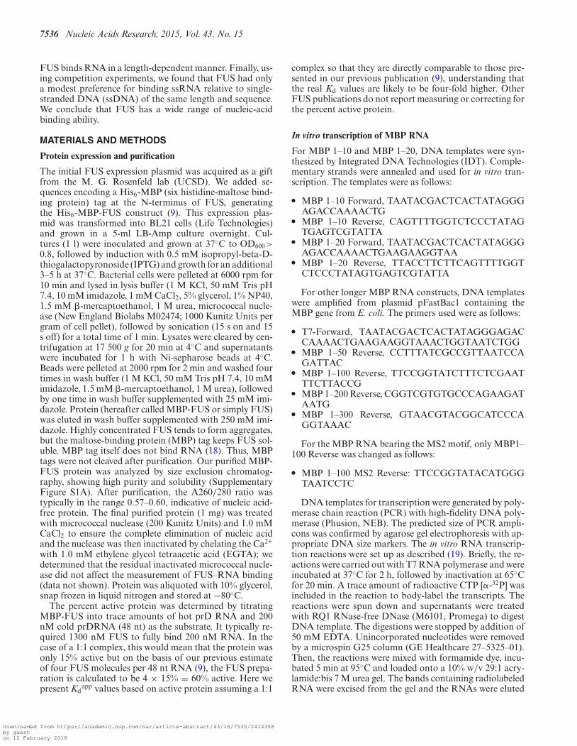
Five different RNA sequences have been reported to bepreferentially bound by FUS protein (12–17). Among these,GGUG, CGCGC and GUGGU are suggested to containa specific sequence motif recognized by FUS (12–16). Onthe other hand, Stem-loop and TERRA form unique sec-ondary and tertiary structures suggested to promote FUSbinding (13,17). We hypothesized that FUS may bind oneof these RNAs with exceptionally higher affinity than theothers. To test this hypothesis, we measured the binding ofE. coli-expressed FUS protein to eight RNAs including thefive published motifs and three negative control sequences(Supplementary Table S1). We also tested prD RNA, oneof many human ncRNAs that recruits FUS in vivo identi-fied in our previous study (11). EMSA was performed withincreasing concentrations of MBP-FUS protein and a traceamount of end-labeled RNA to measure binding affinities(Figure 1A).
Discrete shifted bands were observed, indicating RNA–protein complexes of specific stoichiometry and absence ofaggregation. All nine sequences tested were bound by FUS,
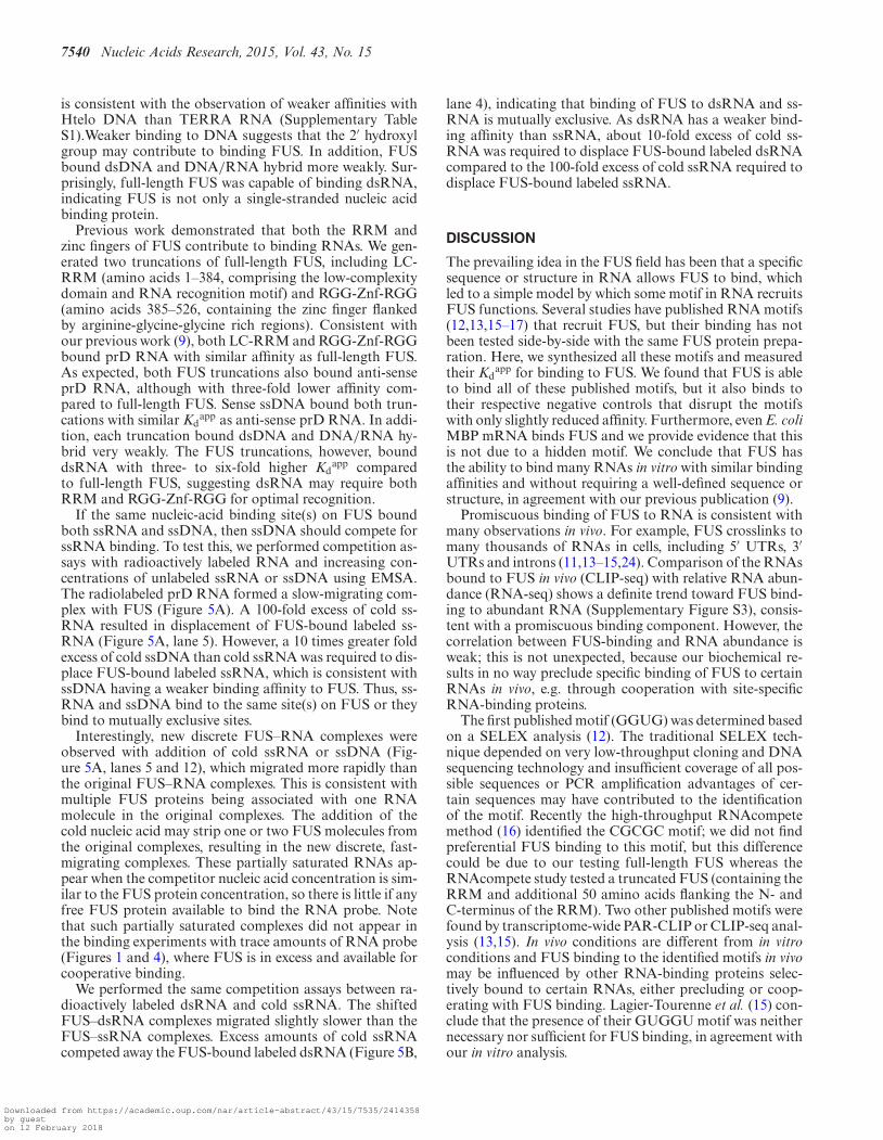
Figure 1. FUS binds many RNAs. (A) A schematic representation of theFUS protein. Blue, low complexity domain. Yellow, RGG domains. Green,RNA-binding domains. (B) A trace amount of TERRA (left), TERRA neg(middle) or prD (right) was incubated with increasing concentrations ofMBP-FUS (0, 15, 31, 62, 125, 188, 250, 375, 500, 750, 1000 and 1500 nM).Binding was analyzed by electrophoretic mobility shift assays (EMSA). (C)Summary of RNA binding data for MBP-FUS with nine different RNAs.Left, Quantification of Fbound (RNA in complexes per total RNA in lane)as a function of MBP-FUS concentration. Right, the apparent dissociationconstant was calculated for each RNA. n and L represent Hill coefficientand length of the RNA, respectively. Uncertainties represent the range oftwo or more replicates.
each with a Kdapp in the range between 100 and 1000 nM
(Figure 1B and Supplementary Figure S1B). The similarbinding affinities of very different RNAs (e.g. CGCGC,stem-loop and GGUG) cast doubt on their specificity forbinding to FUS. This skepticism was reinforced by the smalldifferences in affinity between three of the proposed mo-tifs and their mutated forms (cf. TERRA and TERRA neg,stem-loop and stem-loop neg, GGUG and GGUG neg).Furthermore, the prD RNA binds FUS as well as any ofthe other published RNAs but contains none of the motifs(9).
The EMSA patterns suggested positive cooperativity be-tween FUS and RNA, as it took only two or three proteinconcentration points to proceed from unshifted RNA to thelow-mobility completely shifted complex (Figure 1A). Wequantified and fit the binding data with the Hill equation,which revealed that FUS bound each sequence with pos-itive cooperativity (Figure 1B). The low-mobility complexis thought to contain at least four FUS proteins (9) andthe fact that intermediates with one, two or three boundproteins do not accumulate is expected for highly coopera-tive binding. At higher FUS concentrations, the FUS–RNAcomplexes shifted more toward the well of the gel. This sug-gests that additional FUS molecules are associated with the
Downloaded from https://academic.oup.com/nar/article-abstract/43/15/7535/2414358by gueston 12 February 2018
7538 Nucleic Acids Research, 2015, Vol. 43, No. 15
RNA in the highly retarded species compared to the ini-tial low-mobility FUS–RNA complex. Alternatively, someof these complexes may contain multiple FUS associatedwith multiple RNAs.
Our MBP-FUS protein was purified from E. coli, whileone previous publication carried out EMSA with His6-FUSpurified from insect cells (13). To test for differences in FUSobtained from these expression systems, His6-FUS purifiedfrom insect cells was compared with MBP-FUS purifiedfrom E. coli by EMSA (Supplementary Figure S1C). Thetwo proteins both formed discrete RNA–protein complexesand the protein concentration necessary to shift half of theradioactively labeled RNA was similar. In both cases, theobservation of discrete complexes suggests well-folded pro-tein. Therefore, we used MBP-FUS purified from E. coli forall remaining experiments.
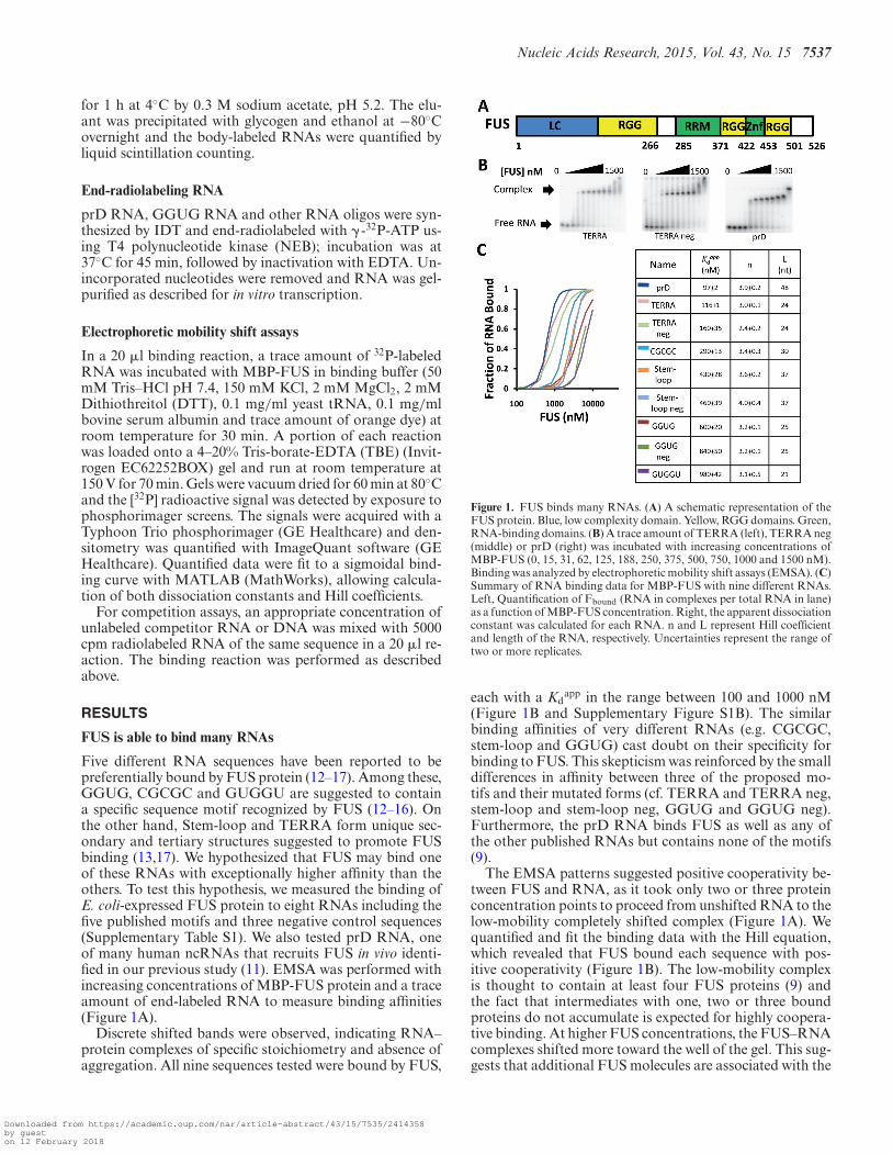
FUS binds RNA in a length-dependent manner
To further test FUS’s specificity for RNA binding, we per-formed EMSAs with portions of the mRNA for the MBPfrom E. coli, an organism that does not possess FUS. Sur-prisingly, the first 200 nt of the E. coli MBP mRNA (MBP1–200) bound FUS with a reasonably high affinity (Kd
app
= 56 ± 2 nM; Figure 2A). The electrophoretic mobility ofthe RNA–protein complex decreased progressively as theFUS concentration was increased, suggesting the loadingof more and more FUS onto the mRNA and low bindingspecificity. The Hill coefficient was 4.8 ± 0.1, indicating thatmultiple FUS proteins bound this non-human sequence ina positively cooperative manner.
Even though MBP–RNA originates from E. coli, it wasstill possible that some sequence or structure hidden in thisRNA could have been responsible for promoting FUS bind-ing. To test this possibility, we in vitro transcribed a seriesof MBP RNAs, including RNA containing the first 10 nt(MBP 1–10), first 20 nt (MBP 1–20) and so on, and thenmeasured their binding to FUS. If MBP 1–200 containedsome sequence or structure necessary to bind FUS, thenthere should be a sudden increase in affinity at the lengthcorresponding to the inclusion of the motif. If no such se-quence or structure existed in MBP 1–200, FUS might bindall the truncated sequences.
As shown in Figure 2B, there was no discrete length cut-off for FUS binding, but rather an incremental increase inaffinity with increasing RNA length. FUS bound MBP1–20 but not MBP 1–10, defining a minimum length for RNAbinding. As the RNA length increased, the binding curvesshifted from right to left, indicating an increase in bindingaffinity (Figure 2B). In other words, Kd
app decreased withincreasing RNA length. Plotting log (Kd) versus log (RNAlength) revealed a linear relationship between dissociationconstant and RNA length with a slope of −1 (Figure 2C),consistent with promiscuous binding (20).
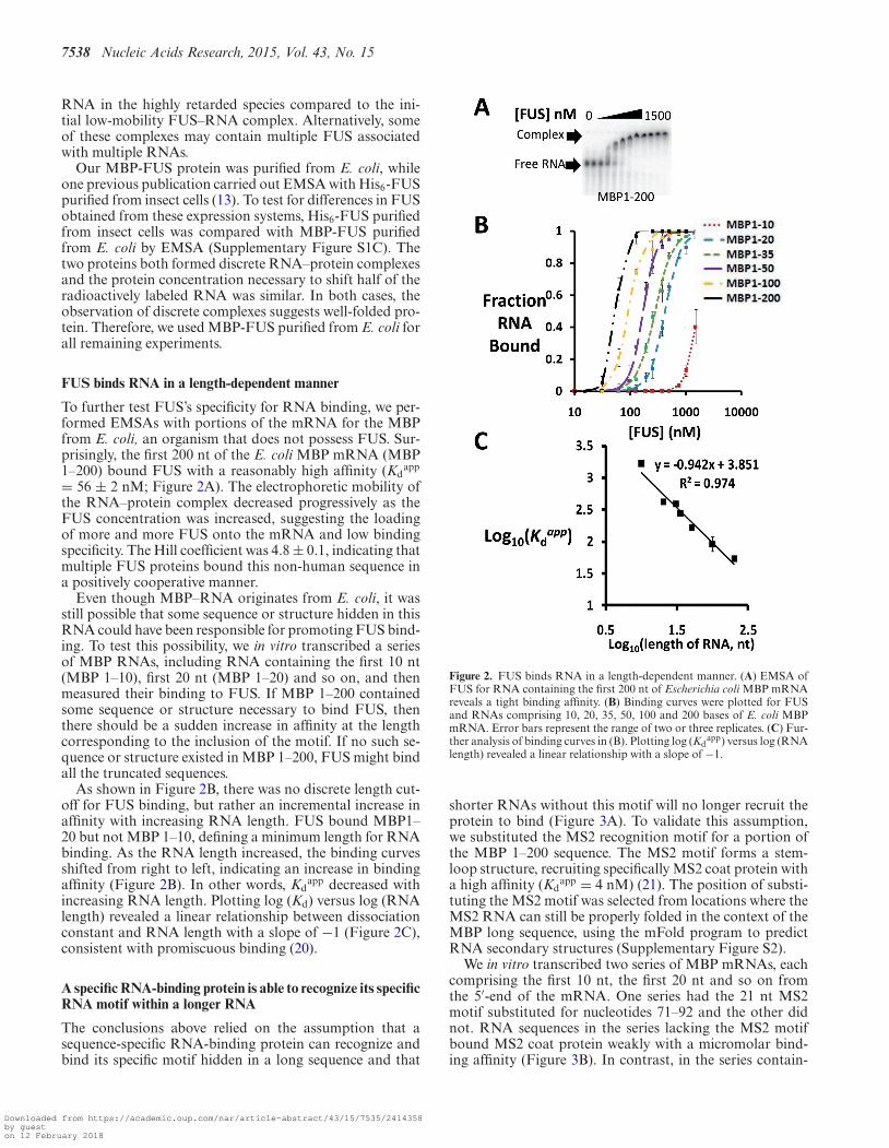
A specific RNA-binding protein is able to recognize its specificRNA motif within a longer RNA
The conclusions above relied on the assumption that asequence-specific RNA-binding protein can recognize andbind its specific motif hidden in a long sequence and that
Figure 2. FUS binds RNA in a length-dependent manner. (A) EMSA ofFUS for RNA containing the first 200 nt of Escherichia coli MBP mRNAreveals a tight binding affinity. (B) Binding curves were plotted for FUSand RNAs comprising 10, 20, 35, 50, 100 and 200 bases of E. coli MBPmRNA. Error bars represent the range of two or three replicates. (C) Fur-ther analysis of binding curves in (B). Plotting log (Kd
app) versus log (RNAlength) revealed a linear relationship with a slope of −1.
shorter RNAs without this motif will no longer recruit theprotein to bind (Figure 3A). To validate this assumption,we substituted the MS2 recognition motif for a portion ofthe MBP 1–200 sequence. The MS2 motif forms a stem-loop structure, recruiting specifically MS2 coat protein witha high affinity (Kd
app = 4 nM) (21). The position of substi-tuting the MS2 motif was selected from locations where theMS2 RNA can still be properly folded in the context of theMBP long sequence, using the mFold program to predictRNA secondary structures (Supplementary Figure S2).
We in vitro transcribed two series of MBP mRNAs, eachcomprising the first 10 nt, the first 20 nt and so on fromthe 5′-end of the mRNA. One series had the 21 nt MS2motif substituted for nucleotides 71–92 and the other didnot. RNA sequences in the series lacking the MS2 motifbound MS2 coat protein weakly with a micromolar bind-ing affinity (Figure 3B). In contrast, in the series contain-
Downloaded from https://academic.oup.com/nar/article-abstract/43/15/7535/2414358by gueston 12 February 2018
Nucleic Acids Research, 2015, Vol. 43, No. 15 7539
Figure 3. MS2 coat-binding protein is able to recognize its specific MS2RNA stem-loop motif inserted into MBP 1–200 RNA. (A) Schematic rep-resentation of the system used. Symbol to the right of each RNA indicateswhether motif-specific binding is expected. (B) Plotting log (Kd
app) ver-sus log (RNA length) for RNA sequences with or without the MS2 motifshowed more than an order of magnitude drop in Kd
app. Dash line con-necting the second point to the third point shows the difference betweenthe Kd
app without and with MS2 motif. Each point represents the averageof two or three replicates.
ing the MS2 motif, the binding affinity increased substan-tially for the third, fourth and fifth RNAs, which containedthe MS2 motif. Even though the binding affinity did not in-crease all the way down to 10 nM, the Kd
app we measuredfor binding to an isolated 21 nt MS2 motif, the Kd
app stilldropped dramatically from several micromolar into the 100nM range (Figure 3B). The reduced affinity of the coat pro-tein for the MS2 site in the context of long RNAs could bedue to sampling of multiple RNA conformations, some ofwhich disrupt the motif. Nevertheless, this control experi-ment supports our conclusion that if there were a specificmotif embedded in a long RNA molecule, it could be foundby testing the binding of a series of truncated versions of thelong RNA.
FUS binds RNA without requiring a specific sequence orstructure
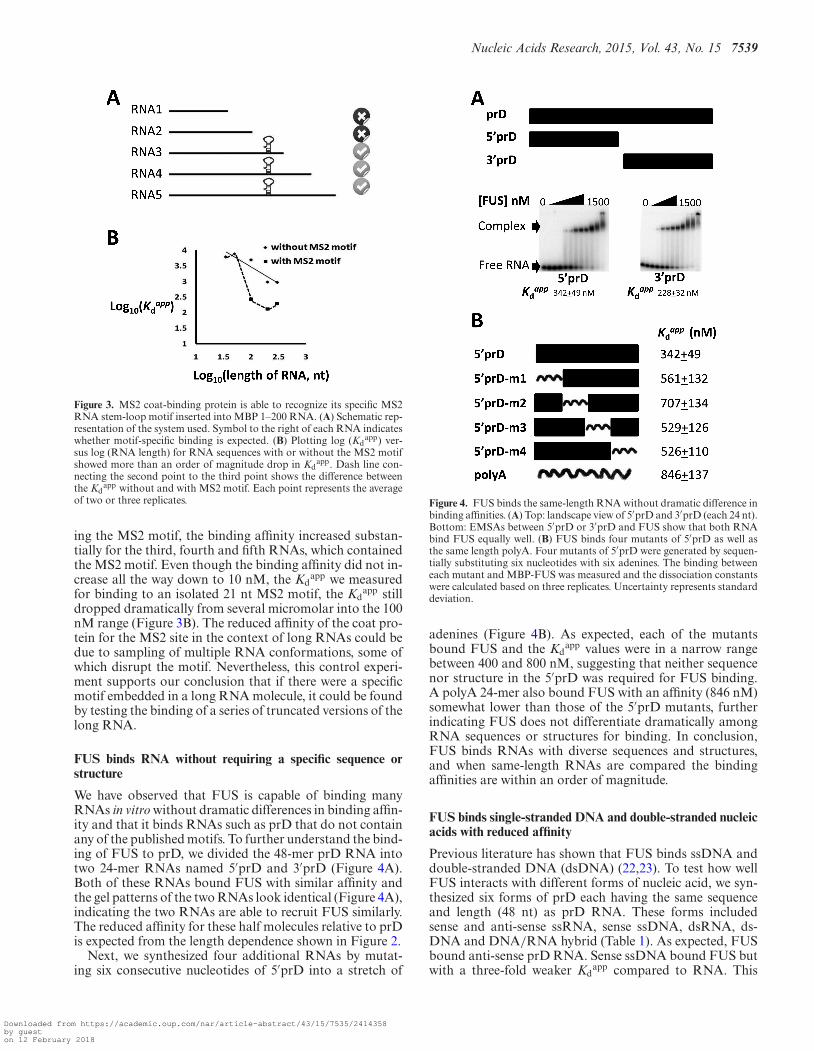
We have observed that FUS is capable of binding manyRNAs in vitro without dramatic differences in binding affin-ity and that it binds RNAs such as prD that do not containany of the published motifs. To further understand the bind-ing of FUS to prD, we divided the 48-mer prD RNA intotwo 24-mer RNAs named 5′prD and 3′prD (Figure 4A).Both of these RNAs bound FUS with similar affinity andthe gel patterns of the two RNAs look identical (Figure 4A),indicating the two RNAs are able to recruit FUS similarly.The reduced affinity for these half molecules relative to prDis expected from the length dependence shown in Figure 2.
Next, we synthesized four additional RNAs by mutat-ing six consecutive nucleotides of 5′prD into a stretch of
Figure 4. FUS binds the same-length RNA without dramatic difference inbinding affinities. (A) Top: landscape view of 5′prD and 3′prD (each 24 nt).Bottom: EMSAs between 5′prD or 3′prD and FUS show that both RNAbind FUS equally well. (B) FUS binds four mutants of 5′prD as well asthe same length polyA. Four mutants of 5′prD were generated by sequen-tially substituting six nucleotides with six adenines. The binding betweeneach mutant and MBP-FUS was measured and the dissociation constantswere calculated based on three replicates. Uncertainty represents standarddeviation.
adenines (Figure 4B). As expected, each of the mutantsbound FUS and the Kd
app values were in a narrow rangebetween 400 and 800 nM, suggesting that neither sequencenor structure in the 5′prD was required for FUS binding.A polyA 24-mer also bound FUS with an affinity (846 nM)somewhat lower than those of the 5′prD mutants, furtherindicating FUS does not differentiate dramatically amongRNA sequences or structures for binding. In conclusion,FUS binds RNAs with diverse sequences and structures,and when same-length RNAs are compared the bindingaffinities are within an order of magnitude.
FUS binds single-stranded DNA and double-stranded nucleicacids with reduced affinity
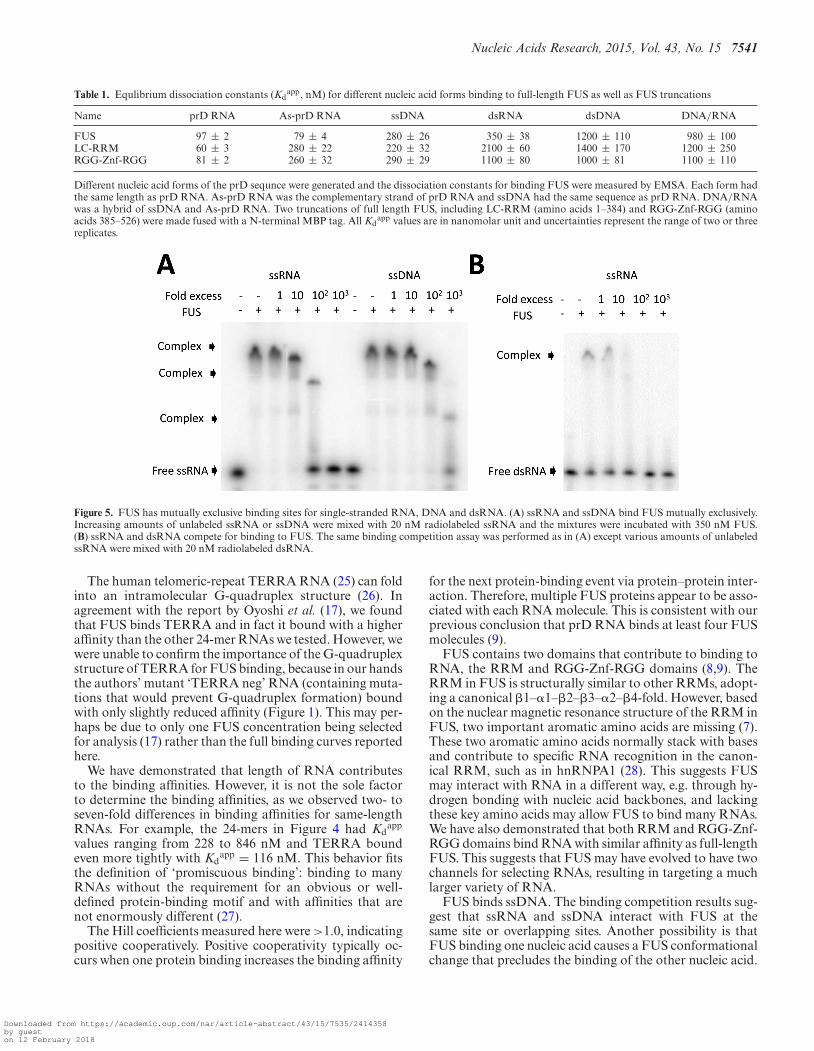
Previous literature has shown that FUS binds ssDNA anddouble-stranded DNA (dsDNA) (22,23). To test how wellFUS interacts with different forms of nucleic acid, we syn-thesized six forms of prD each having the same sequenceand length (48 nt) as prD RNA. These forms includedsense and anti-sense ssRNA, sense ssDNA, dsRNA, ds-DNA and DNA/RNA hybrid (Table 1). As expected, FUSbound anti-sense prD RNA. Sense ssDNA bound FUS butwith a three-fold weaker Kd
app compared to RNA. This
Downloaded from https://academic.oup.com/nar/article-abstract/43/15/7535/2414358by gueston 12 February 2018
7540 Nucleic Acids Research, 2015, Vol. 43, No. 15
is consistent with the observation of weaker affinities withHtelo DNA than TERRA RNA (Supplementary TableS1).Weaker binding to DNA suggests that the 2′ hydroxylgroup may contribute to binding FUS. In addition, FUSbound dsDNA and DNA/RNA hybrid more weakly. Sur-prisingly, full-length FUS was capable of binding dsRNA,indicating FUS is not only a single-stranded nucleic acidbinding protein.
Previous work demonstrated that both the RRM andzinc fingers of FUS contribute to binding RNAs. We gen-erated two truncations of full-length FUS, including LC-RRM (amino acids 1–384, comprising the low-complexitydomain and RNA recognition motif) and RGG-Znf-RGG(amino acids 385–526, containing the zinc finger flankedby arginine-glycine-glycine rich regions). Consistent withour previous work (9), both LC-RRM and RGG-Znf-RGGbound prD RNA with similar affinity as full-length FUS.As expected, both FUS truncations also bound anti-senseprD RNA, although with three-fold lower affinity com-pared to full-length FUS. Sense ssDNA bound both trun-cations with similar Kd
app as anti-sense prD RNA. In addi-tion, each truncation bound dsDNA and DNA/RNA hy-brid very weakly. The FUS truncations, however, bounddsRNA with three- to six-fold higher Kd
app comparedto full-length FUS, suggesting dsRNA may require bothRRM and RGG-Znf-RGG for optimal recognition.
If the same nucleic-acid binding site(s) on FUS boundboth ssRNA and ssDNA, then ssDNA should compete forssRNA binding. To test this, we performed competition as-says with radioactively labeled RNA and increasing con-centrations of unlabeled ssRNA or ssDNA using EMSA.The radiolabeled prD RNA formed a slow-migrating com-plex with FUS (Figure 5A). A 100-fold excess of cold ss-RNA resulted in displacement of FUS-bound labeled ss-RNA (Figure 5A, lane 5). However, a 10 times greater foldexcess of cold ssDNA than cold ssRNA was required to dis-place FUS-bound labeled ssRNA, which is consistent withssDNA having a weaker binding affinity to FUS. Thus, ss-RNA and ssDNA bind to the same site(s) on FUS or theybind to mutually exclusive sites.
Interestingly, new discrete FUS–RNA complexes wereobserved with addition of cold ssRNA or ssDNA (Fig-ure 5A, lanes 5 and 12), which migrated more rapidly thanthe original FUS–RNA complexes. This is consistent withmultiple FUS proteins being associated with one RNAmolecule in the original complexes. The addition of thecold nucleic acid may strip one or two FUS molecules fromthe original complexes, resulting in the new discrete, fast-migrating complexes. These partially saturated RNAs ap-pear when the competitor nucleic acid concentration is sim-ilar to the FUS protein concentration, so there is little if anyfree FUS protein available to bind the RNA probe. Notethat such partially saturated complexes did not appear inthe binding experiments with trace amounts of RNA probe(Figures 1 and 4), where FUS is in excess and available forcooperative binding.
We performed the same competition assays between ra-dioactively labeled dsRNA and cold ssRNA. The shiftedFUS–dsRNA complexes migrated slightly slower than theFUS–ssRNA complexes. Excess amounts of cold ssRNAcompeted away the FUS-bound labeled dsRNA (Figure 5B,
lane 4), indicating that binding of FUS to dsRNA and ss-RNA is mutually exclusive. As dsRNA has a weaker bind-ing affinity than ssRNA, about 10-fold excess of cold ss-RNA was required to displace FUS-bound labeled dsRNAcompared to the 100-fold excess of cold ssRNA required todisplace FUS-bound labeled ssRNA.
DISCUSSION
The prevailing idea in the FUS field has been that a specificsequence or structure in RNA allows FUS to bind, whichled to a simple model by which some motif in RNA recruitsFUS functions. Several studies have published RNA motifs(12,13,15–17) that recruit FUS, but their binding has notbeen tested side-by-side with the same FUS protein prepa-ration. Here, we synthesized all these motifs and measuredtheir Kd
app for binding to FUS. We found that FUS is ableto bind all of these published motifs, but it also binds totheir respective negative controls that disrupt the motifswith only slightly reduced affinity. Furthermore, even E. coliMBP mRNA binds FUS and we provide evidence that thisis not due to a hidden motif. We conclude that FUS hasthe ability to bind many RNAs in vitro with similar bindingaffinities and without requiring a well-defined sequence orstructure, in agreement with our previous publication (9).
Promiscuous binding of FUS to RNA is consistent withmany observations in vivo. For example, FUS crosslinks tomany thousands of RNAs in cells, including 5′ UTRs, 3′UTRs and introns (11,13–15,24). Comparison of the RNAsbound to FUS in vivo (CLIP-seq) with relative RNA abun-dance (RNA-seq) shows a definite trend toward FUS bind-ing to abundant RNA (Supplementary Figure S3), consis-tent with a promiscuous binding component. However, thecorrelation between FUS-binding and RNA abundance isweak; this is not unexpected, because our biochemical re-sults in no way preclude specific binding of FUS to certainRNAs in vivo, e.g. through cooperation with site-specificRNA-binding proteins.
The first published motif (GGUG) was determined basedon a SELEX analysis (12). The traditional SELEX tech-nique depended on very low-throughput cloning and DNAsequencing technology and insufficient coverage of all pos-sible sequences or PCR amplification advantages of cer-tain sequences may have contributed to the identificationof the motif. Recently the high-throughput RNAcompetemethod (16) identified the CGCGC motif; we did not findpreferential FUS binding to this motif, but this differencecould be due to our testing full-length FUS whereas theRNAcompete study tested a truncated FUS (containing theRRM and additional 50 amino acids flanking the N- andC-terminus of the RRM). Two other published motifs werefound by transcriptome-wide PAR-CLIP or CLIP-seq anal-ysis (13,15). In vivo conditions are different from in vitroconditions and FUS binding to the identified motifs in vivomay be influenced by other RNA-binding proteins selec-tively bound to certain RNAs, either precluding or coop-erating with FUS binding. Lagier-Tourenne et al. (15) con-clude that the presence of their GUGGU motif was neithernecessary nor sufficient for FUS binding, in agreement withour in vitro analysis.
Downloaded from https://academic.oup.com/nar/article-abstract/43/15/7535/2414358by gueston 12 February 2018
Nucleic Acids Research, 2015, Vol. 43, No. 15 7541
Table 1. Equlibrium dissociation constants (Kdapp, nM) for different nucleic acid forms binding to full-length FUS as well as FUS truncations
Name prD RNA As-prD RNA ssDNA dsRNA dsDNA DNA/RNA
FUS 97 ± 2 79 ± 4 280 ± 26 350 ± 38 1200 ± 110 980 ± 100LC-RRM 60 ± 3 280 ± 22 220 ± 32 2100 ± 60 1400 ± 170 1200 ± 250RGG-Znf-RGG 81 ± 2 260 ± 32 290 ± 29 1100 ± 80 1000 ± 81 1100 ± 110
Different nucleic acid forms of the prD sequnce were generated and the dissociation constants for binding FUS were measured by EMSA. Each form hadthe same length as prD RNA. As-prD RNA was the complementary strand of prD RNA and ssDNA had the same sequence as prD RNA. DNA/RNAwas a hybrid of ssDNA and As-prD RNA. Two truncations of full length FUS, including LC-RRM (amino acids 1–384) and RGG-Znf-RGG (aminoacids 385–526) were made fused with a N-terminal MBP tag. All Kd
app values are in nanomolar unit and uncertainties represent the range of two or threereplicates.
Figure 5. FUS has mutually exclusive binding sites for single-stranded RNA, DNA and dsRNA. (A) ssRNA and ssDNA bind FUS mutually exclusively.Increasing amounts of unlabeled ssRNA or ssDNA were mixed with 20 nM radiolabeled ssRNA and the mixtures were incubated with 350 nM FUS.(B) ssRNA and dsRNA compete for binding to FUS. The same binding competition assay was performed as in (A) except various amounts of unlabeledssRNA were mixed with 20 nM radiolabeled dsRNA.
The human telomeric-repeat TERRA RNA (25) can foldinto an intramolecular G-quadruplex structure (26). Inagreement with the report by Oyoshi et al. (17), we foundthat FUS binds TERRA and in fact it bound with a higheraffinity than the other 24-mer RNAs we tested. However, wewere unable to confirm the importance of the G-quadruplexstructure of TERRA for FUS binding, because in our handsthe authors’ mutant ‘TERRA neg’ RNA (containing muta-tions that would prevent G-quadruplex formation) boundwith only slightly reduced affinity (Figure 1). This may per-haps be due to only one FUS concentration being selectedfor analysis (17) rather than the full binding curves reportedhere.
We have demonstrated that length of RNA contributesto the binding affinities. However, it is not the sole factorto determine the binding affinities, as we observed two- toseven-fold differences in binding affinities for same-lengthRNAs. For example, the 24-mers in Figure 4 had Kd
app
values ranging from 228 to 846 nM and TERRA boundeven more tightly with Kd
app = 116 nM. This behavior fitsthe definition of ‘promiscuous binding’: binding to manyRNAs without the requirement for an obvious or well-defined protein-binding motif and with affinities that arenot enormously different (27).
The Hill coefficients measured here were >1.0, indicatingpositive cooperatively. Positive cooperativity typically oc-curs when one protein binding increases the binding affinity
for the next protein-binding event via protein–protein inter-action. Therefore, multiple FUS proteins appear to be asso-ciated with each RNA molecule. This is consistent with ourprevious conclusion that prD RNA binds at least four FUSmolecules (9).
FUS contains two domains that contribute to binding toRNA, the RRM and RGG-Znf-RGG domains (8,9). TheRRM in FUS is structurally similar to other RRMs, adopt-ing a canonical �1–�1–�2–�3–�2–�4-fold. However, basedon the nuclear magnetic resonance structure of the RRM inFUS, two important aromatic amino acids are missing (7).These two aromatic amino acids normally stack with basesand contribute to specific RNA recognition in the canon-ical RRM, such as in hnRNPA1 (28). This suggests FUSmay interact with RNA in a different way, e.g. through hy-drogen bonding with nucleic acid backbones, and lackingthese key amino acids may allow FUS to bind many RNAs.We have also demonstrated that both RRM and RGG-Znf-RGG domains bind RNA with similar affinity as full-lengthFUS. This suggests that FUS may have evolved to have twochannels for selecting RNAs, resulting in targeting a muchlarger variety of RNA.
FUS binds ssDNA. The binding competition results sug-gest that ssRNA and ssDNA interact with FUS at thesame site or overlapping sites. Another possibility is thatFUS binding one nucleic acid causes a FUS conformationalchange that precludes the binding of the other nucleic acid.
Downloaded from https://academic.oup.com/nar/article-abstract/43/15/7535/2414358by gueston 12 February 2018
7542 Nucleic Acids Research, 2015, Vol. 43, No. 15
FUS binding ssDNA is consistent with several observationsin the literature. Similar in vitro competition results havebeen shown in other studies (22,29). In fact, FUS proteinhas been isolated and purified by affinity chromatographyon ssDNA (30). The ability of FUS to bind DNA also fitswell with its function associated with DNA damage repair.FUS, being one of the earliest proteins recruited to DNAlesions, interacts directly with the DNA repair factors, suchas HDAC1 and DNA-PK (31,32). Such interactions are re-quired for successful DNA repair (31,32).
FUS is reported to directly interact with the CTD ofRNA polymerase II (11,33). FUS–CTD interactions re-quire RNA, either nascent transcript or noncoding RNA(11). Cooperative binding properties of FUS to RNA mayfacilitate the formation of higher-order assemblies. Theseassemblies orchestrate the phosphorylation status of CTDof RNA pol II (9,11,33). Being able to bind many RNAsmay allow FUS to target a larger diversity of genes neartheir transcription start sites, which is consistent with ourprevious model (11). Also, FUS may bind to long intronsand facilitate their splicing (15).
FUS has the intrinsic ability to bind many RNAs with-out substantial differences in binding affinity, so what de-termines the FUS interactome in vivo? Many other pro-teins, including hnRNP proteins and splicing factors, areassociated with nuclear RNAs and these may precludeFUS binding. On the other hand, some FUS-partner pro-teins may enhance FUS binding specificity. Similar partner-assisted specificity has been documented for protein–nucleicacid interactions including the homeobox (Hox) familytranscription factors, which gain higher DNA sequencespecificity and enhanced affinity when paired with an Exdprotein partner (34). One can also speculate that post-translational modifications, such as arginine methylation(35), may change the conformation of the protein, convey-ing preferences for certain RNAs. Future work is requiredto elucidate the requirements for FUS–RNA interaction inliving systems.
SUPPLEMENTARY DATA
Supplementary Data are available at NAR Online.
ACKNOWLEDGEMENTS
We thank R. Batey (University of Colorado Boulder) forproviding the MS2 coat protein. We thank J. Hoell (Hein-rich Heine University) and T. Tuschl (Rockefeller Uni-versity) for the baculoviral FUS expression construct andaccess to unpublished data, and D. Ray (University ofToronto) and T. Hughes (University of Toronto) for se-quences of their FUS-binding RNAs and helpful conver-sations. We thank C. Davidovich and J.C. Schmidt for theiradvice on experiments.
FUNDING
Howard Hughes Medical Institute (HHMI) (to T.R.C.).Funding for open access charge: HHMI and the Universityof Colorado Boulder Open Access Fund.Conflict of interest statement. T.R.C. is a member of theBoard of Directors of Merck, Inc.
REFERENCES1. Cruz,S.D. and Cleveland,D.W. (2011) Understanding the role of
TDP-43 and FUS/TLS in ALS and beyond. Curr. Opin. Neurobiol.,21, 904–919.
2. Fiesel,F.C. and Kahle,P.J. (2011) TDP-43 and FUS/TLS: cellularfunctions and implications for neurodegeneration. FEBS J., 278,3550–3568.
3. Lagier-Tourenne,C., Polymenidou,M. and Cleveland,D.W. (2010)TDP-43 and FUS/TLS: emerging roles in RNA processing andneurodegeneration. Hum. Mol. Genet., 19, R46–R64.
4. Kiernan,M.C., Vucic,S., Cheah,B.C., Turner,M.R., Eisen,A.,Hardiman,O., Burrell,J.R. and Zoing,M.C. (2011) Amyotrophiclateral sclerosis. Lancet, 377, 942–955.
5. Hewitt,C., Kirby,J., Highley,J.R., Hartley,J.A., Hibberd,R.,Hollinger,H.C., Ince,P.G., McDermott,C.J. and Shaw,P.J. (2010)Novel FUS/TLS mutations and pathology in familial and sporadicamyotrophic lateral sclerosis. Arch. Neurol., 67, 455–461.
6. Deng,H.X., Zhai,H., Bigio,E.H., Yan,J., Fecto,F., Ajroud,K.,Mishra,M., Ajroud-Driss,S., Heller,S., Sufit,R. et al. (2010)FUS-immunoreactive inclusions are a common feature in sporadicand non-SOD1 familial amyotrophic lateral sclerosis. Ann. Neurol.,67, 739–748.
7. Liu,X., Niu,C., Ren,J., Zhang,J., Xie,X., Zhu,H., Feng,W. andGong,W. (2012) The RRM domain of human fused in sarcomaprotein reveals a non-canoical nucleic acid binding site. Biochim.Biophys. Acta, 1832, 375–385.
8. Iko,Y., Kodama,T.S., Kasai,N., Oyama,T., Morita,E.H., Muto,T.,Okumura,M., Fujii,R., Takumi,T., Tate,S. et al. (2004) Domainarchitectures and characterization of an RNA-binding protein, TLS.J. Biol. Chem., 279, 44834–44840.
9. Schwartz,J.C., Wang,X., Podell,E.R. and Cech,T.R. (2013) RNAseeds higher-order assembly of FUS protein. Cell Rep., 5, 918–925.
10. Wang,X., Arai,S., Song,X., Reichart,D., Du,K., Gabriel,P.,Tempst,P., Rosenfeld,M.G., Glass,C.K. and Kurokawa,R. (2008)Induced ncRNAs allosterically modify RNA-binding proteins in cisto inhibit transcription. Nature, 454, 126–131.
11. Schwartz,J.C., Ebmeier,C.C., Podell,E.R., Heimiller,J., Taatjes,D.J.and Cech,T.R. (2012) FUS binds the CTD of RNA polymerase IIand regulates its phosphorylation at Ser2. Genes Dev., 26, 2690–2695.
12. Lerga,A., Hallier,M., Delva,L., Orvain,C., Gallais,I., Marie,J. andMoreau-Gachelin,F. (2001) Identification of an RNA bindingspecificity for the potential splicing factor TLS. J. Biol. Chem., 276,6807–6816.
13. Hoell,J.I., Larsson,E., Runge,S., Nusbaum,J.D., Duggimpudi,S.,Farazi,T.A., Hafner,M., Borkhardt,A., Sander,C. and Tuschl,T.(2011) RNA targets of wild-type and mutant FET family proteins.Nat. Struct. Mol. Biol., 18, 1428–1431.
14. Ishigaki,S., Masuda,A., Fujioka,Y., Iguchi,Y., Katsuno,M.,Shibata,A., Urano,F., Sobue,G. and Ohno,K. (2012)Position-dependent FUS-RNA interactions regulate alternativesplicing events and transcriptions. Sci. Rep., 2, 529.
15. Lagier-Tourenne,C., Polymenidou,M., Hutt,K.R., Vu,A.Q.,Baughn,M., Huelga,S.C., Clutario,K.M., Ling,S.C., Liang,T.Y.,Mazur,C. et al. (2012) Divergent roles of ALS-linked proteinsFUS/TLS and TDP-43 intersect in processing long pre-mRNAs. Nat.Neurosci., 15, 1488–1497.
16. Ray,D., Kazan,H., Cook,K.B., Weirauch,M.T., Najafabadi,H.S.,Li,X., Gueroussov,S., Albu,M., Zheng,H., Yang,A. et al. (2013) Acompendium of RNA-binding motifs for decoding gene regulation.Nature, 499, 172–177.
17. Takahama,K., Takada,A., Tada,S., Shimizu,M., Sayama,K.,Kurokawa,R. and Oyoshi,T. (2013) Regulation of telomere length byG-quadruplex telomere DNA-and TERRA-binding proteinTLS/FUS. Chem. Biol., 20, 341–350.
18. McKay,D.B., Xi,L., Barthel,K.K.B. and Cech,T.R. (2014) Structureand function of steroid receptor RNA activator protein, the proposedpartner of SRA non-coding RNA. J. Mol. Biol., 426, 1766–1785.
19. Milligan,J.F. and Uhlenbeck,O.C. (1989) Synthesis of small RNAsusing T7 RNA polymerase. Methods Enzymol., 180, 51–62.
20. Davidovich,C., Zheng,L., Goodrich,K.J. and Cech,T.R. (2013)Promiscuous RNA binding by polycomb repressive complex 2. Nat.Struct. Mol. Biol., 20, 1250–1257.
Downloaded from https://academic.oup.com/nar/article-abstract/43/15/7535/2414358by gueston 12 February 2018
Nucleic Acids Research, 2015, Vol. 43, No. 15 7543
21. Johansson,H.E., Dertinger,D., LeCuyer,K.A., Behlen,L.S.,Greef,C.H. and Uhlenbeck,O.C. (1998) A thermodynamic analysis ofthe sequence-specific binding of RNA by bacteriophage MS2 coatprotein. Proc. Natl. Acad. Sci. U.S.A., 95, 9244–9249.
22. Bertolotti,A., Lutz,Y., Heard,D.J., Chambon,P. and Tora,L. (1996)hTAF(II)68, a novel RNA/ssDNA-binding protein with homology tothe pro-oncoproteins TLS/FUS and EWS is associated with bothTFIID and RNA polymerase II. EMBO J., 15, 5022–5031.
23. Baechtold,H., Kuroda,M., Sok,J., Ron,D., Lopez,B.S. andAkhmedov,A.T. (1999) Human 75-kDa DNA-pairing protein isidentical to the pro-oncoprotein TLS/FUS and is able to promoteD-loop formation. J. Biol. Chem., 274, 34337–34342.
24. Rogelj,B., Easton,L.E., Bogu,G.K., Stanton,L.W., Rot,G., Curk,T.,Zupan,B., Sugimoto,Y., Modic,M., Haberman,N. et al. (2012)Widespread binding of FUS along nascent RNA regulates alternativesplicing in the brain. Sci. Rep., 2, 603.
25. Azzalin,C.M., Reichenbach,P., Khoriauli,L., Giulotto,E. andLingner,J. (2007) Telomeric repeat-containing RNA and RNAsurveillance factors at mammalian chromosome ends. Science, 318,798–801.
26. Williamson,J.R., Raghuraman,M.K. and Cech,T.R. (1989)Monovalent cation-induced structure of telomeric DNA: theG-quartet model. Cell, 59, 871–880.
27. Davidovich,C., Wang,X., Cifuentes-Rojas,C., Goodrich,K.J.,Gooding,A.R., Lee,J.T. and Cech,T.R. (2015) Toward a consensus onthe binding specificity and promiscuity of PRC2 for RNA. Mol. Cell,57, 552–558.
28. Ding,J., Hayashi,M.K., Zhang,Y., Manche,L., Krainer,A.R. andXu,R.M. (1998) Crystal structure of the two-RRM domain ofhnRNPA1 (UP1) complexed with single-stranded telomeric DNA.Genes Dev., 13, 1102–1115.
29. Tan,A.Y., Riley,T.R., Coady,T., Bussemaker,H.J. and Manley,J.L.(2012) TLS/FUS regulates target gene transcription viasingle-stranded DNA response elements. Proc. Natl. Acad. Sci.U.S.A., 109, 6030–6035.
30. Calvio,C., Neubauer,G., Mann,M. and Lamond,A.I. (1995)Identification of hnRNP P2 as TLS/FUS using electrospray massspectrometry. RNA, 1, 724–733.
31. Wang,W.Y., Pan,L., Su,S.C., Quinn,E.J., Sasaki,M., Jimenez,J.C.,Mackenzie,I.R., Huang,E.J. and Tsai,L.H. (2013) Interaction of FUSand HDAC1 regulates DNA damage response. Nat. Neurosci., 16,1383–1391.
32. Mastrocola,A.S., Kim,S.H., Trinh,A.T., Rodenkirch,L.A. andTibbetts,R.S. (2013) The RNA-binding protein fused in sarcoma(FUS) functions downstream of poly (ADP-ribose) polymerase(PARP) in response to DNA damage. J. Biol. Chem., 288,24731–24741.
33. Kwon,I., Kato,M., Xiang,S., Wu,L., Theodoropoulos,P., Mirzaei,H.,Han,T., Xie,S., Corden,J.L. and McKnight,S.L. (2013)Phosphorylation-regulated binding of RNA polymerase II to fibrouspolymers of low-complexity domains. Cell, 155, 1049–1060.
34. Moretti,R., Donato,L.J., Brezinski,M.L., Stafford,R.L., Hoff,H.,Thorson,J.S., Dervan,P.B. and Ansari,A.Z. (2008) Targeted chemicalwedges reveal the role of allosteric DNA modulation in protein-DNAassembly. ACS Chem. Biol., 3, 220–229.
35. Dormann,D., Madl,T., Valori,C.F., Bentmann,E., Tahirovic,S.,Abou-Ajram,C., Kremmer,E., Ansorge,O., Mackenzie,I.R.,Neumann,M. et al. (2012) Arginine methylation next to the PY-NLSmodulates transportin binding and nuclear import of FUS. EMBOJ., 31, 4258–4275.
Downloaded from https://academic.oup.com/nar/article-abstract/43/15/7535/2414358by gueston 12 February 2018