Embed Size (px)
DESCRIPTION
A new crystal growth cell based on transparent indium tin oxide (ITO) glass-electrodes for electrochemically assisted protein crystal- lization allows for reduced nucleation and crystal quality enhancement. The crystallization behavior of lysozyme and ferritin was monitored as a function of the electric current applied to the growth cell. The X-ray diffraction analysis showed that for specific currents, the crystal quality is substantially improved. No conformational changes were observed in the 3D crystallographic structures determined for crystals grown under different electric current regimes. Finally, the strong crystal adhesion on the surface of ITO electrode because of the electroadhesion allows a sufficiently strong fixing of the protein crystals, to undergo atomic force microscopy investigations in a fluid cell.
Citation preview
Published: July 12, 2011
r 2011 American Chemical Society 3917 dx.doi.org/10.1021/cg200485v | Cryst. Growth Des. 2011, 11, 3917–3922
ARTICLE
pubs.acs.org/crystal
Novel Protein Crystal Growth Electrochemical Cell For ApplicationsIn X-ray Diffraction and Atomic Force MicroscopyGabriela Gil-Alvaradejo,† Rayana R. Ruiz-Arellano,† Christopher Owen,‡ Adela Rodríguez-Romero,†
Enrique Rudi~no-Pi~nera,§ Moriamou K. Antwi,^ Vivian Stojanoff,‡ and Abel Moreno*,†
†Instituto de Química, Universidad Nacional Aut�onoma de M�exico, M�exico, D.F. 04510 Mexico‡Brookhaven National Laboratory, National Synchrotron Light Source Bldg 725D, Upton New York 11873, United States§Departamento de Medicina Molecular y Bioprocesos, Instituto de Biotecnología, Universidad Nacional Aut�onoma de M�exico,Av. Universidad 2001, Cuernavaca, Morelos, 62210, Mexico
^St. Joseph’s College, 245 Clinton Avenue Brooklyn, New York 11205, United States
bS Supporting Information
1. INTRODUCTION
Since Chin et al., pioneering work on the electrochemicallyassisted process of estradiol 17β-dehydrogenase, the idea ofprotein molecules behaving as macroions fell into a long lethargy.1
In the late 1990s, two contributions2,3 revived this research withtheoretical and experimental results on the response of chromo-proteins subjected to external pressure and electric fields. There-after new studies performed by Aubry and co-workers,4,5 as wellas Nanev and Penkova6 applying external electric fields to theprotein crystallization solution, showed that it is possible toreduce lysozyme nucleation, and to control the kinetics of thecrystallization process. The use of internal electric fields mergingcapillary tubes and gels was first proposed by Mirkin et al.,(2003).7 A new concept was introduced byMoreno and Rivera in20058 who used highly oriented pyrolytic graphite electrodes tocrystallize ferritin. The use of different electrodes and configura-tions has been subject of three recent reviews.9�11
Obtaining high quality protein single crystals of a size largeenough for X-ray analysis remains a bottleneck in structuralbiology research. Since the beginning of the 1990s, novel andminiaturized methods are being developed to obtain crystals insmaller droplets and in a shorter time.12 Additional efforts arebeing made to optimize the X-ray data collection and the dataprocessing methods, using powerful personal computers. How-ever, there are only few results focused on the understanding ofthe crystallization process based on physicochemical approachesthat use physical parameters to improve the crystal quality. Some
contributions on the crystal growth process of different proteinsgrown under the influence of electric fields, voltage pulses(at controlled potentials and currents) that use different configura-tions, have been recently published.13,14 There are some approachesalready published combining electric and magnetic fields,15 gelsandmagnetic fields,16,17 or only magnetic fields in solution to controlthe orientation and crystal quality on proteins.18�20
Here we present a new crystal growth cell with parallel indiumtin oxide (ITO) electrodes. The nucleation and crystal growthbehavior of lysozyme and ferritin in the presence of differentelectric currents was monitored at constant temperature. Crystalquality was determined byX-ray crystallography and crystal surfaceanalysis by atomic force microscopy (AFM). We found that foreach protein crystals grown under specific current values showedbetter quality when tested by X-ray diffraction. No conforma-tional changes were found in the 3D crystallographic structuresbecause of the electroadhesion process.
2. EXPERIMENTAL SECTION
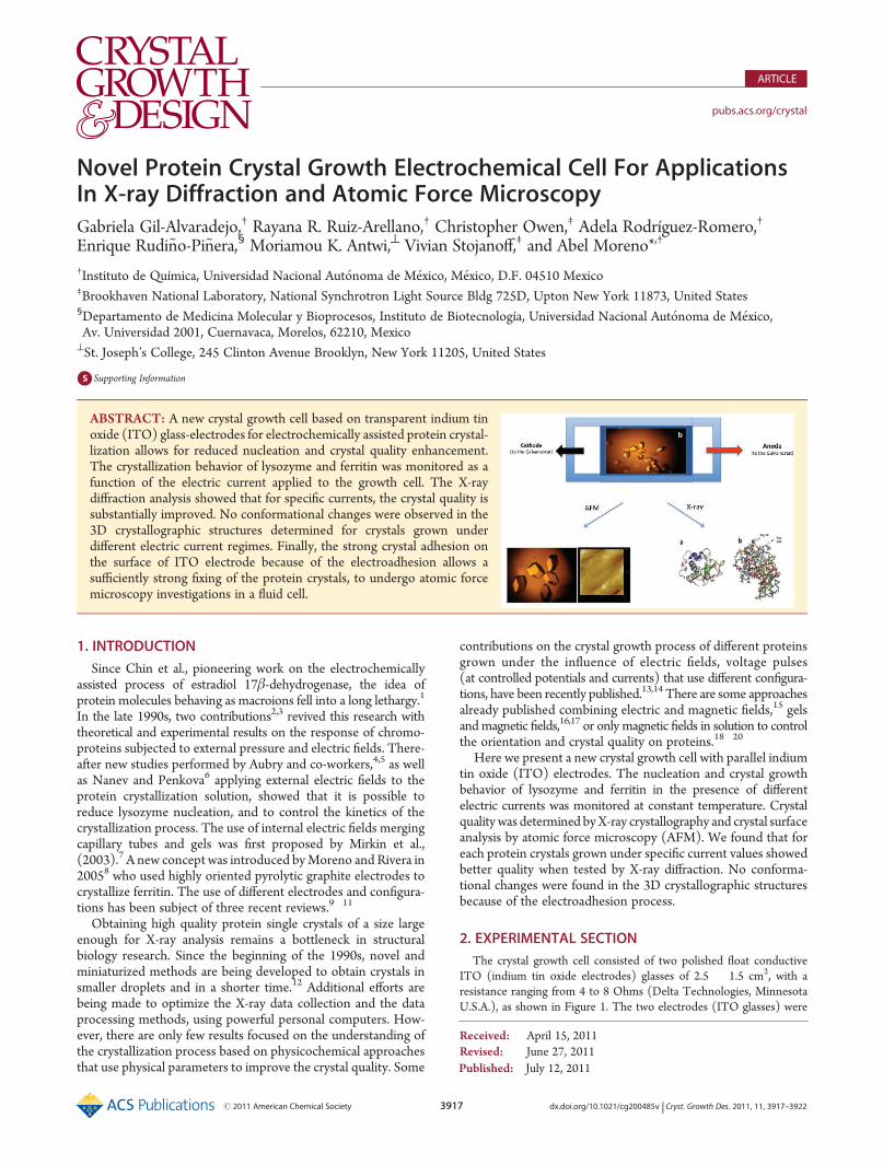
The crystal growth cell consisted of two polished float conductiveITO (indium tin oxide electrodes) glasses of 2.5 � 1.5 cm2, with aresistance ranging from 4 to 8 Ohms (Delta Technologies, MinnesotaU.S.A.), as shown in Figure 1. The two electrodes (ITO glasses) were
Received: April 15, 2011Revised: June 27, 2011
ABSTRACT: A new crystal growth cell based on transparent indium tinoxide (ITO) glass-electrodes for electrochemically assisted protein crystal-lization allows for reduced nucleation and crystal quality enhancement.The crystallization behavior of lysozyme and ferritin was monitored as afunction of the electric current applied to the growth cell. The X-raydiffraction analysis showed that for specific currents, the crystal quality issubstantially improved. No conformational changes were observed in the3D crystallographic structures determined for crystals grown underdifferent electric current regimes. Finally, the strong crystal adhesion onthe surface of ITO electrode because of the electroadhesion allows asufficiently strong fixing of the protein crystals, to undergo atomic forcemicroscopy investigations in a fluid cell.
3918 dx.doi.org/10.1021/cg200485v |Cryst. Growth Des. 2011, 11, 3917–3922
Crystal Growth & Design ARTICLE
placed parallel to each other. The cell was closed using a “U-like” elasticlatex material, sealed with silicon after closing the growth cell. Theconductive ITO coated surfaces were placed inward, facing each other.Each of the ITO electrodes was displaced by 0.5 cm relative to each otherto provide the appropriate connection area with the electric alligatorsbetween the Galvanostat (VIMAR FCC-17) and the electrodes. Eachcell had a volume capacity of approximately 100 μL. The growth cell wasfilled with protein and precipitating agent. The batch crystallizationconditions for each protein are shown in Table 1. After closing the cell,the system was connected to the Galvanostat that supplied a directcurrent, but not alternating one.During the nucleation and crystallization process the cells were hold
at constant temperature. The proteins used for these experiments wereFerritin from horse spleen (Sigma, code F4503), and Hen Egg WhiteLysozyme (Seikagaku Corp., code 100940). All chemicals were analy-tical degree Fluka.For each protein in duplicate experiments, four cells were set up using
a constant current along the crystallization process (which usually takesplace in 48 h): 2, 4, 6, and 8 μA, on a Galvanostat while maintaining aconstant T = 18 �C. The temperature (with an accuracy (0.5 �C) wasfixed by means of a temperature controlled cabinet (T-Incubator GEmodel Profile). The galvanostat was programed for applying a directcurrent at fixed values (2, 4, 6, or 8 μA according to the experiment). Theelectric potential difference (voltage), which is usually called the electrictension was compensated by the galvanostat varying the potentialdifference to keep constant the direct current applied to the growth cellalong the experiment. It is worth mentioning that in all cases there wasno any electrochemical reaction (not redox reaction) between proteinand electrodes tested by cyclic voltammetry using the electrochemicalmodulus of the atomic force microscope (EC-AFM Nanoscope IIIaVeeco Co). To recover the crystals (after 48 h), the silicon must be
melted using an electric heating gun. This will create a gap to facilitatecrystal extraction for further studies.
For atomic force microscopy, a Nanoscope IIIa (Veeco Co.) was usedin contact mode using a fluid cell. All experiments were performed at thesolid/liquid interface at room temperature. For this study, one of theITO electrodes containing the electrodeposited protein crystals was used asa sample holder; being perfectly cut to be adapted to the AFM head andfluid cell for scanning. All images were taken in the contact mode with lowscanning forces of 0.3 N/m to avoid surface damage. Scan speed wastypically 1.97Hz.Optical images were taken in situ using a Zeiss StemiSV11stereoscopic microscope and digital camera coupled to the AFM.
The lysozyme crystals were retrieved and analyzed by X-ray diffrac-tion using a Rigaku/MSC Micromax-007 X-ray generator (copperanode) with an Raxis-IV++ detector. For ferritin crystals, due to the sizeof the crystals, synchrotron radiation was necessary. Crystals were grownin situ ready for X-ray analysis under specific current regimes. The cubic-octahedral planar-shaped of ferritin faces, were perpendicular to the ITOelectrodes allowing them to be scanned by AFM.
For synchrotron radiation, X-ray data up to 2.4 Å resolution werecollected on the X6A beamline at the National Synchrotron Light Source,Brookhaven National Laboratory, Upton, NY. Several data sets of 20, 45,and 180 oscillation images (1 degree) were collected with an ADSCQuantum Detector 270. The X-ray data were integrated and scaled withthe HKL 2000 package.21
3. RESULTS AND DISCUSSION
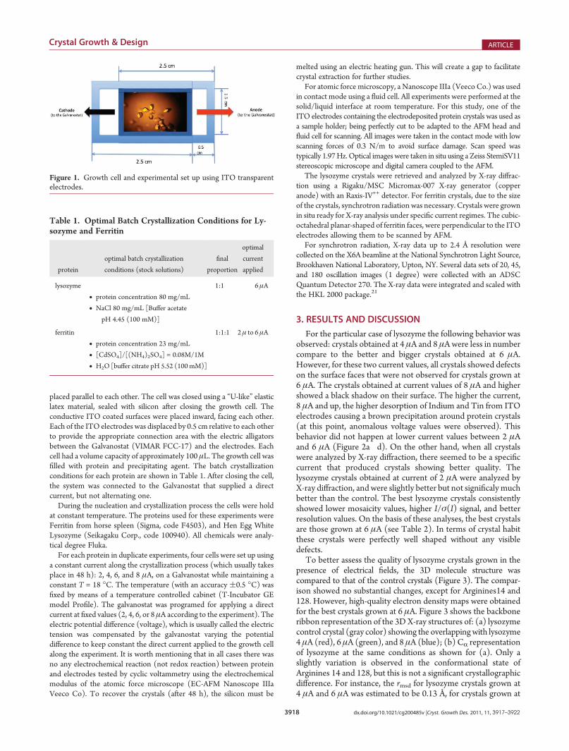
For the particular case of lysozyme the following behavior wasobserved: crystals obtained at 4 μA and 8 μAwere less in numbercompare to the better and bigger crystals obtained at 6 μA.However, for these two current values, all crystals showed defectson the surface faces that were not observed for crystals grown at6 μA. The crystals obtained at current values of 8 μA and highershowed a black shadow on their surface. The higher the current,8 μA and up, the higher desorption of Indium and Tin from ITOelectrodes causing a brown precipitation around protein crystals(at this point, anomalous voltage values were observed). Thisbehavior did not happen at lower current values between 2 μAand 6 μA (Figure 2a�d). On the other hand, when all crystalswere analyzed by X-ray diffraction, there seemed to be a specificcurrent that produced crystals showing better quality. Thelysozyme crystals obtained at current of 2 μA were analyzed byX-ray diffraction, and were slightly better but not significaly muchbetter than the control. The best lysozyme crystals consistentlyshowed lower mosaicity values, higher I/σ(I) signal, and betterresolution values. On the basis of these analyses, the best crystalsare those grown at 6 μA (see Table 2). In terms of crystal habitthese crystals were perfectly well shaped without any visibledefects.
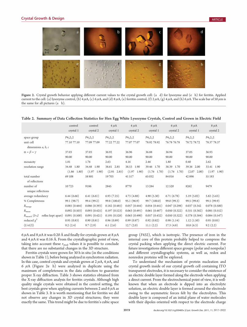
To better assess the quality of lysozyme crystals grown in thepresence of electrical fields, the 3D molecule structure wascompared to that of the control crystals (Figure 3). The compar-ison showed no substantial changes, except for Arginines14 and128. However, high-quality electron density maps were obtainedfor the best crystals grown at 6 μA. Figure 3 shows the backboneribbon representation of the 3DX-ray structures of: (a) lysozymecontrol crystal (gray color) showing the overlappingwith lysozyme4 μA (red), 6 μA (green), and 8 μA (blue); (b) CR representationof lysozyme at the same conditions as shown for (a). Only aslightly variation is observed in the conformational state ofArginines 14 and 128, but this is not a significant crystallographicdifference. For instance, the rmsd for lysozyme crystals grown at4 μA and 6 μA was estimated to be 0.13 Å, for crystals grown at
Figure 1. Growth cell and experimental set up using ITO transparentelectrodes.
Table 1. Optimal Batch Crystallization Conditions for Ly-sozyme and Ferritin
protein
optimal batch crystallization
conditions (stock solutions)
final
proportion
optimal
current
applied
lysozyme
• protein concentration 80 mg/mL
• NaCl 80 mg/mL [Buffer acetate
pH 4.45 (100 mM)]
1:1 6μA
ferritin
• protein concentration 23 mg/mL
• [CdSO4]/[(NH4)2SO4] = 0.08M/1M
• H2O [buffer citrate pH 5.52 (100 mM)]
1:1:1 2μ to 6μA
3919 dx.doi.org/10.1021/cg200485v |Cryst. Growth Des. 2011, 11, 3917–3922
Crystal Growth & Design ARTICLE
6 μA and 8 μA it was 0.20 Å and finally for crystals grown at 8 μAand 4 μA it was 0.16 Å. From the crystallographic point of view,taking into account these rmsd values it is possible to concludethat there are no substantial changes in the 3D structure.
Ferritin crystals were grown for 30 h in situ (in the conditionsshown in Table 1), before being analyzed in synchrotron radiation.In this case, control crystals and crystals grown at 2 μA, 4 μA, and6 μA (Figure 2e�h) were analyzed in duplicate using themaximum of completeness in the data collection to guaranteeproper X-ray diffraction. Table 3 shows statistics obtained fromthe X-ray diffraction analysis for ferritin crystals. Although highquality single crystals were obtained in the control setting, thebest crystals grew when applying currents between 2 and 6 μA asshown in Table 3. It is worth mentioning that for ferritin we didnot observe any changes in 3D crystal structures; they wereexactly the same. This trend might be due to ferritin’s cubic space
group (F432), which is isotropic. The presence of iron in theinternal core of this protein probably helped to compress thecrystal packing when applying the direct electric current. Forfuture investigations different space groups (polar and nonpolar)and different crystallographic systems, as well as, redox andnonredox proteins will be explored.
To understand the mechanism of protein nucleation andcrystal growth inside of our crystal growth cell containing ITOtransparent electrodes, it is necessary to consider the existence ofan electric double layer formed along the electrode when applyinga direct current. From the electrochemical point of view, it is well-known that when an electrode is dipped into an electrolytesolution, an electric double layer is formed around the electrodeowing to the asymmetric forces felt by the electrolytes. Thisdouble layer is composed of an initial plane of water moleculeswith their dipoles oriented with respect to the electrode charge
Figure 2. Crystal growth behavior applying different current values to the crystal growth cell: (a�d) for lysozyme and (e�h) for ferritin. Appliedcurrent to the cell: (a) lysozyme control, (b) 4 μA, (c) 6 μA, and (d) 8 μA, (e) ferritin control, (f) 2 μA, (g) 4 μA, and (h) 6 μA. The scale bar of 50 μm isthe same for all pictures (a�h).
Table 2. Summary of Data Collection Statistics for Hen Egg White Lysozyme Crystals, Control and Grown in Electric Field
control
crystal 1
control
crystal 2
4 μA
crystal 1
4 μA
crystal 2
6 μA
crystal 1
6 μA
crystal 2
8 μA
crystal 1
8 μA
crystal 2
space group P43212 P43212 P43212 P43212 P43212 P43212 P43212 P43212
unit cell
dimensions a, b, c
77.10 77.10 77.09 77.09 77.22 77.22 77.07 77.07 78.92 78.92 76.78 76.78 78.72 78.72 78.37 78.37
R = β = γ 37.03 37.03 36.92 36.96 36.88 36.94 37.05 36.93
90.00 90.00 90.00 90.00 90.00 90.00 90.00 90.00
mosaicity 1.81 1.76 2.63 4.16 2.46 1.80 0.40 2.62
resolution range 34.48�1.80
(1.86�1.80)
34.48�1.90
(1.97�1.90)
38.61�2.85
(2.95�2.85)
38.53�1.90
(1.97�1.90)
39.46�1.70
(1.76�1.70)
38.39�1.70
(1.76�1.70)
39.36�2.00
(2.07�2.00)
33.41�1.90
(1.97�1.90)
total number
of reflections
69 108 58 881 19 703 41 517 65 032 84 016 42 896 55 183
number of
unique reflections
10 723 9186 2845 8770 13 284 12 520 8262 9479
average redundancy 6.44 (6.66) 6.41 (6.61) 6.93 (7.25) 4.73 (4.88) 4.90 (5.30) 6.71 (6.78) 5.19 (5.02) 5.82 (5.63)
% Completeness 99.1 (98.7) 99.4 (99.2) 99.8 (100.0) 95.1 (96.9) 99.7 (100.0) 99.0 (99.3) 99.5 (99.8) 99.5 (99.9)
Rmerge 0.085 (0.464) 0.086 (0.393) 0.182 (0.485) 0.057 (0.444) 0.054 (0.441) 0.047 (0.298) 0.037 (0.342 0.078 (0.500)
Rmeas 0.092 (0.503) 0.093 (0.425) 0.197 (0.523) 0.063 (0.491) 0.061 (0.487) 0.050 (0.322) 0.351 (0.382) 0.085 (0.552)
RmeasA (I+,I� reflns kept apart) 0.091 (0.500) 0.091 (0.421) 0.195 (0.528) 0.063 (0.490) 0.057 (0.432) 0.050 (0.322) 0.378 (0.384) 0.084 (0.547)
reduced χ2 0.95 (0.83) 0.90 (0.81) 0.96 (0.89) 0.99 (0.87) 0.92 (0.82) 0.98 (1.14) 1.12 (1.50) 0.95 (0.85)
ÆI/σ(I)æ 9.2 (2.4) 9.7 (2.9) 6.1 (2.6) 12.7 (2.0) 11.1 (2.2) 17.3 (4.8) 10.8 (6.3) 9.2 (2.2)
3920 dx.doi.org/10.1021/cg200485v |Cryst. Growth Des. 2011, 11, 3917–3922
Crystal Growth & Design ARTICLE
that it is displaced by a second plane formed by hydrated ions ofopposite charge.22 The behavior of the attachment of lysozymemolecules (positively charged molecules at the pH value of thebuffer) to the cathode (a negatively charged electrode) could beexplained based on the double layer effect. The double layereffect of two ITO electrodes dipped in the growth cell containingthe protein solution, and the crystallizing agent may arise fromthe interaction of lysozyme with the ions present in the electricdouble layer around the electrode.
The polarity of ITO electrodes working as cathode (negativelycharged) or anode (positively charged), in this electrolytic cellhas been already represented in Figure 1. This experimentalsetup produces a migration of the positively charged lysozymemolecules (at pH 4.45) toward the cathode (�) or ferritinmolecules (negatively charged at pH 5.52) toward the anode(+). Therefore, the surface of the cathode (�) of this growth cellwill be initially filled with a plane of water molecules with theirdipoles oriented with respect to the electrode charge and subse-quently the positively charged macro-ions of the protein moleculeitself (positively charged at this pH value) will be attached on it.This effect leads to a disturbance of the system owing to the fact
that negatively charged ions (Cl�) are needed for the protein�protein interactions in lysozyme crystallization.23 This generaloverview explains why the crystals grew on the surface of theelectrode when it was used as negatively charged electrode. Forferritin molecules (negatively charged at this pH value) theprocess is exactly the opposite. However, as has been alreadydescribed byMoreno and Rivera,8 Cd2+ ions are fixed on the initialwater plane layer suffering a redox process forming a metal clustersand then the ferritin molecules are deposited on the surface ofmetallic cadmium. Then ferritin molecules are crystallized andgrown by the ammonium sulfate used as precipitating agent.
This electrochemistry-based method of crystallizing proteinsis not an electrocrystallization because there is no redox reactionoccurring between the protein and the electrode, but there is anelectro-focusing or electro-dialysis arising from the applied current.Something similar to this occurs in the classical electrophoresismethod.
It was noticed for both proteins, lysozyme and ferritin, thatcrystals grown in electric field, were strongly attached to thecathode (negatively charged electrode), and to the anode(positively charged electrode) respectively. The crystal adhesion
Figure 3. Ribbon representation of the 3D X-ray structure models of hen egg white lysozyme. (a) control (gray), 4 μA (red), 6 μA (green) and 8 μA(blue); (b) CR representation of the control crystal compared with the same structures shown in a.
Table 3. Summary of Data Collection Statistics for Horse Spleen Ferritin Crystals, Control and Grown in Electric Field
control 2 μA 4 μA 6 μA
space group F432 F432 F432 F432
unit cell dimensions 181.77 181.84 182.38 182.00
mosaicity 0.530 0.308 0.675 0.308
resolution range 25.00�2.27 (2.31�2.27) 25.00�1.97 (2.00�1.97) 25.00�2.24 (2.28�2.24) 25.00�2.07 (2.11�2.07)
total number of reflections 2 804 198 1 189 907 720 448 2 228 266
number of unique reflections 12400 18 819 13 091 16 282
average redundancy 20.6 (21.5) 11.7 (12.1) 5.0 (5.1) 20.8 (20.9)
% completeness 100.0 (100.0) 99.9 (100.0) 99.3 (99.1) 100.0 (100.0)
Rmerge 0.204 0.173 0.134 0.235
ÆI/σ(I)æ 25.0 (2.88) 21.2 (2.03) 13.7 (2.07) 16.7 (1.94)
3921 dx.doi.org/10.1021/cg200485v |Cryst. Growth Des. 2011, 11, 3917–3922
Crystal Growth & Design ARTICLE
was highly efficient, which made it easy to study the surface ofthese crystals with atomic force microscopy. The adhesion ofprotein crystals for research on crystal growth mechanisms, bymeans of atomic force microscopy (AFM), has been a handicapfor AFM investigations for a long time, although there are fewefforts about it already pusblished elsewhere suggesting differentexperimental designs for fixing 2D and 3D protein crystals.24�29
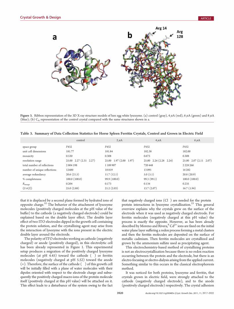
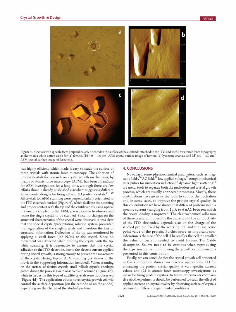
All crystals for AFM scanning were perpendicularly orientated tothe ITO electrode surface (Figure 4), which facilitate the scanningand proper contact with the tip and the cantilever. By using opticalmicroscopy coupled to the AFM, it was possible to observe andlocate the single crystal to be scanned. Since no changes on thestructural characteristics of the crystal were observed, it was clearthat the special crystal/precipitating solution system preventedthe degradation of the single crystals and therefore the loss ofstructural information. Deflection of the tip was monitored byapplying a small force (0.3 N/m) to the crystal. Since nomovement was detected when pushing the crystal with the tip,while scanning, it is reasonable to assume that the crystaladhesion to the ITO electrode, due to the electric current appliedduring crystal growth, is strong enough to prevent the movementof the crystal during typical AFM scanning (as shown in themovie in the Supporting Information included). When scanningon the surface of ferritin crystals small hillock crystals (perhapsgrownduring the process) were observed and scanned (Figure 4b),while in lysozyme this type of satellite crystals were not observed(Figure 4d). The application of this novel crystal growth cell willcontrol the surface deposition (on the cathode or on the anode)depending on the charge of the studied protein.
4. CONCLUSIONS
Nowadays, some physicochemical parameters, such as mag-netic fields,30 AC field,31 low applied voltage,32 nonphotochemicallaser pulses for nucleation induction,33 dynamic light scattering34
are useful tools to separate both the nucleation and crystal growthprocess, which are usually connected processes. Mostly, thesecontributions have given us the tools to control the nucleationand, in some cases, to improve the protein crystal quality. Inthis contribution we have shown that different proteins need aspecific current (ranging from 2 μA to 6 μA), between whichthe crystal quality is improved. The electrochemical adhesionof these crystals, imposed by the current and the conductivityof the ITO electrodes, depends also on the charge of thestudied protein fixed by the working pH, and the isoelectricpoint value of the protein. Further more an important con-sideration is the size of the cell. The smaller the cell the smallerthe value of current needed to avoid Indium Tin Oxidedesorption. So, we need to be cautious when reproducingthe experimental set up following the growth cell dimensionspresented in this contribution.
Finally, we can conclude that the crystal growth cell presentedin this contribution shows two practical applications: (1) forenhancing the protein crystal quality at very specific currentvalues, and (2) in atomic force microscopy investigations asmean for fixing protein crystals. In future experiments compara-tive AFM experiments should be performed to study the effect ofapplied current on crystal quality by observing surface of crystalsobtained in different experimental conditions.
Figure 4. Crystals with specific faces perpendicularly oriented to the surface of the electrode attached to the ITO and useful for atomic force topographyas shown in a white dotted circle for (a) ferritin, (b) 3.0� 3.0 μm2 AFM crystal surface image of ferritin, (c) lysozyme crystals, and (d) 5.0� 5.0 μm2
AFM crystal surface image of lysozyme.
3922 dx.doi.org/10.1021/cg200485v |Cryst. Growth Des. 2011, 11, 3917–3922
Crystal Growth & Design ARTICLE
’ASSOCIATED CONTENT
bS Supporting Information. Video as described in the text.This material is available free of charge via the Internet at http://pubs.acs.org.
’AUTHOR INFORMATION
Corresponding Author*Address: Instituto deQuimica, UniversidadNacional Autonoma deMexico, Circuito Exterior C.U.Mexico, D.F. 04510,Mexico. Phone:+52-55-56224467. Fax: +52-55-56162217. E-mail: [email protected].
’ACKNOWLEDGMENT
The authors acknowledge the X-ray diffraction from the Labor-atorio de Estructura de Proteinas-LANEM at UNAM (Mexico)and the help from M. Sci. Georgina E. Espinosa-P�erez. X-rayexperiments were carried out at the X6A beamline at the NationalSynchrotron Light Source supported by the NIGMS and DOEunder contract GM-0080 and DE-AC02-98CH10886. We ac-knowledge the professional grammar and style English revisiondone by Ms. Antonia S�anchez-Marín. We sincerely thank the helpof Dr. Juan Pablo Reyes-Grajeda from the National Institute of theGenomic Medicine (INMEGEN) for processing high qualitycrystallographic images of Lysozyme crystals grown at differentcurrents. One of the authors (R.R.R-A) acknowledges the PhDschoolarship from the Institute of Science and Technology ofMexico City (ICyTDF) as well as C.L.A.F., and schoolarship asresearch assistant from the SNI-CONACYT(Mexico). Finally oneof the authors (A.M.) acknowledges the finantial support fromDGAPA-UNAM Project PAPIIT No. IN201811-3.
’REFERENCES
(1) Chin, C.; Dence, J. B.; Warren, J. C. J. Biol. Chem. 1976, 251,3700–3705.(2) K€ohler, M.; Friedrich, J.; Fidy, J. Biochim. Biophys. Acta 1998,
1386, 255–288.(3) Fidy, J.; Balog, E.; K€ohler, M. Biochim. Biophys. Acta 1998,
1386, 289–303.(4) Taleb, M.; Didierjean, C.; Jelsch, C.; Mangeot, J. P.; Capelle, B.;
Aubry, A. J. Cryst. Growth 1999, 200, 575–582.(5) Taleb, M.; Didierjean, C.; Jelsch, C.; Mangeot, J. P.; Aubry, A.
J. Cryst. Growth 2001, 232, 250–255.(6) Nanev, C.; Penkova, A. J. Cryst. Growth 2001, 232, 285–293.(7) Mirkin, N.; Frontana-Uribe, B. A.; Rodríguez-Romero, A.;
Hern�andez-Santoyo, A.; Moreno, A. Acta Crystallogr., Sect. D: Biol.Crystallogr. 2003, 59, 1533–1538.(8) Moreno, A.; Rivera, M. Acta Crystallogr., Sect. D: Biol. Crystallogr.
2005, 61, 1678–1681.(9) Al-Haq, M. I.; Lebrasseur, E.; Tsuchiya, H.; Torii, T. Crystallogr.
Rev. 2007, 13, 29–64.(10) Frontana-Uribe, B. A.; Moreno, A. Cryst. Growth Des. 2008, 8,
4194–4199.(11) Zoubida, H.; Veesler, S. Prog. Biophys. Mol. Biol. 2009, 101,
38–44.(12) Bolanos-Garcia, V. M.; Chayen, N. Prog. Biophys. Mol. Biol.
2009, 101, 3–12.(13) Hammadi, Z.; Astier, J. P.; Morin, R.; Veesler, S. Cryst. Growth
Des. 2007, 7, 1472–1475.(14) Koizumi, H.; Fujiwara, K.; Uda, S. Cryst. Growth Des. 2009, 9,
2420–2424.
(15) Sazaki, G.; Moreno, A.; Nakajima, K. J. Cryst. Growth 2004,262, 499–502.
(16) Surade, S.; Ochi, T.; Nietlispach, D.; Chirgadze, D.; Moreno, A.Cryst. Growth Des. 2010, 10, 691–699.
(17) Gavira, J. A.; García-Ruiz, J. M. Cryst. Growth Des. 2009, 9,2610–2615.
(18) Yin, D. C.; Geng, L. Q.; Lu, Q. Q.; Lu, H. M.; Shang, P.;Wakayama, N. I. Cryst. Growth Des. 2009, 9, 5083–5091.
(19) Maki, S.; Ishikawa, K.; Ataka, M. J. Cryst. Growth 2009, 311,4725–4729.
(20) Kimura, F.; Mizutani, K.; Mikami, B.; Kimura, T. Cryst. GrowthDes. 2011, 11, 12–15.
(21) Otwinowski, Z.; Minor, W. Methods in Enzymology: Macromo-lecular Crystallography; Carter, C. W., Jr., Sweet, R. M., Eds.; AcademicPress: New York, 1997; Vol. 276, part A, pp 307�326.
(22) Crow, D. R. In Principles and Applications of Electrochemistry;Chapman and Hall, Ltd.: London, 1979; Chapter 7, pp 150�151.
(23) Vaney, M. C.; Broutin, I.; Retailleau, P.; Douangamath, A.;Lafont, S.; Hamiaux, C.; Prang�e, T.; Ducruix, A.; Ri�es-Kautt, M. ActaCrystallogr., Sect. D: Biol. Crystallogr. 2001, 57, 929–940.
(24) Dorn, I. T.; Hofmann, U. G.; Peltonen, J.; Tamp�e, R. Langmuir1998, 14, 4836–4842.
(25) Yamamoto, D.; Nagura, N.; Omote, S.; Taniguchi, M.; Ando, T.Biophys. J. 2009, 97, 2358–2367.
(26) El-Kirat, K.; Burton, I.; Dufrene, Y. F. J. Microsc. 2005, 218,199–207.
(27) Ko, T. P.; Kuznetsov, Y. G.; Malkin, A. J.; Day, J.; McPherson, A.Acta Crystallogr., Sect. D: Biol. Crystallogr. 2001, 57, 829–839.
(28) Hern�andez-P�erez, T.; Mirkin, N.; Moreno, A.; Rivera, M.Electrochem. Solid State Lett. 2002, 5, 37–39.
(29) Acosta, F.; Eid, D.; Marín-García, L.; Frontana-Uribe, B. A.;Moreno, A. Cryst. Growth Des. 2007, 7, 2187–2191.
(30) Sazaki, G. Prog. Biophys. Mol. Biol. 2009, 101, 45–55.(31) Hou, D.; Chang, H. C. App. Phys. Lett. 2008, 92, 223902.(32) Wakamatsu, T.; Ohnishi, Y. Jpn. J. Appl. Phys. 2011, 50, 048003-1–
048003-2.(33) Lee, I. S.; Evans, J. M. B.; Erdemir, D.; Lee, A. Y.; Garetz, B. A.;
Myerson, A. S. Cryst. Growth Des. 2008, 8, 4255–4261.(34) Saridakis, E.; Dierks, K.; Moreno, A.; Dieckmann, M. W. M.;
Chayen, N. Acta Crystallogr., Sect. D: Biol. Crystallogr. 2002, 58, 1597–1600.