Embed Size (px)
Citation preview
ACCEPTED MANUSCRIPT
ACCEPTED MANUSCRIP
T
1
Normal patterns and pitfalls of FDG uptake in the head and neck
Benjamin R. Gray, MD & Nicholas A. Koontz, MD
Indiana University School of Medicine
Department of Radiology and Imaging Sciences
Goodman Hall
355 West 16th Street, Ste. 4100
Indianapolis, IN 46202
Corresponding Author: Nicholas A. Koontz, MD
The authors report no disclosures, financial or otherwise
Downloaded for Anonymous User (n/a) at Indiana University Ruth Lilly Medical Library from ClinicalKey.com by Elsevier on July 22, 2019.
____________________________________________________
This is the author's manuscript of the article published in final edited form as:
Gray, B. R., & Koontz, N. A. (2019). Normal Patterns and Pitfalls of FDG Uptake in the Head and Neck. Seminars in Ultrasound, CT and MRI. https://doi.org/10.1053/j.sult.2019.07.001
ACCEPTED MANUSCRIPT
ACCEPTED MANUSCRIP
T
2
Abstract:
In order to avoid misdiagnoses, medical imagers should be familiar with the normal patterns and
distribution of FDG activity within the head and neck, as well as the pathophysiology and
imaging-findings of common diagnostic pitfalls related to incidental FDG-avid lesions. The
purpose of this article is to provide an image-rich review of the normal patterns of FDG uptake in
the head and neck, help differentiate benign from malignant incidentally found FDG-avid foci,
and detail important “don’t miss” hypometabolic head and neck lesions on PET/CT and
PET/MRI.
Key Words:
FDG, PET/CT, PET/MRI, radiotracer distribution, benign, malignant, pitfalls
Downloaded for Anonymous User (n/a) at Indiana University Ruth Lilly Medical Library from ClinicalKey.com by Elsevier on July 22, 2019.For personal use only. No other uses without permission. Copyright ©2019. Elsevier Inc. All rights reserved.
ACCEPTED MANUSCRIPT
ACCEPTED MANUSCRIP
T
3
Introduction:
Positron emission tomography/computed tomography (PET/CT) and, more
recently, positron emission tomography-magnetic resonance imaging (PET/MRI) with
fluorine-18-fluorodeoxyglucose (18F-FDG) are vital imaging modalities for the evaluation
of numerous and histopathologically-varied neoplasms within the head and neck.1-3 An
invaluable tool throughout the process of managing oncologic conditions, 18F-FDG
PET/CT is regularly utilized in head and neck cancer imaging for the detection, initial
diagnosis, and staging of tumors, evaluation of treatment response, monitoring for
recurrence, and long-term surveillance.2,4,5 A newer technology, 18F-FDG PET/MRI is
less established in the evaluation of head and neck cancer, but has shown promise
given its superior tissue contrast resolution, lower ionizing radiation dose, and superior
assessment of perineural tumor spread.
Despite their tremendous utility, the accurate interpretation of 18F-FDG PET/CT
and 18F-FDG PET/MRI requires comprehensive awareness and familiarity with many
diagnostic challenges and pitfalls.6-10 Owing to the detailed and challenging anatomy of
the region, as well confounding factors related to prior surgery and radiation, this
requirement is only magnified with head and neck oncologic imaging.6,7,10 The purpose
of this article is to provide a review of the normal patterns of 18F-FDG uptake in the head
and neck, as well as imaging findings of common diagnostic pitfalls related to false-
negative exams and incidentally found 18F-FDG -avid foci in the head and neck on
PET/CT and PET/MRI.
Basics of FDG Physiology and Imaging:
Downloaded for Anonymous User (n/a) at Indiana University Ruth Lilly Medical Library from ClinicalKey.com by Elsevier on July 22, 2019.For personal use only. No other uses without permission. Copyright ©2019. Elsevier Inc. All rights reserved.
ACCEPTED MANUSCRIPT
ACCEPTED MANUSCRIP
T
4
Though a detailed exposition of the physiology, physics, and specific protocols
for 18F-FDG PET/CT and 18F-FDG PET/MRI would far exceed the scope of this article, a
brief preliminary discussion of the modality’s core elements is necessary for
understanding its common interpretive pitfalls. The utility of PET in the evaluation of
malignant tumors is premised on altered glucose metabolism.1,3,11 In order to meet their
energy requirements, metabolically active tumor cells depend on glycolysis, which is
promoted by the activation of the hexose monophosphate pathway and the upregulated
expression of glucose transporter proteins and hexokinase within malignant cells.1,3,11
As an analog of glucose, intravenously administered 18F-FDG is taken up by tumor cells
via glucose transporter proteins and phosphorylated by hexokinase.1,11 However, unlike
glucose-6-phophate, once phosphorylated, 18F-FDG does not undergo further
metabolism in the glycolytic pathway.11 Rather, the phosphorylated 18F-FDG persistently
accumulates1 and, due to the lack of requisite amount of glucose-6-phosphatase,
remains trapped intracellularly within tumor cells.3,11 Consequently, 18F-FDG serves as a
useful representation of the degree of glucose uptake throughout the body when the
patient is subsequently imaged.3
To allow for an appropriate degree of 18F-FDG uptake, imaging of the patient is
typically performed approximately 60 – 90 minutes after 5 - 15 mCi (175 – 550 MBq) of
18F-FDG have been administered intravenously.12 Whole-body CT was first employed in
conjunction with PET imaging primarily to supplement the modality by providing
anatomic correlation and localization of molecular data and to enhance photon
attenuation correction.2,6,11-13 However, with improved technologies and imaging
techniques, PET imaging is now frequently combined not only with low-dose and/or
Downloaded for Anonymous User (n/a) at Indiana University Ruth Lilly Medical Library from ClinicalKey.com by Elsevier on July 22, 2019.For personal use only. No other uses without permission. Copyright ©2019. Elsevier Inc. All rights reserved.
ACCEPTED MANUSCRIPT
ACCEPTED MANUSCRIP
T
5
noncontrast CT but high-resolution, diagnostic-quality CT imaging or magnetic
resonance imaging.5,14 The concurrent acquisition of high-resolution CT imaging or MRI
often obviates the need for obtaining separate diagnostic-quality imaging,5 which is
paramount in head and neck oncology for accurate staging and surgical planning.
PET/CT and PET/MRI Imaging Pitfalls:
While the increased utilization of hybrid 18F-FDG PET/CT has significantly
improved the characterization of primary head and neck malignancies and the
identification of locoregional and metastatic disease, accurate interpretation of the
combined modality necessitates that imagers are familiar with its common imaging
pitfalls. Most commonly, challenges and pitfalls in oncologic head and neck PET/CT or
PET/MRI are related to physiologic uptake of 18F-FDG, incidental foci of increased 18F-
FDG uptake, and false-negative non-FDG avid lesions.6-10,15 Frequently encountered
examples of physiologic 18F-FDG uptake include metabolic activity associated with
normal lymphoid tissue, brown fat, thyroid and salivary gland tissue, and muscle
activity.6-10,16 Incidental foci of increased 18F-FDG uptake can also be seen with vocal
cord paralysis, incidental thyroid and salivary gland lesions, and infectious or
inflammatory processes.6-10,16 Though less common, false-negative lesions may not be
accurately characterized or missed due to location of the lesion and neoplastic
characteristics, such as histopathologic type and tumor size and degree of necrosis.5-
10,12
Incidental FDG Uptake
Downloaded for Anonymous User (n/a) at Indiana University Ruth Lilly Medical Library from ClinicalKey.com by Elsevier on July 22, 2019.For personal use only. No other uses without permission. Copyright ©2019. Elsevier Inc. All rights reserved.
ACCEPTED MANUSCRIPT
ACCEPTED MANUSCRIP
T
6
Thyroid Gland Uptake
On 18F-FDG PET imaging, the thyroid gland demonstrates variable 18F-FDG
uptake and, due to incidental focal or diffusely increased metabolic activity, can present
pitfalls for head and neck oncologic PET/CT or PET/MRI.7,10,15,17 Typically, physiologic
18F-FDG uptake within the thyroid gland is either absent or minimal and homogeneous
in distribution.16-18 Additional patterns of 18F-FDG uptake within the thyroid gland include
symmetric diffusely increased uptake and focal increased uptake, which can both be
seen with physiologic, benign, and pathological conditions.6,7,10,19 Diffuse symmetric
uptake of 18F-FDG within the thyroid gland usually represents either physiologic uptake
or a benign etiology, such as multinodular goiter, Grave disease, or chronic autoimmune
thyroiditis.7,10,17,19 (Figure 1)
When focal 18F-FDG uptake within the region of the thyroid gland is identified on
PET, it is important that the corresponding CT or MR images are carefully scrutinized,
as the uptake can be mistakenly attributed to adjacent cervical nodal uptake.10,16,20
Focal 18F-FDG uptake within the thyroid gland is nonspecific and can be seen with
benign conditions, such as a thyroid adenoma, or in the setting of thyroid
malignancy.6,7,10 The precise risk of malignancy in lesions with focal thyroid 18F-FDG
uptake is equivocal with reported risk of malignancy ranging from 25-63%.10,18,21,22 As a
result, it is recommended that foci of moderate to high 18F-FDG uptake undergo further
diagnostic evaluation with thyroid ultrasound and/or fine-needle aspiration biopsy.6,7,10,18
(Figure 2)
Salivary Gland Uptake
Downloaded for Anonymous User (n/a) at Indiana University Ruth Lilly Medical Library from ClinicalKey.com by Elsevier on July 22, 2019.For personal use only. No other uses without permission. Copyright ©2019. Elsevier Inc. All rights reserved.
ACCEPTED MANUSCRIPT
ACCEPTED MANUSCRIP
T
7
18F-FDG is normally taken up physiologically by the salivary glands and
subsequently excreted through the saliva.7,10 Accordingly, the parotid and
submandibular glands usually demonstrate symmetric low to moderate 18F-FDG
uptake,7,10,16 however the glands may occasionally demonstrate minimal or no uptake.7
The normal physiologic uptake within the parotid and submandibular glands can both
simulate and obscure salivary gland tumors.23 Additional patterns of 18F-FDG uptake
within the salivary glands include diffusely increased uptake and asymmetric or focal
increased uptake, which may present additional challenges to those interpreting
PET/CT and PET/MRI of the head and neck.7,10
There are a number of benign, non-neoplastic conditions that can result in
increased 18F-FDG uptake within the salivary glands.7,10 For example, infectious
etiologies, such as viral, bacterial, and tuberculous infections, radiation-induced
sialadenitis, obstructive/calculous sialadenitis, and inflammatory conditions, such as
sarcoidosis, can cause increased uptake within the salivary glands.6,7,10,24 (Figure 3)
Asymmetric uptake can also be observed secondary to compensatory hypertrophy
following contralateral gland resection.7,10
In addition to non-neoplastic etiologies, salivary gland tumors can demonstrate
increased 18F-FDG uptake, which is usually asymmetric and focal.6,7,10,23,25 (Figure 4)
While high-grade tumors in the salivary gland tend to have higher 18F-FDG uptake than
those of low or intermediate-grade,23 there are benign parotid gland tumors, such as
papillary cystadenoma lymphomatosum (Warthin tumor) and pleomorphic adenoma
(benign mixed tumor), which can also demonstrate increased uptake.6,7,10,23
Additionally, it is important to note that some malignant salivary gland tumors may not
Downloaded for Anonymous User (n/a) at Indiana University Ruth Lilly Medical Library from ClinicalKey.com by Elsevier on July 22, 2019.For personal use only. No other uses without permission. Copyright ©2019. Elsevier Inc. All rights reserved.

ACCEPTED MANUSCRIPT
ACCEPTED MANUSCRIP
T
8
demonstrate significantly elevated 18F-FDG uptake, including low grade
mucoepidermoid and adenoid cystic carcinomas.7,10,23 Thus, the lack of significant
uptake within a salivary gland lesion does not exclude a malignant etiology.7 Due to the
lack of definitive differentiation of benignity versus malignancy based on 18F-FDG
uptake, focal asymmetric salivary gland uptake always requires careful evaluation of the
associated CT or MRI component of the examination, as well as correlation with
pertinent patient history.7 Additional imaging, such as ultrasound or high-resolution MRI
may be complementary in better delineating lesion margins (circumscribed margins
suggest a benign tumor or low grade malignancy; infiltrating margins suggest a high
grade malignancy) and assessing lesion cellularity (low T2 or ADC signal intensity on
MRI suggests a cellular tumor that is more likely to be malignant), but definitive
diagnosis requires imaging-guided biopsy or surgical resection.10,25
Brown Fat
Within the human body, there are two morphologically and functionally distinct
types of adipose tissue: white adipose tissue (WAT) and brown adipose tissue (BAT).26
White adipose tissue has relatively low-level metabolic activity and primarily functions to
provide energy storage and insulation.27-29 However, more than displaying these
relatively inert characteristics, white adipose tissue, in so far as it secretes important
hormones and cytokines and plays an important role in insulin resistance and
inflammation, is now understood to also function as an endocrine organ.26,30 Brown
adipose tissue, while also storing energy,26 mainly serves to produce heat, which can
occur in the setting of cold temperatures (non-shivering thermogenesis) or following
Downloaded for Anonymous User (n/a) at Indiana University Ruth Lilly Medical Library from ClinicalKey.com by Elsevier on July 22, 2019.For personal use only. No other uses without permission. Copyright ©2019. Elsevier Inc. All rights reserved.
ACCEPTED MANUSCRIPT
ACCEPTED MANUSCRIP
T
9
food intake (diet-induced thermogenesis).26,28,29 In addition to its high vascularity,
considerable sympathetic noradrenergic innervation, and adrenergic receptor
expression, brown adipose tissue expresses a mitochondrial uncoupling protein.27-29
This protein facilitates uncoupling of oxidative phosphorylation within mitochondria,
which results in the direct production of heat instead of utilizing adenosine triphosphate
(ATP).27-29 These physiologic processes have important implications, due to the
resulting intense FDG uptake within brown adipose tissue on PET/CT and
PET/MRI.10,27,31
Typically, brown adipose tissue appears on 18F-FDG PET/CT and 18F-FDG
PET/MRI as symmetric, fusiform-shaped, curvilinear foci of FDG uptake that correspond
to areas of fat density (approximately -100 CT units) on CT images or fat signal intensity
(T1 and T2 bright) on MR images.7,10,29 (Figure 5) BAT can be found in the axillae,
intercostal spaces, and para-aortic and perinephric regions of the abdomen.29 However,
most commonly and of critical importance for head and neck cancer imaging, brown
adipose tissue is located within the deep cervical neck and supraclavicular
regions.10,28,29 Though the precise demographics, including prevalence, and the factors
contributing to the presence of brown adipose tissue on 18F-FDG PET/CT and 18F-FDG
PET/MRI remain uncertain, BAT has been observed within the head and neck in
approximately 2.5- 4.0% of imaged patients and has been overall more frequently
observed in children,32 women, and during the winter months.31 Corresponding to the
physiology of brown adipose tissue, proposed mechanisms to reduce 18F-FDG activity
within BAT includes increasing ambient room temperature, application of warm
Downloaded for Anonymous User (n/a) at Indiana University Ruth Lilly Medical Library from ClinicalKey.com by Elsevier on July 22, 2019.For personal use only. No other uses without permission. Copyright ©2019. Elsevier Inc. All rights reserved.
ACCEPTED MANUSCRIPT
ACCEPTED MANUSCRIP
T
10
blankets, and the administration of medications prior to imaging, such as propranolol,
diazepam, and fentanyl.7,29,31,32
Muscle Uptake
Due to the number and small size of muscles within the head and neck,
physiologic muscle uptake of 18F-FDG represents an additional challenge for oncologic
imaging of the head and neck.16 Physiologic muscle uptake of 18F-FDG can be
observed with involuntary muscle activity (Figure 6), voluntary muscle activity (Figure 7),
and in response to insulin administration or recent food ingestion.33 For example, focal
uptake within the extraocular muscles can occur secondary to eye motion.10,16 With
talking and phonation, 18F-FDG uptake may commonly be seen within the genioglossus,
cricopharyngeus, and posterior cricoarytenoid muscles.10 Physiologic uptake associated
with masseteric activity may also be seen with talking or chewing.33 Besides 18F-FDG
uptake within particular muscle groups, diffuse skeletal uptake can also occur
secondary to voluntary activity related to recent strenuous exercise.33
In addition to uptake associated with these voluntary activities, 18F-FDG uptake is
frequently observed within the sternocleidomastoid, longus colli, anterior scalene,
longus capitis, and obliquus capitis inferior muscles due to anxiety associated muscle
contraction.7,9,10,16,33,34 Similarly, scalene and sternocleidomastoid muscle uptake can be
also be seen with coughing or labored breathing.33 In addition, if the patient is imaged
following the administration of insulin or in the postprandial state, the patient may exhibit
diffuse 18F-FDG skeletal muscle uptake.33,35
Downloaded for Anonymous User (n/a) at Indiana University Ruth Lilly Medical Library from ClinicalKey.com by Elsevier on July 22, 2019.For personal use only. No other uses without permission. Copyright ©2019. Elsevier Inc. All rights reserved.
ACCEPTED MANUSCRIPT
ACCEPTED MANUSCRIP
T
11
On 18F-FDG PET/CT and 18F-FDG PET/MRI, muscle uptake is reliably
distinguished by mild to moderate symmetric and linear 18F-FDG uptake that can be
followed on corresponding CT or MR images from the insertion of the muscle to its
origin.6,10,35 The linearity may not always be evident on axial images and scrutinizing the
coronal and sagittal reconstructed CT or acquired multiplanar MR images is helpful to
demonstrate the typically linear configuration of uptake within the muscle or muscle
group. In order to avoid the challenges associated with physiologic muscle uptake,
certain preventative measures ought to be taken prior to imaging. The patient should be
asked to fast for at least 4-6 hours35 and to refrain from intense exercise for at least 2435
to 4833 hours prior imaging. Additionally, during the uptake component of the PET/CT of
PET/MRI examination after 18F-FDG has been administered, the patient should be
asked to remain as calm and relaxed as possible and to avoid any muscle activity, such
as talking, chewing, or performing other physical activities that might promote FDG
uptake.6,10,33,35 In order to help reduce anxiety-related muscle uptake, benzodiazepine
administration prior to the examination can also be considered.10,35,36
Vocal Cord Paralysis
Occasionally, asymmetric 18F-FDG uptake within the vocal cords can present a
diagnostic challenge on PET/CT or PET/MRI. Asymmetric 18F-FDG vocal cord uptake
often occurs in the setting of unilateral vagus or recurrent laryngeal nerve injury, which
may occur secondary to involvement of the nerve by a tumor or injury from prior surgery
or trauma.7,10,37 In order to offset the paralysis of the affected vocal cord, the
functioning, non-paralyzed contralateral vocal cord demonstrates compensatory
Downloaded for Anonymous User (n/a) at Indiana University Ruth Lilly Medical Library from ClinicalKey.com by Elsevier on July 22, 2019.For personal use only. No other uses without permission. Copyright ©2019. Elsevier Inc. All rights reserved.
ACCEPTED MANUSCRIPT
ACCEPTED MANUSCRIP
T
12
increased activity during phonation.7,10,37 (Figure 8) This heightened activity results in
corresponding increased metabolism and glucose, particularly within the thyroarytenoid
and posterior circoarytenoid muscles.10,38 It is imperative to scrutinize the associated
anatomic CT or MR images through the level of the vocal cords, as focal uptake can
simulate a lesion within the normal vocal cord.7,10
On the PET component of PET/CT or PET/MRI, vocal cord paralysis, as a result
of the compensatory increased activity and associated heightened metabolism and
glucose uptake, results in focal avid 18F-FDG uptake within the non-paralyzed vocal
cord.7,10,33,37 If asymmetric focal uptake is identified within a vocal cord, inspection of the
contralateral vocal cord should be performed to assess for common imaging findings of
paralysis or dysfunction. These findings typically include a paramedian location of the
paralyzed vocal cord, dilated ipsilateral laryngeal ventricle (“Spinnaker sail” sign),39
medialized and thickened ipsilateral aryepiglottic fold, dilated ipsilateral pyriform sinus,
and, possibly, posterior cricoarytenoid and thyroarytenoid muscle atrophy.10,39-41
In addition to the characteristic imaging features on CT of vocal cord paralysis,
focal 18F-FDG uptake within the vocal cord should also be correlated with available
pertinent patient clinical information.10 History and physical examination findings of
hoarseness and a documented history of previous radiation or surgery to the neck or
mediastinum may corroborate and explain observed imaging findings.10,41 Additionally,
as vocal cord paralysis can be caused by disease occurring at any location from the
brainstem to the distal aspect of the recurrent laryngeal nerves, it is imperative that the
expected course of the involved vagus (jugular foramen and carotid space) and
recurrent laryngeal nerves (tracheoesophageal groove bilaterally, aortopulmonary
Downloaded for Anonymous User (n/a) at Indiana University Ruth Lilly Medical Library from ClinicalKey.com by Elsevier on July 22, 2019.For personal use only. No other uses without permission. Copyright ©2019. Elsevier Inc. All rights reserved.
ACCEPTED MANUSCRIPT
ACCEPTED MANUSCRIP
T
13
window on the left, and below subclavian artery on the right) are inspected on CT or MR
images to evaluate for contributing pathology.39-41
Lymphoid Tissue and Inflammatory Processes
Focal increased 18F-FDG uptake within the head and neck on PET/CT or
PET/MRI, which can potentially simulate a neoplastic process, is commonly
encountered as result of physiological uptake within lymphoid tissue.6,7,9,16 The tissues
of the head and neck are abound with lymphatic structures, including lymphatic
channels, lymph nodes, and the nasopharyngeal (adenoid), palatine, and base of
tongue (lingual) tonsillar tissue of the Waldeyer ring.6-8,10,16 Physiologic uptake within
lymphoid tissue, which occurs secondary to the accumulation of 18F-FDG within
lymphocytes and macrophages, can symmetric or asymmetric.6,7,10 Unfortunately, the
pattern of uptake is not always helpful as symmetric uptake, though generally
reassuring, can occasionally be seen with malignancy, such as in extra-nodal non-
Hogkin lymphoma and squamous cell carcinoma.6,10 Similarly, focal asymmetric uptake
does not necessarily imply malignancy and can be seen with be seen with benign
variant physiological uptake.6,7,10 In attempting to decipher benign from malignant
etiologies on PET/CT or PET/MRI, it is imperative that the CT or MR images are
carefully inspected for corresponding abnormalities, such as the presence of a mass
lesion, morphological asymmetry, and loss of fat planes with infiltration of adjacent
spaces and structures.10 Ultimately, equivocal findings require correlation with patient
history and clinical evaluation, including endoscopy.10
Downloaded for Anonymous User (n/a) at Indiana University Ruth Lilly Medical Library from ClinicalKey.com by Elsevier on July 22, 2019.For personal use only. No other uses without permission. Copyright ©2019. Elsevier Inc. All rights reserved.
ACCEPTED MANUSCRIPT
ACCEPTED MANUSCRIP
T
14
In addition to physiological uptake within lymphoid tissue, there are numerous
benign infectious, inflammatory, and granulomatous conditions that, as a result of
increased glycolysis within macrophages, also commonly demonstrate focal 18F-FDG
uptake and can mimic head and neck malignancy.6-8,10 Owing to chemotherapy-
associated immunosuppression, focal uptake at sites of infection or within reactive
lymph nodes is frequently encountered with 18F-FDG PET/CT and 18F-FDG PET/MRI of
head and neck malignancies.6,9 Similarly, focal 18F-FDG uptake is commonly seen with
indwelling ports and catheters.6,10,42 Additional etiologies for inflammatory associated
focal uptake within the head and neck include vasculitides, foreign body granulomas,
and active atherosclerotic plaque.6,10 With active atherosclerotic plaque, 18F-FDG
accumulates within plaque associated inflammatory cells, and the degree of uptake
appears to correspond to the severity of vessel wall inflammation.6,10,43 (Figure 9)
The interpretation of 18F-FDG PET/CT and 18F-FDG PET/MRI of head and neck
malignancy is further complicated by the imaging pitfalls associated with postsurgical
and recent radiation inflammatory changes.6,8-10 As a result of tissue injury, surgery
generates an inflammatory response8,42 with the ensuing development of edema,
granulation tissue, and scarring.10 The inflammatory process may have associated
elevated 18F-FDG uptake, which, in addition to postsurgical anatomic changes on CT,
can make evaluation on PET/CT or PET/MRI examinations performed after surgery
challenging.6,8,10 As the uptake associated with postsurgical inflammatory changes
steadily decreases over the few weeks after surgery, the first postoperative 18F-FDG
PET/CT or 18F-FDG PET/MRI should optimally be delayed 4-6 weeks after surgery to
avoid false-positive imaging findings.6,10 Similarly, while 18F-FDG PET/CT has shown to
Downloaded for Anonymous User (n/a) at Indiana University Ruth Lilly Medical Library from ClinicalKey.com by Elsevier on July 22, 2019.For personal use only. No other uses without permission. Copyright ©2019. Elsevier Inc. All rights reserved.
ACCEPTED MANUSCRIPT
ACCEPTED MANUSCRIP
T
15
be of significant utility in the evaluation of head and neck tumor recurrence,5,10 the focal
18F-FDG uptake associated with postradiation inflammatory changes within the field of
radiation can simulate residual tumor or recurrence.4,6,10,12 Though the ideal timing
remains uncertain and controversial,8 the first 18F-FDG PET/CT or 18F-FDG PET/MRI
performed after radiation ought to be delayed at least 2-3 months to hopefully allow for
resolution of the inflammatory changes and associated false-positive PET imaging
findings.10
Common False Negatives:
In addition to incidental 18F-FDG uptake, imagers must be aware of the frequently
encountered causes of false-negative head and neck 18F-FDG PET/CT or 18F-FDG
PET/MRI examinations. Most commonly, false-negative studies within the head and
neck are due to concerning lesions that may not demonstrate significant 18F-FDG
uptake. There are a number of factors that may contribute to the observed lack of
significant 18F-FDG uptake within tumors of the head and neck, including prior
treatment, the tumor’s location, histopathological type, size, and degree of tumor
necrosis.5,8,10,12 For example, false-negative results can be seen when tumors are
located within areas that closely approximate tissues that have high physiologic 18F-
FDG uptake, such as lesions in or near the skull base that are close to the brain or
tumors within the oral cavity that are near tonsillar tissue.6,10 Additionally, head and neck
tumors or lymph nodes that are small in size (approximately 5-8 mm),12 can be missed
or misinterpreted due to limitations of PET spatial resolution and partial volume
effect.6,8,10,12
Downloaded for Anonymous User (n/a) at Indiana University Ruth Lilly Medical Library from ClinicalKey.com by Elsevier on July 22, 2019.For personal use only. No other uses without permission. Copyright ©2019. Elsevier Inc. All rights reserved.
ACCEPTED MANUSCRIPT
ACCEPTED MANUSCRIP
T
16
Besides a tumor’s size and location, false-negative results on head and neck 18F-
FDG PET/CT and 18F-FDG PET/MRI can be seen due to the particular histopathology
type and degree of necrosis. Some types of malignancy that are commonly seen or can
occur within the head and neck, such as well-differentiated thyroid cancer, adenoid
cystic and mucoepidermoid salivary gland tumors, and spindle cell tumors, may
demonstrate low 18F-FDG uptake, consequently producing false-negative findings.8,10
Further, if the tumor or lymph node has significant necrosis, the lesion may demonstrate
falsely-negative absent or low 18F-FDG uptake due to the lack of requisite metabolically
active tissue.5,8,10 (Figure 10) Lastly, false-negative results on head and neck 18F-FDG
PET/CT and 18F-FDG PET/MRI can also be observed on post-treatment imaging
studies. While recent radiation and surgery are common causes of false-positive
findings within the head and neck,6,8-10 radiation and chemotherapy can also contribute
to false-negative results.6 If imaging is performed too early following the completion of
radiation or chemotherapy, falsely-negative absent or low 18F-FDG uptake within the
tumor may be observed due to altered kinetics of 18F-FDG uptake.6,12
In addition to low uptake, false-negative head and neck 18F-FDG PET/CT and
18F-FDG PET/MRI examinations can occur due to misleading symmetric 18F-FDG
accumulation within potentially concerning lesions. While most malignancies within the
head and neck demonstrate asymmetric 18F-FDG uptake, some malignancies may
manifest bilateral disease or may demonstrate metabolic symmetricity. For example,
squamous cell carcinomas of the tongue base or nasopharynx may demonstrate
bilateral 18F-FDG uptake.10 These instances of symmetric and bilateral 18F-FDG uptake
emphasize the importance of carefully evaluating the CT and MR images for the
Downloaded for Anonymous User (n/a) at Indiana University Ruth Lilly Medical Library from ClinicalKey.com by Elsevier on July 22, 2019.For personal use only. No other uses without permission. Copyright ©2019. Elsevier Inc. All rights reserved.
ACCEPTED MANUSCRIPT
ACCEPTED MANUSCRIP
T
17
presence of associated suspicious findings, such as a mass lesion or the loss of normal
fat planes.10
Conclusions:
18F-FDG PET/CT and 18F-FDG PET/MRI are essential tools for oncologic
imaging and of tremendous utility in the diagnosis and staging of head and neck cancer.
However, the accurate interpretation of these modalities necessitates that head and
neck imagers are cognizant of the many diagnostic challenges and pitfalls commonly
found within the head and neck on 18F-FDG PET/CT and 18F-FDG PET/MRI. In order to
avoid misdiagnosis, imagers should be familiar with the pathophysiology and normal
patterns of 18F-FDG uptake within the head and neck, common false-negative (non-
FDG avid) lesions, and troubleshooting of incidental foci of 18F-FDG uptake. Moreover,
head and neck imagers should be aware of the appropriate recommendations to
provide referring clinicians when imaging pitfalls and challenges are encountered.
References
1. Bar-Shalom R, Valdivia AY, Blaufox MD. PET imaging in oncology. Semin Nucl
Med. 2000;30(3):150-185.
2. Blodgett TM, Meltzer CC, Townsend DW. PET/CT: form and function. Radiology.
2007;242(2):360-385.
Downloaded for Anonymous User (n/a) at Indiana University Ruth Lilly Medical Library from ClinicalKey.com by Elsevier on July 22, 2019.For personal use only. No other uses without permission. Copyright ©2019. Elsevier Inc. All rights reserved.
ACCEPTED MANUSCRIPT
ACCEPTED MANUSCRIP
T
18
3. Czernin J, Phelps ME. Positron emission tomography scanning: current and
future applications. Annu Rev Med. 2002;53:89-112.
4. Porceddu SV, Jarmolowski E, Hicks RJ, et al. Utility of positron emission
tomography for the detection of disease in residual neck nodes after
(chemo)radiotherapy in head and neck cancer. Head & neck. 2005;27(3):175-
181.
5. Subramaniam RM, Truong M, Peller P, Sakai O, Mercier G. Fluorodeoxyglucose-
positron-emission tomography imaging of head and neck squamous cell cancer.
AJNR Am J Neuroradiol. 2010;31(4):598-604.
6. Bhargava P, Rahman S, Wendt J. Atlas of confounding factors in head and neck
PET/CT imaging. Clin Nucl Med. 2011;36(5):e20-29.
7. Blodgett TM, Fukui MB, Snyderman CH, et al. Combined PET/CT in the head
and neck: part 1. Physiologic, altered physiologic, and artifactual FDG uptake.
Radiographics. 2005;25(4):897-912.
8. Fukui MB, Blodgett TM, Snyderman CH, et al. Combined PET/CT in the head
and neck: part 2. Diagnostic uses and pitfalls of oncologic imaging.
Radiographics. 2005;25(4):913-930.
9. Kapoor V, Fukui MB, McCook BM. Role of 18FFDG PET/CT in the treatment of
head and neck cancers: posttherapy evaluation and pitfalls. AJR Am J
Roentgenol. 2005;184(2):589-597.
10. Purohit BS, Ailianou A, Dulguerov N, Becker CD, Ratib O, Becker M. FDG-
PET/CT pitfalls in oncological head and neck imaging. Insights Imaging.
2014;5(5):585-602.
Downloaded for Anonymous User (n/a) at Indiana University Ruth Lilly Medical Library from ClinicalKey.com by Elsevier on July 22, 2019.For personal use only. No other uses without permission. Copyright ©2019. Elsevier Inc. All rights reserved.
ACCEPTED MANUSCRIPT
ACCEPTED MANUSCRIP
T
19
11. Kapoor V, McCook BM, Torok FS. An introduction to PET/CT imaging.
Radiographics. 2004;24(2):523-543.
12. Castaigne C, Muylle K, Flamen P. Positron Emission Tomography in Head and
Neck Cancer. In: Hermans R, ed. Head and Neck Cancer Imaging. Berlin,
Heidelberg: Springer Berlin Heidelberg; 2006:329-343.
13. Townsend DW. Positron emission tomography/computed tomography. Semin
Nucl Med. 2008;38(3):152-166.
14. Wong TZ, Paulson EK, Nelson RC, Patz EF, Jr., Coleman RE. Practical
approach to diagnostic CT combined with PET. AJR Am J Roentgenol.
2007;188(3):622-629.
15. Britt CJ, Maas AM, Kennedy TA, Hartig GK. Incidental Findings on FDG PET/CT
in Head and Neck Cancer. Otolaryngol Head Neck Surg. 2018;158(3):484-488.
16. Kostakoglu L, Hardoff R, Mirtcheva R, Goldsmith SJ. PET/CT fusion imaging in
differentiating physiologic from pathologic FDG uptake. Radiographics.
2004;24(5):1411-1431.
17. Bae JS, Chae BJ, Park WC, et al. Incidental thyroid lesions detected by FDG-
PET/CT: prevalence and risk of thyroid cancer. World J Surg Oncol. 2009;7:63.
18. Liu Y. Clinical significance of thyroid uptake on F18-fluorodeoxyglucose positron
emission tomography. Annals of nuclear medicine. 2009;23(1):17-23.
19. Are C, Hsu JF, Schoder H, Shah JP, Larson SM, Shaha AR. FDG-PET detected
thyroid incidentalomas: need for further investigation? Annals of surgical
oncology. 2007;14(1):239-247.
Downloaded for Anonymous User (n/a) at Indiana University Ruth Lilly Medical Library from ClinicalKey.com by Elsevier on July 22, 2019.For personal use only. No other uses without permission. Copyright ©2019. Elsevier Inc. All rights reserved.
ACCEPTED MANUSCRIPT
ACCEPTED MANUSCRIP
T
20
20. Lin EC. Thyroid nodule mimicking cervical adenopathy on FDG positron emission
tomographic imaging. Clinical nuclear medicine. 2002;27(9):656-657.
21. Chen W, Parsons M, Torigian DA, Zhuang H, Alavi A. Evaluation of thyroid FDG
uptake incidentally identified on FDG-PET/CT imaging. Nucl Med Commun.
2009;30(3):240-244.
22. Choi JY, Lee KS, Kim HJ, et al. Focal thyroid lesions incidentally identified by
integrated 18F-FDG PET/CT: clinical significance and improved characterization.
Journal of nuclear medicine : official publication, Society of Nuclear Medicine.
2006;47(4):609-615.
23. Roh JL, Ryu CH, Choi SH, et al. Clinical utility of 18F-FDG PET for patients with
salivary gland malignancies. J Nucl Med. 2007;48(2):240-246.
24. Hadiprodjo D, Ryan T, Truong MT, Mercier G, Subramaniam RM. Parotid gland
tumors: preliminary data for the value of FDG PET/CT diagnostic parameters.
AJR Am J Roentgenol. 2012;198(2):W185-190.
25. Basu S, Houseni M, Alavi A. Significance of incidental fluorodeoxyglucose uptake
in the parotid glands and its impact on patient management. Nucl Med Commun.
2008;29(4):367-373.
26. Coelho M, Oliveira T, Fernandes R. Biochemistry of adipose tissue: an endocrine
organ. Archives of medical science : AMS. 2013;9(2):191-200.
27. Cronin CG, Prakash P, Daniels GH, et al. Brown fat at PET/CT: correlation with
patient characteristics. Radiology. 2012;263(3):836-842.
28. Paidisetty S, Blodgett TM. Brown fat: atypical locations and appearances
encountered in PET/CT. AJR Am J Roentgenol. 2009;193(2):359-366.
Downloaded for Anonymous User (n/a) at Indiana University Ruth Lilly Medical Library from ClinicalKey.com by Elsevier on July 22, 2019.For personal use only. No other uses without permission. Copyright ©2019. Elsevier Inc. All rights reserved.
ACCEPTED MANUSCRIPT
ACCEPTED MANUSCRIP
T
21
29. Yeung HW, Grewal RK, Gonen M, Schoder H, Larson SM. Patterns of (18)F-
FDG uptake in adipose tissue and muscle: a potential source of false-positives
for PET. J Nucl Med. 2003;44(11):1789-1796.
30. Galic S, Oakhill JS, Steinberg GR. Adipose tissue as an endocrine organ. Mol
Cell Endocrinol. 2010;316(2):129-139.
31. Agrawal A, Nair N, Baghel NS. A novel approach for reduction of brown fat
uptake on FDG PET. Br J Radiol. 2009;82(980):626-631.
32. Gelfand MJ, O'Hara S M, Curtwright LA, Maclean JR. Pre-medication to block
[(18)F]FDG uptake in the brown adipose tissue of pediatric and adolescent
patients. Pediatr Radiol. 2005;35(10):984-990.
33. Jackson RS, Schlarman TC, Hubble WL, Osman MM. Prevalence and patterns
of physiologic muscle uptake detected with whole-body 18F-FDG PET. J Nucl
Med Technol. 2006;34(1):29-33.
34. Jacene HA, Goudarzi B, Wahl RL. Scalene muscle uptake: a potential pitfall in
head and neck PET/CT. Eur J Nucl Med Mol Imaging. 2008;35(1):89-94.
35. Karunanithi S, Soundararajan R, Sharma P, Naswa N, Bal C, Kumar R.
Spectrum of Physiologic and Pathologic Skeletal Muscle (18)F-FDG Uptake on
PET/CT. AJR Am J Roentgenol. 2015;205(2):W141-149.
36. Barrington SF, Maisey MN. Skeletal muscle uptake of fluorine-18-FDG: effect of
oral diazepam. J Nucl Med. 1996;37(7):1127-1129.
37. Kamel EM, Goerres GW, Burger C, von Schulthess GK, Steinert HC. Recurrent
laryngeal nerve palsy in patients with lung cancer: detection with PET/CT image
fusion -- report of six cases. Radiology. 2002;224(1):153-156.
Downloaded for Anonymous User (n/a) at Indiana University Ruth Lilly Medical Library from ClinicalKey.com by Elsevier on July 22, 2019.For personal use only. No other uses without permission. Copyright ©2019. Elsevier Inc. All rights reserved.
ACCEPTED MANUSCRIPT
ACCEPTED MANUSCRIP
T
22
38. Romo LV, Curtin HD. Atrophy of the posterior cricoarytenoid muscle as an
indicator of recurrent laryngeal nerve palsy. AJNR Am J Neuroradiol.
1999;20(3):467-471.
39. Koontz NA, Seltman TA, Kralik SF, Mosier KM, Harnsberger HR. Classic signs in
head and neck imaging. Clin Radiol. 2016;71(12):1211-1222.
40. Chin SC, Edelstein S, Chen CY, Som PM. Using CT to localize side and level of
vocal cord paralysis. AJR Am J Roentgenol. 2003;180(4):1165-1170.
41. Paquette CM, Manos DC, Psooy BJ. Unilateral vocal cord paralysis: a review of
CT findings, mediastinal causes, and the course of the recurrent laryngeal
nerves. Radiographics. 2012;32(3):721-740.
42. Bhargava P, Zhuang H, Kumar R, Charron M, Alavi A. Iatrogenic artifacts on
whole-body F-18 FDG PET imaging. Clinical nuclear medicine. 2004;29(7):429-
439.
43. Jezovnik MK, Zidar N, Lezaic L, Gersak B, Poredos P. Identification of inflamed
atherosclerotic lesions in vivo using PET/CT. Inflammation. 2014;37(2):426-434.
Downloaded for Anonymous User (n/a) at Indiana University Ruth Lilly Medical Library from ClinicalKey.com by Elsevier on July 22, 2019.For personal use only. No other uses without permission. Copyright ©2019. Elsevier Inc. All rights reserved.
ACCEPTED MANUSCRIPT
ACCEPTED MANUSCRIP
T
23
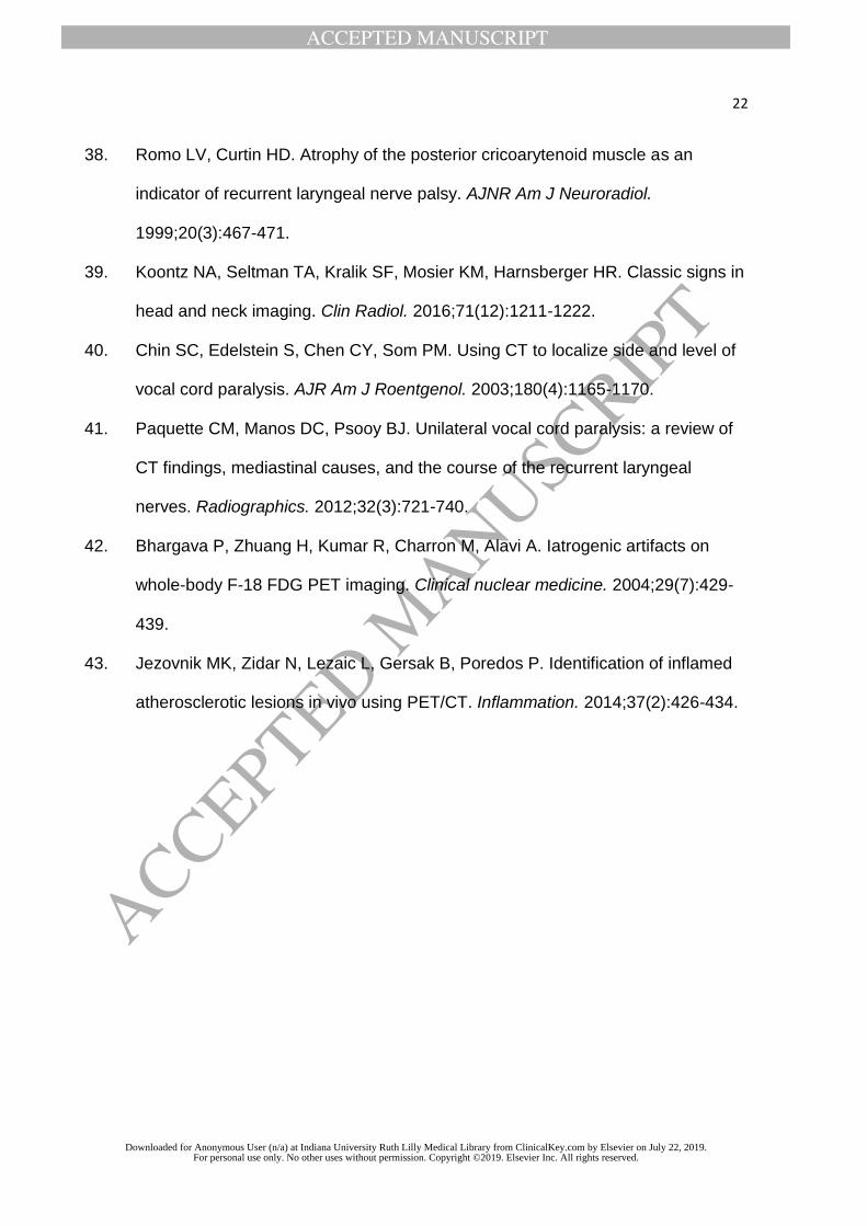
Figure 1. Incidental symmetric, diffusely increased thyroid FDG uptake. Fused axial
FDG-PET/CT (a) in a patient with Hodgkin lymphoma demonstrates diffuse mildly
increased thyroid gland FDG uptake (white arrows). The patient’s history and laboratory
values were compatible with subclinical hypothyroidism. Fused axial FDG-PET/CT (b) in
different patient with a history of Hodgkin lymphoma demonstrates significant diffuse
thyroid gland uptake (white arrowheads). The patient’s history and laboratory values
were also compatible with hypothyroidism.
Downloaded for Anonymous User (n/a) at Indiana University Ruth Lilly Medical Library from ClinicalKey.com by Elsevier on July 22, 2019.For personal use only. No other uses without permission. Copyright ©2019. Elsevier Inc. All rights reserved.
ACCEPTED MANUSCRIPT
ACCEPTED MANUSCRIP
T
24
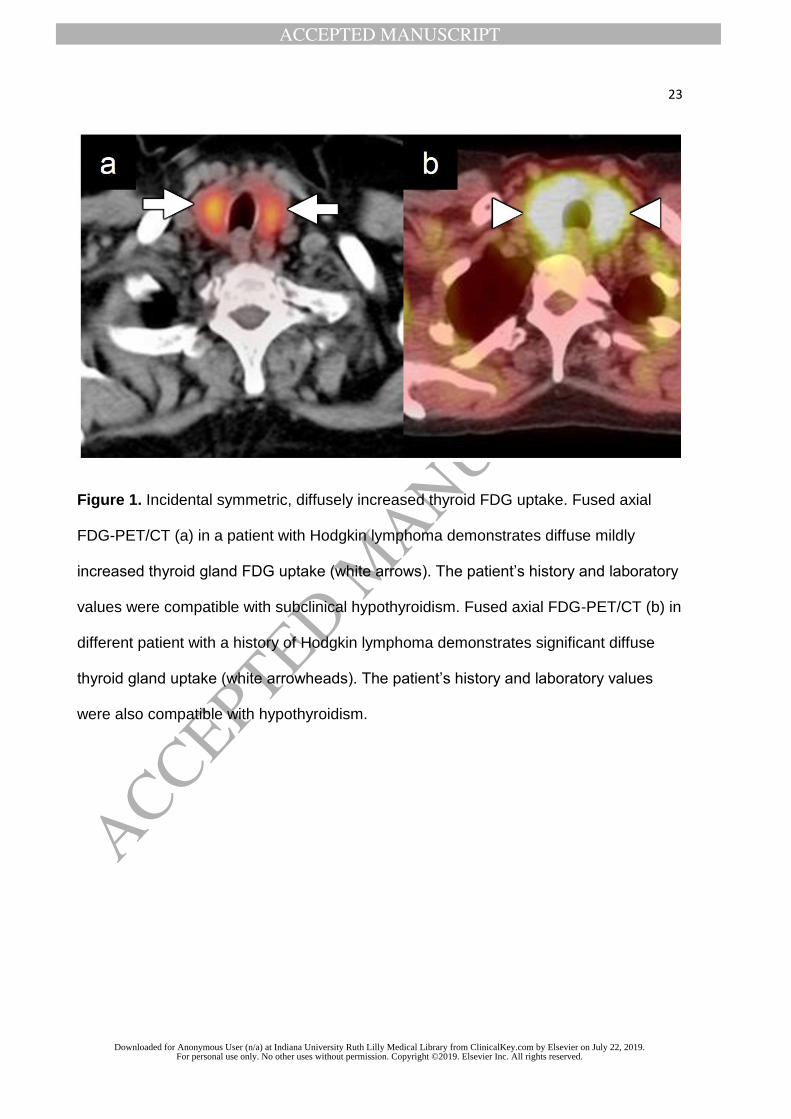
Figure 2. Incidental focal thyroid FDG uptake. Fused axial FDG-PET/CT (a)
demonstrates an enlarged thyroid gland with a small focus of mildly increased uptake
within the left mid gland (white arrow) in a patient with a history of squamous cell
carcinoma of the right maxilla presenting for initial staging. Ultrasound guided biopsy of
the lesion was performed with pathology demonstrating benign thyroid nodule. Fused
axial FDG-PET/CT (b) shows a focus of markedly increased uptake within the left lobe
of the thyroid (white arrowhead) in a patient with a history of breast cancer status post
lumpectomy and chemotherapy presenting for restaging. Ultrasound guided biopsy of
the lesion was performed with pathology demonstrating follicular neoplasm. However,
the lesion was subsequently resected with pathology demonstrating benign follicular
nodule.
Downloaded for Anonymous User (n/a) at Indiana University Ruth Lilly Medical Library from ClinicalKey.com by Elsevier on July 22, 2019.For personal use only. No other uses without permission. Copyright ©2019. Elsevier Inc. All rights reserved.
ACCEPTED MANUSCRIPT
ACCEPTED MANUSCRIP
T
25
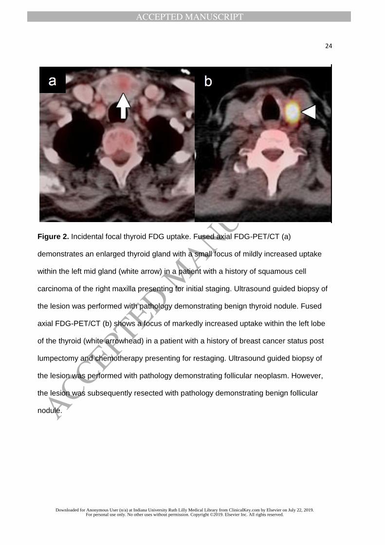
Figure 3. Asymmetric submandibular gland FDG uptake. Fused axial FDG-PET/CT (a)
demonstrates asymmetric increased FDG uptake within the left submandibular gland
(white arrow) relative to the right submandibular gland (white arrowhead). Axial NECT
image in the same patient shows fatty atrophy of the right submandibular gland (black
arrow) with associated large ductal calcifications (black arrowhead) from sialoliths that
have chronically obstructed the submandibular duct. The left submandibular shows
compensatory enlargement, but is otherwise normal in appearance.
Downloaded for Anonymous User (n/a) at Indiana University Ruth Lilly Medical Library from ClinicalKey.com by Elsevier on July 22, 2019.For personal use only. No other uses without permission. Copyright ©2019. Elsevier Inc. All rights reserved.
ACCEPTED MANUSCRIPT
ACCEPTED MANUSCRIP
T
26
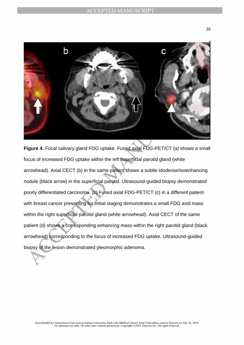
Figure 4. Focal salivary gland FDG uptake. Fused axial FDG-PET/CT (a) shows a small
focus of increased FDG uptake within the left superficial parotid gland (white
arrowhead). Axial CECT (b) in the same patient shows a subtle idodense/isoenhancing
nodule (black arrow) in the superficial parotid. Ultrasound-guided biopsy demonstrated
poorly differentiated carcinoma. (c) Fused axial FDG-PET/CT (c) in a different patient
with breast cancer presenting for initial staging demonstrates a small FDG avid mass
within the right superficial parotid gland (white arrowhead). Axial CECT of the same
patient (d) shows a corresponding enhancing mass within the right parotid gland (black
arrowhead) corresponding to the focus of increased FDG uptake. Ultrasound-guided
biopsy of the lesion demonstrated pleomorphic adenoma.
Downloaded for Anonymous User (n/a) at Indiana University Ruth Lilly Medical Library from ClinicalKey.com by Elsevier on July 22, 2019.For personal use only. No other uses without permission. Copyright ©2019. Elsevier Inc. All rights reserved.
ACCEPTED MANUSCRIPT
ACCEPTED MANUSCRIP
T
27
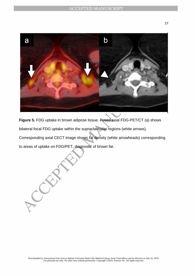
Figure 5. FDG uptake in brown adipose tissue. Fused axial FDG-PET/CT (a) shows
bilateral focal FDG uptake within the supraclavicular regions (white arrows).
Corresponding axial CECT image shows fat density (white arrowheads) corresponding
to areas of uptake on FDG/PET, diagnostic of brown fat.
Downloaded for Anonymous User (n/a) at Indiana University Ruth Lilly Medical Library from ClinicalKey.com by Elsevier on July 22, 2019.For personal use only. No other uses without permission. Copyright ©2019. Elsevier Inc. All rights reserved.

ACCEPTED MANUSCRIPT
ACCEPTED MANUSCRIP
T
28
Figure 6. Involuntary muscular activity with FDG uptake. Fused axial PET/CT (a) shows
symmetric increased FDG uptake along the posterior wall of the nasopharynx (white
arrows). Axial NECT (b) shows corresponding normal appearance of the longus colli
(black arrows), which is confirmed on (c) axial T1-weighted MRI (white arrowheads).
Downloaded for Anonymous User (n/a) at Indiana University Ruth Lilly Medical Library from ClinicalKey.com by Elsevier on July 22, 2019.For personal use only. No other uses without permission. Copyright ©2019. Elsevier Inc. All rights reserved.
ACCEPTED MANUSCRIPT
ACCEPTED MANUSCRIP
T
29
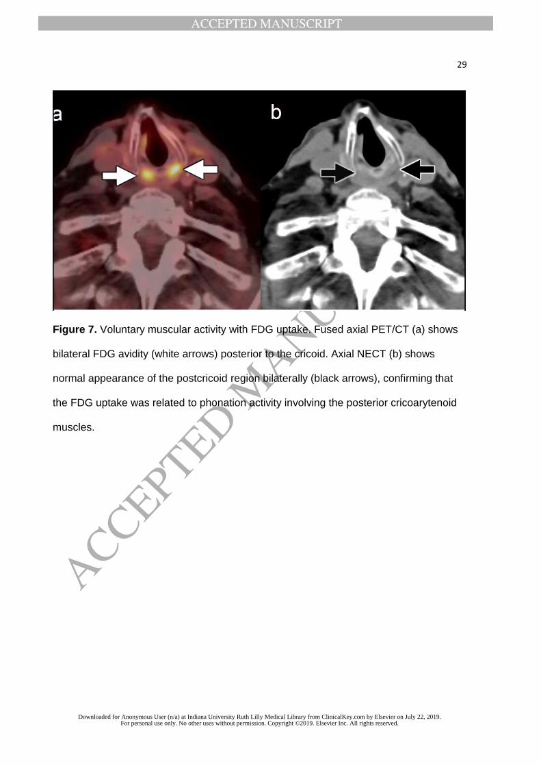
Figure 7. Voluntary muscular activity with FDG uptake. Fused axial PET/CT (a) shows
bilateral FDG avidity (white arrows) posterior to the cricoid. Axial NECT (b) shows
normal appearance of the postcricoid region bilaterally (black arrows), confirming that
the FDG uptake was related to phonation activity involving the posterior cricoarytenoid
muscles.
Downloaded for Anonymous User (n/a) at Indiana University Ruth Lilly Medical Library from ClinicalKey.com by Elsevier on July 22, 2019.For personal use only. No other uses without permission. Copyright ©2019. Elsevier Inc. All rights reserved.
ACCEPTED MANUSCRIPT
ACCEPTED MANUSCRIP
T
30
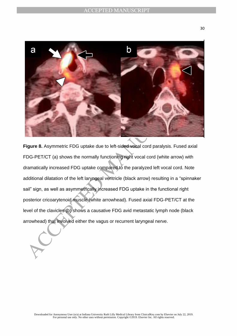
Figure 8. Asymmetric FDG uptake due to left-sided vocal cord paralysis. Fused axial
FDG-PET/CT (a) shows the normally functioning right vocal cord (white arrow) with
dramatically increased FDG uptake compared to the paralyzed left vocal cord. Note
additional dilatation of the left laryngeal ventricle (black arrow) resulting in a “spinnaker
sail” sign, as well as asymmetrically increased FDG uptake in the functional right
posterior cricoarytenoid muscle (white arrowhead). Fused axial FDG-PET/CT at the
level of the clavicles (b) shows a causative FDG avid metastatic lymph node (black
arrowhead) that involved either the vagus or recurrent laryngeal nerve.
Downloaded for Anonymous User (n/a) at Indiana University Ruth Lilly Medical Library from ClinicalKey.com by Elsevier on July 22, 2019.For personal use only. No other uses without permission. Copyright ©2019. Elsevier Inc. All rights reserved.
ACCEPTED MANUSCRIPT
ACCEPTED MANUSCRIP
T
31
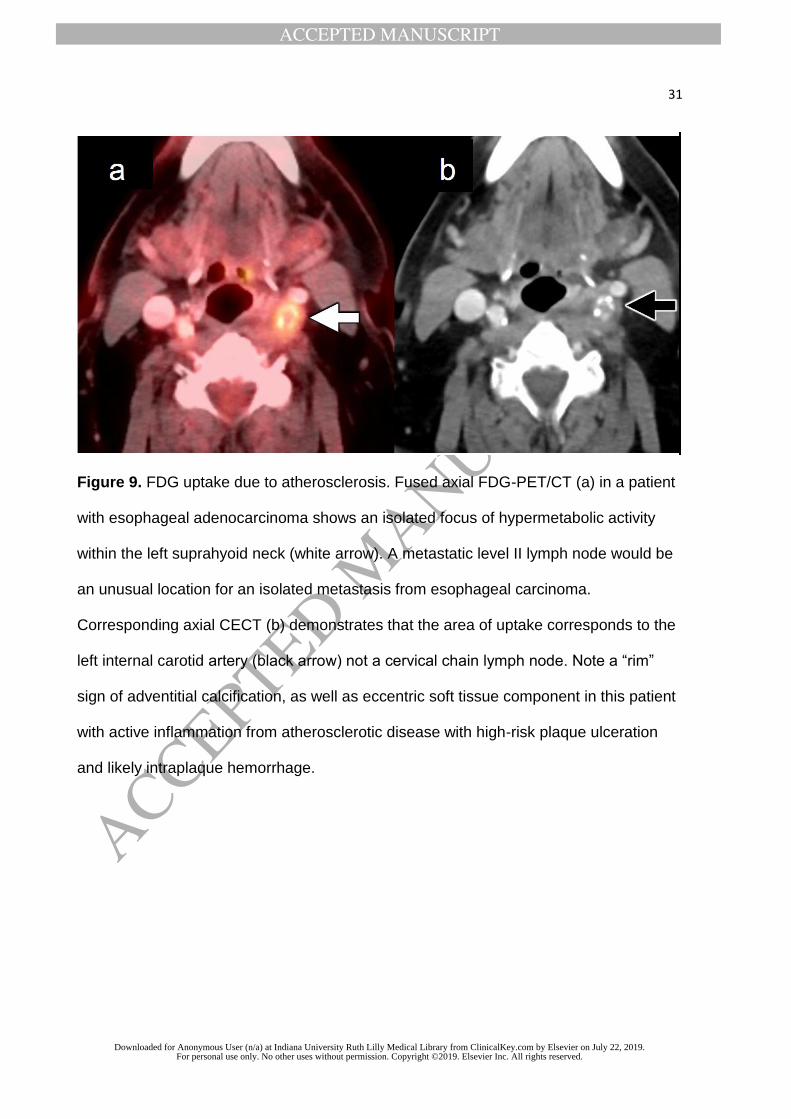
Figure 9. FDG uptake due to atherosclerosis. Fused axial FDG-PET/CT (a) in a patient
with esophageal adenocarcinoma shows an isolated focus of hypermetabolic activity
within the left suprahyoid neck (white arrow). A metastatic level II lymph node would be
an unusual location for an isolated metastasis from esophageal carcinoma.
Corresponding axial CECT (b) demonstrates that the area of uptake corresponds to the
left internal carotid artery (black arrow) not a cervical chain lymph node. Note a “rim”
sign of adventitial calcification, as well as eccentric soft tissue component in this patient
with active inflammation from atherosclerotic disease with high-risk plaque ulceration
and likely intraplaque hemorrhage.
Downloaded for Anonymous User (n/a) at Indiana University Ruth Lilly Medical Library from ClinicalKey.com by Elsevier on July 22, 2019.For personal use only. No other uses without permission. Copyright ©2019. Elsevier Inc. All rights reserved.
ACCEPTED MANUSCRIPT
ACCEPTED MANUSCRIP
T
32
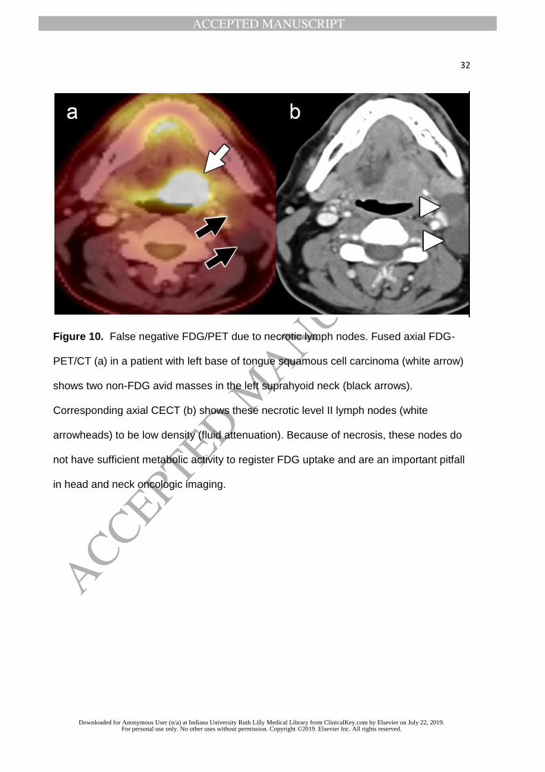
Figure 10. False negative FDG/PET due to necrotic lymph nodes. Fused axial FDG-
PET/CT (a) in a patient with left base of tongue squamous cell carcinoma (white arrow)
shows two non-FDG avid masses in the left suprahyoid neck (black arrows).
Corresponding axial CECT (b) shows these necrotic level II lymph nodes (white
arrowheads) to be low density (fluid attenuation). Because of necrosis, these nodes do
not have sufficient metabolic activity to register FDG uptake and are an important pitfall
in head and neck oncologic imaging.
Downloaded for Anonymous User (n/a) at Indiana University Ruth Lilly Medical Library from ClinicalKey.com by Elsevier on July 22, 2019.For personal use only. No other uses without permission. Copyright ©2019. Elsevier Inc. All rights reserved.
ACCEPTED MANUSCRIPT
ACCEPTED MANUSCRIP
T
33
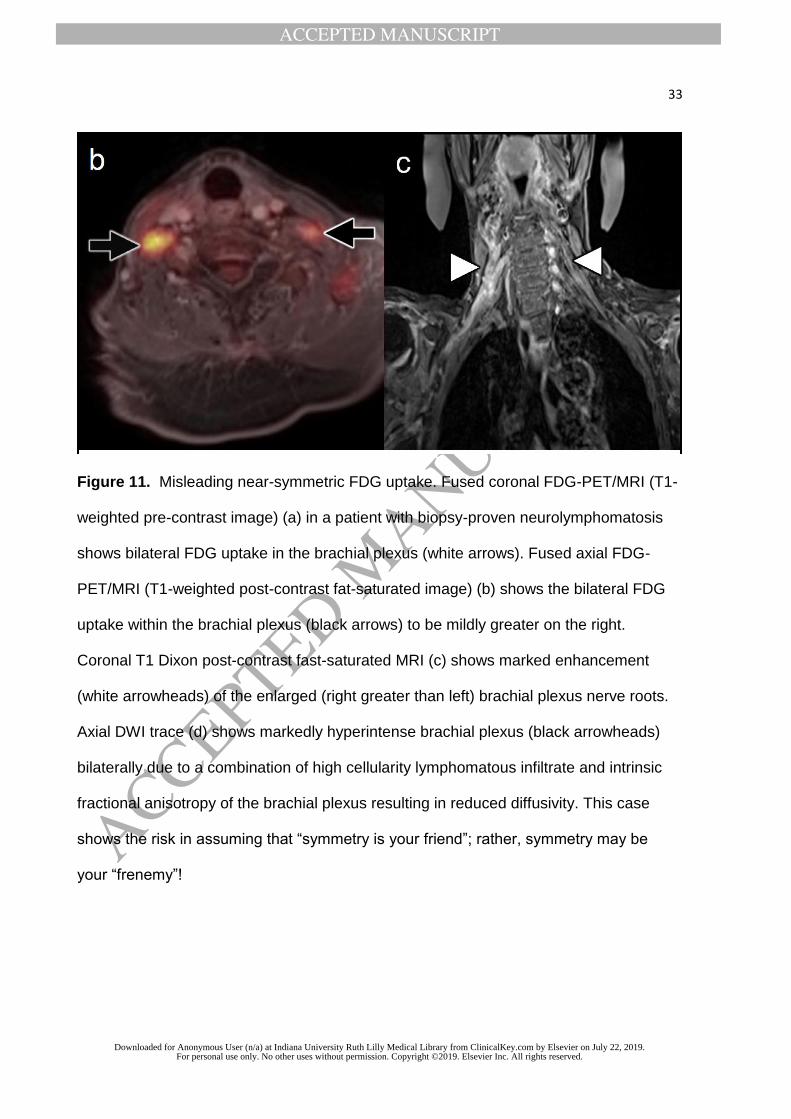
Figure 11. Misleading near-symmetric FDG uptake. Fused coronal FDG-PET/MRI (T1-
weighted pre-contrast image) (a) in a patient with biopsy-proven neurolymphomatosis
shows bilateral FDG uptake in the brachial plexus (white arrows). Fused axial FDG-
PET/MRI (T1-weighted post-contrast fat-saturated image) (b) shows the bilateral FDG
uptake within the brachial plexus (black arrows) to be mildly greater on the right.
Coronal T1 Dixon post-contrast fast-saturated MRI (c) shows marked enhancement
(white arrowheads) of the enlarged (right greater than left) brachial plexus nerve roots.
Axial DWI trace (d) shows markedly hyperintense brachial plexus (black arrowheads)
bilaterally due to a combination of high cellularity lymphomatous infiltrate and intrinsic
fractional anisotropy of the brachial plexus resulting in reduced diffusivity. This case
shows the risk in assuming that “symmetry is your friend”; rather, symmetry may be
your “frenemy”!
Downloaded for Anonymous User (n/a) at Indiana University Ruth Lilly Medical Library from ClinicalKey.com by Elsevier on July 22, 2019.For personal use only. No other uses without permission. Copyright ©2019. Elsevier Inc. All rights reserved.

![[18F]FDG uptake of bone marrow on PET/CT for predicting ......BLR ≥ 0.91 had a distant recurrence rate of 40.7%. Conclusions: BLR on pretreatment [18F]FDG PET/CT were significant](https://img.pdfslide.us/doc/110x75/60de3dd8893f706a1901a451/18ffdg-uptake-of-bone-marrow-on-petct-for-predicting-blr-a-091-had.jpg)


![Pulmonary 18F-FDG uptake helps refine current risk ... · self-propagating scar formation and end-stage fibrosis [10]. 18F-FDG uptake by tissues is a marker of glucose utilization,](https://img.pdfslide.us/doc/110x75/6035c829b976e577c9150e6c/pulmonary-18f-fdg-uptake-helps-refine-current-risk-self-propagating-scar-formation.jpg)
![FDG-PET in Large Vessel Vasculitis...FDG-PET in Large Vessel Vasculitis 61 5. [18 F]FDG-PET and [18 F]FDG-PET/CT [18 F]FDG-PET is an operator-independent, non- invasive imaging modality](https://img.pdfslide.us/doc/110x75/5f6c13132f0609183b646bce/fdg-pet-in-large-vessel-vasculitis-fdg-pet-in-large-vessel-vasculitis-61-5.jpg)