Embed Size (px)
Citation preview

HAL Id: inserm-01293422https://www.hal.inserm.fr/inserm-01293422
Submitted on 24 Mar 2016
HAL is a multi-disciplinary open accessarchive for the deposit and dissemination of sci-entific research documents, whether they are pub-lished or not. The documents may come fromteaching and research institutions in France orabroad, or from public or private research centers.
L’archive ouverte pluridisciplinaire HAL, estdestinée au dépôt et à la diffusion de documentsscientifiques de niveau recherche, publiés ou non,émanant des établissements d’enseignement et derecherche français ou étrangers, des laboratoirespublics ou privés.
No Association Between CEL-HYB Hybrid Allele andChronic Pancreatitis in Asian Populations.
Wen-Bin Zou, Arnaud Boulling, Atsushi Masamune, Prachand Issarapu,Emmanuelle Masson, Hao Wu, Xiao-Tian Sun, Liang-Hao Hu, Dai-Zhan
Zhou, Lin He, et al.
To cite this version:Wen-Bin Zou, Arnaud Boulling, Atsushi Masamune, Prachand Issarapu, Emmanuelle Masson, et al..No Association Between CEL-HYB Hybrid Allele and Chronic Pancreatitis in Asian Populations..Gastroenterology, WB Saunders, 2016, �10.1053/j.gastro.2016.02.071�. �inserm-01293422�

1
No Association Between CEL–HYB Hybrid Allele and Chronic Pancreatitis
in Asian Populations
Short Title: CEL-HYB Variants and Chronic Pancreatitis
Wen-Bin Zou,1,2,3,4,*
Arnaud Boulling,2,3,5,*
Atsushi Masamune,6,*
Prachand Issarapu,7,*
Emmanuelle Masson,2,8
Hao Wu,1,4
Xiao-Tian Sun,1,4
Liang-Hao Hu,1,4
Dai-Zhan Zhou,9
Lin He,9
Yann Fichou,2,3
Eriko Nakano,6 Shin Hamada,
6 Yoichi Kakuta,
6 Kiyoshi Kume,
6
Hiroyuki Isayama,10
Sumit Paliwal,7 K Radha Mani,
7 Seema Bhaskar,
7 David N. Cooper,
11
Claude Férec,2,3,5,8
Tooru Shimosegawa6,#
, Giriraj R Chandak,7,12#
Jian-Min Chen,2,3,5,#
Zhao-Shen Li,1,4,#
and Zhuan Liao1,4,#
*Authors share co-first authorship.
#Authors share co-senior authorship.
1Department of Gastroenterology, Changhai Hospital, the Second Military Medical
University, Shanghai, China; 2Institut National de la Santé et de la Recherche Médicale
(INSERM), U1078, Brest, France; 3Etablissement Français du Sang (EFS) – Bretagne, Brest,
France; 4Shanghai Institute of Pancreatic Diseases, Shanghai, China;
5Faculté de Médecine et
des Sciences de la Santé, Université de Bretagne Occidentale (UBO), Brest, France; 6Division
of Gastroenterology, Tohoku University Graduate School of Medicine, Sendai, Japan;
7Genomic Research on Complex Diseases, CSIR-Centre for Cellular and Molecular Biology
(CSIR-CCMB), Hyderabad, India; 8Laboratoire de Génétique Moléculaire et
d’Histocompatibilité, Centre Hospitalier Universitaire (CHU) Brest, Hôpital Morvan, Brest,
France; 9Key Laboratory of Developmental Genetics and Neuropsychiatric Diseases (Ministry
of Education), Bio-X Institutes, Shanghai Jiao Tong University, Shanghai, China;

2
10Department of Gastroenterology, Graduate School of Medicine, the University of Tokyo,
Tokyo, Japan; 11
Institute of Medical Genetics, School of Medicine, Cardiff University,
Cardiff, United Kingdom; 12
Human Genetics Division, Genome Institute of Singapore,
Biopolis, Singapore
Grant Support
WBZ is a joint PhD student between Changhai Hospital and INSERM U1078 and received a
one-year scholarship (the year 2015) from the China Scholarship Council (No.
201403170271). Support for this study came from the National Natural Science Foundation of
China (Grant Nos. 81470884, 81422010 [Z.L.]), the Shanghai Rising-Star Program (Grant
No.13QA1404600 [Z.L.]), the HIROMI Medical Research Foundation (A.M.), the Mother
and Child Health Foundation (A.M.), the Smoking Research Foundation (A.M.), the Pancreas
Research Foundation of Japan (E.N.), the Ministry of Health, Labour and Welfare of Japan
(Principal investigators: Yoichi Matsubara and Yoshifumi Takeyama), Council of Scientific
and Industrial Research, Ministry of Science and Technology, Government of India (Grant
No. BSC0121 [G.R.C.]), the Conseil Régional de Bretagne, the Association des Pancréatites
Chroniques Héréditaires, the Association de Transfusion Sanguine et de Biogénétique Gaetan
Saleun, and the Institut National de la Santé et de la Recherche Médicale (INSERM), France.
Abbreviations used in this paper: CP, chronic pancreatitis; NAHR, non-allelic homologous
recombination; NMD, nonsense-mediated mRNA decay; RT-PCR, reverse transcription-PCR;
SNP, single-nucleotide polymorphism; VNTR, variable number tandem repeat; WT, wild-
type.

3
Correspondence to:
Zhuan Liao, MD, Department of Gastroenterology, Changhai Hospital, the Second Military
Medical University, 168 Changhai Road, Shanghai 200433, China. e-mail:
[email protected].; Tel.: 86-21-31161344; Fax: 0086-21-55621735.
Zhao-Shen Li, MD, Department of Gastroenterology, Changhai Hospital, the Second Military
Medical University, 168 Changhai Road, Shanghai 200433, China. e-mail:
[email protected]; Tel.: 86-21-31161335; Fax: 0086-21-55621735.
Jian-Min Chen, MD, PhD, INSERM U1078 and EFS – Bretagne, 46 rue Félix Le Dantec,
29218 Brest 29218, France; [email protected]; Tel.: +33-2-98449333; Fax: +33-
2-98430555
Conflicts of Interest
The authors are unaware of any conflicts of interest.
Author Contributions
JMC, ZSL and ZL designed and directed the overall project, with the assistance of CF. AM
and TS directed the Japanese study whereas GRC directed the Indian study. WBZ, AM, PI
performed the genetic analysis, with substantial contributions from EM, HW, XTS, LHH,
DZZ, LH, EN, SH, YK, KK, HI, SP, KRM and SB. EM established the modified genotyping
method. WBZ and AB performed the functional analysis, with contributions from YF. JMC
and ZL wrote the manuscript, with substantial contributions from WBZ, AB, EM, AM and
GRC. DNC, CF and ZSL critically revised the manuscript with important intellectual input.

4
AM, GRC, ZSL and ZL recruited study subjects. AM, CF, GRC and ZL obtained the funding.
All authors approved the final manuscript.

5
ABSTRACT
A hybrid allele between the carboxyl ester lipase gene (CEL) and its pseudogene, CELP
(called CEL–HYB), generated by non-allelic homologous recombination between CEL intron
10 and CELP intron 10′, was found to increase susceptibility to chronic pancreatitis in a case–
control study of patients of European ancestry. We attempted to replicate this finding in 3
independent cohorts from China, Japan, and India, but failed to detect the CEL–HYB allele in
any of these populations. The CEL–HYB allele might therefore be an ethnic-specific risk
factor for chronic pancreatitis. An alternative hybrid allele (CEL–HYB2) was identified in all
3 Asian populations (1.7% combined carrier frequency), but was not associated with chronic
pancreatitis.
Keywords: Disease Susceptibility; Human Genetics; Nonsense-Mediated mRNA Decay;
Pancreatic Acinar Cells.
6
The etiology of chronic pancreatitis (CP) is complex and involves a subtle interplay between
genetic and environmental factors. Most of the hitherto reported genes/loci affecting disease
susceptibility encode proteins of the protease-antiprotease system of the pancreatic acinar
cells.1-7
A hybrid allele (CEL–HYB), involving the CEL gene (encoding pancreatic carboxyl
ester lipase8) and its tandemly linked pseudogene (CELP), was recently reported to be
significantly overrepresented in CP cases as compared with controls, firstly in a discovery
cohort of patients with familial CP and then in three replication cohorts of patients with
idiopathic CP.9 The CEL–HYB allele resulted from non-allelic homologous recombination
(NAHR) occurring between CEL intron 10 and CELP intron 10′; replacement of the eleventh
and last exon of CEL by CELP exon 11′ would yield a premature stop codon within the third
“pseudo” 33-bp variable number tandem repeat (VNTR)9 (Figure 1A). The mutant enzyme is
more stable than its wild-type (WT) counterpart and induced autophagy in cellular models,9
suggesting a novel pathogenic mechanism. However, since the patients analyzed in the
original study9 were solely of European ancestry, replication in independent populations of
different ethnicity was warranted.10
The detection of the CEL–HYB allele is dependent upon a long-range duplex PCR
assay.9 Using French CEL–HYB positive samples, we found that the originally described
PCR assay9 yielded better results under slightly modified conditions (see Supplementary
Material and Supplementary Figure 1). Consequently, these modified conditions were
employed to screen for the CEL–HYB allele in three Asian populations. We first analyzed a
cohort of Han Chinese patients with idiopathic CP. The previously described CEL–HYB-
specific 3.2-kb band was detected in 2.4% (19/799) of patients and 1.9% (20/1028) of controls
(P = 0.64; Table 1). Sequencing of the 3.2-kb band-containing PCR products however
revealed that none of these Chinese CEL–HYB alleles corresponded to that expected; instead,
they invariably resulted from an NAHR event occurring within a 239-bp sequence tract
7
affecting the intron 9/exon 10 boundary of CEL and the intron 9′/exon 10′ boundary of CELP
(Supplementary Figure 2). Hereafter, we term the previously reported disease-associated
CEL–HYB allele9 as CEL–HYB1 so as to distinguish it from this newly described allele,
CEL–HYB2.
Closer inspection of the 39 Chinese CEL–HYB2 alleles revealed that they could be
divided into two subtypes by reference to three single nucleotide polymorphisms (SNPs)
present within the substituting CELP sequence (Supplementary Figure 2); 38 harbored the G
allele of rs10901232A/G, the T allele of rs10901233C/T and the G allele of rs671412A/G
(termed CEL–HYB2a), the sole exception harboring the alternative alleles of these three
SNPs (termed CEL–HYB2b). We further genotyped two idiopathic CP cohorts from Japan
and India. Results from the Japanese cohort were remarkably similar to those from the
Chinese cohort whereas in the Indian cohort, only one control carried a CEL–HYB allele, and
this corresponded to CEL–HYB2b (Table 1). Note that a variant similar to CEL–HYB2 was
mentioned in passing by Fjeld et al.9 to be present in one European case and four controls,
although no sequence details were presented. Sequencing of two corresponding French CEL–
HYB positive samples revealed their identity to be CEL–HYB2b. In short, the PCR assay
employed efficiently detects all three CEL hybrid alleles, the differentiation of which relies
upon the sequencing of the respective PCR products.
Both CEL–HYB2a and CEL–HYB2b variants harbor premature stop codons within their
chimeric exons 10 (Figure 1A); their corresponding mRNAs could therefore be subject to
significant degradation by nonsense-mediated mRNA decay (NMD). However, this might not
be the case for CEL–HYB1 because mRNAs that harbor a stop codon in the final exon
usually escape degradation by NMD.11
To explore this possibility, we first sought to compare
the mRNA expression levels of CEL–HYB2a versus CEL–HYB1 in vitro (Supplementary
Material and Supplementary Tables 1 and 2). Briefly, we PCR amplified the full-length

8
genomic CEL–HYB2a and CEL–HYB1 sequences from their corresponding carriers and
cloned the resulting PCR products into the pcDNA3.1/V5-His-TOPO vector. Reverse
transcription-PCR (RT-PCR) analyses of mRNAs from subsequently transfected HEK293T
cells indicated lower CEL–HYB2a mRNA expression as compared to CEL–HYB1 (Figure
1B). Further quantitative RT-PCR analyses demonstrated that the mRNA expression of CEL–
HYB2a accounted for only 60% of that of CEL–HYB1 (Figure 1C). Then, we tested whether
the mRNA expression level of CEL–HYB2a could be increased by treatment of the
transfected cells with cycloheximide, a known NMD inhibitor12
and found this to be the case
(Figure 1D). Hence, we conclude that mRNA expression from the CEL–HYB2a allele is
significantly reduced by NMD. This conclusion probably also applies to CEL–HYB2b owing
to its high sequence similarity with CEL–HYB2a (Figure 1A).
In summary, contrary to our expectation, we failed to identify the CEL–HYB1 allele in
any of the three Asian cohorts. Based on the allele frequency of CEL–HYB1 in healthy
German and French populations (0.4%),9 the power to detect at least one CEL–HYB1 carrier
among the cases is estimated to be >86%, even for the smallest cohort. Given that most rare
variants (defined as a minor allele frequency of <0.5%) are population-specific,13
CEL–HYB1
may be an ethnicity-specific disease risk factor, although this remains to be confirmed by
replication in an independent cohort of European ancestry (e.g., the North American
Pancreatitis Study 214
). The other unexpected finding was the identification of an alternative
CEL–HYB2 allele in all three Asian populations. The significant degradation of CEL–HYB2a
mRNA by NMD and the observation that pancreatic exocrine function has been found to be
normal in Cel-knockout mice15
are consistent with the apparent lack of any association
between CEL–HYB2a and CP.

9
Figure Legend
Figure 1. (A) Key differences between CEL–HYB1 (originally termed CEL–HYB9), CEL–
HYB2 and the wild-type CEL gene in terms of the defining exon 10, intron 10 and exon 11
sequences. The gene structure of CEL (in black) and that of the replacement CELP sequences
(in green) within CEL–HYB1 and CEL–HYB2 were described in accordance with GenBank
accession number AF072711.1. In CEL, the VNTRs within exon 11 are indicated in purple;
nucleotide positions of the exon 10 boundaries and the beginning of exon 11 are numbered by
reference to the A of the translational initiation codon ATG as c.1; the translational
termination codon is denoted by a vertical blue bar, with the last coding nucleotide and the
amino acid position of the translational termination codon being numbered above and below
the bar. In CEL–HYB1, CEL–HYB2a and CEL–HYB2b, the presumed premature stop
codons are indicated in a similar manner. (B) A representative gel showing the RT-PCR
analyses of HEK293T cells transfected with expression constructs carrying the full-length
CEL–HYB1 and CEL–HYB2a genomic sequences. Sanger sequencing of the approximately
2.2-kb CEL–HYB1 and CEL–HYB2a products revealed that all introns were spliced
correctly. CV, control vector. (C) Relative mRNA expression levels of CEL–HYB2a versus
CEL–HYB1 in vitro as determined by quantitative RT-PCR analyses. CV, control vector. (D)
Relative mRNA expression levels of CEL–HYB2a in transfected cells with (grey) and
without (black) cycloheximide treatment as determined by quantitative RT-PCR analyses. **,
P < 0.01.
10
References
1. Whitcomb DC, et al. Nat Genet 1996;14:141-145.
2. Le Maréchal C, Masson E, et al. Nat Genet 2006;38:1372-1374.
3. Witt H, et al. Nat Genet 2000;25:213-216.
4. Witt H, Sahin-Tóth M, et al. Nat Genet. 2006;38:668-673.
5. Rosendahl J, Witt H, Szmola R, et al. Nat Genet 2008;40:78-82.
6. Masson E, et al. Hum Genet 2008;123:83-91.
7. Witt H, et al. Nat Genet 2013;45:1216-1220.
8. Holmes RS, Cox LA. Cholesterol 2011;2011:781643.
9. Fjeld K, et al. Nat Genet 2015;47:518-522.
10. MacArthur DG, et al. Nature 2014;508:469-476.
11. Karam R, et al. Biochim Biophys Acta 2013;1829:624-633.
12. Pereverzev AP, et al. Sci Rep 2015;5:7729.
13. Tennessen JA, Bigham AW, O'Connor TD, et al. Science 2012;337:64-69.
14. Whitcomb DC, et al. Nat Genet 2012;44:1349-1354.
15. Vesterhus M, et al. Pancreatology 2010;10:467-476.
Author names in bold designate shared co-first authorship.

11

12
Table 1. Carrier frequencies of the CEL–HYB2 variant in Chinese, Japanese and Indian
subjects with idiopathic chronic pancreatitis*
Populations Case Control P valuea
+/n % +/n %
Chinese 19/799 2.4 20 (1)/1028 1.9 0.64
Japanese 4b/248 1.6 7 (2)/403 1.7 0.85
Indian 0/280 0 1 (1)/225 0.4 0.91
Combined 23/1327 1.7 28 (4)/1656 1.7 0.96 * Number of subjects carrying the less frequent CEL–HYB2 subtype, CEL–HYB2b, is
indicated in parentheses wherever applicable.
a Two-tailed Fisher’s exact test.
b One patient is a homozygote.

13
Supplementary Material
Subjects Chinese, Japanese and Indian patients with idiopathic chronic pancreatitis (i.e. absence of
a positive family history and any known precipitating factor such as alcohol abuse) and
healthy controls from their corresponding general populations were recruited by the three
leading Asian pancreatitis groups. A clinical diagnosis of chronic pancreatitis was based on
two or more of the following criteria: (i) presence of a typical history of recurrent pancreatitis;
(ii) radiological findings such as pancreatic calcification and/or pancreatic irregularities
revealed by endoscopic retrograde pancreatography or by magnetic resonance imaging of the
pancreas; and (iii) pathological sonographic findings. Informed consent was obtained from
each patient and the study was approved by the respective ethics committees of the three
leading Asian pancreatitis groups.
Screening for the CEL–HYB Allele Screening for the disease-associated CEL–HYB allele (termed CEL–HYB1 in the present
study) was performed in accordance with the previously reported method9 but with some
modified reaction conditions. Specifically, we performed the PCR reactions in total volumes
of 10 μl containing 2 × GC buffer, 0.4 μM primer L11F (5'-
GTCCCTCACTCATTCTTCTATGGCAAC-3'), 0.2 μM of the primers IAR (5'-
TCCAAAGCCCTAGCAGTAACGA-3') and CELP VNTR-rev (5'-
CTGTGGAGGGCATGGAACT-3'), 0.4 mM each dNTP, 0.5 U LA Taq DNA polymerase and
10-50 ng genomic DNA. These conditions work better than those originally described for
genotyping the French CEL–HYB1 positive samples (Supplementary Figure 1).
Sequencing the Positive Samples Samples showing the CEL–HYB-specific 3.2-kb band
9 were subjected to Sanger
sequencing using the previously published primers, S10F and S10R.9 Crossover regions of the
CEL–HYB alleles were assigned by evaluating the nucleotide positions that serve to
discriminate the CEL and CELP sequences within the region of interest. To this end, we
aligned CEL exon 10 and its partial flanking sequences against the corresponding CELP
sequences, the discriminant nucleotides being clearly indicated (Supplementary Figure 2).
The assessment of significance of the differences between the carrier frequencies of the
CEL–HYB alleles in patients and controls was performed by means of two-tailed Fisher’s
exact test using StatCalc (2 × 2 Tables) from Epi Info™ 7
(http://wwwn.cdc.gov/epiinfo/index.htm). The difference was regarded as being statistically
significant when the P value was ≤ 0.05.
Construction of Expression Vectors for mRNA Expression Analysis in the
Context of the Full-Length CEL–HYB1 and CEL–HYB2a Genomic
Sequences We PCR amplified the full-length (approximately 10-kb) genomic CEL–HYB1 and CEL–
HYB2a sequences (going from the 5'-untranslated region of CEL to the 3'-untranslated region
of the replacement CELP) from their corresponding carriers and cloned the resulting PCR
products into the pcDNA3.1/V5-His-TOPO vector using previously described methods.16,17
Specifically, the long-range PCR was performed using 50 ng DNA in a 50 μL reaction
mixture with 2.5 U TaKaRa LA Taq DNA polymerase, 8 μL dNTP Mixture (400 µM final), 5
µL 10 × PCR buffer system, and 1 μM each primer of primer pair A1 (Supplementary Table
1). Thermal cycling conditions were: initial denaturation at 94°C for 1 min, followed by 30
14
cycles of denaturation at 98°C for 10 s, annealing and extension at 68°C for 12 min, and a
final extension step at 72°C for 10 min. PCR products of the expected size were purified
using the NucleoSpin® Gel kit (Macherey Nagel) after gel electrophoresis. 3'-A overhangs
were added to the purified products before being cloned into the pcDNA3.1/V5-His-TOPO
vector (Invitrogen) in accordance with the manufacturer’s instructions. Transformation was
performed using XL10-Gold Ultracompetent Cells (Stratagene). Transformed cells were
spread onto LB agar plates with 50 μg/ml ampicillin and incubated at 37°C overnight.
Plasmid constructs containing inserts in the right orientation were selected for
simultaneously by two PCRs using the HotStarTaq Master Mix Kit (Qiagen). The first PCR
amplifies a 235-bp fragment extending from the T7 promoter/priming site (located within the
vector) to the exon 1/intron 1 boundary of CEL (i.e. primer pair A2 in Supplementary Table
1). The second PCR amplifies an approximately 1.3-kb fragment going from the beginning of
CEL exon 10 to the BGH reverse priming site (located within the vector) (i.e. primer pair A3
in Supplementary Table 1). Selected plasmids were then sequenced using primer S10F (see
Supplementary Figure 2) to pick up those carrying the desired CEL–HYB1 or CEL–HYB2a
variant.
Cell Culture, Transfection and Cycloheximide Treatment Human embryonic kidney (HEK) 293T cells were cultured in DMEM nutrient mixture
with 10% fetal calf serum. 3.5 x 10⁵ cells were seeded per well of 6-well plates 24 hours prior
to transfection. For conventional RT-PCR analyses (see below), 1 μg pcDNA3.1-CEL–HYB1
or pcDNA3.1-CEL–HYB2a plasmid, which were mixed with 3 μL Lipofectamine 2000
Reagent (Invitrogen), were used for transfection per well. For real-time quantitative RT-PCR
analyses (see below), 500 ng pcDNA3.1-CEL–HYB1 or pcDNA3.1-CEL–HYB2a plasmid
were mixed with 500 ng pGL3-GP2 minigene for transfection. Forty-eight hours after
transfection, total RNA was extracted using the RNeasy Mini Kit (Qiagen). For the NMD
inhibition experiment, 50 μg/mL (final concentration) cycloheximide (Sigma) were added to
the cells 4 hours before RNA extraction.
Reverse Transcription Reverse transcription (RT) was performed using the SuperScript II Reverse Transcriptase
(Life Technologies) with 1 µg total RNA, 500 µM dNTPs, 5 µM 20mer-oligo(dT) and 10 mM
DTT (Eurogentec). The resulting cDNAs were treated with 2U RNAseH (Life Technologies)
to degrade the remaining RNA.
Conventional RT-PCR and Sequencing of the Resulting Products Conventional RT-PCR was performed in a 25-μl reaction mixture containing 0.5 U KAPA
HiFi HotStart DNA Polymerase (Kapa Biosystems), 1 μl cDNA and 0.3 µM each primer of
primer pair A4 (Supplementary Table 1). Note that this primer pair was designed to amplify a
full-length CEL–HYB1 or CEL–HYB2a transcript. The PCR program comprised an initial
denaturation step at 95°C for 5 min, followed by 30 cycles of denaturation at 98°C for 20 s,
annealing at 72°C for 15 s, and extension at 72°C for 3 min, and a final extension step at 72°C
for 5 min.
RT-PCR products were cleaned using ExoSAP-IT® before being sequenced with five
primers (Supplementary Table 2) by means of the BigDye Terminator v1.1 Cycle Sequencing
Kit (Life Technologies).
Real-Time Quantitative RT-PCR Real-time quantitative RT-PCR analyses were performed essentially as described
elsewhere.16
In brief, pcDNA3.1-CEL–HYB1 or pcDNA3.1-CEL–HYB2a was co-transfected

15
with the previously constructed pGL3-GP2 minigene.16
The target and reference genes were
PCR amplified separately; target gene expression was determined using pGL3-GP2 minigene
expression as a reference by means of the ∆∆Ct method in accordance with the Pfaffl
efficiency-corrected calculation model.18
Real-time quantitative RT-PCR was performed in a
25μL mixture containing 12.5 μL HotStarTaq Master Mix Kit (Qiagen), 1 μL 1:25 diluted
cDNA, 0.5 µM SYTO9 (Life Technologies) and 0.3 μM each primer (primer pair A5 was
employed for both CEL–HYB1 and CEL–HYB2a whilst primer pair A6 was used for the
reference gene; Supplementary Table 1). The PCR program comprised an initial denaturation
step at 95°C for 15 min, followed by 40 cycles of denaturation at 94°C for 45 s, annealing at
55°C for 30 s and extension at 72°C for 30 s. Quantitative analysis of each RT-PCR
amplification was performed in triplicate on a Lightcycler 480II (Roche, Paris, France) using
“relative quantification” and “2nd derivative maxima” options.
The difference between the expression levels of CEL–HYB1 and CEL–HYB2a (results
from six independent transfection experiments) and that between expression levels of CEL–
HYB2a with and without cycloheximide treatment (results from three independent
transfection experiments) were assessed for significance by the Student’s t-test. The
difference was regarded as being statistically significant when the P value was ≤ 0.05.
Supplementary References 16. Boulling A, Chen JM, Callebaut I, et al. Is the SPINK1 p.Asn34Ser missense mutation per
se the true culprit within its associated haplotype? WebmedCentral GENETICS
2012;3:WMC003084
[http://static.webmedcentral.com/wmcpdf/Article_WMC003084.pdf].
17. Zou WB, Boulling A, Masson E, et al. Clarifying the clinical relevance of SPINK1
intronic variants in chronic pancreatitis. Gut. 2015 Dec 30. pii: gutjnl-2015-311168. doi:
10.1136/gutjnl-2015-311168.
18. Pfaffl MW. A new mathematical model for relative quantification in real-time RT-PCR.
Nucleic Acids Res 2001;29:e45.
Author names in bold designate shared co-first authorship.
Supplementary Figure Legends
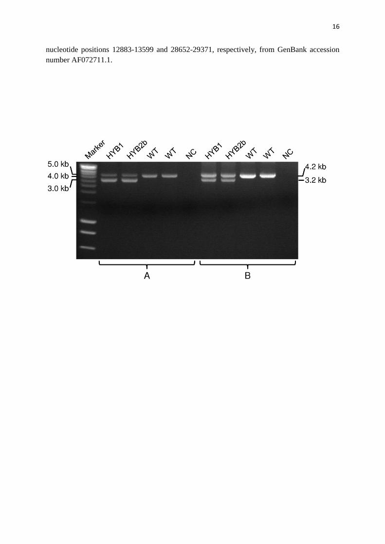
Supplementary Figure 1. Comparison of the originally reported PCR assay (A) and the
currently used PCR assay (B) for detecting the CEL–HYB1 (as well as the CEL–HYB2b)
allele (3.2 kb) in French heterozygotes. WT, wild-type (4.2 kb). NC, negative control.
Supplementary Figure 2. Alignment of reference CEL and CELP sequences in the context of
exon 10 (in bold) and partial flanking sequences. The crossover region for the CEL–HYB
deletion allele9 (termed here CEL–HYB1) is doubly underlined whereas that for CEL–HYB2
is singly underlined. The discriminant positions between the two paralogous sequences are
indicated by numbered upwardly pointing arrows. The CEL–HYB2a variant harbors the G
allele of rs10901232A/G, the T allele of rs10901233C/T and the G allele of rs671412A/G
whereas the CEL–HYB2b variant harbors the alternative alleles of these three SNPs (the three
affected sites are indicated by circles). The premature translation termination codon in the
chimeric exon 10 of the CEL–HYB2b variant is indicated by a box. The two primers used for
sequencing the breakpoint junctions of the CEL–HYB alleles, S10F and S10R,9 are indicated
by horizontal dotted arrows. The CEL and CELP sequences shown in the Figure correspond to
16
nucleotide positions 12883-13599 and 28652-29371, respectively, from GenBank accession
number AF072711.1.

17

18
Supplementary Table 1. PCR primer pairs used in this study
Primer
pair Sequence (5' > 3') Location Amplified sequence
Amplicon size
(bp)
A1 Forward: TCCATAAATACCCGAGGCCC 5'-untranslated region of CEL Full-length genomic
CEL–HYB sequences
9676 (HYB1)
9678 (HYB2a) Reverse: CTTCCTGCAGCTTAGCCTTG 3'-untranslated region of CELP
A2
Forward: TAATACGACTCACTATAGGG T7 promoter/priming site 5' extremity of the
constructs 235
Reverse: CTCTGCTGGGCTCTTACCTT Exon 1/intron 1 boundary of
genomic CEL
A3 Forward: CAAGACCTACGCCTACCTGT Exon 10 of genomic CEL 3' extremity of the
constructs
1334 (HYB1)
1336 (HYB2a) Reverse: TAGAAGGCACAGTCGAGG BGH reverse priming site
A4
Forward: GGAGACCCAAGCTGGCTAGT pcDNA3.1/5'-untranslated region Full length CEL–HYB
transcripts
2159 (HYB1)
2161 (HYB2a) Reverse: CCCTCTCGGCCTCTTGAG Between the stop codon and
polyadenylation signal of CELP
A5 Forward: GGAGACCCAAGCTGGCTAGT pcDNA3.1/5'-untranslated region 5' extremity of CEL–
HYB cDNA 280
Reverse: ATGTCCACAGAGTCACCCAG Exon 2 of CEL
A6a
Forward: ACTGTTGGTAAAGCCACCAT pGL3-control/5'-untranslated region Exons 8 and 9 of GP2 479
Reverse: TGTATCTTATCATGTCTGCTCGAA pGL3-control/3'-untranslated region a Primer sequences were taken from ref. 16.

19
Supplementary Table 2. Primers used for sequencing the full-length CEL–HYB1 and
CEL–HYB2a transcripts
Primer Sequence (5' > 3') Location
1 GGAGACCCAAGCTGGCTAGT pcDNA3.1/5'-untranslated region
2 GGACCTGCCCGTTATGATCT Exon 4 of CEL
3 TGGCTGAGAAGGTGGGTTG Exon 7 of CEL
4 CAAGACCTACGCCTACCTGT Exon 10 of CEL
5 CCCTCTCGGCCTCTTGAG Between the stop codon and
polyadenylation signal of CELP