Embed Size (px)
Citation preview
Bacteria, Yeast, Worms, and Flies: Exploiting simple modelorganisms to investigate human mitochondrial diseases
Shane L. Rea1,*, Brett H. Graham2, Eiko Nakamaru-Ogiso3, Adwitiya Kar1, and Marni J.Falk4
1Barshop Institute for Longevity and Aging Studies, and Department of Physiology, University ofTexas Health Science Center at San Antonio, San Antonio, TX 78240, USA2Department of Molecular and Human Genetics, Baylor College of Medicine and Texas Children’sHospital, Houston, TX 77030, USA3Johnson Research Foundation, Department of Biochemistry and Biophysics, School of Medicine,University of Pennsylvania, Philadelphia, PA 19104, USA4Division of Human Genetics, Department of Pediatrics, The Children’s Hospital of Philadelphiaand University of Pennsylvania School of Medicine, Philadelphia, PA 19104, USA
AbstractThe extensive conservation of mitochondrial structure, composition, and function across evolutionoffers a unique opportunity to expand our understanding of human mitochondrial biology anddisease. By investigating the biology of much simpler model organisms, it is often possible toanswer questions that are unreachable at the clinical level. Here, we review the relative utility offour different model organisms, namely the bacteria Escherichia coli, the yeast Saccharomycescerevisiae, the nematode Caenorhabditis elegans and the fruit fly Drosophila melanogaster, instudying the role of mitochondrial proteins relevant to human disease. E. coli are single cell,prokaryotic bacteria that have proven to be a useful model system in which to investigatemitochondrial respiratory chain protein structure and function. S. cerevisiae is a single-celledeukaryote that can grow equally well by mitochondrial-dependent respiration or by ethanolfermentation, a property that has proven to be a veritable boon for investigating mitochondrialfunctionality. C. elegans is a multi-cellular, microscopic worm that is organized into five majortissues and has proven to be a robust model animal for in vitro and in vivo studies of primary
*Corresponding Author: Shane L. Rea, Ph.D., 15355 Lambda Drive, STCBM Building, Rm. 2.200.04, Texas Research Park, SanAntonio, TX 78245-3207, Fax: 210-562-5028; [email protected].
CallOuts: Three to four 30–40 word call-out quotations from the text should be highlighted to emphasize important aspects of thearticle.“Here, we review the relative utility of four model organisms, namely the bacteria Escherichia coli, the yeast Saccharomycescerevisiae, the microscopic roundworm Caenorhabditis elegans, and the fruit fly Drosophila melanogaster, emphasizing insights eachhas afforded in the pursuit of improved understanding of the role and functions of mitochondrial gene mutations and proteins relevantto human disease”“Utilizing evolutionary homology allows key components held in common across divergent classes of organisms to be recognized andstudied”“In the context of the newly reported structural framework, future mutagenesis and structure/function studies of E. coli complex I willlikely yield not only fundamental insight into energy coupling mechanisms in complex I but also provide detailed understanding at themolecular level of presumed pathogenic human mutations.”“The peculiarities afforded by yeast as a model organism are accelerating our understanding of mitochondrial function and clarifyingthe relevance of mitochondrial function to human disease.”“Many therapeutic agents of purported benefit in human RC disease are amenable to mechanistic study in C. elegans models ofprimary respiratory chain dysfunction.”“Over the past ten years mutations in several Drosophila orthologs of human mitochondrial disease genes, or genes critical tofundamental mitochondrial functions, have been described.”
NIH Public AccessAuthor ManuscriptDev Disabil Res Rev. Author manuscript; available in PMC 2013 April 17.
Published in final edited form as:Dev Disabil Res Rev. 2010 June ; 16(2): 200–218. doi:10.1002/ddrr.114.
NIH
-PA Author Manuscript
NIH
-PA Author Manuscript
NIH
-PA Author Manuscript
respiratory chain dysfunction and its potential therapies in humans. Studied for over a century, D.melanogaster is a classic metazoan model system offering an abundance of genetic tools andreagents that facilitates investigations of mitochondrial biology using both forward and reversegenetics. The respective strengths and limitations of each species relative to mitochondrial studiesare explored. In addition, an overview is provided of major discoveries made in mitochondrialbiology in each of these four model systems.
KeywordsEscherichia coli; Saccharomyces cerevisiae; Caenhorabditis elegans; Drosophila melanogaster;mitochondria; model organisms
I. IntroductionMitochondria are organelles present in all eukaryotic species. Nevertheless, all eukaryotesdo not contain prototypic mitochondria, nor are all mitochondria the same within a singleorganism. The suite of functions performed by mitochondria varies, depending both onenvironmental conditions and tissue demands. These findings imply that mitochondria aredynamic organelles which, even today, remain under evolutionary pressure. Best scientificestimates trace the origin of mitochondria to a purple eubacteria-like organism thatestablished itself in an endosymbiosis with a primitive nucleated cell approximately 2 billionyears ago (Margulis 1996). The transition from this ancestral purple eubacteria into modernmitochondria has been accompanied by major macromolecular rearrangements. Entiremetabolic pathways have vanished, most genetic information necessary for mitochondrialstructure and function has been transferred to the nucleus, and new protein functions havebeen acquired within mitochondria largely from their co-evolving eukaryotic host. Indeed,proteins such as the ATP/ADP translocase, the entire family of metabolite carrier proteins,and Mia40 of the intermembrane space protein-assembly machinery were all acquired on theroad to becoming modern mitochondria (Chacinska et al. 2009; Gabaldon and Huynen2004).
Approximately 1,100 mouse mitochondrial proteins have been identified, which representon the order of 85% of the total predicted mitochondrial protein content (Pagliarini et al.2008). It is highly likely that a similar complement of mitochondrial proteins exist inhumans (Pagliarini et al. 2008). To date, 184 mitochondria-localized proteins have beenimplicated in human disorders (Chinault et al. 2009; Schwimmer et al. 2006). Although thevast majority of mitochondrial proteins appear to have been at least tentatively identified,the means by which mutated versions of these proteins lead to mitochondrial diseases thathave a broad spectrum of pathogenesis and phenotypic presentations remain largelyunexplained. Just as model organisms have helped us identify many of the knownmitochondrial proteins, they can also be used to discern their roles in the etiology ofmitochondrial diseases to improve our understanding of causative factors for the nearly 60%of mitochondrial disorders which have no definitive etiology (Dimmer et al. 2002). Here, wewill review the relative utility of four model organisms, namely the bacteria Escherichiacoli, the yeast Saccharomyces cerevisiae, the roundworm Caenorhabditis elegans and thefruitfly Drosophila melanogaster, emphasizing insights each has afforded in the pursuit ofimproved understanding of the role and functions of mitochondrial proteins relevant tohuman disease.
Rea et al. Page 2
Dev Disabil Res Rev. Author manuscript; available in PMC 2013 April 17.
NIH
-PA Author Manuscript
NIH
-PA Author Manuscript
NIH
-PA Author Manuscript
II. The Bacteria, Escherichia ColiIIA. Utility of bacteria as a model system in which to study mitochondrial composition andfunction
Bacteria are single cell, prokaryotic organisms that are typically several microns in length,which is similar in size to mitochondria. They take on a range of shapes including spheres,rods, and spirals (Figure 2A, left image). The cell membrane in gram-negative bacteria, suchas Escherichia coli, is separated by the periplasm from a peptidoglycan layer and an outermembrane. In contrast, there is no periplasm in gram-positive bacteria and their cell wall isclosely juxtaposed to the cytoplasmic membrane. Oxidative phosphorylation exists in manygenera of bacteria (Haddock 1980; Zannoni 2004). Recent focus on bacterial electrontransport mechanisms stems from their structural simplicity in comparison with eukaryotecounterparts, their short generation time on the order of 20–40 minutes, and their relativeease of gene manipulation. Bacteria have proven a useful model system in which toinvestigate protein structure and function, although these single cell organisms cannot beused to directly study manifestations of human diseases. Knowledge gained in the bacteriamodel can often be applied to homologous proteins in more complex higher organisms.
IIB. Electron transport chains in bacteriaElectron transport systems are located in the cytoplasmic membrane of numerous species ofaerobic, or facultative, bacteria. Specific components involved in electron transfer can behighly diversified between organisms, or even within the same organism depending ongrowth conditions. In contrast to mitochondria, individual bacteria utilize multiple electrontransport chains, often simultaneously. Bacteria have a number of different dehydrogenases,oxidases and reductases that are used for this purpose, as well as a variety of differentelectron donors and acceptors (Zannoni 2004). Individual enzymes for susbstrate oxidationand reduction are variably expressed according to environmental conditions. For example,when E. coli are grown aerobically, many of their electron transport components are distinctfrom those found in mammalian mitochondria. E. coli uses two different NADHdehydrogenases. One is the proton-translocating enzyme, NDH-1, which is very similar tomitochondrial complex I; the other is a single polypeptide enzyme, alternative NADH-quinone oxidoreductase (NDH-2), which does not translocate protons (Yagi et al. 1998).They also have two different quinol oxidases. One is the cytochrome bo3 complex, whichcontains a heme-copper center related to the cytochrome aa3 family that includesmitochondrial complex IV; the other is the cytochrome bd complex, which contains hemeproteins and a chlorine-Fe-protein (Unden and Bongaerts 1997). Cytochrome bd has a muchhigher affinity for oxygen than cytochrome bo3 and is induced under lower oxygen pressure.However, E. coli have no detectable c-type cytochromes (Unden and Bongaerts 1997). Sincebc1 complex (complex III) is also absent, ubiquinol is oxidized directly in E. coli. Thus, E.coli clearly has a truncated electron transfer chain relative to that in mitochondria. On theother hand, the electron transport system of the soil bacteria, Paracoccus denitrificans, hasmany features that are similar to those in mitochondria: complex I/NDH-1, succinatedehydrogenase (complex II), bc1 complex, and cytochorome aa3 (Baker et al. 1998; Zannoni2004). P. denitrificans does not have a NDH-2 type enzyme, but does have other ba3 andcbb3 oxidases (Baker et al. 1998).
IIC. Modeling mitochondrial respiratory complexes in bacteriaLacking an intricate coding background and mitochondrial assembly pathways, bacterialenzymes typically involve a much simpler subunit composition than those in mitochondria.Nonetheless, bacterial enzymes maintain all fundamental functions established for theirmitochondrial “relatives”. The number of protein subunits in mammalian and bacterialrespiratory chain complexes, respectively, includes: complex I, 45:14; complex II, 4:4;
Rea et al. Page 3
Dev Disabil Res Rev. Author manuscript; available in PMC 2013 April 17.
NIH
-PA Author Manuscript
NIH
-PA Author Manuscript
NIH
-PA Author Manuscript
complex III, 11:3; complex IV, 13:4. Electron flow from reducing substrates to oxygen iscoupled to transmembrane proton movements, just as it is in the mitochondrial respiratorychain.
Complex I, NADH:ubiquinone oxidoreductase, is the main entrance for electrons into therespiratory chains of many bacteria and mitochondria of most eukaryotes. It couples thetransfer of electrons from NADH to ubiquinone with the translocation of protons across themembrane. The bacterial complex I, in general, is made up of 14 different subunits,representing a minimal structural form of complex I (Yagi et al. 1998) (Figure 2A, rightimage). In E. coli, the genes encoding two of the subunits are fused to form one gene,nuoCD. The 14 E. coli complex I subunits together have a molecular mass of approximately530 kDa (Friedrich 1998). Seven are peripheral proteins including the subunits that bind allknown redox groups of complex I, namely one FMN and eight or nine iron-sulfur clusters(Friedrich 1998). The remaining seven subunits are hydrophobic membrane proteins, whichhave recently been shown to fold into 63 α-helices across the cell membrane (Efremov et al.2010); little is known about their function, but they are most likely involved in quinonereduction and proton translocation (Friedrich 1998). In contrast, mammalian complex I has45 different protein subunits with a total molecular mass of approximately 1 MDa (Carroll etal. 2006). The majority of these subunits have no known function (Brandt 2006). However,by comparison with their simpler bacterial homologues, it is apparent that the core catalyticstructure of mammalian complex I that carries out electron transfer and proton pumpingfunctions involves only 14 subunits, all of which are homologous to the 14 bacterialcomplex I subunits (Brandt 2006). The homologs of the seven hydrophobic bacterialmembrane subunits (NuoA, H, J, K, L, M, and N) are encoded by mitochondrial DNA(mtDNA) in all eukaryotes as ND3, ND1, ND6, ND4L, ND5, ND4, and ND2, respectively.Electron microscopy has established that both mitochondrial and bacterial complex I have acharacteristic L-shaped structure that consists of two domains, a peripheral arm and amembrane domain (Brandt 2006; Zannoni 2004). Both complexes have similar electron-transfer and energy-transduction pathways, and are sensitive to the same inhibitors such aspiericidin A, capcaisin or acetogenins, suggesting that the bacterial complex I may serve as auseful model system for the study of the human enzyme complex I.
In fact, utilizing P. denitrificans, Thermus thermophilus, Rhodobacter capsulatus and E. coli,several groups succeeded in identifying 8–9 iron-sulfur clusters, N1a, N1b, N2, N3, N4, N5,N6a, N6b, and N7 in each of the peripheral subcomplex arms of complex I (Chevallet et al.1997; Dupuis et al. 1998; Flemming et al. 2003; Nakamaru-Ogiso et al. 2008; Nakamaru-Ogiso et al. 2002; Nakamaru-Ogiso et al. 2005; Rasmussen et al. 2001; Velazquez et al.2005; Yano et al. 1999; Yano et al. 2003; Yano et al. 1994; Yano et al. 1995). Thoseassignments were recently confirmed by demonstration of the X-ray crystal structure of thehydrophilic peripheral part of T. thermophilus complex I that was determined at 3.3angstrom (Å) resolution (Sazanov and Hinchliffe 2006). In addition, recent informationabout the structure of the membrane domain subunits became available although at lowerresolution (4.5 Å and 3.9 Å) (Efremov et al. 2010). A speculative arrangement (andtopology) of the membrane segment of E. coli complex I, which was previously proposedbased on the projection structure of the membrane domain and detergent-based fractionationstudy (Baranova et al. 2007; Holt et al. 2003), has now been confirmed by the X-ray crystalstructure of bacterial complex I (Efremov et al. 2010). Subunits NuoH (ND1), NuoA (ND3),NuoJ (ND6), and NuoK (ND4L) are present in the vicinity of the peripheral arm, whereasthe NuoL (ND5) and NuoM (ND4) subunits are distantly located from the peripheralsegment. NuoN (ND2) is located in the middle of the membrane arm. Since similarsubcomplex patterns of ND subunits have been obtained with bovine heart complex I afterdetergent treatment (Sazanov and Walker 2000), this recent insight into structuralorganization can now be directly applied to mammalian complex I. Thus, it is expected that
Rea et al. Page 4
Dev Disabil Res Rev. Author manuscript; available in PMC 2013 April 17.
NIH
-PA Author Manuscript
NIH
-PA Author Manuscript
NIH
-PA Author Manuscript
investigation into the roles of membrane-bound complex I subunits in proton translocation,ubiquinone/inhibitor binding sites, and the mechanisms linking mutations in these subunitsto human mitochondrial diseases will now be quickly advanced.
Complex III, also called ubiquinol:cytochrome c oxidoreductase or the bc1 complex, is a keycomponent for both bacterial respiration and photosynthesis. The simplest form of thecytochrome bc1 is found in prokaryotes and is comprised of three redox-active subunits,which bear two b-type hemes, one c-type heme, and one [2Fe-2S] cluster (Rieske protein) asprosthetic groups (Zannoni 2004). Photosynthetic bacteria like Rhodobacter species haveprovided powerful models for studying the function and structure of this enzyme. In recentyears, extensive use of spontaneous and site-directed mutants, investigation of newinhibitors, and engineering of novel bc1 complexes have provided us with a wealth ofinformation on the functional mechanisms, subunit interactions, and assembly of thisimportant enzyme. Recent resolution of the structure of various mitochondrial bc1complexes in different crystallographic forms (Berry et al. 2000) has consolidated previousfindings, and raised new issues, such as the unique mobility of the Rieske protein subunitduring Qo site catalysis (Lee et al. 2006; Lee et al. 2008). Multidisciplinary approachescombining physiologic, molecular genetic, biochemical, and biophysical techniques havebeen extremely successful in the bacterial bc1 complex to serve as a guiding light for allorganisms and their organelles.
Complex IV, cytochrome c oxidase, is a large transmembrane protein complex found in bothbacteria and mitochondria. It is the last enzyme in the electron transport chain ofmitochondria. It receives an electron from each of four cytochrome c molecules, transfersthem to one oxygen molecule, and then converts molecular oxygen to two molecules ofwater. While the canonical mitochondrial complexes have been investigated for almost fivedecades, the corresponding bacterial enzymes have been established only recently asattractive model systems in which to address basic reactions in electron transfer and energytransduction. Two different preparations have previously been studied for the aa3-typecytochrome c oxidase isolated from P. denitrificans (Richter and Ludwig 2009): it wasoriginally isolated as a two-subunit enzyme in the presence of Triton X-100 (Richter andLudwig 2009) and later isolated as a four-subunit complex using dodecyl maltoside forsolubilization (Richter and Ludwig 2009). These studies demonstrated that cytochrome coxidase subunits I–III (but not subunit IV) exhibit high sequence identities with theircorresponding mtDNA-encoded subunits of the eukaryotic enzyme. Not unexpectedly, bothbacterial preparations were indistinguishable from the 13-subunit enzyme isolated frommammalian mitochondria in terms of their energy transduction properties and basic 3-Dstructure as deduced from X-ray crystal analyses. Furthermore, subunits III and IV do notcontribute to the redox-related signals observed by fourier transform infrared (FTIR)spectroscopy (Hellwig et al. 1998), nor do they influence the electron transfer reaction toany appreciable extent. Recent research questions ranging from primary steps in cofactorinsertion to supramolecular architecture of electron transfer complexes, can also befavorably addressed in prokaryotic systems to improve understanding of prototypicstructures and mechanisms common to all family members.
IID. Investigating complex I dysfunction by modeling point mutations in mtDNA-encodedND subunits
Mitochondrial DNA contains 37 genes, thirteen of which encode polypeptides that are allessential components of the respiratory chain. Indeed, these 13 proteins are highly conservedsubunits in respiratory enzyme complexes (complex I, III, IV, and or ATP synthase) frombacteria to mitochondria. Seven of these proteins constitute the core hydrophobic membranesubunits of complex I. A number of point mutations reported in these subunits have beenimplicated in human mitochondrial diseases including Leber Hereditary Optic Neuropathy
Rea et al. Page 5
Dev Disabil Res Rev. Author manuscript; available in PMC 2013 April 17.
NIH
-PA Author Manuscript
NIH
-PA Author Manuscript
NIH
-PA Author Manuscript
(LHON) and Mitochondrial Encephalomyopathy, Lactic Acidosis, and Stroke-like episodes(MELAS) (DiMauro and Schon 2003). Complex I dysfunction has also been implicated in avariety of human diseases including heart failure, diabetes mellitus, and severalneurodegenerative diseases such as Parkinson’s disease (DiMauro and Schon 2003; Singeret al. 1993). Although complex I dysfunction is postulated to promote apoptosis throughenhanced reactive oxygen species (ROS) production from the damaged enzyme (Sheeranand Pepe 2006), the pathophysiology of complex I-related diseases is complex andmultifactorial. However, at least identifying the detrimental mtDNA mutations offers arational approach to investigate pathogenesis. Analysis of the bacterial enzymes has a clearadvantage when the target protein is mtDNA-encoded in eukaryotes. Studying the effectsand implications of engineered mutations in the ND subunits of the complex I enzymerelative to its structure, respiratory chain function, and/or reactive oxygen species productionis a prerequisite to understand how individual disease mutations may contribute to diseasestates.
In that respect, E. coli complex I is a powerful model system in which to exploit theconvenient tool of gene manipulation (especially site-specific mutagenesis), which isvirtually impossible in mammalian complex I for mtDNA-encoded subunits such as ND5.Even commonly used RNA interference technique cannot be applied to knockdownmtDNA-encoded subunits. Transmitochondrial cybrid systems are useful but offer verylimited capacity to carry out mechanistic studies at the enzyme level. Fortunately, humandisease mutations in mitochondrial complex I involve amino acid residues that are mostlyconserved in E. coli complex I. In the case of ND5, they are perfectly conserved (Figure 3,red font). Some are also conserved with other multi-subunit secondary Na+/H+ antiporters,which show high sequence similarity to ND5 and to other transporter module subunits, suchas ND2 and ND4 of complex I. F124L causes Leigh Disease, E145G causes MELAS,A171V, G239S and G465E cause LHON, M237L causes MELAS/Leigh /LHON overlapsyndrome, and D393N/G causes MELAS/Leigh Disease. Very recently, mutational analysesof the corresponding mutations at E144 and D400 of the E. coli NuoL (ND5) subunitsuggested that these residues are critical for the normal complex I functions of electrontransfer and/or proton pumping (Nakamaru-Ogiso et al. submitted). The A52T mutation inND1 that causes LHON was introduced into its homologous subunit, NQO8 of P.denitrificans complex I, revealing that the mutated residue plays an important role inubiquinone reduction by complex I (Zickermann et al. 1998). Other ND1 pathogenicmutations, such as the E24K LHON/MELAS mutation and the R25Q, G131S, and E214KMELAS mutations, were also found to play an important role for complex I activities (Sinhaet al. 2009). The equivalent E. coli mutant of the common M64V LHON mutation in ND6demonstrated a mild effect on E. coli complex I activity (Kao et al. 2005). These datademonstrate the feasibility and utility of modeling amino acid substitutions caused bymtDNA mutations in homologous positions in the prokaryotic enzyme.
However, there are clearly some limitations to the utilization of the bacterial system.Although the residue associated with the E143K LHON mutation in ND1 is almostcompletely conserved, the corresponding E. coli mutant (E157K) actually augmentedperipheral complex I activity to 160% of control (Sinha et al. 2009). Slight mitochondrialproliferation with abnormal mitochondria has previously been reported in affected humanpatients (Valentino et al. 2004). Introducing human pathogenic mutations at non-conservedresidues in ND1 has recently been attempted in its homologus subunit, NuoH, of E. colicomplex I (Maliniemi et al. 2009). The overall effects of mutations were milder in thissystem such that results did not support the pathogenicity of the sole ND1-L285P/NuoH-V297P mutation or the suppressor effect of the ND1-Y277C/NuoH-L289C mutation on theformer one, which has been previously suggested from the study of clinical/biochemicalphenotypes in affected family members carrying these mutations (Howell et al. 1991). As
Rea et al. Page 6
Dev Disabil Res Rev. Author manuscript; available in PMC 2013 April 17.
NIH
-PA Author Manuscript
NIH
-PA Author Manuscript
NIH
-PA Author Manuscript
another example, Hofhaus et al. (Howell et al. 1991) and Park et al. (Park et al. 2007)reported that NADH dehydrogenase-dependent respiration measured in digitonin-permeabilized cybrid cells was specifically decreased by 40% in cells harboring ahomoplasmic ND4 R340H mutation that is associated with LHON. The corresponding E.coli R369H mutant in NuoM showed only slightly decreased complex I activity (70% ofcontrol) (Torres-Bacete et al. 2007), while the corresponding R. capsulatus R368H mutant inNuoM showed a clear impairment in oxidative phosphorylation capacity (Lunardi et al.1998).
In conclusion, the bacterial system can be beneficial for comprehensive studies investigatingeffects of mtDNA mutations known to cause human complex I deficient mitochondrialdiseases. However, much more detailed information is first needed on the basic structure andfunction of complex I. Without basic knowledge, it will be extremely difficult to assesswhether pathogenic human mutations are in functionally-important regions (such asinhibitor/ubiquinone binding sites and/or proton-pumping sites) and what effects oncomplex I function might be anticipated. Knowledge of mitochondrial complex I is still toolimited to completely understand human mitochondrial diseases involving complex Idysfunction. However, for the first time, the X-ray crystal structures of the entire complex Ifrom T. thermophilus as well as the membrane domain of E. coli complex I have beendetermined (Efremov et al. 2010). This structural framework provided surprising newinformation that NuoL contains an unusually long, conserved, amphipathic helix thatextends ~110 Å to a position near the catalytic site from the end of the membrane arm(Efremov et al. 2010), suggesting that it constitutes a mechanical link capable oftransmitting conformational changes. This structural information suggests an unprecedented,unique, and unexpected role for ND5 (NuoL) in the redox-driven proton pumpingmechanism of complex I. In the context of the newly reported structural framework, futuremutagenesis and structure/function studies of E. coli complex I will likely yield not onlyfundamental insight into energy coupling mechanisms in complex I but also provide detailedunderstanding at the molecular level of presumed pathogenic human mutations.
III. The Yeast Saccharomyces cerevisiaeThe term ‘yeast’ is often used synonomously with Saccharomyces cerevisiae – the powerfulgenetic model that also doubles as the well-known Baker’s and Brewer’s Yeast. S.cerevisiae is, however, only one of 1,500 yeast species that comprise the sub-kingdomFungi. These organisms are some of the simplest eukaryotes known. Most yeast reproduceasexually by budding, although a few, such as Schizosaccaromyces pombe, do so by binaryfission. Typical yeast, such as S. cerevisiae, measure only 5 to 10 microns in diameter(Figure 2B).
IIIA. Comparison of yeast and mammalian mitochondrial biologyAs a single-celled organism, S. cerevisiae has no physiologic relevance to humans.However, it is a genetically-tractable eukaryote replete with many mitochondrial proteinsthat are orthologous to human proteins. Since the last common ancestor of fungi and animalsdates back to approximately 450 million years ago (Margulis 1996), an estimated 75% of theevolutionary history of mitochondria in these two sub-kingdoms has been shared. Recentestimates suggest that up to 700 proteins in S. cerevisiae are required for mitochondrialfunction (Pagliarini et al. 2008; Perocchi et al. 2008; Reinders et al. 2006; Steinmetz et al.2002). Of the 601 known yeast mitochondria proteins, 222 have clear human orthologs (seeTable I for database links). Indeed, most of the recent approaches used to identifymitochondrial proteins in mammals (Pagliarini et al. 2008; Smith et al. 2007) were piloted inyeast. These include large-scale gene expression analyses (Epstein et al. 2001), massspectrometry-based proteomics (Reinders and Sickmann 2007), sub-cellular localization
Rea et al. Page 7
Dev Disabil Res Rev. Author manuscript; available in PMC 2013 April 17.
NIH
-PA Author Manuscript
NIH
-PA Author Manuscript
NIH
-PA Author Manuscript
analysis using fluorescent reporters (Westermann and Neupert 2000), deletion phenotyping(Pan et al. 2004), and many computational methods designed to identify mitochondrialproteins by virtue of signature trafficking sequences (Nakai and Horton 1999). S. cerevisiaehas also proven to be a powerful tool for studying diverse cellular processes such asmitochondrial biogenesis (Bolotin-Fukuhara and Grivell 1992; Chacinska et al. 2009),mitochondrial DNA (mtDNA) packaging and inheritance (Contamine and Picard 2000),retrograde regulation (Jazwinski 2005), mitochondrial metabolism (Christensen andMacKenzie 2006), and electron transport chain assembly and activity (Barrientos 2003). Inboth humans and yeast, the functions of mitochondria are now recognized to extend farbeyond the synthesis of energy in the form of ATP. Rather, mitochondrial proteinsparticipate in diverse processes ranging from ion flux regulation (Cardoso et al. 2010),ubiquinone production (Tran and Clarke 2007b), pyrimidine biosynthesis and single-carbonmetabolism (Christensen and MacKenzie 2006) to amino acid catabolism (Metzler 1977),iron sulfur cluster assembly (Sheftel et al. 2010), and protein quality control (Koppen andLanger 2007; Rep and Grivell 1996).
Several properties of S. cerevisiae made it the early model of choice for studies ofmitochondria. Foremost, S. cerevisiae is a facultative anaerobe that preferentially fermentsglucose to ethanol under aerobic conditions. When glucose becomes limiting, ethanol isoxidized to produce reducing equivalents that fuel mitochondrial respiration. Thus,relatively few mitochondrial proteins are essential for viability in S. cerevisiae. Thosemitochondrial proteins that do remain essential are restricted to protein import andmaturation, iron sulfur cluster assembly, flavin mononucleotide synthesis, and uracilbiosynthesis (Chacinska et al. 2009; Dimmer et al. 2002). Coupled with the ability to survivein either haploid or diploid states, their capacity to toggle between two metabolic statesmeans many genetic disruptions that adversely affect mitochondrial functions in otherspecies can be easily maintained and studied in S. cerevisiae. An additional advantageafforded by S. cerevisiae is the ease with which genes can be knocked out or heterologousproteins knocked-in and expressed. Such approaches have been used successfully in thequest to identify novel gene candidates responsible for human mitochondrial disorders. Twoillustrative examples are provided by the nuclear-encoded COX10 and BCS1L genes, wheregene polymorphisms were suspected of causing a tubulopathy and leukodystrophy disorderand GRACILE syndrome, respectively. In both cases, yeast ‘complementation assays’confirmed the role of these genes and polymorphisms in each disorder (Hinson et al. 2007).
The mitochondrial proteome has been shown to markedly differ between mouse tissues(Pagliarini et al. 2008). Analysis of 14 different tissues revealed that, on average, each tissueexpressed only 700 of the known 1,098 mouse mitochondrial proteins (considering a 10%false discovery rate). Mitochondrial protein composition between tissue pairs were found tooverlap by an average of 75%. However, a core set of approximately 300 proteins waspresent in every tissue, primarily representing proteins involved in oxidativephosphorylation (OXPHOS), the citric acid cycle, and folate metabolism. Analysis across500 fully-sequenced species suggested much of this core mitochondrial proteome has beenretained throughout evolution. Consistent with this idea, when S. cerevisiae is cultured ondifferent non-fermentable carbon sources (e.g., lactate, ethanol and glycerol) a core set ofmitochondrial proteins also can be discerned. On top of this core, there is a food type-specific set of mitochondrial proteins that is expressed under each condition (Steinmetz et al.2002). Furthermore, 58 of 601 known yeast mitochondrial proteins are orthologous to genescausative of human disorders, most of which correspond to core components of intermediarymetabolism and energy transduction.
Rea et al. Page 8
Dev Disabil Res Rev. Author manuscript; available in PMC 2013 April 17.
NIH
-PA Author Manuscript
NIH
-PA Author Manuscript
NIH
-PA Author Manuscript
IIIB. Modeling Human Mitochondrial Diseases in Yeast – Past, Present & FutureIn humans, diseases associated with mitochondrial dysfunction can be broadly grouped intothose arising from genes of nuclear origin and those arising from mutations in mitochondrialDNA (mtDNA). Diseases of nuclear origin can be further classified into diseases due to (i)defective mitochondrial biogenesis, (ii) defective mitochondrial protein import and export,(iii) defective assembly of the oxidative phosphorylation (OXPHOS) system, (iv) defectiveactivity of the OXPHOS system, (v) defective enzymes of intermediary metabolism, (iv)defective mtDNA maintenance, and (vi) defective quality control proteins. S. cerevisiae hasbeen used to model several human diseases spanning each of these classes and, in manycases, played an important role in facilitating identification of the genetic mutation or thecausative mechanism underlying the human disease. A list of several such examples isprovided in Table II. Several excellent reviews also provide detailed historical accounts ofthe role yeast have played in understanding human mitochondrial disorders (Barrientos2003; Bassett et al. 1996; Fontanesi et al. 2009; Foury and Kucej 2002; Schwimmer et al.2006).
The explosive growth of molecular biology in the 1990s, and of systems biology morerecently, has led to the introduction of novel models and techniques for the study of humanmitochondrial disorders. This has, in some respects relegated yeast to ‘just another tool in abox of many’. Nonetheless, yeast continues to yield major advances in the study ofmitochondria. In the following section, we highlight several areas where the peculiaritiesafforded by yeast as a model organism are accelerating our understanding of mitochondrialfunction and clarifying the relevance of mitochondrial function to human disease. We endthis section on a speculative note, looking at least one yeast research endeavor that mighthold promise for understanding previously unappreciated forms of mitochondrial diseases.
IIIC. Yeast models of mitochondrial diseasei. Complex I deficiency – when nothing is something—In human mitochondria, 88proteins collectively form the 5 complexes of the energy-generating electron transport chain(ETC). Although this number varies in yeast by species, S. cerevisiae contains 90 proteinsthat are currently annotated as respiratory chain-associated (MITOP, see Table 1), includinga small number of paralogues and respiratory chain assembly factors. Two basic types ofETC proteins can be distinguished: (i) ‘structural core proteins’, which play an integral rolein electron transfer or positive charge-translocation and derive from the originalendosymbiont; and (ii) ‘supernumerary proteins’, representing all remaining proteins oflargely unknown function that evolved as ‘add-ons’ to the ancient structural-core ETC. Theeubacterial genome of present day mitochondria reflect their evolutionary past: in humans,the ancestral genome has been reduced to 13 core ETC protein-coding genes (ND1 to ND6and ND4L of complex I, CYB of complex III, COI to COIII of complex IV, and ATP6 andATP8 of complex V). In S. cerevisiae, the mitochondrial genome encodes just eight proteins(CYTB, COX1, COX2, COX3, ATP6, ATP8, ATP9, and a ribosomal protein, VAR1)(Bouchier et al. 2009; Tsang and Lemire 2003). These tiny genomes also demonstrate thateven the smallest of ETC complexes cannot assemble spontaneously. Rather, it becomesclear that multiple factors are needed to control the assembly of each complex when oneconsiders that (i) approximately half of all ETC proteins are integral membrane proteins, (ii)subunits decorate structural-cores on both cytosolic- and matrix-side of the inner membrane,(iii) most subunits have to navigate two membranes before undergoing additional peptideprocessing, and (iv) prosthetic groups are loaded into structural-core proteins after theyreach their final destination. Yeast has played a pivotal role in elucidating the complicatedprocess required to temporally and spatially control the assembly of each ETC complex.
Rea et al. Page 9
Dev Disabil Res Rev. Author manuscript; available in PMC 2013 April 17.
NIH
-PA Author Manuscript
NIH
-PA Author Manuscript
NIH
-PA Author Manuscript
NADH:ubiquinone oxidoreductase (complex I) deficiency is associated with a wide range ofclinical phenotypes including Leigh Syndrome, Leber Hereditary Optic Neuropathy, andmitochondrial encephalopathy, lactic acidosis, and stroke (MELAS). With the advent oflarge scale sequencing tools it came as a surprise to find that mutations in complex Istructural genes were absent in approximately 60% of cases where NADH:ubiquinoneoxidoreductase deficiency could be confirmed biochemically (Smeitink et al. 2001). Suchfindings suggested auxiliary proteins were required for complex I assembly.
Many yeast species contain alternative NADH dehydrogenases that operate alongsidecomplex I. These enzymes provide additional pathways for the transfer of reducingequivalents from NADH to the ETC without the concomitant pumping of protons across theinner mitochondrial membrane (Kerscher 2000). In some yeast species, complex I functionhas been lost entirely. Such is the case for S. cerevisiae, where three alternative NADHdehydrogenases operate to deliver reducing equivalents from the cytosol (NDE1, NDE2) ormatrix (NDI1) to the mitochondrial ETC (Fang and Beattie 2003). Reasoning that loss ofcomplex I should remove any evolutionary pressure to retain its assembly factors,Shoubridge and colleagues (Ogilvie et al. 2005) used an in silico approach to subtractivelyidentify genes that had been differentially lost in S. cerevisiae but retained in Y. lipolyticaand D. hansenii, which have both retained complex I functionality. This approach led to theidentification of B17.2L (encoded by NDUFAF2 in humans), which is a protein necessaryfor the maturation of an 830 kDA subassembly of complex I. The authors not only showedthat a patient presenting with a progressive encephalopathy had a null mutation inNDUFAF2 but that complex I dysfunction in fibroblasts from this patient was completelyreversible by retroviral-mediated expression of B17.2L.
In a similar but broader fashion, Mootha and colleagues (Pagliarini et al. 2008) appliedphylogenetic profiling across 42 fully-sequenced eukaryotic species to show that a set of 15core complex I proteins had been independently lost four times across the course ofevolution (two yeast and two protozoan clades). These authors also identified a set of 19proteins that shared the same phylogenetic profile as the 15 core complex I proteins, makingthem candidates for complex I assembly factors. Biochemical analyses confirmed one ofthese proteins, C8Orf38, was indeed required for complex I activity and that mutations inthis gene caused a novel form of Leigh Syndrome. Later studies confirmed that a secondpredicted protein, C20orf7, was also a bona fide complex I assembly factor (Gerards et al.2009; Sugiana et al. 2008). Intriguingly, several of the remaining 17 predicted assemblyfactors encode metabolic enzymes of known function. It is unclear if these proteins evolvedas an attachment to the structural-core to enhance substrate tunneling or if they serve as truefolding factors.
ii. Fumarase – An old enzyme with a new function—Studies using the yeast S.cerevisiae have significantly advanced recent understanding of the tumor susceptibilitysyndrome, Hereditary Leiomyomatosis and Renal Cell Cancer (HLRCC), characterized bybenign cutaneous and uterine leiomyomas, uterine leiomyosarcomas, and renal cellcarcinomas (Launonen et al. 2001). HLRCC is almost always associated with bi-allelicinactivation of the citric acid cycle enzyme fumarase, which reversibly hydrates fumarate tomalate. While the majority of fumarase is delivered to the mitochondrial matrix, a smallfraction remains cytosolic (Karniely et al. 2006). This phenomenon arises because somefumarase molecules mature in the cytosol before being imported into mitochondria. Earlymaturation is not a problem for most mitochondria-targeted proteins since targeting pre-sequences direct uptake of mature proteins into the mitochondria. The mature fold forfumarate effectively masks its mitochondria import pre-sequence, however, leaving theprotein stranded in the cytosol. The interesting observation that cytosolic fumarase isconserved from yeast to man strongly implies that cytosolic fumarase is not just the by-
Rea et al. Page 10
Dev Disabil Res Rev. Author manuscript; available in PMC 2013 April 17.
NIH
-PA Author Manuscript
NIH
-PA Author Manuscript
NIH
-PA Author Manuscript
product of inefficient protein import but likely has an important role. Until recently,cytosolic fumarase’s function was assumed to be the scavenging of excess fumarateproduced by two cellular sources: fumarylacetoacetate hydrolase in tyrosine catabolism, andarginosuccinate lyase in the urea cycle (Metzler 1977). HLRCC was speculated to resultfrom loss of fumarase activity leading to elevated cytosolic fumarate, which cancompetitively inhibit HIF prolyl hydroxylase (HPH) and stabilize the transcriptionalactivator hypoxia-inducible factor (HIF) by preventing its proteasomal degradation(Koivunen et al. 2007). HIF plays an important role in regulating angiogenesis-regulatedgenes, such as VEGF (Michiels et al. 2001).
In a seminal study, Yogev and colleagues (Yogev et al. 2010) recently employed S.cerevisiae to unequivocally investigate the role of cytosolic fumarase. Utilizing a procedureeffectively inaccessible to almost all other model organisms (Mireau et al. 2003), S.cerevisiae cells were re-engineered to express only mitochondria-localized fumarase. Byknocking out the nuclear fumarase gene and knocking-in a mitochondrial DNA-encodedversion, Yogev and colleagues discovered that cytosolic fumarase functions as part of anuclear DNA damage response (DDR) pathway, which is normally activated followingformation of double-strand breaks (DSBs). Hydroxyurea- or ionizing radiation-inducedDSBs lead to the rapid translocation of cytosolic fumarase into the nucleus. Yeast mutantsdeficient for cytosolic fumarase displayed both delayed and abrogated activation of both theDDR-activated histone variant γH2A(X) that marks the sites of DNA breakage, and theRad53p/CHK2 checkpoint protein. Intriguingly, addition of fumarate alone was sufficient torescue the DNA repair deficit, whereas malate or a catalytically inactive form of fumarasewas not. The DDR response function of cytosolic fumarase was found to be conserved inhuman HeLa cells. The precise role played by nuclear fumarate remains unclear.
iii. Ubiquinone Biosynthesis – A New Function for Coq4p—Ubiquinone (a.k.a.Coenzyme Q, CoQ10 in humans, or simply ‘Q’) is a mobile, one- or two-electron carriermolecule that is synthesized on the inner mitochondrial membrane and is essential fortransferring reducing equivalents from complexes I and II to complex III in the ETC.Ubiquinone also serves as a direct electron acceptor for the uracil biosynthetic enzymedihydroorotate dehydrogenase (Denis-Duphil 1989), and as an indirect electron acceptor fornine acyl-CoA dehydrogenases involved in lipid or amino acid metabolism (Ghisla andThorpe 2004). The latter is accomplished by way of electron-transferring flavoprotein (ETF)and the CoQ10-linked mitochondrial ETF dehydrogenase (ETFDH). Ubiquinone is presentin virtually all cell membranes, where it plays an important antioxidant role. Indeed,ubiquinone has both electron-scavenging and electron-donating capabilities (Santos-Ocanaet al. 2002; Villalba and Navas 2000). In humans, CoQ deficiency presents with four majorclinical manifestations: 1) encephalomyopathy, characterized by recurrent myoglobinuria,encephalopathy, and ragged-red fibers; 2) infantile multi-systemic disease, usually withprominent nephropathy and encephalopathy; 3) cerebellar ataxia with marked cerebellaratrophy; and 4) pure myopathy (Quinzii et al. 2008). Primary CoQ10 deficiencies due tomutations in ubiquinone biosynthetic genes (COQ2, PDSS1, PDSS2, and ADCK3) havebeen identified in patients with either the infantile multi-systemic or the cerebellar ataxiaphenotypes (DiMauro et al. 2007; Gironi et al. 2004; Mollet et al. 2008). In contrast,secondary CoQ10 deficiencies due to mutations in genes not directly related to ubiquinonebiosynthesis (APTX, ETFDH, and BRAF), have been identified in patients with either thecerebellar ataxia or pure myopathy phenotypes, as well in cardiofaciocutaneous syndrome(Aeby et al. 2007; Gempel et al. 2007; Musumeci et al. 2001).
Our knowledge of ubiquinone biosynthesis in humans is derived almost entirely fromstudies in the yeast S. cerevisiae and Schizosaccharomyces pombe, as well as the bacteriaEscherichia coli. Ubiquinone is comprised of a fully-substituted benzoquinone head group
Rea et al. Page 11
Dev Disabil Res Rev. Author manuscript; available in PMC 2013 April 17.
NIH
-PA Author Manuscript
NIH
-PA Author Manuscript
NIH
-PA Author Manuscript
attached to a polyisoprenoid tail. The number of isoprenoid units is species specific,consisting of 10 in humans and 6 in S. cerevisiae. Nine complementation groups (COQ1through COQ9) define the Q-biosynthetic route in S. cerevisiae (Tzagoloff and Dieckmann1990). Mammalian orthologs of yeast COQ genes have been identified by sequencehomology and confirmed by functional complementation (Tran and Clarke 2007a).Biosynthesis of ubiquinone begins with the formation of the isoprenoid tail that is catalyzedby the polyprenyl diphosphate synthase, Coq1p (in humans, this function is undertaken by arelated protein comprised of a heterodimer of PDSS1 and PDSS2 subunits). Next, thepolyprenyl diphosphate tail is condensed with 4-hydroxybenzoic acid (derived fromtyrosine) to form 4-hydroxy-3-polyprenylbenzoic acid (HHB) in a reaction catalyzed byCoq2p. Modification of the benzoquinone head group then follows a series of steps thatremain only partially characterized and occur in an unknown order. These reactions includetwo O-methylation steps catalyzed by Coq3p, a C-methylation step catalyzed by Coq5p, twohydroxylation steps probably involving Coq6p, and a monoxygenase step catalyzed byCoq7p/CLK-1. Coq4p, Coq8p (a probable kinase, and orthologous to ADCK3 in humans)and Coq9p have unknown roles. An additional protein, Coq10, appears to be involved inubiquinone trafficking, rather than its biosynthesis (Tran and Clarke 2007a).
In humans, more than half of patients that present with CoQ10 deficiency have unidentifiedgenetic mutations (Quinzii et al. 2008). Based on studies in yeast, it is almost certain thatdisruption of any gene involved in ubiquinone biosynthesis will be pathogenic in people.Coq4p is a particularly good candidate since genetic disruption of this locus in S. cerevisiaeis known to also result in the biochemical loss of Coq3p, Coq6p, Coq7p, and Coq9p(Marbois et al. 2009). The primary sequence of Coq4p was recently found to contain aputative zinc ligand motif HDxxH-(x)11-E motif (Marbois et al. 2009) but, oddly, noenzymatic domains associated with the kind of catalytic activities necessary for modificationof the benzoquinone head group. HA-tagged Coq9p can co-immunoprecipitate Coq4p, alongwith Coq5p, Coq6p and Coq7p. Based on these findings, Clarke and colleagues (Marbois etal. 2009) hypothesized that Coq4p forms a scaffold on the matrix side of the innermitochondrial membrane upon which much of the ubiquinone biosynthesis machinery maybe decorated.
The Northeast Structural Genomics (neSG) consortium is one of four large-scale, NIHfunded centers operating under the ‘Protein Structure Initiative (PSI-2)’. This programinitially aimed to reduce the cost and time required to determine three-dimensional proteinstructures. As part of this initiative, Alr8543, a cyanobacteria protein that contains theCoq4p fold, was recently solved (Forouhar et al. 2009). In an effort to identify the functionof Coq4p, and for the purposes of this review to illustrate how studies in S. cerevisiae can beused to further our understanding of human diseases, we have generated a homology modelof Coq4p from S. cerevisiae using Alr8543 as a template (Schwede et al. 2003). The largeevolutionary distance between Coq4p and Alr8543 permits easy identification of structurallyand functionally important residues (Figure 1A). From the crystal structure, Alr8543assembles into a homodimer. Interestingly, geranylgeranyl monophosphate co-crystallizedwith the protein in a deep binding pocket. This molecule is a polymer of four isoprenoidunits. Also in the crystal structure, a magnesium ion (Mg2+) was chelated by the putativezinc (Zn2+)-ligand motif (Figure 1B). The COQ4 complementation group in S. cerevisiaecontains 3 loss-of-function, nonsense mutations: coq4-1 (E226K), coq4-2 (E121K), andcoq4-3 (G120E). The quality of our modeling, as well as the reliability of using Alr8543 as atemplate, can be assessed by mapping these mutations onto the yeast Coq4p structuralprediction. Since these are loss-of-function alleles we would expect each mutation to cause aprotein defect of obvious consequence. Indeed, this appears to be the case (see details inFigure 1C). Based on these findings, we predict that Coq4p does indeed act as an anchorprotein primarily for the polyisoprenoid tail of HHB. We suggest that Coq4p acts to tether
Rea et al. Page 12
Dev Disabil Res Rev. Author manuscript; available in PMC 2013 April 17.
NIH
-PA Author Manuscript
NIH
-PA Author Manuscript
NIH
-PA Author Manuscript
HHB to the inner mitochondrial membrane by its distal four or five isoprenoid tail units,thus allowing the benzoquinone head group to rotate freely for sequential modification byCoq5p, Coq6p, Coq7p and Coq9p. From the structure, it is also very likely that the surfaceof Coq4p may form a scaffold for attachment of the latter proteins.
iv. Barth Syndrome: Tafazzin and cld-1—Barth syndrome (BTHS) is an X-linkeddisorder caused by inactivation of Tafazzin, the protein encoded by the Taz G4.5 gene(Bione et al. 1996). Clinically, Barth Syndrome is characterized by cardiomyopathy,neutropenia, and delayed growth (Barth et al. 1983; Spencer et al. 2006). Tafazzin mutationshamper the remodeling of cardiolipin (diphosphatidylglycerol), which is a tetra-acylphospholipid that is almost exclusively located in the inner mitochondrial membrane whereit constitutes up to 25% of the total lipid. Cardiolipin has been implicated in diversemitochondrial functions including membrane biogenesis, electron transport chainfunctionality, apoptosis, and lipid-protein interface interactions (Chicco and Sparagna 2006).In eukaryotes, the final step in cardiolipin biosynthesis involves linkingphosphatidylglycerol with the activated diacyl group of cytidinediphosphate diacylglycerol(Schlame et al. 1993) in a reaction catalyzed by the enzyme cardiolipin synthase (Crd1p inS. cerevisiae). Although the reaction of cardiolipin synthase was first demonstrated in ratliver (Tamai and Greenberg 1990), the systematic characterization of cardiolipin synthaseshas largely been undertaken using the yeast S. cerevisiae.
As there are four distinct fatty acyl groups in cardiolipin, there is enormous potential forcomplexity in the distribution within molecular species. For most animal tissues, however,cardiolipin contains almost exclusively C18 fatty acids, 80% of which is typically linoleicacid (18:2(n-6)). Testis cardiolipin is an exception that contains mainly palmitic acid(C16:0), while brain cardiolipin contains many different fatty acids includingpolyunsaturated arachidonic acid (20:4(ω-6)) and docosahexanoic acid (22:6(n-3)). In S.cerevisiae, cardiolipin is mainly comprised of 16:1 and 18:1 fatty acids. It appears that theultimate fatty acid composition of cardiolipin in eukaryotes is attained by re-modeling. Suchremodeling could be achieved in theory by the coenzyme A (CoA)-dependent deacylation-reacylation cycle known as the Lands cycle. However, it is now believed that the mainremodeling route is via CoA-independent transacylation between different phospholipids, areaction in which Tafazzin plays a central role (Houtkooper et al. 2009).
Barth Syndrome is marked by the presence of aberrant cardiolipin molecular species(Schlame and Ren 2006). Tetra-acylated cardiolipin levels are low, levels of tri-acylatedmonolysocardiolipin (MLCL) are elevated and the acyl chain composition of the remainingCL is more saturated. Tafazzin encodes a phospholipid transacylase (Neuwald 1997) thattransfers an acyl chain from phosphatidylcholine (PC) to MLCL (Ma et al. 1999; Schlameand Rustow 1990; Xu et al. 2003). Identification of Tafazzin quickly set the stage for S.cerevisiae to become a model system for studying Barth Syndrome, since it has anorthologous gene. Mutant Δtaz1 yeast exhibit phospholipid defects similar to those observedin Barth Syndrome cells – they accumulate aberrant cardiolipin species and have decreasedlevels of mature cardiolipin (Gu et al. 2004; Vaz et al. 2003). Cardiolipin remodeling inΔtaz1 mutants was shown to be essential for the stability of mitochondrial membranesfollowing exposure to elevated temperature or osmotic stress (Koshkin and Greenberg 2000;Koshkin and Greenberg 2002). Interestingly, when complementation assays using thehuman Tafazzin gene were performed in Δtaz mutants, only splice variants lacking exon 5were able to fully rescue the aberrant cardiolipin profile. Constructs expressing humanTafazzin lacking exon 5 preferentially incorporated C18:1, not C18:2 as is typical of humancardiolipin. Furthermore, variants lacking exon 5 restored mitochondrial coupling to Δtazmutants when challenged with hypotonic stress (Ma et al. 2004). These findings led to therealization that, in humans, Tafazzin splice variants that lack exon 5 are likely the active
Rea et al. Page 13
Dev Disabil Res Rev. Author manuscript; available in PMC 2013 April 17.
NIH
-PA Author Manuscript
NIH
-PA Author Manuscript
NIH
-PA Author Manuscript
variant in vivo, and that preferential incorporation of C18:2 likely reflects differences insubstrate availability rather that substrate selectivity (Houtkooper et al. 2009).
The re-modeling reaction of Tafazzin could theoretically proceed by a cyclic mechanism - inwhich case only trace amounts of either lyso-phosphatidylcholine or MLCL would berequired. Recently, a phospholipase specific for cardiolipin was identified in yeast(cardiolipin-specific deacylase 1, CLD1). This enzyme hydrolyzes newly synthesizedcardiolipin with a strong substrate preference for palmitoyl acyl groups and functionsupstream of tafazzin to generate MLCL (Beranek et al. 2009). Interestingly, deletion ofCLD1 in the Δtaz background did not decrease the total amount of cardiolipin or lead to anelevation in MLCL levels, yet still resulted in a mitochondrial pathology. This implies thatin Barth Syndrome patients, dysfunction of mitochondria probably does not result fromexcessive MLCL accumulation but, rather, from defective cardiolipin re-modeling.
v. Mitochondria-associated Membranes (MAM)—One of the obvious advantages ofusing yeast as a model organism is that it provides an excellent tool for exploratory research- science that has the potential to result in unexpected insight into previously inexplicablehuman disorders. In this light, recent studies have shown that there are structural andfunctional contact points between mitochondria and endoplasmic reticulum (ER). Interactionof endoplasmic reticulum with mitochondria (termed mitochondrial-associated membranes,MAM), was first described by Copeland and Dalton in 1959 (Copeland and Dalton 1959)and has since been confirmed both biochemically and structurally (via electron microscopy)in mice (Ardail et al. 1993), rats (Camici and Corazzi 1995) and yeast (Gaigg et al. 1995;Zinser et al. 1991). How MAM relates to cell function remains an area of active exploration.Recent studies emphasize a role for MAM in the biosynthesis and trafficking ofphosphatidylcholine and phosphatidylserine between ER and mitochondrial membranes(Voelker 2003), as well as in the transmission of physiological and pathological Ca2+ signalsbetween both organelles (Giorgi et al. 2009; Schon and Area-Gomez 2010; Simmen et al.2010). MAM’s central role in non-vesicular inter-organelle lipid trafficking, and its directimpact on cell physiology, first came into light with a seminal paper on theneurodegenerative disorder neuronal ceroid lipofuscinosis (NCL) (Vance et al. 1997). NCLsare a group of autosomal recessive lysosomal storage diseases resulting from one of 160known mutations in eight NCL genes (CLN1–CLN3, CLN5–CLN7 and CLN10) (Lyly et al.2009). Pathologically, NCL is characterized by the intracellular accumulation ofautofluorescent lipopigment and the progressive loss of neocortical neurons. The majorcomponent of stored lysosomal material is either subunit c of mitochondrial ATP synthaseor sphingolipid activator proteins A and D (Cooper et al. 2006). Vance and colleagues(Vance et al. 1997) used an mdm/mdm (CLN8) mutant mouse model to show aberrantbiochemical separation of MAM from livers of older, but not younger, mice. Moreover, theamount of the MAM-specific protein phosphatidylethanolamine N-methyltransferase-2(PEMT2) was found to be reduced by 60% in mdm/mdm liver homogenates of all ages,compared to control animals, despite normal levels of PEMT2 mRNA. The activity of twoadditional phospholipid biosynthetic enzymes, CTP:phosphocholine cytidylyltransferase andphosphatidylserine synthase, were also reduced by 50% in mnd/mnd liver microsomes.Consistent with disruption of MAM enzymatic activities, 2–3 fold greater uptake ofexogenously-applied phosphatidylcholine and phosphatidylserine occurred in mutant mnd/mnd cells relative to control cells.
While it is unclear the extent to which MAM disruption contributes to the NCL phenotype,reduced phosphatidylcholine levels in mitochondria may directly impact the biosynthesis ofcardiolipin, which in turn, would disrupt the mitochondrial electron transport chain. To whatextent S. cerevisiae might be used as a model to investigate the consequences of MAMdisruption in NCL disorders also remains an open question. S. cerevisiae lack an obvious
Rea et al. Page 14
Dev Disabil Res Rev. Author manuscript; available in PMC 2013 April 17.
NIH
-PA Author Manuscript
NIH
-PA Author Manuscript
NIH
-PA Author Manuscript
CLN8 ortholog but does have other CLN orthologs. Moreover, phosphatidylserine transportfrom MAM to mitochondria is already known to be regulated by the Met30p proteinubiquitin ligase (Voelker 2005) - indicating that the power of yeast genetics simply awaitsapplication to an important human disorder.
IV. The Nematode Caenhorhabditis elegansIVA. Worm basics
C. elegans is a multi-cellular animal that measures only a millimeter in length (Figure 2C).Adults contain 959-cells that are organized into five main tissues including gastrointestinaltract, reproductive system, muscle, nerve, and cuticle. These non-parasitic garden wormshave been utilized for nearly three decades as a model animal in which to investigateneurobiology and development through the study of basic cellular processes like apoptosis,cell signaling, and aging (Brenner 1974; Hartman et al. 2001; Ishii 2001; Rea and Johnson2003; Senoo-Matsuda et al. 2003). Its ease of manipulation is based on each animalproceeding from an egg through four larval stages to reach adulthood in less than three days(Wood 1988). Adults live for a total of three weeks (at 20°C) and are hermaphroditic, eachproducing approximately 300 genetically identical offspring. Males do exist, as a result ofloss of an X chromosome, thereby enabling facile genetic crossing between strains. The C.elegans genome was among the first to be fully sequenced in 1998, sharing greater than 83%homology with the human genome (Lai et al. 2000). The worm genome can be easilymanipulated to produce single gene mutants that may be studied for a wide range of cellularand whole animal phenotypes (Brenner 1974; Hoffenberg 2003; Wood 1988). Nematodesprimarily feed on E. coli bacteria, a fact that is commonly manipulated to knock-down genesthrough the phenomenon of RNA interference (RNAi) - a tool first discovered in C. elegans(Kamath et al. 2001; Timmons et al. 2001). Their transparent nature allows for cellularlocalization of fluorescence-tagged genes. The Caenorhabditis Genetics Center (CGC,Minneapolis, MN), a public strain repository, as well as two centralized knockout consortia,facilitate study of this low-cost animal model system. Remarkably, a centralized publicdatabase, “wormbase”, provides comprehensive links to obtain access to all previouslygenerated mutant strains, known information on each genetic mutant strain, as well asavailable community resources (www.wormbase.org) (Harris et al. 2010).
IVB. Mitochondria conservation between mammals and C. elegansExtensive evolutionary conservation exists in mitochondrial composition and functionbetween humans and C. elegans (Falk et al. 2009). Their 13,794 base pair mitochondrialDNA (mtDNA) genome is also highly conserved, encoding 12 of the 13 proteins found inmammalian mtDNA with the exception being the complex V gene, ATP8. Mitochondrialgenome content has been shown to be essential for normal worm development, with a largeincrease in mtDNA content occurring at the later larval stages (Tsang and Lemire 2002).Understanding specific genetic causes of human mitochondrial dysfunction can be advancedby exploiting the high degree of evolutionary conservation present in the components of the5 complexes of the respiratory chain (RC). Similarities between species are largely predictedin silico using publicly available data from multiple genome sequencing and annotationwebsites (http://ucsc.genome.edu; www.wormbase.org; www.genome.jp/kegg). However,the exact composition of each respiratory complex within different species still remains amatter of investigation. For example, complex I, the largest and most commonly involvedcomplex in human respiratory chain disease, is estimated by mass spectroscopy to consist of38 nuclear-encoded subunits and 7 mtDNA-encoded subunits (Hirst et al. 2003). Of these,all mitochondrial encoded components and at least 32 nuclear encoded components areconserved with the nematode, C. elegans (Falk et al. 2009). Utilizing evolutionary homologyallows key components held in common across divergent classes of organisms to be
Rea et al. Page 15
Dev Disabil Res Rev. Author manuscript; available in PMC 2013 April 17.
NIH
-PA Author Manuscript
NIH
-PA Author Manuscript
NIH
-PA Author Manuscript
recognized and studied. Intact mitochondria can also be isolated from worm populations topermit performance of classical in vitro assays of mitochondrial respiratory capacity andenzyme activities (Falk et al. 2006; Grad et al. 2007).
IVC. Modeling respiratory chain dysfunction in C. elegansSeveral well-characterized strains have been described that harbor classical gene mutationsin nuclear-encoded subunits of the respiratory chain. These include missense and/or deletionmutants in subunits of complex I (gas-1(fc21)); nuo-1), complex II (mev-1(kn1)), complexIII (isp-1(qm150)), as well as in CoQ biosynthesis (clk-1) (Felkai et al. 1999; Feng et al.2001; Ishii et al. 1998; Kayser et al. 2001; Tsang et al. 2001) (Table III). Dilution of RNAito knockdown expression of a given nuclear-encoded mitochondrial protein can also betitrated to modulate phenotypic effects (Rea et al. 2007).
Recent research in nematode models of mitochondrial dysfunction has revealed thatcomplex I-dependent respiratory capacity mediates whole organism response to volatileanesthesia (Falk et al. 2006). Systematic knockdown of 28 highly-conserved complex Isubunits and two complex I assembly factors has further revealed widely differentinfluences on energy production, anesthetic sensitivity, and free radical generation amongdifferent components of this one megadalton complex that serves as an entry point formetabolic substrates into the respiratory chain (Falk et al. 2009). RNAi that knocked-down acomplex IV subunit gene also revealed complex I activity to be impaired, apparentlythrough disruption of respiratory chain supercomplex formation (Suthammarak et al. 2009).
IVD. Investigating in vivo mitochondrial functions in C. elegansOne of the most frequently investigated phenotypes of C. elegans mitochondrial mutants isaltered longevity (Ventura et al. 2006). Genome-wide RNAi screens have identified a widerange of mitochondrial mutations that actually prolong life (Dillin et al. 2002; Lee et al.2003). In contrast, a well-characterized complex I subunit mutant (gas-1(fc21)) is short-livedat 20°C (Kayser et al. 2004). Extensive investigations of the potential role of oxidative stressin altered longevity of mitochondrial mutants has been performed to test “the free radicaltheory of aging”, which collectively suggest that oxidative damage can be experimentallydissociated from aging (Honda and Honda 2002; Ishii 2000; Lee et al. 2003; Rea et al. 2007;Van Raamsdonk and Hekimi 2010). Similarly, slow physiologic rates and prolongedlifespan were recently reported in 11 different clk mutants, all of which had mitochondrialdysfunction and evidence of decreased energy utilization but no systematically increasedoxidative stress resistance or reduced oxidative damage, which suggests that impairedenergy metabolism can cause increased lifespan without reducing reactive oxidant species(ROS) production (Van Raamsdonk et al. 2010). Interestingly, recent comparison of RNAi-mediated gene knock-down and classical mutations in the same RC subunit genes suggestedthat prolonged lifespan in nematodes can be induced by distinct aspects of mitochondrialbiology; whereas the former induces a stress and autophagy response, the latter altersrespiratory capacity and ROS metabolism (Yang and Hekimi 2010).
Mitochondria-targeted fluorescent dyes can be fed to C. elegans to permit quantitation ofmitochondrial specific parameters, such as oxidant burden and mitochondrial membranepotential. Recent whole nematode fluorescence quantitation studies suggested that animalshaving prolonged lifespan have reduced mitochondrial membrane potential, which has beenproposed to be mediated by relative uncoupling of oxidation from phosphorylation in theseanimals (Lemire et al. 2009). In addition, targeted fluorescence microscopy quantitation offluorescence in mitochondrial-dense terminal pharyngeal bulbs (Figure 3C, right image)offers a means for targeted analysis of mitochondrial functions in living animals withoutinterference of non-specific binding of these lipophilic dyes to other structures (Dingley et
Rea et al. Page 16
Dev Disabil Res Rev. Author manuscript; available in PMC 2013 April 17.
NIH
-PA Author Manuscript
NIH
-PA Author Manuscript
NIH
-PA Author Manuscript
al. 2010). Such in vivo studies remove confounding experimental factors inherent to in vitrosystems, such as buffer composition, oxygen tension, etc. Studies in the short-lived complexI mutant, gas-1(fc21), suggest it has increased in vivo mitochondrial matrix oxidant burdenand decreased mitochondrial membrane potential (Dingley et al. 2010).
Gene expression analyses in nematode models have revealed a highly specific adaptiveresponse occurs in the setting of primary RC dysfunction (Falk et al. 2008). Specifically, RCdysfunction results in global cellular transcriptional alterations interpretable at the level ofbiochemical pathways, as assessed by cluster analyses such as gene set enrichment analysis(GSEA). 15 biochemical pathways were concordantly upregulated on transcriptionalprofiling between C. elegans models of primary RC dysfunction due to mutations in nuclear-encoded subunits of complexes I (gas-1), II (mev-1), and III (isp-1) and a murine model ofprimary RC dysfunction due to a Pdss2 mutation that impairs CoQ biosynthesis (Peng et al.2008). RC dysfunction stimulates the constituent components of oxidative phosphorylation(OXPHOS), TCA cycle, and many pathways (e.g., glycolysis, amino acid and fatty acidmetabolism) that furnish substrate to it, as well as key cellular defense pathways such asglutathione and P450 metabolism. Such similarities spanning evolution constitute atranscriptional ‘‘signature’’ of RC disease.
IVE. Investigating mechanism and efficacy of mitochondrial therapies in C. elegansIntroduction of an alternative pathway for lactate oxidation was shown to markedly improvethe phenotype of a nuo-1 (NDUFV1 homolog) complex I subunit mutant worm.Specifically, this was accomplished by introduction of the yeast CYB2 gene, which encodesan L-lactate:cytochrome c oxidoreductase that oxidizes lactate and bypasses complex I todonate electrons directly into the RC. Cyb2p expression increased complex I mutant wormlifespan, fertility, respiration rate, and ATP content, suggesting that metabolic imbalanceleading to lactic acidosis and energy depletions are central pathogenic mechanisms ofmitochondrial dysfunction that can be treated by alternative lactate oxidation (Grad et al.2005).
Many pharmacologic agents have discernible effects on variable endpoints when fed to C.elegans. Riboflavin (B2) but not thiamine (B1) improved animal fitness (progeny) andmitochondrial function (increased complex I and complex IV assembly, decreased lacticacid levels) following administration to the nuo-1 complex I subunit mutant (Grad andLemire 2004; Grad and Lemire 2006). N-acetyl-l-cysteine (NAC) and ascorbatesignificantly ameliorated oxidative stress when fed to C. elegans complex II mutants (Huangand Lemire 2009). Probucol offered significant protection from oxidative stress in a C.elegans model of Parkinson’s disease (PD) attributed to its antioxidant properties, althoughNAC had no discernible benefit (Ved et al. 2005). 3-hydroxybutyrate, which is generated bythe ketogenic diet, fully rescued wild-type nematodes treated with a complex I inhibitor,rotenone, and partially rescued transgenic nematode models of multiple genetic forms of PD(Ved et al. 2005). Both resveratrol and nicotinic acid extended lifespan in wild-type C.elegans (Gruber et al. 2007; Hashimoto et al. 2009), but have not previously been studied inRC dysfunction. Many therapeutic agents of purported benefit in human RC disease are thusamenable to mechanistic study in C. elegans models of primary RC dysfunction.
V. The Fruit Fly Drosophila melanogasterVA. Utility of Drosophila as a genetic model system to study mitochondrial biology anddisease
As a genetic model system, the fruit fly, Drosophila melanogaster, offers many advantages(Figure 2D). The biology and genetics of Drosophila have been studied for over a centuryand have been extensively documented (Ashburner et al. 2005; Demerec 1994). There is an
Rea et al. Page 17
Dev Disabil Res Rev. Author manuscript; available in PMC 2013 April 17.
NIH
-PA Author Manuscript
NIH
-PA Author Manuscript
NIH
-PA Author Manuscript
extensive genetic “toolbox” for Drosophila that includes manipulatable transposableelements for mutagenesis and transgenesis, a GAL4/UAS bipartite system that allowsdifferential spatiotemporal transgeneic expression, extensive genetic reagents that enablemosaic clonal analysis (i.e., the generation and analysis of homozygous mutant clones of anessential gene within heterozygous animals via mitotic recombination) in both developingand adult tissues, and a genome-comprehensive transgenic RNAi library (Bellen et al. 2004;Brand and Perrimon 1993; Dietzl et al. 2007; Perrimon 1998; Ryder and Russell 2003;Thibault et al. 2004; Xu and Harrison 1994). Over the past thirty years, studies inDrosophila have elucidated many fundamental mechanisms of cellular growth anddevelopment that are conserved among higher eukaryotes, including humans (Gilbert 2008).During the past decade, the utility of modeling human diseases in the fly has been repeatedlydemonstrated, in particular for neurodegenerative disorders (Lu and Vogel 2009).Drosophila models of Parkinson’s disease (Park et al. 2009), amyotrophic lateral sclerosis(Tsuda et al. 2008), and spinocerebellar ataxia (Al-Ramahi et al. 2007; Lam et al. 2006)have provided insights into pathogenetic mechanisms. The Drosophila models ofParkinson’s disease are especially notable examples given that mitochondrial complex Ideficiency plays a role in disease pathogenesis (Lu and Vogel 2009) and that mutants for theorthologs of the familial Parkinson’s disease genes PARKIN (Greene et al. 2003; Pesah etal. 2004) and PINK1 (PTEN-Induced Putative Kinase 1) (Clark et al. 2006; Park et al. 2006)demonstrate distinctive mitochondrial phenotypes, suggesting a role for these genes inmodulating mitochondrial morphology and function. The power of Drosophila models toprovide novel insights into human disease pathogenesis is also exemplified by a geneticscreen for modifiers of neurodegeneration in a model for spinocerebellar ataxia that, inaddition to chaperones and the proteasome, implicated multiple other cellular processesincluding RNA processing, transcriptional regulation, and cellular detoxification inmodulating disease severity (Fernandez-Funez et al. 2000). In summary, extensiveconservation of fundamental cellular processes with mammalian systems, an establishedtrack record of modeling human diseases, short generation time, and the availability of manysophisticated genetic tools make Drosophila an attractive model system for performinggenetic analyses and screens to model human mitochondrial diseases.
VB. Forward genetic screens in fruit flies identified mutants in mitochondrial genesIn the 1960s and 1970s, classical forward genetic screens were performed in Drosophila toisolate mutants that exhibit abnormal behaviors or responses to environmental stress (Benzer1971; Homyk 1977; Homyk et al. 1980). Three of the mutants exhibiting temporaryparalysis when exposed to mechanical stress (increased “bang” sensitivity), stress sensitiveB (sesB), knockdown (kdn), and technical knockout (tko), have subsequently beendetermined to be mutants of nuclear-encoded mitochondrial genes. sesB encodes the flyortholog of the mitochondrial adenine nucleotide translocator (ANT). Studies of sesBmutants have demonstrated abnormalities in synaptic vesicle recycling under conditions ofhigh frequency stimulation as well as dysfunction of the Malpighian tubules (Rikhy et al.2003; Terhzaz et al.; Trotta et al. 2004). In humans, ANT1 mutations have been associatedwith autosomal dominant progressive external ophthalmoplegia with mitochondrial DNAdeletions (Kaukonen et al. 2000) as well as autosomal recessive hypertrophiccardiomyopathy, myopathy, and lactic acidosis (Palmieri et al. 2005). kdn encodes the Krebcycle enzyme, citrate synthase. Flies mutant for kdn exhibit increased bang sensitivity andseizure-like phenotypes (Fergestad et al. 2006). tko encodes the S12 subunit of themitochondrial ribosome (MRPS12) (Royden et al. 1987). tko mutants exhibit increased bangsensitivity, abnormal responses to auditory stimulation, and fertility defects, thus providing amodel of mitochondrial disease with perturbations of mitochondrial protein translation(Toivonen et al. 2001). A recent transcriptome analysis of tko mutants demonstratedupregulation of several pathways including catabolic pathways, pathways involved in gut
Rea et al. Page 18
Dev Disabil Res Rev. Author manuscript; available in PMC 2013 April 17.
NIH
-PA Author Manuscript
NIH
-PA Author Manuscript
NIH
-PA Author Manuscript
transport and absorption of lipids and proteins, and alterations of oxidative metabolism. Inaddition, many genes involved in gametogenesis and courtship behaviors weredownregulated. Overall, the authors hypothesized that these gene expression changesrepresent a programmed response to cellular energy starvation (Fernandez-Ayala et al.2010).
A more recent genetic screen resulting in the identification of multiple mitochondrialmutants involved screening for mutants that exhibit deficiencies of eye developmentsecondary to abnormal cell cycle arrest in the developing eye imaginal disc (Liao et al.2006) (Figure 2D). Ten of 23 complementation groups isolated were determined to be genesthat encode mitochondrial proteins, including components of complexes I, III, IV and V ofthe respiratory chain, mitochondrial ribosomal subunits, and mitochondrial tRNAsynthetases (Liao et al. 2006). Further analyses of two of the mutants of the respiratorychain, pdsw (NDUFB10 ortholog) and cytochrome c oxidase subunit Va (CoVa, COX5Aortholog) demonstrated that these mutants lead to cell cycle arrest via two separateretrograde signaling pathways affecting the G1-S checkpoint regulator, cyclin E (Owusu-Ansah et al. 2008). The complex I subunit (pdsw) mutant exhibited increased oxidativestress and activation of the JNK stress pathway leading to activation of the cyclin E inhibitorDacapo. The complex IV subunit (CoVa) mutant demonstrated decreased ATP content withrelatively increased AMP causing AMP kinase-dependent downregulation of cyclin E(Owusu-Ansah et al. 2008).
VC. Additional models of mitochondrial dysfunction and disease in D. melanogasteri. Mutants involving components of the respiratory chain—Drosophila mutants ofcomponents of complex II (succinate dehydrogenase) have been reported. Mutant alleles ofSdhA have been isolated in a genetic screen designed to identify abnormal vision-dependentbehaviors (i.e., abnormal phototaxis). These mutants are recessive lethal, but eye-specificmosaics exhibit retinal degeneration that can be partially suppressed by transgenic ectopicexpression of CuZn-superoxide dismutase or by exposure to the antioxidant vitamin E,suggesting that reactive oxygen species play a role in pathogenesis (Mast et al. 2008).Additionally, a screen designed to isolate mutants exhibiting increased sensitivity tohyperoxia led to the identification of a transposable element (P element) insertion allele ofSdhB. This mutant exhibits complex II deficiency, impaired succinate-dependentmitochondrial respiration, decreased lifespan, abnormal muscle mitochondrial morphology,and increased production of hydrogen peroxide (Walker et al. 2006). Also, a temperature-sensitive paralytic mutant was determined to harbor a mutant in levy, the ortholog ofCOX6A1 (complex IV subunit). This mutant also exhibits many mutant phenotypesreminiscent of mitochondrial disease including partial complex IV deficiency, neurologicdysfunction (increased bang sensitivity), and progressive neurodegeneration (Liu et al.2007).
ii. Mutants of mitochondrial DNA (mtDNA) and mtDNA replication—Analysis ofthe sesB mutant resulted in the identification and isolation of a spontaneous maternallyinherited enhancer of increased bang sensitivity. Sequencing of the mtDNA revealed anearly homoplasmic mutation in ATP6 (complex V/ATP synthase subunit) that mutates aglycine residue conserved between flies and human. This mutant exhibits pathologicalfeatures that model Leigh disease (subacute necrotizing encephalopathy) includingvacuolization of the brain, myodegeneration, abnormal mitochondrial morphology anddeficiency of ATP synthase (Celotto et al. 2006). Fly models of mtDNA maintenanceinvolving the mitochondrial DNA polymerase γ have also been described. A mutant thatexhibits an abnormal larval phototaxis behavior was determined to have a mutation in tamas,the catalytic subunit of DNA polymerase γ. This mutant exhibits larval locomotion defects
Rea et al. Page 19
Dev Disabil Res Rev. Author manuscript; available in PMC 2013 April 17.
NIH
-PA Author Manuscript
NIH
-PA Author Manuscript
NIH
-PA Author Manuscript
and abnormal development of the adult compound eye (Iyengar et al. 1999). In addition,overexpression of the catalytic subunit of DNA polymerase γ in transgenic flies causesmtDNA depletion and developmental defects (Lefai et al. 2000). Subsequently, mutants ofthe accessory subunit of DNA polymerase γ were identified that also exhibit mtDNAdepletion and developmental defects/lethality (Iyengar et al. 2002).
iii. Mutants involving genes that regulate mitochondrial dynamics—There is arelatively recently recognized class of mitochondrial disease involving perturbation ofmitochondrial dynamics and/or morphology that leads to abnormal distribution ofmitochondria in cells and disease. For example, the axonal form of Charcot-Marie-Toothdisease (hereditary motor and sensory neuropathy type 2A) is caused by heterozygousmutations in MITOFUSIN-2 (MFN2), one of two isoforms that mediate fusion of themitochondrial outer membrane (MOM) (Liesa et al. 2009). Flies mutant for Dynamin relatedprotein 1 (Drp1), which is required for mitochondrial fission, exhibit increased bangsensitivity, defects in synaptic neurotransmission, and defective transport of mitochondria tomotor neuron presynaptic termini (Verstreken et al. 2005). Similarly, mutants of orthologsof genes that regulate mitochondrial fusion, Rhomboid-7 and opa1-like (ortholog of OPA1,a cause of autosomal dominant optic atrophy), exhibit defects of mitochondrial fusion(McQuibban et al. 2006). Rhomboid-7 mutants also exhibit muscle defects with a droopywing phenotype and impaired retinal function exhibited by electroretinogram abnormalities(McQuibban et al. 2006). Opa1-like mutants exhibit abnormal eye development in adult eyemosaics associated with mitochondrial fragmentation and increased reactive oxygen species,which is partially rescued by both transgenic and exogenous antioxidant treatment (Yaroshet al. 2008).
iv. Fly model of Barth syndrome provides pathogenic insights—To develop a flymodel of Barth disease, hypomorphic alleles of tafazzin were generated by impreciseexcision of a P element insertion. These mutant animals exhibit normal rates of de novocardiolipin synthesis but demonstrate defects of cardiolipin remodeling resulting in relativecardiolipin depletion in combination with accumulation of cardiolipin deacylatedintermediates, monolyso-cardiolipins (Malhotra et al. 2009; Xu et al. 2006). In addition,these mutants exhibit abnormal mitochondrial morphology in muscle, locomotor defects,and male infertility (Malhotra et al. 2009; Xu et al. 2006). Using a candidate gene approach,the male infertility phenotype was suppressed by inactivation of the gene encoding acalcium-independent phospholipase A2, iPLA2-VIA, which mediates cardiolipin deacylation(Malhotra et al. 2009). Furthermore, treatment of lymphoblasts derived from Barth patientswith an iPLA2 inhibitor, bromoenol lactone, partially restores cardiolipin homeostasis(Malhotra et al. 2009). Therefore, this fly model of Barth syndrome has provided newinsight into disease pathogenesis as well as identified a potential therapeutic target.
v. Drosophila porin mutant models mitochondrial dysfunction and disease—The voltage-dependent anion channel (VDAC or porin) is an integral membrane proteinpresent in the mitochondrial outer membrane (MOM). VDAC is a monomeric, voltage-gatedchannel that allows passage of molecules up to 5 kDa. Multiple isoforms have beenidentified in numerous eukaryotic species. VDAC provides the predominant pathway forsmall metabolites such as ATP, ADP, phosphocreatine, and small ions across the MOM.While classically viewed as the major determinant of MOM permeability since the 1970s,studies over the past 15 years have clearly demonstrated that VDAC also interacts with bothcytosolic and MOM proteins (reviewed in (Shoshan-Barmatz et al. 2010). For example,VDAC integrates mitochondrial and cytoplasmic energy metabolism by binding hexokinasesto effectively link glycolysis with mitochondrial oxidative phosphorylation, and is also themitochondrial docking site for glycerol kinase and creatine kinase (Shoshan-Barmatz et al.
Rea et al. Page 20
Dev Disabil Res Rev. Author manuscript; available in PMC 2013 April 17.
NIH
-PA Author Manuscript
NIH
-PA Author Manuscript
NIH
-PA Author Manuscript
2010). D. melanogaster contains a cluster of four genes (porin, CG17137 [Porin2],CG17139, and CG17140), which encode proteins that are homologous to known VDACs(Graham and Craigen 2005). porin exhibits the greatest homology to mammalian VDACsand is ubiquitously expressed in the fruit fly, while the other three fly VDACs have a morespatially restricted expression pattern predominantly in the male reproductive tract (Grahamand Craigen 2005). Using imprecise excision of a P element inserted in the 5’ UTR of porin,a series of hypomorphic alleles has been generated and characterized (Graham et al. 2010).Flies deficient for porin exhibit a range of mutant phenotypes including partialdevelopmental lethality, defective mitochondrial respiration, partial complex I deficiency,fertility defects, skeletal muscle abnormalities, and neurological dysfunction. The fertilitydefects include reduced fertility and fecundity in females and infertility in males associatedwith sperm immotility. The muscle phenotype consists of abnormal mitochondrialmorphology with unusual inclusions by electron microscopy, as well as a functional deficitmanifested as a defect in a climbing assay. The neurological dysfunction in porin-deficientflies is demonstrated by an increased sensitivity to mechanical stress (“bang sensitivity”),progressive retinal dysfunction demonstrated by abnormal ERGs, and an abnormalelectrophysiological response at the larval neuromuscular junction (NMJ). The neurologicaldysfunction at the larval NMJ is also associated with abnormal distribution of mitochondriawithin the motor neuron. Using a mitochondria-targeted GFP transgene expressed in themotor neuron, a relative paucity of mitochondria in the presynaptic termini and axons withconcomitant accumulation of mitochondria in the motor neuronal cell bodies wasdemonstrated (Graham et al. 2010). The phenotypes of defective energy metabolism, maleinfertility with sperm immotility, and neurological dysfunction in porin-deficient fruit fliesare reminiscent of abnormal phenotypes seen in mitochondrial diseases. These studiessuggest that porin deficiency in the fly is a valid model relevant both for primarymitochondrial diseases and for common adult-onset neurological and metabolic diseases inwhich mitochondrial dysfunction has been implicated to play a pathogenic role.
VD. Future of modeling mitochondrial disease in D. melanogasterAs described in this review, over the past ten years mutants have been described in severalDrosophila orthologs of human mitochondrial disease genes or genes critical to fundamentalmitochondrial functions. Characterizations of these mutant lines have provided manyinsights into the pathogenesis of mitochondrial diseases, as well as identified some potentialtherapeutic targets and strategies. Additional insights into disease pathogenesis and potentialtherapeutic strategies will undoubtedly come from the continued utilization of Drosophila’sgreatest strength as a model system: genetic screens. Future forward genetic screens as wellas modifier screens in models of mitochondrial disease that are designed to identifysuppressors of mutant phenotypes promise to advance understanding of fundamentalmitochondrial functions as well as identify new therapeutic targets and strategies for humanmitochondrial diseases.
AcknowledgmentsThis work was supported by Ellison Medical Foundation grant #AG-NS-0519-08 (S.L.R.) and The NationalInstitutes of Health K08-DK073545 (M.J.F.). We thank Dr. Thomas Fox (Cornell University) for generouslyproviding the yeast images in Figure 2A.
ReferencesAeby A, Sznajer Y, Cave H, Rebuffat E, Van Coster R, Rigal O, Van Bogaert P. Cardiofaciocutaneous
(CFC) syndrome associated with muscular coenzyme Q10 deficiency. J Inherit Metab Dis. 2007;30(5):827. [PubMed: 17703371]
Rea et al. Page 21
Dev Disabil Res Rev. Author manuscript; available in PMC 2013 April 17.
NIH
-PA Author Manuscript
NIH
-PA Author Manuscript
NIH
-PA Author Manuscript
Al-Ramahi I, Perez AM, Lim J, Zhang M, Sorensen R, de Haro M, Branco J, Pulst SM, Zoghbi HY,Botas J. dAtaxin-2 mediates expanded Ataxin-1-induced neurodegeneration in a Drosophila modelof SCA1. PLoS Genet. 2007; 3(12):e234. [PubMed: 18166084]
Ardail D, Gasnier F, Lerme F, Simonot C, Louisot P, Gateau-Roesch O. Involvement of mitochondrialcontact sites in the subcellular compartmentalization of phospholipid biosynthetic enzymes. J BiolChem. 1993; 268(34):25985–25992. [PubMed: 8245031]
Ashburner, M.; Golic, KG.; Hawley, RS. Drosophila: A Laboratory Handbok. Cold Spring Harbor,NY: Cold Spring Harbor Laboratory Press; 2005.
Baker SC, Ferguson SJ, Ludwig B, Page MD, Richter OM, van Spanning RJ. Molecular genetics ofthe genus Paracoccus: metabolically versatile bacteria with bioenergetic flexibility. Microbiol MolBiol Rev. 1998; 62(4):1046–1078. [PubMed: 9841665]
Baranova EA, Morgan DJ, Sazanov LA. Single particle analysis confirms distal location of subunitsNuoL and NuoM in Escherichia coli complex I. J Struct Biol. 2007; 159(2):238–242. [PubMed:17360196]
Barrientos A. Yeast models of human mitochondrial diseases. IUBMB Life. 2003; 55(2):83–95.[PubMed: 12749690]
Barth PG, Scholte HR, Berden JA, Van der Klei-Van Moorsel JM, Luyt-Houwen IE, Van 't Veer-Korthof ET, Van der Harten JJ, Sobotka-Plojhar MA. An X-linked mitochondrial disease affectingcardiac muscle, skeletal muscle and neutrophil leucocytes. J Neurol Sci. 1983; 62(1–3):327–355.[PubMed: 6142097]
Bassett DE Jr, Boguski MS, Hieter P. Yeast genes and human disease. Nature. 1996; 379(6566):589–590. [PubMed: 8628392]
Bellen HJ, Levis RW, Liao G, He Y, Carlson JW, Tsang G, Evans-Holm M, Hiesinger PR, SchulzeKL, Rubin GM, et al. The BDGP gene disruption project: single transposon insertions associatedwith 40% of Drosophila genes. Genetics. 2004; 167(2):761–781. [PubMed: 15238527]
Benzer S. From the gene to behavior. Jama. 1971; 218(7):1015–1022. [PubMed: 4942064]
Beranek A, Rechberger G, Knauer H, Wolinski H, Kohlwein SD, Leber R. Identification of acardiolipin-specific phospholipase encoded by the gene CLD1 (YGR110W) in yeast. J Biol Chem.2009; 284(17):11572–11578. [PubMed: 19244244]
Berry EA, Guergova-Kuras M, Huang LS, Crofts AR. Structure and function of cytochrome bccomplexes. Annu Rev Biochem. 2000; 69:1005–1075. [PubMed: 10966481]
Bione S, D'Adamo P, Maestrini E, Gedeon AK, Bolhuis PA, Toniolo D. A novel X-linked gene, G4.5.is responsible for Barth syndrome. Nat Genet. 1996; 12(4):385–389. [PubMed: 8630491]
Bolotin-Fukuhara M, Grivell LA. Genetic approaches to the study of mitochondrial biogenesis inyeast. Antonie van Leeuwenhoek. 1992; 62(1–2):131–153. [PubMed: 1444332]
Bouchier C, Ma L, Creno S, Dujon B, Fairhead C. Complete mitochondrial genome sequences of threeNakaseomyces species reveal invasion by palindromic GC clusters and considerable sizeexpansion. FEMS Yeast Res. 2009; 9(8):1283–1292. [PubMed: 19758332]
Brand AH, Perrimon N. Targeted gene expression as a means of altering cell fates and generatingdominant phenotypes. Development. 1993; 118(2):401–415. [PubMed: 8223268]
Brandt U. Energy converting NADH:quinone oxidoreductase (complex I). Annu Rev Biochem. 2006;75:69–92. [PubMed: 16756485]
Brenner S. The genetics of Caenorhabditis elegans. Genetics. 1974; 77(1):71–94. [PubMed: 4366476]
Camici O, Corazzi L. Import of phosphatidylethanolamine for the assembly of rat brain mitochondrialmembranes. J Membr Biol. 1995; 148(2):169–176. [PubMed: 8606365]
Cardoso AR, Queliconi BB, Kowaltowski AJ. Mitochondrial ion transport pathways: Role inmetabolic diseases. Biochim Biophys Acta. 2010
Carroll J, Fearnley IM, Skehel JM, Shannon RJ, Hirst J, Walker JE. Bovine complex I is a complex of45 different subunits. J Biol Chem. 2006; 281(43):32724–32727. [PubMed: 16950771]
Celotto AM, Frank AC, McGrath SW, Fergestad T, Van Voorhies WA, Buttle KF, Mannella CA,Palladino MJ. Mitochondrial encephalomyopathy in Drosophila. J Neurosci. 2006; 26(3):810–820.[PubMed: 16421301]
Rea et al. Page 22
Dev Disabil Res Rev. Author manuscript; available in PMC 2013 April 17.
NIH
-PA Author Manuscript
NIH
-PA Author Manuscript
NIH
-PA Author Manuscript
Chacinska A, Koehler CM, Milenkovic D, Lithgow T, Pfanner N. Importing mitochondrial proteins:machineries and mechanisms. Cell. 2009; 138(4):628–644. [PubMed: 19703392]
Chevallet M, Dupuis A, Lunardi J, van Belzen R, Albracht SP, Issartel JP. The NuoI subunit of theRhodobacter capsulatus respiratory Complex I (equivalent to the bovine TYKY subunit) isrequired for proper assembly of the membraneous and peripheral domains of the enzyme. Eur JBiochem. 1997; 250(2):451–458. [PubMed: 9428698]
Chicco AJ, Sparagna GC. Role of cardiolipin alterations in mitochondrial dysfunction and disease10.1152/ajpcell.00243.2006. Am J Physiol Cell Physiol. 2006 00243.02006.
Chinault AC, Shaw CA, Brundage EK, Tang L-Y, Wong L-JC. Application of dual-genomeoligonucleotidearray-based comparative genomic hybridization to the molecular diagnosis ofmitochondrial DNA deletion and depletion syndromes. Genetics in Medicine. 2009; 11(7):518–526. 510.1097/GIM.1090b1013e3181abd1083c. [PubMed: 19546809]
Christensen KE, MacKenzie RE. Mitochondrial one-carbon metabolism is adapted to the specificneeds of yeast, plants and mammals. Bioessays. 2006; 28(6):595–605. [PubMed: 16700064]
Clark IE, Dodson MW, Jiang C, Cao JH, Huh JR, Seol JH, Yoo SJ, Hay BA, Guo M. Drosophila pink1is required for mitochondrial function and interacts genetically with parkin. Nature. 2006;441(7097):1162–1166. [PubMed: 16672981]
Cohen JS, Fox TD. Expression of green fluorescent protein from a recoded gene inserted intoSaccharomyces cerevisiae mitochondrial DNA. Mitochondrion. 2001; 1(2):181–189. [PubMed:16120277]
Contamine V, Picard M. Maintenance and integrity of the mitochondrial genome: a plethora of nucleargenes in the budding yeast. Microbiol Mol Biol Rev. 2000; 64(2):281–315. [PubMed: 10839818]
Cooper JD, Russell C, Mitchison HM. Progress towards understanding disease mechanisms in smallvertebrate models of neuronal ceroid lipofuscinosis. Biochim Biophys Acta. 2006; 1762(10):873–889. [PubMed: 17023146]
Copeland DE, Dalton AJ. An association between mitochondria and the endoplasmic reticulum in cellsof the pseudobranch gland of a teleost. J Biophys Biochem Cytol. 1959; 5(3):393–396. [PubMed:13664679]
Demerec, M. Biology of Drosophila. Cold Spring Harbor, NY: Cold Spring Harbor Laboratory Press;1994.
Denis-Duphil M. Pyrimidine biosynthesis in Saccharomyces cerevisiae: the ura2 cluster gene, itsmultifunctional enzyme product, and other structural or regulatory genes involved in de novoUMP synthesis. Biochem Cell Biol. 1989; 67(9):612–631. [PubMed: 2679804]
Dietzl G, Chen D, Schnorrer F, Su KC, Barinova Y, Fellner M, Gasser B, Kinsey K, Oppel S,Scheiblauer S, et al. A genome-wide transgenic RNAi library for conditional gene inactivation inDrosophila. Nature. 2007; 448(7150):151–156. [PubMed: 17625558]
Dillin A, Hsu AL, Arantes-Oliveira N, Lehrer-Graiwer J, Hsin H, Fraser AG, Kamath RS, Ahringer J,Kenyon C. Rates of behavior and aging specified by mitochondrial function during development.Science. 2002; 298(5602):2398–2401. [PubMed: 12471266]
DiMauro S, Quinzii CM, Hirano M. Mutations in coenzyme Q10 biosynthetic genes. J Clin Invest.2007; 117(3):587–589. [PubMed: 17332886]
DiMauro S, Schon EA. Mitochondrial respiratory-chain diseases. N Engl J Med. 2003; 348(26):2656–2668. [PubMed: 12826641]
Dimmer KS, Fritz S, Fuchs F, Messerschmitt M, Weinbach N, Neupert W, Westermann B. GeneticBasis of Mitochondrial Function and Morphology in Saccharomyces cerevisiae. Mol Biol Cell.2002; 13(3):847–853. [PubMed: 11907266]
Dingley S, Polyak E, Lightfoot R, Ostrovsky J, Rao M, Greco T, Ischiropoulos H, Falk MJ.Mitochondrial respiratory chain dysfunction variably increases oxidant stress in Caenorhabditiselegans. Mitochondrion. 2010; 10(2):125–136. [PubMed: 19900588]
Dupuis A, Chevallet M, Darrouzet E, Duborjal H, Lunardi J, Issartel JP. The complex I fromRhodobacter capsulatus. Biochim Biophys Acta. 1998; 1364(2):147–165. [PubMed: 9593868]
Efremov RG, Baradaran R, Sazanov LA. The architecture of respiratory complex I. Nature. 2010;465(7297):441–445. [PubMed: 20505720]
Rea et al. Page 23
Dev Disabil Res Rev. Author manuscript; available in PMC 2013 April 17.
NIH
-PA Author Manuscript
NIH
-PA Author Manuscript
NIH
-PA Author Manuscript
Epstein CB, Waddle JA, Hale Wt, Dave V, Thornton J, Macatee TL, Garner HR, Butow RA. Genome-wide responses to mitochondrial dysfunction. Mol Biol Cell. 2001; 12(2):297–308. [PubMed:11179416]
Falk MJ, Kayser EB, Morgan PG, Sedensky MM. Mitochondrial complex I function modulatesvolatile anesthetic sensitivity in C. elegans. Curr Biol. 2006; 16(16):1641–1645. [PubMed:16920626]
Falk MJ, Rosenjack JR, Polyak E, Suthammarak W, Chen Z, Morgan PG, Sedensky MM. SubcomplexIlambda specifically controls integrated mitochondrial functions in Caenorhabditis elegans. PLoSOne. 2009; 4(8):e6607. [PubMed: 19672299]
Falk MJ, Zhang Z, Rosenjack JR, Nissim I, Daikhin E, Nissim I, Sedensky MM, Yudkoff M, MorganPG. Metabolic pathway profiling of mitochondrial respiratory chain mutants in C. elegans. MolGenet Metab. 2008; 93(4):388–397. [PubMed: 18178500]
Fang J, Beattie DS. External alternative NADH dehydrogenase of Saccharomyces cerevisiae: apotential source of superoxide. Free Radic Biol Med. 2003; 34(4):478–488. [PubMed: 12566073]
Felkai S, Ewbank JJ, Lemieux J, Labbe JC, Brown GG, Hekimi S. CLK-1 controls respiration,behavior and aging in the nematode Caenorhabditis elegans. EMBO J. 1999; 18(7):1783–1792.[PubMed: 10202142]
Feng J, Bussiere F, Hekimi S. Mitochondrial electron transport is a key determinant of life span inCaenorhabditis elegans. Dev Cell. 2001; 1(5):633–644. [PubMed: 11709184]
Fergestad T, Bostwick B, Ganetzky B. Metabolic disruption in Drosophila bang-sensitive seizuremutants. Genetics. 2006; 173(3):1357–1364. [PubMed: 16648587]
Fernandez-Ayala DJ, Chen S, Kemppainen E, O'Dell KM, Jacobs HT. Gene expression in aDrosophila model of mitochondrial disease. PLoS One. 2010; 5(1):e8549. [PubMed: 20066047]
Fernandez-Funez P, Nino-Rosales ML, de Gouyon B, She WC, Luchak JM, Martinez P, Turiegano E,Benito J, Capovilla M, Skinner PJ, et al. Identification of genes that modify ataxin-1-inducedneurodegeneration. Nature. 2000; 408(6808):101–106. [PubMed: 11081516]
Flemming D, Schlitt A, Spehr V, Bischof T, Friedrich T. Iron-sulfur cluster N2 of the Escherichia coliNADH:ubiquinone oxidoreductase (complex I) is located on subunit NuoB. J Biol Chem. 2003;278(48):47602–47609. [PubMed: 12975362]
Fontanesi F, Diaz F, Barrientos A. Evaluation of the mitochondrial respiratory chain and oxidativephosphorylation system using yeast models of OXPHOS deficiencies. Curr Protoc Hum Genet.2009; Chapter 19(Unit19):15.
Forouhar F, Neely H, Seetharaman J, Xiao X, Ciccosanti C, Patel D, Belote RL, Everett JK, Nair R,Acton TB, et al. PDB ID: 3KB4 Crystal Structure Of The Alr8543 Protein In Complex WithGeranylgeranyl Monophosphate And Magnesium Ion From Nostoc Sp. Pcc 7120, NortheastStructural Genomics Consortium Target Nsr141. 2009
Foury F, Kucej M. Yeast mitochondrial biogenesis: a model system for humans? Current Opinion inChemical Biology. 2002; 6(1):106–111. [PubMed: 11827833]
Friedrich T. The NADH:ubiquinone oxidoreductase (complex I) from Escherichia coli. BiochimBiophys Acta. 1998; 1364(2):134–146. [PubMed: 9593861]
Gabaldon T, Huynen MA. Shaping the mitochondrial proteome. Biochim Biophys Acta. 2004;1659(2–3):212–220. [PubMed: 15576054]
Gaigg B, Simbeni R, Hrastnik C, Paltauf F, Daum G. Characterization of a microsomal subfractionassociated with mitochondria of the yeast, Saccharomyces cerevisiae. Involvement in synthesisand import of phospholipids into mitochondria. Biochim Biophys Acta. 1995; 1234(2):214–220.[PubMed: 7696296]
Gempel K, Topaloglu H, Talim B, Schneiderat P, Schoser BG, Hans VH, Palmafy B, Kale G, TokatliA, Quinzii C, et al. The myopathic form of coenzyme Q10 deficiency is caused by mutations in theelectron-transferring-flavoprotein dehydrogenase (ETFDH) gene. Brain. 2007; 130(Pt 8):2037–2044. [PubMed: 17412732]
Gerards M, Sluiter W, van den Bosch BJ, de Wit E, Calis CM, Frentzen M, Akbari H,Schoonderwoerd K, Scholte HR, Jongbloed RJ, et al. Defective complex I assembly due toC20orf7 mutations as a new cause of Leigh syndrome. J Med Genet. 2009
Rea et al. Page 24
Dev Disabil Res Rev. Author manuscript; available in PMC 2013 April 17.
NIH
-PA Author Manuscript
NIH
-PA Author Manuscript
NIH
-PA Author Manuscript
Ghisla S, Thorpe C. Acyl-CoA dehydrogenases - A mechanistic overview. European Journal ofBiochemistry. 2004; 271(3):494–508. [PubMed: 14728676]
Gilbert LI. Drosophila is an inclusive model for human diseases, growth and development. Mol CellEndocrinol. 2008; 293(1–2):25–31. [PubMed: 18374475]
Giorgi C, De Stefani D, Bononi A, Rizzuto R, Pinton P. Structural and functional link between themitochondrial network and the endoplasmic reticulum. Int J Biochem Cell Biol. 2009; 41(10):1817–1827. [PubMed: 19389485]
Gironi M, Lamperti C, Nemni R, Moggio M, Comi G, Guerini FR, Ferrante P, Canal N, Naini A,Bresolin N, et al. Late-onset cerebellar ataxia with hypogonadism and muscle coenzyme Q10deficiency. Neurology. 2004; 62(5):818–820. [PubMed: 15007142]
Grad LI, Lemire BD. Mitochondrial complex I mutations in Caenorhabditis elegans producecytochrome c oxidase deficiency, oxidative stress and vitamin-responsive lactic acidosis. HumMol Genet. 2004; 13(3):303–314. [PubMed: 14662656]
Grad LI, Lemire BD. Riboflavin enhances the assembly of mitochondrial cytochrome c oxidase in C.elegans NADH-ubiquinone oxidoreductase mutants. Biochim Biophys Acta. 2006; 1757(2):115–122. [PubMed: 16443191]
Grad LI, Sayles LC, Lemire BD. Introduction of an additional pathway for lactate oxidation in thetreatment of lactic acidosis and mitochondrial dysfunction in Caenorhabditis elegans. Proc NatlAcad Sci U S A. 2005; 102(51):18367–18372. [PubMed: 16344465]
Grad LI, Sayles LC, Lemire BD. Isolation and functional analysis of mitochondria from the nematodeCaenorhabditis elegans. Methods Mol Biol. 2007; 372:51–66. [PubMed: 18314717]
Graham BH, Craigen WJ. Mitochondrial voltage-dependent anion channel gene family in Drosophilamelanogaster: complex patterns of evolution, genomic organization, and developmentalexpression. Mol Genet Metab. 2005; 85(4):308–317. [PubMed: 15886041]
Graham BH, Li Z, Alesii EP, Versteken P, Lee C, Wang J, Craigen WJ. Neurologic dysfunction andmale infertility in Drosophila porin mutants: a new model for mitochondrial dysfunction anddisease. J Biol Chem. 2010; 285(15):11143–11153. [PubMed: 20110367]
Greene JC, Whitworth AJ, Kuo I, Andrews LA, Feany MB, Pallanck LJ. Mitochondrial pathology andapoptotic muscle degeneration in Drosophila parkin mutants. Proc Natl Acad Sci U S A. 2003;100(7):4078–4083. [PubMed: 12642658]
Gruber J, Tang SY, Halliwell B. Evidence for a trade-off between survival and fitness caused byresveratrol treatment of Caenorhabditis elegans. Ann N Y Acad Sci. 2007; 1100:530–542.[PubMed: 17460219]
Gu Z, Valianpour F, Chen S, Vaz FM, Hakkaart GA, Wanders RJ, Greenberg ML. Aberrantcardiolipin metabolism in the yeast taz1 mutant: a model for Barth syndrome. Mol Microbiol.2004; 51(1):149–158. [PubMed: 14651618]
Haddock BA. Microbial energetics. Philos Trans R Soc Lond B Biol Sci. 1980; 290(1040):329–339.[PubMed: 6106247]
Harris TW, Antoshechkin I, Bieri T, Blasiar D, Chan J, Chen WJ, De La Cruz N, Davis P, DuesburyM, Fang R, et al. WormBase: a comprehensive resource for nematode research. Nucleic AcidsRes. 2010; 38(Database issue):D463–D467. [PubMed: 19910365]
Hartman PS, Ishii N, Kayser EB, Morgan PG, Sedensky MM. Mitochondrial mutations differentiallyaffect aging, mutability and anesthetic sensitivity in Caenorhabditis elegans. Mech Ageing Dev.2001; 122(11):1187–1201. [PubMed: 11389932]
Hashimoto T, Horikawa M, Nomura T, Sakamoto K. Nicotinamide adenine dinucleotide extends thelifespan of Caenorhabditis elegans mediated by sir-2.1 and daf-16. Biogerontology. 2009
Hellwig P, Ostermeier C, Michel H, Ludwig B, Mantele W. Electrochemically induced FT-IRdifference spectra of the two- and four-subunit cytochrome c oxidase from P. denitrificans revealidentical conformational changes upon redox transitions. Biochim Biophys Acta. 1998; 1409(2):107–112. [PubMed: 9838069]
Hinson JT, Fantin VR, Schonberger J, Breivik N, Siem G, McDonough B, Sharma P, Keogh I,Godinho R, Santos F, et al. Missense mutations in the BCS1L gene as a cause of the Bjornstadsyndrome. N Engl J Med. 2007; 356(8):809–819. [PubMed: 17314340]
Rea et al. Page 25
Dev Disabil Res Rev. Author manuscript; available in PMC 2013 April 17.
NIH
-PA Author Manuscript
NIH
-PA Author Manuscript
NIH
-PA Author Manuscript
Hirst J, Carroll J, Fearnley IM, Shannon RJ, Walker JE. The nuclear encoded subunits of complex Ifrom bovine heart mitochondria. Biochim Biophys Acta. 2003; 1604(3):135–150. [PubMed:12837546]
Hoffenberg R. Brenner, the worm and the prize. Clin Med. 2003; 3(3):285–286. [PubMed: 12848268]
Holt PJ, Morgan DJ, Sazanov LA. The location of NuoL and NuoM subunits in the membrane domainof the Escherichia coli complex I: implications for the mechanism of proton pumping. J BiolChem. 2003; 278(44):43114–43120. [PubMed: 12923180]
Homyk T Jr. BEHAVIORAL MUTANTS OF DROSOPHILA MELANOGASTER. II.BEHAVIORAL ANALYSIS AND FOCUS MAPPING. Genetics. 1977; 87(1):105–128.[PubMed: 17248753]
Homyk T Jr, Szidonya J, Suzuki DT. Behavioral mutants of Drosophila melanogaster. III. Isolationand mapping of mutations by direct visual observations of behavioral phenotypes. Mol Gen Genet.1980; 177(4):553–565. [PubMed: 6770227]
Honda Y, Honda S. Oxidative stress and life span determination in the nematode Caenorhabditiselegans. Ann N Y Acad Sci. 2002; 959:466–474. [PubMed: 11976220]
Houtkooper RH, Turkenburg M, Poll-The BT, Karall D, Perez-Cerda C, Morrone A, Malvagia S,Wanders RJ, Kulik W, Vaz FM. The enigmatic role of tafazzin in cardiolipin metabolism. BiochimBiophys Acta. 2009; 1788(10):2003–2014. [PubMed: 19619503]
Howell N, Bindoff LA, McCullough DA, Kubacka I, Poulton J, Mackey D, Taylor L, Turnbull DM.Leber hereditary optic neuropathy: identification of the same mitochondrial ND1 mutation in sixpedigrees. Am J Hum Genet. 1991; 49(5):939–950. [PubMed: 1928099]
Huang J, Lemire BD. Mutations in the C. elegans succinate dehydrogenase iron-sulfur subunit promotesuperoxide generation and premature aging. J Mol Biol. 2009; 387(3):559–569. [PubMed:19233206]
Ishii N. Oxidative stress and aging in Caenorhabditis elegans. Free Radic Res. 2000; 33(6):857–864.[PubMed: 11237107]
Ishii N. Oxidative Stress and Aging in Caenorhabditis Elegans. Free Radic Res. 2001; 33(6):857–864.[PubMed: 11237107]
Ishii N, Fujii M, Hartman PS, Tsuda M, Yasuda K, Senoo-Matsuda N, Yanase S, Ayusawa D, SuzukiK. A mutation in succinate dehydrogenase cytochrome b causes oxidative stress and ageing innematodes. Nature. 1998; 394(6694):694–697. [PubMed: 9716135]
Iyengar B, Luo N, Farr CL, Kaguni LS, Campos AR. The accessory subunit of DNA polymerasegamma is essential for mitochondrial DNA maintenance and development in Drosophilamelanogaster. Proc Natl Acad Sci U S A. 2002; 99(7):4483–4488. [PubMed: 11917141]
Iyengar B, Roote J, Campos AR. The tamas gene, identified as a mutation that disrupts larval behaviorin Drosophila melanogaster, codes for the mitochondrial DNA polymerase catalytic subunit(DNApol-gamma125). Genetics. 1999; 153(4):1809–1824. [PubMed: 10581287]
Jazwinski SM. The retrograde response links metabolism with stress responses, chromatin-dependentgene activation, and genome stability in yeast aging. Gene. 2005; 354:22–27. [PubMed:15890475]
Kamath RS, Martinez-Campos M, Zipperlen P, Fraser AG, Ahringer J. Effectiveness of specific RNA-mediated interference through ingested double-stranded RNA in Caenorhabditis elegans. GenomeBiol. 2001; 2(1) RESEARCH0002.
Kao MC, Di Bernardo S, Nakamaru-Ogiso E, Miyoshi H, Matsuno-Yagi A, Yagi T. Characterizationof the membrane domain subunit NuoJ (ND6) of the NADH-quinone oxidoreductase fromEscherichia coli by chromosomal DNA manipulation. Biochemistry. 2005; 44(9):3562–3571.[PubMed: 15736965]
Karniely S, Regev-Rudzki N, Pines O. The Presequence of Fumarase is Exposed to the Cytosol duringImport into Mitochondria. Journal of Molecular Biology. 2006; 358(2):396–405. [PubMed:16530220]
Kaukonen J, Juselius JK, Tiranti V, Kyttala A, Zeviani M, Comi GP, Keranen S, Peltonen L,Suomalainen A. Role of adenine nucleotide translocator 1 in mtDNA maintenance. Science.2000; 289(5480):782–785. [PubMed: 10926541]
Rea et al. Page 26
Dev Disabil Res Rev. Author manuscript; available in PMC 2013 April 17.
NIH
-PA Author Manuscript
NIH
-PA Author Manuscript
NIH
-PA Author Manuscript
Kayser EB, Morgan PG, Hoppel CL, Sedensky MM. Mitochondrial expression and function of GAS-1in Caenorhabditis elegans. J Biol Chem. 2001; 276(23):20551–20558. [PubMed: 11278828]
Kayser EB, Sedensky MM, Morgan PG. The effects of complex I function and oxidative damage onlifespan and anesthetic sensitivity in Caenorhabditis elegans. Mech Ageing Dev. 2004; 125(6):455–464. [PubMed: 15178135]
Kerscher SJ. Diversity and origin of alternative NADH:ubiquinone oxidoreductases. Biochim BiophysActa. 2000; 1459(2–3):274–283. [PubMed: 11004440]
Koivunen P, Hirsila M, Remes AM, Hassinen IE, Kivirikko KI, Myllyharju J. Inhibition of Hypoxia-inducible Factor (HIF) Hydroxylases by Citric Acid Cycle Intermediates: POSSIBLE LINKSBETWEEN CELL METABOLISM AND STABILIZATION OF HIF. J Biol Chem. 2007;282(7):4524–4532. [PubMed: 17182618]
Koppen M, Langer T. Protein degradation within mitochondria: versatile activities of AAA proteasesand other peptidases. Crit Rev Biochem Mol Biol. 2007; 42(3):221–242. [PubMed: 17562452]
Koshkin V, Greenberg ML. Oxidative phosphorylation in cardiolipin-lacking yeast mitochondria.Biochem J. 2000; 347(Pt 3):687–691. [PubMed: 10769171]
Koshkin V, Greenberg ML. Cardiolipin prevents rate-dependent uncoupling and provides osmoticstability in yeast mitochondria. Biochem J. 2002; 364(Pt 1):317–322. [PubMed: 11988106]
Lai CH, Chou CY, Ch'ang LY, Liu CS, Lin W. Identification of novel human genes evolutionarilyconserved in Caenorhabditis elegans by comparative proteomics. Genome Res. 2000; 10(5):703–713. [PubMed: 10810093]
Lam YC, Bowman AB, Jafar-Nejad P, Lim J, Richman R, Fryer JD, Hyun ED, Duvick LA, Orr HT,Botas J, et al. ATAXIN-1 interacts with the repressor Capicua in its native complex to causeSCA1 neuropathology. Cell. 2006; 127(7):1335–1347. [PubMed: 17190598]
Launonen V, Vierimaa O, Kiuru M, Isola J, Roth S, Pukkala E, Sistonen P, Herva R, Aaltonen LA.Inherited susceptibility to uterine leiomyomas and renal cell cancer. Proc Natl Acad Sci U S A.2001; 98(6):3387–3392. [PubMed: 11248088]
Lee DW, Ozturk Y, Mamedova A, Osyczka A, Cooley JW, Daldal F. A functional hybrid between thecytochrome bc1 complex and its physiological membrane-anchored electron acceptorcytochrome cy in Rhodobacter capsulatus. Biochim Biophys Acta. 2006; 1757(5–6):346–352.[PubMed: 16781662]
Lee DW, Ozturk Y, Osyczka A, Cooley JW, Daldal F. Cytochrome bc1-cy fusion complexes revealthe distance constraints for functional electron transfer between photosynthesis components. JBiol Chem. 2008; 283(20):13973–13982. [PubMed: 18343816]
Lee SS, Lee RY, Fraser AG, Kamath RS, Ahringer J, Ruvkun G. A systematic RNAi screen identifiesa critical role for mitochondria in C. elegans longevity. Nat Genet. 2003; 33(1):40–48. [PubMed:12447374]
Lefai E, Calleja M, Ruiz de Mena I, Lagina AT 3rd, Kaguni LS, Garesse R. Overexpression of thecatalytic subunit of DNA polymerase gamma results in depletion of mitochondrial DNA inDrosophila melanogaster. Mol Gen Genet. 2000; 264(1–2):37–46. [PubMed: 11016831]
Lemire BD, Behrendt M, DeCorby A, Gaskova D. C. elegans longevity pathways converge to decreasemitochondrial membrane potential. Mech Ageing Dev. 2009; 130(7):461–465. [PubMed:19442682]
Liao TS, Call GB, Guptan P, Cespedes A, Marshall J, Yackle K, Owusu-Ansah E, Mandal S, FangQA, Goodstein GL, et al. An efficient genetic screen in Drosophila to identify nuclear-encodedgenes with mitochondrial function. Genetics. 2006; 174(1):525–533. [PubMed: 16849596]
Liesa M, Palacin M, Zorzano A. Mitochondrial dynamics in mammalian health and disease. PhysiolRev. 2009; 89(3):799–845. [PubMed: 19584314]
Liu W, Gnanasambandam R, Benjamin J, Kaur G, Getman PB, Siegel AJ, Shortridge RD, Singh S.Mutations in cytochrome c oxidase subunit VIa cause neurodegeneration and motor dysfunctionin Drosophila. Genetics. 2007; 176(2):937–946. [PubMed: 17435251]
Lu B, Vogel H. Drosophila models of neurodegenerative diseases. Annu Rev Pathol. 2009; 4:315–342.[PubMed: 18842101]
Lunardi J, Darrouzet E, Dupuis A, Issartel JP. The nuoM arg368his mutation in NADH:ubiquinoneoxidoreductase from Rhodobacter capsulatus: a model for the human nd4-11778 mtDNA
Rea et al. Page 27
Dev Disabil Res Rev. Author manuscript; available in PMC 2013 April 17.
NIH
-PA Author Manuscript
NIH
-PA Author Manuscript
NIH
-PA Author Manuscript
mutation associated with Leber's hereditary optic neuropathy. Biochim Biophys Acta. 1998;1407(2):114–124. [PubMed: 9685604]
Lyly A, von Schantz C, Heine C, Schmiedt ML, Sipila T, Jalanko A, Kyttala A. Novel interactions ofCLN5 support molecular networking between Neuronal Ceroid Lipofuscinosis proteins. BMCCell Biol. 2009; 10:83. [PubMed: 19941651]
Ma BJ, Taylor WA, Dolinsky VW, Hatch GM. Acylation of monolysocardiolipin in rat heart. J LipidRes. 1999; 40(10):1837–1845. [PubMed: 10508203]
Ma L, Vaz FM, Gu Z, Wanders RJ, Greenberg ML. The human TAZ gene complements mitochondrialdysfunction in the yeast taz1Delta mutant. Implications for Barth syndrome. J Biol Chem. 2004;279(43):44394–44399. [PubMed: 15304507]
Malhotra A, Edelman-Novemsky I, Xu Y, Plesken H, Ma J, Schlame M, Ren M. Role of calcium-independent phospholipase A2 in the pathogenesis of Barth syndrome. Proc Natl Acad Sci U SA. 2009; 106(7):2337–2341. [PubMed: 19164547]
Maliniemi P, Kervinen M, Hassinen IE. Modeling of human pathogenic mutations in Escherichia colicomplex I reveals a sensitive region in the fourth inside loop of NuoH. Mitochondrion. 2009;9(6):394–401. [PubMed: 19616643]
Marbois B, Gin P, Gulmezian M, Clarke CF. The yeast Coq4 polypeptide organizes a mitochondrialprotein complex essential for coenzyme Q biosynthesis. Biochim Biophys Acta. 2009; 1791(1):69–75. [PubMed: 19022396]
Margulis L. Archaeal-eubacterial mergers in the origin of Eukarya: phylogenetic classification of life.Proceedings of the National Academy of Sciences of the United States of America. 1996; 93(3):1071–1076. [PubMed: 8577716]
Mast JD, Tomalty KM, Vogel H, Clandinin TR. Reactive oxygen species act remotely to causesynapse loss in a Drosophila model of developmental mitochondrial encephalopathy.Development. 2008; 135(15):2669–2679. [PubMed: 18599508]
McQuibban GA, Lee JR, Zheng L, Juusola M, Freeman M. Normal mitochondrial dynamics requiresrhomboid-7 and affects Drosophila lifespan and neuronal function. Curr Biol. 2006; 16(10):982–989. [PubMed: 16713954]
Metzler, DE. Biochemistry: The chemical reaction of living cells. New York: Academic Press; 1977.
Michiels C, Minet E, Michel G, Mottet D, Piret JP, Raes M. HIF-1 and AP-1 cooperate to increasegene expression in hypoxia: role of MAP kinases. IUBMB Life. 2001; 52(1–2):49–53. [PubMed:11795593]
Mireau H, Arnal N, Fox TD. Expression of Barstar as a selectable marker in yeast mitochondria. MolGenet Genomics. 2003; 270(1):1–8. [PubMed: 12928865]
Mollet J, Delahodde A, Serre V, Chretien D, Schlemmer D, Lombes A, Boddaert N, Desguerre I, deLonlay P, de Baulny HO, et al. CABC1 gene mutations cause ubiquinone deficiency withcerebellar ataxia and seizures. Am J Hum Genet. 2008; 82(3):623–630. [PubMed: 18319072]
Musumeci O, Naini A, Slonim AE, Skavin N, Hadjigeorgiou GL, Krawiecki N, Weissman BM, TsaoCY, Mendell JR, Shanske S, et al. Familial cerebellar ataxia with muscle coenzyme Q10deficiency. Neurology. 2001; 56(7):849–855. [PubMed: 11294920]
Nakai K, Horton P. PSORT: a program for detecting sorting signals in proteins and predicting theirsubcellular localization. Trends Biochem Sci. 1999; 24(1):34–36. [PubMed: 10087920]
Nakamaru-Ogiso E, Matsuno-Yagi A, Yoshikawa S, Yagi T, Ohnishi T. Iron-sulfur cluster N5 iscoordinated by an HXXXCXXCXXXXXC motif in the NuoG subunit of Escherichia coliNADH:quinone oxidoreductase (complex I). J Biol Chem. 2008; 283(38):25979–25987.[PubMed: 18603533]
Nakamaru-Ogiso E, Yano T, Ohnishi T, Yagi T. Characterization of the iron-sulfur cluster coordinatedby a cysteine cluster motif (CXXCXXXCX27C) in the Nqo3 subunit in the proton-translocatingNADH-quinone oxidoreductase (NDH-1) of Thermus thermophilus HB-8. J Biol Chem. 2002;277(3):1680–1688. [PubMed: 11704668]
Nakamaru-Ogiso E, Yano T, Yagi T, Ohnishi T. Characterization of the iron-sulfur cluster N7 (N1c) inthe subunit NuoG of the proton-translocating NADH-quinone oxidoreductase from Escherichiacoli. J Biol Chem. 2005; 280(1):301–307. [PubMed: 15520003]
Rea et al. Page 28
Dev Disabil Res Rev. Author manuscript; available in PMC 2013 April 17.
NIH
-PA Author Manuscript
NIH
-PA Author Manuscript
NIH
-PA Author Manuscript
Neuwald AF. Barth syndrome may be due to an acyltransferase deficiency. Curr Biol. 1997;7(8):R465–R466. [PubMed: 9259571]
Ogilvie I, Kennaway NG, Shoubridge EA. A molecular chaperone for mitochondrial complex Iassembly is mutated in a progressive encephalopathy. J Clin Invest. 2005; 115(10):2784–2792.[PubMed: 16200211]
Owusu-Ansah E, Yavari A, Mandal S, Banerjee U. Distinct mitochondrial retrograde signals controlthe G1-S cell cycle checkpoint. Nat Genet. 2008; 40(3):356–361. [PubMed: 18246068]
Pagliarini DJ, Calvo SE, Chang B, Sheth SA, Vafai SB, Ong SE, Walford GA, Sugiana C, Boneh A,Chen WK, et al. A mitochondrial protein compendium elucidates complex I disease biology.Cell. 2008; 134(1):112–123. [PubMed: 18614015]
Palmieri L, Alberio S, Pisano I, Lodi T, Meznaric-Petrusa M, Zidar J, Santoro A, Scarcia P, FontanesiF, Lamantea E, et al. Complete loss-of-function of the heart/muscle-specific adenine nucleotidetranslocator is associated with mitochondrial myopathy and cardiomyopathy. Hum Mol Genet.2005; 14(20):3079–3088. [PubMed: 16155110]
Pan X, Yuan DS, Xiang D, Wang X, Sookhai-Mahadeo S, Bader JS, Hieter P, Spencer F, Boeke JD. ARobust Toolkit for Functional Profiling of the Yeast Genome. Molecular Cell. 2004; 16(3):487–496. [PubMed: 15525520]
Park J, Kim Y, Chung J. Mitochondrial dysfunction and Parkinson's disease genes: insights fromDrosophila. Dis Model Mech. 2009; 2(7–8):336–340. [PubMed: 19553694]
Park J, Lee SB, Lee S, Kim Y, Song S, Kim S, Bae E, Kim J, Shong M, Kim JM, et al. Mitochondrialdysfunction in Drosophila PINK1 mutants is complemented by parkin. Nature. 2006; 441(7097):1157–1161. [PubMed: 16672980]
Park JS, Li YF, Bai Y. Yeast NDI1 improves oxidative phosphorylation capacity and increasesprotection against oxidative stress and cell death in cells carrying a Leber's hereditary opticneuropathy mutation. Biochim Biophys Acta. 2007; 1772(5):533–542. [PubMed: 17320357]
Peng M, Falk MJ, Haase VH, King R, Polyak E, Selak M, Yudkoff M, Hancock WW, Meade R, SaikiR, et al. Primary coenzyme Q deficiency in Pdss2 mutant mice causes isolated renal disease.PLoS Genet. 2008; 4(4):e1000061. [PubMed: 18437205]
Perocchi F, Mancera E, Steinmetz LM. Systematic screens for human disease genes, from yeast tohuman and back. Mol Biosyst. 2008; 4(1):18–29. [PubMed: 18075670]
Perrimon N. Creating mosaics in Drosophila. Int J Dev Biol. 1998; 42(3):243–247. [PubMed:9654004]
Pesah Y, Pham T, Burgess H, Middlebrooks B, Verstreken P, Zhou Y, Harding M, Bellen H, MardonG. Drosophila parkin mutants have decreased mass and cell size and increased sensitivity tooxygen radical stress. Development. 2004; 131(9):2183–2194. [PubMed: 15073152]
Quinzii CM, Lopez LC, Naini A, DiMauro S, Hirano M. Human CoQ10 deficiencies. Biofactors.2008; 32(1–4):113–118. [PubMed: 19096106]
Rasmussen T, Scheide D, Brors B, Kintscher L, Weiss H, Friedrich T. Identification of twotetranuclear FeS clusters on the ferredoxin-type subunit of NADH:ubiquinone oxidoreductase(complex I). Biochemistry. 2001; 40(20):6124–6131. [PubMed: 11352750]
Rea S, Johnson TE. A metabolic model for life span determination in Caenorhabditis elegans. DevCell. 2003; 5(2):197–203. [PubMed: 12919672]
Rea SL, Ventura N, Johnson TE. Relationship between mitochondrial electron transport chaindysfunction, development, and life extension in Caenorhabditis elegans. PLoS Biol. 2007;5(10):e259. [PubMed: 17914900]
Reinders J, Sickmann A. Proteomics of yeast mitochondria. Methods Mol Biol. 2007; 372:543–557.[PubMed: 18314750]
Reinders J, Zahedi RP, Pfanner N, Meisinger C, Sickmann A. Toward the complete yeastmitochondrial proteome: multidimensional separation techniques for mitochondrial proteomics. JProteome Res. 2006; 5(7):1543–1554. [PubMed: 16823961]
Rep M, Grivell LA. The role of protein degradation in mitochondrial function and biogenesis. CurrGenet. 1996; 30(5):367–380. [PubMed: 8929388]
Rea et al. Page 29
Dev Disabil Res Rev. Author manuscript; available in PMC 2013 April 17.
NIH
-PA Author Manuscript
NIH
-PA Author Manuscript
NIH
-PA Author Manuscript
Richter OM, Ludwig B. Electron transfer and energy transduction in the terminal part of therespiratory chain - lessons from bacterial model systems. Biochim Biophys Acta. 2009; 1787(6):626–634. [PubMed: 19268423]
Rikhy R, Ramaswami M, Krishnan KS. A temperature-sensitive allele of Drosophila sesB revealsacute functions for the mitochondrial adenine nucleotide translocase in synaptic transmission anddynamin regulation. Genetics. 2003; 165(3):1243–1253. [PubMed: 14668379]
Royden CS, Pirrotta V, Jan LY. The tko locus, site of a behavioral mutation in D. melanogaster, codesfor a protein homologous to prokaryotic ribosomal protein S12. Cell. 1987; 51(2):165–173.[PubMed: 3117373]
Ryder E, Russell S. Transposable elements as tools for genomics and genetics in Drosophila. BriefFunct Genomic Proteomic. 2003; 2(1):57–71. [PubMed: 15239944]
Santos-Ocana C, Do TQ, Padilla S, Navas P, Clarke CF. Uptake of exogenous coenzyme Q andtransport to mitochondria is required for bc1 complex stability in yeast coq mutants. J BiolChem. 2002; 277(13):10973–10981. [PubMed: 11788608]
Sazanov LA, Hinchliffe P. Structure of the hydrophilic domain of respiratory complex I from Thermusthermophilus. Science. 2006; 311(5766):1430–1436. [PubMed: 16469879]
Sazanov LA, Walker JE. Cryo-electron crystallography of two sub-complexes of bovine complex Ireveals the relationship between the membrane and peripheral arms. J Mol Biol. 2000; 302(2):455–464. [PubMed: 10970745]
Schlame M, Brody S, Hostetler KY. Mitochondrial cardiolipin in diverse eukaryotes. Comparison ofbiosynthetic reactions and molecular acyl species. Eur J Biochem. 1993; 212(3):727–735.[PubMed: 8385010]
Schlame M, Ren M. Barth syndrome, a human disorder of cardiolipin metabolism. FEBS Lett. 2006;580(23):5450–5455. [PubMed: 16973164]
Schlame M, Rustow B. Lysocardiolipin formation and reacylation in isolated rat liver mitochondria.Biochem J. 1990; 272(3):589–595. [PubMed: 2268287]
Schon EA, Area-Gomez E. Is Alzheimer's Disease a Disorder of Mitochondria-AssociatedMembranes? J Alzheimers Dis. 2010
Schwede T, Kopp J, Guex N, Peitsch MC. SWISS-MODEL: An automated protein homology-modeling server. Nucleic Acids Res. 2003; 31(13):3381–3385. [PubMed: 12824332]
Schwimmer C, Rak M, Lefebvre-Legendre L, Duvezin-Caubet S, Plane G, di Rago JP. Yeast modelsof human mitochondrial diseases: from molecular mechanisms to drug screening. Biotechnol J.2006; 1(3):270–281. [PubMed: 16897707]
Senoo-Matsuda N, Hartman PS, Akatsuka A, Yoshimura S, Ishii N. A complex II defect affectsmitochondrial structure, leading to ced-3- and ced-4-dependent apoptosis and aging. J BiolChem. 2003; 278(24):22031–22036. [PubMed: 12672828]
Sheeran FL, Pepe S. Energy deficiency in the failing heart: linking increased reactive oxygen speciesand disruption of oxidative phosphorylation rate. Biochim Biophys Acta. 2006; 1757(5–6):543–552. [PubMed: 16631107]
Sheftel A, Stehling O, Lill R. Iron-sulfur proteins in health and disease. Trends Endocrinol Metab.2010
Shoshan-Barmatz V, De Pinto V, Zweckstetter M, Raviv Z, Keinan N, Arbel N. VDAC, a multi-functional mitochondrial protein regulating cell life and death. Mol Aspects Med. 2010; 31(3):227–285. [PubMed: 20346371]
Simmen T, Lynes EM, Gesson K, Thomas G. Oxidative protein folding in the endoplasmic reticulum:Tight links to the mitochondria-associated membrane (MAM). Biochim Biophys Acta. 2010
Singer TP, Ramsay RR, Sonsalla PK, Nicklas WJ, Heikkila RE. Biochemical mechanisms underlyingMPTP-induced and idiopathic parkinsonism. New vistas. Adv Neurol. 1993; 60:300–305.[PubMed: 8380523]
Sinha PK, Torres-Bacete J, Nakamaru-Ogiso E, Castro-Guerrero N, Matsuno-Yagi A, Yagi T. Criticalroles of subunit NuoH (ND1) in the assembly of peripheral subunits with the membrane domainof Escherichia coli NDH-1. J Biol Chem. 2009; 284(15):9814–9823. [PubMed: 19189973]
Smeitink J, Sengers R, Trijbels F, van den Heuvel L. Human NADH:ubiquinone oxidoreductase. JBioenerg Biomembr. 2001; 33(3):259–266. [PubMed: 11695836]
Rea et al. Page 30
Dev Disabil Res Rev. Author manuscript; available in PMC 2013 April 17.
NIH
-PA Author Manuscript
NIH
-PA Author Manuscript
NIH
-PA Author Manuscript
Smith DG, Gawryluk RM, Spencer DF, Pearlman RE, Siu KW, Gray MW. Exploring themitochondrial proteome of the ciliate protozoon Tetrahymena thermophila: direct analysis bytandem mass spectrometry. J Mol Biol. 2007; 374(3):837–863. [PubMed: 17959197]
Spencer CT, Bryant RM, Day J, Gonzalez IL, Colan SD, Thompson WR, Berthy J, Redfearn SP,Byrne BJ. Cardiac and clinical phenotype in Barth syndrome. Pediatrics. 2006; 118(2):e337–e346. [PubMed: 16847078]
Steinmetz LM, Scharfe C, Deutschbauer AM, Mokranjac D, Herman ZS, Jones T, Chu AM, GiaeverG, Prokisch H, Oefner PJ, et al. Systematic screen for human disease genes in yeast. 2002; 31(4):400–404.
Sugiana C, Pagliarini DJ, McKenzie M, Kirby DM, Salemi R, Abu-Amero KK, Dahl HH, HutchisonWM, Vascotto KA, Smith SM, et al. Mutation of C20orf7 disrupts complex I assembly andcauses lethal neonatal mitochondrial disease. Am J Hum Genet. 2008; 83(4):468–478. [PubMed:18940309]
Suthammarak W, Yang YY, Morgan PG, Sedensky MM. Complex I function is defective in complexIV-deficient Caenorhabditis elegans. J Biol Chem. 2009; 284(10):6425–6435. [PubMed:19074434]
Tamai KT, Greenberg ML. Biochemical characterization and regulation of cardiolipin synthase inSaccharomyces cerevisiae. Biochim Biophys Acta. 1990; 1046(2):214–222. [PubMed: 2171667]
Terhzaz S, Cabrero P, Chintapalli VR, Davies SA, Dow JA. Mislocalization of mitochondria andcompromised renal function and oxidative stress resistance in Drosophila SesB mutants. PhysiolGenomics. 41(1):33–41. [PubMed: 20009008]
Thibault ST, Singer MA, Miyazaki WY, Milash B, Dompe NA, Singh CM, Buchholz R, Demsky M,Fawcett R, Francis-Lang HL, et al. A complementary transposon tool kit for Drosophilamelanogaster using P and piggyBac. Nat Genet. 2004; 36(3):283–287. [PubMed: 14981521]
Timmons L, Court DL, Fire A. Ingestion of bacterially expressed dsRNAs can produce specific andpotent genetic interference in Caenorhabditis elegans. Gene. 2001; 263(1–2):103–112. [PubMed:11223248]
Toivonen JM, O'Dell KM, Petit N, Irvine SC, Knight GK, Lehtonen M, Longmuir M, Luoto K,Touraille S, Wang Z, et al. Technical knockout, a Drosophila model of mitochondrial deafness.Genetics. 2001; 159(1):241–254. [PubMed: 11560901]
Torres-Bacete J, Nakamaru-Ogiso E, Matsuno-Yagi A, Yagi T. Characterization of the NuoM (ND4)subunit in Escherichia coli NDH-1: conserved charged residues essential for energy-coupledactivities. J Biol Chem. 2007; 282(51):36914–36922. [PubMed: 17977822]
Tran UC, Clarke CF. Endogenous synthesis of coenzyme Q in eukaryotes. Mitochondrion. 2007a;7(Suppl):S62–S71. [PubMed: 17482885]
Tran UC, Clarke CF. Endogenous synthesis of coenzyme Q in eukaryotes. Mitochondrion. 2007b;7(Supplement 1):S62–S71. [PubMed: 17482885]
Trotta N, Rodesch CK, Fergestad T, Broadie K. Cellular bases of activity-dependent paralysis inDrosophila stress-sensitive mutants. J Neurobiol. 2004; 60(3):328–347. [PubMed: 15281071]
Tsang WY, Lemire BD. Mitochondrial genome content is regulated during nematode development.Biochem Biophys Res Commun. 2002; 291(1):8–16. [PubMed: 11829454]
Tsang WY, Lemire BD. The role of mitochondria in the life of the nematode, Caenorhabditis elegans.Biochimica et Biophysica Acta (BBA) - Molecular Basis of Disease. 2003; 1638(2):91–105.
Tsang WY, Sayles LC, Grad LI, Pilgrim DB, Lemire BD. Mitochondrial respiratory chain deficiencyin Caenorhabditis elegans results in developmental arrest and increased life span. J Biol Chem.2001; 276(34):32240–32246. [PubMed: 11410594]
Tsuda H, Han SM, Yang Y, Tong C, Lin YQ, Mohan K, Haueter C, Zoghbi A, Harati Y, Kwan J, et al.The amyotrophic lateral sclerosis 8 protein VAPB is cleaved, secreted, and acts as a ligand forEph receptors. Cell. 2008; 133(6):963–977. [PubMed: 18555774]
Tzagoloff A, Dieckmann CL. PET genes of Saccharomyces cerevisiae. Microbiol Rev. 1990; 54(3):211–225. [PubMed: 2215420]
Unden G, Bongaerts J. Alternative respiratory pathways of Escherichia coli: energetics andtranscriptional regulation in response to electron acceptors. Biochim Biophys Acta. 1997;1320(3):217–234. [PubMed: 9230919]
Rea et al. Page 31
Dev Disabil Res Rev. Author manuscript; available in PMC 2013 April 17.
NIH
-PA Author Manuscript
NIH
-PA Author Manuscript
NIH
-PA Author Manuscript
Valentino ML, Barboni P, Ghelli A, Bucchi L, Rengo C, Achilli A, Torroni A, Lugaresi A, Lodi R,Barbiroli B, et al. The ND1 gene of complex I is a mutational hot spot for Leber's hereditaryoptic neuropathy. Ann Neurol. 2004; 56(5):631–641. [PubMed: 15505787]
Van Raamsdonk JM, Hekimi S. Reactive Oxygen Species and Aging in <I>Caenorhabditis elegans</I>: Causal or Casual Relationship? Antioxid Redox Signal. 2010
Van Raamsdonk JM, Meng Y, Camp D, Yang W, Jia X, Benard C, Hekimi S. Decreased EnergyMetabolism Extends Life Span in Caenorhabditis elegans Without Reducing Oxidative Damage.Genetics. 2010; 185(2):559–571. [PubMed: 20382831]
Vance JE, Stone SJ, Faust JR. Abnormalities in mitochondria-associated membranes and phospholipidbiosynthetic enzymes in the mnd/mnd mouse model of neuronal ceroid lipofuscinosis. BiochimBiophys Acta. 1997; 1344(3):286–299. [PubMed: 9059519]
Vaz FM, Houtkooper RH, Valianpour F, Barth PG, Wanders RJ. Only one splice variant of the humanTAZ gene encodes a functional protein with a role in cardiolipin metabolism. J Biol Chem. 2003;278(44):43089–43094. [PubMed: 12930833]
Ved R, Saha S, Westlund B, Perier C, Burnam L, Sluder A, Hoener M, Rodrigues CM, Alfonso A,Steer C, et al. Similar patterns of mitochondrial vulnerability and rescue induced by geneticmodification of alpha-synuclein, parkin, and DJ-1 in Caenorhabditis elegans. J Biol Chem. 2005;280(52):42655–42668. [PubMed: 16239214]
Velazquez I, Nakamaru-Ogiso E, Yano T, Ohnishi T, Yagi T. Amino acid residues associated withcluster N3 in the NuoF subunit of the proton-translocating NADH-quinone oxidoreductase fromEscherichia coli. FEBS Lett. 2005; 579(14):3164–3168. [PubMed: 15922336]
Ventura N, Rea SL, Testi R. Long-lived C. elegans mitochondrial mutants as a model for humanmitochondrial-associated diseases. Exp Gerontol. 2006; 41(10):974–991. [PubMed: 16945497]
Verstreken P, Ly CV, Venken KJ, Koh TW, Zhou Y, Bellen HJ. Synaptic mitochondria are critical formobilization of reserve pool vesicles at Drosophila neuromuscular junctions. Neuron. 2005;47(3):365–378. [PubMed: 16055061]
Villalba JM, Navas P. Plasma membrane redox system in the control of stress-induced apoptosis.Antioxid Redox Signal. 2000; 2(2):213–230. [PubMed: 11229527]
Voelker DR. New perspectives on the regulation of intermembrane glycerophospholipid traffic. J LipidRes. 2003; 44(3):441–449. [PubMed: 12562848]
Voelker DR. Bridging gaps in phospholipid transport. Trends Biochem Sci. 2005; 30(7):396–404.[PubMed: 15951180]
Walker DW, Hajek P, Muffat J, Knoepfle D, Cornelison S, Attardi G, Benzer S. Hypersensitivity tooxygen and shortened lifespan in a Drosophila mitochondrial complex II mutant. Proc Natl AcadSci U S A. 2006; 103(44):16382–16387. [PubMed: 17056719]
Westermann B, Neupert W. Mitochondria-targeted green fluorescent proteins: convenient tools for thestudy of organelle biogenesis in Saccharomyces cerevisiae. Yeast. 2000; 16(15):1421–1427.[PubMed: 11054823]
Wood, WB. The Nematode Caenorhabditis elegans. Cold Spring Harbor, N.Y.: Cold Spring HarborLaboratory; 1988. p. xiii667 p.
Xu T, Harrison SD. Mosaic analysis using FLP recombinase. Methods Cell Biol. 1994; 44:655–681.[PubMed: 7707974]
Xu Y, Condell M, Plesken H, Edelman-Novemsky I, Ma J, Ren M, Schlame M. A Drosophila modelof Barth syndrome. Proc Natl Acad Sci U S A. 2006; 103(31):11584–11588. [PubMed:16855048]
Xu Y, Kelley RI, Blanck TJ, Schlame M. Remodeling of cardiolipin by phospholipid transacylation. JBiol Chem. 2003; 278(51):51380–51385. [PubMed: 14551214]
Yagi T, Yano T, Di Bernardo S, Matsuno-Yagi A. Procaryotic complex I (NDH-1), an overview.Biochim Biophys Acta. 1998; 1364(2):125–133. [PubMed: 9593856]
Yang W, Hekimi S. Two modes of mitochondrial dysfunction lead independently to lifespan extensionin Caenorhabditis elegans. Aging Cell. 2010; 9(3):433–447. [PubMed: 20346072]
Yano T, Magnitsky S, Sled VD, Ohnishi T, Yagi T. Characterization of the putative 2×[4Fe-4S]-binding NQO9 subunit of the proton-translocating NADH-quinone oxidoreductase (NDH-1) of
Rea et al. Page 32
Dev Disabil Res Rev. Author manuscript; available in PMC 2013 April 17.
NIH
-PA Author Manuscript
NIH
-PA Author Manuscript
NIH
-PA Author Manuscript
Paracoccus denitrificans. Expression, reconstitution, and EPR characterization. J Biol Chem.1999; 274(40):28598–28605. [PubMed: 10497226]
Yano T, Sklar J, Nakamaru-Ogiso E, Takahashi Y, Yagi T, Ohnishi T. Characterization of cluster N5as a fast-relaxing [4Fe-4S] cluster in the Nqo3 subunit of the proton-translocating NADH-ubiquinone oxidoreductase from Paracoccus denitrificans. J Biol Chem. 2003; 278(18):15514–15522. [PubMed: 12600982]
Yano T, Sled VD, Ohnishi T, Yagi T. Expression of the 25-kilodalton iron-sulfur subunit of theenergy-transducing NADH-ubiquinone oxidoreductase of Paracoccus denitrificans.Biochemistry. 1994; 33(2):494–499. [PubMed: 8286379]
Yano T, Yagi T, Sled VD, Ohnishi T. Expression and characterization of the 66-kilodalton (NQO3)iron-sulfur subunit of the proton-translocating NADH-quinone oxidoreductase of Paracoccusdenitrificans. J Biol Chem. 1995; 270(31):18264–18270. [PubMed: 7629145]
Yarosh W, Monserrate J, Tong JJ, Tse S, Le PK, Nguyen K, Brachmann CB, Wallace DC, Huang T.The molecular mechanisms of OPA1-mediated optic atrophy in Drosophila model and prospectsfor antioxidant treatment. PLoS Genet. 2008; 4(1):e6. [PubMed: 18193945]
Yogev O, Yogev O, Singer E, Shaulian E, Goldberg M, Fox TD, Pines O. Fumarase: A MitochondrialMetabolic Enzyme and a Cytosolic/Nuclear Component of the DNA Damage Response. PLoSBiol. 2010; 8(3):e1000328. [PubMed: 20231875]
Zannoni, D. Respiration in Archaea and Bacteria. Dordrecht ; Boston: Kluwer Academic Publishers;2004.
Zickermann V, Barquera B, Wikstrom M, Finel M. Analysis of the pathogenic human mitochondrialmutation ND1/3460, and mutations of strictly conserved residues in its vicinity, using thebacterium Paracoccus denitrificans. Biochemistry. 1998; 37(34):11792–11796. [PubMed:9718301]
Zinser E, Sperka-Gottlieb CD, Fasch EV, Kohlwein SD, Paltauf F, Daum G. Phospholipid synthesisand lipid composition of subcellular membranes in the unicellular eukaryote Saccharomycescerevisiae. J Bacteriol. 1991; 173(6):2026–2034. [PubMed: 2002005]
Rea et al. Page 33
Dev Disabil Res Rev. Author manuscript; available in PMC 2013 April 17.
NIH
-PA Author Manuscript
NIH
-PA Author Manuscript
NIH
-PA Author Manuscript
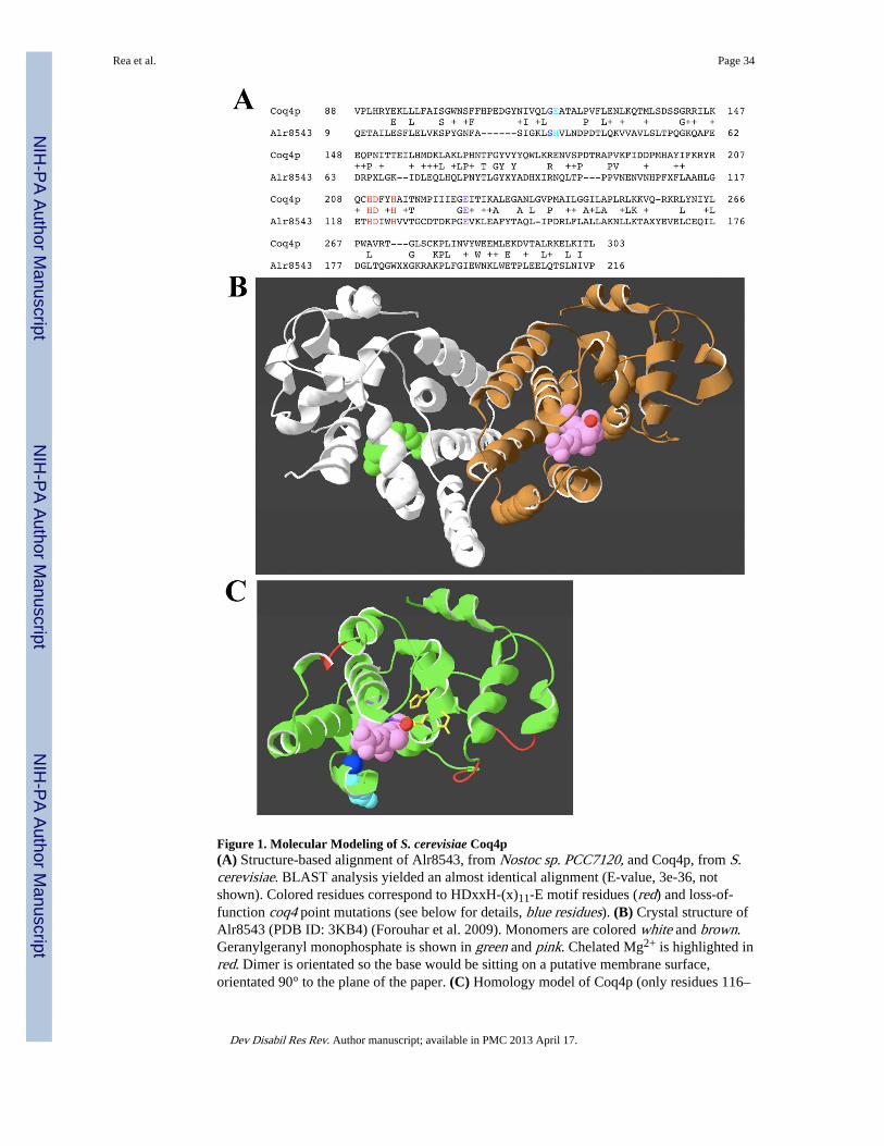
Figure 1. Molecular Modeling of S. cerevisiae Coq4p(A) Structure-based alignment of Alr8543, from Nostoc sp. PCC7120, and Coq4p, from S.cerevisiae. BLAST analysis yielded an almost identical alignment (E-value, 3e-36, notshown). Colored residues correspond to HDxxH-(x)11-E motif residues (red) and loss-of-function coq4 point mutations (see below for details, blue residues). (B) Crystal structure ofAlr8543 (PDB ID: 3KB4) (Forouhar et al. 2009). Monomers are colored white and brown.Geranylgeranyl monophosphate is shown in green and pink. Chelated Mg2+ is highlighted inred. Dimer is orientated so the base would be sitting on a putative membrane surface,orientated 90° to the plane of the paper. (C) Homology model of Coq4p (only residues 116–
Rea et al. Page 34
Dev Disabil Res Rev. Author manuscript; available in PMC 2013 April 17.
NIH
-PA Author Manuscript
NIH
-PA Author Manuscript
NIH
-PA Author Manuscript
302 were modeled). Red loops mark sites of amino acid insertions relative to Alr8543. Thefollowing features are highlighted: Geranylgeranyl monophosphate (pink); chelated Mg2+
(red ball); E226K (coq4-1), E121K (coq2-4) and G120E (coq4-3) point mutations (purple,azure and navy blue, respectively); H, D and H of the predicted Zinc-ligand bindingHDxxH-(x)11-E motif (yellow).
Rea et al. Page 35
Dev Disabil Res Rev. Author manuscript; available in PMC 2013 April 17.
NIH
-PA Author Manuscript
NIH
-PA Author Manuscript
NIH
-PA Author Manuscript
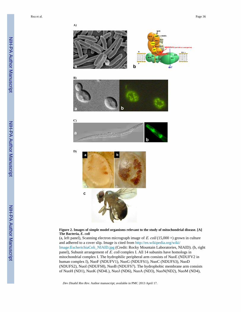
Figure 2. Images of simple model organisms relevant to the study of mitochondrial disease. [A]The Bacteria, E. coli(a, left panel), Scanning electron micrograph image of E. coli (15,000 ×) grown in cultureand adhered to a cover slip. Image is cited from http://en.wikipedia.org/wiki/Image:EscherichiaColi_NIAID.jpg (Credit: Rocky Mountain Laboratories, NIAID). (b, rightpanel), Subunit arrangement of E. coli complex I. All 14 subunits have homologs inmitochondrial complex I. The hydrophilic peripheral arm consists of NuoE (NDUFV2 inhuman complex I), NuoF (NDUFV1), NuoG (NDUFS1), NuoC (NDUFS3), NuoD(NDUFS2), NuoI (NDUFS8), NuoB (NDUFS7). The hydrophobic membrane arm consistsof NuoH (ND1), NuoK (ND4L), NuoJ (ND6), NuoA (ND3), NuoN(ND2), NuoM (ND4),
Rea et al. Page 36
Dev Disabil Res Rev. Author manuscript; available in PMC 2013 April 17.
NIH
-PA Author Manuscript
NIH
-PA Author Manuscript
NIH
-PA Author Manuscript
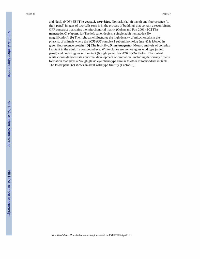
and NuoL (ND5). [B] The yeast, S. cerevisiae. Nomaski (a, left panel) and fluorescence (b,right panel) images of two cells (one is in the process of budding) that contain a recombinantGFP construct that stains the mitochondrial matrix (Cohen and Fox 2001). [C] Thenematode, C. elegans. (a) The left panel depicts a single adult nematode (50×magnification). (b) The right panel illustrates the high density of mitochondria in thepharynx of animals where the NDUFS2 complex I subunit homolog (gas-1) is labeled ingreen fluorescence protein. [D] The fruit fly, D. melanogaster. Mosaic analysis of complexI mutant in the adult fly compound eye. White clones are homozygous wild type (a, leftpanel) and homozygous null mutant (b, right panel) for NDUFS3 ortholog. The mutantwhite clones demonstrate abnormal development of ommatidia, including deficiency of lensformation that gives a “rough glass” eye phenotype similar to other mitochondrial mutants.The lower panel (c) shows an adult wild type fruit fly (Canton-S).
Rea et al. Page 37
Dev Disabil Res Rev. Author manuscript; available in PMC 2013 April 17.
NIH
-PA Author Manuscript
NIH
-PA Author Manuscript
NIH
-PA Author Manuscript
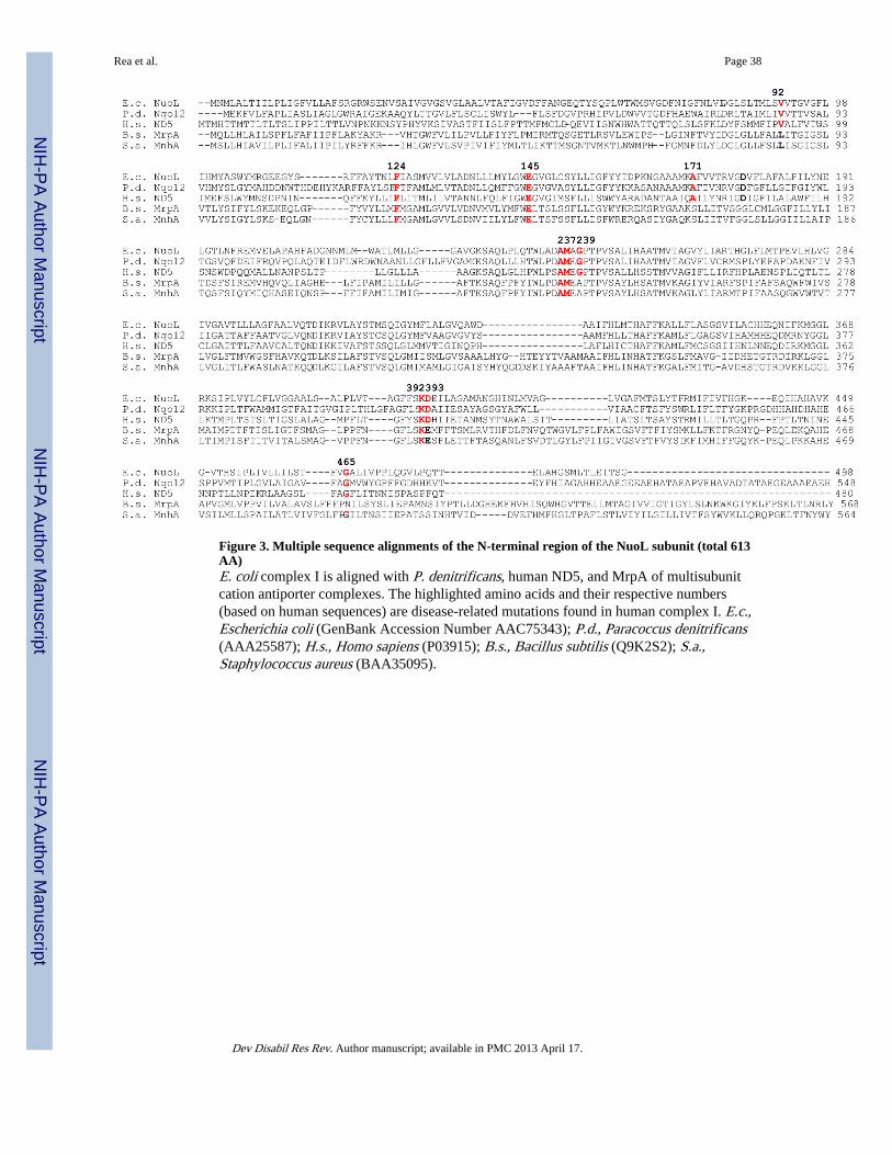
Figure 3. Multiple sequence alignments of the N-terminal region of the NuoL subunit (total 613AA)E. coli complex I is aligned with P. denitrificans, human ND5, and MrpA of multisubunitcation antiporter complexes. The highlighted amino acids and their respective numbers(based on human sequences) are disease-related mutations found in human complex I. E.c.,Escherichia coli (GenBank Accession Number AAC75343); P.d., Paracoccus denitrificans(AAA25587); H.s., Homo sapiens (P03915); B.s., Bacillus subtilis (Q9K2S2); S.a.,Staphylococcus aureus (BAA35095).
Rea et al. Page 38
Dev Disabil Res Rev. Author manuscript; available in PMC 2013 April 17.
NIH
-PA Author Manuscript
NIH
-PA Author Manuscript
NIH
-PA Author Manuscript
NIH
-PA Author Manuscript
NIH
-PA Author Manuscript
NIH
-PA Author Manuscript
Rea et al. Page 39
Table IYeast mitochondrial databases
Excellent online resources are available to facilitate study of mitochondria in the yeast S. cerevisiae.
Database Description Website
MitoP2 (Mitochondrial Proteome) Comprehensive list of mitochondrial proteins of S.cerevisiae, mouse, Arabidopsis thaliana, neurosporaand human
http://www.mitop.de
Yeast Mitochondrial Protein Database List of S. cerevisiae mtDNA-encoded proteins,nDNA-encoded proteins, and proteins that play arole in mitochondrial functionality
http://bmerc-www.bu.edu/projects/mito/
YDPM (Yeast Deletion Project &Mitochondria)
Database supporting proteomic, mRNA expression,and deletion studies relevant to mitochondrialfunction in S. cerevisiae
http://www.deletion.stanford.edu/
Dev Disabil Res Rev. Author manuscript; available in PMC 2013 April 17.
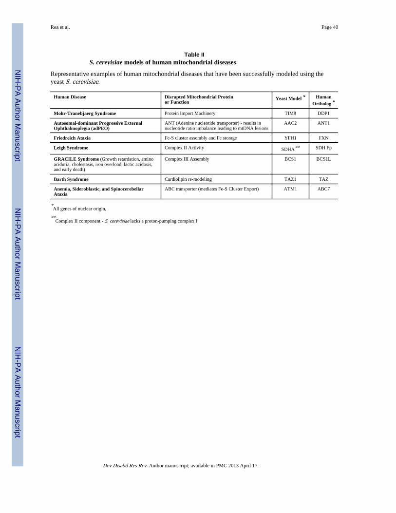
NIH
-PA Author Manuscript
NIH
-PA Author Manuscript
NIH
-PA Author Manuscript
Rea et al. Page 40
Table IIS. cerevisiae models of human mitochondrial diseases
Representative examples of human mitochondrial diseases that have been successfully modeled using theyeast S. cerevisiae.
Human Disease Disrupted Mitochondrial Proteinor Function
Yeast Model * HumanOrtholog *
Mohr-Tranebjaerg Syndrome Protein Import Machinery TIM8 DDP1
Autosomal-dominant Progressive ExternalOphthalmoplegia (adPEO)
ANT (Adenine nucleotide transporter) - results innucleotide ratio imbalance leading to mtDNA lesions
AAC2 ANT1
Friedreich Ataxia Fe-S cluster assembly and Fe storage YFH1 FXN
Leigh Syndrome Complex II Activity SDHA** SDH Fp
GRACILE Syndrome (Growth retardation, aminoaciduria, cholestasis, iron overload, lactic acidosis,and early death)
Complex III Assembly BCS1 BCS1L
Barth Syndrome Cardiolipin re-modeling TAZ1 TAZ
Anemia, Sideroblastic, and SpinocerebellarAtaxia
ABC transporter (mediates Fe-S Cluster Export) ATM1 ABC7
*All genes of nuclear origin,
**Complex II component - S. cerevisiae lacks a proton-pumping complex I
Dev Disabil Res Rev. Author manuscript; available in PMC 2013 April 17.
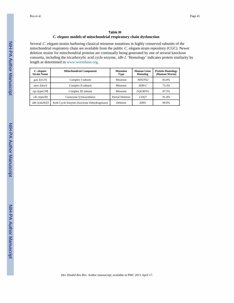
NIH
-PA Author Manuscript
NIH
-PA Author Manuscript
NIH
-PA Author Manuscript
Rea et al. Page 41
Table IIIC. elegans models of mitochondrial respiratory chain dysfunction
Several C. elegans strains harboring classical missense mutations in highly conserved subunits of themitochondrial respiratory chain are available from the public C. elegans strain repository (CGC). Newerdeletion strains for mitochondrial proteins are continually being generated by one of several knockoutconsortia, including the tricarboxylic acid cycle enzyme, idh-1. ‘Homology’ indicates protein similarity bylength as determined in www.wormbase.org.
C. elegansStrain Name
Mitochondrial Component MutationType
Human GeneHomolog
Protein Homology(Human-Worm)
gas-1(fc21) Complex I subunit Missense NDUFS2 83.4%
mev-1(kn1) Complex II subunit Missense SDH-C 75.3%
isp-1(qm150) Complex III subunit Missense UQCRFS1 97.5%
clk-1(qm30) Coenzyme Q biosynthesis Partial Deletion COQ7 91.4%
idh-1(ok2832) Kreb Cycle Enzyme (Isocitrate Dehydrogenase) Deletion IDH1 99.0%
Dev Disabil Res Rev. Author manuscript; available in PMC 2013 April 17.


![Third report on jurisdictional immunities of States and ... · by Mr. Motoo Ogiso, Special Rapporteur [Original: English] [11 April 1990] CONTENTS Page Note 5 Paragraphs INTRODUCTION](https://img.pdfslide.us/doc/110x75/5f1fdfc4ed7d2d656929a6e5/third-report-on-jurisdictional-immunities-of-states-and-by-mr-motoo-ogiso.jpg)


![Second report on jurisdictional immunities of States and ... · by Mr. Motoo Ogiso, Special Rapporteur [Original: English] [11 and 24 April 1989} CONTENTS Note 59 Paragraphs INTRODUCTION](https://img.pdfslide.us/doc/110x75/5f1fdfc4ed7d2d656929a6e6/second-report-on-jurisdictional-immunities-of-states-and-by-mr-motoo-ogiso.jpg)