Embed Size (px)
Citation preview

JOURNAL OF NEUROTRAUMAVolume 23, Number 3/4, 2006© Mary Ann Liebert, Inc.Pp. 295–308
New Insights into Neuronal Regeneration: The Role of AxonalProtein Synthesis in Pathfinding and Axonal Extension
JEFFERY L. TWISS1,2 and JAN VAN MINNEN3
ABSTRACT
Protein synthesis in dendrites has become an accepted cellular mechanism that contributes to ac-tivity-dependent responses in the post-synaptic neuron. Although it was argued that protein syn-thesis does not occur in axons, early studies from a number of groups provided evidence for thepresence of RNAs and active protein synthesis machinery in both invertebrate and vertebrate ax-ons. Work over the past decade has confirmed these early findings and has proven the capabilityof axons to locally synthesize some of their own proteins. The functional significance of this local-ized protein synthesis remained largely unknown until recent years. Recent studies have shown thatmRNA translation in developing and mature axons plays a role in axonal growth. In developing ax-ons, protein synthesis allows the distal axon to autonomously respond to guidance cues by rapidlychanging its direction of outgrowth. In addition, local proteolysis of axonal proteins contributes ax-onal guidance and growth cone initiation. This local synthesis and degradation of proteins are likelyto provide novel insights into how growing axons navigate through their complex environment. Inmature axons, injury triggers formation of a growth cone through localized protein synthesis, andmoreover, in these injured axons locally synthesized proteins provide a retrogradely transportedsignal that can enhance regenerative responses. The intrinsic capability for axons to autonomouslyregulate local protein levels can be modulated by exogenous stimuli providing opportunities for en-hancing regeneration. In this review, the concept of axonal protein synthesis is discussed from a his-torical perspective. Further, the implications of axonal protein synthesis and proteolysis for neuralrepair are considered.
Key words: axonal growth; axon guidance; mRNA localization; protein synthesis
295
1Nemours Biomedical Research, Alfred I duPont Hospital for Children, Wilmington, Delaware.2Department of Biological Sciences, University of Delaware, Newark, Delaware.3Department of Molecular and Cellular Neurobiology, Institute of Neurosciences, Vrije Universiteit, Amsterdam, The Nether-
lands.
INTRODUCTION
NEURONS ARE EXTREMELY POLARIZED CELLS, consist-ing of somal, dendritic, and axonal domains. A clas-
sic view in neuroscience has been that the cell body sup-
plies the axon, including its most distal presynaptic ter-minal, with all the proteins that are necessary for its main-tenance and plasticity. The axons of the larger vertebratescan reach lengths of 1 m or more. For a neuronal cellbody of 80 �m in diameter, the volume of a meter-long

axon would exceed that of the cell body by more than300 fold. Thus, growing and maintaining a process of thisproportion is a huge undertaking for the neuron. Axonalproteins can be anterogradely transported at rates up to40 cm per day (Ochs, 1972). This microtubule-based, fastaxonal transport carries vesicles and vesicle-associatedproteins, such as receptor molecules. However, most cy-tosolic proteins, including cytoskeletal proteins, are trans-ported by slower means, of which the faster component(SCb) moves at up 2–4 mm per day and the slower com-ponent (SCa) moves at 0.1–1 mm per day. Proteins trans-ported in SCb include actin, neuronal myosin, clathrin,enzymes involved in the intermediary metabolism, andcalmodulin. Neurofilament proteins and tubulin are trans-ported in the slower SCa component (Alvarez et al.,2000). These cytoskeletal proteins, which are needed forstructure of the axon, would take at least 300 days totravel 1 m from the soma to the terminal reaches of theaxon. Considering the limited half-life of some proteins(e.g., tubulin t1/2 � 1 week [Forgue and Dahl, 1978]), itis hard to imagine that these proteins could reach the dis-tal axon in sufficient quantities to be of biological sig-nificance. Indeed, studies by Nixon (1980), using ra-dioactive precursors, demonstrated that some cell bodysynthesized proteins destined for axonal transport are de-graded in the first few millimeters of the mouse opticnerve. Presuming that these data can be extended verte-brates, either the neuron must expend significant energygenerating extra proteins to account for degradation dur-ing transport or it has other means to replenish these pro-teins lost during transport.
The distal axon also has the capacity to autonomouslyrespond to pathfinding and activity dependent stimuli(Song and Poo, 2001). Considering the enormous differ-ences in volume between cell body vs. axon and the dis-tance separating cellular components, it almost is a mir-acle that the distal axon not only survives but alsoperforms autonomous functions. An alternative mecha-nism to provide the distal axon with proteins was put for-ward in the 1970s. A few studies lent support to the ideathat glial cells could supply the axons with proteins(Lasek et al., 1974; Lasek et al., 1977; Tytell and Lasek,1984). However, these studies did not provide an answerto how neuronal specific proteins, such as neurofila-ments, are supplied to the axon. A simple and economi-cal way to locally provide proteins would be for the axonto synthesize some of its own proteins. Such localizedprotein synthesis is now a generally accepted mechanismin dendrites, where it was shown to play a critical role inactivity-dependent processes associated with memoryformation (e.g., long-term potentiation [LTP] and long-term depression [LTD]) (Jiang and Schuman, 2002). Thepossibility of axonal or presynaptic protein synthesis was
dismissed for a long time by much of the neurosciencecommunity since ultrastructural analyses did not revealany of the cellular machinery necessary for protein syn-thesis (i.e., ribosomes, rough endoplasmic reticulum[RER], and Golgi apparatus) in mature axons and presy-naptic terminals (Peters et al., 1991). Nevertheless, asmall but tenacious group of researchers have advocatedfor the likelihood of axonal protein synthesis for over 40years (Alvarez et al., 2000; Giuditta et al., 2002).
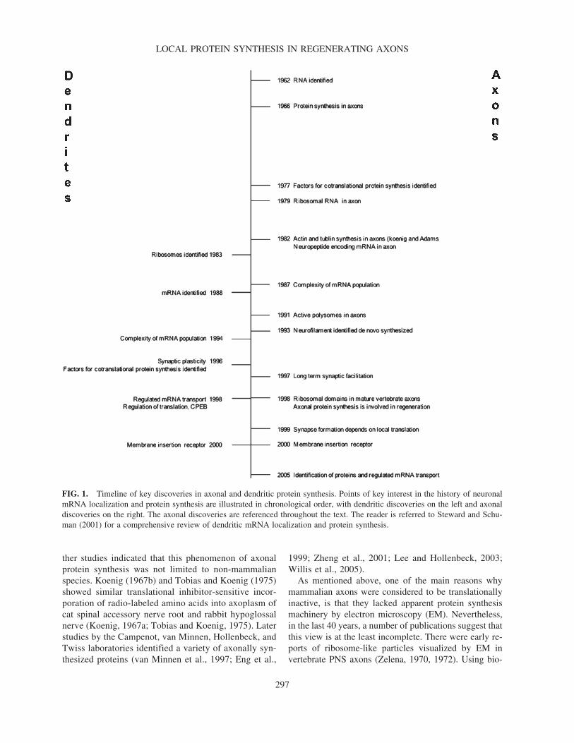
In the next section, the landmark studies that have cul-minated in a body of evidence to abandon the earlierviews that axons are incapable of protein synthesis willbe highlighted. The key discoveries that lead to the con-cept of axonal protein synthesis are presented in a chrono-logical order in Figure 1. For comparison, similar dis-coveries in the dendritic field are chronicled. Many of theaxonal studies, especially those in the early days, werecarried out in two model systems, the Mauthner axon ofthe goldfish and the giant axon of the squid, where thelarge caliber axons (40–80 �m and 1 mm in diameter,respectively) provided pure axoplasm for analyses. Withthe recent advancements in sensitivity of molecular bio-logical and biochemical techniques, axons from highervertebrates have become more amenable to analyses.
HISTORICAL PERSPECTIVE OF AXONALPROTEIN SYNTHESIS
The first publications that hinted at the phenomenonof axonal protein synthesis date back as far as the early1960s. In 1962, considerable amounts of RNA was firstdetected in the Mauthner cell axon of the goldfish (Ed-strom et al., 1962). Similar findings were subsequentlyreported in the cat and rabbit (Koenig, 1965a,b,1967a,b). Although early studies were unable to detectmRNAs and ribosomal RNAs (rRNA) in mature axons(Lasek et al., 1973), Koenig and Giuditta were able laterto identify rRNA in Mauthner and squid axons (Koenig,1979; Giuditta et al., 1980a). Much later, ribosomeswere detected in other preparations, including matureaxons from the Lymnaea and Heliosoma snail species(Davis et al., 1992; van Minnen, 1994). Further studiesin the goldfish and squid systems were the first to sug-gest that axonal ribosomes are functional. Incorporationof radio-labeled amino acids and sensitivity to inhibitorsof translation argued that axonal ribosomes are utilizedfor protein synthesis in pure preparations of axoplasm(Edström, 1966; Fischer and Litvak, 1967; Giuditta etal., 1968; Edstrom and Sjostrand, 1969; Alvarez andChen, 1972). Synaptosomes of presynaptic terminals ofsquid photoreceptor neurons were also shown to syn-thesize a variety of proteins (Crispino et al., 1993). Fur-
TWISS AND VAN MINNEN
296
ther studies indicated that this phenomenon of axonalprotein synthesis was not limited to non-mammalianspecies. Koenig (1967b) and Tobias and Koenig (1975)showed similar translational inhibitor-sensitive incor-poration of radio-labeled amino acids into axoplasm ofcat spinal accessory nerve root and rabbit hypoglossalnerve (Koenig, 1967a; Tobias and Koenig, 1975). Laterstudies by the Campenot, van Minnen, Hollenbeck, andTwiss laboratories identified a variety of axonally syn-thesized proteins (van Minnen et al., 1997; Eng et al.,
1999; Zheng et al., 2001; Lee and Hollenbeck, 2003;Willis et al., 2005).
As mentioned above, one of the main reasons whymammalian axons were considered to be translationallyinactive, is that they lacked apparent protein synthesismachinery by electron microscopy (EM). Nevertheless,in the last 40 years, a number of publications suggest thatthis view is at the least incomplete. There were early re-ports of ribosome-like particles visualized by EM in vertebrate PNS axons (Zelena, 1970, 1972). Using bio-
LOCAL PROTEIN SYNTHESIS IN REGENERATING AXONS
297
FIG. 1. Timeline of key discoveries in axonal and dendritic protein synthesis. Points of key interest in the history of neuronalmRNA localization and protein synthesis are illustrated in chronological order, with dendritic discoveries on the left and axonaldiscoveries on the right. The axonal discoveries are referenced throughout the text. The reader is referred to Steward and Schu-man (2001) for a comprehensive review of dendritic mRNA localization and protein synthesis.

chemical methods, ribosomal RNA [rRNA] was detectedin the axonal compartment of the Mauthner cell and squidgiant axon (Koenig, 1979; Giuditta et al., 1980b). Morerefined EM methods, including electron spectroscopicimaging (ESI) to enhance detection of nucleic acids, aswell as immunolabeling and in situ hybridization studieson purified axoplasm and primary neuronal culture prepa-rations were used to demonstrate the presence of ribo-somes in both invertebrate and vertebrate axons (Martinet al., 1989; Pannese and Ledda, 1991; Koenig and Mar-tin, 1996; Bassell et al., 1998; Koenig et al., 2000; Zhenget al., 2001). In addition to ribosomes, other componentsof the protein synthesizing machinery have been identi-fied in axons. In 1977, Giuditta et al. showed that thesquid giant axon also contained translation factors neededfor protein synthesis (Giuditta et al., 1977). More recentconfocal microscopy of adult rat ventral root axonsdemonstrated translation initiation factors, ribosomal pro-teins and rRNA in injured mammalian axons (Zheng etal., 2001). Furthermore, ribosomes that were detected inaxoplasmic wholemount preparations from Mauthneraxon and myelinated fibers of mature rabbit and rat PNSaxons appear concentrated in subdomains associated withthe cortical F-actin layer of the axon (Koenig and Mar-tin, 1996; Koenig et al., 2000). These “periaxoplasmicplaques” are sensitive to RNase (Koenig et al., 2000), andrecent studies have shown that the motor proteins,Myosin Va and Kinesin II, are also concentrated in theseregions (Sotelo-Silveira et al., 2004).
mRNAs IN THE AXONAL DOMAIN
One of the earliest studies that reported a complex, het-erogeneous population of mRNAs in the axonal com-partment was conducted using the squid giant axon. Ca-pano et al. (1987) demonstrated that this invertebrateaxon contains at least 200 mRNA species. Follow-upstudies showed that this mRNA population includedthose encoding neurofilament, �-actin, �-tubulin, ki-nesin, enolase, nuclear-encoded mitochondrial proteinsand heat-shock proteins (HSP) (Kaplan et al., 1992; Gioioet al., 1994, 2001; Chun et al., 1995, 1996). From stud-ies on other mollusks (Aplysia and Lymnaea) and mam-mals it became apparent that axons and axon terminalsof neuropeptide-producing neurons contain largeamounts of neuropeptide-encoding mRNAs (van Minnen,1994). The functional significance of this phenomenonwas not clear, but follow-up studies have shown that neu-ropeptide-encoding mRNAs can be translated in axonsand that this locally synthesized protein can then be se-creted (van Minnen et al., 1997; Wayne et al., 2004).mRNA and protein-based studies aimed at identifying ax-
onally synthesized proteins have now greatly expandedthe list of axonal transcripts. Surprisingly, among the listof axonal mRNAs are species encoding ribosomal pro-teins and other components of the protein synthesizingmachinery, such as translation factors and chaperone pro-teins of the endoplasmic reticulum (ER) (Moccia et al.,2003; Willis et al., 2005). These findings suggest that theaxon can synthesize components of it’s own protein syn-thesizing and trafficking machinery raising the questionof whether this may represent a novel means to regulatethe axonal protein synthesis apparatus.
�-actin mRNA has consistently been detected in manydifferent axonal mRNA preparations. In fibroblasts andmyoblasts, �-actin and �-actin mRNAs show distinctsubcellular localization (Lawrence and Singer, 1986;Sundell and Singer, 1990; Hill and Gunning, 1993). Bas-sell et al. (1998) showed differential localization of actinmRNAs in cortical neurons, with �-actin extending intoaxons and �-actin being restricted to the cell body. Thistransport of neuronal �-actin mRNA from cell body toaxon is determined by a cis-element, or “zip code,” inthe transcript’s 3� untranslated region (UTR) that is notpresent in �-actin mRNA (Kislauskis et al., 1993; Hill etal., 1994). A protein binding to the �-actin mRNA zipcode, the zip code binding protein (ZBP), is needed foraxonal localization of �-actin mRNA (Zhang et al., 2001;Gu et al., 2002). It is not clear what mechanisms maydrive other neuronal mRNAs into axons. Specific cellu-lar mechanisms must exist to target these mRNAs to ax-ons since not all neuronal transcripts localize to axons(nor to dendrites). Bi et al. (2003) have used transfectionof differentiated P19 cells and dorsal root ganglion neu-rons (DRG) to show that the 5� UTR drives localizationof �-opioid receptor mRNAs into axonal processes. ForTau mRNA, which localizes to developing axonal pro-cesses, a cis-element in the 3� UTR is recognized by RNAbinding proteins including the Elav protein HuD (Amaldiand Pierandrei-Amaldi, 1997; Aronov et al., 2002;Larcher et al., 2004). There appears to be no consensussequence in the 5� or 3� UTRs that dictates axonal mRNAlocalization, but rather secondary structure of the mRNAdetermines its interaction with RNA binding proteins(Bassell and Kelic, 2004).
Most evidence indicates that mammalian axonal mRNAs, at least the vast majority, are anterogradelytransported from the cell body (Willis et al., 2005). Imag-ing studies indicate that axonal RNA granules are trans-ported at up to approximately 10 cm/day in axons, whichis about half that of fast anterograde transport of cellbody–derived vesicular proteins (Zhang et al., 2001;Knowles et al., 1996). If these rates are comparable forall axonal mRNAs, then the mRNAs encoding mollus-can neuropeptide proteins would be delivered to axons at
TWISS AND VAN MINNEN
298

rates slower than the same neuropeptide proteins that aresynthesized in the cell body and anterogradely trans-ported. On the other hand, delivery of mRNAs encodingcytoskeletal proteins to distal axons would easily exceedthe rate of delivering their encoded proteins that are syn-thesized in the cell body and normally delivered in SCaand SCb axonal transport modes. For the mRNAs whoseprotein products normally move in fast axonal transport,such as the neuropeptide proteins, axonal mRNAs couldbe used to provide the distal axon with some autonomyto locally modulate levels of these secreted proteins. Sup-port for this possibility came from studies from Lee andWayne (2004) who demonstrated that Aplysia bag cellaxons were able to release processed ELH by both theconstitutive and regulated secretory pathways. As notedbelow, local protein synthesis can be used by developingaxons to autonomously respond to extracellular stimuli.
ROLE OF LOCALIZED PROTEINSYNTHESIS DURING GROWTH CONEMOTILITY AND AXONAL GUIDANCE
A unifying theme in axonal protein synthesis is thatlocally translated proteins play a role in axonal growth.For instance, studies by the Bassell group showed the lo-calization of �-actin mRNA to neuronal growth cones(Bassell et al., 1998), and that brain-derived neurotrophicfactor (BDNF) and neurotrophin 3 (NT3) can increasethe localization of �-actin mRNA in these growth cones(Zhang et al., 1999). Since new actin polymerization isneeded for growth cone motility (Okabe and Hirokawa,1990, 1991), neurotrophin-dependent localization of �-actin mRNA and the ensuing protein synthesis in thegrowth cone is likely to play a critical role in axonal ex-tension. Localization of �-actin mRNA to the leadingedge of migrating fibroblasts (Lawrence and Singer,1986) suggests that localized synthesis of actin is a gen-eral mechanism of cell motility.
A locally renewable source of cytoskeletal proteins atthe growth cone could conceivably provide the distalaxon with some autonomy in rapidly responding to en-vironmental stimuli (Fig. 2A). Exciting clues supportingthis concept of localized protein synthesis contributing toaxonal growth were provided by three independentgroups studying axonal guidance. Using a preparation ofXenopus retinal neurons, Campbell et al. (2001) showedthat pathfinding stimuli can locally regulate protein syn-thesis in distal axons. Subsequent work by Ming et al.(2002) indicated that growth cones of Xenopus motorneurons require local protein synthesis for resensitizationin responding to gradients of guidance cues. Pathfindingstimuli that regulate this local protein synthesis include
Netrin-1, semaphorin 3A (Sema3A), and BDNF. Impor-tantly, analyses of axons isolated from the cell bodyproved that the growth cone responses to these stimulirequired localized axonal protein synthesis (Campbelland Holt, 2001; Ming et al., 2002). Further studies fromBrittis et al. (2002) showed that EphA2, a receptor thatdetects axonal guidance cues, is locally synthesized ingrowth cones of commissural axons as they cross the mid-line in developing spinal cord. Together, these studies in-dicate that axonally synthesized proteins can play a crit-ical role in axonal pathfinding to help guide developingaxons to their target tissues.
The intracellular signals that lead to translational reg-ulation in response to axonal guidance cues have beenthe subject of recent investigation. Erk 1/2 and p38MAPK
are activated by axonal guidance cues and these signalscan differentially regulate axonal protein synthesis orproteolysis (Campbell and Holt, 2003). Sema3A locallyactivates Erk 1/2 leading to protein synthesis dependentturning of axons, while Netrin-1 activates both Erk 1/2and p38MAPK leading to protein synthesis and proteoly-sis dependent turning of axons. Sema3A can also increaserates of retrograde and anterograde axonal transport inDRG neurons through activation of axonal protein syn-thesis (Li et al., 2004). However, this effect of Sema3Aon axonal transport appears to be through the fyn andCDK5 kinases leading to phosphorylation of the transla-tion factor eIF4E (Sasaki et al., 2002; Li et al., 2004),rather than the dependence on Erk 1/2 as seen in the de-veloping retinal axons (Campbell and Holt, 2001, 2003;Li et al., 2004). Motor axons responding to gradients ofBDNF or Netrin-1 undergo consecutive rounds of de-sensitization with a corresponding decrease in intra-ax-onal Ca2� signaling (Ming et al., 2002). Most recently,Piper et al. (2005) used a collapse assay in retinal axonsto show that desensitization to Sema3A and Netrin-1 re-quires receptor endocytosis but the subsequent resensiti-zation phase is protein synthesis dependent.
The locally synthesized proteins that are at the basisof the above changes in axonal growth and responsive-ness remain a mystery. Work in migrating fibroblasts in-dicates that local synthesis of �-actin protein provides di-rectionality to cellular motility (Shestakova et al., 2001).Thus, local synthesis of �-actin protein in growth conesmay serve as nidus to alter direction of growth in re-sponse to pathfinding stimuli. However, localized syn-thesis of �-actin alone cannot fully explain these au-tonomous responses of developing axons since thedesensitization/resensitization that required axonal pro-tein synthesis in chick retinal axons shows specificity toparticular guidance cues (Piper et al., 2005). It will bevery interesting to see how this specificity is imparted tothe axonal translational apparatus. Specific means to reg-
LOCAL PROTEIN SYNTHESIS IN REGENERATING AXONS
299

ulate localized translation of EphA2 mRNA through cy-toplasmic polyadenylation was demonstrated in commis-sural axons (Brittis et al., 2002). Use of internal ribosomeentry sites (IRES), which can be preferentially utilizedwhen cap-dependent translation is turned off or attentu-ated, is another means to potentially impart specificity to
localized protein synthesis (Huang and Richter, 2004).Interestingly, two axonal mRNAs, HSP70 and grp78/BiPmRNAs can be translated by cap-independent means inother cellular systems (Macejak and Sarnow, 1991;Rubtsova et al., 2003; Thoma et al., 2004; Willis et al.,2005).
TWISS AND VAN MINNEN
300

INTRINSIC MODULATIONS OF LOCALSYNTHESIS AND PROTEIN
DEGRADATION DURING AXONAL REGENERATION
The developing vertebrate neuronal preparations haveprovided robust systems to analyze axonal protein synthe-sis. Work from Koenig’s group indicated that mature ver-tebrate axons contain at least some components of the trans-lational machinery (Koenig et al., 2000). As noted earlier,localized protein synthesis in mature axons was hypothe-sized to replenish proteins that are degraded over the courseof their transport from the cell body (Alvarez et al., 2000).However, the actual function that this localized protein syn-thesis provides to the axon remained speculative withoutany means to test this hypothesis. Using an in vivo periph-eral nerve injury model, Gaete et al. (1998) suggested thata localized source of mRNA translation is required for ax-onal regeneration. An earlier report suggested that local-ized protein synthesis may contribute to axonal regenera-tion in injury-conditioned neurons (Edbladh et al., 1994).A culture preparation of dissociated injury-conditioned ratsensory neurons, initially generated by Smith and Skene(1997), provided a model system that both verified the ca-pability of adult axons to synthesize proteins and addresseda functionality (Zheng et al., 2001). These DRG neuronsshow rapid, directed axonal growth over the first day invitro that requires new protein synthesis but not new mRNAsynthesis (Smith and Skene, 1997; Twiss et al., 2000). Ax-ons isolated from these injury-conditioned neurons synthe-size proteins in culture and their growth cones rapidly re-tract upon inhibition of local protein synthesis (Zheng etal., 2001). This axonal retraction is consistent with the re-traction of growth cones seen when axonal localization of
�-actin mRNA is blocked in cultures of developing corti-cal neurons (Zhang et al., 2001). Thus, the robust axonalprotein synthesis of developing neurons can be reactivatedafter injury, presumably providing cytoskeletal stability tofacilitate growth of regenerating axons.
Very recently it was shown by the Fawcett group thatgrowth cone formation requires both protein synthesisand proteolysis in axons (Verma et al., 2005) (Fig. 2B).Pharmacological inhibition of the target of rapamycin[TOR], p38MAPK or Erk 1/2 pathways could block growthcone formation effectively linking these signal transduc-tion pathways to modulation of local protein synthesisand/or proteolysis during growth cone formation (Vermaet al., 2005). In the developing retinal axons, p38MAPK isactivated by stimuli that regulate proteolysis through cas-pase 3, while Erk 1/2 activation leads to localized pro-tein synthesis (Campbell and Holt, 2003). Since an in-crease in axoplasmic [Ca2�] and subsequent localizedactivation of proteolysis has been shown to initiategrowth cone formation (Ziv and Spira, 1997; Gitler andSpira, 2002), it is tempting to speculate that a localizedincrease of Ca2� is at the basis of these observed in-creases in protein synthesis and proteolysis. Taken to-gether, the signaling pathways that underlie these intrin-sic determinants of axonal regeneration may provide newavenues for facilitating growth of CNS axons.
PROTEIN SYNTHESIS IN REGENERATING AXONS
In addition to �-actin, synthesis of proteins encodingother cytoskeletal elements has been frequently detectedin developing and invertebrate axons (Piper and Holt,
LOCAL PROTEIN SYNTHESIS IN REGENERATING AXONS
301
FIG. 2. Regulatory events in axonal protein synthesis. (A) Schematic of a developing neuron, with cell body, axon, and growthcone shown. In developing axons, transport of mRNAs from cell body into axons and localized protein synthesis in the growthcone can be regulated by neurotrophins (Knowles and Kosik, 1997; Zhang et al., 1999, 2001; Ming et al., 2002). Axon turningin response to the pathfinding cues, Sema3A and Netrin-1, requires localized protein synthesis in axons (Campbell and Holt,2001; Ming et al., 2002). Netrin-1 also activates localized proteolysis to initiate a chemotactic response and growth cone retrac-tion in response to LPA requires localized proteolysis in axons (black arrows) (Campbell and Holt, 2001, 2003). (B) Schematicof an injured neuron with residual proximal axon, axotomy site, and distal axon undergoing Wallerian degeneration (gray bars)with loss of contact to its target is shown. In mature axons, axotomy can trigger translation of mRNAs in the proximal axon.Translation of axonal Importin �1 and vimentin mRNAs that form a protein complex to carry signaling proteins (designated asX), including activated Erk back to the nucleus (Hanz et al., 2003; Perlson et al., 2005). Formation of this retrograde signalingcomplex is needed to trigger an enhanced regenerative response (Hanz et al., 2003). Axotomy also elicits localized proteolysis,and both local protein synthesis and proteolysis in axons is needed to initiate growth cone formation (Verma et al., 2005). Thecues that axotomy uses to activate local protein synthesis and proteolysis in axons (designated as “?”) is presently unknown. (C)Schematic of a regenerating neuron. In regenerating axons, local sources of neurotrophins increase transport of mRNAs from cellbody into the axons (Willis et al., 2005). Neurotrophins and other stimuli (designated as ?) are likely to regulate protein synthe-sis in regenerating axons as well. There is evidence for activity-dependent regulation of axonal protein synthesis in mature ax-ons terminals (Zhang and Poo, 2002), and BDNF has been shown to alter translational efficiency of some mRNAs (Schratt etal., 2004).

2004). However, there is accumulating evidence that ax-ons can synthesize a variety of protein types. Lee andHollenbeck (2003) recently reported that approximately5% of neuronal protein synthesis occurs in axons of de-veloping sympathetic neurons (Lee and Hollenbeck,2003). Using isolated processes of Aplysia sensory neu-rons, Moccia et al. (2003) detected approximately 200distinct mRNAs in sequence analyses of individualcDNA clones. Protein synthesis in axons of other inver-tebrate species may be similarly complex. As notedabove, a similarly complex population of mRNAs hasbeen demonstrated in squid giant axons (Capano et al.,1987). Localized translation of a number of proteins, in-cluding HSP70, was detected by proteomics-based analy-ses of presynaptic terminals from the squid optic lobe(Jimenez et al., 2002). Recent studies argue that proteinsynthesis in regenerating vertebrate axons is also quitecomplex and further shed light on new functions for ax-onal protein synthesis. Proteomics-based analyses of pro-teins translated in mammalian sensory axons showed thata large number of different proteins are locally synthe-sized in addition to cytoskeletal proteins (Willis et al.,2005). To date, the emerging catalog of axonally syn-thesized proteins includes integral membrane proteins,HSPs and other chaperone proteins, anti-oxidant proteins,metabolic proteins, as well as proteins that have beenlinked to neurodegenerative disorders (Capano et al.,1987; Jimenez et al., 2002; Lee and Hollenbeck, 2003;Piper and Holt, 2004; Willis et al., 2005). Future analy-ses of locally synthesized proteins are certain to yield in-sight into new functional roles for axonal protein syn-thesis in regeneration. For example, upregulation ofHSP27 after axotomy appears to be neuroprotective(Costigan et al., 1998; Lewis et al., 1999; Benn et al.,2002). HSP27 mRNA extends into regenerating DRG ax-ons suggesting that it may play a more direct role in nerveregeneration (Willis et al., 2005).
One presumes that these locally synthesized proteinsprovide the growing axon with molecules that it cannotderive in sufficient time through anterograde transport of cell body synthesized proteins. Consistent with thisnotion, injury-conditioned DRG neurons can extendprocesses up to 1 mm over a 12–18-h period in vitro(Lankford et al., 1996; Smith and Skene, 1997). This rateof axonal outgrowth is actually faster than the knownrates of transport of some of the axonally synthesizedcytoskeletal proteins (e.g., tubulin) (Zheng et al., 2001).Beyond locally generating structural proteins, studiesfrom the Fainzilber lab indicate that assembly of a ret-rograde signaling complex requires axonal protein syn-thesis and proteolysis (Fig. 2B). Axotomy triggers thetranslation of Importin � and vimentin mRNAs in PNSaxons. These newly synthesized proteins assemble into
a retrograde signaling complex that is needed to initiatea regenerative response (Hanz et al., 2003; Perlson et al.,2005). Locally synthesized vimentin protein is proteo-lyzed and the cleaved vimentin serves as a scaffold toretrogradely transport activated Erk to the cell body(Perlson et al., 2005). Axonal protein synthesis not onlyprovides autonomy to the axon, but also serves as ameans to signal the cell body after axotomy. Local pro-teolysis of other axonal proteins is also needed forgrowth cone formation in Aplysia (Gitler and Spira,2002). The translation of mRNAs encoding ubiquitin C-terminal hydrolase-L1 (Uch-L1) and chaperone proteinsin axons may provide a further link between localizedproteolysis and protein synthesis (Willis et al., 2005).Thus, protein synthesis and protein degradation, al-though seeming to generate completely opposite out-comes, may serve complementary roles during axonalinjury and regeneration.
Similar assembly of Importin complexes followingNMDA receptor activation, with retrograde signaling tothe nucleus, was demonstrated in dendrites of hip-pocampal neurons (Thompson et al., 2004). This arguesthat plasticity and injury share common mediators. Suchan overlap between injury and plasticity mechanisms waspreviously suggested from work in Aplysia (Ambron andWalters, 1996; Ambron et al., 1996). In slice prepara-tions, LTP-inducing stimuli can initiate actin polymer-ization in dendritic spines and preventing this polymer-ization blocks consolidation of LTP (Kruker et al., 2000;Lin et al., 2005). ZBP-bound �-actin mRNA also local-izes into dendrites after depolarization or NMDA recep-tor activation (Tiruchinapalli et al., 2003). Thus, althoughphysiologically distinct, axons and dendrites may indeedshare common molecular mechanisms of mRNA local-ization/translation. Indeed, muscle-derived BDNF can in-crease presynaptic release of neurotransmitters throughactivation of localized protein synthesis in axons (Zhangand Poo, 2002).
CONCLUSION
Similar to development, regenerating axons need tonavigate their environment. mRNA translation in the ax-onal compartment appears to provide diverse functionsto the neuron. It is intriguing to speculate that localizedprotein synthesis in axons and growth cones may be amechanism to harness for coaxing axons to regenerate inthe CNS. As mentioned above, DRG neurons that areprimed for enhanced axonal growth in vitro show robustaxonal protein synthesis (Zheng et al., 2001; Hanz et al.,2003; Willis et al., 2005). Such axonal injury can alsoprime adult DRG neurons for growth into CNS white
TWISS AND VAN MINNEN
302

matter (Neumann and Woolf, 1999). Work from the Fil-bin lab has shown that priming with neurotrophin or di-rect increases in neuronal cAMP can be used to over-come inhibitory effect of CNS myelin for axonaloutgrowth from adult neurons (Cai et al., 2001; Qiu etal., 2002; Lu et al., 2004; Pearse et al., 2004). In the cul-tured DRG neurons, treatment with nerve growth factor(NGF) or BDNF increases transport of �-actin, vimentin,and peripherin mRNAs into the growing axons (Fig. 2C)(Willis et al., 2005). Neurotrophins have also been shownto increase translation of some neuronal proteins, bothwithin cell body and axonal processes (Zhang et al., 1999;Ming et al., 2002; Schratt et al., 2004). It is noteworthythat transport of the majority of the axonal mRNAs an-alyzed by Willis et al. (2005) was not affected by neu-
rotrophin treatment. This suggests that localized transla-tion may be the key regulatory event for altering axonallevels of the proteins encoded by these transcripts (Fig.2C). Thus, trophic agents, including pathfinding stimuli,may provide a means to exogenously regulate axonal pro-tein synthesis in injured CNS axons. In line with this idea,ultrastructural analyses of central DRG processes regen-erating into a Schwann cell graft shows arrays of polyso-mal and free ribosomes in the axoplasm (Fig. 3).
The realization that axons are capable of synthesiz-ing a sizable portion of their own proteins raises ques-tions about the basic biology of this process. It is amystery how the axon can target locally synthesizedproteins for secretion and to membrane locations giventhat RER and Golgi apparatus are not ultrastructurally
LOCAL PROTEIN SYNTHESIS IN REGENERATING AXONS
303
FIG. 3. Ribosome particles in regenerating CNS axons. Electron microscopy image of an axon that was taken from a Schwanncell containing graft that was placed in the spinal cord to bridge an surgically induced lesion (Oudega et al., 1997). Numerouspolyribosomes (arrows) can be identified in the axoplasm. This event could be observed in 0.5–1% of the regenerating axons.Mi, mitochondrion; My, myelin sheath. Scale bar � 100 nm.

visualized in axons (Zelena, 1970, 1972; Peters et al.,1991; Spencer et al., 2000; Bassell et al., 1998; Zhenget al., 2001). Nevertheless, targeting of axonally syn-thesized transmembrane proteins has been demon-strated. Spencer et al. (2000) injected an exogenousmRNA encoding a G-protein coupled receptor into de-nucleated Lymnaea axons (Spencer et al., 2000). Theinjected axons translated the mRNA, inserted the newlysynthesized protein into the axonal membrane, andshowed ligand-induced depolarization. Brittis et al.(2002) further showed that locally synthesized EphA2mRNA is translated in developing chick spinal cord ax-ons and the nascent protein is inserted into the growthcone membrane. It will be of great interest to delineatethe mechanisms of protein trafficking into the axonalmembrane. Additionally, there is extensive overlap inthe patterns of proteins synthesized in the axons vs. theneuronal cell body. The question emerges whether theneuron functionally distinguishes locally synthesizedproteins from those synthesized in and anterogradelytransported from the cell body. For example, even inthe rapidly growing axons of injury-conditioned DRGneurons, the vast majority of axonal �-actin is derivedfrom anterograde transport of actin protein synthesizedin the cell body (D.E. Willis and J.L. Twiss, unpub-lished data). Thus, the need for delivering mRNAs intothe axons might not be as simple as overcoming thelimitations of speed of protein transport mechanisms.As further understanding of axonal protein synthesis isgained, the cellular mechanisms regulating this processmay provide additional targets for modulating growthand repair of the injured nervous system.
From the summary above, it can be concluded thatthe axonal compartments of both vertebrate and inver-tebrate axons have the ability to synthesize proteins.The amount of protein synthesis may vary from animalto animal, and is likely to depend on the physiologicalstate of the axon. To generalize, for reasons presentlyunknown, in large vertebrate (Mauthner) and inverte-brate axons, protein synthesis appears to proceed at ahigher rate than in mature mammalian axons. This as-sumption is based on the facts that ribosomes, mRNAsand translational machinery are readily detectable inthese axons, whereas they are very scanty in adult mam-malian axons. Mammalian axons however are able to“recruit” the ability to synthesize proteins under par-ticular physiological conditions including developmentand regeneration. Understanding the mechanisms bywhich axons acquire the ability to recruit the syntheticmachinery and translocate specific mRNAs to the ax-onal domain holds great promise for the future to ma-nipulate these processes.
ACKNOWLEDGMENTS
We wish to thank Dr. Martin Oudega for providingspinal cord grafts for ultrastructural analyses. Dianna E.Willis, Ph.D., Tanuja T. Merianda, Ph.D., Erna van Niek-erk, and Deepika Vuppalanchi provided constructivecomments on this work and shared unpublished data.J.L.T. is supported by grants from NIH (NS041596,NS045880, and RR020173) and programmatic fundingfrom the Nemours Foundation.
REFERENCES
ALVAREZ, J., and CHEN, W.Y. (1972). Injection of leucineinto a myelinic axon: incorporation in the axoplasm andtransfer to associated cells. Acta Physiol. Lat. Am. 22,266–269.
ALVAREZ, J., GIUDITTA, A., and KOENIG, E. (2000). Pro-tein synthesis in axons and terminals: significance for main-tenance, plasticity and regulation of phenotype. With a cri-tique of slow transport theory. Prog. Neurobiol. 62, 1–62.
AMALDI, F., and PIERANDREI-AMALDI, P. (1997). TOPgenes: a translationally controlled class of genes includingthose coding for ribosomal proteins. Prog. Mol. Subcell. Biol.18, 1–17.
AMBRON, R.T., and WALTERS, E.T. (1996). Priming eventsand retrograde injury signals. A new perspective on the cel-lular and molecular biology of nerve regeneration. Mol. Neu-robiol. 13, 61–79.
AMBRON, R.T., ZHANG, X.P., GUNSTREAM, J.D.,POVELONES, M., and WALTERS, E.T. (1996). Intrinsicinjury signals enhance growth, survival, and excitability ofAplysia neurons. J. Neurosci. 16, 7469–7477.
ARONOV, S., ARANDA, G., BEHAR, L., and GINZBURG,I. (2002). Visualization of translated tau protein in the axonsof neuronal P19 cells and characterization of tau RNP gran-ules. J. Cell Sci. 115, 3817–3827.
BASSELL, G.J., and KELIC, S. (2004). Binding proteins formRNA localization and local translation, and their dysfunc-tion in genetic neurological disease. Curr. Opin. Neurobiol.14, 574–581.
BASSELL, G.J., ZHANG, H., BYRD, A.L., et al. (1998). Sort-ing of beta-actin mRNA and protein to neurites and growthcones in culture. J. Neurosci. 18, 251–265.
BENN, S.C., PERRELET, D., KATO, A.C., et al. (2002).Hsp27 upregulation and phosphorylation is required for in-jured sensory and motor neuron survival. Neuron 36, 45–56.
BRITTIS, P.A., LU, Q., and FLANAGAN, J.G. (2002). Axonalprotein synthesis provides a mechanism for localized regu-lation at an intermediate target. Cell 110, 223–235.
TWISS AND VAN MINNEN
304

CAI, D., QUI, J., CAI, Z., MCATEE, M., BREGMAN, B., andFILBIN, M. (2001). Neuronal cyclic AMP controls the de-velopmental loss in ability of axons to regenerate. J. Neu-rosci. 21, 4731–4739.
CAMPBELL, D., and HOLT, C. (2001). Chemotropic re-sponses of reginal growth cones mediated by rapid local pro-tein synthesis and degradation. Neuron 32, 1013–1016.
CAMPBELL, D.S., and HOLT, C.E. (2003). Apoptotic pathway and MAPKs differentially regulate chemotropicresponses of retinal growth cones. Neuron 37, 939–952.
CAPANO, C.P., GIUDITTA, A., CASTIGLI, E., and KA-PLAN, B. (1987). Occurrence and complexity of polyadeny-lated RNA in squid axoplasm. J. Neurochem. 49, 698–704.
CHUN, J.T., GIOIO, A.E., CRISPINO, M., GIUDITTA, A.,and KAPLAN, B.B. (1995). Characterization of squid eno-lase mRNA: sequence analysis, tissue distribution, and ax-onal localization. Neurochem. Res. 20, 923–930.
CHUN, J.-T., GIOIO, A., CRISPINO, M., CAPANO, C., GIU-DITTA, A., and KAPLAN, B. (1996). Differential compart-mentalization of mRNAs in squid giant axon. J. Neurochem.67, 1806–1812.
COSTIGAN, M., MANNION, R.J., KENDALL, G., et al.(1998). Heat shock protein 27: developmental regulation andexpression after peripheral nerve injury. J. Neurosci. 18,5891–5900.
CRISPINO, M., CAPANO, C.P., KAPLAN, B.B., and GIU-DITTA, A. (1993). Neurofilament proteins are synthesizedin nerve endings from squid brain. J. Neurochem. 61,1144–1146.
DAVIS, L., DOU, P., DEWIT, M., and KATER, S. (1992). Pro-tein synthesis within neuronal growth cones. J. Neurosci. 12,4867–4877.
EDBLADH, M., TONGE, D., GOLDING, J., EKSTROM,P.A., and EDSTROM, A. (1994). Early regeneration in vitroof adult mouse sciatic axons is dependent on local proteinsynthesis but may not involve neurotrophins. Neurosci. Lett.168, 37–40.
EDSTRÖM, A. (1966). Amino acid incorporation in isolatedMauthner nerve fibre components. J. Neurochem. 13,315–321.
EDSTROM, A., and SJOSTRAND, J. (1969). Protein synthe-sis in the isolated Mauthner nerve fibre of goldfish. J. Neu-rochem. 16, 67–81.
EDSTROM, J.E., EICHNER, D., and EDSTROM, A. (1962).The ribonucleic acid of axons and myelin sheaths from Mau-thner neurons. Biochim. Biophys. Acta 61, 178–184.
ENG, H., LUND, K., and CAMPENOT, R.B. (1999). Synthe-sis of beta-tubulin, actin, and other proteins in axons of sym-pathetic neurons in compartmented cultures. J. Neurosci. 19,1–9.
FISCHER, S., and LITVAK, S. (1967). The incorporation ofmicroinjected 14C-amino acids into TCA insoluble fractionsof the giant axon of the squid. J. Cell. Physiol. 70, 69–74.
FORGUE, S.T., and DAHL, J.L. (1978). The turnover of tubu-lin in rat brain. J. Neurochem. 31, 1289–1297.
GIOIO, A., CHUN, J.-T., CRISPINO, M., CAPANO, C., GIU-DITTA, A., and KAPLAN, B. (1994). Kinesin mRNA is pre-sent in the squid giant axon. J. Neurochem. 63, 13–18.
GIOIO, A.E., EYMAN, M., ZHANG, H., SCOTTO LAVINA,Z., GIUDITTA, A., and KAPLAN, B.B. (2001). Local syn-thesis of nuclear-encoded mitochondrial proteins in thepresynaptic terminal. J. Neurosci. Res. 64, 447–453.
GITLER, D., and SPIRA, M. (2002). Short window of oppor-tunity for Calpain-induced growth cone formation after axo-tomy in Aplysia neurons. J. Neurobiol. 52, 267–279.
GIUDITTA, A., DETTBARN, W.-D., and BRZIN, M. (1968).Protein synthesis in the isolated giant axon of the squid. Proc.Natl. Acad. Sci. USA 59, 1284–1287.
GIUDITTA, A., METAFORA, S., FLESANI, A., and RIO,A.D. (1977). Factors for protein synthesis in the axoplasmof giant squid axon. J. Neurochem. 28, 1393–1395.
GIUDITTA, A., CUPELLO, A., and LAZZARINI, G. (1980a).Ribosomal RNA in the axoplasm of the squid giant axon. J.Neurochem. 34, 1757–1760.
GIUDITTA, A., CUPELLO, A., and LAZZARINI, G. (1980b).Ribosomal RNA in axoplasm of squid giant axon. J. Neu-rochem. 34, 1757–1760.
GIUDITTA, A., KAPLAN, B.B., VAN MINNEN, J., and AL-VAREZ, J. (2002). Axonal and presynaptic protein synthe-sis: new insights into the biology of the neuron. Trends Neu-rosci. 25, 400–404.
GU, W., PAN, F., ZHANG, H., BASSELL, G., and SINGER,R. (2002). A predominantly nuclear protein affecting cyto-plasmic localization of beta-actin mRNA in fibroblasts andneurons. J. Cell Biol. 156, 41–52.
HANZ, S., PERLSON, E., WILLIS, D., et al. (2003). Axo-plasmic importins enable retrograde injury signaling in le-sioned nerve. Neuron 40, 1095–1104.
HILL, M.A., and GUNNING, P. (1993). Beta and gamma actinmRNAs are differentially located within myoblasts. J. CellBiol. 122, 825–832.
HILL, M.A., SCHEDLICH, L., and GUNNING, P. (1994).Serum-induced signal transduction determines the peripherallocation of beta-actin mRNA within the cell. J. Cell Biol.126, 1221–1229.
HUANG, Y.S., and RICHTER, J.D. (2004). Regulation of lo-cal mRNA translation. Curr. Opin. Cell Biol. 16, 308–313.
JIANG, C., and SCHUMAN, E.M. (2002). Regulation andfunction of local protein synthesis in neuronal dendrites.Trends Biochem. Sci. 27, 506–513.
LOCAL PROTEIN SYNTHESIS IN REGENERATING AXONS
305

JIMENEZ, C.R., EYMAN, M., LAVINA, Z.S., et al. (2002).Protein synthesis in synaptosomes: a proteomics analysis. J.Neurochem. 81, 735–744.
KAPLAN, B., GIOIO, A., CAPANO, C.P., CRISPINO, M., andGIUDITTA, A. (1992). �-Actin and �-tubulin are compo-nents of a heterogenous mRNA population present in thesquid giant axon. Mol. Cell. Neurosci. 3, 133–144.
KISLAUSKIS, E.H., LI, Z., SINGER, R.H., and TANEJA, K.L.(1993). Isoform-specific 3�-untranslated sequences sort al-pha-cardiac and beta-cytoplasmic actin messenger RNAs todifferent cytoplasmic compartments. J. Cell Biol. 123,165–172.
KNOWLES, R.B., and KOSIK, K.S. (1997). Neurotrophin-3signals redistribute RNA in neurons. Proc. Natl. Acad. Sci.USA 94, 14804–14808.
KNOWLES, R.B., SABRY, J.H., MARTONE, M.E., et al.(1996). Translocation of RNA granules in living neurons. J.Neurosci. 16, 7812–7820.
KOENIG, E. (1965a). Synthetic mechanisms in the axon—I:Local axonal synthesis of acetylcholinesterase. J. Neu-rochem. 12, 343–355.
KOENIG, E. (1965b). Synthetic mechanisms in the axon—II:RNA in myelin-free axons of the cat. J. Neurochem. 12,357–361.
KOENIG, E. (1967a). Synthetic mechanisms in the axon—III:Stimulation of acetylcholinesterase synthesis by actino-mycin-D in the hypoglossal nerve. J. Neurochem. 14,429–435.
KOENIG, E. (1967b). Synthetic mechanisms in the axon—IV:In vitro incorporation of [3H]precursors into axonal proteinand RNA. J. Neurochem. 14, 437–446.
KOENIG, E. (1979). Ribosomal RNA in Mauthner axon: im-plications for a protein synthesizing machinery in the myeli-nated axon. Brain Res. 174, 95–107.
KOENIG, E., and MARTIN, R. (1996). Cortical plaque-like structures identify ribosome-containing domains in the Mauthner cell axon. J. Neurosci. 16, 1400–1411.
KOENIG, E., MARTIN, R., TITMUS, M., and SOTELO-SIL-VEIRA, J.R. (2000). Cryptic peripheral ribosomal domainsdistributed intermittently along mammalian myelinated ax-ons. J. Neurosci. 20, 8390–8400.
KRUKER, T., SIGGINS, G.R., and HALPAIN, S. (2000). Dy-namic actin filments are required for stable long-term po-tentiation (LTP) in area CA1 of the hippocampus. Proc. Natl.Acad. Sci. USA 97, 6856–6861.
LANKFORD, K.L., KENNEY, A.M., and KOCSIS, J.D.(1996). Cellular mechanisms regulating neurite initiation.Prog. Brain Res. 108, 55–81.
LARCHER, J.C., GASMI, L., VIRANAICKEN, W., et al.(2004). Ilf3 and NF90 associate with the axonal targeting el-ement of Tau mRNA. FASEB J. 18, 1761–1763.
LASEK, R., DABROWSKI, C., and NORDLANDER, R.(1973). Analysis of axoplasmic RNA from invertebrate gi-ant axons. Nature 244, 162–165.
LASEK, R., GAINER, H., and PRZYBYLSKIGI, R. (1974).Transfer of newly synthesized proteins from Schwann cellsto the squid giant axon. Proc. Natl. Acad. Sci. USA 71,1118–1192.
LASEK, R., GAINER, H., and BARKER, J. (1977). Cell-to-celltransfer of glial proteins to the squid giant axon. The glia-neu-ron protein trnasfer hypothesis. J. Cell Biol. 74, 501–523.
LAWRENCE, J.B., and SINGER, R.H. (1986). Intracellular lo-calization of messenger RNAs for cytoskeletal proteins. Cell45, 407–415.
LEE, S., and HOLLENBECK, P. (2003). Organization andtranslation of mRNA in sympathetic axons. J. Cell Sci. 116,4467–4478.
LEE, W., and WAYNE, N. L. (2004). Secretion of locally syn-thesized neurohormone from neurites of peptidergic neurons.J. Neurochem. 88, 532–537.
LEWIS, S.E., MANNION, R.J., WHITE, F.A., et al. (1999). Arole for HSP27 in sensory neuron survival. J. Neurosci. 19,8945–8953.
LI, C., SASAKI, Y., TAKEI, K., et al. (2004). Correlation betweensemaphorin3A-induced facilitation of axonal transport and lo-cal activation of a translation initiation factor eukaryotic trans-lation initiation factor 4E. J. Neurosci. 24, 6161–6167.
LIN, B., KRAMAR, E.A., BI, X., BRUCHER, F.A., GALL,C.M., and LYNCH, G. (2005). Theta stimulation polymer-izes actin in dendritic spines of hippocampus. J. Neurosci.25, 2062–2069.
LU, P., YANG, H., JONES, L.L., FILBIN, M.T., andTUSZYNSKI, M.H. (2004). Combinatorial therapy with neu-rotrophins and cAMP promotes axonal regeneration beyondsites of spinal cord injury. J. Neurosci. 24, 6402–6409.
MACEJAK, D., and SARNOW, P. (1991). Internal initiationof translation mediated by the 5� leader of a cellular mRNA.Nature 353, 90–94.
MARTIN, R., FRITZ, W., and GUIDITTA, A. (1989). Visual-ization of polysomes in the postsynaptic area of the squid gi-ant synapse by electron spectroscoic imaging. J. Neurocytol.18, 11–18.
MING, G.L., WONG, S.T., HENLEY, J., et al. (2002). Adap-tation in the chemotactic guidance of nerve growth cones.Nature 417, 411–418.
MOCCIA, R., CHEN, D., LYLES, V., et al. (2003). An unbi-ased cDNA library prepared from isolated Aplysia sensoryneuron processes is enriched for cytoskeletal and transla-tional mRNAs. J. Neurosci. 23, 9409–9417.
NEUMANN, S., and WOOLF, C.J. (1999). Regeneration ofdorsal column fibers into and beyond lesion site followingadult spinal cord injury. Neuron 23, 83–91.
TWISS AND VAN MINNEN
306

NIXON, R.A. (1980). Protein degradation in the mouse visualsystem: I. Degradation of axonally transported and retinalproteins. Brain Res. 200, 69–83.
OCHS, S. (1972). Fast transport of materials in mammaliannerve fibers. Science 176, 252–260.
OKABE, S., and HIROKAWA, N. (1990). Turnover of fluo-rescently labelled tubulin and actin in the axon. Nature 343,479–482.
OKABE, S., and HIROKAWA, N. (1991). Actin dynamics ingrowth cones. J. Neurosci. 11, 1918–1929.
OUDEGA, M., XU, X.M., GUENARD, V., KLEITMAN, N.,and BUNGE, M.B. (1997). A combination of insulin-likegrowth factor-I and platelet-derived growth factor enhancesmyelination but diminishes axonal regeneration intoSchwann cell grafts in the adult rat spinal cord. Glia 19,247–258.
PANNESE, E., and LEDDA, M. (1991). Ribosomes in myeli-nated axons of the rabbit spinal ganglion neurons. J. Submi-cro. Cytol. Pathol. 23, 33–38.
PEARSE, D.D., PEREIRA, F.C., MARCILLO, A.E., et al.(2004). cAMP and Schwann cells promote axonal growth andfunctional recovery after spinal cord injury. Nat. Med. 10,610–616.
PERLSON, E., HANZ, S., BEN-YAAKOV, K., SEGAL-RUDER, Y., SEGAR, R., and FAINZILBER, M. (2005). Vi-mentin-dependent spatial translocation of an activated MAPkinse in injured nerve. Neuron 45, 715–726.
PETERS, A., PALAY, S., and WEBSTER, H.D.-F. (1991) Thefine structure of the nervous system, 3rd ed. Oxford Univer-sity Press: New York.
PIPER, M., and HOLT, C. (2004). RNA translation in axons.Annu. Rev. Dev. Biol. 20, 505–523.
PIPER, M., SALIH, S., WEINL, C., HOLT, C.E., and HAR-RIS, W.A. (2005). Endocytosis dependent desensitizationand protein synthesis-dependent resensitization in retionalgrowth cone adaptation. Nat. Neurosci. 8, 179–186.
QIU, J., CAI, D., DAI, H., et al. (2002). Spinal axon regener-ation induced by elevation of cAMP. Neuron 34, 895–903.
RUBTSOVA, P., SIZOVA, D., DMITRIEV, S., IVANOV, D.,PRASSOLOV, V., and SHATSKY, I. (2003). Distinctiveproperties of the 5�-untranslated region of human HSP70mRNA. J. Biol. Chem. 278, 22350–22356.
SASAKI, Y., CHENG, C., UCHIDA, Y., et al. (2002). Fyn andCdk5 mediate semaphorin-3A signaling, which is involvedin regulation of dendrite orientation in cerebral cortex. Neu-ron 35, 907–920.
SCHRATT, G.M., NIGH, E.A., CHEN, W.G., HU, L., andGREENBERG, M.E. (2004). BDNF regulates the translationof a select group of mRNAs by a mammalian target of ra-pamycin-phosphatidylinositol 3-kinase–dependent pathwayduring neuronal development. J. Neurosci. 24, 7366–7377.
SHESTAKOVA, E.A., SINGER, R.H., and CONDEELIS, J.(2001). The physiological significance of beta-actin mRNAlocalization in determining cell polarity and directional motil-ity. Proc. Natl. Acad. Sci. USA 98, 7045–7050.
SMITH, D.S., and SKENE, P. (1997). A transcription-depen-dent switch controls competence of adult neurons for distinctmodes of axon growth. J. Neurosci. 17, 646–658.
SONG, H.-J., and POO, M.-M. (2001). The cell biology of neu-ronal navigation. Nat. Cell Biol. 3, E81–E88.
SOTELO-SILVEIRA, J.R., CALLIARI, A., CARDENAS, M.,KOENIG, E., and SOTELO, J.R. (2004). Myosin Va and ki-nesin II motor proteins are concentrated in ribosomal do-mains (periaxoplasmic ribosomal plaques) of myelinated ax-ons. J. Neurobiol. 60, 187–196.
SPENCER, G.E., SYED, N.I., VAN KESTEREN, E.,LUKOWIAK, K., GERAERTS, W.P., and VAN MINNEN,J. (2000). Synthesis and functional integration of a neuro-transmitter receptor in isolated invertebrate axons. J. Neuro-biol. 44, 72–81.
SUNDELL, C., and SINGER, R. (1990). Actin mRNA local-izes in the absence of protein synthesis. J. Cell Biol. 111,2397–2403.
THOMA, C., BERGAMINI, G., GALY, B., HUNDSDOER-FER, P., and HENTZE, M.W. (2004). Enhancement of IRES-mediated translation of the c-myc and BiP mRNAs by thepoly(A) tail is independent of intact eIF4G and PABP. Mol.Cell 15, 925–935.
THOMPSON, K.R., OTIS, K.O., CHEN, D.Y., ZHAO, Y.,O’DELL, T.J., and MARTIN, K.C. (2004). Synapse to nu-cleus signaling during long-term synaptic plasticity; a rolefor the classical active nuclear import pathway. Neuron 44,997–1009.
TIRUCHINAPALLI, D.M., OLEYNIKOV, Y., KELIC, S., etal. (2003). Activity-dependent trafficking and dynamic lo-calization of zipcode binding protein 1 and beta-actin mRNAin dendrites and spines of hippocampal neurons. J. Neurosci.23, 3251–3261.
TOBIAS, G., and KOENIG, E. (1975). Axonal protein synthe-sizing activity during the early outgrowth period followingneurotomy. Exp. Neurol. 49, 221–234.
TWISS, J., SMITH, D., CHANG, B., and SHOOTER, E.(2000). Translational control of ribosomal protein L4 is re-quired for rapid neurite extension. Neurobiol. Dis. 7,416–428.
TYTELL, M., and LASEK, R. (1984). Glial polypeptides trans-ferred into the squid giant axon. Brain Res. 324, 223–232.
VAN MINNEN, J. (1994). RNA in the axonal domain: a newdimension in neuronal functioning? Histochem. J. 26,377–391.
VAN MINNEN, J., BERGMAN, J., KESTEREN, E.V., et al.(1997). De novo protein synthesis in isolated axons of in-dentified neurons. Neuroscience 80, 1–7.
LOCAL PROTEIN SYNTHESIS IN REGENERATING AXONS
307

VERMA, P., CHIERZI, S., CODD, A.M., et al. (2005). Axonalprotein synthesis and degradation are necessary for efficientgrowth cone regeneration. J. Neurosci. 25, 331–342.
WAYNE, N.L., LEE, W., MICHEL, S., DYER, J., andSOSSIN, W.S. (2004). Activity-dependent regulation of neu-rohormone synthesis and its impact on reproductive behav-ior in aplysia. Biol. Reprod. 70, 277–281.
WILLIS, D.E., LI, K.W., ZHENG, J.-Q., et al. (2005). Differ-ential transport and local translation of cytoskeletal, injury-response, and neurodegeneration protein mRNAs in axons.J. Neurosci. 25, 778–791.
ZELENA, J. (1970). Ribosome-like particles in myelinated ax-ons of the rat. Brain Res. 24, 359–363.
ZELENA, J. (1972). Ribosomes in myelinated axons of dorsalroot ganglia. Zeitsch. Zellfors. Mikroskop. Anat. 124, 217–229.
ZHANG, X., and POO, M.M. (2002). Localized synaptic po-tentiation by BDNF requires local protein synthesis in thedeveloping axon. Neuron 36, 675–688.
ZHANG, H.L., SINGER, R.H., and BASSELL, G.J. (1999).Neurotrophin regulation of beta-actin mRNA and protein lo-calization within growth cones. J. Cell Biol. 147, 59–70.
ZHANG, H.L., EOM, T., OLEYNIKOV, Y., et al. (2001). Neu-rotrophin-induced transport of a beta-actin mRNP complexincreases beta-actin levels and stimulates growth cone motil-ity. Neuron 31, 261–275.
ZHENG, J.-Q., KELLY, T., CHANG, B., et al. (2001). A func-tional role for intra-axonal protein synthesis during axonalregeneration from adult sensory neurons. J. Neurosci. 21,9291–9303.
ZIV, N., and SPIRA, M. (1997). Localized and transient ele-vations of intracellular Ca2� induce dedifferentiation of axonal segments into growth cones. J. Neurosci. 17,3568–3579.
Address reprint requests to:Dr. Jeffery L. Twiss
Nemours Biomedical ResearchAlfred I duPont Hospital for Children
1600 Rockland Rd.Wilmington, DE 19803
E-mail: [email protected]
TWISS AND VAN MINNEN
308