Embed Size (px)
Citation preview
Axonal PPARc Promotes Neuronal RegenerationAfter Injury
Juan Pablo Lezana,1,2† Shachar Y. Dagan,3† Ari Robinson,4 Ronald S. Goldstein,4
Mike Fainzilber,3 Francisca C. Bronfman,1 Miguel Bronfman2‡
1 Department of Physiology, Millenium Nucleus in Regenerative Biology (MINREB) and CARE Center,Pontificia Universidad Cat�olica De Chile, Santiago, Chile
2 Department of Cellular and Molecular Biology, CARE Center, Pontificia Universidad Cat�olica De Chile,Santiago, Chile
3 Department of Biological Chemistry, Weizmann Institute of Science, Rehovot 76100, Israel
4 Mina and Everard Goodman Faculty of Life Sciences, Bar-Ilan University, Ramat-Gan 52900, Israel
Received 14 April 2015; revised 15 September 2015; accepted 1 October 2015
ABSTRACT: PPARc is a ligand-activated nuclear
receptor best known for its involvement in adipogenesis
and glucose homeostasis. PPARc activity has also been
associated with neuroprotection in different neurological
disorders, but the mechanisms involved in PPARc effects
in the nervous system are still unknown. Here we
describe a new functional role for PPARc in neuronal
responses to injury. We found both PPAR transcripts
and protein within sensory axons and observed an
increase in PPARc protein levels after sciatic nerve
crush. This was correlated with increased retrograde
transport of PPARc after injury, increased association of
PPARc with the molecular motor dynein, and increased
nuclear accumulation of PPARc in cell bodies of sensory
neurons. Furthermore, PPARc antagonists attenuated
the response of sensory neurons to sciatic nerve injury,
and inhibited axonal growth of both sensory and cortical
neurons in culture. Thus, axonal PPARc is involved in
neuronal injury responses required for axonal regenera-
tion. Since PPARc is a major molecular target of the
thiazolidinedione (TZD) class of drugs used in the treat-
ment of type II diabetes, several pharmaceutical agents
with acceptable safety profiles in humans are available.
Our findings provide motivation and rationale for the
evaluation of such agents for efficacy in central and
peripheral nerve injuries. VC 2015 Wiley Periodicals, Inc. Develop
Neurobiol 00: 000–000, 2015
Keywords: PPARc; regeneration; axon; retrograde
transport; dynein
INTRODUCTION
The peroxisomal proliferator activated receptor c(PPARc) is a ligand-activated nuclear receptor best
known for its roles in the regulation of lipid
Correspondence to: F.C. Bronfman ([email protected]).†Equally contributing first authors.‡Deceased 6th January 2014.Contract grant sponsor: Fondecyt; contract grant number:
11201146 (to F.B.).Contract grant sponsors: CONICYT PFB-12/2007 and Millen-
nium-Nucleus-P07/011-F (to F.B.).Contract grant sponsor: CONICYT PhD fellowship and CONI-
CYT Research Supporting Grant; contract grant number: 21110307(to J.P.L.).
Contract grant sponsor: European Research Council (projectNeuroGrowth), Dr. Miriam and Sheldon G. Adelson MedicalResearch Foundation and Wings for Life Spinal Cord ResearchFoundation (to M.F.).
Contract grant sponsor: Israel Science Foundation; contractgrant numbers: 1284/13 (to M.F.) and 238/11 (to R.S.G.).
Contract grant sponsor: The Chaya Professorial Chair in Molec-ular Neuroscience at the Weizmann Institute of Science (to M.F.).
Additional Supporting Information may be found in the onlineversion of this article.� 2015 Wiley Periodicals, Inc.Published online 00 Month 2015 in Wiley Online Library(wileyonlinelibrary.com).DOI 10.1002/dneu.22353
1
metabolism, adipocyte differentiation, and mainte-
nance of insulin sensitivity. Indeed PPARc binds var-
ious fatty acids and oxidized lipids, and is the
molecular target of the thiazolidinedione (TZD) class
of drugs used in the treatment of type II diabetes.
After binding a ligand, PPARc heterodimerizes with
the retinoid X receptor to increase gene transcription
(Ahmadian et al., 2013).
A number of studies have also suggested that PPARcmay promote neuroprotection in a broad spectrum of
pathologies, including Alzheimer’s and Parkinson’s
diseases (Chen et al., 2012). The mechanism(s) under-
lying PPARc-mediated neuroprotection are unclear,
and might be associated with anti-inflammatory effects
on glial cells (Garcia-Bueno et al., 2005) although other
studies have shown that neuronal expression of PPARcis protective in cerebral ischemia (Bernardo and Min-
ghetti, 2008; Zhao et al., 2009). Moreover, TZDs can
promote neuronal protection and axonal extension in
neural cell lines and primary neuron cultures in vitro(Fuenzalida et al., 2005, 2007; Quintanilla et al., 2013;
Chiang et al., 2014). Taken together, these studies sug-
gest that PPARc might be involved in axonal physiol-
ogy in normal and pathological conditions.
Axonal expression of transcription factors has
been implicated in retrograde signaling after nerve
injury (Ji and Jaffrey, 2014; Rishal and Fainzilber,
2014). In this process, local axonal synthesis of
importin b1 and other adaptor proteins enable linkage
of diverse signaling molecules to the dynein retro-
grade motor in injured peripheral nerve (Perry et al.,
2012). Analyses of the retrograde signaling proteome
in injured sciatic nerve together with the transcrip-
tome response in dorsal root ganglia has suggested a
number of transcription factors, including the PPAR
family, as candidate dynein-transported injury signals
in axons (Michaelevski et al., 2010; Ben-Yaakov
et al., 2012; Rishal and Fainzilber, 2014). We there-
fore examined here whether axonal injury affects lev-
els and retrograde transport of PPARc and, whether
axonal PPARc is required for axonal regeneration.
Here we show that PPARc protein and mRNA are
present in rat sensory neuron axons and that the pro-
tein is retrogradely transported from axons back to the
soma. Upon injury, axonal PPARc protein is upregu-
lated and its association with the molecular motor
dynein is increased. Pharmacological inhibition of
PPARc reduced axonal regeneration in mouse and
stem cell-derived human neurons in culture. Consis-
tently with these results, PPARc antagonists also
reduced axonal growth after axotomy of rat cortical
neurons while the PPARc agonist rosiglitazone
increased the regeneration of axotomized cortical neu-
rons. These findings indicate that axonal PPARc is
involved in the regenerative response triggered by
axonal injury in peripheral and central neurons, and
provide a rationale for future clinical testing of
PPARc-targeting drugs in nerve injuries.
METHODS
Reagents, Drugs, and Antibodies
Culture media, chemicals and serum were from Invitrogen.
Rosiglitazone, GW9662 and T0070907 (T007) were provided
by Cayman Chemical (MI). Stock solutions of drugs were
prepared in Me2SO and added to the culture medium (0.01%
final Me2SO concentration). For PPARc immunofluorescence
the chicken polyclonal antibody for PPARc was used
(GW21258, Sigma-Aldrich, MO). For immunofluorescence
of sciatic nerve sections and neuronal cultures the following
antibodies were used; mouse monoclonal anti-neurofilament
200 (from Sigma-Aldrich, MO), rabbit polyclonal anti-P0
was a kind gift from Dr. Alejandro Roth (Universidad de
Chile), mouse monoclonal anti-b-III tubulin (Sigma-Aldrich,
MO), Goat anti-calcitonin gene related peptide (CGRP) anti-
body (ab36001, Abcam, Cambridge, UK), rabbit anti-p75
antibody (Upstate, NY). Other antibodies used for immuno-
precipitation and Western blot were the following; mouse
monoclonal anti-PPARc recognizing both isoforms
(ab41928, Abcam, Cambridge, UK), rabbit polyclonal anti-
S100 (Sigma-Aldrich, MO), mouse monoclonal anti-RCC1
(C6, Santa Cruz, CA), mouse monoclonal anti-Erk 1/2
(Abcam, Cambridge, UK), mouse monoclonal anti-dynein
(ab23905, Abcam, Cambridge, UK). Goat serum (Cell Sig-
naling, CA), cytosine arabinoside (AraC; Sigma-Aldrich,
MO), deoxynucleotide probes were from Integrated DNA
Technologies, Inc (IA). Terminal transferase, DIG-labeled
deoxyuridine-triphosphate (DIG-dUTP) and anti-DIG anti-
bodies conjugated to fluorescein (FITC) were from Roche
(Germany). Prolong Gold Antifade Reagent was from Invi-
trogen (CA). Protein A-agarose (Santa Cruz Biotechnology,
CA). SuperSignal West Pico Chemiluminescent Substrate
was from Pierce (Life Technologies, CA). Polydimethylsilox-
ane (PDMS) was from Dow Corning Corporation (MI).
Sciatic Nerve Immunofluorescence
Rat (Sprague-Dawley) sciatic nerves were excised, fixed 30
min at room temperature with 4% paraformaldehyde,
passed through a sucrose gradient (5%, 10%, and 20%)
overnight, and frozen in Tissue-Tek optimal cutting tem-
perature compound (Sakura, Tokyo, Japan). Cryostat sec-
tions (10 lm or 8 lm for longitudinal and cross-sciatic
nerve sections, respectively) were washed with cold PBS
and then blocked and permeabilized with 0.25% Triton X-
100 and 5% fish gelatin in PBS solution for 3 h at room
temperature. Sections were incubated with primary anti-
bodies overnight in the cold, washed and incubated with
secondary antibodies for 1 h at room temperature. Finally,
2 Lezana et al.
Developmental Neurobiology

sections were mounted in mounting medium and analyzed
by fluorescent microscopy.
Immunofluorescence of DRGs Ganglia
Dorsal Root Ganglia (L4-L6) were excised from adult rat
(200–250 g), fixed for 30 min, treated in sucrose gradient and
frozen with Tissue-Tek (Sakura, Tokyo, Japan). DRG cross
sections were obtained by cryostat sectioning (10 lm sec-
tions) and washed with PBS and then blocked and permeabil-
ized with 0.25% Triton X-100 and 5% fish gelatin in PBS
solution for 3 h at room temperature. Sections were incubated
with the primary antibodies (chicken polyclonal antibody for
PPARc or mouse monoclonal anti-neurofilament 200 both
from Sigma-Aldrich, St Louis, MO) and mounted as
described for sciatic nerve immunofluorescent.
Rat Sciatic Nerve In Situ Hybridization
One hundred picomoles of each deoxynucleotide probe
for rat PPARc mRNA (NM_013124, Genbank) were
labeled by incubation with 55 units of terminal transfer-
ase in 25 lL of tailing buffer, 9 mmol of ATP, and 1
mmol of DIG-labeled deoxyuridine-triphosphate. Sciatic
nerve cross-sections were rinsed with PBS and then
incubated at 508C in a prehybridization solution contain-
ing Denhardt’s 13 and 43 saline sodium-citrate buffer
(SSC) consisting of 0.6 M NaCl and 60 mM sodium
citrate at pH 7.0. Sections were then hybridized over-
night at 568C using 10 picomoles/mL of DIG-labeled
probe in a buffer containing 50% formamide, 1 mg/mL
dextran sulfate, and 10 mM dithiotreitol, 0.06 M Tris,
pH 7.5. Following hybridization, tissue sections were
rinsed with 23 SSC followed by 13 SSC, 10 min each
at 428C. Control experiments were performed in the
presence of 1003 excess nonlabeled probes or using a
DIG-labeled random probe. The presence of DIG-
labeling in rat sciatic nerve sections was detected after
overnight incubation with anti-DIG antibodies conjugated
to fluorescein (FITC). Finally tissue sections were
mounted with Prolong Antifade Reagent (Life Technolo-
gies, USA) in slides and analyzed by fluorescence
microscopy.
Transcription factor binding site analysis for genes con-
taining PPAR response elements regulated by sciatic nerve
injury was performed according to previous studies
(Michaelevski et al., 2010; Ben-Yaakov et al., 2012).
Axonal Transport in Normal and InjuredRat or Mouse Sciatic Nerve
All surgical procedures were performed under deep anes-
thesia. Rats weighting approximately 250 g were anesthe-
tized by intraperitoneal injection of a ketamine/xylazine
mixture (50 mg/kg and 10 mg/kg, respectively). Two liga-
tions 10 mm apart, were applied at mid thigh level of the
sciatic nerve to allow accumulation of transported mole-
cules at the ligation site (Delcroix et al., 1999). In this
model, accumulation of a substance on the proximal side
(closer to cell bodies) of the ligation provides evidence for
anterograde transport and accumulation on the distal side
of the double ligation indicates retrograde transport. The
nonligated contralateral sciatic nerve served as control.
Goat anti-calcitonin gene related peptide (CGRP) antibody
and rabbit anti-p75 antibody were used as anterograde and
retrograde transport markers, respectively.
Axonal injuries (crush) were generated in rat sciatic
nerve by applying firm force with N�5 fine forceps for 10 s,
twice, 2 mm distally from the fascia of the paraspinal
muscles. Ligatures were applied at the mid-thigh level of
the sciatic nerve, and the tissue was then processed for
immunofluorescence at 2, 4, or 6 h after injury. In sham-
operated rats the nerve was exposed, but not injured. For
immunoprecipitations, 500 lg axoplasm obtained from
control and injured nerve was precleared for 2 h with Pro-
tein A-agarose (Santa Cruz). Following overnight incuba-
tion with primary antibody (5 lg) the axoplasm was
incubated with Protein A-agarose beads for 2 h at room
temperature and then precipitated at 48C and washed four
times with ice-cold buffer (20 mM HEPES/KOH, pH 7.3;
110 mM KAc; 5 mM MgAc; 0.5 mM EGTA). Proteins
were eluted by boiling in sample buffer and subjected to
Western blotting.
Axoplasm Preparation, ProteinExtraction, and Immunoblotting
Axoplasm-enriched samples were obtained immediately
after dissection of the sciatic nerve, by gentle squeezing of
sciatic nerve segments in nuclear transport buffer (NTB,
20 mM HEPES/KOH, pH 7.3; 110 mM KAc; 5 mM MgAc;
0.5 mM EGTA) as previously described (Rishal et al.,
2010). For Western blot, axoplasm samples were resolved
in 10% SDS-PAGE and transferred to nitrocellulose mem-
brane. After reaction with the antibodies the membrane was
developed with SuperSignal Femto West Chemilumines-
cent Substrate (Life Technologies).
Culture and Immunofluorescence ofMouse Primary Sensory Neurons
Dorsal Root Ganglia (DRG) were dissected from adult (8–12
weeks old) male C57BL/6 mice (Harlan Laboratories, Israel)
and cultured on laminin-coated Costar 24-well plastic plates
as previously described (Ben-Yaakov et al., 2012). Mouse
DRG neuronal cultures were imaged using an ImageXpress
microscope (Molecular Devices), followed by determination
of morphological parameters by MetaXpress4 (Molecular
Devices). The parameters examined include total neurite
length, defined as the sum of lengths of all processes per
neuron, percentage of growing neurons, defined as the per-
centage of neurons with maximal process length exceeding
50 mm, and number of branching points per neuron. At least
500 neurons were quantified per repeat for each treatment.
Comparison of PPARc accumulation in growing and
PPARc Promotes Neuronal Regeneration After Injury 3
Developmental Neurobiology
nongrowing DRG neurons was done using Cell Profiler 2.0
(Carpenter et al., 2006).
Culture and Regeneration Analysis ofHuman Axons
NP1 human neural precursors were purchased from Neuro-
mics (MN) and passaged up to eight times before generation
of neurons. For terminal neuronal differentiation, medium
was changed to neurobasal medium, 2% B27 supplement,
GIBCO, penicillin streptomycin, supplemented with 10 ng/
mL NGF, 5 ng/mL NT3, and 10 ng/mL BDNF (all from Alo-
mone Labs, Israel) one day after plating. After 12 days, neu-
rons were transfected with a GFP expression plasmid
(Addgene#11154) using Xfect reagent. Two days after trans-
fection, cells were removed with a scraper (without trypsin)
and passed through a P1000 tip 10 times to strip off axons.
Cells were then re-plated in 48-well plates previously coated
with ECM gel (matrigel equivalent, Sigma-Aldrich E1270,
1–3 h at 48C) with 250 lL differentiation medium per well
(Shin et al., 2006; Dhara et al., 2008). Cultures contained
95% neurofilament-immunopositive neurons, the majority of
which were CNS-type.
Cultures and Immunofluorescence of RatPrimary Cortical Neurons
Primary cortical neurons were prepared from E18 rat
embryos as described previously (Taylor et al., 2003).
Briefly, the cortex was dissected and dissociated to single
cells by gentle trituration, resuspended in MEM/HS (Mini-
mum Essential Medium supplemented with 10% horse
serum, 20% D-glucose, and 0.5 mM glutamine) and seeded
on poly-L-lysine (1 mg/mL) for culture in microfluidic
chambers devices. For immunostaining, coverslips were
washed with PBS, fixed with 4% paraformaldehyde and
processed for immunofluorescence labeling as previously
described (Fuenzalida et al., 2005). To study axonal regen-
eration after axotomy in cortical neurons, axons of nine
DIV cultures were lesioned by vacuum aspiration in the
distal axons compartment as described previously (Taylor
et al., 2009) and treatment was performed for 96 h with
rosiglitazone (1 mM) or two different agonist for PPARc,
GW9662 (10 mM) and T0070907 (10 mM) added to the dis-
tal axons compartment.
Preparation of CompartmentalizedCultures from Cortical Neurons andQuantification of Axonal Growth afterAxotomy
Microfluidic chambers for compartmentalized cultures were
produced as described (Park et al., 2006) and fixed to cover-
slips coated with Poly-L-lysine (1 mg/mL). Approximately,
30,000 neurons were seeded in the cell body chamber. Total
volume differential between the two compartments was
maintained at 20 mL to ensure fluidic isolation during experi-
ments. Four hours after seeding the cells, the culture medium
was replaced with Neurobasal medium supplemented with
3% B27 and 0.5 mM glutamine. Proliferation of non-
neuronal cells was limited by the use of cytosine arabinoside
at 3 DIV. The neurons were grown for 9 to 10 days in vitro(DIV) to allow axons to project into the distal axons. Neurite
growth in compartmentalized cultures of cortical neurons
was determined by counting the number of axons positive for
b-III-tubulin crossing through a line located every 100 lm
distal to the microgrooves. The distance from microgrooves
was calculated using ImageJ software (NIH). Quantifications
were performed on three independent experiments, each one
comprising three microfluidic chambers per treatment.
Immunofluorescence of Rat PrimaryCortical Neurons
For immunostaining of cells, coverslips with the primary
cultures were washed with PBS, cultures were fixed using
4% paraformaldehyde, permeabilized using 0.1% Triton X-
100, blocked with 7% normal goat serum and immuno-
stained with chicken polyclonal antibody for PPARc(GW21158, Sigma-Aldrich, St. Louis, MO).
Statistical Analyses
Results are expressed as mean 6 SE. Statistical analyses
were carried out using Student’s t-test or one-way analysis
of variance (ANOVA) followed by Tukey’s multiple com-
parisons test. All experiments were replicated independ-
ently (new animals for each repeat) at least three times.
RESULTS
PPARc Protein is Present andRetrogradely Transported in Axons of RatSciatic Nerve
In order to evaluate the presence of PPARc in axons, we
examined the localization of PPARc in rat sciatic nerve
using a specific antibody. As shown in Figure 1(A,B),
PPARc expression is observed in neurofilament-positive
axons in both cross-sections and dissociated fibers of
sciatic nerve (see Supporting Information Fig. S1 for
verification of antibody specificity). Co-localization
with P0-positive sciatic nerve cells indicates that PPARcis also present in Schwann cells. Western blots of axo-
plasm further confirmed the presence of PPARc in
axons [Fig. 1(C)].
Nerve ligation enables analysis of the net directional-
ity of axonal transport of molecules of interest. The
ligation separates proximal axonal segments that remain
connected to the cell bodies from downstream distal
regions. Proteins accumulating on the proximal side of
the ligation are anterogradely transported, while those
4 Lezana et al.
Developmental Neurobiology

accumulating on the distal are retrogradely transported.
To study whether PPARc is retrogradely transported we
performed ligation experiments that revealed accumula-
tion of PPARc protein on the distal side after 24 h of
ligation, indicating that PPARc is normally transported
retrogradely along the axon towards the cell body [Fig.
Figure 1 PPARc is present in the axons of rat sciatic nerve. (A) Rat sciatic nerve was fixed in 4% paraformaldehyde and
transverse sections were immunostained for PPARc and neurofilament heavy chain (NFH). Colocalization of both proteins is
evident in the merged image (Scale bar: 10 lm). (B) Nerve fibers from rat sciatic nerve were gently isolated, fixed as above
and immunostained for PPARc, NFH, and P0 (a Schwann cells marker). Co-localization of NFH with PPARc is visible in the
merged image. Co-localization between PPARc/P0, and NFH/P0 is also shown (scale bar: 50 lm). (C) Isolated axoplasm
from rat sciatic nerve was obtained by gentle squeezing the nerve segments in physiological buffer and immunoblotted with
an antibody against PPARc. Axoplasm purity was verified by the absence of S100 (Schwann cell marker) and RCC1 (nuclear
marker) proteins. Positive controls were whole sciatic nerve extract for NFH, S100 and RCC1 and adipose tissue extract for
PPARc. (D) To assess axonal transport of PPARc in the intact sciatic nerve, we used a ligation procedure that distinguishes
between anterograde and retrograde flow. After 24 h of ligation, sciatic nerve segments were exposed, gently cut and fixed in
4% paraformaldehyde. Longitudinal sections were obtained by cryostat sectioning (scale bar: 200 lm). Axonal transport of
endogenous PPARc was compared with the neuropeptide calcitonin gene-related peptide (CGRP, anterograde transport
marker), and with the p75 receptor (retrograde transport marker). CGRP immunoreactivity predominantly accumulates proxi-
mal; whereas, PPARc and p75 accumulate distal to the double ligature. [Color figure can be viewed in the online issue, which
is available at wileyonlinelibrary.com.]
PPARc Promotes Neuronal Regeneration After Injury 5
Developmental Neurobiology

1(D)]. In these experiments, the calcitonin gene-related
peptide (CGRP) was used as a marker for anterograde
transport (Kashihara et al., 1989) and p75 as marker for
retrograde transport (Yano and Chao, 2004).
Axonal Injury Increases PPARc Levels inAxons and Nucleus of Sensory Neurons
In order to evaluate the effect of axonal injury on
PPARc levels, we performed immunofluorescence for
PPARc on the region proximal to the lesion site in
crushed rat sciatic nerve, 6 h after the lesion [Fig.
2(A)]. The immunofluorescence showed increased
immunoreactivity for PPARc compared with control
uncrushed contralateral nerve [Fig. 2(A)]. Similar
results were obtained on Western blots for PPARc in
axoplasm samples where the increased levels were
quantified [Fig. 2(B)]. This increase in PPARc levels
is consistent with local translation of PPARc in injured
axons. We therefore used FISH to identify PPARcmRNA in non-crushed sciatic nerve. We observed
robust signal co-localized with neurofilament immuno-
staining [Fig. 2(C)], indicating that mRNAs encoding
PPARc are indeed present in sciatic nerve axons.
Axonal transport has an important role during the
injury response and subsequent axon regeneration
(Eva et al., 2012; Rishal and Fainzilber, 2014). Dynein
is the molecular motor responsible for retrograde axo-
nal transport in neurons, and association of transcrip-
tion factors with the dynein complex has been
reported in axonal retrograde injury signaling (Rishal
and Fainzilber, 2014). In order to determine whether
PPARc associates with dynein, dynein was immuno-
precipitated from control and injured (6 h postlesion)
rat sciatic nerve axoplasm, followed by Western blot-
ting for PPARc. Consistent with the results described
in Figure 2, we observed increased co-precipitation of
PPARc with dynein in axoplasm from injured-sciatic
nerve [Fig. 3(A)] compared with noninjured condi-
tions. This result was also confirmed by reciprocal co-
immunoprecipitation of dynein with PPARc [Fig.
3(B)]. Taken together, our results suggest that newly
synthesized PPARc associates with dynein for retro-
grade transport after injury. A time course analysis fur-
ther revealed increased accumulation of PPARc distal
to the ligation over 2 to 6 h after lesion [Fig. 3(C,D)],
supporting the notion of an increase in retrogradely
transported PPARc in nerve injury.
Injury-induced increases in PPARc expression and
retrograde transport should correlate with increased
nuclear accumulation of PPARc in the affected neurons
together with a PPAR transcriptional response. Indeed,
L4-L6 DRG sections exhibited an increase in the
nuclear localization of PPARc in injured sensory neu-
rons [Fig. 4(A,B)]. Interestingly, recent studies identi-
fied the transcriptional signature of PPARs in
microarray analysis of DRG tissue following sciatic
nerve damage (Michaelevski et al., 2010; Ben-Yaakov
et al., 2012). A heat map representation of the PPAR-
responsive genes identified in those studies [Fig. 4(C)]
shows a delayed kinetics of gene regulation after injury,
consistent with the nuclear uptake kinetics observed for
PPARc [Fig. 4(A,B)]. Taken together these data indi-
cate that axonal PPARc is retrogradely transported to
the soma and nucleus to influence the injury response.
In additional analyses, we observed that the vast
majority of actively growing adult mouse DRG neu-
rons in culture (defined as those with process lengths
exceeding 50 mm) display PPARc nuclear accumula-
tion, while only about 20% of non-growing neurons
have nuclear PPARc (Supporting Information Fig.
S2). Furthermore, adding the PPARc antagonists
GW9662 and T0070907 (Lee et al., 2002; Leesnitzer
et al., 2002) to adult mouse DRG cultures reduced
the percent of growing neurons by approximately
half after 40 h in vitro [Supporting Information Fig.
S3 and Fig. 5(A,C)]. In addition, we observed reduc-
tions in the total neurite length per cell in GW9662
and T0070907-treated cultures [Supporting Informa-
tion Fig. S3 and Fig. 5(A,B)].
Inhibition of Axonal PPARc DecreasesRegenerative Responses in DifferentCellular Models
All the data presented above suggest that increased
activation of axonal PPARc increases the regenera-
tive response of sensory neurons after injury. We
tested this hypothesis in a conditioning lesion para-
digm, which is a well-established model for monitor-
ing retrograde injury signaling in neurons (Smith and
Skene, 1997). To perform this experiment, 28 ng of
GW9662, 69 ng of T0070907 or DMSO as control
(1:500) were injected at the lesion site in parallel
with nerve crush and 48 h later neuronal cultures
were performed from L4-L6 DRGs. Neurons were
then allowed to grow for 18 h in vitro in normal F12
medium. As shown in Figure 6, both PPARc antago-
nists reduced both total neurite outgrowth and the
percent of growing neurons.
To extend the above-mentioned observation to other
species and culture systems we asked whether PPARcinhibition would also affect human neurite growth in
culture, using an in vitro axotomy model of neurons
differentiated from NP1 Neuronal Progenitors (hNP1
cells) (Dhara and Stice, 2008). After 12 days of termi-
nal differentiation, human neurons were axotomized
and re-plated to allow neuronal process regeneration.
6 Lezana et al.
Developmental Neurobiology
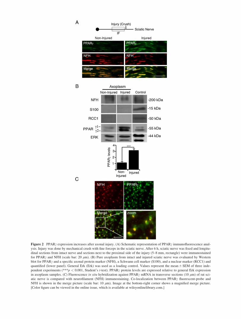
Figure 2 PPARc expression increases after axonal injury. (A) Schematic representation of PPARc immunofluorescence anal-
ysis. Injury was done by mechanical crush with fine forceps in the sciatic nerve. After 6 h, sciatic nerve was fixed and longitu-
dinal sections from intact nerve and sections next to the proximal side of the injury (5–8 mm, rectangle) were immunostained
for PPARc and NFH (scale bar: 20 lm). (B) Pure axoplasm from intact and injured sciatic nerve was evaluated by Western
blot for PPARc and a specific axonal protein marker (NFH), a Schwann cell marker (S100), and a nuclear marker (RCC1) and
quantified (lower panel). General Erk (Erk) was used as a loading control. Values represent the mean 6 SEM of three inde-
pendent experiments (***p < 0.001, Student’s t-test). PPARc protein levels are expressed relative to general Erk expression
in axoplasm samples. (C) Fluorescence in situ hybridization against PPARc mRNA in transverse sections (10 lm) of rat sci-
atic nerve is compared with neurofilament (NFH) immunostaining. Co-localization between PPARc fluorescent-probe and
NFH is shown in the merge picture (scale bar: 10 lm). Image at the bottom-right corner shows a magnified merge picture.
[Color figure can be viewed in the online issue, which is available at wileyonlinelibrary.com.]

Figure 3 PPARc interacts with dynein and its retrograde transport increases after nerve injury. (A and B) To study whether
axonal PPARc interacted with dynein, 500 lg axoplasm from naive and injured sciatic nerve (6 h after injury) was subjected
to dynein (A) or PPARc (B) immunoprecipitation and Western blotting against PPARc (A) and dynein (B) was performed.
Precipitation with an unrelated total IgG serves as a control. Levels of PPARc or dynein co-immunoprecipitacion were quanti-
fied. There is an increased amount of PPARc interacting with dynein or dynein interacting with PPARc after injury consistent
with the fact that PPARc protein levels are increased after injury (see Fig. 2). Numbers were standardized against the levels of
co-immunoprecipitated PPARc (A) or dynein (B) measured by Western blotting. Values represent the mean 1-SEM of three
independent experiments (**p < 0.01, Student’s t-test). NI, non-injury. I, injury. IP, immunoprecipitation. IgG, non-related
IgG. WB, Western blot. (C) PPARc retrograde transport is increased after injury. Sciatic nerve was ligated, and distal to liga-
tion, an injury (mechanical crush) was performed. After 2, 4 and 6 h, longitudinal sections of sciatic nerve were assessed by
immunofluorescence for PPARc in both side of the ligature. (D) Quantification of PPARc fluorescence in the proximal (site of
ligature closer to cell bodies, grey bars) and distal side (black bars) of the ligature at 2, 4, and 6 h postinjury. Values represent
the mean 6 SEM of three independent experiments (**p < 0.01, Student’s t-test).
8 Lezana et al.
Developmental Neurobiology

Similar to results from rodent neurons, GW9962 treat-
ment of axotomized human neurons reduced the num-
ber of neurite-extending cells and the lengths of
neuronal processes (Supporting Information Fig. S4).
Finally, we examined whether PPARc might play a
role in regeneration of rodent CNS neurons as well.
We used microfluidic chambers to study the presence
of PPARc in the axons of rat E18 cortical neurons.
Immunofluorescence staining of compartmentalized
cultures at 10 DIV revealed the presence of PPARc in
distal cortical axons. The axonal localization of
PPARc was confirmed by co-staining with b-III-
tubulin (Supporting Information Fig. S5). Next, we
evaluated whether axonal activation by the PPARcagonist rosiglitazone (Wright et al., 2014) affects the
regeneration of axotomized axons. Neurons were
Figure 4 Nuclear localization of PPARc in sensory neurons after sciatic nerve injury. (A) Rat L4-L5 DRGs were fixed 6 and
12 h after sciatic nerve crush and transverse sections were immunostained for NFH (red), PPARc (green) and DAPI (blue).
(Scale bar: 20 lm). The right column shows the nuclear localization of PPARc in the highlighted section of the respective
PPARc picture. (B) Quantification of positive nuclei for PPARc in DRGs tissue sections. Over 100 nuclei were quantified for
each replicate. The experiment was repeated three times with similar results. Values represent the mean 1-SEM of three inde-
pendent experiments (*p < 0.05, **p < 0.01, ANOVA followed by Tukey’s multiple comparisons test). (C) Expression of
DRG injury-regulated genes with PPAR response elements plotted from the microarray data of Michaelevski et al. (2010).
Expression data is shown as a heat map of fold changes (color code is shown above). [Color figure can be viewed in the online
issue, which is available at wileyonlinelibrary.com.]
PPARc Promotes Neuronal Regeneration After Injury 9
Developmental Neurobiology

axotomized by aspiration of the liquid in the axonal
chamber (Supporting Information Fig. S6) and then
allowed to regrow after supplementation of the axonal
compartment with PPARc agonists and/or antagonists
as indicated (Fig. 7). Cultures treated with rosiglita-
zone for 96 h displayed extensive axonal regeneration
compared with the control situation. This effect was
abolished by treatment of distal axons with the PPARcinhibitors GW9662 and/or T0070907, suggesting that
rosiglitazone enhances axonal regeneration in a
PPARc-dependent manner (Fig. 7).
DISCUSSION
Activation of the transcriptional response to nervous
system injury requires retrograde signaling from axo-
nal lesion sites to neuronal cell bodies. These tran-
scriptional changes enable an increase in the intrinsic
growth capacity of injured neurons, hence are critical
for functional regeneration. A variety of transcription
factors have been implicated in neuronal regenera-
tion, typically acting within the soma as part of the
cell body response. However recent studies have sug-
gested that transcription factors are also found within
axons, and may traffic with retrograde signaling com-
plexes to elicit and modulate events in the soma (Ji
and Jaffrey, 2014; Rishal and Fainzilber, 2014).
Analyses of the responses of DRG sensory neurons to
sciatic nerve lesion suggested that the PPAR family
of transcription factors might be candidate retrograde
injury signals (Michaelevski et al., 2010; Ben-
Yaakov et al., 2012). Here we have confirmed this
prediction by showing that PPARc is located within
axons in the sciatic nerve and in sensory and cortical
neurons in cultures. Sciatic nerve axons contain both
PPARc protein and mRNA. Upon injury, increases in
protein levels, dynein motor association and retrograde
transport of PPARc were observed, suggesting that it
is recruited to motor-driven complexes for transport
back to the cell body. This was further supported by
observations of increased nuclear localization of
PPARc in the nucleus of DRG neurons after sciatic
nerve crush and in growing sensory neurons in culture
together with increased expression of genes bearing
the PPAR response element. Furthermore, the PPARcinhibitors GW9662 and T0070907 (Lee et al., 2002;
Leesnitzer et al., 2002) both reduced the conditioning
lesion response of sensory neurons subjected to in vivoaxonal lesions in the sciatic nerve. We also observed
inhibition of neurite regeneration in axotomized
human neurons treated with GW9662, consistent with
Figure 5 Assessing PPARc antagonist effects on DRG
neurons in culture. (A) Adult L4-L6 DRG neurons were
cultured for 40 h in medium containing GW9662 (10 lM)
or T0070907 (5 lM) or DMSO as control (scale bar: 200
lm). (B) Average of total neurite length and percent grow-
ing neurons (neurons with maximal process length exceed-
ing 50 lm). Values represent the mean 1 SEM of three
independent experiments (*p < 0.05, ** p < 0.01,
***p< 0.001, ANOVA followed by Tukey’s multiple com-
parisons test). D, DMSO. G, GW9662. T, T0070907.
Figure 6 Inhibition of PPARc reduces neuronal response
to sciatic nerve injury. (A) Sciatic nerve of adult mice was
injected with 28 ng GW9662, 69 ng T0070907 or DMSO
as control in parallel with nerve crush. The contralateral
side was left uninjured as a control. Two days after the
crush L4-L6 DRG neurons were extracted and cultured for
18 h (scale bar: 200 lm). (B and C) Average of total neurite
length (B) and percent growing neurons (neurons with max-
imal process length exceeding 50 lm) (C). Values represent
the mean 6 SEM of at least three experiments (*p < 0.05,
**p < 0.01, ANOVA followed by Tukey’s multiple com-
parisons test). D, DMSO. G, GW9662. T, T0070907.
10 Lezana et al.
Developmental Neurobiology
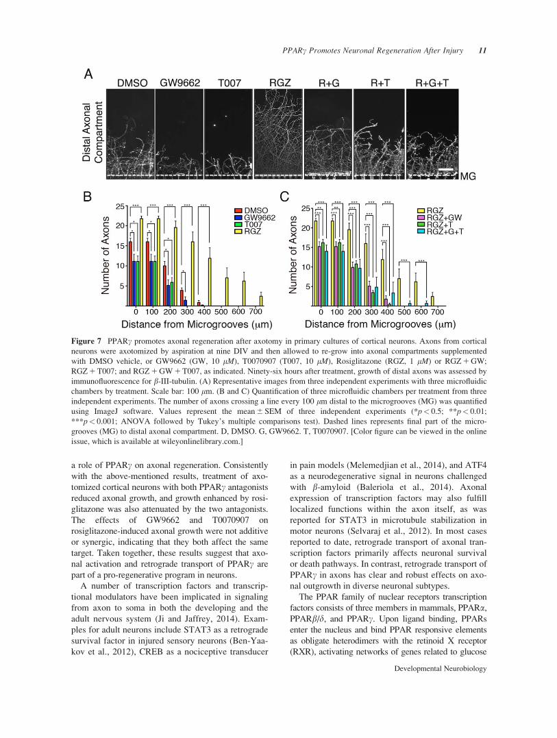
a role of PPARc on axonal regeneration. Consistently
with the above-mentioned results, treatment of axo-
tomized cortical neurons with both PPARc antagonists
reduced axonal growth, and growth enhanced by rosi-
glitazone was also attenuated by the two antagonists.
The effects of GW9662 and T0070907 on
rosiglitazone-induced axonal growth were not additive
or synergic, indicating that they both affect the same
target. Taken together, these results suggest that axo-
nal activation and retrograde transport of PPARc are
part of a pro-regenerative program in neurons.
A number of transcription factors and transcrip-
tional modulators have been implicated in signaling
from axon to soma in both the developing and the
adult nervous system (Ji and Jaffrey, 2014). Exam-
ples for adult neurons include STAT3 as a retrograde
survival factor in injured sensory neurons (Ben-Yaa-
kov et al., 2012), CREB as a nociceptive transducer
in pain models (Melemedjian et al., 2014), and ATF4
as a neurodegenerative signal in neurons challenged
with b-amyloid (Baleriola et al., 2014). Axonal
expression of transcription factors may also fulfill
localized functions within the axon itself, as was
reported for STAT3 in microtubule stabilization in
motor neurons (Selvaraj et al., 2012). In most cases
reported to date, retrograde transport of axonal tran-
scription factors primarily affects neuronal survival
or death pathways. In contrast, retrograde transport of
PPARc in axons has clear and robust effects on axo-
nal outgrowth in diverse neuronal subtypes.
The PPAR family of nuclear receptors transcription
factors consists of three members in mammals, PPARa,
PPARb/d, and PPARc. Upon ligand binding, PPARs
enter the nucleus and bind PPAR responsive elements
as obligate heterodimers with the retinoid X receptor
(RXR), activating networks of genes related to glucose
Figure 7 PPARc promotes axonal regeneration after axotomy in primary cultures of cortical neurons. Axons from cortical
neurons were axotomized by aspiration at nine DIV and then allowed to re-grow into axonal compartments supplemented
with DMSO vehicle, or GW9662 (GW, 10 lM), T0070907 (T007, 10 lM), Rosiglitazone (RGZ, 1 lM) or RGZ 1 GW;
RGZ 1 T007; and RGZ 1 GW 1 T007, as indicated. Ninety-six hours after treatment, growth of distal axons was assessed by
immunofluorescence for b-III-tubulin. (A) Representative images from three independent experiments with three microfluidic
chambers by treatment. Scale bar: 100 lm. (B and C) Quantification of three microfluidic chambers per treatment from three
independent experiments. The number of axons crossing a line every 100 lm distal to the microgrooves (MG) was quantified
using ImageJ software. Values represent the mean 6 SEM of three independent experiments (*p< 0.5; **p< 0.01;
***p< 0.001; ANOVA followed by Tukey’s multiple comparisons test). Dashed lines represents final part of the micro-
grooves (MG) to distal axonal compartment. D, DMSO. G, GW9662. T, T0070907. [Color figure can be viewed in the online
issue, which is available at wileyonlinelibrary.com.]
PPARc Promotes Neuronal Regeneration After Injury 11
Developmental Neurobiology
and lipid metabolism as well as inflammation in differ-
ent tissues (Ahmadian et al., 2013). The PPARs are
known as receptors for different fatty acids and lipids
metabolites, hence are mainly considered to be lipid
sensors regulating cellular metabolism. Natural or
endogenous ligands for PPARc include unsaturated
fatty acids, oxidized phospholipids, eicosanoids and
nitroalkens. The relatively large size of the ligand-
binding region in PPARc allows the binding of different
types of lipid molecules. For example, the PPARcligand-binding domain can accommodate at least two
oxidized fatty acids, and different ligands are able to
generate graded transcriptional responses by recruiting
different subsets of the requisite co-activators (Hughes
et al., 2012). These characteristics might enhance the
versatility of PPARc-dependent injury response mecha-
nisms. For example, injury-induced elevations in axonal
calcium could initially enhance the activity of calcium
dependent lipases such as PLA2. Subsequent fatty acid
release from membrane-associated phospholipids would
then generate a variety of potential PPARc ligands by
oxidation or modification of fatty acids due to calcium-
induced oxidative stress (Leslie, 1997; Villegas et al.,
2014). Thus, axonal injury can lead to significant pro-
duction of diverse PPARc agonists, suggesting that axo-
nal localization of PPARc might facilitate efficient
coordination of regeneration.
In additional to transcriptional effects in the
nucleus, PPARc might influence axonal growth via
other downstream targets in axons or soma. For
example, PPARc and its co-activator PGC-1a are
implicated in the promotion of mitochondrial biogen-
esis, and axonal or dendritic targeting of mitochon-
dria is required for growth. Increased PGC-1aexpression in neurons increases mitochondrial den-
sity and regulates mitochondrial transport and oxida-
tion state after injury (O’Donnell et al., 2013; van
Spronsen et al., 2013; Corona and Duchen, 2015).
Another possibility is that downstream targets of
PPARc modulate the c-jun-NH2 terminal kinase
(JNK) pathway to modulate axonal growth, as previ-
ously shown for long-term treatment of hippocampal
neurons with TZDs (Quintanilla et al., 2013). The
JNK pathway is known to affect a series of transcrip-
tion factors and cytoskeleton substrates in axonal
regeneration (Waetzig et al., 2006; Coffey, 2014).
The activation of PPARc in axons upon nerve lesion
and its effects on axonal outgrowth raise interest in the
translational potential of targeting the PPARc pathway
for enhancing nerve growth and regeneration. PPARc is
a master regulator of metabolism and an important drug
target in diabetes, as the specific target of thiazolidine-
diones (TZDs), a drug class of full agonists for PPARcwhich includes rosiglitazone (Wright et al., 2014).
Although TZDs are widely used in the clinic, a number
of undesirable side effects have motivated significant
efforts to develop new pharmaceutical agents targeting
PPARc (Ahmadian et al., 2013), including selective
PPARc modulators (SPPARMs) that act as PPARc par-
tial agonists (Grygiel-Gorniak, 2014). Thus, there is a
rich pharmacological toolkit for PPARc, including a
number of agents with acceptable safety profiles in
humans that could potentially be evaluated for efficacy
in central and peripheral nerve injuries. Moreover, a
series of recent studies have reported neuroprotective
effects of TZDs in stroke and in a number of neurode-
generative disease models (Chen et al., 2012). These
effects were attributed to anti-inflammatory activities
reported for PPARc in glial cells (Bernardo and
Minghetti, 2008; Zhao et al., 2009). Our results how-
ever, strongly support direct effects of PPARc on neuro-
nal regeneration, and raise the possibility that TZD-
induced neuroprotection might also be due to specific
effects on neurons. We expect that the findings reported
in this study will open new avenues for both basic
understanding and translational exploitation of the roles
of PPARc in the nervous system.
ACKNOWLEDGMENTS
The authors dedicate this study to Miguel Bronfman who
sadly passed away in January 2014. The authors are grateful
for his many contributions to Chilean science and his scien-
tific legacy. The authors thank Dr. Efrat Shoham for excellent
technical support, Dr Ariadna Pacheco for her initial contri-
bution to this study, Dr Felipe Court for technical help with
the isolation of axonal fibers for immunostaining and the lab
of Dr Katia Gysling for assistance with in situ hybridization.
REFERENCES
Ahmadian M, Suh JM, Hah N, Liddle C, Atkins AR,
Downes M, Evans RM. 2013. PPARgamma signaling and
metabolism: the good, the bad and the future. Nat Med
19:557–566.
Baleriola J, Walker CA, Jean YY, Crary JF, Troy CM,
Nagy PL, Hengst U. 2014. Axonally synthesized ATF4
transmits a neurodegenerative signal across brain regions.
Cell 158:1159–1172.
Ben-Yaakov K, Dagan SY, Segal-Ruder Y, Shalem O,
Vuppalanchi D, Willis DE, Yudin D, et al. 2012. Axonal
transcription factors signal retrogradely in lesioned
peripheral nerve. EMBO J. 31:1350–1363.
Bernardo A, Minghetti. L. 2008. Regulation of glial cell
functions by PPAR-gamma natural and synthetic ago-
nists. PPAR Res 2008:864140.
Carpenter AE, Jones TR, Lamprecht MR, Clarke C, Kang
IH, Friman O, Guertin DA, et al. 2006. CellProfiler:
12 Lezana et al.
Developmental Neurobiology
Image analysis software for identifying and quantifying
cell phenotypes. Genome Biol 7:R100.
Chen YC, Wu JS, Tsai HD, Huang CY, Chen JJ, Sun GY,
Lin TN. 2012. Peroxisome proliferator-activated receptor
gamma (PPAR-gamma) and neurodegenerative disorders.
Mol Neurobiol 46:114–124.
Chiang MC, Cheng YC, Chen HM, Liang YJ, Yen CH.
2014. Rosiglitazone promotes neurite outgrowth and
mitochondrial function in N2A cells via PPARgamma
pathway. Mitochondrion 14:7–17.
Coffey ET. 2014. Nuclear and cytosolic JNK signalling in
neurons. Nat Rev 15:285–299.
Corona JC, Duchen MR. 2015. PPARgamma and PGC-
1alpha as Therapeutic Targets in Parkinson’s. Neurochem
Res 40:308–316.
Delcroix JD, Averill S, Fernandes K, Tomlinson DR,
Priestley JV, Fernyhough. P. 1999. Axonal transport of
activating transcription factor-2 is modulated by nerve
growth factor in nociceptive neurons. J Neurosci. 19:RC24.
Dhara SK, Hasneen K, Machacek DW, Boyd NL, Rao RR,
Stice. SL. 2008. Human neural progenitor cells derived
from embryonic stem cells in feeder-free cultures. Differ-
entiation 76:454–464.
Dhara SK, Stice. SL. 2008. Neural differentiation of human
embryonic stem cells. J Cell Biochem 105:633–640.
Eva R, Crisp S, Marland JR, Norman JC, Kanamarlapudi
V, Ffrench-Constant C, Fawcett. JW. 2012. ARF6
directs axon transport and traffic of integrins and regu-
lates axon growth in adult DRG neurons. J Neurosci 32:
10352–10364.
Fuenzalida K, Quintanilla R, Ramos P, Piderit D, Fuentealba
RA, Martinez G, Inestrosa NC, et al. 2007. Peroxisome
proliferator-activated receptor gamma up-regulates the
Bcl-2 anti-apoptotic protein in neurons and induces mito-
chondrial stabilization and protection against oxidative
stress and apoptosis. J Biol Chem 282:37006–37015.
Fuenzalida KM, Aguilera MC, Piderit DG, Ramos PC,
Contador D, Quinones V, Rigotti A, et al. 2005. Peroxi-
some proliferator-activated receptor gamma is a novel
target of the nerve growth factor signaling pathway in
PC12 cells. J Biol Chem 280:9604–9609.
Garcia-Bueno B, Madrigal JL, Lizasoain I, Moro MA,
Lorenzo P, Leza JC. 2005. Peroxisome proliferator-acti-
vated receptor gamma activation decreases neuroinflamma-
tion in brain after stress in rats. Biol Psychiatry 57:885–894.
Grygiel-Gorniak B. 2014. Peroxisome proliferator-activated
receptors and their ligands: nutritional and clinical implica-
tions–A review. Nutr J 13:17.
Hughes TS, Chalmers MJ, Novick S, Kuruvilla DS, Chang
MR, Kamenecka TM, Rance M, et al. 2012. Ligand and
receptor dynamics contribute to the mechanism of graded
PPARgamma agonism. Structure 20:139–150.
Ji SJ, Jaffrey SR. 2014. Axonal transcription factors: Novel
regulators of growth cone-to-nucleus signaling. Dev Neu-
robiol 74:245–258.
Kashihara Y, Sakaguchi M, Kuno. M. 1989. Axonal trans-
port and distribution of endogenous calcitonin gene-related
peptide in rat peripheral nerve. J Neurosci 9:3796–3802.
Lee G, Elwood F, McNally J, Weiszmann J, Lindstrom M,
Amaral K, Nakamura M, et al. 2002. T0070907, a selec-
tive ligand for peroxisome proliferator-activated receptor
gamma, functions as an antagonist of biochemical and
cellular activities. J Biol Chem 277:19649–19657.
Leesnitzer LM, Parks DJ, Bledsoe RK, Cobb JE, Collins
JL, Consler TG, Davis RG, et al. 2002. Functional conse-
quences of cysteine modification in the ligand binding
sites of peroxisome proliferator activated receptors by
GW9662. Biochemistry 41:6640–6650.
Leslie CC. 1997. Properties and regulation of cytosolic
phospholipase A2. J Biol Chem 272:16709–16712.
Melemedjian OK, Tillu DV, Moy JK, Asiedu MN, Mandell
EK, Ghosh S, Dussor G, et al. 2014. Local translation and
retrograde axonal transport of CREB regulates IL-6-
induced nociceptive plasticity. Mol Pain 10:45.
Michaelevski I, Segal-Ruder Y, Rozenbaum M,
Medzihradszky KF, Shalem O, Coppola G, Horn-Saban
S, et al. 2010. Signaling to transcription networks in the
neuronal retrograde injury response. Sci Signal 3:ra53.
O’Donnell KC, Vargas ME, Sagasti A. 2013. WldS and
PGC-1alpha regulate mitochondrial transport and oxidation
state after axonal injury. J Neurosci 33:14778–14790.
Park JW, Vahidi B, Taylor AM, Rhee SW, Jeon NL. 2006.
Microfluidic culture platform for neuroscience research.
Nat Protoc 1:2128–2136.
Perry RB, Doron-Mandel E, Iavnilovitch E, Rishal I,
Dagan SY, Tsoory M, Coppola G, et al. 2012. Subcellular
knockout of importin beta1 perturbs axonal retrograde
signaling. Neuron 75:294–305.
Quintanilla RA, Godoy JA, Alfaro I, Cabezas D, von
Bernhardi R, Bronfman M, Inestrosa. NC. 2013. Thiazoli-
dinediones promote axonal growth through the activation
of the JNK pathway. PLoS One 8:e65140.
Rishal I, Fainzilber. M. 2014. Axon-soma communication
in neuronal injury. Nat Rev Neurosci 15:32–42.
Rishal I, Michaelevski I, Rozenbaum M, Shinder V,
Medzihradszky KF, Burlingame AL, Fainzilber M. 2010.
Axoplasm isolation from peripheral nerve. Dev Neurobiol
70:126–133.
Selvaraj BT, Frank N, Bender FL, Asan E, Sendtner M.
2012. Local axonal function of STAT3 rescues axon
degeneration in the pmn model of motoneuron disease.
J Cell Biol 199:437–451.
Shin S, Mitalipova M, Noggle S, Tibbitts D, Venable A,
Rao R, Stice SL. 2006. Long-term proliferation of human
embryonic stem cell-derived neuroepithelial cells using
defined adherent culture conditions. Stem Cells 24:125–
138.
Smith DS, Skene JH. 1997. A transcription-dependent
switch controls competence of adult neurons for distinct
modes of axon growth. J Neurosci 17:646–658.
Taylor AM, Berchtold NC, Perreau VM, Tu CH, Li Jeon N,
Cotman CW. 2009. Axonal mRNA in uninjured and
regenerating cortical mammalian axons. J Neurosci 29:
4697–4707.
Taylor AM, Rhee SW, Tu CH, Cribbs DH, Cotman
CW, Jeon NL. 2003. Microfluidic multicompartment
PPARc Promotes Neuronal Regeneration After Injury 13
Developmental Neurobiology
device for neuroscience research. Langmuir 19:
1551–1556.
van Spronsen M, Mikhaylova M, Lipka J, Schlager MA,
van den Heuvel DJ, Kuijpers M, Wulf PS, et al. 2013.
TRAK/Milton motor-adaptor proteins steer mitochondrial
trafficking to axons and dendrites. Neuron 77:485–502.
Villegas R, Martinez NW, Lillo J, Pihan P, Hernandez D,
Twiss JL, Court FA. 2014. Calcium release from intra-
axonal endoplasmic reticulum leads to axon degeneration
through mitochondrial dysfunction. J Neurosci 34:7179–
7189.
Waetzig V, Zhao Y, Herdegen. T. 2006. The bright side of
JNKs-multitalented mediators in neuronal sprouting,
brain development and nerve fiber regeneration. Prog
Neurobiol 80:84–97.
Wright MB, Bortolini Tadayyon MM, Bopst M. 2014.
Minireview: Challenges and opportunities in develop-
ment of PPAR agonists. Mol Endocrinol 28:1756–
1768.
Yano H, Chao. MV. 2004. Mechanisms of neurotro-
phin receptor vesicular transport. J Neurobiol 58:
244–257.
Zhao X, Strong R, Zhang J, Sun G, Tsien JZ, Cui Z, Grotta
JC, et al. 2009. Neuronal PPARgamma deficiency
increases susceptibility to brain damage after cerebral
ischemia. J Neurosci 29:6186–6195.
14 Lezana et al.
Developmental Neurobiology