Embed Size (px)
Citation preview
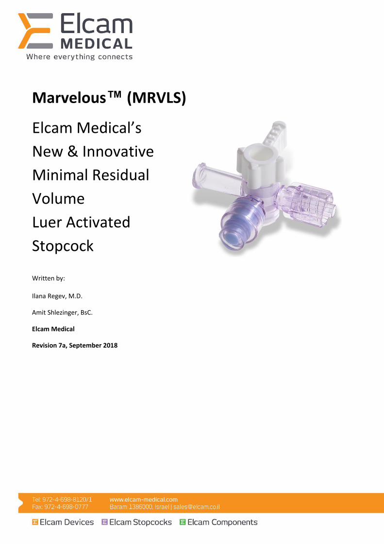
Marvelous™ (MRVLS)
Elcam Medical’s
New & Innovative
Minimal Residual
Volume
Luer Activated
Stopcock Written by:
Ilana Regev, M.D.
Amit Shlezinger, BsC.
Elcam Medical
Revision 7a, September 2018
Abstract
Catheter Related Blood Stream Infections
(CRBSI) are a well-known and increasingly
disturbing problem in hospitals, especially
in Intensive Care Units (ICUs) and other units
utilizing catheters and their accessories for
patient care.
Reducing the risk of microbial colonization and
proliferation on stopcocks, a valve assembled
into a catheter set enabling access to it,
can contribute to reducing the rates of
catheter colonization. Additionally, needle
stick injuries and resulting contamination are
a well-known risk for medical staff. Reducing
the risk of needle sticks can increase the safety
of the medical staff and patients.
Elcam Medical, the leading provider
of stopcocks to the U.S. and European OEM
markets, has currently designed the
Marvelous™ with the purpose
of increasing the safety of patients and
medical staff.
Elcam Medical’s Marvelous™is a standard
Stopcock (STP) with two additional features
that were designed in order to increase
patient safety relative to infection prevention:
1) the Luer- activated Valve that serves as
a bacterial barrier, enables access to the line
without having to open it, practically creating
a closed system and allows needle free
connection; and 2) the minimal residual
volume feature that automatically flushes the
side port along with the flow line, minimizes
the STP's dead space and contributes to
preventing bacterial colonization.
In order to prove the safety of the Marvelous™ for human usage in general, and the functionality of the minimal residual volume feature in particular, Elcam Medical conducted tests performed by well-established laboratories. The Flushing study was designed for the purpose of demonstrating that the Marvelous™ minimal dead space and minimal residual volume feature is as safe as other devices in the market with regard to blood residuals after flushing. This was achieved by quantifying blood residuals in the Marvelous™ versus other corresponding devices at the sampling port after line and port flushing. The Bacterial contamination of residual blood solutions study was designed as a continuation of the Flushing study, for the purpose of characterizing the bacterial growth in saline containing low concentrations of blood at different time periods. The Marvelous™ was tested for mechanical hemolysis by simulating hospital procedure, and its Biocompatibility was evaluated in accordance with the FDA’s Memorandum and with International Standard ISO 10993,-1. All the test results show that the Marvelous™ is safe for its intended use and establishes the functionality of the minimal residual volume feature.
Pathogenesis The management of critically ill patients, often hospitalized in intensive care units (ICU), essentially involves the use of Central Venous Catheters (CVCs) for the administration of intravenous (IV) fluids, medications, blood products and parenteral nutrition. They also enable monitoring of the hemodynamic status of critical patients and blood sampling. CVC sets include one or more stopcocks (STPs). These are valves serving as ports of entry into the circulatory system of the patient and are used for different interventions. CVCs, although indispensable in the critical care setup, pose risks of serious complications, of which the most common are infections (1-4). Infections can be localized along the insertion-site of the catheter tunnel, spread systemically as they disperse into the bloodstream or metastasize to distant body sites and seed infection in specific organs (3).
According to The American Center for Disease Control and Prevention (CDC), more than 80,000 catheter-related bloodstream infections are reported each year from American ICUs alone (a total of 250,000 cases per year) (5). CRBSI are associated with mortality rates of 10% to 20%, increased morbidity, prolonged hospitalization and increased medical care costs –
$3700-$29,000 attributed cost per infection (5, 6).
The pathogenesis of catheter-related infections is complex and multifactorial. Two important mechanisms have been demonstrated to be involved: 1. Insertion site-related: skin microorganisms at the insertion site of the catheter migrate along the catheter tract and colonize the catheter and inner tissues, rendering development of local infection (7-8). 2. Catheter hub-related: the hub of the catheter is the part to which tubes or syringes are connected, usually through a stopcock,
for administration of fluids or medications and for monitoring. Contamination of the hub by frequent
manipulations may result in intraluminal
colonization of the catheters (9-12) , with
possible infectious complications.
The relative importance of the
two mechanisms of catheter contamination
depends on several factors and is subject
to continuing debate (6). Three different
groups (Salzman, Linares and Cicco) that
studied microbial colonization of catheters
found the hub to be the most common source
of catheter infection and bacteremia
(54%,70% and 45% respectively) (9,12,13).
The Stopcock (STP) is a multi-directional rotating valve, assembled into a catheter set (sometimes including several units in the same set) and used during diverse interventions for the administration of different medical fluids, parenteral feeding and blood sampling. A typical stopcock is designed with a proximal (male) port and a distal port, serving as an inlet and outlet of the line, and a side(female) port, used to be accessed with a syringe for injecting or sampling as described above. Due to the design of the stopcock, there is a ‘dead space’ at the junction between syringe attachment point at the side port and the flow of fluids between the distal and proximal ports. This space cannot be efficiently cleared of all the fluid remnants passing through it despite standard flushing procedures, resulting in an accumulation of residue debris. During the period in which a catheter is constantly present in a blood vessel, this accumulation inside the STP encourages bacterial colonizing in the ‘dead space’ area triggering CRBSI (14). Recurrent manipulations expose the stopcock to increased contamination, making it a major source of nosocomial infections (15).
A CDC report from 2002 declared that
stopcock contamination is common, occurring
in 45%-50% of CRBSI cases (5).
Reducing the chance of microbial colonization
on stopcocks can contribute to reducing the
rates of catheter colonization.
The Minimal Residual Volume Luer -
Activated Swabbable Stopcock
(Marvelous™)
Elcam Medical is a world-class producer
of medical equipment in the area
of disposable flow control devices.
In light of the pronounced risks associated
with hospital-acquired infections in patients
who need therapy using stopcocks,
the company has currently designed
an innovative Stopcock, the Marvelous™ with
the purpose of increasing patients and medical
staff safety, especially those patients treated
in the intensive care, oncology and dialysis
units, who comprise more than 50% of the
total STP users. The Marvelous™ is a standard
STP (as seen in Figure A) with two additional
features.
The first component is a Luer Activated Valve
which is attached to the product by ultrasonic
welding, replacing one or both female ports
(luers). This feature is depicted in Figure B
(the text describing the new component
is highlighted in yellow in the text box).
The Luer-activated valve component
is purchased by Elcam from Halkey-Roberts
and its design is based on Halkey-Roberts’s
needleless injection site valve that was cleared
under 510(k) K002689 on March 06, 2001. The
Luer-activated valve is illustrated in Figure C.
The valve has an elastomeric "stem" that
maintains the female port (luer) closed,
canceling the need to close it with a cap,
in order to avoid leakage and/or port's
contamination.
The elastomeric stem has a slit that enables
the “stem” to be in an either open or closed
position. Once a male luer is introduced into
the closed port through the Luer-activated
valve component, the fluid path automatically
opens to allow injection or blood sampling.
Once the male luer is taken out, the female
port is automatically closed (due to the valve's
elastomeric stem that seals itself).
The valve actually serves as a needleless
injection site integrated in the stopcock.
Besides eliminating the usage of needles,
thereby minimizing needle stick occurrences,
the end user benefits by not having to use
caps or manipulate the stopcock’s handle,
in order to close the port before and after
injection or blood sampling. Swabbing the
valve’s top (injection site) with aseptic fluid
such as alcohol prior to male luer insertion
supports the safety requirements.
The Luer Activated Valve enables a needle
free manipulation of the stopcock and
therefore improves the staff's safety. The
valve creates a bacterial barrier closed
system that contributes to fighting infections
by preventing contamination.
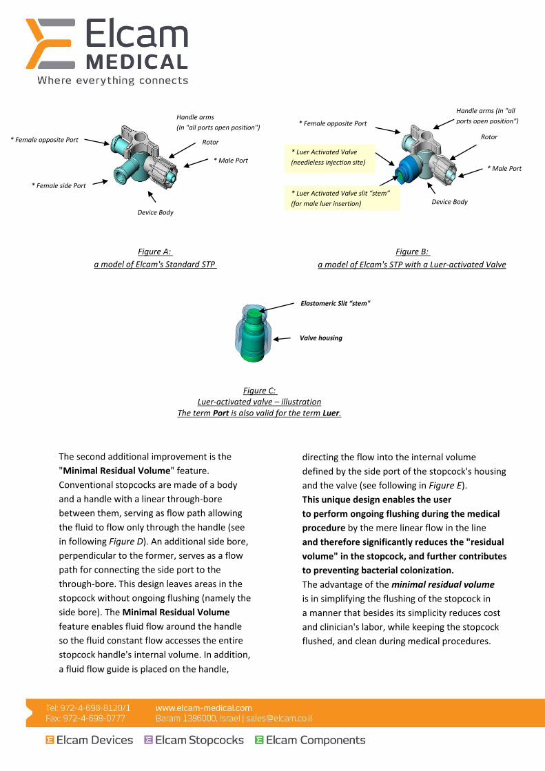
Figure B:
a model of Elcam's STP with a Luer-activated Valve
* Female opposite Port
* Luer Activated Valve
(needleless injection site)
* Luer Activated Valve slit “stem”
(for male luer insertion)
Handle arms (In "all
ports open position")
Rotor
* Male Port
Device Body
Figure A:
STP a model of Elcam's Standard
* Female side Port
* Female opposite Port
Handle arms
(In "all ports open position")
Rotor
* Male Port
Device Body
Figure C: Luer-activated valve – illustration
The term Port is also valid for the term Luer.
Elastomeric Slit “stem"
Valve housing
The second additional improvement is the
"Minimal Residual Volume" feature.
Conventional stopcocks are made of a body
and a handle with a linear through-bore
between them, serving as flow path allowing
the fluid to flow only through the handle (see
in following Figure D). An additional side bore,
perpendicular to the former, serves as a flow
path for connecting the side port to the
through-bore. This design leaves areas in the
stopcock without ongoing flushing (namely the
side bore). The Minimal Residual Volume
feature enables fluid flow around the handle
so the fluid constant flow accesses the entire
stopcock handle's internal volume. In addition,
a fluid flow guide is placed on the handle,
directing the flow into the internal volume
defined by the side port of the stopcock's housing
and the valve (see following in Figure E).
This unique design enables the user
to perform ongoing flushing during the medical
procedure by the mere linear flow in the line
and therefore significantly reduces the "residual
volume" in the stopcock, and further contributes
to preventing bacterial colonization.
The advantage of the minimal residual volume
is in simplifying the flushing of the stopcock in
a manner that besides its simplicity reduces cost
and clinician's labor, while keeping the stopcock
flushed, and clean during medical procedures.
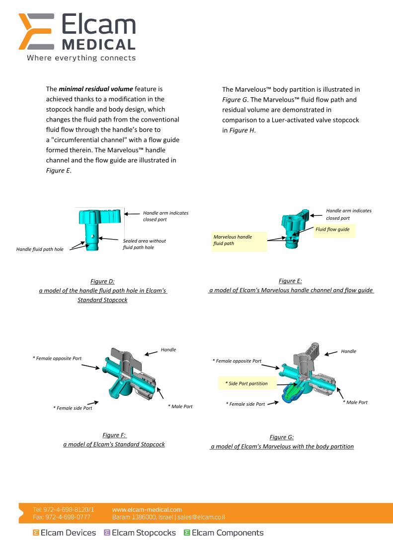
The minimal residual volume feature is
achieved thanks to a modification in the
stopcock handle and body design, which
changes the fluid path from the conventional
fluid flow through the handle’s bore to
a "circumferential channel" with a flow guide
formed therein. The Marvelous™ handle
channel and the flow guide are illustrated in
Figure E.
The Marvelous™ body partition is illustrated in
Figure G. The Marvelous™ fluid flow path and
residual volume are demonstrated in
comparison to a Luer-activated valve stopcock
in Figure H.
Figure D:
a model of the handle fluid path hole in Elcam's
Standard Stopcock
Handle arm indicates closed port
Handle fluid path hole
Sealed area without fluid path hole
Figure E:
a model of Elcam's Marvelous handle channel and flow guide
Marvelous handle fluid path
Handle arm indicates
closed port
Fluid flow guide
Figure F:
a model of Elcam's Standard Stopcock
Handle
* Female side Port
* Female opposite Port
* Male Port
Figure G:
a model of Elcam's Marvelous with the body partition
* Side Port partition
Handle
* Female side Port
* Male Port
* Female opposite Port
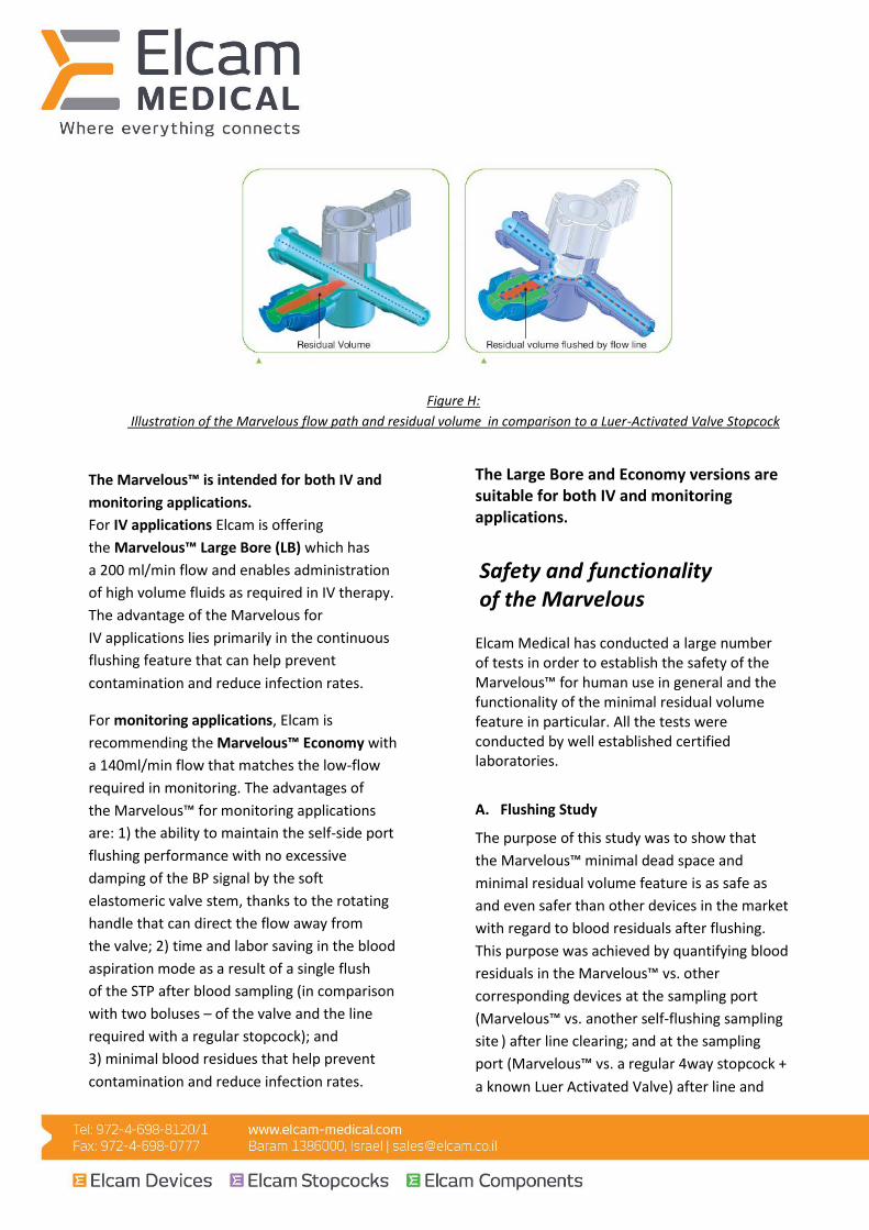
Figure H:
Illustration of the Marvelous flow path and residual volume in comparison to a Luer-Activated Valve Stopcock
The Marvelous™ is intended for both IV and
monitoring applications.
For IV applications Elcam is offering
the Marvelous™ Large Bore (LB) which has
a 200 ml/min flow and enables administration
of high volume fluids as required in IV therapy.
The advantage of the Marvelous for
IV applications lies primarily in the continuous
flushing feature that can help prevent
contamination and reduce infection rates.
For monitoring applications, Elcam is
recommending the Marvelous™ Economy with
a 140ml/min flow that matches the low-flow
required in monitoring. The advantages of
the Marvelous™ for monitoring applications
are: 1) the ability to maintain the self-side port
flushing performance with no excessive
damping of the BP signal by the soft
elastomeric valve stem, thanks to the rotating
handle that can direct the flow away from
the valve; 2) time and labor saving in the blood
aspiration mode as a result of a single flush
of the STP after blood sampling (in comparison
with two boluses – of the valve and the line
required with a regular stopcock); and
3) minimal blood residues that help prevent
contamination and reduce infection rates.
The Large Bore and Economy versions are suitable for both IV and monitoring applications.
Safety and functionality of the Marvelous
Elcam Medical has conducted a large number of tests in order to establish the safety of the Marvelous™ for human use in general and the functionality of the minimal residual volume feature in particular. All the tests were conducted by well established certified laboratories.
A. Flushing Study
The purpose of this study was to show that
the Marvelous™ minimal dead space and
minimal residual volume feature is as safe as
and even safer than other devices in the market
with regard to blood residuals after flushing.
This purpose was achieved by quantifying blood
residuals in the Marvelous™ vs. other
corresponding devices at the sampling port
(Marvelous™ vs. another self-flushing sampling
site ) after line clearing; and at the sampling
port (Marvelous™ vs. a regular 4way stopcock +
a known Luer Activated Valve) after line and

port clearing. The test procedure was
designed to mimic real-life hospital
procedures(16).
One study compared the Marvelous™ to two
common sampling ports in monitoring sets
with an in-line reservoir:
An arterial line including a sampling port and a reservoir, was filled with saline and its tip was dipped in sheep blood. Saline mixed with blood was drawn up the line by manipulating the reservoir. The amount of blood drawn was set per medical practice to be 4 times the volume between the tip and the sampling port, in order to assure pure blood at the sampling port. Some blood was withdrawn through the port. The mixed blood was returned down the line by an in-line reservoir. The Line and port were flushed by an in-line flush device for X seconds [X= 4, 6, 8]. The Blood residuals samples were collected through the tested sampling port. Blood residuals were quantified by DAS* photo spectroscopy at 234nm wavelength. Each tested group included 10 products.
No significant differences in Blood residues
(LOQ*<0.00001 mg/L) (Alpha=0.05) were
found between the three types of products
after in-line flushing for 4, 6 & 8 seconds
(Wash Time). Therefore we concluded that
the Marvelous™ is as good as or better than
the other marketed sampling ports of
reservoir including monitoring sets
(Graph1).
A second study compared the Marvelous™ to
some common luer activated valves, attached
to stopcocks in monitoring sets without an
in-line reservoir:
An arterial line including a sampling port in the form of a luer activated valve attached to a stopcock, was filled with saline and its tip was dipped in blood. Saline mixed with blood was drawn up the line by using a syringe at the top of the line. The amount of blood drawn was set per medical practice to be 4 times the volume between the tip and the sampling port, to assure pure blood at the sampling port. Blood was drawn through the tested port. The line and port were flushed separately by an in-line flush device for X seconds [X=1, 2, 3, 4] each. The blood residuals samples were collected through the tested sampling port. Blood residuals were quantified by DAS* photo spectroscopy at 234nm wavelength. Each tested group included 3-10 products. All of the product groups showed significant differences in blood residue concentrations (LOQ**<0.00001 mg/L) (Alpha=0.05) between 1 and 2 seconds wash times. There were no significant differences in blood residue concentrations between 2, 3 and 4 sec. wash times. In all wash time flushes (1,2,3,4 sec) there were significant differences in blood residues concentration between the different analyzed products (LOQ**<0.00001 mg/L) (Alpha=0.05)
*DAS –Diode Array Spectrophotometer
** LOQ – Limit Of Quantitation –a resolution index - the lowest value detected by the photo spectrometer.
The Marvelous™ was always at the lower end of the curve (lower blood residue concentration), Therefore we concluded that the Marvelous™ is as good as or even better than the other marketed sampling ports in monitoring sets without a reservoir (Graph 2).
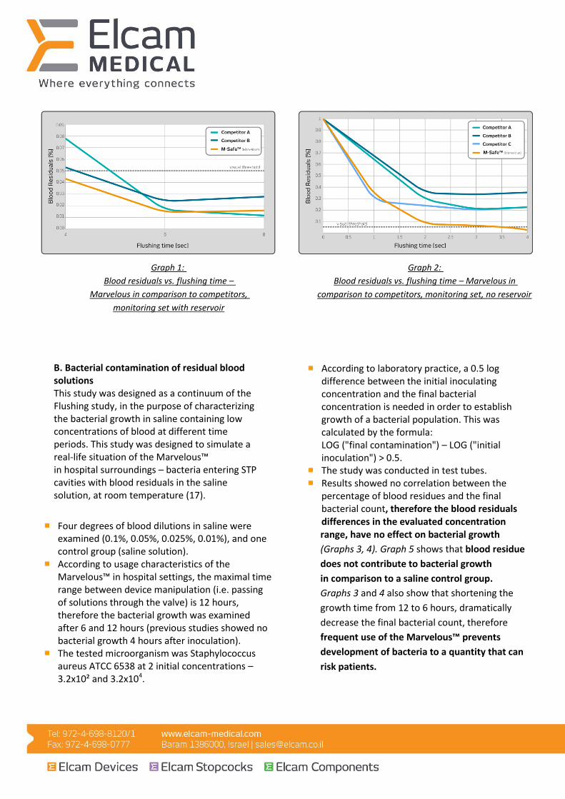
Graph 1:
–flushing time Blood residuals vs.
in comparison to competitors, arvelousM
monitoring set with reservoir
Graph 2:
Blood residuals vs. flushing time – Marvelous in
comparison to competitors, monitoring set, no reservoir
B. Bacterial contamination of residual blood solutions This study was designed as a continuum of the Flushing study, in the purpose of characterizing the bacterial growth in saline containing low concentrations of blood at different time periods. This study was designed to simulate a real-life situation of the Marvelous™ in hospital surroundings – bacteria entering STP cavities with blood residuals in the saline solution, at room temperature (17).
Four degrees of blood dilutions in saline were examined (0.1%, 0.05%, 0.025%, 0.01%), and one control group (saline solution). According to usage characteristics of the Marvelous™ in hospital settings, the maximal time range between device manipulation (i.e. passing of solutions through the valve) is 12 hours, therefore the bacterial growth was examined after 6 and 12 hours (previous studies showed no bacterial growth 4 hours after inoculation). The tested microorganism was Staphylococcus aureus ATCC 6538 at 2 initial concentrations – 3.2x10² and 3.2x10⁴.
According to laboratory practice, a 0.5 log difference between the initial inoculating concentration and the final bacterial concentration is needed in order to establish growth of a bacterial population. This was calculated by the formula: LOG ("final contamination") – LOG ("initial inoculation") > 0.5. The study was conducted in test tubes. Results showed no correlation between the percentage of blood residues and the final bacterial count, therefore the blood residuals differences in the evaluated concentration range, have no effect on bacterial growth
(Graphs 3, 4). Graph 5 shows that blood residue
does not contribute to bacterial growth
in comparison to a saline control group.
Graphs 3 and 4 also show that shortening the
growth time from 12 to 6 hours, dramatically
decrease the final bacterial count, therefore
frequent use of the Marvelous™ prevents
development of bacteria to a quantity that can
risk patients.
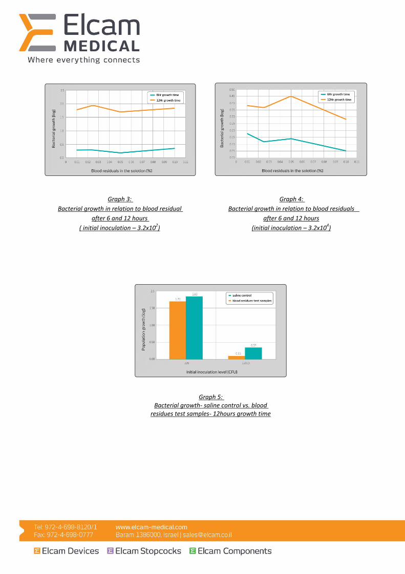
Graph 3:
Bacterial growth in relation to blood residual
after 6 and 12 hours
( initial inoculation – 3.2x10²)
Graph 4:
Bacterial growth in relation to blood residuals
after 6 and 12 hours
(initial inoculation – 3.2x10⁴)
Graph 5: Bacterial growth- saline control vs. blood
residues test samples- 12hours growth time
D. Biocompatibility Biocompatibility evaluation was performed in accordance with the FDA’s Memorandum – #G95, 1- May 1, 1995 and with International Standard ISO 10993,-1 under the classification of “Externally Communicating Device, Blood path-indirect, Prolonged (up to 96 hours) Contact Duration”. Based on that classification, the following types of tests were performed and successfully passed: Cytotoxicity, Systemic Toxicity, Intracutaneous Reactivity, Sensitization and Chemical Hemolysis. All tests were performed by Namsa US Laboratory.
C. Mechanical Hemolysis
The Marvelous™ was tested
for mechanical hemolysis by simulating hospital
procedure. Blood was drawn from a reservoir
of cow blood using a Marvelous™ at three
different flow rates - 1ml/0.5sec, 1ml/2.5sec and
1ml/5sec; Blood was also drawn at a constant
flow of 1ml/5sec while the Marvelous™ handle
was deviated by 10° and 25° from its intended
position.
In the control group blood was drawn using
either a 23G needle and a 3ml syringe or a 3ml
syringe alone. A regular STP was used as the gold
standard for hemolysis. The study groups
included 10 products each. The current
international standard for chemical hemolysis is
5%, but a recent study claimed that chemical
hemolysis during contact with polyethylene is
0.7%, and the acceptance criteria was set at 0.7%
in accordance with the polyethylene STP handles.
The hemolysis level was determined using a DAS
– Diode Array Spectrophotometer.
The results were all under 0.42% hemolysis, well
below the acceptance criteria. In comparison
to the gold standard no statistically significant
difference was observed. We conclude that
Elcam Medical's Marvelous™ does not cause
mechanical hemolysis.
Shelf life: The Marvelous™ has been tested from
both mechanical and efficacy aspects and was
approved for a shelf life of 5 years.
Elcam Medical has been manufacturing various
stopcocks for the medical market during the past
25 years in full compliance with FDA CFR 21 820
Quality System Regulations and ISO 13485:2016.
In accordance with all the studies and tests conducted by Elcam Medical and detailed
in this document, we conclude that the minimal residual volume feature is functional
as stated and that the Marvelous™ is safe for human use.
Regulatory status:
The Marvelous™ is 510(k) cleared and CE
approved.
Elcam holds marketing approvals from the Israeli Ministry of Health, as well as from the US FDA and The European MDD (Medical Devices Directive) - Annex II Section 3 of the Council Directive 93/42/EEC.
References
1. Collignon PJ. Intravascular catheter associated sepsis: a common problem.
The Australian Study on Intravascular Catheter Associated Sepsis. Med J Aust 1994;161:374-8.
2. Heiselman D. Nosocomial bloodstream infections in critically ill patients. JAMA 1994;272:1819-20.
3. Norwood S, Ruby A, Civetta J, Cortes V. Catheter-related infections and associated septicemia.
Chest 1991;99:968-75.
4. Pittet P, Wendzel RP. Nosocomial bloodstream infections. Secular trends in mortality rates and contribution
to total hospital deaths. Arch Intern Med 1995;155:1177-84.
5. O’Grady NP. Et al. Guidelines for the Prevention of Intravascular Catheter-Related Infections. CDC, 2002 Report.
6. Pearson ML. Intravascular device-related infections: an overview. Am J Infect Control 1996;24:262-93.
7.Ljungman P, Hagglund H, Bhjorkstrand B, Lonnqvist B, Ringden O. Perioperative teicoplanin for prevention
of Gram-positive infections in neutropenic patients with indwelling central venous catheters: a randomized, controlled study. Support Care Cancer
1997;5:485-8.
8. Snydman DR, Pober BR, Murray SA, Gorbea HF, Majka JA, Perry LK.
Predictive value of surveillance skin cultures in total parenteral nutrition-related infection.
Prospective epidemiologic study using semi-quantitative cultures. Lancet 1982;1385-8.
9. Salzman MB, Isenberg HD, Shapiro JF, Lipsitz PJ, Rubin LG. A prospective study of the catheter hub as the portal of entry
for microorganisms causing catheter-related sepsis in neonates. J Infect Dis 1993;167:487-90.
10.Peters G, Locci R, Pulverer G. Adherence and growth of coagulase-negative staphylococci in surfaces of intravenous catheters. J Infect Dis 1982;146:479-
82.
11. Sitges-Serra A, Linares J, Perez JL, Jaurrieta E, Lorente L. A randomized trial on the effect of tubing changes on hub contamination and catheter sepsis
during parenteral nutrition. J Parenter Enteral Nutr 1985;9:322-5. 12. Linares J, Sitges-Serra A, Garau J, Perez JL, Martin R. Pathogenesis of catheter sepsis: a prospective study with
quantitative and semiquantitative cultures of catheter hub and segments. J Clin Microbiol 1985;9:322-5.
13.deCicco M, Chiaradia V, Veronesi A, et al. Source and route of microbial colonization of parenteral nutrition catheters. Lancet 1989;2:1258-61.
14. Medical Anti-Bacterial Stopcock, Business plan, Elcam Medical, June 2004.
15. McArthur et al., Stopcock Contamination in an ICU, The American Journal of Nursing, Jan. 1975.
16. Blood residues analysis/ Igal Bar-Ilan Ph.D / Migal Ltd for Elcam Medical.
17. Bacterial growth characterization in saline solutions containing low concentration of blood in different time periods/ Aminolab Ltd. For Elcam Medical.