Embed Size (px)
Citation preview

Neurology · Neurosurgery · Medical Oncology · Radiotherapy · Paediatric Neuro-
oncology · Neuropathology · Neuroradiology · Neuroimaging · Nursing · Patient Issues
EDITORIALRiccardo Soffietti
REVIEW ARTICLESPrognostic Utility of Neuraxis Imaging in Lepto-meningeal Metastasis: A Retrospective Case SeriesMarc C Chamberlain
Surgery of Malignant Gliomas Using ModernTechnologyMartin Proescholdt, Christian Doenitz, Alexander Brawanski
Emerging Immune Therapeutics Targeting Glioblas-toma-Mediated Immune Suppression: Dark Beforethe DawnShuo Xu, Amy B Heimberger
Virology of Malignant Brain TumoursSteven Lehrer, Sheryl Green, Lakshmi Ramanathan,Kenneth E Rosenzweig, Angela Rendo
Volume 3 (2013) // Issue 1 // e-ISSN 2224-3453
www.kup.at/journals/eano/index.html
Member of the
THE EUROPEAN ASSOCIATION OF
NEUROONCOLOGY
COLUMNSCase ReportsNurses and Health-Related GroupsPatient IssuesCalendar of EventsOngoing TrialsHotspots in Neuro-OncologySNO News

2 EUR ASSOC NEUROONCOL MAG 2013; 3 (1)
Table ofContent
EDITORIAL
Riccardo Soffietti 5
REVIEW ARTICLES
Prognostic Utility of Neuraxis Imaging in Leptomeningeal Metastasis:A Retrospective Case Series 6
Marc C Chamberlain
Surgery of Malignant Gliomas Using Modern Technology 11
Martin Proescholdt, Christian Doenitz, Alexander Brawanski
Emerging Immune Therapeutics Targeting Glioblastoma-Mediated ImmuneSuppression: Dark Before the Dawn 15
Shuo Xu, Amy B Heimberger
Virology of Malignant Brain Tumours 23
Steven Lehrer, Sheryl Green, Lakshmi Ramanathan, Kenneth E Rosenzweig, Angela Rendo
COLUMNS
Case Reports
Low-Grade Glioma, Refractory Epilepsy, VPA Encephalopathy, and Chemo-therapy Supporting Seizure Control: A Complex Case 25
Josef Pichler, Gabriele Schwarz
Primary CNS Lymphoma: An Unusual Case of Prolonged Response to Steroidsand Extended Survival (21 Years) 27
Chiara Bosa, Luca Bertero, Elisa Trevisan, Roberta Rudà
Nurses and Health-Related Groups
Psychosocial Care for Neuro-Oncology Patients, Results of a Survey onBehalf of EANO 29
Hanneke Zwinkels
Editor-in-ChiefRiccardo Soffietti
Section EditorsCase reports:Stefan OberndorferGuidelines:Riccardo SoffiettiNurses:Hanneke Zwinkels
Editorial BoardStefan OberndorferKhe Hoang XuanMichael WellerWolfgang WickUfuk Abacioglu (radiotherapy)Lorenzo Bello (neurosurgery)Olivier Chinot (medical oncology)
Patient issues:Kathy OliverOngoing trials:Ufuk AbaciogluHotspots in Neuro-Oncology:Michael Weller
Managing EditorWolfgang Grisold
J. M. Kros (neuropathology)Giorgio Perilongo(pediatric neuro-oncology)Marion Smits(neuro-radiology)Hanneke Zwinkels (nurses)Kathy Oliver (patient issues)
EANO MAGAZINE
EUR ASSOC NEUROONCOL MAG 2013; 3 (1) 3
Patient Issues
The Role and Support of Caregivers: We Also Ride the Brain TumourRoller Coaster 31
Kathy Oliver
Calendar of Events 33
Ongoing Trials
Interview with Dr Florence Laigle-Donadey about the “Surgery versus Biopsyfor Potentially Operable GBM in the Elderly” trial 35
Ufuk Abacioglu
Hotspots in Neuro-Oncology 37Michael Weller
SNO NewsA Forum for Sharing the Latest Laboratory and Clinical Research: 17th AnnualScientific Meeting and Education Day of the Society for Neuro-Oncology 38Nicholas Butowski
Instructions for Authors 39
Front Page: Quantification of the preoperative tumour volume in malignant gliomas. A 3-dimensionalsegment which can be quantified volumetrically, Fig. 3c, from Martin Proescholdt, Christian Doenitz,Alexander Brawanski: Surgery of Malignant Gliomas Using Modern Technology, p. 13.
Table ofContent
Publisher’s Office:Krause & Pachernegg GmbHMag. Irene Schinnerle-mail: [email protected] für Medizin und Wirtschaft3003 Gablitz, Mozartgasse 10, AustriaTel. +43/2231/61258-0, Fax +43/2231/61258-10
Policy:The European Association of NeuroOncologyMagazine welcomes submission of clinically andoriginal papers, reviews etc in the fields of neuro-logy, neurosurgery, medical oncology, radiotherapy,pediatric neurooncology, neuropathology, neuro-radiology, neuroimaging, nursing, patient issues, etc.
Disclaimer:Authors, editors, and the publisher do not acceptresponsibility for any loss or damage arising fromactions or decisions based on information con-tained in this publication: ultimate responsibilityfor the treatment of patients and interpretation ofpublished material lies with the medical practi-tioner. Statements and opinions expressed in arti-cles herein are those of the authors and not neces-sarily those of the editors or publisher. Great careis devoted to the compilation of the articles. Evenso, however, errors in data processing cannotalways be avoided. In view of this and becausedevelopments in medical science advance veryquickly, it is recommended that the reader con-ducts his own independent inquiries and/or re-search as regards the stated diagnostic methods,measurements of medication etc. The editors and
publisher disclaim any responsibility or liabilityfor the correctness of such material and do notguarantee, warrant or endorse any product orservice advertised in this publication nor do theyguarantee any claim made by the manufacturerof such product or service. The use of generaldescriptive names, trade names, trademarks etcin this publication even if not specifically identi-fied, does not imply that these names are notprotected by the relevant laws and regulations.
Conflict of interest, ethical approval:A conflict-of-interest statement must be com-pleted for each author of each submitted article.All original research involving human subjectsmust be accompanied by evidence of prior ethicscommittee approval. Authors must supply evi-dence of informed consent of research partici-pants (patients).
Copyright:© Krause und Pachernegg GmbH. All rightsreserved. No part of this publication may be re-produced or transmitted in any form or by anymeans, electronic or mechanic, including photo-copy, recording, or any information storage andretrieval system, without written permission fromKrause und Pachernegg.
Use of texts and files:For personal, non-commercial use and infor-mation only. Not to be reproduced without per-mission of Krause & Pachernegg GmbH.The European Association of NeuroOncologyMagazine is an open-access journal without com-mercial funding.
IMPRINT
European Association ofNeuroOncology Magazine
ISSN-Online: e-ISSN 2224-3453
Official Organ of the European Associationof Neurooncology
Editor-in-Chief:Riccardo Soffietti, MDDivision of Neuro-OncologyDepartment of NeuroscienceUniversity and San Giovanni Battista Hospital10126 Turin, Via Cherasco 15, ItalyTel. +39/(0)11/633-4904, Fax +39/011/696-3487e-mail: [email protected]
Managing Editor:Wolfgang Grisold, MDDepartment of NeurologySozialmedizinisches Zentrum Süd –Kaiser-Franz-Josef-Spital1100 Vienna, Kundratstraße 3, AustriaTel. +43/1/60191-2001, Fax +43/1/60191-2009e-mail: [email protected]
Responsible for the content.Please send queries to:European Association of Neurooncologyc/o Vienna Medical AcademyAlser Straße 4, 1090 Vienna, AustriaTel. +43/1/40 51 383-0, Fax +43/1/40 78 274e-mail: [email protected]

EUR ASSOC NEUROONCOL MAG 2013; 3 (1)
Editorial
5
Ric
card
o So
ffiet
ti, M
D
Editorial
Dear colleagues,
before moving on to the new initiatives of EANO in 2013, I want to inform you regarding twoevents that took place in the final months of 2012. The first was the Oncopolicy Forum 2012,organized by ECCO in Brussels in October, consisting of a 1-day meeting on “the Future of Perso-nalized Cancer Medicine in Europe”. The chairmen of oncologic societies (including EANO),major cancer centres, and patient organizations met with representatives of the European Commis-sion for Research, Innovation and Science and health economy experts to discuss how to improvethe organization of research, education, and ethical issues within Europe. Three major needsemerged: (1) the need for a central authority in the EU to work on quality assurance of new diag-nostic and imaging techniques to uniform validation and reimbursement procedures, (2) the needto define centres of excellence for a future multidisciplinary personalized cancer medicine, and (3)the need of patients as partners for research, especially in rare diseases. All these issues are amongthe objectives that we are pursuing. The second event I want to inform you about is the meeting ofthe new EANO Executive Board (elected in Marseille in September) in Frankfurt in December,where we defined the actions to be taken in 2013. We reached a final agreement on several admin-istrative issues such as tax payment and secretariat activities. In particular, from January the ViennaMedical Academy, ie, the agency organizing our meetings, will act as EANO secretariat as well.We will soon move to a final update of the bylaws which will be circulated among members fordefinitive approval. New task forces to develop guidelines on malignant gliomas, primary centralnervous system lymphomas, and brain metastases are being organized.
In 2013, EANO will be directly involved in the organization of two international meetings. The firstwill be held in Prague from March 22–23 and comprise an update on the major issues under dis-cussion in the neuro-oncological field. The second, probably most important event will be the4th Quadriennal Meeting of the World Federation of Neuro-Oncology in San Francisco from No-vember 21–24. In this regard, we are actively working together with colleagues form SNO (US)and ASNO (Japan) to define the structure of the World Federation of Neuro-Oncology. Notably,at the beginning, national groups from various areas of the world could apply.
Last but not least, the EANO Magazine and the website are going very well and I would like to inviteany individual member to contribute cases, news, or any other material (by contacting Dr WolfgangGrisold or Dr Khê Hoang-Xuan).
My best wishes for a Happy and Productive New Year!
Riccardo Soffietti, MD
EANO President (2012–2014)

Prognostic Utility of Imaging in Leptomeningeal Metastasis
6 EUR ASSOC NEUROONCOL MAG 2013; 3 (1)
Prognostic Utility of Neuraxis Imaging in Lepto-meningeal Metastasis: A Retrospective Case Series
Marc C Chamberlain
Received on November 21, 2012; accepted after revision on December 12, 2012;Pre-Publishing Online on December 18, 2012From the Division of Neuro-Oncology, Department of Neurology, University ofWashington, Seattle, WA, USACorrespondence to: Marc C. Chamberlain, MD, Division of Neuro-Oncology,Department of Neurology, University of Washington, Fred Hutchinson CancerResearch Center, Seattle Cancer Care Alliance, 825 Eastlake Avenue E, MS G4-940,Seattle, WA 98109, USA; e-mail: [email protected]
Abstract: Objective: Correlate imaging andsurvival in a retrospective series of patientswith leptomeningeal metastasis (LM).
Methods: 240 patients with LM (125 solid tu-mour patients with positive CSF cytology; 40solid tumour patients with negative CSF cytol-ogy and MRI consistent with LM; 50 lymphomaand 25 leukaemia patients with positive CSFflow cytometry), all considered for treatment,underwent prior to treatment neuraxis MRI andradio-isotope CSF flow studies.
Results: Survival was significantly shortenedin patients with large volume MRI-defined dis-ease and in patients with CSF flow obstructionirrespective of primary tumour histology. Addi-tionally, cause of death differed wherein pa-tients with large volume of disease or ob-structed CSF flow more often died of progres-sive LM disease.
Conclusions: Neuraxis imaging utilizing brainand spine MRI as well as radio-isotope CSFflow studies has prognostic significance and is
predictive of median overall survival in thislarge cohort of patients all considered for treat-ment with LM. Eur Assoc NeuroOncol Mag2012; 3 (1): 6–10.
Key words: leptomeningeal metastasis (LM),neuraxis imaging, brain and spine contrast MRI,CSF radio-isotope flow study, survival
Introduction
Leptomeningeal metastasis (LM) is the third most commoncentral nervous system (CNS) metastatic complication of can-cer occurring in 2–5 % of all patients with solid tumour can-cers [1–8]. There is general agreement that in patients consid-ered for LM-directed therapy, CNS staging is indicated as forexample articulated in the CNS tumour section of the Na-tional Comprehensive Cancer Network guidelines [8]. How-ever, there is limited consensus regarding the extent of CNSimaging required to assess a patient with LM prior to treat-ment as there have been few studies that correlate CNSimaging abnormalities with survival in patients with LM andconsequently the relevance of imaging is unknown. Atpresent, there are no large prospective or retrospective studiesthat have compared results of pre-treatment imaging with sur-vival in patients with LM [9–23]. This retrospective case se-ries of 240 patients with solid tumours (exclusive of primarybrain tumours) and haematological cancer-related LM corre-lates brain and spine MRI findings as well as radio-isotopeCSF flow study findings prior to treatment with overall sur-vival in patients considered eligible for LM-directed therapy.
Methods
Patient PopulationThe retrospective analysis commenced in January 1987 andclosed in December 2011. 240 adult patients with a medianage of 58 years (range 20–86) with LM defined by CSF posi-tive for cancer (defined as positive or suspicious by cytopa-thology; atypical was considered negative) with one patientgroup exception (solid cancers with negative CSF cytology;
vide infra) were evaluated and considered for LM-directedtreatment (Table 1). The intent in all patients was to proceedwith intra-CSF chemotherapy and CNS site-specific radio-therapy or systemic chemotherapy when clinically appropri-ate. Patients with LM defined clinically and with negativeCSF cytology or flow cytometry and normal neuraxis imagingand patients with primary brain tumours were not included inthis retrospective imaging analysis. Approximately two thirdsof the current patients have previously been reported in othercontexts not however specifically addressing pre-treatmentneuroimaging findings or correlation with survival [24–31].In addition to excluding patients with negative CSF cytologyor flow as well as normal neuraxis MR imaging, patients notconsidered candidates for LM-directed treatment (defined bya low Karnofsky performance status < 60; expected limitedsurvival, and progressive systemic disease) were not evalu-ated in this analysis. One category of solid tumour-related LMconsidered in the analysis was defined by an LM compatibleclinical syndrome, negative CSF cytology, and neuraxisimaging demonstrating radiographic abnormalities consistentwith LM. All but 25 patients (8 solid tumours, 17 haemato-logic malignancies) were symptomatic with signs and symp-toms of LM.
All patients underwent a similar pre-treatment LM evaluationincluding CSF assessment (cytology for solid tumours or flowcytometry and cytology for haematological cancers), con-trast-enhanced brain and entire spine MR imaging, and radio-isotope 111-Indium CSF flow study as previously reported [9–23]. LM was confirmed in all patients (except for a group of40 patients with solid cancer and radiographic-only LM) byeither positive CSF cytology (in instances of solid tumoursand haematologic cancers) or flow cytometry (in haematologiccancers). A majority of patients (85 %) had an Ommaya ven-tricular access device implanted to facilitate administration ofintra-CSF chemotherapy.
The primary tumour histology in patients with solid tumour-related LM (n = 165; 69 % of all patients in the analysis) wasbreast (45 %) and non-small cell lung cancer (34 %) (Table 1).
EUR ASSOC NEUROONCOL MAG 2013; 3 (1)
Prognostic Utility of Imaging in Leptomeningeal Metastasis
7
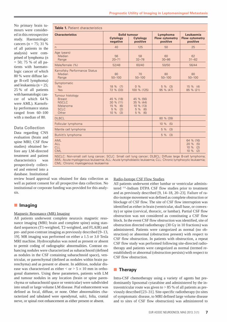
No primary brain tu-mours were consider-ed in this retrospectivestudy. Haematologiccancers (n = 75; 31 %of all patients in theanalysis) were com-prised of lymphoma (n= 50; 75 % of all pa-tients with haemato-logic cancer of which80 % were diffuse lar-ge B-cell lymphoma)and leukaemia (n = 25;25 % of all patientswith haematologic can-cer of which 64 %were AML). Karnofs-ky performance statusranged from 60–100with a median of 80.
Data CollectionData regarding CNSevaluation (brain andspine MRI; CSF flowstudies) obtained be-fore any LM-directedtreatment and patientcharacteristics wasprospectively collect-ed and entered into adatabase. Institutionalreview board approval was obtained for data collection aswell as patient consent for all prospective data collection. Noinstitutional or corporate funding was provided for this analy-sis.
Imaging
Magnetic Resonance (MR) ImagingAll patients underwent complete neuraxis magnetic reso-nance imaging (MRI; brain and complete spine) using stan-dard sequences (T1-weighted, T2-weighted, and FLAIR) andpre- and post-contrast imaging as previously described [9–13,19]. MR imaging was performed on either a 1.5 or 3.0 TeslaMRI machine. Hydrocephalus was noted as present or absentto permit coding of radiographic abnormalities. Contrast en-hancing nodules were characterized as subarachnoid (definedas nodules in the CSF containing subarachnoid space), ven-tricular, or parenchymal (defined as nodules within brain pa-renchyma) and as present or absent. In addition, nodular dis-ease was characterized as either < or > 5 × 10 mm in ortho-gonal diameters. Using these parameters, patients with LMand tumour nodules in any location (brain or spine paren-chyma or subarachnoid space or ventricular) were subdividedinto small or large volume LM disease. Pial enhancement wasdefined as focal, diffuse, or none. Other abnormalities cha-racterized and tabulated were ependymal, sulci, folia, cranialnerve, or spinal root enhancement as either present or absent.
Radio-Isotope CSF Flow StudiesAll patients underwent either lumbar or ventricular adminis-tered 111-Indium DTPA CSF flow studies prior to treatmentand as previously described [9, 14–18, 20–23]. Failure of ra-dio-isotope movement was defined as complete obstruction orblockage of CSF flow. The site of CSF flow interruption wasidentified as either in brain (ventricular, skull base, or convex-ity) or spine (cervical, thoracic, or lumbar). Partial CSF flowobstruction was not considered as constituting a CSF flowblock. In the event CSF flow obstruction was identified, site ofobstruction directed radiotherapy (30 Gy in 10 fractions) wasadministered. Patients were categorized as normal (no ob-struction) or abnormal (obstruction present) with respect toCSF flow obstruction. In patients with obstruction, a repeatCSF flow study was performed following site-directed radio-therapy and patients were categorized as normal (termed re-established) or abnormal (obstruction persists) with respect toCSF flow obstruction.
Therapy
Intra-CSF chemotherapy using a variety of agents but pre-dominantly liposomal cytarabine and administered by the in-traventricular route was given to > 85 % of all patients as pre-viously described [23–31]. Site-specific radiotherapy (to sitesof symptomatic disease, to MRI defined large volume diseaseand to sites of CSF flow obstruction) was administered to
Table 1. Patient characteristics
Characteristics Solid tumour Lymphoma LeukaemiaCytology Cytology Flow cytometry Flow cytometrynegative positive positive positive
n 40 125 50 25
Age (years)Median 56 58 60 62Range 20–71 32–78 30–86 31–82
Male/female (%) 52/48 60/40 50/50 56/44
Karnofsky Performance StatusMedian 80 70 80 80Range 50–100 50–100 50–100 50–100
SymptomaticNo 18 % (7) 0 % 5 % (3) 15 % (4)Yes 72 % (33) 100 % (125) 95 % (47) 85 % (21)
Tumour histologyBreast 45 % (18) 45 % (56)NSCLC 30 % (11) 35 % (44)Melanoma 15 % (6) 10 % (13)SCLC 5 % (2) 5 % (6)Other 10 % (3) 5 % (6)
DLBCL 80 % (39)
Follicular lymphoma 10 % (5)
Mantle cell lymphoma 5 % (3)
Burkitt’s lymphoma 5 % (3)
AML 64 % (16)ALL 20 % (5)CLL 10 % (2)CML 10 % (2)
NSCLC: Non-small cell lung cancer; SCLC: Small cell lung cancer; DLBCL: Diffuse large B-cell lymphoma;AML: Acute myelogenous leukaemia; ALL: Acute lymphoblastic leukaemia; CLL: Chronic lymphocytic leukaemia;CML: Chronic myelogenous leukaemia

Prognostic Utility of Imaging in Leptomeningeal Metastasis
8 EUR ASSOC NEUROONCOL MAG 2013; 3 (1)
45 % of patients. Systemic chemotherapy was used in the ma-jority (90 %) of patients with haematological cancers and inapproximately 27 % of solid tumour-related LM patients.
Survival AnalysisOverall survival (OS) was defined as the time from LM diag-nosis to death or last follow-up when patients were still alive.Survival rates were determined using the Kaplan Meiermethod and survival curves were compared using the log-ranktest. Statistical analysis were performed using the SAS Soft-ware (USA, Cary, NC) V9.2.
ResultsFour categories of patients with LM were retrospectivelyanalyzed; solid tumour-related LM with (n = 125) or without(n = 40) positive CSF cytology, lymphoma (n = 50), and leu-kaemia (n = 25; Table 2). Both categories of haematologiccancers (lymphoma and leukaemia) were positive by CSFflow cytometry and in 40 % positive as well by CSF cytology.In 4 patients (5 % of all patients with haematologic cancers)with haematologic malignancies CSF flow cytometry wasnegative and LM was determined by CSF cytology.
Patient categories were further divided into normal or abnor-mal MRI findings (Table 2). Abnormal MRI findings werethen divided into small or large volume disease as defined bymeasurable tumour nodules < or > 5 × 10 mm in orthogonaldiameter. Solid tumour-related LM had a higher incidence ofpatients with abnormal MRI findings as well as patients withlarge volume disease as compared to haematological cancer-related LM. Patients were also characterized by having nor-mal or abnormal (ie, obstructed) radio-isotope CSF flow stud-ies. One further category included patients with initial ob-structed CSF flow that following radiotherapy converted tonormal CSF flow (re-established) as determined by post-ra-diotherapy CSF flow study (Table 2).
Median overall survival (mOS) was similar (p = 0.3) in bothcategories (CSF positive and CSF cytology negative) of pa-tients with solid tumour-related LM (Table 3). However, sur-vival in patients with solid tumour-related LM with large vol-ume disease was significantly less than in patients with eithernormal MRI findings or small volume disease (p = 0.03).Similarly, mOS was not significantly different in solid tumourpatients with normal or re-established CSF flow studies (p =0.2). There was a significant difference in patients with ob-
Table 3. Median overall survival with respect to CNS imaging
Pre-treatment imaging Solid tumour Lymphoma LeukaemiaCytology negative Cytology positive
n 40 125 50 25
MRI (brain + spine)
AbnormalSmall volume disease 3 months 3.5 months 5 months 6 monthsLarge volume disease 2 months 2 months 3 months 2 months
Normal 3 months 3.5 months 5 months 6 months
CSF flow study
Abnormal 2 months 2 months 2 months 2 monthsRe-established 3 months 3.5 months 5 months 6 monthsNormal 3 months 3.5 months 5 months 6 months
Table 2. CNS imaging
Pre-treatment imaging Solid tumour Lymphoma LeukaemiaCytology negative Cytology positive
n 40 125 50 25
MRI (brain + spine)Abnormal 40 (100 %) 50 (40 %) 10 (20 %) 4 (16 %)
Small volume disease 10 (25 %) 25 (50 %) 8 (80 %) 3 (75 %)Large volume disease 30 (75 %) 25 (50 %) 2 (20 %) 1 (25 %)
Normal 0 (0 %) 75 (60 %) 40 (80 %) 21 (84 %)
CSF flow study
Abnormal 10 (25 %) 35 (28 %) 5 (10 %) 2 (8 %)Re-established 4 (40 %) 12 (34 %) 2 (40 %) 1 (50 %)Normal 30 (75 %) 90 (72 %) 45 (90 %) 23 (92 %)
Small volume disease: number of patients (percent) with MRI abnormalities and without tumour nodules or nodules < 10 mm in diameter;Large volume disease: number of patients (percent) with MRI abnormalities and tumour nodules > 5 × 10 mm in diameter; Abnormal:number of patients (percent) with obstructed CSF flow study; Re-established: number of patients (percent of total obstructed) post-radiotherapy with normal CSF flow study

EUR ASSOC NEUROONCOL MAG 2013; 3 (1)
Prognostic Utility of Imaging in Leptomeningeal Metastasis
9
structed (abnormal) CSF flow that could not be corrected byradiotherapy compared to the other 2 categories (normal orre-established; p = 0.04).
Comparable findings were seen with haematological cancer-related LM wherein normal or small volume MRI abnormali-ties defined a longer surviving cohort of patients relative topatients with large volume disease (p = 0.018). In addition,CSF obstruction not corrected by radiotherapy characterizeda haematological cancer patient category with worse outcomethan those with normal or re-established CSF flow studies(p = 0.015).
Cause of death, an arguably subjective analysis, showed simi-lar trends across all categories of patients wherein patientswith either large volume disease defined by MRI or non-cor-rected CSF flow obstruction by radio-isotope imaging moreoften died of LM (2-fold increase) compared to patients withnormal or small volume MRI disease and normal or re-estab-lished CSF flow (Table 4). By contrast, patients with normalor small volume MRI disease and normal or re-establishedCSF flow more often (3-fold increase) died of systemic dis-ease progression.
Discussion
It has previously been suggested that there are categories ofpatients with LM that are not candidates for LM-directedtherapy [8]. As outlined in the NCCN CNS tumour guidelinesvide supra, these include patients with poor performance,likely short life expectancy, carcinomatous encephalopathy,uncorrected CSF flow obstruction, and large CNS tumourburden [8]. These recommendations are primarily based uponexpert opinion with a paucity of literature-based evidence.The current retrospective study selected patients consideredeligible for LM-directed therapy based upon these recom-mendations and excluded patients a priori not considered byclinical criteria to warrant intra-CSF chemotherapy. What re-mains problematic in treating patients with LM is decidingwhom to treat and the current large retrospective study pro-vides some illumination in this regard.
Previous work has suggested CSF flow studies are informa-tive with respect to outcome and the current study corrobo-rates these findings in a considerably larger patient data set[14–18, 20–23]. CSF flow obstruction as defined by radioiso-tope studies appears prognostic as patients with non-correctedCSF flow obstruction survive a significantly shorter time thanpatients with normal or re-established CSF flow irrespective oftumour histology (solid tumour or haematological cancer-re-lated LM). In part the impoverished survival seen in patientswith CSF flow obstruction is reflective of tumour burden aswell as the pharmacologic barrier posed by interrupted CSFflow dynamics that mitigates intra-CSF chemotherapy admin-istration. Whether intra-CSF chemotherapy alters survival inpatients with LM is as yet undetermined and controversial asthere has never been a large prospective randomized trial thatshows a survival benefit for the receipt of intra-CSF chemo-therapy [32, 33]. In that CSF flow obstruction was not predictedby MRI aside from the finding of hydrocephalus (nor by pa-tients presenting symptoms) in the current study (data notshown), radio-isotope CSF flow studies appear complimentaryto MRI in determining outcome in patients otherwise consid-ered for LM-directed therapy. The current study supports theparadigm of utilizing CSF flow studies in patients with LMconsidered for treatment regardless if intra-CSF chemotherapyis used as survival is negatively impacted with evidence of in-terrupted CSF flow. Importantly, the current findings, ie thatCSF flow obstruction is prognostic, require validation in a pro-spective study of LM wherein radio-isotope CSF flow studiesare incorporated into pre-treatment evaluation. It was alsonoted that patients with non-correctable CSF flow obstructionmore often succumb to LM as a cause of death than patientswith normal or re-established CSF flow. Though the currentstudy represents the largest data set of patients withhaematological cancer-related LM (n = 75), the total number ofpatients, particularly with obstructed CSF flow, is compara-tively small (n = 7) and therefore may not be generalizable.
MRI-based imaging in patients with LM has primarily beenutilized to define brain involvement and when spine MRI isused, its use is mostly to define clinically site-relevant diseaseinvolvement [10–13]. The current study is unique in defining
Table 4. Cause of death with respect to CNS imaging
Pre-treatment imaging Solid tumour Lymphoma Leukaemia
Cytology negative Cytology positive
n 40 125 50 25
Cause of death (%) LM SD LM + SD LM SD LM + SD LM SD LM + SD LM SD LM + SD
MRI (brain + spine)
AbnormalSmall volume disease 23 53 25 25 48 27 26 51 25 24 52 23Large volume disease 48 15 37 53 12 35 47 16 37 51 14 35
Normal 25 51 24 23 55 22 24 52 24 25 50 25
CSF flow studyAbnormal 51 14 35 47 16 37 53 12 35 48 15 37Re-established 25 50 25 25 50 25 25 50 25 25 50 25Normal 25 50 25 25 50 25 25 50 25 25 50 25
LM: leptomeningeal metastasis; SD: systemic disease; LM + SD: combined LM and systemic disease

Prognostic Utility of Imaging in Leptomeningeal Metastasis
10 EUR ASSOC NEUROONCOL MAG 2013; 3 (1)
the total burden of CNS disease in patients with LM as allpatients underwent both brain and whole spine MRI. Al-though the data is not shown, there was limited concordancebetween symptoms and MRI findings whether in brain orspine. Consequently, CNS disease burden is not predicted byLM-related symptoms and therefore neuraxis imaging is re-quired to adequately stage the CNS. More important, how-ever, is the correlation between survival and MRI-defined dis-ease burden. In patients with large volume disease defined inthis study as patients with tumour nodule(s) > 5 × 10 mm insize, survival is significantly shortened relative to patientswith normal or small volume MRI disease. Whether tumournodules > 5 × 10 mm in diameter define all categories of largetumour burden is unknown as there has never been a studyattempting to quantify LM disease burden. Five by 10 milli-metre diameters were selected as nodules of this size or largerwere easily and reproducibly measured by MRI. Other com-mon radiologic findings by MRI of LM for example leptomen-ingeal, cranial nerve, or spinal nerve root enhancement do notlend themselves to quantification. An improved radiographicmethod to quantify LM disease burden would be a welcometool in assessing LM disease. Also noted in patients with largevolume disease burden, cause of death was more often a resultof LM as compared to patients with normal or small volumeMRI disease that more frequently died due to systemic dis-ease progression.
In conclusion, neuraxis imaging utilizing brain and spineMRI as well as radio-isotope CSF flow studies may have prog-nostic significance and appears predictive of median overallsurvival in this large cohort of patients with LM. The study islimited by the retrospective design, the novel definition ofMRI large volume disease, and multiple small categories ofpatients upon which these conclusions are based. However,pending a larger prospective trial the current retrospectivedata set is the most robust data available regarding the utilityof CNS imaging in predicting survival in patients with LM.
Conflict of Interest
The author has no financial disclosures.
References:1. Chamberlain MC. Leptomeningeal metasta-sis. Curr Opin Oncol 2010; 22: 627–35.2. Shapiro WR, Johanson CE, Boogerd W. Treat-ment modalities for leptomeningeal metasta-ses. Semin Oncol 2009; 36: S46–S54.3. Mammoser AG, Groves MD. Biology andtherapy of neoplastic meningitis. Curr OncolRep 2010; 12: 41–9.4. Glantz MJ, Jaeckle KA, Chamberlain MC,et al. A randomized controlled trial comparingintrathecal sustained-release cytarabine(DepoCyt) to intrathecal methotrexate in pa-tients with neoplastic meningitis from solidtumors. Clin Cancer Res 1999; 5: 3394–402.5. Glantz MJ, LaFollette S, Jaeckle KA, et al.Randomized trial of a slow release vs. a stand-ard formulation of cytarabine for the intrathecaltreatment of lymphomatous meningitis. J ClinOncol 1999; 17: 3110–6.6. Strik H, Prommel P. Diagnosis and individual-ized therapy of neoplastic meningitis. ExpertRev Anticancer Ther 2010; 10: 1137–48.7. Jaeckle KA. Neoplastic meningitis from sys-temic malignancies: diagnosis, prognosis, andtreatment. Semin Oncol 2006; 33: 312–23.8. Brem SS, Bierman PJ, Black P, et al. Centralnervous system cancers: clinical practiceguidelines in oncology. J Natl Compr CancNetw 2008; 6: 456–504.9. Chamberlain MC, Glantz M, Groves MD, etal. Diagnostic tools for neoplastic meningitis:detecting disease, identifying patient risk, anddetermining benefit of treatment. Semin Oncol2009; 36 (Suppl 2): S35–S45.10. Clarke JL, Perez HR, Jacks LM, et al. Lep-tomeningeal metastases in the MRI era. Neu-rology 2010; 74: 1449–54.11. Collie DA, Brush JP, Lammie GA, et al.Imaging features of leptomeningeal metasta-ses. Clin Radiol 1999; 54: 765–71.12. Straathof CS, de Bruin HG, Dippel DW, etal. The diagnostic accuracy of magnetic reso-nance imaging and cerebrospinal fluid cytologyin leptomeningeal metastasis. J Neurol 1999;246: 810–4.13. Freilich RJ, Krol G, DeAngelis LM. Neuro-imaging and cerebrospinal fluid cytology in thediagnosis of leptomeningeal metastasis. AnnNeurol 1995; 38: 51–7.14. Grossman SA, Trump CL, Chen DCP, et al.Cerebrospinal flow abnormalities in patientswith neoplastic meningitis. Am J Med 1982;73: 641–7.15. Glantz MJ, Hall WA, Cole BF, et al. Diagno-sis, management, and survival of patients withleptomeningeal cancer based on cerebrospinalfluid-flow status. Cancer 1995; 75: 2919–31.16. Chamberlain MC, Corey-Bloom J. Leptomen-ingeal metastasis: Indium-DTPA CSF flow stud-ies. Neurology 1991; 41: 1765–9.17. Mason WP, Yeh SD, DeAngelis LM. 111Indium-diethylenetriamine pentaacetic acid cerebro-
spinal fluid flow studies predict distribution ofintrathecally administered chemotherapy andoutcome in patients with leptomeningealmetastases. Neurology 1998; 50: 438–44.
18. Chamberlain MC. Spinal 111Indium-DTPACSF flow studies in leptomeningeal metasta-sis. J Neurooncol 1995; 25: 135–41.
19. Chamberlain MC. Comparative spine imag-ing in leptomeningeal metastases. J Neuro-oncol 1995; 23: 233–8.
20. Chamberlain MC. Radioisotope CSF flowstudies in leptomeningeal metastases. JNeurooncol 1998; 38: 135–40.
21. Chamberlain MC, Glantz M. Utility of CSFradioisotope flow studies in leptomeningealmetastases: A review. J Nucl Med 2002; 5:2101–12.
22. Chamberlain MC, Sandy A, Press GA. Lep-tomeningeal metastasis: A comparison ofgadolinium-enhanced MR and contrast-en-hanced CT of the brain. Neurology 1990; 40:435–8.
23. Chamberlain MC, Kormanik P. Prognosticsignificance of 111Indium-DTPA CSF flow stud-ies. Neurology 1996; 46: 1674–7.
24. Chamberlain MC, Kormanik P. Leptomenin-geal metastases due to melanoma: Combinedmodality therapy. Int J Oncol 1996; 9: 505–10.
25. Chamberlain MC, Kormanik PA. Carcinoma-tous meningitis secondary to non-small celllung cancer: Combined modality therapy. ArchNeurol 1998; 55: 506–12.
26. Chamberlain MC, Kormanik PA. Non-AIDSrelated lymphomatous meningitis: Combinedmodality therapy. Neurology 1997; 49: 1728–31.
27. Chamberlain MC, Kormanik P, Glantz M.Recurrent primary central nervous system lym-phoma complicated by lymphomatous menin-gitis. Oncol Rep 1998; 5: 521–3.
28. Chamberlain MC. Combined modality treat-ment of leptomeningeal gliomatosis. Neurosur-gery 2003; 52: 324–30.
29. Chamberlain MC, Chalmers L. Acute bin-ocular blindness. Cancer 2007; 109: 1851–4.
30. Chamberlain M, Glantz MJ. Myelomatousmeningitis: multimodal therapy. Cancer 2008;112: 1562–7.
31. Chamberlain MC, Johnston S, Van Horn A,et al. Recurrent lymphomatous meningitistreated with intra-CSF rituximab and liposomalara-C. J Neurooncol 2009; 91: 271–7.
32. Siegal T. Leptomeningeal metastases: ra-tionale for systemic chemotherapy or what isthe role of intra-CF chemotherapy? J Neuro-oncol 1998; 38: 151–7.
33. Boogerd W, van den Bent MJ, Koehler PJ,et al. The relevance of intraventricular chemo-therapy for leptomeningeal metastasis in breastcancer: a randomized study. Eur J Cancer 2004;40: 2726–33.
EUR ASSOC NEUROONCOL MAG 2013; 3 (1)
High-Tech Surgery of Gliomas
11
Surgery of Malignant Gliomas UsingModern Technology
Martin Proescholdt, Christian Doenitz, Alexander Brawanski
Received on September 25, 2012; accepted on September 30, 2012; Pre-PublishingOnline on October 22, 2012
From the Department of Neurosurgery, Regensburg University Medical Center,GermanyCorrespondence to: Martin Proescholdt, MD, Department of Neurosurgery,Regensburg University Medical Center, Franz-Josef-Strauß-Allee 11, 93053 Regens-burg, Germany; e-mail: [email protected]
Therapy of Malignant Gliomas – A Formi-dable Challenge
Gliomas of astrocytic, oligodendroglial, and ependymal dif-ferentiation comprise with an incidence of 6/100,000/yearabout 70 % of all intrinsic brain tumours [1]. The WHO clas-sification system distinguishes 4 grades of malignancy [2]characterized by morphologic features such as mitotic activ-ity, microvascular proliferation, and intratumoural necroses.The most frequent glioma of the adulthood, the glioblastomamultiforme, is a highly malignant neoplasm which displays anexceptionally poor prognosis with a median survival time of15 months [3]. Two years after diagnosis, only 8.2 % of allpatients are still alive [4]. The management of glioblastomaconsists of 3 main elements: (1) microsurgical resection isfollowed by (2) concomitant treatment with radiotherapy plus(3) temozolomide chemotherapy [5]. In this context, the ex-tent of surgical resection (EOR) has increasingly been recog-nized as an important prognostic factor in this patient popula-tion [6]. A prospective, randomized multicentre trial in glio-blastoma patients has demonstrated that complete resection ofthe contrast-enhancing tumour leads to an overall survival of16.7 months compared to 11.8 months after subtotal resection[7]. However, there are 2 major limitations to radical surgicalresection: (1) glioblastomas display a highly infiltrativegrowth pattern [8] which renders complete resection virtuallyimpossible. Careful histological studies revealed a tumourcell spread into the contralateral hemisphere in about 30 % ofall patients at the time of diagnosis [9, 10]. Thus, even themost radical surgical approach will not lead to curative treat-ment [11]. (2) The functional anatomy of the brain consists ofcortical and subcortical structures such as the primary motorcortex, Wernicke and Broca speech centres, or the internalcapsule, which need to be preserved during surgical resectionto avoid serious postoperative neurological deficits. Since pa-
tients with permanent neurological deficits have a signifi-cantly worse survival prognosis [12], the avoidance of anydamage to these eloquent structures is mandatory in the surgi-cal treatment of glioblastoma [13].
Preoperative Work-Up
Traditionally, surgery planning was conducted utilizing ana-tomical landmarks [14]. In the past, the identification of elo-quent areas was performed in a generalized, rigid fashionbased on the functional studies by Wilder Penfield, frequentlyleading to an inadequate assessment of the surgical risk in theindividual patient [15]. The major reason for this inaccuracyis the significant individual variability of cortical organization[16]. In addition, recent studies have demonstrated a high de-gree of functional plasticity of the brain, which causes a sig-nificant shift of eloquent areas to distant sites especially underthe condition of intracerebral tumour growth [17]. Preopera-tive application of functional MRI (fMRI) and Diffusion Ten-sor Imaging (DTI) allows the detection of eloquent corticaland subcortical structures with high sensitivity and specificity[18, 19]. With the advent of computer-based analysis toolsallowing the fusion of patho-anatomical, functional, andmetabolic imaging data, it is now possible to plan and executea precise and safe resection trajectory, thus achieving maxi-mal EOR with minimal surgical morbidity (Figure 1). In thecase of a large, infiltrative tumour, which needs to be biopsiedin order to establish a histological diagnosis, it is of para-mount importance to target the area of the lesion with the sus-pected highest grade of malignancy. In a study conducted in81 patients who received stereotactic biopsy followed by re-section of the tumour within 60 days, the biopsy-based diag-nosis was incorrect in 38 %, emphasizing the limitations ofstereotactic biopsy as a diagnostic tool [20]. The applicationof Positron Emission Tomography (PET) scanning utilizingtracers such as [F-18]fluoroethyltyrosine allows to detect meta-bolically active areas within a larger tumour mass. The inte-gration of these molecular imaging data into the target plan-ning process can significantly increase the diagnostic yield ofstereotactic biopsies in patients with diffuse gliomas [21, 22].In addition, the differentiation between tumour progress andradiation-induced necrosis or pseudoprogression can be fa-cilitated by PET scanning, supporting adequate clinical man-agement and avoidance of unnecessary treatment measures
Abstract: Recent evidence has demonstratedthat the extent of surgical resection (EOR) is animportant prognostic factor in patients with ma-lignant gliomas. However, the infiltrative growthpattern and the functional anatomy of the brainconsisting of eloquent cortical and subcorticalstructures pose a significant limitation to thesurgical resection of these tumours. The gain of
knowledge regarding function, biology, and patho-physiology of the brain has resulted in the adventof advanced technology to the neurosurgeon, al-lowing maximal resection with minimal opera-tive morbidity. We have reviewed the current lit-erature concerning intraoperative imaging, fluo-rescence-guided resection, and awake cranioto-my to generate a comprehensive overview of the
most recent developments in this field. In addi-tion, we have provided data from our own insti-tution confirming the beneficial effects of thismultimodal approach. Eur Assoc NeuroOncolMag 2013; 3 (1): 11–4.
Key words: resection, glioma, technology, MRI,awake craniotomy

High-Tech Surgery of Gliomas
12 EUR ASSOC NEUROONCOL MAG 2013; 3 (1)
such as repeated surgical resection [23]. Finally, detailed neu-ropsychological evaluation is helpful in unmasking subclini-cal tumour-related impairments to improve the prognostica-tion of the postoperative course of the disease [24].
Intraoperative Technique
One of the most substantial obstacles to an extensive resectionof gliomas is the infiltrative growth pattern of these tumours.The development of 5-aminolevulinic acid (5-ALA) as a tu-mour-specific fluorescence marker has caused a breakthroughin the resection of malignant gliomas [25]. The substanceleads to an intracellular accumulation of fluorescent porphy-rins which can be detected intraoperatively using a micro-scope equipped with a violet-blue excitation light source(Figure 2). A prospective, randomized controlled multicentretrial has demonstrated a significantly better EOR in the 5-ALA group compared to the control arm resected with con-ventional light [26]. The intraoperative localization of the tu-mour in addition to the adjacent, eloquent areas of the brain isgreatly facilitated by the use of neuronavigation, which hasalso been termed frameless stereotaxy [27]. This technique isbased on MRI imaging conducted with fiducial markersplaced on particular landmarks of the patient’s skull. Prior tocraniotomy, an LED-emitting detection system linked to acomputer containing the imaging data set is used to calibratethe surgical instrument set, which then allows the visualiza-tion of the resection process intraoperatively. This approachhas significantly improved the safety and extent of resectionin glioma patients [28, 29]. However, the accuracy of neuro-navigation-based resection, which is solely based on preop-erative imaging, decreases during the course of the proceduredue to “brain shift” caused by the release of cerebrospinalfluid, brain swelling, and surgical manoeuvres [30]. To ac-count for this aspect, real-time intraoperative imaging is re-quired. Consequently, intraoperative MRI (iMRI) has been
developed as an advanced technique for imaging-based resec-tion control in glioma surgery [31]. A recent, controlled, pro-spective clinical trial has demonstrated that the use of iMRIleads to a better extent of resection and improved 6-monthsurvival rates in the iMRI group compared to the controlpopulation. Interestingly, the occurrence of postoperativeneurological deficits was not significantly different betweenthe 2 study groups [32]. However, iMRI is complex, requiringeither transport of the patient to the scanner during the opera-tion or a completely antimagnetic setting in the operatingroom. Surgery time is prolonged due to the scanning proce-dure and iMRI systems are expensive and not available in themajority of neurosurgical centres [33, 34]. A valid alternativeis the use of intraoperative ultrasound (IOUS), which allowsreal-time detection of infiltrative tumour margins [35]. How-ever, IOUS-based resection control, albeit possible, is influ-enced by surgery-related artefacts and depends significantlyon the experience of the surgeon [36]. As an alternative toimage-based surgery, awake craniotomy with intraoperativecortical and subcortical stimulation has been established as“gold standard” to achieve maximal EOR with minimal mor-bidity [37]. The procedure involves tumour resection in theawake patient, allowing serial neurocognitive tests concern-ing motor or language function combined with direct electri-
R L
R L
Figure 1. Fusion of [F-18]fluoroethyltyrosine: PET (red), functional MRI (green), andDTI tractography (yellow) in a patient with a left frontal anaplastic astrocytoma.Note the close vicinity of eloquent cortical and subcortical structures to the tumourborders.
Figure 2. Resection cavity in a patient with glioblastoma following 5-aminolevuli-nic acid application as a fluorescence marker. (A) Under conventional light, incon-spicuous adjacent white matter is visible. (B) Fluorescence illumination reveals ahigh-intensity signal indicating a residual tumour. Reprinted from [Stummer W,Novotny A, Stepp H, et al. Fluorescence-guided resection of glioblastomamultiforme by using 5-aminolevulinic acid-induced porphyrins: a prospec-tive study in 52 consecutive patients. J Neurosurg 2000; 93: 1003–13]with permission from the American Association of Neurological Surgeons.

EUR ASSOC NEUROONCOL MAG 2013; 3 (1)
High-Tech Surgery of Gliomas
13
cal stimulation of the brain to unmask eloquent cortical andsubcortical structures. Using this approach, a better EOR canbe achieved while avoiding damage to functionally relevantbrain structures [38, 39]. In order to avoid stress for the pa-tient and to gain the best surgical results, a team of highlytrained and experienced physicians consisting of anaesthesi-ologists, neuropsychologists, and neurosurgeons is manda-tory [40]. In addition, especially if awake craniotomy is not anoption, intraoperatively evoked potential monitoring is highlyuseful to detect damage to eloquent structures early during theprocedure, allowing to correct the surgical trajectory in atimely fashion [41].
Results from a Single Centre – High-TechSurgery, Is It Worthwhile?
Although it is self-evident to embrace the concept of high-tech surgery, limited resources in today’s medical practicemay prompt the question of whether this multimodal ap-proach is of any clinical benefit to glioma patients. Employingthe entire armamentarium outlined in this review except foriMRI, we volumetrically analyzed the EOR and clinical out-come in 44 patients with malignant gliomas (5 anaplastic as-trocytoma, 39 glioblastoma) receiving surgical resection atour department. Mean age was 62.5 years, 61.4 % of all pa-tients presenting with focal neurological deficits. Preopera-tive tumour size and EOR were quantified volumetricallybased on MRI imaging (Iplan Cranio, Brainlab, Feldkirchen,Germany; Figure 3). In addition, surgical morbidity and mor-tality as well as the improvement of neurological performancewere registered. There was no perioperative mortality, surgi-cal morbidity was recorded in 9 % of all cases, caused bywound infection and CSF fistula, respectively. Complete re-section (ie, no residual contrast enhancement in the postop-erative scan) was achieved in 62 % of all cases, in 93 % of thepatients an EOR > 90 % was accomplished. Of all patientspresenting with neurological impairment, 52 % showed sig-nificant improvement. Only one patient developed transientdouble vision postoperatively, which completely dissipatedafter one week. These data confirm that the employment ofadvanced pre- and intraoperative technologies allows a safeand extensive resection in malignant glioma patients with alow rate of surgical morbidity.
R L
Conclusion
Basic science research efforts during the decade of the brainhas created an enormous gain of knowledge regarding func-tion, biology, and pathophysiology of the brain [42]. This hascaused a shift of paradigm in clinical neurosciences, includ-ing the surgical treatment of malignant gliomas. The advent ofmodern technology has revolutionized the preoperative work-up, surgical trajectory planning, and intraoperative monitor-ing with significant benefits for the patients regarding neuro-functional improvement and overall survival. In the treatmentof malignant glioma, combined efforts of all involved medicalspecialties are mandatory to achieve the best results for theindividual patient [43, 44]. Modern neurosurgery can contrib-ute to this treatment structure by providing maximal EORcombined with minimal morbidity.
Conflict of Interest
The authors report no conflict of interest.
Funding
The study was not funded by any source.
References:1. Ricard D, Idbaih A, Ducray F, et al. Primarybrain tumours in adults. Lancet 2012; 379:1984–96.
2. Louis DN, Ohgaki H, Wiestler OD, et al.The 2007 WHO classification of tumours ofthe central nervous system. Acta Neuro-pathol (Berl) 2007; 114: 97–109.
3. Wen PY , Kesari S. Malignant gliomas inadults. N Engl J Med 2008; 359: 492–507.
4. Tran B, Rosenthal MA. Survival compari-son between glioblastoma multiforme andother incurable cancers. J Clin Neurosci2010; 17: 417–21.
5. Stupp R, Mason WP, van den Bent MJ, etal. Radiotherapy plus concomitant and adju-vant temozolomide for glioblastoma. N EnglJ Med 2005; 352: 987–96.
6. Sanai N, Polley MY, McDermott MW, et al.An extent of resection threshold for newlydiagnosed glioblastomas. J Neurosurg 2011;115: 3–8.
7. Pichlmeier U, Bink A, Schackert G, et al.Resection and survival in glioblastoma multi-forme: an RTOG recursive partitioning analy-sis of ALA study patients. Neuro Oncol 2008;10: 1025–34.8. Claes A, Idema AJ, Wesseling P. Diffuseglioma growth: a guerilla war. Acta Neuro-pathol (Berl) 2007; 114: 443–58.9. Sahm F, Capper D, Jeibmann A, et al. Ad-dressing diffuse glioma as a systemic braindisease with single-cell analysis. Arch Neurol2012; 69: 523–6.10. Burger PC. Pathologic anatomy and CTcorrelations in the glioblastoma multiforme.Appl Neurophysiol 1983; 46: 180–7.11. Dandy WE. Removal of the right hemi-sphere for certain tumors with hemiplegia:preliminary report. JAMA 1928; 90: 823–5.12. Bauchet L, Mathieu-Daude H, Fabbro-Peray P, et al. Oncological patterns of careand outcome for 952 patients with newly di-agnosed glioblastoma in 2004. Neuro Oncol2010; 12: 725–35.
Figure 3. Quantification of the preoperative tumour volume in malignant gliomas. (A) Axial T1-weighted MRI scan of a patient with a left frontal glioblastoma. (B) Thecontrast-enhancing part of each section is outlined and subsequently fused to generate (C) a 3-dimensional segment which can be quantified volumetrically.

High-Tech Surgery of Gliomas
14 EUR ASSOC NEUROONCOL MAG 2013; 3 (1)
13. Stummer W, Tonn JC, Mehdorn HM, etal. Counterbalancing risks and gains fromextended resections in malignant gliomasurgery: a supplemental analysis from therandomized 5-aminolevulinic acid glioma re-section study. Clinical article. J Neurosurg2011; 114: 613–23.14. Berger MS, Hadjipanayis CG. Surgery ofintrinsic cerebral tumors. Neurosurgery 2007;61 (Suppl): 279–304.15. Mazzola L, Isnard J, Peyron R, et al.Stimulation of the human cortex and the ex-perience of pain: Wilder Penfield’s observa-tions revisited. Brain 2012; 135: 631–40.16. Ojemann GA. Individual variability in cor-tical localization of language. J Neurosurg1979; 50: 164–9.17. Duffau H. Brain plasticity: from patho-physiological mechanisms to therapeutic ap-plications. J Clin Neurosci 2006; 13: 885–97.18. Gupta A, Shah A, Young RJ, et al. Imag-ing of brain tumors: functional magneticresonance imaging and diffusion tensor im-aging. Neuroimaging Clin N Am 2010; 20:379–400.19. Castellano A, Bello L, Michelozzi C, etal. Role of diffusion tensor magnetic reso-nance tractography in predicting the extentof resection in glioma surgery. Neuro Oncol2012; 14: 192–202.20. Jackson RJ, Fuller GN, Abi-Said D, et al.Limitations of stereotactic biopsy in the ini-tial management of gliomas. Neuro Oncol2001; 3: 193–200.21. Pirotte B, Goldman S, Bidaut LM, et al.Use of positron emission tomography (PET)
in stereotactic conditions for brain biopsy.Acta Neurochir (Wien) 1995; 134: 79–82.22. Kunz M, Thon N, Eigenbrod S, et al. Hotspots in dynamic (18)FET-PET delineate ma-lignant tumor parts within suspected WHOgrade II gliomas. Neuro Oncol 2011; 13:307–16.23. Caroline I, Rosenthal MA. Imaging mo-dalities in high-grade gliomas: pseudopro-gression, recurrence, or necrosis? J ClinNeurosci 2012; 19: 633–7.24. Wu AS, Witgert ME, Lang FF, et al.Neurocognitive function before and aftersurgery for insular gliomas. J Neurosurg2011; 115: 1115–25.25. Stummer W, Stocker S, Wagner S, et al.Intraoperative detection of malignant glio-mas by 5-aminolevulinic acid-induced por-phyrin fluorescence. Neurosurgery 1998; 42:518–525.26. Stummer W, Pichlmeier U, Meinel T, etal. Fluorescence-guided surgery with 5-ami-nolevulinic acid for resection of malignantglioma: a randomised controlled multicentrephase III trial. Lancet Oncol 2006; 7: 392–401.27. Willems PW, van der Sprenkel JW, Tul-leken CA, et al. Neuronavigation and sur-gery of intracerebral tumours. J Neurol 2006;253: 1123–36.28. Kurimoto M, Hayashi N, Kamiyama H, etal. Impact of neuronavigation and image-guided extensive resection for adult patientswith supratentorial malignant astrocytomas:a single-institution retrospective study. Mi-nim Invasive Neurosurg 2004; 47: 278–83.
29. Wirtz CR, Albert FK, Schwaderer M, etal. The benefit of neuronavigation for neuro-surgery analyzed by its impact on glioblas-toma surgery. Neurol Res 2000; 22: 354–60.
30. Ohue S, Kumon Y, Nagato S, et al. Evalu-ation of intraoperative brain shift using anultrasound-linked navigation system for braintumor surgery. Neurol Med Chir (Tokyo) 2010;50: 291–300.
31. Kubben PL, ter Meulen KJ, Schijns OE,et al. Intraoperative MRI-guided resection ofglioblastoma multiforme: a systematic re-view. Lancet Oncol 2011; 12: 1062–70.
32. Senft C, Bink A, Franz K, et al. Intraopera-tive MRI guidance and extent of resection inglioma surgery: a randomised, controlledtrial. Lancet Oncol 2011; 12: 997–1003.
33. Oh DS , Black PM. A low-field intraop-erative MRI system for glioma surgery: is itworthwhile? Neurosurg Clin N Amer 2005;16: 135–41.
34. Seifert V. Intraoperative MRI in neurosur-gery: technical overkill or the future of brainsurgery? Neurol India 2003; 51: 329–32.
35. Gerganov VM, Samii A, Giordano M, etal. Two-dimensional high-end ultrasound im-aging compared to intraoperative MRI dur-ing resection of low-grade gliomas. J ClinNeurosci 2011; 18: 669–73.
36. Hammoud MA, Ligon BL, elSouki R, etal. Use of intraoperative ultrasound for lo-calizing tumors and determining the extentof resection: a comparative study with mag-netic resonance imaging. J Neurosurg 1996;84: 737–41.
37. Kim SS, McCutcheon IE, Suki D, et al.Awake craniotomy for brain tumors neareloquent cortex: correlation of intraopera-tive cortical mapping with neurological out-comes in 309 consecutive patients. Neuro-surgery 2009; 64: 836–45.
38. De Benedictis A, Moritz-Gasser S, DuffauH. Awake mapping optimizes the extent ofresection for low-grade gliomas in eloquentareas. Neurosurgery 2010; 66: 1074–84.
39. Pereira LC, Oliveira KM, L’Abbate GL, etal. Outcome of fully awake craniotomy forlesions near the eloquent cortex: analysis ofa prospective surgical series of 79 supraten-torial primary brain tumors with long follow-up. Acta Neurochir (Wien) 2009; 151: 1215–30.
40. Brydges G, Atkinson R, Perry MJ, et al.Awake craniotomy: a practice overview.AANA J 2012; 80: 61–8.
41. Kombos T, Picht T, Derdilopoulos A, etal. Impact of intraoperative neurophysiologi-cal monitoring on surgery of high-grade glio-mas. J Clin Neurophysiol 2009; 26: 422–5.
42. Laws ER Jr. The decade of the brain:1990 to 2000. Neurosurgery 2000; 47: 1257–60.
43. Hofer S, Roelcke U, Herrmann R. [Newaspects of interdisciplinary therapy for ma-lignant gliomas in adults]. Schweiz MedWochenschr 1999; 129: 1332–41.
44. Taylor LP. Diagnosis, treatment, and prog-nosis of glioma: five new things. Neurology2010; 75 (Suppl 1): S28–S32.

EUR ASSOC NEUROONCOL MAG 2013; 3 (1)
Emerging Immune Therapeutics Targeting Glioblastoma-Mediated Immune Suppression
15
Emerging Immune Therapeutics TargetingGlioblastoma-Mediated Immune Suppression:
Dark Before the DawnShuo Xu1,2, Amy B Heimberger2
Received on December 10, 2012; accepted on December 21, 2012
From the 1Department of Neurosurgery, Qilu Hospital of Shandong University,Jinan, China; 2Department of Neurosurgery, The University of Texas M.D.Anderson Cancer Center, Houston, TX, USACorrespondence to: Amy B Heimberger, MD, Department of Neurosurgery,Unit 422, The University of Texas M.D. Anderson Cancer Center, Houston, TX,USA 77030-1402; e-mail [email protected]
Abstract: As the most common and particularlydevastating primary brain malignancy, glio-blastoma exerts profound immunosuppressionon the anti-tumour weapons of the immunesystem, which also poses a tremendous obstacleto immunotherapy. By targeting glioblastoma-mediated immune suppression, enthusiasm and
confidence are accumulating based not only onthe encouraging results of current clinical trialsbut also largely on promising preclinical find-ings. In this article, we summarize causes ofglioblastoma-mediated immune suppression, re-view the current and potential approachesagainst several key immunosuppressive regula-
Immunosuppression and Its Influenceon Glioblastoma Treatment
Despite the marked advances in basic scientific research andclinical practice over the last several decades, improvementsin progression-free survival (PFS) and overall survival (OS)have been modest in patients with glioblastoma – the mostcommon and particularly devastating primary brain malig-nancy [1–4]. The failures of conventional glioblastoma treat-ments are attributed to the complex and heterogeneous tu-mour composition, aggressive diffuse infiltration, exuberantangiogenesis, and the tumour’s capacity to escape therapies[5, 6]. Glioblastomas express a variety of tumour-associatedand tumour-specific antigens such as interleukin- (IL-) 13RA,EGFRvIII, EphA2, survivin, and CMV, etc. By inducing anti-tumour immune responses, glioblastoma immunotherapyprovides an alternative treatment strategy, with the theoreticaladvantage of tumour specificity.
Considering the presence of the blood-brain barrier, lack oflymphatic drainage, and the paucity of resident specializedantigen-presenting cells (APC) within the central nervoussystem (CNS), “immunological privilege” was once believedto be an inherent property of the brain. This concept has beensignificantly revised by the evidence of dynamic immune re-sponses found in various physiological and pathophysiologi-cal circumstances inside the CNS [7]. In the context of gliob-lastoma immune responses, although there can be significantimmune cell infiltration (including microglia/macrophages,lymphocytes, and dendritic cells), the anti-tumour immuneresponses are markedly impaired and can actually be tumour-promoting [8]. In fact, the immune responses within the gliob-lastoma microenvironment can be profoundly immunosup-pressive and include recruitment and induction of regulatoryT cells (Tregs) [9–11]; expression of immune checkpoints
tors, and discuss the challenges and future ofimmunotherapy in glioblastoma treatment. EurAssoc NeuroOncol Mag 2013; 3 (1): 15–22.
Key words: glioblastoma, immunosuppression,immunotherapy, clinical trials
(such as B7.H1/PD-L1) [12–14] and cytotoxic T-lymphocyte-associated antigen 4 (CTLA-4) [15]; down-regulation or ab-sence of tumour-specific antigens; immunosuppressive cyto-kine secretion, such as transforming growth factor- (TGF-) β,IL-10, and vascular endothelial growth factor (VEGF) [16,17]; and recruitment and skewing of tumour-supportive macro-phages (M2 vs M1) [18, 19].
Immune therapeutic strategies that induce immune effectorresponses have demonstrated marked increases in PFS andOS in phase-II clinical trials of glioblastoma patients [20–25].However, several of these trials have included in the enrol-ment criterion a requirement for gross-total resection in orderto minimize glioblastoma-mediated immune suppression.Unfortunately, not all glioblastoma patients are subjected toextensive resections or may have medical contraindications.If glioblastoma-mediated immune suppression could be con-trolled, then theoretically, immune recognition and clearanceshould occur.
Current Clinical Trials Targeting Glioblas-toma-Mediated Immunosuppression
Numerous glioblastoma immunotherapeutic clinical trials areunderway, with most designed to prime/amplify the host anti-tumour immune responses rather than to abrogate or reverseglioblastoma-mediated immunosuppression. According to theClinicalTrials.gov database (http://www.clinicaltrials.gov/, up-dated to November 2012), there are less than 10 completed oractive glioblastoma immunosuppression-targeted clinical trialsdocumented among more than 120 clinical trials related toglioblastoma immunotherapeutics. Among the limited glio-blastoma immunosuppression-targeted clinical trials, most arephase-I studies evaluating the pharmacokinetic and toxicologi-cal properties of certain reagents, or exploratory studies deter-mining the correlation between immune status modulation anddrug intervention (Table 1).
TGF-βββββ PathwayTransforming growth factor β2 (TGF-β2) is a potent cytokinewith multiple biological activities [26] that has been found to

Emerging Immune Therapeutics Targeting Glioblastoma-Mediated Immune Suppression
16 EUR ASSOC NEUROONCOL MAG 2013; 3 (1)
be over-expressed in more than 90 %of high-grade gliomas [27–29], mak-ing it an attractive target for glioblas-toma treatment. Inhibition of TGF-β2in tumour tissue leads to reversal oftumour-induced immune suppressionas well as inhibition of tumour growth,invasion, and metastasis [30, 31].Trabedersen (AP 12009) is a synthe-tic antisense phosphorothioate oligo-deoxynucleotide complementary tothe human TGF-β2 mRNA, whosesafety and efficacy has been shownthrough various pharmacokinetic andtoxicology studies, both in vitro andin vivo, as well as in phase-I/-II dose-escalation studies [32–34]. The over-all outcome was negative for the pre-specified primary endpoint in therandomized phase-IIb study com-paring a dose of either 10 μM or 80μM of trabedersen with standardchemotherapy in a cohort of 145 pa-tients with recurrent or refractoryanaplastic astrocytoma or glioblas-toma (Trial Registration: Clini-calTrials.gov NCT00431561). Nev-ertheless, survival at 2 and 3 years forglioblastoma patients in a small sub-group (≤ 55 years old, with an initialKarnofsky Performance scale scoreof > 80) who received 10 μM trabe-dersen was 3-fold higher than forthose receiving chemotherapy [35].Despite the controversy over the in-terpretation of the clinical trial data[36, 37], a multinational phase-IIIstudy entitled SAPPHIRE was under-way investigating the efficacy andsafety of 10 μM trabedersen com-pared with standard chemotherapy inadult patients with confirmed recur-rent or refractory anaplastic astrocy-toma or secondary glioblastoma, butit was terminated due to slow patientrecruitment (Trial Registration: Cli-nicalTrials.gov NCT00761280).
In addition to trabedersen, severalother drugs targeting the TGF-β path-way are now being tested in phase-I/IIclinical trials. For instance, GC1008is an antibody capable of neutralizingTGF-β, and a phase-II clinical trial isopen to determine its safety, tolera-bility, pharmacokinetics, and phar-macodynamics in primary malignantglioma patients (Trial Registration:ClinicalTrials.gov NCT01472731).LY2157299 is another new smallmolecule drug antagonizing the TGF-
Tab
le 1
. C
linic
al a
nd p
recl
inic
al t
rial t
arge
ts a
nd r
esul
ts f
or g
liobl
asto
ma-
med
iate
d im
mun
osup
pres
sion
. The
Can
cer
Gen
ome
Atla
s gl
iobl
asto
ma
data
base
of
mR
NA
dat
a (A
gile
nt m
icro
arra
y,sa
mpl
e nu
mbe
r: 50
0, u
pdat
ed to
11/
30/2
012)
was
use
d as
the
sour
ce o
f dat
a fo
r exp
ress
ion
and
surv
ival
eva
luat
ion,
and
dat
a w
ere
anal
yzed
thro
ugh
the
open
acc
ess
cBio
Can
cer G
enom
ics
Port
alat
ww
w.c
biop
orta
l.org
. The
glio
blas
tom
a tis
sue
anal
yzed
con
tain
s gl
iom
a ce
lls a
s w
ell a
s tu
mou
r-sup
port
ive
stro
mal
and
infil
trat
ing
imm
une
cells
. To
defin
e th
e ex
pres
sion
alte
rnat
ion
of c
erta
inm
RN
As,
the
z-sc
ore
thre
shol
d (th
e nu
mbe
r of s
tand
ard
devi
atio
ns a
bove
the
mea
n ex
pres
sion
leve
l of t
he s
elec
ted
gene
) was
set
to ±
1. T
he u
p-re
gula
ted
(up)
and
dow
n-re
gula
ted
(dow
n) p
erce
ntag
esof
the
mR
NA
s w
ithin
all
avai
labl
e gl
iobl
asto
ma
sam
ples
and
the
ir re
spec
tive
corr
elat
ions
with
ove
rall
surv
ival
(OS)
/dis
ease
-free
sur
viva
l (D
FS) a
re li
sted
.Ta
rget
sC
lass
ific
atio
nm
RN
AO
vera
llD
isea
se f
ree
Th
erap
euti
cT
her
apeu
tic
Clin
ical
Tria
l.go
vP
has
eR
esu
lts
alte
rnat
ion
surv
ival
surv
ival
stra
teg
yag
ents
acce
ss #
wit
hin
GB
Mco
rrel
atio
nco
rrel
atio
n
TGF-
βIm
mun
osup
pres
sive
Up:
11.
2 %
Up:
ns
Up:
ns
Ant
i-sen
seTr
abed
erse
nN
CT0
0431
561
II/II
IIS
urvi
val b
enef
it fo
r a
sub-
cyto
kine
Dow
n: 1
6.0
%D
own:
pos
itive
Dow
n: p
ositi
veol
igon
ucle
otid
e;(A
P 1
2009
)N
CT0
0761
280
grou
p of
GB
M p
atie
nts
(p =
0.0
27)
(p =
0.0
12)
antib
ody
bloc
kade
GC
1008
NC
T014
7273
1I
(pha
se I
I)TG
FBR
IIm
mun
osup
pres
sive
Up:
7.8
%U
p: n
sU
p: n
sS
mal
l mol
ecul
eLY
2157
299
NC
T012
2027
1I/
II–
cyto
kine
rec
epto
rD
own:
13.
0 %
Dow
n: n
sD
own:
ns
inhi
bito
rIL
-2R
αTr
eg m
arke
rU
p: 2
5.0
%U
p: n
sU
p: n
egat
ive
Ant
ibod
y bl
ocka
deB
asili
xim
abN
CT0
0626
015
I/II
Wel
l-tol
erat
ed;
perip
hera
l(C
D25
)D
own:
11.
6 %
Dow
n: n
s(p
= 0
.004
8)D
acliz
umab
NC
T006
2648
3Tr
eg r
educ
tion
Dow
n: n
sV
EG
F-A
Imm
unos
uppr
essi
veU
p: 1
5.6
%U
p: n
sU
p: n
egat
ive
Ant
ibod
y bl
ocka
deB
evac
izum
abN
CT0
1091
792
0–
cyto
kine
Dow
n: 2
0.6
%D
own:
ns
(p =
0.0
16)
Dow
n: n
sC
TLA
-4Im
mun
e ch
eckp
oint
Up:
12.
2 %
Up:
ns
Up:
ns
Ant
ibod
y bl
ocka
deIp
ilim
umab
––
–D
own:
12.
0 %
Dow
n: n
sD
own:
ns
Trem
elim
umab
PD
-1Im
mun
e ch
eckp
oint
Up:
8.4
%U
p: n
sU
p: n
sA
ntib
ody
bloc
kade
MD
X-1
106
––
–D
own:
12.
4 %
Dow
n: n
sD
own:
ns
PD
-L1
Imm
une
chec
kpoi
ntU
p: 8
.8 %
Up:
ns
Up:
neg
ativ
eA
ntib
ody
bloc
kade
MD
X11
05-0
1–
––
Dow
n: 1
8.0
%D
own:
pos
itive
(p =
0.0
63)
(p =
0.0
44)
Dow
n: p
ositi
veST
AT3
Tran
scrip
tion
Up:
13.
0 %
Up:
neg
ativ
eU
p: n
egat
ive
Sm
all m
olec
ule
WP
1066
––
–re
gula
tor
Dow
n: 1
7.0
%(p
= 0
.009
5)(p
= 0
.042
)in
hibi
tor
Dow
n: n
sD
own:
ns
IDO
Imm
unos
uppr
essi
veU
p: 9
.8 %
Up:
ns
Up:
ns
Sm
all m
olec
ule
1-M
T–
––
enzy
me
Dow
n: 1
.6 %
Dow
n: n
sD
own:
ns
inhi
bito
rA
rg-1
Imm
unos
uppr
essi
veU
p: 1
4.0
%U
p: n
sU
p: n
sS
mal
l mol
ecul
eno
r-NO
HA
––
enzy
me
Dow
n: 1
7.8
%D
own:
ns
Dow
n: n
sin
hibi
tor
ns: n
on-s
igni
fican
t; p
ositi
ve: t
he O
S/D
FS o
f an
alte
red
gene
set
is la
rger
tha
n fo
r un
alte
red
ones
, p <
0.0
5; n
egat
ive:
the
OS
/DFS
of
an a
ltere
d ge
ne s
et is
sm
alle
r th
an f
or u
nalte
red
ones
, p <
0.0
5.

EUR ASSOC NEUROONCOL MAG 2013; 3 (1)
Emerging Immune Therapeutics Targeting Glioblastoma-Mediated Immune Suppression
17
β receptor I/II kinase [38] that is being tested in a phase-I/IIclinical trial with temozolomide-based radiochemotherapy inpatients with newly diagnosed malignant glioma (Trial Regis-tration: ClinicalTrials.gov NCT01220271).
CD25/IL-2RαααααAn increase in the Treg fraction and amount of Treg infiltra-tion has been verified as an immunosuppression-related char-acteristic in the peripheral blood and tumour tissue of gliomapatients, especially in high-grade gliomas [9–11]. Expressionof alpha chain of the IL-2 receptor (CD25/IL-2Rα) not onlyserves as a Treg phenotypic marker, but it also empowers theTreg to induce IL-2 cytokine deprivation-mediated apoptosisof effector T cells [39, 40]. Preclinical studies conducted byDr John Sampson’s group have shown that systemic Treg celldepletion with anti-mouse CD25 mAb (Clone PC61) can sig-nificantly extend survival in an SMA-560 glioma syn-geneic mouse model system [41]. Inspired by this, severalanti-CD25-based phase-I/II clinical trials in glioblastoma pa-tients have been initiated with basiliximab (Trial Registration:ClinicalTrials.gov NCT00626015) and daclizumab (TrialRegistration: ClinicalTrials.gov NCT00626483). Both basili-ximab and daclizumab are mouse-human chimeric mono-clonal antibodies that specifically block IL-2-to-IL-2Rα bind-ing and were initially designed and approved by the US Foodand Drug Administration (FDA) to prevent acute rejection af-ter organ transplantation. In contrast to the PC61 antibodywhich depletes murine CD25+ T cells, basiliximab and dac-lizumab act through a non-depleting mechanism [42]. Thepurpose of the current clinical trials is to study the safety andcombinatorial approaches for patients with resected glioblas-toma. Previous attempts to selectively eliminate Tregs withdenileukin diftitox (ONTAK, a fusion protein of diphtheriatoxin and IL-2) and LMB-2 (a fusion protein of an anti-IL-2Rα monoclonal antibody and exotoxin) resulted in mixedsuccess and off-target limitations [43]. Of note, a recentlypublished randomized placebo-controlled pilot study indi-cates that daclizumab treatment is well-tolerated with nosymptoms of autoimmune toxicity and resulted in a signifi-cant reduction in the frequency of circulating CD4+FoxP3+
Tregs cells relative to saline controls [44], indicating that fur-ther large-scale studies are warranted.
VEGFDynamic endothelial cell proliferation and abnormal vesselformation are among the major characteristics of glioma pa-thology, which are mainly driven by elevated VEGF signal-ling in the tumour micromilieu [45, 46]. Via ligand interactionwith VEGFR, VEGF initiates PI3K/Akt, MEK/Erk, and othersignalling pathways, which trigger endothelial cell adhesion,migration, and growth [41]. Along with its proangiogenesiseffect, VEGF induces immunosuppression and other regula-tory functions to promote glioma progression [48, 49]. Theanti-VEGF monoclonal antibody bevacizumab has been ap-proved by the FDA for the treatment of glioblastoma since2009. Although there is still considerable debate regardingthe overall survival benefit of bevacizumab and whether ornot it induces more infiltrative glioma recurrence in some pa-tients [50], it has been shown to prolong PFS and controlperitumoural oedema [51]. A phase-0 clinical trial has beenopened for newly diagnosed glioblastoma patients to evaluate
the respective roles of radiotherapy, temozolomide (TMZ), andbevacizumab on Treg shift and modulation of the immune sys-tem (Trial Registration: ClinicalTrials.gov NCT01091792).
Several studies have suggested that bevacizumab may improveimmunological responses by abrogating VEGF-induced inhibi-tion on dendritic cells, reconstitution of the lymphocyte com-partment, modulation of cytokine secretion, and decreasing theTreg fraction [52–55]. Consequently, a phase-II clinical trial ofEGFRvIII peptide in combination with bevacizumab (TrialRegistration: ClinicalTrials.gov NCT00671970) has been initi-ated. However, human glioblastoma tumours and their murinexenografts have been shown to have markedly increased ex-pression of immune suppressive signal transducer and activatorof transcription 3 (STAT3) upon failing to respond to bevacizu-mab therapy [56], suggesting that sustained use of bevacizu-mab may hinder immune therapeutic approaches. To date, nopublished studies have evaluated the synergistic activity ofbevacizumab and immunotherapy in murine glioma modelsystems.
Proposed Therapeutics Targeting Glioblas-toma-Mediated Immunosuppression
Unfortunately, most present clinical trials abrogating glio-blastoma-mediated immune suppression suffer from smallsample size, administration optimization, and bias induced bypatient selection. It is too early to draw conclusions regardingtherapeutic success or failure of these approaches based onthese limited clinical trials. However, the observations of cli-nical symptom alleviation, survival advantage in select pa-tients, low toxicity, and supplementary effect to standardtreatments have generated enthusiasm. Moreover, with theidentification of key hubs in glioblastoma-mediated immuno-suppression, preclinical findings have indicated broad andencouraging opportunities to enhance anti-tumour immunity.Some of these targets have been well-tested in other types ofcancer. Given extensive preclinical data and target expressionanalysis, we are clearly on the precipice of evaluating thera-peutic efficacy of several approaches targeting glioblastoma-mediated immune suppression.
CTLA-4T-cell activation is initiated through antigens presented in thecontext of the major histocompatibility complex (MHC) tothe T-cell receptor (TCR; Figure 1). The T-cell response statusand amplitude are regulated by the balance between co-stimu-latory and inhibitory signals [57, 58]. Specifically, CD28 sig-nalling by binding to co-stimulatory molecules such as CD80(B7.1) and CD86 (B7.2) provides the second signal for T-cellproliferation, resulting in pro-inflammatory cytokine secre-tion and cytotoxic killing. On the contrary, inhibitory signals,or so-called immune checkpoints, down-regulate the activa-tion signal and induce T-cell inactivation, anergy, andapoptosis to maintain immune homeostasis. In the case ofglioblastoma, these immune checkpoint haemostatic mecha-nisms can be up-regulated inappropriately by the tumour tolimit immune recognition and elimination of the malignancy[13, 14, 59]. Well-characterized checkpoint molecules thatplay a role in tumour immune suppression include the cyto-

Emerging Immune Therapeutics Targeting Glioblastoma-Mediated Immune Suppression
18 EUR ASSOC NEUROONCOL MAG 2013; 3 (1)
toxic T-lymphocyte-associated antigen-4 (CTLA-4) and pro-grammed cell death protein 1 (PD-1) [60, 61].
CTLA-4 is expressed on T cells and exhibits a higher affinityfor CD80/CD86 than the co-stimulatory receptor CD28[62–64]. Although the exact suppressive mechanism ofCTLA-4 is still uncertain, it is proposed that CTLA-4 counter-acts CD28 activity by both competitively inhibiting CD80/CD86 binding and actively transmitting inhibitory signals[65, 66]. Moreover, CTLA-4 knockout or blockade signifi-cantly inhibits the ability of Tregs to regulate both auto-immunity and antitumour immunity [67, 68], which indicatesthat CTLA-4 can also enhance Treg function to indirectly in-duce effector T-cell inhibition.
An anti-human CTLA-4 antibody, ipilimumab, has recentlybeen approved by the FDA for the treatment of stage-IVmelanoma [69], which begets enthusiasm for CTLA-4 block-ade in the treatment of other cancers. Furthermore, radiogra-phic responses have been observed in melanoma patients withCNS metastasis treated with ipilimumab. In preclinical stud-ies with the murine SMA-560 intracranial glioma model, sys-temic administration of anti-CTLA-4 Ab (Clone 9H10) con-ferred long-term survival in 80 % of treated mice, withouteliciting experimental allergic encephalomyelitis. CTLA-4blockade re-established a normal CD4 fraction, restored T-
cell proliferation, and abrogated dysregulated Tregs [15]. Re-cently, Fong et al revealed that dynamic expression of CTLA-4on peripheral blood CD4+ and CD8+ T lymphocytes was sig-nificantly associated with survival of glioblastoma patientsreceiving dendritic cell vaccination [59], which further sup-ports enthusiasm for targeting CTLA-4 in glioblastoma pa-tients. Thus, a phase-II/-III randomized, double-blinded pla-cebo-controlled clinical trial (RTOG 1125) was devised tocompare PFS and OS between newly diagnosed glioblastomapatients receiving standard-of-care adjuvant temozolomide tothose treated with adjuvant temozolomide and ipilimumab;however, the study was cancelled when financial support bythe sponsor was withdrawn.
PD-1Programmed cell death protein 1 (PD-1) is another immunecheckpoint receptor expressed by activated T cells. WhileCTLA-4 regulates T cells at the initial stages of activation,PD-1 dampens the immune response during later stages of T-cell activation by interacting with its ligands, programmedcell death protein 1 ligand 1 (PD-L1/B7-H1) and PD-L2 (B7-DC), expressed on hematopoietic cells, including antigen-presenting cells (APCs), T cells, B cells, macrophages, andstromal cells [70–73]. Its ligand PD-L1 is also expressed byglioblastoma cells, including the cancer stem cells [12–14,74], indicating this PD-1/PD-L1 pathway is a relevant thera-
Figure 1. Schema demonstrating immune activation and the role ofimmune suppression and checkpoints. Glioblastoma exerts profoundimmunosuppressive effects via multiple, redundant mechanisms, in-cluding the TGF-β/VEGF signalling pathway, IL-2Rα/CD25-mediated IL-2 deprivation, CTLA-4/PD-1/PD-L1 immune checkpoint interaction,STAT3 activation, as well as by IDO/Arg-1 enzymatic suppression. All ofthese mechanisms have been investigated as potential therapeutic tar-gets for glioblastoma treatment in various clinical and preclinical trials.

EUR ASSOC NEUROONCOL MAG 2013; 3 (1)
Emerging Immune Therapeutics Targeting Glioblastoma-Mediated Immune Suppression
19
peutic target for glioblastoma patients. In addition, PD-1 par-ticipates in the regulation of humoral immunity [75, 76] andinnate immunity [77], suggesting that this mechanism mightexert an even broader regulatory effect on immune responsesthan CTLA-4. Of note, PD-1 is highly expressed on Tregs andenhances Treg proliferation [78].
Inspiringly, the results from 2 phase-I clinical trials (Trial Regis-tration: ClinicalTrials.gov NCT00730639 and NCT00729664)using monoclonal antibodies that antagonize the PD-1/PD-L1pathway (MDX-1106 for PD-1 and MDX1105-01 for PD-L1)were reported recently for several tumour types, including ad-vanced melanoma, non-small-cell lung, prostate, renal, colo-rectal, ovarian, pancreatic, gastric, and breast cancers. Anti-body-mediated blockade of the PD-1/PD-L1 pathway induceddurable tumour regression (objective response rate of 6–28 %)without severe toxicity (grade-3 or -4 drug-related adverseevent rate of 9–14 %) [79, 80]. Based on these encouraging re-sults, the strategy of blocking the PD-1/PD-L1 pathway mightprovide a new benchmark for anti-cancer immunotherapy [81,82]. Given the PD-L1 expression on glioma cells, the estab-lished role of the PD-1/PD-L1 pathway in glioma immuno-suppression, and the relationship between PD-L1 expressionon tumour cells and the anti-PD-1 therapy objective response[80], a clinical trial of these agents in glioblastoma patients,including stratification based on expression, warrants consid-eration. Preclinical studies have demonstrated prolonged sur-vival in the intracranial GL261 glioma C57/BL6 murinemodel using the combination of anti-PD-1 therapy and radia-tion therapy when compared with either modality alone [83].
STAT3STAT3 is a key transcription factor that drives the fundamen-tal components of malignancy and invasion (including in glio-mas) and is considered a master transcriptional regulator oftumourigenesis [84]. Although not mutated in glioma, STAT3is phosphorylated and therefore activated in nearly all glio-mas. Many growth factors and cytokines, including IL-6 thatis expressed in the CNS, activate Jak2, which subsequentlyactivates STAT3 by phosphorylation of the tyrosine residue inthe transactivation domain. Phosphorylated STAT3 (p-STAT3)then translocates into the nucleus and induces a variety of ef-fector molecules. STAT3 is frequently over-expressed in tu-mour cells, including gliomas [85], and drives tumourigenesisby preventing apoptosis (by increasing survivin, Bcl-xl, andMcl-1 expression) and enhancing proliferation (by increasingc-Myc and cyclin D1/D2 expression), angiogenesis (by in-creasing VEGF and hypoxia-inducible factor- [HIF-] 1α ex-pression), and invasiveness (by increasing matrix metallo-proteinase- [MMP-] 2/9 expression) [86, 87] – including spe-cifically within gliomas [19, 88–90].
STAT3 has also been strongly implicated as a key regulator ofimmunosuppression in patients with cancer [91]. The p-STAT3pathway is activated in the immune cells, especially the im-mune cells that reside within the tumour microenvironment[92], which down-regulates the anti-tumour immune re-sponses of the immune cells. Others have shown that the up-regulation of p-STAT3 reduces the expression of MHC II,CD80, CD86, and IL-12 in dendritic cells, rendering themunable to stimulate T cells and generate effective anti-tumour
immunity [92] and that STAT3 is a transcriptional regulator ofFoxP3 expression in Tregs [93]. We have shown that humanglioblastoma can specifically polarize macrophages via theSTAT3 pathway toward an immunosuppressive and tumour-supportive M2 phenotype [19] that then contributes to angio-genesis and tumour invasion and is a negative prognosticatorfor long-term survival in genetically engineered murinemodel systems of glioma [18].
Recent studies have indicated that STAT3 is essential forglioma cancer stem cell (gCSC) maintenance [94, 95]. gCSCshave the capacity for self-renewal, and their main feature istheir ability to initiate a tumour in mice. gCSCs have beenshown to recapitulate the characteristics of glioblastoma [96],and they are also believed to confer the resistance to chemo-therapy and radiation observed in glioblastoma [97, 98]. Theyare also profoundly immune suppressive and can expressCTLA-4 and B7-H1 [99]. Moreover, gCSCs elaborate a vari-ety of immune suppressive cytokines such as galectin-3 andTGF-β that inhibit T-cell proliferation and activation, induceFoxP3+ Tregs, trigger T-cell apoptosis, and induce macro-phages/microglia to become polarized to the tumour-support-ive p-STAT3-expressing M2 phenotype [19, 100]. Many thinkthat without targeting the gCSC population of cells therapeu-tically, malignant gliomas will continue to persist and recur.
WP1066 blocks the nuclear translocation of p-STAT3 into thenucleus [101, 102] and achieves excellent penetration into theCNS. Therapeutic efficacy with WP1066 has been demon-strated against head and neck carcinoma [103], pancreaticcancer [104, 105], bladder cancer [106], B-cell non-Hodg-kin’s lymphoma and myeloma [107], and chronic myelog-enous leukaemia [108]. WP1066 has demonstrated therapeu-tic efficacy against metastatic [109] and established CNSmelanoma in murine models [110]. The therapeutic effects ofWP1066 can be partially ablated with in vivo depletions of theCD4+ and CD8+ population or by implanting B16 melanomaor GL261 glioma in nude mouse model systems, indicatingthat part of the therapeutic effect of WP1066 is immunolo-gically mediated [110]. WP1066 has demonstrated therapeu-tic efficacy against subcutaneously implanted U-87 MG cells[89] and in 2 distinct Ntv-A murine models of gliomas, thoseinduced by RCAS-PDGFB + RCAS-bcl-2 [89] and by RCAS-PDGFB + RCAS-STAT3 [88].
We have also demonstrated that STAT3 blockade with WP1066can significantly modulate tumour-mediated immune sup-pression. Specifically, WP1066 can induce the expression ofco-stimulatory molecules on peripheral macrophages and tu-mour-infiltrating microglia ex vivo from glioblastoma pa-tients. WP1066 treatment of the peripheral blood from glio-blastoma patients who are immunologically anergic resulted inmarked production of pro-inflammatory cytokines includingIL-2, IL-4, IL-12, and IL-15. STAT3 blockade with WP1066was sufficiently potent to induce proliferation of effector Tcells from glioblastoma patients who were refractory to CD3stimulation, and mechanistically, this was found to be second-ary to the activation of ZAP-70 in the T cells and inhibition ofTregs [111]. Furthermore, the immunosuppressive propertiesof GSCs were markedly diminished when the GSCs weretreated with either siRNA targeting STAT3 or with WP1066

Emerging Immune Therapeutics Targeting Glioblastoma-Mediated Immune Suppression
20 EUR ASSOC NEUROONCOL MAG 2013; 3 (1)
[19, 100]. Specifically, the gCSC-mediated effects of inhibit-ing T-cell proliferation and inducing Tregs were markedly di-minished when the STAT3 pathway was blocked in thegCSCs. Furthermore, WP1066 could inhibit the gCSC-condi-tioned medium polarization of the macrophage/microglia tothe M2 immune suppressive, tumour-propagative phenotype,and inhibited the secretion of the immunosuppressivecytokines interleukin-10 (IL-10) and TGF-β1 by the M2macrophages/microglia. Combined, these data indicate thatWP1066 can reverse both innate and adaptive tumour-medi-ated immune suppression. Cumulatively, these data providecompelling rationale for therapeutically targeting the STAT3pathway. It is anticipated that WP1066 will be entering clini-cal trials for glioblastoma patients within the next year.
Indoleamine 2,3-DioxygenaseIndoleamine 2,3-dioxygenase-1 (IDO) is an inducible enzymethat converts tryptophan to kynurenine. It has been demon-strated to be a potent immunosuppressive regulator expressedby cancer cells as well as by infiltrating immune cells, includ-ing dendritic cells and myeloid-derived suppressor cells(MDSCs) [112, 113]. Up-regulated by high IFN-γ and TGF-βwithin the tumour microenvironment, IDO utilizes variousmechanisms to induce local immunosuppression, includingdepletion of the essential amino acid tryptophan, tryptophantoxic metabolite accumulation, NF-κB signalling activationthat leads to effector T-cell suppression and Treg induction,up-regulation of inhibitory TGF-β and CTLA-4, and modula-tion of the maturation and/or function of dendritic cells [114].In addition, IDO expression induces Treg recruitment and in-hibition of T-cell-mediated glioma immunity, which suggestsa critical role for IDO-mediated immunosuppression in gliob-lastoma [115]. Recently, Dr Maciej Lesniak’s group demon-strated that IDO down-regulation in glioma predicts a betterprognosis in both mouse models and human patients.
The potential of IDO as a therapeutic target in glioblastomatreatment has been investigated in several preclinical studies[116, 117]. The agent 1-methyl-tryptophan (1-MT) is a com-petitive inhibitor for IDO [118]. Several phase-I clinical trialsbased on 1-MT are underway in advanced solid tumours, in-cluding breast, lung, and pancreatic cancers as well asmelanoma. Although there is no current clinical trials of IDOinhibition in glioblastoma patients, this is also a potential ap-proach in glioblastoma treatment. Of note, selective inhibitionof IDO is expected to exert a therapeutic effect without sig-nificant side effects because IDO is an inducible enzyme[119].
Arginase-1As a marker of tumour-supporting M2 macrophages [120,121] and MDSCs [122], arginase-1 (Arg-1) has been shown toexert immunosuppressive effects through the consumption ofL-arginine, a critical cofactor for sustained T-cell activation[123, 124]. Increased Arg-1 level and activity are observed inthe plasma obtained from patients with glioblastoma, suggest-ing the functional dysregulation of circulating neutrophils andMDSCs. Interestingly, in vitro T-cell suppression induced byperipheral blood mononuclear cells (PBMCs) from glioblas-toma patients could be reversed by the specific Arg-1 inhibi-tor nor-NOHA or by arginine supplementation [125, 126].
Other TargetsOther potential targets for glioblastoma-mediated immunesuppression include IL-10 [127, 128], GITR [129, 130], andinducible nitric oxide synthase (iNOS) [131]. Although thereare no current clinical trials in glioblastoma patients targetingthese molecules, agents targeting these are being evaluated inother types of cancers and have shown promise.
Perspectives
A fundamental challenge for targeting glioblastoma-mediatedimmune suppression is tumour heterogeneity. This featureundoubtedly contributes to the tumour’s aggressiveness andposes a tremendous obstacle to the glioblastoma treatment,including immunotherapy [132]. With the plethora of immu-nosuppressive mechanisms described to date and the possibil-ity of more yet to be identified, multiple, redundant mecha-nisms are utilized by glioblastoma to escape host immune at-tack. It would be anticipated that the targeting of one immu-nosuppressive mechanism would result in the rapid appro-priation of alternative mechanisms. A key reason for immu-notherapeutic failure is the dynamic immunoediting and im-mune escape within the glioma environment [133]. It is verylikely that an optimized arsenal of multiple immunothera-peutics targeting corresponding mechanisms and used incombination with other standard approaches will be neces-sary to exert a sustained suppression of malignancy.
Alternatively, agents could be used that target networks ofimmune suppression. MicroRNAs (miRNAs) are a group ofsmall non-coding RNA molecules, which post-transcriptio-nally regulate genes by binding to the 3’-untranslated region(UTR). Interestingly, one single miRNA can regulate multipletarget genes and vice versa. Because miRNAs have been dem-onstrated to modulate tumour cell proliferation and apoptosisand to act as oncogenes or tumour-suppressor genes, the con-nection between tumour-mediated immune suppression andmiRNAs has yet to be explored. Our preliminary studies haveshown that miRNA dysregulation is present in the gliomamicroenvironment and that it participates in the glioma-medi-ated immunosuppression. Moreover, selective miRNA ad-ministration or inhibition can exert potent anti-glioma thera-peutic effects via the immune system and can target multipleimmunosuppressive mechanisms simultaneously (unpub-lished data, 2012). Thus, these miRNAs may represent a newclass of immune therapeutics that have the potential to modu-late multiple mechanisms of glioblastoma-mediated immunesuppression.
Conclusions
Complex and profound glioblastoma-associated immunosup-pression abrogates anticancer immune responses. The limitedsuccess of current glioblastoma immunotherapy further pro-vokes the necessity for new approaches. Recent preclinicalfindings targeting the key hubs of glioblastoma-mediated im-munosuppression along with lessons learned from the othercancers indicate broader and more encouraging opportunities.Despite the tremendous challenges and difficulties ahead,multiple-target or personalized glioblastoma-mediated im-mune suppression therapeutic targeting combined with other
EUR ASSOC NEUROONCOL MAG 2013; 3 (1)
Emerging Immune Therapeutics Targeting Glioblastoma-Mediated Immune Suppression
21
standard approaches will undoubtedly bring the dawn to glio-blastoma treatment.
Acknowledgments
We thank Audria Patrick and David M Wildrick, PhD, for edi-torial assistance. Grant support was graciously provided byThe Anthony Bullock III Foundation (ABH), Cynthia andGeorge Mitchell Foundation (ABH), the Dr Marnie RoseFoundation (ABH), and the National Institutes of HealthCA120813-01, P50 CA127001 (ABH), and the China Schol-arship Council (SX).
Conflict of Interest
The authors state that no conflicts of interest exist.
References:1. Dolecek TA, Propp JM, Stroup NE, et al.CBTRUS statistical report: primary brain andcentral nervous system tumors diagnosed inthe United States in 2005–2009. Neuro Oncol2012; 14 (Suppl 5): v1–v49.
2. Ricard D, Idbaih A, Ducray F, et al. Primarybrain tumours in adults. Lancet 2012; 379:1984–96.
3. Stupp R, Mason WP, van den Bent MJ, et al.Radiotherapy plus concomitant and adjuvanttemozolomide for glioblastoma. N Engl J Med2005; 352: 987–96.
4. Wen PY, Kesari S. Malignant gliomas inadults. N Engl J Med 2008; 359: 492–507.
5. Huse JT, Holland EC. Targeting brain cancer:advances in the molecular pathology of malig-nant glioma and medulloblastoma. Nat RevCancer 2010; 10: 319–31.
6. Westphal M, Lamszus K. The neurobiologyof gliomas: from cell biology to the develop-ment of therapeutic approaches. Nat RevNeurosci 2011; 12: 495–508.
7. Carson MJ, Doose JM, Melchior B, et al.CNS immune privilege: hiding in plain sight.Immunol Rev 2006; 213: 48–65.
8. Hussain SF, Yang D, Suki D, et al. The role ofhuman glioma-infiltrating microglia/macropha-ges in mediating antitumor immune responses.Neuro Oncol 2006; 8: 261–79.
9. El Andaloussi A, Lesniak MS. CD4+ CD25+FoxP3+ T-cell infiltration and heme oxygenase-1 expression correlate with tumor grade in hu-man gliomas. J Neurooncol 2007; 83: 145–52.
10. Fecci PE, Mitchell DA, Whitesides JF, et al.Increased regulatory T-cell fraction amidst adiminished CD4 compartment explains cellularimmune defects in patients with malignantglioma. Cancer Res 2006; 66: 3294–302.
11. Heimberger AB, Abou-Ghazal M, Reina-Ortiz C, et al. Incidence and prognostic impactof FoxP3+ regulatory T cells in human gliomas.Clin Cancer Res 2008; 14: 5166–72.
12. Parsa AT, Waldron JS, Panner A, et al. Lossof tumor suppressor PTEN function increasesB7-H1 expression and immunoresistance inglioma. Nat Med 2007; 13: 84–8.
13. Wintterle S, Schreiner B, Mitsdoerffer M,et al. Expression of the B7-related moleculeB7-H1 by glioma cells: a potential mechanismof immune paralysis. Cancer Res 2003; 63:7462–7.14. Yao Y, Tao R, Wang X, et al. B7-H1 is corre-lated with malignancy-grade gliomas but is notexpressed exclusively on tumor stem-like cells.Neuro Oncol 2009; 11: 757–66.15. Fecci PE, Ochiai H, Mitchell DA, et al. Sys-temic CTLA-4 blockade ameliorates glioma-in-
duced changes to the CD4+ T cell compartmentwithout affecting regulatory T-cell function.Clin Cancer Res 2007; 13: 2158–67.
16. Albesiano E, Han JE, Lim M. Mechanismsof local immunoresistance in glioma. Neuro-surg Clin N Am 2010; 21: 17–29.17. Waziri A. Glioblastoma-derived mecha-nisms of systemic immunosuppression. Neuro-surg Clin N Am 2010; 21: 31–42.
18. Kong LY, Wu AS, Doucette T, et al. Intra-tumoral mediated immunosuppression is prog-nostic in genetically engineered murine mod-els of glioma and correlates to immunothera-peutic responses. Clin Cancer Res 2010; 16:5722–33.
19. Wu A, Wei J, Kong LY, et al. Glioma cancerstem cells induce immunosuppressive macro-phages/microglia. Neuro Oncol 2010; 12:1113–25.
20. Sampson JH, Aldape KD, Archer GE, et al.Greater chemotherapy-induced lymphopeniaenhances tumor-specific immune responsesthat eliminate EGFRvIII-expressing tumor cellsin patients with glioblastoma. Neuro Oncol2011; 13: 324–33.
21. Sampson JH, Heimberger AB, Archer GE,et al. Immunologic escape after prolonged pro-gression-free survival with epidermal growthfactor receptor variant III peptide vaccinationin patients with newly diagnosed glioblas-toma. J Clin Oncol 2010; 28: 4722–9.22. De Vleeschouwer S, Fieuws S, RutkowskiS, et al. Postoperative adjuvant dendritic cell-based immunotherapy in patients with relaps-ed glioblastoma multiforme. Clin Cancer Res2008; 14: 3098–104.23. Liau LM, Prins RM, Kiertscher SM, et al.Dendritic cell vaccination in glioblastoma pa-tients induces systemic and intracranial T-cellresponses modulated by the local central nerv-ous system tumor microenvironment. ClinCancer Res 2005; 11: 5515–25.24. Yu J, Liu G, Ying H, et al. Vaccination withtumor lysate-pulsed dendritic cells elicits anti-gen-specific, cytotoxic T-cells in patients withmalignant glioma. Cancer Res 2004; 64: 4973–9.25. Yu J, Wheeler C, Zeltzer P, et al. Vaccina-tion of malignant glioma patients with peptide-pulsed dendritic cells elicits systemic cytotox-icity and intracranial T-cell infiltration. CancerRes 2001; 61: 842–7.26. Wick W, Naumann U, Weller M. Transform-ing growth factor-beta: a molecular target forthe future therapy of glioblastoma. Curr PharmDes 2006; 12: 341–9.27. Fontana A, Bodmer S, Frei K, et al. Expres-sion of TGF-beta 2 in human glioblastoma: arole in resistance to immune rejection? CibaFound Symp 1991; 157: 232–8.
28. Kjellman C, Olofsson SP, Hansson O, et al.Expression of TGF-beta isoforms, TGF-betareceptors, and SMAD molecules at differentstages of human glioma. Int J Cancer 2000; 89:251–8.
29. Maxwell M, Galanopoulos T, Neville-Golden J, et al. Effect of the expression oftransforming growth factor-beta 2 in primaryhuman glioblastomas on immunosuppressionand loss of immune surveillance. J Neurosurg1992; 76: 799–804.
30. Jachimczak P, Bogdahn U, Schneider J,et al. The effect of transforming growth factor-beta 2-specific phosphorothioate-anti-senseoligodeoxynucleotides in reversing cellular im-munosuppression in malignant glioma. J Neuro-surg 1993; 78: 944–51.
31. Massague J. TGFbeta in cancer. Cell 2008;134: 215–30.
32. Hau P, Jachimczak P, Bogdahn U. Treatmentof malignant gliomas with TGF-beta2 antisen-se oligonucleotides. Expert Rev AnticancerTher 2009; 9: 1663–74.
33. Hau P, Jachimczak P, Schlingensiepen R,et al. Inhibition of TGF-beta2 with AP 12009 inrecurrent malignant gliomas: from preclinicalto phase I/II studies. Oligonucleotides 2007;17: 201–12.
34. Schlingensiepen R, Goldbrunner M, SzyrachMN, et al. Intracerebral and intrathecal infu-sion of the TGF-beta 2-specific antisense phos-phorothioate oligonucleotide AP 12009 in rab-bits and primates: toxicology and safety. Oligo-nucleotides 2005; 15: 94–104.
35. Bogdahn U, Hau P, Stockhammer G, et al.Targeted therapy for high-grade glioma withthe TGF-beta2 inhibitor trabedersen: results ofa randomized and controlled phase IIb study.Neuro Oncol 2011; 3: 132–42.
36. Chamberlain MC. Convection-enhanced de-livery of a transforming growth factor-beta2 in-hibitor trabedersen for recurrent high-gradegliomas: efficacy real or imagined?, in refer-ence to Bogdahn et al. (Neuro-Oncology 2011;13: 132–142). Neuro Oncol 2011; 13: 558–9.
37. Wick W, Weller M. Trabedersen to targettransforming growth factor-beta: when thejourney is not the reward, in reference toBogdahn et al. (Neuro-Oncology 2011; 13:132–142). Neuro Oncol 2011; 13: 559–60.
38. Bueno L, de Alwis DP, Pitou C, et al. Semi-mechanistic modelling of the tumour growthinhibitory effects of LY2157299, a new type Ireceptor TGF-beta kinase antagonist, in mice.Eur J Cancer 2008; 44: 142–50.
39. Pandiyan P, Zheng L, Ishihara S, et al.CD4+CD25+Foxp3+ regulatory T cells inducecytokine deprivation-mediated apoptosis of ef-fector CD4+ T cells. Nat Immunol 2007; 8:1353–62.
40. Vignali DA, Collison LW, Workman CJ.How regulatory T cells work. Nat Rev Immunol2008; 8: 523–32.
41. Fecci PE, Sweeney AE, Grossi PM, et al.Systemic anti-CD25 monoclonal antibody ad-ministration safely enhances immunity inmurine glioma without eliminating regulatoryT cells. Clin Cancer Res 2006; 12: 4294–305.42. O’Mahony D, Morris JC, Quinn C, et al. Apilot study of CTLA-4 blockade after cancervaccine failure in patients with advanced ma-lignancy. Clin Cancer Res 2007; 13: 958–64.
43. Choi BD, Fecci PE, Sampson JH. RegulatoryT Cells Move in When Gliomas Say “I DO”. ClinCancer Res 2012; 18: 6086–8.
44. Sampson JH, Schmittling RJ, Archer GE,et al. A pilot study of IL-2Ralpha blockade dur-ing lymphopenia depletes regulatory T-cellsand correlates with enhanced immunity in pa-tients with glioblastoma. PLoS One 2012; 7:e31046.
45. Samoto K, Ikezaki K, Ono M, et al. Expres-sion of vascular endothelial growth factor andits possible relation with neovascularization inhuman brain tumors. Cancer Res 1995; 55:1189–93.
46. Schmidt NO, Westphal M, Hagel C, et al.Levels of vascular endothelial growth factor,hepatocyte growth factor/scatter factor andbasic fibroblast growth factor in human glio-mas and their relation to angiogenesis. Int JCancer 1999; 84: 10–8.
47. Miletic H, Niclou SP, Johansson M, et al.Anti-VEGF therapies for malignant glioma:treatment effects and escape mechanisms.Expert Opin Ther Targets 2009; 13: 455–68.
48. Johnson B, Osada T, Clay T, et al. Physiol-ogy and therapeutics of vascular endothelialgrowth factor in tumor immunosuppression.Curr Mol Med 2009; 9: 702–7.
49. Johnson BF, Clay TM, Hobeika AC, et al.Vascular endothelial growth factor and immu-nosuppression in cancer: current knowledgeand potential for new therapy. Expert Opin BiolTher 2007; 7: 449–60.
50. Wick W, Wick A, Weiler M, et al. Patternsof progression in malignant glioma followinganti-VEGF therapy: perceptions and evidence.Curr Neurol Neurosci Rep 2011; 11: 305–12.
51. Norden AD, Drappatz J, Wen PY. Antiangio-genic therapies for high-grade glioma. Nat RevNeurol 2009; 5: 610–20.
52. Manzoni M, Rovati B, Ronzoni M, et al.Immunological effects of bevacizumab-basedtreatment in metastatic colorectal cancer. On-cology 2010; 79: 187–96.
53. Rini BI, Weinberg V, Fong L, et al. Combina-tion immunotherapy with prostatic acid phos-phatase pulsed antigen-presenting cells (pro-venge) plus bevacizumab in patients with sero-logic progression of prostate cancer after de-finitive local therapy. Cancer 2006; 107: 67–74.
54. Tsavaris N, Voutsas IF, Kosmas C, et al.Combined treatment with bevacizumab andstandard chemotherapy restores abnormal im-mune parameters in advanced colorectal can-cer patients. Invest New Drugs 2012; 30: 395–402.
55. Wada J, Suzuki H, Fuchino R, et al. Thecontribution of vascular endothelial growthfactor to the induction of regulatory T-cells inmalignant effusions. Anticancer Res 2009; 29:881–8.
56. de Groot J, Liang J, Kong LY, et al. Modu-lating antiangiogenic resistance by inhibitingthe signal transducer and activator of transcrip-tion 3 pathway in glioblastoma. Oncotarget2012; 3: 1036–48.
57. Greenwald RJ, Freeman GJ, Sharpe AH.The B7 family revisited. Annu Rev Immunol2005; 23: 515–48.
58. Zou W, Chen L. Inhibitory B7-family mol-ecules in the tumour microenvironment. NatRev Immunol 2008; 8: 467–77.
59. Fong B, Jin R, Wang X, et al. Monitoring ofregulatory T cell frequencies and expression ofCTLA-4 on T cells, before and after DC vaccina-tion, can predict survival in GBM patients. PLoSOne 2012; 7: e32614.
60. Kwek SS, Cha E, Fong L. Unmasking the im-mune recognition of prostate cancer with CTLA4blockade. Nat Rev Cancer 2012; 12: 289–97.
61. Pardoll DM. The blockade of immune check-points in cancer immunotherapy. Nat Rev Can-cer 2012; 12: 252–64.
62. Linsley PS, Greene JL, Brady W, et al.Human B7-1 (CD80) and B7-2 (CD86) bind withsimilar avidities but distinct kinetics to CD28and CTLA-4 receptors. Immunity 1994; 1: 793–801.
63. Salama AK, Hodi FS. Cytotoxic T-lympho-cyte-associated antigen-4. Clin Cancer Res2011; 17: 4622–8.
64. Walunas TL, Lenschow DJ, Bakker CY,et al. CTLA-4 can function as a negative regu-lator of T cell activation. Immunity 1994; 1:405–13.
65. Egen JG, Allison JP. Cytotoxic T lympho-cyte antigen-4 accumulation in the immuno-logical synapse is regulated by TCR signalstrength. Immunity 2002; 16: 23–35.
Emerging Immune Therapeutics Targeting Glioblastoma-Mediated Immune Suppression
22 EUR ASSOC NEUROONCOL MAG 2013; 3 (1)
66. Schneider H, Downey J, Smith A, et al.Reversal of the TCR stop signal by CTLA-4.Science 2006; 313: 1972–5.
67. Peggs KS, Quezada SA, Chambers CA,et al. Blockade of CTLA-4 on both effector andregulatory T cell compartments contributes tothe antitumor activity of anti-CTLA-4 antibod-ies. J Exp Med 2009; 206: 1717–25.
68. Wing K, Onishi Y, Prieto-Martin P, et al.CTLA-4 control over Foxp3+ regulatory T cellfunction. Science 2008; 322: 271–5.
69. Hodi FS, O’Day SJ, McDermott DF, et al.Improved survival with ipilimumab in patientswith metastatic melanoma. N Engl J Med2010; 363: 711–23.
70. Keir ME, Butte MJ, Freeman GJ, et al.PD-1 and its ligands in tolerance and immunity.Annu Rev Immunol 2008; 26: 677–704.
71. Dong H, Strome SE, Salomao DR, et al.Tumor-associated B7-H1 promotes T-cell apop-tosis: a potential mechanism of immune eva-sion. Nat Med 2002; 8: 793–800.
72. Nishimura H, Nose M, Hiai H, et al. Devel-opment of lupus-like autoimmune diseases bydisruption of the PD-1 gene encoding an ITIMmotif-carrying immunoreceptor. Immunity1999; 11: 141–51.
73. Woo SR, Turnis ME, Goldberg MV, et al.Immune inhibitory molecules LAG-3 and PD-1synergistically regulate T-cell function to pro-mote tumoral immune escape. Cancer Res2012; 72: 917–27.
74. Jacobs JF, Idema AJ, Bol KF, et al. Regula-tory T cells and the PD-L1/PD-1 pathway medi-ate immune suppression in malignant humanbrain tumors. Neuro Oncol 2009; 11: 394–402.
75. Good-Jacobson KL, Szumilas CG, Chen L,et al. PD-1 regulates germinal center B cell sur-vival and the formation and affinity of long-liv-ed plasma cells. Nat Immunol 2010; 11: 535–42.
76. Okazaki T, Maeda A, Nishimura H, et al.PD-1 immunoreceptor inhibits B cell receptor-mediated signaling by recruiting src homology2-domain-containing tyrosine phosphatase 2to phosphotyrosine. Proc Natl Acad Sci USA2001; 98: 13866–71.
77. Rodriguez-Garcia M, Porichis F, de JongOG, et al. Expression of PD-L1 and PD-L2 onhuman macrophages is up-regulated by HIV-1and differentially modulated by IL-10. J LeukocBiol 2011; 89: 507–15.
78. Francisco LM, Salinas VH, Brown KE, et al.PD-L1 regulates the development, mainte-nance, and function of induced regulatory Tcells. J Exp Med 2009; 206: 3015–29.
79. Brahmer JR, Tykodi SS, Chow LQ, et al.Safety and activity of anti-PD-L1 antibody inpatients with advanced cancer. N Engl J Med2012; 366: 2455–65.
80. Topalian SL, Hodi FS, Brahmer JR, et al.Safety, activity, and immune correlates of anti-PD-1 antibody in cancer. N Engl J Med 2012;366: 2443–54.
81. Bluestone JA, Small EJ. The future of can-cer treatment: will it include immunotherapy?Cancer Cell 2012; 22: 7–8.
82. Kirk R. From ASCO-immunotherapy: pro-gramming cancer cell death. Nat Rev ClinOncol 2012; 9: 427.
83. Zeng J, See A, Phallen J, et al. Combininganti-PD-1 (B7-H1) Immunotherapy with stereo-tactic radiosurgery in a mouse orthotopic glio-blastoma model. Abstract: 16th Annual Scien-tific Meeting & Education Day of the Societyfor Neuro-Oncology, Orange County, CA, No-vember 17, 2011.
84. Carro MS, Lim WK, Alvarez MJ, et al. Thetranscriptional network for mesenchymaltransformation of brain tumours. Nature 2010;463: 318–25.
85. Abou-Ghazal M, Yang DS, Qiao W, et al.The incidence, correlation with tumor-infiltrat-ing inflammation, and prognosis of phosphor-ylated STAT3 expression in human gliomas.Clin Cancer Res 2008; 14: 8228–35.
86. Huang S. Regulation of metastases by sig-nal transducer and activator of transcription 3signaling pathway: clinical implications. ClinCancer Res 2007; 13: 1362–6.
87. Yu H, Jove R. The STATs of cancer – newmolecular targets come of age. Nat Rev Can-cer 2004; 4: 97–105.
88. Doucette TA, Kong LY, Yang Y, et al. Signaltransducer and activator of transcription 3 pro-motes angiogenesis and drives malignant pro-gression in glioma. Neuro Oncol 2012; 14:1136–45.
89. Iwamaru A, Szymanski S, Iwado E, et al.A novel inhibitor of the STAT3 pathway indu-ces apoptosis in malignant glioma cells both invitro and in vivo. Oncogene 2007; 26: 2435–44.
90. Wei J, Wu A, Kong LY, et al. Hypoxia po-tentiates glioma-mediated immunosuppres-sion. PLoS One 2011; 6: e16195.
91. Yu H, Kortylewski M, Pardoll D. Crosstalkbetween cancer and immune cells: role ofSTAT3 in the tumour microenvironment. NatRev Immunol 2007; 7: 41–51.
92. Kortylewski M, Kujawski M, Wang T, et al.Inhibiting Stat3 signaling in the hematopoieticsystem elicits multicomponent antitumor im-munity. Nat Med 2005; 11: 1314–21.
93. Zorn E, Nelson EA, Mohseni M, et al. IL-2regulates FOXP3 expression in human CD4+CD25+ regulatory T cells through a STAT-de-pendent mechanism and induces the expan-sion of these cells in vivo. Blood 2006; 108:1571–9.
94. Colman H, Sai K, Wang S, et al. Effect of asmall molecule inhibitor of the JAK2/STAT3pathway on self-renewal of glioblastoma stemcells. J Clin Oncol 2008; 26 (Suppl): 89s.
95. Sherry MM, Reeves A, Wu JK, et al. STAT3is required for proliferation and maintenanceof multipotency in glioblastoma stem cells.Stem Cells 2009; 27: 2383–92.
96. Sanai N, Alvarez-Buylla A, Berger MS.Neural stem cells and the origin of gliomas.N Engl J Med 2005; 353: 811–22.
97. Bao S, Wu Q, McLendon RE, et al. Gliomastem cells promote radioresistance by prefer-ential activation of the DNA damage response.Nature 2006; 444: 756–60.
98. Murat A, Migliavacca E, Gorlia T, et al.Stem cell-related “self-renewal” signature andhigh epidermal growth factor receptor expres-sion associated with resistance to concomitantchemoradiotherapy in glioblastoma. J ClinOncol 2008; 26: 3015–24.
99. Wei J, Barr J, Kong LY, et al. Glioma-asso-ciated cancer-initiating cells induce immuno-suppression. Clin Cancer Res 2010; 16: 461–73.
100. Wei J, Barr J, Kong LY, et al. Glioblastomacancer-initiating cells inhibit T-cell proliferationand effector responses by the signal transduc-ers and activators of transcription 3 pathway.Mol Cancer Ther 2010; 9: 67–78.
101. Priebe W, Fokt I, Szymanski S, et al. De-sign, synthesis and structure-activity relation-ships of novel Jak2/STAT3 signaling inhibitors.Abstract: 97th Annual Meeting of the American
Association for Cancer Research, Washington,USA, April 1, 2006.
102. Madden T, Kazerooni R, Myer J, et al. Thepreclinical pharmacology of WP1066, a potentsmall molecule inhibitor of the JAK2/STAT3pathway. Abstract: 97th Annual Meeting of theAmerican Association for Cancer Research.Washington, USA, April 1, 2006.
103. Kupferman ME, Zhou G, Zhao M, et al.A novel inhibitor of STAT3 signaling in head andneck squamous cell carcinoma. Abstract: 97th
American Association of Cancer Research An-nual Meeting, Washington, USA, April 1, 2006.
104. Bao JJ, Fokt I, Szymanski S, et al. Inhibi-tion of constitutively active STAT3 by WP1066suppresses proliferation and induces apoptosisin pancreatic cancer cells. Clin Cancer Res2005; 11: 9026S–9027S.
105. Guha S, Chakraborty A, Szymanski S, et al.WP1066, a potent inhibitor of Jak2/STAT3 path-way inhibits pancreatic tumor growth both invitro and in vivo. Abstract: 98th American Asso-ciation of Cancer Research Annual Meeting,Los Angeles, USA, April 14, 2007.
106. Chakraborty A, Guha S, Helgason T, et al.A novel Jak2/STAT3 pathway inhibitor pro-motes apoptosis and blocks growth of bladdercancer cells. Abstract: 98th American Associa-tion of Cancer Research Annual Meeting, LosAngeles, USA, April 14, 2007.
107. Kong LY, Kapuria V, Bartholomeusz G, et al.Antitumor activity and mechanism of action ofa novel Stat3 Iihibitor, WP1066, against humanB-Cell non-Hodgkin’s lymphoma and multiplemyeloma. Blood (ASH Annual Meeting Ab-stracts) 2005; 106: 1489.
108. Samanta A, Kantarjian H, Priebe W, et al.Cross talk between Jak2 and Lyn in Bcr-Ablsignaling pathway in cells from Imatinib-sensi-tive and resistant chronic myelogenous leu-kemia (CML). Abstract: 98th American Associa-tion of Cancer Research Annual Meeting, LosAngeles, USA, April 14, 2007.
109. Hatiboglu MA, Kong LY, Wei J, et al. Thetumor microenvironment expression of p-STAT3 influences the efficacy of cyclophospha-mide with WP1066 in murine melanoma mod-els. Int J Cancer 2012; 131: 8–17.
110. Kong LY, Abou-Ghazal MK, Wei J, et al.A novel inhibitor of signal transducers and ac-tivators of transcription 3 activation is effica-cious against established central nervous sys-tem melanoma and inhibits regulatory T cells.Clin Cancer Res 2008; 14: 5759–68.
111. Hussain SF, Kong LY, Jordan J, et al. Anovel small molecule inhibitor of signal trans-ducers and activators of transcription 3 rever-ses immune tolerance in malignant glioma pa-tients. Cancer Res 2007; 67: 9630–6.
112. Munn DH, Mellor AL. Indoleamine 2,3-dioxygenase and tumor-induced tolerance.J Clin Invest 2007; 117: 1147–54.
113. Wainwright DA, Nigam P, Thaci B, et al.Recent developments on immunotherapy forbrain cancer. Expert Opin Emerg Drugs 2012;17: 181–202.
114. Adams S, Braidy N, Bessesde A, et al.The kynurenine pathway in brain tumor patho-genesis. Cancer Res 2012; 72: 5649–57.
115. Wainwright DA, Balyasnikova IV, ChangAL, et al. IDO expression in brain tumors in-creases the recruitment of regulatory T cellsand negatively impacts survival. Clin CancerRes 2012; 18: 6110–21.
116. Avril T, Saikali S, Vauleon E, et al. Distincteffects of human glioblastoma immunoregu-latory molecules programmed cell death lig-
and-1 (PDL-1) and indoleamine 2,3-dioxyge-nase (IDO) on tumour-specific T cell functions.J Neuroimmunol 2010; 225: 22–33.
117. Miyazaki T, Moritake K, Yamada K, et al.Indoleamine 2,3-dioxygenase as a new targetfor malignant glioma therapy. Laboratory in-vestigation. J Neurosurg 2009; 111: 230–7.
118. Cady SG, Sono M. 1-Methyl-DL-tryp-tophan, beta-(3-benzofuranyl)-DL-alanine (theoxygen analog of tryptophan), and beta-[3-benzo(b)thienyl]-DL-alanine (the sulfur analogof tryptophan) are competitive inhibitors forindoleamine 2,3-dioxygenase. Arch BiochemBiophys 1991; 291: 326–33.
119. Huang L, Baban B, Johnson BA 3rd, et al.Dendritic cells, indoleamine 2,3 dioxygenaseand acquired immune privilege. Int Rev Immu-nol 2010; 29: 133–55.120. Heusinkveld M, van der Burg SH. Identifi-cation and manipulation of tumor associatedmacrophages in human cancers. J Transl Med2011; 9: 216.
121. Mantovani A, Sozzani S, Locati M, et al.Macrophage polarization: tumor-associatedmacrophages as a paradigm for polarized M2mononuclear phagocytes. Trends Immunol2002; 23: 549–55.
122. Lechner MG, Megiel C, Russell SM, et al.Functional characterization of human Cd33+and Cd11b+ myeloid-derived suppressor cellsubsets induced from peripheral blood mono-nuclear cells co-cultured with a diverse set ofhuman tumor cell lines. J Transl Med 2011; 9:90.
123. Munder M. Arginase: an emerging keyplayer in the mammalian immune system. BrJ Pharmacol 2009; 158: 638–51.
124. Rodriguez PC, Zea AH, DeSalvo J, et al. L-arginine consumption by macrophages modu-lates the expression of CD3 zeta chain in Tlymphocytes. J Immunol 2003; 171: 1232–9.
125. Raychaudhuri B, Rayman P, Ireland J, et al.Myeloid-derived suppressor cell accumulationand function in patients with newly diagnosedglioblastoma. Neuro Oncol 2011; 13: 591–9.
126. Sippel TR, White J, Nag K, et al. Neu-trophil degranulation and immunosuppressionin patients with GBM: restoration of cellularimmune function by targeting arginase I. ClinCancer Res 2011; 17: 6992–7002.127. Akasaki Y, Kikuchi T, Irie M, et al. Cotrans-fection of Poly(I: C) and siRNA of IL-10 intofusions of dendritic and glioma cells enhancesantitumor T helper type 1 induction in patientswith glioma. J Immunother 2011; 34: 121–8.
128. Hishii M, Nitta T, Ishida H, et al. Humanglioma-derived interleukin-10 inhibits antitu-mor immune responses in vitro. Neurosurgery1995; 37: 1160–6.
129. Ko K, Yamazaki S, Nakamura K, et al.Treatment of advanced tumors with agonisticanti-GITR mAb and its effects on tumor-infil-trating Foxp3+CD25+CD4+ regulatory T cells.J Exp Med 2005; 202: 885–91.
130. Nocentini G, Ronchetti S, Cuzzocrea S, etal. GITR/GITRL: more than an effector T cell co-stimulatory system. Eur J Immunol 2007; 37:1165–9.131. Badn W, Hegardt P, Fellert MA, et al. In-hibition of inducible nitric oxide synthase en-hances anti-tumour immune responses in ratsimmunized with IFN-gamma-secreting gliomacells. Scand J Immunol 2007; 65: 289–97.
132. Bonavia R, Inda MM, Cavenee WK, et al.Heterogeneity maintenance in glioblastoma: asocial network. Cancer Res 2011; 71: 4055–60.
133. Dunn GP, Fecci PE, Curry WT. Cancer im-munoediting in malignant glioma. Neurosur-gery 2012; 71: 201–22.

EUR ASSOC NEUROONCOL MAG 2013; 3 (1)
Virology of Malignant Brain Tumours
23
Virology of Malignant Brain TumoursSteven Lehrer1, Sheryl Green1, Lakshmi Ramanathan2, Kenneth E Rosenzweig1, Angela Rendo2
Received on October 27, 2012; accepted on November 13, 2012; Pre-PublishingOnline on December 18, 2012
From the Departments of 1Radiation Oncology and 2Pathology, Mount Sinai Schoolof Medicine, New York, USACorrespondence to: Steven Lehrer, MD, Radiation Oncology, Mount SinaiMedical Center, 1 Gustave L. Levy Place, New York, NY 10029, USA;e-mail: [email protected]
Introduction
Glioblastoma multiforme is the most common and most ag-gressive type of primary malignant brain tumour, accountingfor 52 % of all primary brain tumour cases and 20 % of allintracranial tumours [1]. The only effective chemotherapy istemodar [2]. Glioblastoma multiforme is more common inmales and appears to be sporadic, without any genetic predis-position. No links have been found between glioblastomamultiforme and smoking or diet. The relation to cell phones isstill uncertain [3]. An association between malignant braintumour and malaria may indicate anopheles mosquito trans-mission of an etiologic agent, possibly a virus [4].
Cytomegalovirus (CMV) may be a risk factor [5–7], thoughthe CMV-glioblastoma association is controversial [5]. CMVdoes transform normal cells into cancerous cells [8, 9], andhas been implicated as a risk factor in cancers of the cervix[10], prostate [11], and colon [12]. In addition, CMV se-quences and viral gene expression exist in most, if not all,malignant gliomas [13].
Risk Factors and Cancer
A risk factor and cancer can interact in 3 ways. The first is thesimplest. When a rare form of cancer is associated with a rareexposure, the link between the risk and the cancer stands outclearly. The association can often be discerned accurately byobservation alone. A striking example is scrotal cancer. In1775, a London surgeon, Sir Percivall Pott, discovered thatscrotal cancer was much more common in chimney sweepsthan in the general population. The link between an unusualmalignancy and an uncommon profession was so striking thatPott did not even need statistics to prove the association. Pottdiscovered one of the first clear links between an environmen-tal carcinogen and a particular type of cancer [14].
Abstract: Glioblastoma multiforme is the mostcommon and most aggressive type of primarymalignant brain tumour, accounting for 52 % ofall primary brain tumour cases and 20 % of allintracranial tumours. Evidence for a viral causeof glioblastoma has been postulated, possiblySV40 or more likely cytomegalovirus (CMV). Aviral cause is difficult to substantiate, since CMVinfection is so common and malignant brain tu-
mours are so rare. One possible basis for a CMV-glioblastoma association may be the “hit-and-run” hypothesis. CMV might be capable, undercertain conditions, of acting as a cell mutagen.Age at infection may be one of these conditions,since the incidence of both glioblastoma multi-forme and CMV infection are related to socio-economic status. CMV infection in early child-hood, more common in lower socioeconomic
groups, may be protective against glioblastomamultiforme, whereas CMV infection in laterchildhood or adulthood may be a risk factor forglioblastoma. If so, glioblastoma occurrencewould resemble paralytic polio, where low so-cioeconomic status, poor hygiene, and early in-fection are protective. Eur Assoc NeuroOncolMag 2013; 3 (1): 23–4.
A more vexing situation occurs when a common exposure isassociated with a common form of cancer. An example is to-bacco smoking and lung cancer. In the mid-1920s, smokingwas so common and lung cancer so prevalent that it was ini-tially impossible to definitively identify a statistical link be-tween the 2. No one knew whether the intersection of the 2phenomena was causal or accidental, until smoking was lateridentified as a major cause of many cancers through carefulclinical studies in the 1950s and 1960s [14].
The most complex intersection between a risk factor and can-cer often occurs in the third instance, when a common expo-sure is associated with a rare form of cancer. This is cancerepidemiology’s most difficult problem. Cell phones and braintumours are one example. A second is the possible relation-ship of CMV to glioblastoma [14].
Problematic CytomegalovirusInvolvement
Cobbs et al reported that a high percentage of malignant glio-mas are infected by CMV and multiple CMV gene productsare expressed in these tumours [15]. Mitchell et al found that80 % of patients with newly diagnosed glioblastoma multi-forme have detectable cytomegalovirus DNA in their periph-eral blood, while seropositive normal donors and other surgi-cal patients did not exhibit detectable virus [16]. Mitchell et alsuggested an association of CMV with malignant gliomas andproposed that subclinical CMV viremia is a previously unrec-ognized manifestation of glioblastoma multiforme.
In our own studies, we have collected peripheral blood in an-ticoagulated tubes from 10 patients with newly diagnosedglioblastoma multiforme referred for radiation therapy [17].We used standard methods for detecting CMV by reversetranscriptase-polymerase chain reaction (RT-PCR) [18] andperipheral blood culture [19]. None of our 10 patients had cir-culating CMV detected. Mitchell et al reported that 80 % ofpatients with newly diagnosed glioblastoma multiforme havedetectable cytomegalovirus DNA in their peripheral blood[16]. The chance of a single glioblastoma patient not havingdetectable cytomegalovirus would be 20 % or 0.2. Therefore,the chance of none of 10 patients having detectable cytomega-lovirus would be 0.210 or p = 0.000000124.
Virology of Malignant Brain Tumours
24 EUR ASSOC NEUROONCOL MAG 2013; 3 (1)
Moreover, CMV seropositivity data and glioblastoma inci-dence data do not support a CMV-glioblastoma association,since CMV seroprevalence rates are not consistently relatedto glioblastoma incidence rates [20]. CMV seroprevalence is,however, related to socioeconomic status. CMV infection issignificantly lower in whites than in blacks or Hispanics(Mexican Americans), while glioblastoma incidence ishigher. CMV seroprevalence rates are significantly higher inwomen than men, although glioblastoma is more common inmen. Therefore, a possible CMV-glioblastoma associationcannot be readily substantiated with CMV seropositivityrates.
Possible Basis for CMV Involvement
One possible basis for a CMV-glioblastoma association is the“hit-and-run” hypothesis [21]. CMV might be capable, undercertain conditions, of acting as a cell mutagen.
Age at infection may be one of these conditions, since the in-cidence of both glioblastoma multiforme and CMV infectionare related to socioeconomic status, as described above. CMVinfection in early childhood, more common in lower socio-economic groups, may be protective against glioblastoma,whereas CMV infection in later childhood or adulthood maybe a risk factor for glioblastoma. If so, glioblastoma occur-rence would resemble paralytic polio, where low socioeco-nomic status, poor hygiene, and early infection are protective[22].
Conflict of Interest
None.
References:1. Ohgaki H, Kleihues P. Epidemiology andetiology of gliomas. Acta Neuropathol 2005;109: 93–108.
2. Chamberlain MC, Glantz MJ, Chalmers L,et al. Early necrosis following concurrenttemodar and radiotherapy in patients withglioblastoma. J Neurooncol 2007; 82: 81–3.
3. Little MP, Rajaraman P, Curtis RE, et al.Mobile phone use and glioma risk: compari-son of epidemiological study results withincidence trends in the United States. BMJ2012; 344: e1147.
4. Lehrer S. Anopheles mosquito transmis-sion of brain tumor. Med Hypotheses 2010;74: 167–8.
5. Miller G. Brain cancer. A viral link to glio-blastoma? Science 2009; 323: 30–1.
6. Tognon M, Casalone R, Martini F, et al.Large T antigen coding sequences of twoDNA tumor viruses, BK and SV40, and non-random chromosome changes in two glio-blastoma cell lines. Cancer Genet Cytogenet1996; 90: 17–23.
7. Lucas KG, Bao L, Bruggeman R, et al. Thedetection of CMV pp65 and IE1 in glioblas-toma multiforme. J Neurooncol 2011; 103:231–8.
8. Geder KM, Lausch R, O’Neill F, et al. On-cogenic transformation of human embryolung cells by human cytomegalovirus. Sci-ence 1976; 192: 1134–7.
9. Geder L, Kreider J, Rapp F. Human cellstransformed in vitro by human cytomegalovi-rus: tumorigenicity in athymic nude mice. JNatl Cancer Inst 1977; 58: 1003–9.
10. Fletcher K, Cordiner JW, Macnab JC.Detection of sequences that hybridize tohuman cytomegalovirus DNA in cervicalneoplastic tissue. Dis Markers 1986; 4:219–29.
11. Sanford EJ, Geder L, Laychock A, et al.Evidence for the association of cytomegalo-virus with carcinoma of the prostate. J Urol1977; 118: 789–92.
12. Huang ES, Roche JK. CytomegalovirusD.N.A. and adenocarcinoma of the colon:
Evidence for latent viral infection. Lancet1978; 1: 957–60.13. Dziurzynski K, Chang SM, HeimbergerAB, et al. Consensus on the role of humancytomegalovirus in glioblastoma. Neuro-Oncology 2012; 14: 246–55.14. Mukherjee S. Do cellphones cause braincancer? New York Times Magazine, April 13,2011; 30.15. Cobbs CS, Harkins L, Samanta M, et al.Human cytomegalovirus infection and ex-pression in human malignant glioma. CancerRes 2002; 62: 3347–50.16. Mitchell DA, Xie W, Schmittling R, et al.Sensitive detection of human cytomegalovi-rus in tumors and peripheral blood of pa-tients diagnosed with glioblastoma. NeuroOncol 2008; 10: 10–8.17. Lehrer S, Labombardi V, Green S, et al.No circulating cytomegalovirus in five pa-tients with glioblastoma multiforme. Anti-cancer Res 2011; 31: 959–60.18. Gozlan J, Salord JM, Chouaid C, et al.Human cytomegalovirus (HCMV) late-mRNAdetection in peripheral blood of AIDS pa-tients: diagnostic value for HCMV diseasecompared with those of viral culture andHCMV DNA detection. J Clin Microbiol1993; 31: 1943.19. Shibata D, Martin WJ, Appleman MD,et al. Detection of cytomegalovirus DNA inperipheral blood of patients infected withhuman immunodeficiency virus. J Infect Dis1988; 158: 1185–92.20. Lehrer S, Green S, Ramanathan L, et al.No consistent relationship of glioblastomaincidence and cytomegalovirus seropositiv-ity in whites, blacks, and Hispanics. Anti-cancer Res 2012; 32: 1113–5.21. Skinner GR. Transformation of primaryhamster embryo fibroblasts by type 2 sim-plex virus: evidence for a “hit and run”mechanism. Br J Exp Pathol 1976; 57: 361–76.22. Lehrer S. Cytomegalovirus infection inearly childhood may be protective againstglioblastoma multiforme, while later infec-tion is a risk factor. Med Hypotheses 2012;78: 657–8.

EUR ASSOC NEUROONCOL MAG 2013; 3 (1)
Case Report
25
Introduction
Seizures in patients with low-grade glioma are frequent andthe most common leading symptom. Treatment of seizures insuch patients can be challenging and sometimes a combina-tion of several antiepileptic drugs is necessary.
We report on a patient with a long history of a low-gradeglioma and seizures who developed a life-threatening compli-cation of antiepileptic drug therapy.
Case Report
A 73-year-old male suffered from a fibrillary astrocytoma(WHO 2) of the left frontal lobe since 1989. Initial diagnosiswas confirmed by biopsy. After surgery, focal radiotherapywith a dose of 60 Gy was performed. Initial focal seizureswere controlled with phenytoin for a few years, but antiepi-leptic drug treatment had to be switched to carbamazepineand clonazepam due to seizure recurrence. In 2002, levetir-acetam was added. The patient was followed up at other hos-pitals until July 2010, therefore seizure control could not beassessed. At this time, first radiological signs of progressionof the tumour with new contrast enhancement lead to re-op-eration with partial resection. Post-OP histological diagnosisshowed pure necrosis.
Over the next 16 months, seizure frequency increased and inFebruary 2012, the patient was admitted to our centre due tofrequent, daily focal seizures with and without impairment ofconsciousness. At this time, medication consisted of levetir-acetam 3000 mg/d, topiramate 450 mg/d, and phenytoin 200mg/d. Phenytoin was discontinued as it was suspected toworsen the patient’s gait disturbance, which was primarily at-tributed to post-radiation white matter changes. Lacosamideand subsequently valproic acid were added without any effecton seizure frequency. MRI at this time showed a residual tu-mour with no clear signs of tumour progression. As a conse-quence of intractable seizures despite triple AED therapy,chemotherapy with temozolomide was recommended but notinitiated due to acute deterioration of the clinical condition.Surgery was not recommended due to the risk of causing neu-rological deficits. Almost 2 weeks after the start of valproicacid at a dose of 1000 mg/d the patient became somnolent tostuporous with swallowing difficulties and developed aspira-tion pneumonia. Due to an elevated ammonium level of 69µmol/l (normal: 12–47 µmol/l) and continuous generalizedslowing in the EEG, valproate-induced hyperammonemic en-cephalopathy was diagnosed. After abrupt discontinuation of
valproic acid and with antibiotic therapy the patient recov-ered. Triple therapy consisting of levetiracetam, topiramate,and phenytoin was again reintroduced and the seizures werewell controlled until September 2012. At this time the patientexperienced new seizure aggravation combined with cogni-tive decline. MRI now indicated tumour progression withsigns of malignancy (Figure 1). Chemotherapy with temozo-lomide (dose-dense one week on/one week off [120–150 mg/m2]) was started and after one month seizures disappearedcompletely. Neurological status indicated no focal deficit, butmoderate cognitive impairment, mild gait disorder, and aKarnofsky Performance Score (KPS) of 60.
Conclusions
Seizures are sometimes difficult to treat in patients with low-grade glioma. Multiple antiepileptic drug therapies harbourthe risk of serious side effects as demonstrated in our patient.Valproate-induced encephalopathy is a rare but serious com-plication especially in older people. An increased risk in the
Low-Grade Glioma, Refractory Epilepsy, VPAEncephalopathy, and Chemotherapy Supporting
Seizure Control: A Complex CaseJosef Pichler1, Gabriele Schwarz2
From the Departments of 1Internal Medicine and Neurooncology and2Neurology, Upper-Austrian Provincial Wagner-Jauregg Neurology Hospital, Linz, Austria
a
c
b
d
Figure 1: (a, b) MRI: FLAIR; (c) T1 with contrast media showing hyperintensecortical and subcortical signals with contrast enhancement at the left centralregion. (d) FET-PET increased FET metabolism in the left central region.
Case Report
26 EUR ASSOC NEUROONCOL MAG 2013; 3 (1)
presence of topiramate is reported and may be a causative fac-tor in this case [2].
Chemotherapy can positively affect seizure control in low-grade gliomas with or even without radiological signs of tu-mour response [1, 3, 4]. This treatment option can be takeninto account especially in patients with uncontrolled seizures.
Some remarks on this case are necessary. Nowadays, earlyradiotherapy of low-grade glioma is not the standard of care.Moreover, today 50 Gy are recommended in low-grade gliomain order to reduce late toxicity as occurred in our patient witha high dose of 60 Gy (radionecrosis and white matter changesafter 21 years). Surveillance at a centre with both epilepsy andneuro-oncology expertise is crucial in such patients.
During treatment with temozolomide, cognition as well asgait improved and the patient has been on therapy for 2months with a KPS of 70.
References:1. Van Breemen MS, Rijsman RM, TaphoornMJ, et al. Efficacy of anti-epileptic drugs inpatients with gliomas and seizures. J Neurol2009; 256: 1519–26.
2. Noh Y, Kim DW, Chu K, et al. Topiramateincreases the risk of valproic acid-inducedencephalopathy. Epilepsia 2013; 54: e1–e4.
3. Sherman JH, Moldovan K, Yeoh HK, et al.Impact of temozolomide chemotherapy onseizure frequency in patients with low-gradegliomas. J Neurosurg 2011; 114: 1617–21.
4. Hu A, Xu Z, Kim RY, et al. Seizure control:a secondary benefit of chemotherapeutictemozolomide in brain cancer patients. Epi-lepsy Res 2011; 95: 270–2.
Correspondence to:Josef Pichler, MDDepartment of Internal Medicine and NeurooncologyUpper-Austrian-Provincial Wagner-Jauregg NeurologyHospitalWagner-Jauregg-Weg 154020 LinzAustriae-mail: [email protected]

EUR ASSOC NEUROONCOL MAG 2013; 3 (1)
Case Report
27
Primary CNS Lymphoma: An Unusual Case ofProlonged Response to Steroids and Extended
Survival (21 Years)Chiara Bosa, Luca Bertero, Elisa Trevisan, Roberta Rudà
From the Department of Neuro-Oncology, University and Città della Salute e della Scienza, Turin, Italy
Case Report
In 1991, a 27-year-old man presented at the emergency de-partment of a country hospital with diplopia, tickling paraes-thesia, and a right-sided hemiparesis. He underwent contrast-enhanced brain MRI which documented a lesion with inho-mogeneous enhancement in the right paramedian pontine re-gion. Other examinations (CSF analysis with isoelectricfocussing, EMG, SSEP) were negative and steroids (dexa-methasone 8 mg/d) were then administered with clinical im-provement.
The MRI examinations at 1 and 6 months after steroid therapywere stable and the patient remained asymptomatic.
In 2007, 16 years after the first episode, the man was admittedto our hospital with dizziness and diplopia. Contrast-en-hanced MRI showed multiple enhancing areas in the right cer-ebellar hemisphere, the floor of the fourth ventricle, and themesencephalic-diencephalic region. The original pontine le-sion on brain MRI was unchanged and spine MRI was normal(Figures 1A, 1B). The patient was extensively investigated(systemic work-up and CSF analysis), and the spectrum ofsteroid-responsive non-neoplastic lesions in the CNS (multi-ple sclerosis, neurosarcoidosis, Behçet disease, and atypicaltuberculosis) was ruled out. Primary central nervous systemlymphoma (PCNSL) was then suspected. An HIV serum testwas negative. The patient refused a biopsy, then was treated
Figure 1: T1-weighted MR imageswith gadolinium at first relapse in2007.
Figure 2: T1-weighted MR imageswith gadolinium at second relapsein 2009.
A B
A B

Case Report
28 EUR ASSOC NEUROONCOL MAG 2013; 3 (1)
with steroids and complete clinical and radiological remis-sion was achieved.
In April 2009, at the age of 45, the patient presented again atthe emergency department with headache, vomiting, and dizzi-ness. We performed contrast-enhanced brain MRI whichshowed a new, large enhancing lesion in the left cerebellumwith distortion of the fourth ventricle but no evidence of hy-drocephalus (Figures 2A, 2B).
The lesion was surgically partially resected and the histologi-cal diagnosis was that of diffuse large B-cell lymphoma (Fig-ure 3). The examinations performed for the staging of lym-phoma (contrast-enhanced spine MRI, CSF analysis, chest-ab-domen CT scan, whole-body PET scan, testicular ultrasound,and bone marrow biopsy) ruled out systemic involvement.
The patient started chemotherapy with high-dose me-thotrexate (3.5 gr/m2 body surface every 2 weeks). He comp-
Correspondence to:Roberta Rudà, MDDepartment of Neuro-OncologyUniversity and San Giovanni Battista HospitalVia Cherasco 15, 10126 Torino, ItalyE-mail: [email protected]
Further Reading:Algazi AP, Kadoch C, Rubenstein JL. Biologyand treatment of primary central nervoussystem lymphoma. Neurotherapeutics 2009;6: 587–97.
Clarke JL, Deangelis LM. Primary centralnervous system lymphoma. Handb ClinNeurol 2012; 105: 517–27.
Ferreri AJ. How I treat primary CNS lym-phoma. Blood 2011; 118: 510–22.
Gerstner ER, Batchelor TT. Primary centralnervous system lymphoma. Arch Neurol2010; 67: 291–7.
Schäfer N, Glas M, Herrlinger U. PrimaryCNS lymphoma: a clinician’s guide. ExpertRev Neurother 2012; 12: 1197–206.
Figure 4: T1-weighted MR images with gadolinium after treatment with high-dose methotrexate and whole-brain radio-therapy.
Figure 3. Hematoxylin and eosin staining, CD20 staining, and Ki67 staining.
A B
leted 6 cycles with good toler-ance and nCR (near completeresponse) on MRI. Whole-brain radiotherapy (WBRT;36 Gy + boost of 9 Gy) wasthen performed. At the firstbrain MRI performed afterthe end of treatment (Decem-ber 2009) complete responsewas observed (Figures 4A,4B) and subsequent serialMRI examinations showedno recurrence (last MRI per-formed in January 2012). Thepatient is now neurologicallynormal without cognitivedeficits and is continuing fol-low-up with MRI every 6months.
Comment
Primary CNS lymphoma,when affecting young andimmunocompetent subjectsand being located in criticalareas for biopsy, can presentdiagnostic problems at onset.Treatment with steroids canlead to a regression of lesionsin up to 40 % of patients, thusdeferring the definite histo-logical diagnosis.
Our case shows some pecu-liar features: a long diseasehistory (overall, 21 years)with multiple recurrences in-volving exclusively the brainstem, cerebellum, and thala-mus; an unusual and pro-tracted response to steroids
alone and complete response to standard chemo- andradiotherapies even if performed very late (18 years after theonset of symptoms).

EUR ASSOC NEUROONCOL MAG 2013; 3 (1)
Nurses and Health-Related Groups
29
Psychosocial Care for Neuro-Oncology Patients,Results of a Survey on Behalf of EANO
Hanneke Zwinkels
Introduction
Patients with malignant brain tumours are confronted with adisease with a poor perspective and hardly any chance forcure. With poor prognosis and possible cognitive and physicaldecline brain tumours affect quality of life and this reflects aneed for psychosocial and supportive care. Clinicians respon-sible for treatment within a multidisciplinary team are able torefer patients and their partners for psychosocial care to on-cology nurses, social workers, (neuro-) psychologists, physio-therapists, and speech therapists. These health care profes-sionals can play a key role by guiding patients and their familycarers from diagnosis until death, paying attention to the sideeffects of treatment and its influence on quality of life. Al-though this supportive care is still limited to certain medicalcentres, there is increasing interest in neuro-oncology as asub-specialty for other health care professionals. EANOwould like to encourage collaborations between differentspecialties and the focus on the availability of supportive care.
Background
A lot of – mostly observational – studies reflect the increasingattention for supportive care needs in patients with brain tu-mours. Recently, 2 reviews were published describing theknowledge on patients’ psychosocial needs and the type of carethat has been found to be beneficial. Looking at the patient’sand caregiver’s perspective, Sterckx et al [1] state that both pa-tients and caregivers report many emotional, instrumental, andinformational needs, but that they are dissatisfied with the re-ceived support or because of lack of adequate support. From aclinical perspective, Ford et al [2] conclude that there is a needto refine understanding of patients’ and caregivers’ experiencesand needs, and to better tune care to their needs.
But do we know which care is available and where? From aUK study in which clinicians from multidisciplinary neuro-oncology teams were approached to get insight into availablecare, it became clear that more than 80 % of the respondents(n = 86) reported having referral access to neurologists, physio-therapy, speech therapy, and clinical trials. Fewer clinicians(60–70 %) were able to refer to an epilepsy nurse, a socialworker, counsellor, neuropsychologist or support group, orfor rehabilitation, occupational therapy, or complementarytherapies. The least accessible service was clinical psychol-ogy (50 %) [3].
Methods
On behalf of EANO, e-mails were sent out to its memberswith the aim to evaluate which health care professionals pro-vide this supportive care in an attached survey. In this way,
clarity on the available supportive care throughout Europecould be established. Analysis of the received surveys wasperformed with SPSS 17.0.
Results
Approximately 350 e-mails were sent, which resulted in re-ceived surveys on delivered psychosocial care from 60 healthcare professionals (HCP); 20 % were clinicians, 42 % nurses,10 % social workers, 18 % psychologists, 6 % neuropsycho-logists, 2 % speech therapists, and 2 % physiotherapists. Thenumber of respondents per country: the Netherlands 24 (13nurses), United Kingdom 8 (7 nurses), France 7 (2 nurses),Italy 6 (1 nurse), Belgium 4, Germany 3 (1 nurse), Spain 3,Sweden 2, Denmark 2 (1 nurse), and, finally, Austria 1. Themean age of clinicians (n = 12) was higher than that of nurses,the mean age of nurses (n = 25) was higher than the mean ageof paramedical HCPs (n = 23).
Most HCPs provide care for patients with primary as well assecondary brain tumours (78 %), whereas 3 % only providecare for secondary brain tumours. 40 % of the HCPs providecare for patients with spinal cord tumours while 33.3 % do sooccasionally. 25 % of the total respondents provide care forchildren.
Psychosocial and supportive care most importantly consist ofsupport and information on symptoms and conditions, socialaspects and coping; to a certain extent, other aspects such asmedication, anti-epileptic drugs, anxiety and depression, end-of-life phase situations and existential problems were men-tioned (Table 1), depending on the profession of the HCP. Inall surveys received from nurses, psychosocial and supportivecare was offered for both patients and their partners, otherHCPs such as physiotherapists and psychologists mainly sup-port patients.
Both nurses and paramedical HCPs provide psychosocial sup-port for end-of-life and existential issues, 84 % of nurses (n = 25)
Table 1. Various provider groups and their spectra of supportand information.
Information Patient care Supportiveof patient during care onon disease end of life existential
problems
No Yes No Yes No Yes
Nurse/nurse practitioner 1 24 4 21 3 22Health care provider 13 10 10 13 3 20Clinician 0 12 5 7 6 6
Total 14 46 19 41 12 48

Nurses and Health-Related Groups
30 EUR ASSOC NEUROONCOL MAG 2013; 3 (1)
compared to 56 % of paramedical HCPs (n = 23) provide careduring the end-of-life phase. Two nurses practice in a homecare setting, all other HCPs work at an academic or non-aca-demic hospital.
Conclusion
The results of the received surveys show that in Western Eu-rope there are multidisciplinary teams providing psychosocialcare, in the Netherlands and the UK there are more nursesthan other health care professionals available for this type ofcare. However, the e-mails sent out were sometimes unde-liverable (approximately 60 of 350), and EANO has lessmembers in Eastern Europe. The results described do not giveenough insight into available psychosocial care in Europe,however, for Western European countries it seems that thereare more HCPs available.
Recommendations
Psychosocial oncology focuses on a whole-person approachto cancer care, addressing the social, psychological, emo-tional, spiritual, and functional aspects of the patient journeythrough a multidisciplinary team and service providers fromvarious care settings. The disciplines include oncology, nurs-ing, social work, nutrition, psychology, palliative care, psy-chiatry, rehabilitation, volunteer services, and spiritual care.Psychosocial oncology is an essential service to improve thequality of life for people affected by cancer. The availabilityof health care professionals working in multidisciplinaryneuro-oncology teams could be increased to improve psycho-social neuro-oncology.
Neuro-oncology specialist nurses play a key role by acting asa point of contact for patients, their caregivers, and all otherhealthcare professionals involved in the clinical managementof brain tumour patients. Oncology nurses are the profession-als most often available for patients and families as they expe-rience cancer across all settings of care. Oncology nurses playa fundamental role in cancer care, with the potential to influ-ence and improve the quality of care. The position of theneuro-oncology nurses in the multidisciplinary setting is notclearly defined, although their role is recognized as beingvaluable and expanding. To improve care and care manage-ment of neuro-oncology patients development of expertiseand education of neuro-oncology nurses is essential [4].
Correspondence to:Hanneke Zwinkels, RN, MA ANPMedical Center HaaglandenPO Box 432, NL-2501CK The Haguee-mail: [email protected]
References:1. Sterckx W, Coolbrandt A, Dierckx de Cas-terlé B, et al. The impact of a high-gradeglioma on everyday life: A systematic re-view from the patient’s and caregiver’s per-spective. Eur J Oncol Nurs 2013; 17: 107–17.
2. Ford E, Catt S, Chalmers A, et al. System-atic review of supportive care needs in pa-tients with primary malignant brain tumors.Neuro Oncol 2012; 14: 392–404.
3. Catt SL, Anderson JL, Chalmers AJ, et al.A UK-wide survey of follow-up practices forpatients with high-grade glioma treated withradical intent. J Eval Clin Pract 2011; 17: 1–6.
4. Bartolo M, Zucchella C, Pace A, et al. Im-proving neuro-oncological patients care: ba-sic and practical concepts for nurse special-ist in neuro-rehabilitation. J Exp Clin CancerRes 2012; 31: 82.

EUR ASSOC NEUROONCOL MAG 2013; 3 (1) 31
Patient Issues
“It’s like standing on the shore watching him out at sea.Sometimes the waves bring him back closer to me andsometimes the tide takes him away again.”
These words, written by a caregiver, hauntingly capture theemotional ups and downs of looking after a person with amalignant brain tumour. Brimming with pathos, tinged withstrain, and symbolic of a journey that is unique in the atlas ofdiseases, these 2 simple sentences highlight the fact thatcaregivers also ride the brain tumour roller coaster.
One of the most striking things about brain tumours is that,unlike other cancers, they intersect 3 major disease areas: can-cer, neurological disease, and rare disease. This triumviratepresents a huge challenge to both the patient and the care-giver.
Understanding the Caregiver’sPerspective
The stresses resulting from a brain tumour diagnosis are sig-nificant and fairly well-documented for patients. But the ef-fects of this devastating diagnosis on the caregiver were his-torically not so widely understood or recognised.
In recent years, however, there have been a number of studiesfocussing on the brain tumour caregiver’s role, especiallywith regard to high-grade glioma.
A team from the Department of Oncology at the UniversityHospital in Leuven, Belgium, led by Wendy Sterckx, carriedout a systematic review from the patient’s and caregiver’s per-spectives on the impact of a high-grade glioma on everydaylife. The results were published in the European Journal ofOncology in 2012 [1]. The review states:
“Caregivers associate the diagnosis [of a brain tumour]with a loss of safety in daily life. Suffering and powerless-ness loom large ... Feelings of being overwhelmed, denial,anger and isolation are described. In an attempt to copewith the diagnosis, caregivers take every day as it comesand do not want to plan ahead. They try to prepare them-selves for the worst and want to be ready for when the pa-tient deteriorates.”
This same Leuven review mentions that the patient’s cogni-tive and neuropsychiatric symptoms are the greatest chal-lenges for caregivers.
Furthermore, the paper quotes other studies – notably Fox andLantz, Sherwood et al, Janda et al, and Schubart et al – whenit reports:
The Role and Support of Caregivers:We Also Ride the Brain Tumour Roller Coaster
Kathy Oliver
“[A brain tumour] is a family disease. Caregivers’ dis-tress is reflected in burden and depressive symptoms, bothrelated to the reduced independence and neuro-psychiat-ric symptoms of the patient. Compared with caregivers ofother brain tumour patients, caregivers of patients with aglioblastoma have a worse quality of life and may havemore psychosocial needs because the disease process isfaster and they have less time to adapt.”
A 2010 Australian study of high-grade glioma brain tumourcaregivers from a team in Perth pointed out that:
“... Caregivers in this study reported experiences similarto those described by caregivers of people with other can-cers. What differed for this group [of brain tumourcaregivers] was the rapidity of change and the need forimmediate information and support to assist with caringfor a person with high grade glioma” [2].
Living the New Normal
After diagnosis, brain tumour patients and their caregiversmust try to rebuild their lives and learn to live the “new nor-mal”.
For the caregiver this may include a major readjustment infamily dynamics and roles. Caregivers may have to learn howto move and handle a patient, organise equipment, monitorand administer medications, become the family’s main trans-port person, oversee the patient’s personal care, and help thepatient cope with treatment side effects.
Caregiving can be incredibly intense and extremely isolating.On the other hand, many caregivers have also said that the in-spiration and courage shown by the brain tumour patient maybring them to a new level of deep love, admiration, and close-ness with the patient. Time together is cherished in a way thatit has never been before.
Caregivers and the IBTA Survey
The International Brain Tumour Alliance (IBTA) carried out apatient and caregiver satisfaction survey in 2010 which asked:“Was information about your diagnosis and prognosis pro-vided to you in a considerate and sensitive manner and in asuitable physical environment?” [3].
Although 59 % of those surveyed responded “yes”, a numberhighlighted less than satisfactory situations. They included alack of compassion shown by medical staff, bad news beingbroken in inappropriate surroundings, and a nihilistic attitudeon the part of doctors.
32 EUR ASSOC NEUROONCOL MAG 2013; 3 (1)
Patient Issues
online discussion groups where patients and caregivers canfind information, support, friendship, and understanding.
The Cancer Institute New South Wales (Australia) websitefeatures an excellent set of 16 brain tumour fact sheets and 11resource sheets. These cover topics such as anger manage-ment, stress, concentration, communication, and fatigue, andare not just aimed at the brain tumour patient but at the care-giver as well.
Lessons Learned
There is no exact science to caregiving. It seems to hoversomewhere between treading on eggshells, juggling dozens ofballs at once, and having unlimited reservoirs of physical andmental strength.
As one of the caregivers to our son Colin for over 7 years, untilhe died in August 2011 from his brain tumour, I learned manylessons.
I learned that caregiving must be a partnership so that the pa-tient can make his or her own decisions and choices as muchas possible in order to feel that they are still in control.
I learned that it is also vital not to forget the needs of otherpeople in the family; to accept help from relatives, friends,and neighbours when they offer it; to maintain continuity andas much of a sense of normality as possible.
Finally, I learned that the hope and encouragement you giveas a caregiver to the patient can go a long way in helping alle-viate some of the nightmarish aspects of the brain tumourjourney. As Dr Jerome Groopman, a leading cancer research-er, said:
“Hope acknowledges the significant obstacles and deeppitfalls along that path. True hope has no room for delu-sion. ... Hope gives us the courage to confront our cir-cumstances and the capacity to surmount them” [4].
One respondent wrote:
“We did not understand the terminal prognosis, it was notproperly explained ... [This was] harmful to the survivingpartner as expectations were not properly set. The surviv-ing partner is a ‘patient’ in this sense also, but is nottreated as such. I think there is a need for counselling be-ing given to the partner-carer in parallel with the treat-ment for the patient.”
Helping Caregivers Cope
So what can caregivers do to help themselves cope on thisjourney?
Information, befriending, and support for caregivers is avail-able from brain tumour charities. However, not every countryis fortunate enough to have these organisations. A directory ofbrain tumour charities known to the IBTA is available at
www.theibta.org/websitelinks.html
A 2012 article in Brain Tumour magazine by Dr Martin Kleinof the VU University Medical Center in Amsterdam says:
“Research to date indicates that, while continuing to facesignificant caregiving stress, caregivers can benefitgreatly from structured psychosocial interventions, lead-ing to clinically significant improvements in the care-giver’s quality of life.”
We at the IBTA have seen that it may be useful for families, ifthey wish, to be paired in a buddy system with other familieson the same journey so there is peer-to-peer support. This issomething that brain tumour patient support groups often fa-cilitate. But unfortunately, not all families are put in touchwith these groups by clinicians at the time of diagnosis.
Some neuro-oncology departments run their own supportgroup meetings which facilitate such introductions. In manycases, it is the specialist neuro-oncology nurses who run thesegroups.
Klein also mentions the use of
“... E-Communities by which caregivers are connected totheir peers ... over the Internet.”
There are, in fact, a number of highly regarded, reliable webresources for brain tumour caregivers.
Used by patients and caregivers worldwide, the US-basedMusella Foundation’s website – www.virtualtrials.com –houses a section on survivors’ stories which can help givepeople hope. The website also hosts a number of excellent
Correspondence to:Kathy OliverInternational Brain Tumour AlliancePO Box 244Tadworth, Surrey KT20 5WQ, United Kingdome-mail: [email protected]
Notes:1. Sterckx W, Coolbrandt A, Dierckx deCasterlé B, et al. The impact of a high-gradeglioma on everyday life: a systematic reviewfrom the patient and caregiver perspective. EurJ Oncol Nurs 2013; 17: 107–17.
2. McConigley R, Halkett G, Lobb E, et al.Caring for someone with high-grade glioma: a
time of rapid change for caregivers. PalliatMed 2010; 24: 473–9.
3. Klein M. Caregiver burden in relation to abrain tumour diagnosis. Brain Tumour 2012:58–9.
4. Groopman J. The anatomy of hope. RandomHouse, New York, 2004.

EUR ASSOC NEUROONCOL MAG 2013; 3 (1) 33
Calendar of Events
2013
February 24–27 Intraoperative Imaging Society (IOIS) St Louis, MO, USA http://www.soc-neuro-onc.org/en/cev/904th Annual Meeting
March 4–8 Neurosurgery Week London, UK http://www.rcseng.ac.uk/courses/course-search/neurosurgery-week
March 21–23 19th Annual Blood-Brain Barrier Stevenson, WA, USA http://www.ohsu.edu/xd/research/Consortium Meeting centers-institutes/neurology/blood-brain-
barrier/news-and-events/meeting2013.cfm
March 21–24 10th Meeting of Asian Society for Neuro- Mumbai, India http://www.asno2013.org/?utm_source=Oncology jointly with the 5th Annual MailingList&utm_medium=email&utm_Meeting of Indian Society of campaign=ENewsDec2012NeuroOncology
March 22–23 EORTC EANO ESMO 2013: Trends Prague, Czech Republic http://www.ecco-org.eu/Conferences/in Central Nervous System Malignancies Conferences/EORTC_EANO_ESMO.aspx
April 4–6 XXIV Congreso Sociedad Latino- Bogota, Colombia http://www.theibta.org/LatinAmericanamericana Oncología Pediátrica 2013.pdf?utm_source=MailingList&utm_
medium=email&utm_campaign=ENewsDec2012
April 6–10 American Association for Cancer Washington, DC, USA http://www.aacr.org/Research Annual Meeting
April 10–11 AHNS 2013 Annual Meeting during Orlando, USA http://www.ahns.info/meetings/index.phpthe Combined Otolaryngology SocietyMeetings
April 12–13 3. ASORS-Jahreskongress Supportive Berlin, Germany http://www.kongresseonline.de/Therapie und Rehabilitation bei Krebs ASORS_2013/html/impressum.html
April 17–20 3rd International CNS Germ Cell Cambridge, UK http://www.cclg.org.uk/events/Tumour Symposium 3rd-international-cns-germ-cell-tumour-
symposium-2
May 9–12 19. Jahreskongress der Deutschen Berlin, Germany http://www.degro.org/degro2013/Gesellschaft für Radioonkologie
May 16–17 2013 Pediatric Neuro-Oncology Basic Ft Lauderdale, FL, USA http://www.soc-neuro-onc.org/en/cev/88and Translational Research Conference
May 27–30 16th Quadrennial Meeting of the World Tokyo, Japan http://www.soc-neuro-onc.org/en/cev/89Society for Stereotactic & FunctionalNeurosurgery
May 29–June 1 Interventional Oncology Sans Frontières Cernobbio, Italy http://www.ecco-org.eu/Global/Events/(IOSFC): Creativity, Novelty, and Other-Events/IOSFC2013.aspxClinical Excellence
May 30–June 2 13th World Congress of the European Prague, Czech Republic http://www.eapc-2013.org/Association for Palliative Care
May 31–June 4 2013 ASCO Annual Meeting Chicago, IL, USA http://www.asco.org
June 7 The Brain Tumor Biotech Summit New York, NY, USA http://www.braintumorbiotechsummit.org/
June 8–14 North American Association of Central Austin, TX, USACancer Registries (NAACCR) 2013Annual Conference
June 22–28 Methods in Clinical Cancer Research Flims, Switzerland http://www.ecco-org.eu/Education/Clinical-trials-workshop/Flims-workshops/FLIMS-15.aspx
June 24–25 Cancer and Metabolism 2013 Amsterdam, http://www.ecco-org.eu/Global/Events/The Netherlands Other-Events/Cancer-and-Metabolism-
2013.aspx
September 4–7 XXIV Brazilian Congress of Neuro- Rio de Janeiro, Brazil http://www.kenes-group.com/Events/xxiv-physiology brazilian-congress-of-neurophysiology/
September 8–13 XV WFNS World Congress of Neuro- Seoul, South Korea http://www.wfns2013.org/surgery
September XXI World Congress of Neurology Vienna, Austria http://www.wcn-neurology.com21–26
September Multinational Association of Supportive Berlin, Germany http://www2.kenes.com/mascc2013/Pages/27–29 Care in Cancer/International Society of Home.aspx
Oral Oncology 2013 InternationalCancer Care Symposium

EUR ASSOC NEUROONCOL MAG 2013; 3 (1)
Ongoing Trials
35
Interview with Dr Florence Laigle-Donadey aboutthe “Surgery versus Biopsy for Potentially Operable
GBM in the Elderly” trialUfuk Abacioglu
From the Department of Radiation Oncology, Neolife Medical Center, Istanbul, Turkey
Q: Dear Dr Laigle-Donadey, can you tell us about the ongo-ing “Surgery versus Biopsy for potentially operable GBM inthe elderly” trial? I guess this is one of the first randomisedtrials to assess the role of resection in a specific GBM popula-tion. What is the rationale and background for this trial?
A: The incidence of gliomas is increasing in the elderly popu-lation. The lack of robust guidelines issued from clinical trialsin this population may lead to an inappropriate pattern of care,left to the discretion of the responsible physician. This is par-ticularly true for surgical decisions in this age group, as weknow there is a very heterogeneous pattern of neurosurgicalcare for these patients in France and other countries.
Indeed, it is often believed that elderly patients recover moreslowly from surgery and are at a higher risk for post-operativeneurologic deterioration. Nevertheless, retrospective studieshave found that resection of primary brain tumours could beperformed safely in older patients with a positive impact onsurvival, especially after complete resection.
To date, the value of debulking surgery for GBM in the elderlyhas been shown in a small Finnish randomised clinical trialreported by Vuorinen et al, in which 23 patients aged > 65years with malignant glioma (83 % with GBM) were ran-domly assigned to biopsy only or to surgical resection, fol-lowed by radiotherapy. Median survival time was signifi-cantly longer with resection (5.6 months) compared to 2.8months with biopsy. When compared to biopsy, resection wasalso associated with improved quality of life. These data areencouraging, but they are preliminary because of the verysmall number of evaluated patients.
A large prospective randomised study evaluating the impactof surgery of malignant gliomas on survival and quality of lifein elderly patients is strongly needed and we decided to con-duct it.
Q: How is the study designed and what are the inclusion cri-teria?
A: Inclusion criteria are patient age ≥ 70, a radiological pat-tern of probably high-grade glioma and candidacy for surgicaltreatment with a preoperative KPS > 50.
Q: What is your definition of the elderly? Why did you choosethe age of 70 as a cut-off?
A: We chose 70 years as cut-off for homogeneous purposesbecause this age is conventionally chosen by French teamsregarding elderly patients suffering from malignant gliomas
(confer previously published studies such as RSP [Keime-Guibert F, et al] and TAG [Gállego Pérez-Larraya J, et al]).
Q: Which groups and how many centres participate in thetrial?
A: This is a national French trial supported by the Associationdes Neuro-Oncologues d’Expression Française (ANOCEF)and by Assistance Publique Hôpitaux de Paris (AP-HP).Overall, there are 12 participating institutions in France.
Q: Recent studies focusing on the treatment of GBM in theelderly population have revealed important results and yourstudy will give more information about this disease. Can youcritique the rationale of your study, taking into account theresults of these studies?
A: Obviously, there is a great dynamism of clinical research inthe elderly population, and a lot of emerging publications; Iwill not comment on recent works in radiotherapy and chemo-therapy in this population but I just will focus on surgicalstudies: Ewert et al recently showed that the extent of micro-surgical resection for patients treated with adjuvant radio-therapy and chemotherapy seems to be predictive of a betteroutcome. This month, Grossmann et al showed that awake-craniotomy is a well-tolerated and safe procedure even in el-derly patients and that gross total resection in elderly patientswith high-grade gliomas was associated with prolonged sur-vival. However, all these data were based on retrospectiveanalyses and it is crucial to confirm prospectively the role ofsurgery by means of a study such as ours.
Q: Is there any translational or biological component to thistrial?
A: Yes, there is an accompanying translational research pack-age to this trial. We are searching for the usual diagnostic,prognostic, and predictive biomarkers.
Q: How is the accrual proceeding and when do you expect toreach the accrual goal? When can we get the first results?
A: It is a very difficult study to conduct, because it is a hugechallenge for patients and their families to participate in arandomised trial with 2 very different procedures (on the onehand a “simple” biopsy and on the other hand debulking sur-gery). To date, we have accrued 66 of the 135 required pa-tients according to the design of the study. We do not expectfirst preliminary results within the next 2 years.
Thank you very much!

Ongoing Trials
36 EUR ASSOC NEUROONCOL MAG 2013; 3 (1)
Further Reading:Ewelt C, Goeppert M, Rapp M, et al. Glioblastoma multiforme of the elderly: the prognosticeffect of resection on survival. J Neurooncol 2011; 103: 611–8.
Gállego Pérez-Larraya J, Ducray F, Chinot O, et al. Temozolomide in elderly patients withnewly diagnosed glioblastoma and poor performance status: an ANOCEF phase II trial. J ClinOncol 2011; 29: 3050–5.
Grossman R, Nossek E, Sitt R, et al. Outcome of elderly patients undergoing awake-cranioto-my for tumor resection. Ann Surg Oncol 2012 [Epub ahead of print].
Keime-Guibert F, Chinot O, Taillandier L, et al.; Association of French-Speaking Neuro-Oncologists.Radiotherapy for glioblastoma in the elderly. N Engl J Med 2007; 3: 1527–35.
Vuorinen V, Hinkka S, Färkkilä M, et al. Debulking or biopsy of malignant glioma in elderly people– a randomised study. Acta Neurochir (Wien) 2003; 145: 5–10.
Dr Florence Laigle-Donadey is the national referent inves-tigator (along with Philippe Cornu, the national neurosur-geon coordinator) of the trial entitled, “Surgery versus Bi-opsy for Potentially Operable GBM in the Elderly”.
Contact Details:Dr Florence Laigle-DonadeyService de Neurologie MazarinHôpital Pitié-Salpêtrière47–83 Boulevard de l’Hôpital75013 ParisFrancee-mail: [email protected]
Dr Philippe CornuService de NeurochirurgieHôpital Pitié-Salpêtrière47–83 Boulevard de l’Hôpital75013 ParisFrancee-mail: [email protected]
Correspondence to:Ufuk Abacioglu, MDDepartment of Radiation Oncology, Neolife Medical CenterYucel Sok # 6, 1. Levent, Besiktas, 34340 Istanbul, Turkeye-mail: [email protected]

EUR ASSOC NEUROONCOL MAG 2013; 3 (1)
High-Tech Surgery of Gliomas
37
Hotspots in Neuro-OncologyMichael Weller
From the Department of Neurology, University Hospital Zurich, Switzerland
Radiotherapy and Temozolomide in Ana-plastic Astrocytoma: A RetrospectiveMulticenter Study by the Central Ner-vous System Study Group of AIRO (Ital-ian Association of Radiation Oncology)
Scoccianti S, et al. Neuro Oncol 2012; 14: 798–807.
Too many neuro-oncologists change their personal standardsof care over time without adequate support from clinical tri-als. A classical example for this observation is the increasinguse of temozolomide in addition to radiotherapy in the first-line treatment of anaplastic astrocytoma. The largest recenttrial in that patient population, the German NOA-04 trial, hadcompared radiotherapy alone with alkylating agent chemo-therapy alone and found no difference between these 2 treat-ments for any endpoint. In the June issue of Neuro-Oncology,an Italian consortium reported a retrospective study of 295patients with newly diagnosed anaplastic astrocytoma treatedfrom 2002–2007. All patients had received radiotherapy,67 % received postoperative temozolomide. The majority hadboth concomitant and adjuvant temozolomide. The increasinguse of temozolomide over the time frame covered did not pro-duce an improvement in survival. This interesting analysisemphasizes the need to base changes in standards of care onadequate randomized trials. At present, the ongoingCATNON trial is closest to answering the question of whetheranaplastic astrocytoma patients should be treated with com-bined modality treatment upfront.
Clinical and Molecular Characteristics ofCongenital Glioblastoma
Macy ME, et al. Neuro Oncol 2012; 14: 931–41.
In the July issue, a rare variant of glioblastoma, congenitalglioblastoma, was studied and reviewed. The authors reportedan analysis of 5 such patients, 1 died at surgery, the other 4 pa-tients had adequate tumour control with a median progres-sion-free survival of 36 months (range: 30–110 months) withchemotherapy alone. There were no characteristic patterns onmolecular characterization using Affymetrix microarrays andexhibited expression profiles related both to paediatric andadult glioblastoma.
Incidence, Treatment and Survival ofPatients with Craniopharyngioma in theSurveillance, Epidemiology and EndResults Program
Zacharia BE, et al. Neuro Oncol 2012; 14: 1070–8.
Craniopharyngioma is a rare brain tumour which may occurin almost any age group and where treatment decisions are
often controversial, specifically regarding the extent of resec-tion and the role and timing of radiotherapy. In the August is-sue, Zacharia et al presented an overview of 644 patients withcraniopharyngioma diagnosed between 2004 and 2008 withdata derived from the Surveillance, Epidemiology and EndResults program (SEER) database. One- and 3-year survivalrates were 92 % and 86 %. On multivariate modelling, youn-ger age, smaller tumours, subtotal resection, and administra-tion of radiotherapy were favourable prognostic factors,whereas black race was associated with less favourable out-come. Altogether, this publication provides a contemporaryoverview on the incidence, clinical presentation, and currentpatterns of care for this rare brain tumour which may servewell for the design and conduct of future clinical trials in thisdisease.
Phase II Trial of Lapatinib in Adult andPediatric Patients with Neurofibro-matosis Type 2 and ProgressiveVestibular Schwannomas
Karajannis MA, et al. Neuro Oncol 2012; 14: 1163–70.
Medical treatment options for patients with neurofibromato-sis type 2 (NF2) and schwannomas progressing after surgeryand radiotherapy are scarce. In the September issue, Ka-rajannis et al reported the results of a single-institution phase-II study to assess the response of patients affected by thesetumours to lapatinib, a reversible inhibitor of epidermalgrowth factor receptor and ErbB2. Response was defined as a> 15-% decrease in tumour volume. This endpoint was evalu-able in 17 patients. Hearing performance was available as asecondary endpoint in 13 patients. Four of 17 available pa-tients showed an objective response with a median time to re-sponse of 4.5 months. Four of 13 patients available for hear-ing had a response, too. Toxicity was in general minor and nomodifications of the dose of 900 mg/m2 twice daily (paediat-ric patients), respectively 1500 mg flat daily for adult patientswere made. Overall, this study demonstrates some objectiveactivity of lapatinib in NF2 patients with progressive vestibu-lar schwannomas. While this efficacy is limited, it adds to thecurrent repertoire of pharmacological agents with some ben-efit in these patients that are notoriously difficult to treat.Combinations of lapatinib with vascular endothelial growthfactor antagonists seem to be the next step.
Correspondence to:Michael Weller, MDDepartment of Neurology, University Hospital ZurichFrauenklinikstrasse 268091 Zurich, Switzerlande-mail: [email protected]

38 EUR ASSOC NEUROONCOL MAG 2013; 3 (1)
SNO News
A Forum for Sharing the Latest Laboratory andClinical Research: 17th Annual Scientific Meeting and
Education Day of the Society for Neuro-OncologyNicholas Butowski
The 17th Annual Scientific Meeting of the Society for Neuro-Oncology was held November 15–18, 2012, in Washington,DC. We thank Drs E Antonio Chiocca, Balveen Kaur, VinayPuduvalli, and Michael Glantz for composing a comprehen-sive programme highlighting cutting-edge laboratory andclinical research in the field of neuro-oncology. The meetingresulted in a stimulating exchange of ideas among neuro-oncologists, medical oncologists, neurosurgeons, neuropa-thologists, radiation oncologists, neuroradiologists, paediatri-cians, laboratory scientists, nurses, and other specialists in-volved in the research, diagnosis, care, and treatment of pa-tients with tumours of the central nervous system. In additionto the thought-provoking Education Day and the abstract-driven scientific sessions of the main meeting, SNO offered anumber of innovative features at the 2012 meeting, includinga special biomarkers course, a keynote address from Dr BertVogelstein, a new Public Service Award, the inaugural AbhijitGuha Award and Lecture, expanded sessions for Young Inves-tigators, and an evening satellite symposium on 1p/19q co-deleted anaplastic gliomas.
Education Day
The meeting began on Thursday, November 15, with an op-portune and relevant programme for the Education Day con-sisting of concurrent morning sessions entitled “TargetedTherapies Against Primary Brain Tumours” and “Quality ofLife/Symptom Management”. The afternoon session offereda novel programme titled “SNO Course on the Basics ofBiomarkers 2012”.
SNO Annual Meeting
The formal meeting launched on Friday, November 16, withSunrise Sessions on the following topics: (1) EANO and SNOJoint Session: From Guidelines to New Trials in Low-GradeGliomas: The American and European Views, (2) NF2 Up-date: Hearing Restoration and Foundations for the Future, (3)Energetics and Metabolism, and (4) Re-engineered T Cellsand Bone Marrow Cells. The first plenary session began withan official meeting welcome by Dr Chiocca followed by aPublic Service Award presented to Edward Shaw and by pres-entation of top-Scoring Abstracts. Subsequent to these eventsthe first Abhijit Guha Award was bestowed to James Rutka,and we heard an invigorating keynote lecture given by BertVogelstein.
A Young Investigators Luncheon Roundtable was held atnoon on Friday at which trainees and early-phase independentinvestigators participated in informal discussions with seniorinvestigators at roundtables organized into a variety of differ-
ent areas. Afternoon concurrent sessions included (1) Medi-cal, Neuro- and Radiation Oncology and (2) Basic Sciences.The next set of concurrent sessions included (1) SymptomManagement, Neuro-cognitive and Quality of Life; and (2)Molecular Epidemiology, -Omics, and Prognostic Markers.After an exciting, first of its kind, town-hall style meeting onthe management of 1p/19q co-deleted anaplastic gliomas, theevening opened for poster sessions, which was an opportunityfor lively discussion and debate.
Saturday, November 17, Sunrise Sessions featured the follow-ing topics: (1) Asian Society for Neuro-Oncology Session, (2)The CMV and Glioma Connection, (3) Mechanisms of Glio-blastoma Immunoevasion, and (4) Pituitary Tumours: Biol-ogy and Treatment. Next in line was the presentation of theVictor Levin Award and Lecture by Gregory Cairncross andRobert Jenkins. The first afternoon concurrent sessions were(1) Cell Biology/Signalling and Epidemiology or (2) Pathol-ogy and Radiology with subsequent afternoon concurrent ses-sions on (1) Angiogenesis/Invasion and (2) Surgery and Im-munology. The second lively poster session took place afterthe oral sessions concluded for the day. That evening, theSNO Banquet beheld itself as the social highlight of the meet-ing.
Sunday, November 18, witnessed the first SNO highlights ses-sion – an invitation-only press programme that highlightednew advances in neuro-oncology, major trial results, and sig-nificant advances in supportive care and patient quality of life.Sunrise Sessions included (1) Oncolytic Viruses: Clinical Tri-als Update, (2) The Radiobiology of CNS Tumours, (3) TheBiology of Brain Metastases, and (4) MicroRNA Biology inCNS Tumours. These were followed by a plenary session pre-senting Top Scoring Abstracts and a RANO session prior tothe meeting adjournment.
We look forward to seeing members of EANO at the 4th Qua-drennial Meeting of the World Federation of Neuro-Oncologyin conjunction with the 18th Annual Scientific Meeting of theSociety for Neuro-Oncology next November 21–24, 2013, inSan Francisco, California.
Correspondence to:Nicholas Butowski, MDDivision of Neuro-OncologyDepartment of Neurological SurgeryUniversity of California, San Francisco400 Parnassus Ave #0372San Francisco, CA 94143USAe-mail: [email protected]

EUR ASSOC NEUROONCOL MAG 2013; 3 (1)
Neurologic Complications in Multiple Myeloma and Plasmacytoma
39
●●●●● Send manuscript and related parts to the editorialoffice. Manuscripts cannot be submitted by fax.
2. General instructions●●●●● Type manuscripts double-spaced, including
references, figure legends, and tables, on one sideof the page only. Original contributions generallyshould not exceed seven pages when typeset. As areference for manuscript length, there should beno more than l figure or 1 table for every 750 words.The manuscript should not exceed 35,000keystrokes including title page, abstract, key words,references, legends, and tables. Authors shouldeliminate redundancy, emphasise the centralmessage, and provide only the data necessary toconvey their message. Manuscripts may exceedseven pages when exigencies of design orcomplexities of research require it and length isapproved by editors.
●●●●● Leave 1-inch margins on all sides. Do not usejustified margins.
●●●●● Cite each figure and table in text in numerical order.●●●●● Cite each reference in the text in numerical order
and list it in the reference section. In the text,reference numbers may be repeated but not omitted.
●●●●● Use SI units of measure in all manuscripts. Forexample, molar (M) should be changed to mol/l;mg/dl to mmol/l; and cm to mm. Units of measurepreviously reported as percentages (e. g.,hematocrit) are expressed as a decimal fraction.Measurements currently not converted to SI unitsin biomedical applications are blood and oxygenpressures, enzyme activity, H+ concentration,temperature, and volume. The SI unit should beused in the text, followed by the conventionallyused measurement in parentheses. Conversionsshould be made by the author before the manuscriptis submitted for peer review.
●●●●● Assemble manuscript in this order: – title page, –abstract page (including key words), – text, –acknowledgements, – conflict of interest, –references, – figure legends, – tables, – figures.
3. Title pageThe title page must include●●●●● Full title and the first author’s surname with a
short title (total keystrokes must not exceed 60)to be typeset at the top of the journal page.
●●●●● Authors’ names and affiliations, name andcomplete address for correspondence (be sure toinclude street address as well as post office boxfor corresponding author), and address forreprints if different from address for corres-pondence.
●●●●● Telephone number and e-mail address.
4. Abstract●●●●● Do not cite references in the abstract.●●●●● Limit use of acronyms and abbreviations.●●●●● Be concise (250 words maximum).
5. Text●●●●● Follow the guidelines in the General Instructions
section.●●●●● Abbreviations must be defined at first mention
in the text, tables, and each figure.●●●●● Acknowledgements. The acknowledgement
section recognises all sources of support forresearch, plus substantial contributions ofindividuals. When expressing appreciation toanother scientist for assistance with research orthe manuscript, enclose written permission sincesuch an acknowledgement may imply endorse-ment of data and conclusions. All persons
acknowledged must have read and approvedmention of their names in the article.
6. Conflict of interestPlease indicate any existing conflicts of interest,which refer to the present or the past 3 years.Examples: The author states that no conflict ofinterests exists. OR: The corresponding authordiscloses the following connections: Prof. Dr.Mustermann works for company XY as an advisor /receives a consultancy fee from company XY.
7. References●●●●● Accuracy of reference data is the author’s
responsibility. Verify all entries against originalsources, especially journal titles, including pagenumbers, publication dates, accents, diacriticalmarks, and spelling in languages other thanEnglish.
●●●●● The first three authors must be listed followedby “et al”. Manuscripts cannot be sent to revie-wers until this requirement is met.
●●●●● Cite references in numerical order according totheir first mention in text. Ensure accuracy inspelling and details of publication.
●●●●● Personal communications, unpublished obser-vations, and submitted manuscripts are notlegitimate references. They must be cited in thetext as “(unpublished data, 19xx)”.
●●●●● Abstracts may be cited only if they are the solesource and must be identified in the reference as“Abstract”.
●●●●● „In press“ citations must have been accepted forpublication and the name of the journal or bookpublisher included.
8. Illustrations, tables, video sequences●●●●● Please refrain from using illustrations which have
already been published in other media. If this isnot possible, we will gladly enquire about obtainingreprint permission for you. This may be subject toa fee that will not be covered by the publisher!
●●●●● Tables and illustrations: to be submitted on separatepages, consecutively numbered, list the relevantlegends on a separate page. All abbreviations andsymbols used must be explained in the legends.Illustrations must have a resolution of at least 300dpi and be saved in individual files as *.jpg, *.tif or*.eps.
●●●●● Illustrations with video sequences should be savedas *.avi, the freeze frame for the respective sequenceas *.jpg, *.tif or *.eps. Important references shouldbe marked with indicator arrows where appropriate.
9. PermissionsAddress requests for permission to reproduce figures,tables, or portions of articles originally published inthe EANO Neurooncology Online Magazine to
Permissions DeskVerlag Krause & Pachernegg GmbHA-3003 Gablitz, Mozartgasse 10,Tel. +43/2231/61258-0,Fax +43/2231/61258-10
For additional information, contact theeditorial office:Verlag Krause & Pachernegg GmbHA-3003 Gablitz, Austria, Tel. +43/2231/61258-25,Fax +43/2232/61258-10,E-Mail: [email protected], www.kup.at
EANO Neuro-Oncology Online MagazineInstructions for Authors
Policy:Published quarterly since September 2011, theEANO Neuro-Oncology Online Magazine is theofficial open-access online journal of the EuropeanAssociation of Neuro-Oncology (EANO).
Dedicated to providing superior and rapid publi-cation of information in all areas of neuro-oncol-ogy, this education-oriented journal contains peer-reviewed articles and reviews, case reports, congressreports, letters, society news and announcementsfrom around the world with a special focus onEurope and the EANO member states.
The goal of the EANO Neuro-Oncology OnlineMagazine is to provide the European neuro-oncology community, in particular of the EANOmember states, with high-quality rapid publicationof information in all fields of neuro-oncology viaopen online access.
Areas covered include, but are not limited to,neurology, neurosurgery, medical oncology, radio-therapy, pediatric neurooncology, neuropathology,neuroradiology, neuroimaging, nursing, and patientissues.
Submitted manuscripts should not containpreviously published material or be under considera-tion for publication elsewhere. Manuscripts shouldconform to the „Uniform Requirements forManuscripts Submitted to Biomedical Journals“(http://www.icmje.org/urm_main.html).
Address new and revised manuscripts andcorrespondence to the editorial office:Verlag Krause & Pachernegg GmbH,A-3003 Gablitz, Mozartgasse 10,Tel. +43/2231/61258-25, Fax +43/2231/61258-10,E-Mail: [email protected]
Author’s Checklist1. What and where to submit● Copyright transfer agreement hand-signed by
all authors (to be mailed or faxed to the editorialoffice).
●●●●● Authors who submit manuscripts to the journalmust– provide signatures from all persons ack-
nowledged, stating that they have seen andapproved mention of their names in thearticle;
– cite all sources of support for research. Ifauthors have potential conflicts of interestthat relate to the manuscript, these must bestated;
– indicate in a statement of submission that allauthors have read and approved submissionof the manuscript, and that the manuscripthas not been published and is not beingconsidered for publication elsewhere inwhole or in part in any language except asan abstract.
●●●●● If an author’s prior work is in preparation, hasbeen previously submitted or published, or iscurrently in press, and is potentially overlappingor provides information essential to the referee’sunderstanding of a submitted manuscript, sub-mit one copy of that work with the manuscript.
●●●●● Electronic submission of the manuscript at allreview stages is mandatory. The submissionpackage must comprise the latest version of thecomplete manuscript (including tables andfigures), author information, acknowledgements,and references.