Embed Size (px)
Citation preview
Biophysicat Chemistry, 37 (1990) 257-263 Elsevier
257
BIOCHE 01483
Nested allostery in scorpion hemocyanin ( Pandhws impenztor)
Heinz Decker Zoologisckes Institut der lJniversiti* Miincken, Luisenstr. 14, D-&XXI Mihcken. F.R.G.
Received 23 January 1990 Revised manuscript received 2 March 1990
Accepted 2 March 1990
Cooperativity; Allostery; Hemocyanin
The oxygen-binding behavior of the 24-meric hemocyanin of the scorpion Pandinw imperator and its dependence on allosteric effecters such as protons can be successfully described by the nesting model; the MWC model is not acceptable. The affinitiesof the four assumed conformations of the allosteric unit, the 12-n&c half-molecule, are not dependent on pH whereas the three allosteric equilibrium constants decrease with decreasing proton concentration. Comparison with the oxygen-binding behavior of the 24-meric tarantula hemocyanin (Eutypeima cakjornictun) reveals that the affinity values for the various conformations seem to be conserved for chelicerata hemocyanin.
1. Introduction
Hemocyanins are giant respiratory macromole- cules responsible for oxygen transport in many species of arthropods and molluscs. Extremely high Hill coefficients (nn > 6) were reported as well as strong sensitivities to protons [l-4]. Arthropod hemocyanins show obvious hierarchies in their structures. They consist of integral multi- ples of substructures containing six subunits, i.e., 1 .6-mer, 2.6-mer, 4.6mer, 6 - 6-mer and 8 _ 6- mer. Each subunit (average molecular mass - 75 kDa) reversibly binds one molecule of oxygen by bridging two copper atoms.
With the exception of hexamers, arthropod he- mocyanins cannot be described by the simple ‘two-state’ model of Monod, Wyman and Changeux [5]. In order to reveal the molecular mechanism of allosteric interactions, the nesting model with hierarchies of allosteric equilibria was developed [6,7]. This model successfully explains
Correspondence address: H. Decker, Zoologisches lnstitut da Universitgt Mtchen. Luisenstr. 14, D-8009 Mtchen, F.R.G.
home- and heterotropic interactions within two arthropod hemocyanins from the tarantula Ewy- pelma califomicum and the lobster Homarus americanus [6-81.
The structure of the hemocyanin from the scorpion Pandinus imperator has recently been resolved [9]. Its molecular mass of 1.75 MDa results from the aggregation of 24 subunits. Seven to eight different subunit types can be dis- tinguished by crossed immunoelectrophoresis. Electron microscopy reveals a structure similar to those of hemocyanins from the spider E. cali- fornicum end the scorpion Androctonw australis [lO,ll]. Two hexamers are closely connected. A deep cleft separates the two dodecameric half- molecules which are connected edge to edge only. The obvious hierarchical structure of P. imperator hemocyanin ,suggests this molecule as another test of the nesting model. I therefore measured oxygen-binding curves at different pH values in order to reveal home- and heterotropic interac- tions. The effects are discussed with particular respect to the results obtained for i$uyp&a he- mocyanin [8].
0301-4622/90/.$03.50 0 1990 Ehevier Bcience Publishers B.V. (Biomedical Division)
258 H. Decker/Allostuy in arthropod hemocyanins
2. Theory polynomial of a given model:
2.1. The nesting model
The nesting model and its application to the Eutypelma and Homarus hemocyanin were de- scribed previously [6-8,121. In analogy, the half- molecule is considered to be the allosteric unit for the Pundinus hemocyanin (fig. 1). As in the MWC model, the allosteric units can adopt two confor- mations, r and t. Due to the interactions of the two nested half-molecules within the native 24 mer, each half-molecule is supposed to exist in four different conformations designated as rR, tR, rT and tT. In the absence of oxygen, the equi- librium between the concentrations of the half- molecules in the conformations rR and tR is de- scribed by I,, = [tR,]/[rR,] and analogously by I, = [tTol/[rTJ. me parameter 4~ = [TMRoI (= [tT,,] + [rTO])/([tRO] + [rR,]) represents the equilibrium of the concentrations of native mole- cules being in the R and T state. The intrinsic association constants for binding oxygen, which characterize the four different conformations, are kru, k,u, k, and k,r.
According to Wyman [13], the fractional saturation B can be calculated from the binding
@(x)=n;l*(P’x)/P (I)
where ni denotes the total number of &and mole- cules X bound and P’ the first derivative of the binding polynomial P with respect to the l&and activity x.
The binding polynomial for the whole (native) molecule, the 2xKmer, composed of two allosteri- tally coupled half-molecules, the N-mers, is given by
P PxN-mer = l/C1 + h3) ’ pi + LCl/(l + 4)) - ‘4
(2)
where PR and PT represent the binding polynomi- als for the dodecamers when the native molecule is in the R or T state, respectively. The squaring of terms in binding polynomials is due to the pres- ence of two dimerized half-molecules.
The binding polynomials for the dodecameric half-molecule are given by
P, = l/(1 -I- I& * (1 + k&l2
+ hw/(l + ~LO) - (I+ k,d2 (3)
P, = l/(1 + I,,,) - (1 + k&l2
+ &,A + b,o) + (1 + k-rx )I2 (4)
0 8% .-: l 0. . l c . 0. .
T
rT rT \
1 @ -. .- l ._ e . . . . *.:. rl IT
/ / .- .*. . l 0 88 . . . :,: ,T t1
ItTl ‘T=[rT1
Fig. 1. The nesting model as applied to the scorpion hemocyanin: CR, tR, rT and tT denote the ‘allosteric unit’ being in the rR, tR. rT and tT conformation, respectively. (0) The overall R state; (0) the overall T state of native hemacyanin. No functional heterogentity
is assumed for the. individual subunits.
H. Decker/AIlostety in nrthropod hemocyanins 259
Heterotropic effecters are assumed to act on different levels, namely: (a) on the level of the ‘allosteric units’ due to the influence on the allo- steric equilibrium constants I,,0 = [tR,]/[rR,] and/or 1,, = [tT&[rTJ; (b) on the level of the allosterically coupled ‘allosteric units’ due to their influence on the dodecamer-dodecamer interao tion expressed by L, = ([rTO] + [tT,,])/([rRe] + PR,l).
Since negative cooperativity was not observed in our experiments, no functional heterogeneity was assumed to occur on the subunit level.
2. Materials and me&ads
2. I. Sample preparation
P. imperator hemolymph was obtained by puncturing the pericard. All samples were im- mediately diluted 1: 2 with 0.1 M Tris buffer containing 10 mM CaCl, and 10 mM MgCl, at pH 8.0 and 20 Q C in order to stabilize the protein. The samples were then centrifuged for 10 min to remove blood cells. The superuatant was used as the stock solution and stored at 4°C. The protein concentration was between 20 and 30 mg/ml. The homogeneity was checked by analytical ultra- centrifugation using a Beckman model E appara- tus.
2.2. Oxygen-binding curves
Continuous oxygen-equilibrium curves were determined by the fluorimetric-polarographic method [14]. For the preparation of the sample, an aliquot of the stock solution was diluted in ap- propriate buffer to yield a protein concentration of about 0.1 mg/ml. The buffer had been adjusted to the desired pH. The binding curves of Pandinw hemocyanin were measured in 0.1 M Tris-HCl in the presence of 10 mM CaCl, and 10 mM MgCl,. The oxygen partial pressure is calculated to a precision of 1 Torr and saturation to a precision of 0.5% At all pH values the stability of the material was tested by analytical ultracentrifuga- tion (Beckman model E).
2.3. Data analysis
The resulting continuous binding curves were digitized using a digitizing tablet (Cybernetic Re- search and Production, Freiburg) and an ATARI 1040 ST computer system. The data were analyzed by optimizing the parameter values via a nonlinear least-squares method based on Marquardt al- gorithms.
3. Results
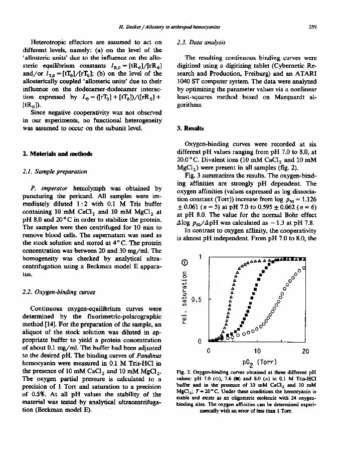
Oxygen-binding curves were recorded at six different pH values ranging from pH 7.0 to 8.0, at 20.0 “C. Divalent ions (10 mM CaCl, and 10 mM MgC12) were present in all samples (fig. 2).
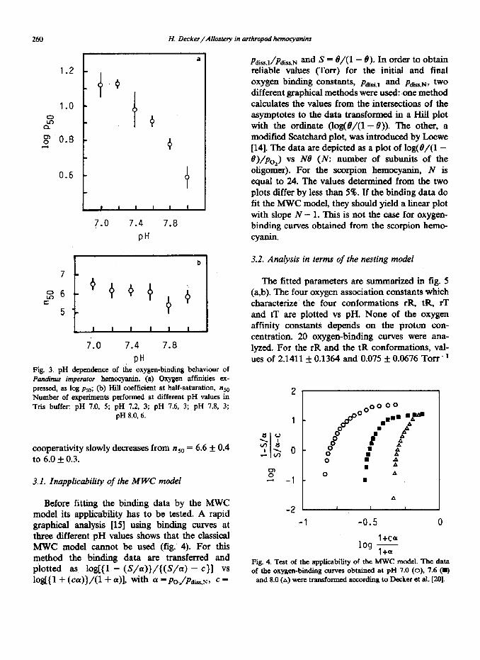
Fig. 3 su mrnarizes the results. The oxygen-bind- ing affinities are strongly pH dependent. The oxygen affinities (values expressed as log clissocia- tion constant (TOIT)) increase from log pso = 1.126 f 0.061 (n = 5) at pH 7.0 to 0.595 f 0.062 (n = 6) at pH 8.0. The value for the normal Bohr effect Alog pso/ApH was calculated as - 1.3 at pH 7.8.
In contrast to oxygen affinity, the cooperativity is almost pH independent. From pH 7.0 to 8.0, the
*&AA AA
A ~flarrAmA=
4.t .” ON
A .i 0 O0 A
A : 0" f C 0
: n
f C 8 2 : 8 t :
A ..OO . OO .OOO
&&8? I I
0 10
p02 (Tort-)
20
Fig. 2. Oxygen-binding curves obtained at three different pH vahw: pH 7.0 (0), 7.6 (=) and 8.0 (A) in 0.1 M Tris-HCI buffer and in the prcaence of 10 mM CaCl, and 10 mM MgCl,; T= 20°C. Under these. conditions the heanccyanin is stable and exists as an oligomcric molecule with 24 oxygcn- binding sites. The oxygen affinities can be determined experi-
mentaUywithanerroroflessthan1Torr.
2643 H. Decker/Allostery in arthropod hemocyanins
a
‘-* - 4 ,o 1.0 -
5: CL t 0 0" 0.8 - d 0
1 ’ ’ ’ ’ ’ ’ I 7.0 7.4 7.8
PH
7
t26 c
5 I
b
L ’ 1 I I I I
7.0 7.4 7.8
PH Fig. 3. pH dependence of the oxygen-binding behaviour of Pan&us imperator hemocyanin. (a) Oxygen affinities ex- pressed, as log psO; (b) Hill coefficient at half-saturation, nM Number of experiments performed at different pH values in Tris buffer: pH 7.0, 5; pH 7.2, 3; pH 7.6, 3; pH 7.8, 3;
pH 8.0, 6.
cooperativity slowly decreases from n 50 = 6.6 f 0.4 to 6.0 f 0.3.
3. I. Inapplicability of the M WC model
Before fitting the binding data by the MWC model its applicability has to be tested. A rapid graphical analysis [15] using binding curves at three different pH values shows that the classical MWC model cannot be used (fig. 4). For this method the binding data are transferred and plotted as, log({l - (S/a)}/{(S/a) - c}] vs log[{I + (ca))/U + a>l, with a =&/Pdis,N, C =
pdiss,JpdisrrN and S = t9/(1 - 19). In order to obtain reliable values (Torr) for the initial and final OXy@ll binding COIlShIltS, P~,J and pdi~,N, tW0
different graphical methods were used: one method calculates the values from the intersections of the asymptotes to the data transformed in a Hill plot with the ordinate (log(B/(l - 19)). The other, a modified Scatchard plot, was introduced by Loewe [14]. The data are depicted as a plot of log(8/(1- 8)/po2) vs iVB (N: number of subunits of the oligomer}. For the scorpion hemocyanin, N is equal to 24. The values determined from the two plots differ by less than 5%. If the binding data do fit the MWC model, they should yield a linear plot with slope N - 1. This is not the case for oxygen- binding curves obtained from the scorpion hemo- cyanin.
3.2. Analysis in terms of the nesting model
The fitted parameters are summarized in fig. 5 (a,b). The four oxygen association constants which characterize the four conformations rR, tR, rT and tT are plotted vs pH. None of the oxygen affinity constants depends on the proton con- centration. 20 oxygen-binding curves were ana- lyzed. For the rR and the tR conformations, val- ues of 2.14115 0.1364 and 0.075 f 0.0676 Torr-’
*1
-1 -0.5 0
l+cor lo!2 1+
a
Fig. 4. Test of the applicability of the MWC model. The data of the oxygen-binding curves obtained at pH 7.0 (0), 7.6 (q
and 8.0 (A) were transformed according to Dcckcr et at. [20].
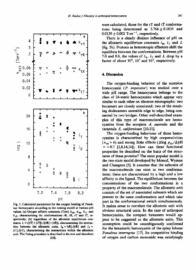
H. Decker/AIlostety in arthropod hemoqvanins 261
16
8
7.0 7.4 7.8 8.2
PH Fig. 5. Calculated parameters for the oxygen binding of Pandi-
values. (a) Oxygen affinity constants (Torr) krR, k,,, k, and k ,T, characterizing the conformations rR, tR, rT and tT, rc- spectively; @) logarithms of the allosteric equilibrium con- stants L = ([tq + [rTl)/([tR] + [rR]), characterizing the interac- tion between the allostnic units, I,= [tR]/[rR] and I, = [tTjl/[rTj, characterizing the interactions within the allosteric unit. The fitting procedure is described iu the text and elscwherc
WI.
were calculated, those for the rT and tT conforrna- tions being determined as 3.766 f 0.1835 and 0.0139 f 0.002 Torr-‘,. respectively.
There is a clearly distinct influence of pH on the allosteric equilibrium constants I,, 1, and L (fig. 5b). Protons as heterotropic effecters shift the equilibria between the conformations. Between pH 7.0 and 8.0, the values of l,, I, and L drop by a factor of about 106, lo7 and 105, respectively.
4. Discussion
The oxygen-binding behavior of the scorpion hemocyanin (P. imperator) was studied over a wide pH range. The hemocyanin belongs to the class of 24meric hemocyanins which appear very similar to each other on electron micrographs: two hexamers are closely associated; two of the result- ing dodecamers assemble edge to edge, being con- nected by two bridges. Other well-described exam- ples of this type of macromolecule are hemo- cyanins from the scorpion A. australis and the tarantula E. calijbrnicum [lO,ll].
The oxygen-binding behaviour of these hemo- cyanins is characterized by high cooperativities (nso > 6) and strong Bohr effects (Alog pso/ApH -z -0.7 [2,8,14,16]). How can these functional properties be described on the basis of the struc- tures of these proteins? The most popular model is the two-state model developed by Monod, Wyman and Changeux [5]. It assumes that the subunits of the macromolecule can exist in two conforma- tions: these are characterized by a high and a low affinity to the ligand. The equilibrium between the concentrations of the two conformations is a property of the macromolecule. The allosteric unit consists of the set of associated subunits which are present in the same conformation and which take part in the conformational switch simultaneously. It makes sense to correlate the allosteric unit with obvious structural units. In the case of arthropod hemocyanins, the compact hexamers would ap pear to be suggested as the allosteric units. This assumption could be unambiguously confirmed for the hexameric hemocyanin of the spiny lobster Panulirus interruptus [17]. Its competitive binding of oxygen and carbon monoxide was satisfyingly

262 ?I. Decker/Mastery in arthropod hemoqanins
described by the two-state model with the allo- steric unit containing six subunits. However, for 24meric arthropod hemocyanins, the simple two- state model is not applicable as shown in the present study for Pandinw hemocyanin and as previously reported for that of Ewypehna [15].
The recently introduced nesting model corre- lates the obvious hierarchies in the structures of arthropod hemocyanins with the function of these mole&es [6,7]. It has been shown in several cases [4,10,11,18] that the half-molecules of arthropod hemocyanins are the smallest structurally repeat- ing units. They are regarded as allosteric units which are nested and allosterically coupled within the native molecule.
For the 24-merit Pandinw hemocyanin, the 1Zmeric half-molecule was assumed to be the allosteric unit (ref. 9; and Decker and Heimerl, unpublished observations), which can adopt four different conformations: rR or tR when the native molecule is in the R state, and rT or tT when the native molecule is in the T state. Comparison of the calculated values for the oxygen affinities and allosteric equilibrium constants obtained for Pan- dinus and Eurypelma hemocyanins reveals a com- mon feature (table 1). The values for the affinities of the conformations appear to be conserved among cheliceratean hemocyanins. Additionally, allosteric effecters such as protons quantitatively influence the ratios between the concentrations of the conformations in the same way: when the concentration of protons is increased, the values of the allosteric equilibrium constants I,, 1, and
Table 1
Gxygen afftity w&ants calculated for Ewypelma cahforni- cum ad Pandims imperator hemocyanins according to the nesting model
The data are expressed as the association constants (TOIT-‘).
Confor- Eutypehna cal~ornieum ’ Pudnus imperator mation (n = 41) (n-20)
rR 1.990*0.070 2.141 kO.136 tR 0.049 f 0.012 0.075 iO.068
rT 3.457 f 0.056 3.766 *0.184 tT 0.014 f 0.004 0.014* 0.002
’ Values taken from ref. 8.
L also increase. The numerical values may be considered to be characteristic of the particular hemocyanin and may therefore be different. Al- though several arguments favor the nesting model [S], its validity can only be proved by the experi- mental elucidation of postulated conformations and conformational transitions. For Eurypelrna hemocyanin, four different conformations have recently been detected after labelling the hemo- cyanin with a fluorescent dye [19].
Acknowledgements
This work was supported by the Deutsche For- schungsgemeinschaft (De 414/l-10). The excellent technical assistance of Mrs R. Drechsel is grate- fully appreciated. I like to thank R. Sterner and Professor S.J. Gill for several discussions on the nesting theory and Professor H.K. MacWilliams for help with the English language.
References
K.E. Van Holde and KJ. Miller, Q. Rev. Biophys. 15 (1982) 1. C.P. Mangum, in: The biology of the crustacea, vol. 5, ed. L.H: Mantel (Academic Press, London, 1983) p. 373. H.D. Ellerton, N.F. Ellerton and H.A. Robinson, Proc. Biophys. Mol. Biol. 41 (1983) 143. J. Markl, Biol. Bull. 171 (1965) 90. J. Monod, J. Wyman and J.-P. Changcux, J. Mol. Biol. 12 (1965) 88. H. Decker, C.H. Robert and SJ. Gill, in: Invertebrate oxygen carriers, ed. B. Linzen (Springer, Berlin, 1986) p. 383. C.H. Robert, H. Decker, B. Richey, S.J. Gill and J. Wy- man, Proc. Natl. Acad. Sci. U.S.A. 84 (1987) 1891. H. Decker and R. Sterner, J. Mol. Biol. 211 (1990) 281. R. Heimerl and H. Decker, Verh. Dtsch. 2001. Gesch. 82 (1989) 226.
10 J. Lamy, M.M.C. Bijholt, P.-Y. Sizaret, J. Lamy and E.FJ. van Bnaggen, Biochemistry 20 (1981) 1849.
11 J. Ma&l, B. Kempter, B. L&en, MM-C. Bijholt aad R.F.J. van Bruggen, Hoppe-Seyler’s Z. Physiol. Chem. 362 (1981) 1631.
12 H. Decker, P.R. Connelly, C.H. Robert and SJ. Gii, Biochemistry 27 (1988) 6901.
13 J. Wyman. Adv. Protein Chcm. 19 (1964) 223. 14 R. me, J. Comp. Physiol. 128 (1978) 161. ’
H. Decker/Allostery in arthropod hemocyanins 263
15 I-I. Decker, A. Savel, B. Linzen and K.E. van Holde, Life Chem. Rep. Suppl. 1 (1983) 251.
16 J. Lamy, J. Lamy, J. Bonaventura and C. Bonaventura Biochemistry 19 (1980) 3033.
17 P.R Connelly, CR. Johnson, C.H. Robert, H.J. Bak and S.J. Gill, J. Mol. Biol. 207 (1989) 829.
18 W. Stiicker, U. Raeder, M.M.C. Bijholt, T. Wichertjes, E.F.J. van Bruggen and J. Ma&l, J. Comp. Physiol. B 158 (1988) 271.
19 T. Leidescher and H. Decker, Eur. J. Bicchem. 187 (1990) 617.

![A Preliminary Study for the Detection of Gelatinolytic ... · maurus palmatus, Androctonus australis and Pandinus imperator [10]. Almost nothing is known about components of Androctonus](https://img.pdfslide.us/doc/110x75/5ec48663998d1877571aefc5/a-preliminary-study-for-the-detection-of-gelatinolytic-maurus-palmatus-androctonus.jpg)