Embed Size (px)
DESCRIPTION
Nerve Growth Factor (NGF) Up-regulation in the Cerebrospinal Fluid of Newborns With Myelomeningocele
Citation preview
European Review for Medical and Pharmacological Sciences
894
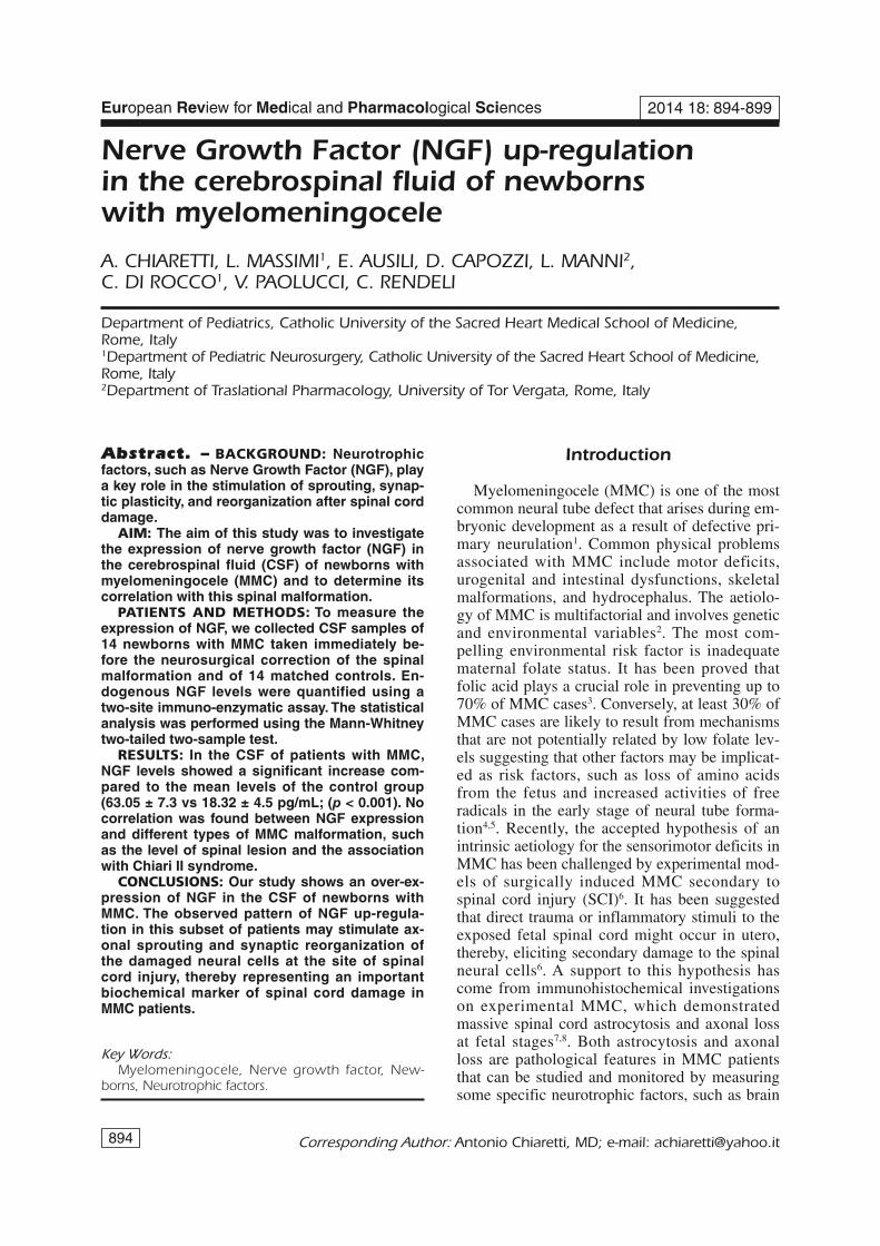
Nerve Growth Factor (NGF) up-regulationin the cerebrospinal fluid of newbornswith myelomeningocele
A. CHIARETTI, L. MASSIMI1, E. AUSILI, D. CAPOZZI, L. MANNI2,C. DI ROCCO1, V. PAOLUCCI, C. RENDELI
Department of Pediatrics, Catholic University of the Sacred Heart Medical School of Medicine,Rome, Italy1Department of Pediatric Neurosurgery, Catholic University of the Sacred Heart School of Medicine,Rome, Italy2Department of Traslational Pharmacology, University of Tor Vergata, Rome, Italy
Corresponding Author: Antonio Chiaretti, MD; e-mail: [email protected]
Introduction
Myelomeningocele (MMC) is one of the mostcommon neural tube defect that arises during em-bryonic development as a result of defective pri-mary neurulation1. Common physical problemsassociated with MMC include motor deficits,urogenital and intestinal dysfunctions, skeletalmalformations, and hydrocephalus. The aetiolo-gy of MMC is multifactorial and involves geneticand environmental variables2. The most com-pelling environmental risk factor is inadequatematernal folate status. It has been proved thatfolic acid plays a crucial role in preventing up to70% of MMC cases3. Conversely, at least 30% ofMMC cases are likely to result from mechanismsthat are not potentially related by low folate lev-els suggesting that other factors may be implicat-ed as risk factors, such as loss of amino acidsfrom the fetus and increased activities of freeradicals in the early stage of neural tube forma-tion4,5. Recently, the accepted hypothesis of anintrinsic aetiology for the sensorimotor deficits inMMC has been challenged by experimental mod-els of surgically induced MMC secondary tospinal cord injury (SCI)6. It has been suggestedthat direct trauma or inflammatory stimuli to theexposed fetal spinal cord might occur in utero,thereby, eliciting secondary damage to the spinalneural cells6. A support to this hypothesis hascome from immunohistochemical investigationson experimental MMC, which demonstratedmassive spinal cord astrocytosis and axonal lossat fetal stages7,8. Both astrocytosis and axonalloss are pathological features in MMC patientsthat can be studied and monitored by measuringsome specific neurotrophic factors, such as brain
2014 18: 894-899
Abstract. – BACKGROUND: Neurotrophicfactors, such as Nerve Growth Factor (NGF), playa key role in the stimulation of sprouting, synap-tic plasticity, and reorganization after spinal corddamage.
AIM: The aim of this study was to investigatethe expression of nerve growth factor (NGF) inthe cerebrospinal fluid (CSF) of newborns withmyelomeningocele (MMC) and to determine itscorrelation with this spinal malformation.
PATIENTS AND METHODS: To measure theexpression of NGF, we collected CSF samples of14 newborns with MMC taken immediately be-fore the neurosurgical correction of the spinalmalformation and of 14 matched controls. En-dogenous NGF levels were quantified using atwo-site immuno-enzymatic assay. The statisticalanalysis was performed using the Mann-Whitneytwo-tailed two-sample test.
RESULTS: In the CSF of patients with MMC,NGF levels showed a significant increase com-pared to the mean levels of the control group(63.05 ± 7.3 vs 18.32 ± 4.5 pg/mL; (p < 0.001). Nocorrelation was found between NGF expressionand different types of MMC malformation, suchas the level of spinal lesion and the associationwith Chiari II syndrome.
CONCLUSIONS: Our study shows an over-ex-pression of NGF in the CSF of newborns withMMC. The observed pattern of NGF up-regula-tion in this subset of patients may stimulate ax-onal sprouting and synaptic reorganization ofthe damaged neural cells at the site of spinalcord injury, thereby representing an importantbiochemical marker of spinal cord damage inMMC patients.
Key Words:Myelomeningocele, Nerve growth factor, New-
borns, Neurotrophic factors.

derived neurotrophic factor (BDNF), glial de-rived neurotrophic factor (GDNF), and nervegrowth factor (NGF). These factors play a keyrole in the stimulation of sprouting, synapticplasticity and reorganization after SCI and headinjury, providing neuroprotection and enhancingsome neuroregenerative activities9-12. NGF pro-tects the damaged neurons by promoting up-reg-ulation of doublecortin expression, and intraven-tricular NGF administration has been shown toimprove cerebral blood flow in hypoxic-ischemicbrain injury13,14. Acting on spinal cholinergicneurons NGF plays a key role also in bladderdysfunction following SCI for its action on neu-rochemical and electrophysiological reorganiza-tion of the micturition reflex15. Moreover, im-planting NGF at the site of dorsal hemisection ofthe spinal cord improves the corticospinal axonalsprouts and bladder function after spinal corddamage16. All these experimental studies demon-strated the key role of this neurotrophin in neu-ronal survival and axonal regeneration after SCI,but there are no reports on endogenous changesof NGF in the CSF of newborns with MMC.Based on those previous experiences demonstrat-ing the NGF role on neural cell differentiationand neuroprotection, the aim of this study was toinvestigate the expression of NGF in the CSF ofnewborns with MMC to determine its correlationwith this malformation.
Patients and Methods
This study evaluated the expression of NGF inthe CSF of newborns affected by MMC and ad-mitted to our Institution between January 2005and January 2012. All these infants were born atterm from non consanguineous parents, after un-complicated pregnancy and with normal birthweight. All of them underwent the neurosurgicalcorrection of the malformation within 24 h fromthe birth. As controls, CSF samples collected inthe same period from newborns who underwentlumbar puncture to rule out meningitis wereused. Patients and control subjects were matchedfor age, sex and weight, respectively. To measurethe levels of NGF, we collected CSF sampleswithdrawn from the myelomeningocelic sac im-mediately before starting the neurosurgical oper-ation. All patients were not under any medica-tions that could affect the NGF expression. AllCSF samples were centrifuged for 10 minutes at5000 rpm, and the supernatant was immediately
stored at -70°C until analysis. The study was ap-proved by the Ethical Committee of the Hospital.The parents of all patients involved providedwritten informed consent.
NGF AssaysNGF was quantified using a two-site im-
munoassay kit from Promega Corporation(Madison, WI, USA). 96-well plates were coat-ed with 100 (L/well of monoclonal anti-NGFantibody. After overnight incubation at 4°C, theantibody was removed from the plates and thesamples were incubated in coated wells (100(L/well) for 6 hours at room temperature. Theplates were then washed 5 times with buffer[0.05 M carbonate buffer (pH 9.5), 1% BSA(bovine serum albumin)] and the antigen wasincubated overnight with polyclonal anti-humanNGF antibody at 4°C. The plates were washedagain with buffer [0.05 M carbonate buffer (pH9.5), 1% BSA] and incubated with anti-chickenIgY HRP (horseradish peroxidase-conjugated)conjugate for 2 hours at room temperature. Theplates were incubated with a tetramethylbenzi-dine (TMB)/peroxidase substrate solution for 15minutes, and 1 M phosphoric acid was added(100 (l/well). The colorimetric reaction productwas measured at 450 nm. NGF concentrationswere interpolated from a NGF standard curveranging from 15.6 to 1,000 pg/ml of purifiedhuman NGF. The sensitivity of this assay was 3pg/ml and cross-reactivity with other relatedneurotrophins was less than 5%. All assays wereperformed in triplicate and NGF concentrationwas expressed as pg/mL.
Statistical AnalysisStatistical analysis of the data was performed
using StatSoft (Tulsa, OK, USA) package con-sidering the experimental conditions as a mainfactor. Analysis of variance was performed usingthe Tukey-Kramer test. The non-parametricMann-Whitney two-tailed, two-sample test wasused to perform statistical comparisons betweenchildren with MMC and the control group. Be-cause the study population was small, we did notperform multivariate analyses to adjust for the ef-fect of each parameter in the presence of the oth-ers. Spearman correlation coefficients were usedto analyze the correlations between NGF levels,age, and clinical findings of the patients. A p-val-ue of 0.05 was considered significant.
895
NGF up-regulation in myelomeningocele

896
A. Chiaretti, L. Massimi, E. Ausili, D. Capozzi, L. Manni, C. Di Rocco, V. Paolucci, C. Rendeli
Type of Level of Chiari II- Time of NGFPatients Sex lesion the lesion Hydrocephalus malformation surgery (pg/mL)
1 M MMC Lumbosacral Yes Yes Within 24 h 80.52 M MMC Lumbosacral No No Within 24 h 62.53 F MMC Lumbosacral No No Within 24 h 68.54 F MMC Lumbosacral No No Within 24 h 55.55 M MMC Lumbosacral Yes Yes Within 24 h 60.06 F MMC Lumbosacral No No Within 24 h 75.57 M MMC Lumbosacral Yes Yes Within 24 h 52.28 M MMC Lumbosacral Yes Yes Within 24 h 63.59 F MMC Lumbosacral Yes Yes Within 24 h 74.510 F MMC Lumbosacral No No Within 24 h 41.511 F MMC Lumbosacral Yes Yes Within 24 h 56.512 M MMC Lumbosacral No No Within 24 h 78.013 F MMC Lumbosacral No No Within 24 h 49.014 F MMC Lumbosacral No No Within 24 h 65.0
Table I. Clinical and demographic characteristics of newborns with myelomeningocele together with NGF levels in the CSF.
Figure 1. NGF levels in the CSF of newborns withmyelomeningocele and in controls. Data represent meanvalue (± S.D.) in pg/mL and asterisk indicates significantdifference among experimental groups (p < 0.001).
MMC compared with controls. Although wefound no correlation between NGF expressionand different types of MMC malformation, theobserved NGF up-regulation in infants withMMC may represent a reactive response to theloss and damage of astrocytes and other neuralcells at the site of MMC lesion, resulting in anincreased biosynthesis of this neurotrophin in theCSF. In experimental animal models of SCI andMMC, a moderate recovery can be found afterthe spinal lesion. This recovery can be partly at-tributed to sprouting of spared and injured axons,rostral and caudal to the lesion, due to the neuro-protective action of neurotrophins17. NGF is
Results
Between January 2005 and January 2012, 29newborns with MMC were born in our Hospitaland were admitted to our Pediatric and Neurosur-gical Departments for the treatment of their mal-formation. 14 of them (6 males and 8 females)were included in this study since they showed anunruptured myelomeningocelic sac that allowedthe analysis of uncontaminated CSF. All patientsunderwent neurosurgical operation within 24hours from the birth. The control group consistedof 14 newborns who underwent lumbar punctureto rule out meningitis. None of the controls hadevidence of meningitis (as determined by negativecultures, chemical analysis and lack of pleocyto-sis). Table I reports the clinical and demographiccharacteristics of the MMC newborns togetherwith the levels of NGF in their CSF. In newbornswith MMC the NGF levels were significantlyhigher than in controls: NGF 63.05 ± 7.3 respectto 18.32 ± 4.5 pg/mL (p < 0.001) (Figure 1). Onthe contrary, no correlation was found betweenNGF expression in the CSF and different types ofMMC malformation, such as the level of spinal le-sion and the association with Chiari II syndrome(69.5 ± 5.5 vs 60.1± 6.5 pg/mL and 64.5 ± 5.0 vs61.9 ± 5.5 pg/mL, respectively) (Figures 2 and 3).Due to the age of infants it was not possible to es-tablish any correlation between NGF expressionand motor and bladder dysfunction of patients.
Discussion
Our study demonstrates a significant increaseof NGF levels in the CSF of newborns with

jured spinal neurons showed a significant in-crease in collateral sprouting and in the numberof contacts with propriospinal interneurons thatcorrelated significantly with the functional recov-ery17,19. In addition, local infusion of NGF, earlyafter SCI, significantly improved motor functionand reduced edema formation and nerve cell,glial cell, and axonal damage, stimulating theneuroprotective mechanisms at the level of thespinal cord injury16. Moreover, the transplanta-tion with NGF-human umbilical cord blood cellsinto the spinal cord injured rat showed a positiveeffects on axonal regeneration with associatedmotor and autonomic improvement12,20,21. NGF isalso involved in the pathogenesis of lower uri-nary tract disease, especially in conditions withaltered neural function, such as neurogenic blad-der22. This neurotrophin stimulates neural plastic-ity and bladder function and these activities arerelated to its properties on altering the sodiumand potassium channels in bladder afferent fiberscausing the alteration of the micturition reflex22.Our results are in keeping with these previous ex-perimental findings confirming that the up-regu-lation of this neurotrophin participates in thepathophysiology of spinal cord injury in MMCmalformation. Although we have analyzed only14 newborns with MMC, the expression of NGFin their CSF doesn’t seem to correlate with theseverity of clinical manifestations and with thekind of spinal lesion, because NGF expressionwas not associated neither with the presence ofChiari II malformation and nor with the level ofspinal lesion in these patients. Further researches,which include a larger number of patients, willbe necessary to confirm our observation and topossibly establish if NGF determination in theCSF of infants with MMC may be an usefulmarker both of the severity of spinal cord dam-age and also for the motor and bladder dysfunc-tion of the patients. Moreover, the finding of anearly increase of NGF in the CSF of newbornswith MMC suggests that this neurotrophin can beconsidered a molecular marker of spinal corddamage and could be useful as a specific diag-nostic tool for the severity of MMC lesion and toshed light on the molecular pathogenesis ofMMC and other human spinal cord disorders.The effect of NGF in the morphogenesis of neur-al tube occurs in very early stage of life by pro-moting the growth and the differentiation of neu-roectoblasts and endoblasts20,23. The physiopatho-logical role of neuronal cells in the developing ofMMC damage has not been investigated previ-
897
NGF up-regulation in myelomeningocele
known to be one of the powerful survival factorsfor spinal neurons and several studies haveshown the increase of this factor in experimentalmodel of SCI, head trauma and meningoen-cephalitis6,18. Similarly, animals receiving NGFand other neurotrophins at the cell bodies of in-
Figure 2. NGF levels in the CSF of newborns with lumbar(L) and sacral (S) MMC lesion. Data represent mean value(± SD) in pg/mL.
Figure 3. NGF levels in the CSF of newborns with orwithout Chiari II malformation. Data represent mean value(± SD) in pg/mL

ously, but there are some experimental datashowing an astrocytic hypertrophy observed afteraxonal injury in different spinal cord damage,such as MMC and multiple sclerosis (MS), inwhich increased levels of neurotrophins are re-ported and related to motor disability and dysau-tonomyc dysfunction of the patients24,25,26.
Conclusions
According to those observations, our resultssuggest that NGF determination in the CSF ofnewborns with MMC might be used as an indexof axonal damage during the development ofspinal lesion. NGF up-regulation might be anexpression of functional mechanism aimed atstimulate axonal sprouting and synaptic reorga-nization of the damaged neural cells at the siteof spinal cord injury. Although additionalprospective and long-term studies are neededfor a better understanding of neurotrophin pat-tern modulation in newborns with MMC, webelieve that NGF and other neurotrophic fac-tors may have promising clinical applicationsin these patients.
–––––––––––––––––-––––Conflict of InterestThe Authors declare that there are no conflicts of interest.
References
1) COPP AJ, GREENE ND, MURDOCH JN. The geneticbases of mammalian neurulation. Nat Rev Genet2003; 4: 784-793.
2) JURILOFF DM, HARRIS MJ. A consideration of the evi-dence that genetic defects in planar cell polaritycontribute to the etiology of human neural tubedefects. Birth Defects Res A Clin Mol Teratol2012; 94: 824-840.
3) BOTTO L, MOORE CA, KHOURY MJ, ERICKSON JD.Neural tube defects. N Engl J Med 1999; 341:1509-1519.
4) ARSLAN M, MELEK M, DEMIR H, ESEOGLU M, GUDUBO, DEMIR I, CETIN C. Relationship of antioxidantenzyme activities with myelomeningocele. TurkNeurosurg 2012; 22: 300-304.
5) KALE A, KALE E. The role of amino acids in spinabifida. Clin Exp Obstet Gynecol 2012; 39: 374-375.
6) KASAHARA K, NAKAGAWA T, KUBOTA T. Neuronal lossand expression of neurotrophic factors in a modelof rat chronic compressive spinal cord injury.Spine 2006; 31: 2059-2066.
898
A. Chiaretti, L. Massimi, E. Ausili, D. Capozzi, L. Manni, C. Di Rocco, V. Paolucci, C. Rendeli
7) MEULI M, MEULI-SIMMEN C, YINGLING CD, HUTCHINSGM, HOFFMAN KM, HARRISON MR, ADZICK NS. Cre-ation of myelomeningocele in utero: a model offunctional damage from spinal cord exposure infetal sheep. J Pediatr Surg 1995; 30: 1028-1032.
8) VAN REGEMORTER N, GHEUENS J, NOPPE M, VAMOS E,SELLER MJ, LOWENTHAL A. Value of glial fibrillaryacidic protein determination in amniotic fluid forprenatal diagnosis of neural tube defects. ClinChim Acta 1987; 165: 83-88.
9) BRADBURY EJ, KHEMANI S, KING VR, PRIESTLEY JV,MCMAHON SB. NT-3 promotes growth of lesionedadult rat sensory axons ascending in the dorsalcolumns of the spinal cord. Eur J Neurosci 1999;11: 3873-3883.
10) CHIARETTI A, ANTONELLI A, RICCARDI R, GENOVESE O,PEZZOTTI P, DI ROCCO C, TORTOROLO L, PIEDIMONTE G.Nerve growth factor expression correlates withseverity and outcome of traumatic brain injury inchildren. Eur J Paediatr Neurol 2008; 12: 195-204.
11) HENDERSON CE, CAMU W, METTLING C, GOUIN A,POULSEN K, KARIHALOO M, RULLAMAS J, EVANS T,MCMAHON SB, ARMANNI MP, BERKEMEIER L, PHILLIPS HS,ROSENTHAL A. Neurotrophins promote motor neu-ron survival and are present in embryonic limbbud. Nature 1993; 363: 266-270.
12) TUSZYNSKI MH, MURAI K, BLESCH A, GRILL R, MILLER I.Functional characterization of NGF-secreting cellgrafts to the acutely injured spinal cord. CellTransplant 1997; 6: 361-368.
13) CHIARETTI A, ANTONELLI A, GENOVESE O, FERNANDEZ E,GIUDA D, MARIOTTI P, PULITANÒ S, PIASTRA M, POLIDORIG, COLAFATI ES, ALOE L. Intraventricular nervegrowth factor infusion improves cerebral bloodflow and stimulates doublecortin expression intwo infants with hypoxic-ischemic brain injury.Neurol Res 2008; 30: 223-228.
14) CHIARETTI A, GENOVESE O, RICCARDI R, DI ROCCO C, DIGIUDA D, MARIOTTI P, RICCARDI R. Intraventricularnerve growth factor infusion: a possible treatmentfor neurological deficits following hypoxic-is-chemic brain injury in infants. Neurol Res 2005;27: 741-746.
15) VIZZARD MA. Changes in urinary bladder neu-rotrophic factor mRNA and NGF protein followingurinary bladder dysfunction. Exp Neurol 2000;161: 273-284.
16) FERNANDEZ E, PALLINI R, LAURETTI L, MERCANTI D, SERRAA, CALISSANO P. Spinal cord transection in adultrats: effects of local infusion of nerve growth fac-tor on the corticospinal tract axons. Neurosurgery1993; 33: 889-893.
17) QIN DX, ZOU XL, LUO W, ZHANG W, ZHANG HT, LI XL,ZHANG H, WANG XY, WANG TH. Expression of someneurotrophins in the spinal motoneurons aftercord hemisection in adult rats. Neurosci Lett2006; 410: 222-227.
18) CHIARETTI A, ANTONELLI A, PIASTRA M, GENOVESE O,POLIDORI G, ALOE L. Expression of neurotrophic

899
NGF up-regulation in myelomeningocele
factors in cerebrospinal fluid and plasma of chil-dren with viral and bacterial meningoencephalitis.Acta Paediatr 2004; 93: 1178-1184.
19) SHARMA HS. Post-traumatic application of brain de-rived neurotrophic factor and glial derived neu-rotrophic factor on the rat spinal cord enhancesneuroprotection and improves motor function. Ac-ta Neurochir Suppl 2006; 96: 329-334.
20) BHARGAVA S. Role of nerve growth factor and its re-ceptor in the morphogenesis of neural tube inearly chick embryo. Gen Comp Endocrinol 2007;2: 39-43.
21) BROWN A, RICCI MJ, WEAVER LC. NGF message andprotein distribution in the injured rat spinal cord.Exp Neurol 2004; 188: 115-127.
22) STEERS WD, TUTTLE JB. Mechanisms of disease: therole of nerve growth factor in the pathophysiologyof bladder disorders. Nat Clin Pract Urol 2006; 3:101-110.
23) CAYUSO J, ULLOA F, COX B, BRISCOE J, MARTI’ E. TheSonic hedgehog pathway independently controlsthe patterning, proliferation and survival of neu-roepithelial cells by regulating Gli activity. Devel-opment 2006; 133: 517-528.
24) KALINOWSKA-ŁYSZCZARZ A, PAWLAK MA, MICHALAK S,LOSY J. Cognitive deficit is related to immune-cellbeta-NGF in multiple sclerosis patients. J NeurolSci 2012; 321: 43-48.
25) PETZOLD A, STIEFEL D, COPP AJ. Amniotic fluid brain-specific proteins are biomarkers for spinal cord in-jury in experimental myelomeningocele. J Neu-rochem 2005; 95: 594-598.
26) SEMRA YK, SEIDI OA, SHARIEF MK. Heightened in-trathecal release of axonal cytoskeletal proteinsin multiple sclerosis is associated with progres-sive disease and clinical disability. J Neuroim-munol 2002; 122: 132-139.