Embed Size (px)
Citation preview
The Cerebrospinal Fluid
Dr.Sahar Aldisi
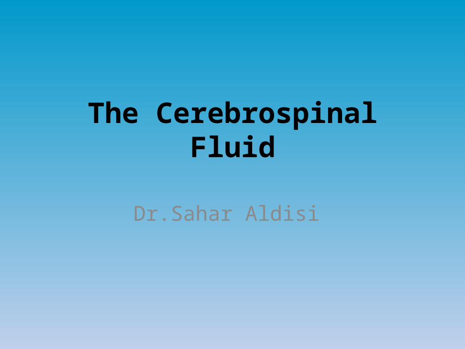
The cerebrospinal Fluid [CSF] is a clear, colorless transparent, tissue fluid present in the cerebral ventricles, spinal canal, and subarachnoid spaces.
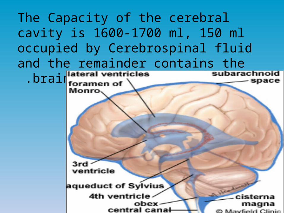
The Capacity of the cerebral cavity is 1600-1700 ml, 150 ml occupied by Cerebrospinal fluid and the remainder contains the brain and the
spinal cord .

Functions of Cerebrospinal Fluid: 1-Cushioning between the brain and cranium The main function of CSF is cushioning the brain within its solid
vault.The brain floats in the fluid due to the equal gravity between it
and the CSF .So that any blow to the head (without intensity) will move the entire brain simultaneously with the skull protecting the whole brain from being momentarily contorted.

A contrecoup is also a characteristic of CSF. It means that when a blow is intense or severe it may not damage the brain on the side of the head where the blow is struck but on the opposite side.
The reason behind that is that when the blow is struck, the fluid on that side
is so incompressible making it push the brain at the same time in union with the skull .While on the opposite side, a sudden movement of the whole skull causes the skull to pull away from the brain creating a vacuum space in the cranial vault leading to a sudden vacuum collapse and the brain strikes the inner surface of the skull when it is no longer accelerated by the blow.

2-Act as a reservoir and regulates the contents of the cranium
3-Serves as a medium for nutritional exchange in CNS4-Transport hormones and hormone releasing factors5-Removes the metabolic waste products through
absorption

CSF composition
Proteins 20-40 mg/100 mlGlucose 50-65 mg/100 mlCholesterol 0.2 mg/100 mlNa+ 147 meq/Kg H2OCa+ 2.3 meq/kg H2OUrea 12.0 mg/100 mlCreatinine 1.5 mg/100 mlLactic acid 18.0 mg/100 ml
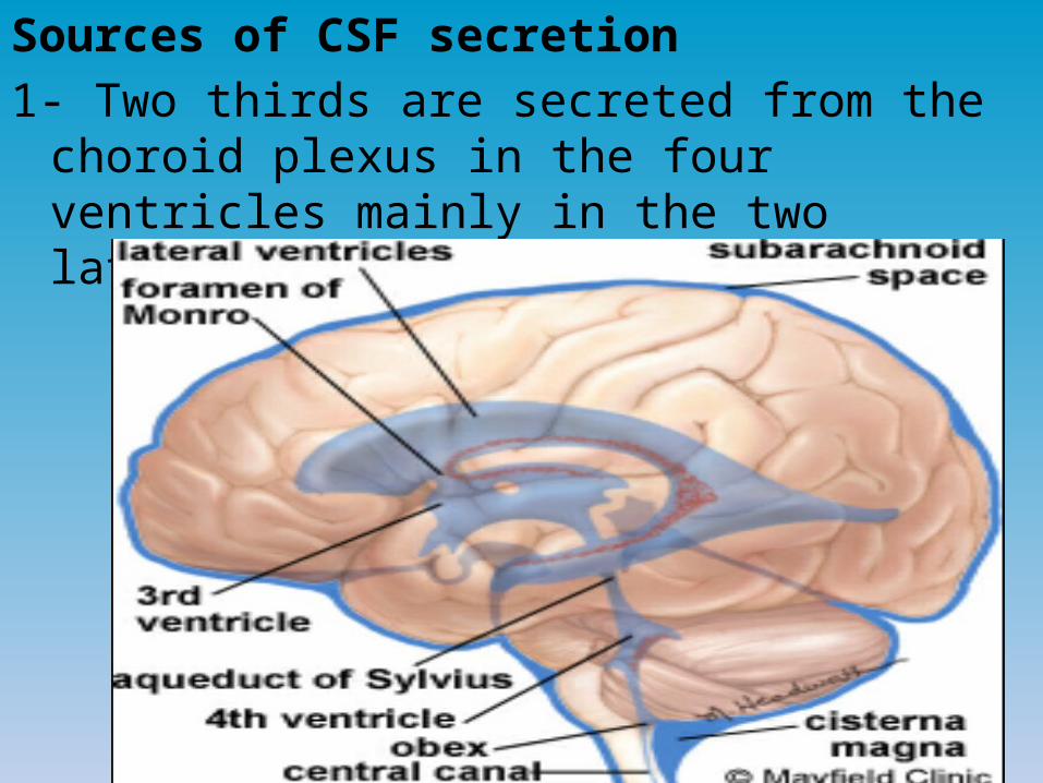
Sources of CSF secretion1- Two thirds are secreted from the choroid plexus in
the four ventricles mainly in the two lateral ones.
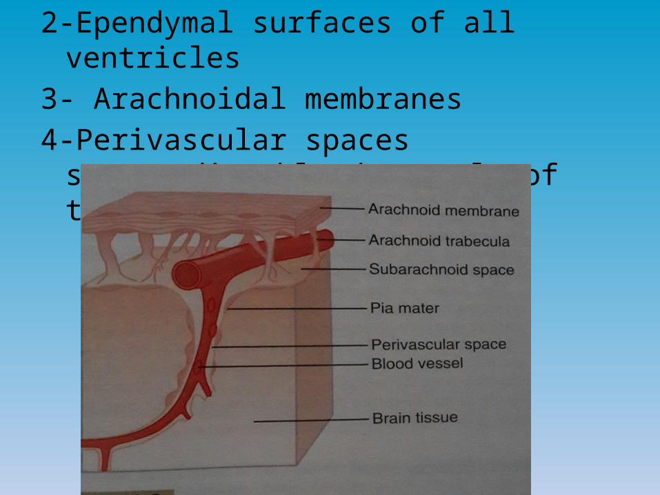
2-Ependymal surfaces of all ventricles3- Arachnoidal membranes4-Perivascular spaces surrounding blood vessels
of the brain.

Locations of CSF:1-In the ventricles of the brain.2-Cisterns around the outside of the brain.3-Subarachnoid Space around both the brain and
spinal cord. All chambers are connected with one another keeping
the pressure in all of them at a constant level.
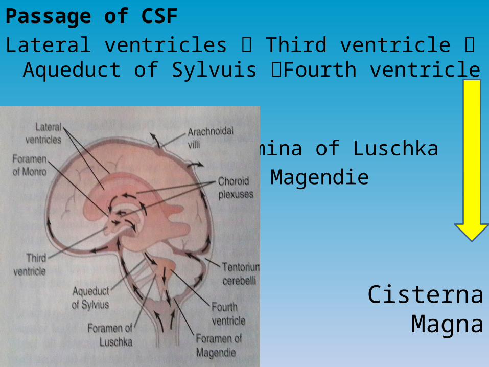
Passage of CSFLateral ventricles Third ventricle Aqueduct of
Sylvuis Fourth ventricle
Foramina of Luschka Foramina of Magendie
Cisterna Magna
Cisterna Magma (a space behind the medulla and beneath cerebellum) and is continuous with subarachnoid space surrounding the entire brain and spinal cord and the cerebrum.

Secretion depends on active transport of sodium ions through epithelial cells lining the outside of the plexus.
Sodium ions pulling large amounts of Chloride ions due to positive charge attraction of negative ions of chloride.
Thereby increasing the osmotically action of NaCl in CSF causing immediate osmosis of water through the membrane providing the fluid of secretion.
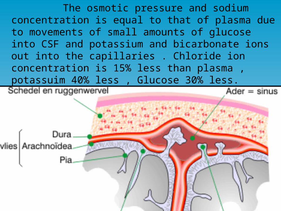
The osmotic pressure and sodium concentration is equal to that of plasma due to movements of small amounts of glucose into CSF and potassium and bicarbonate ions out into the capillaries . Chloride ion concentration is 15% less than plasma , potassuim 40% less , Glucose 30% less.
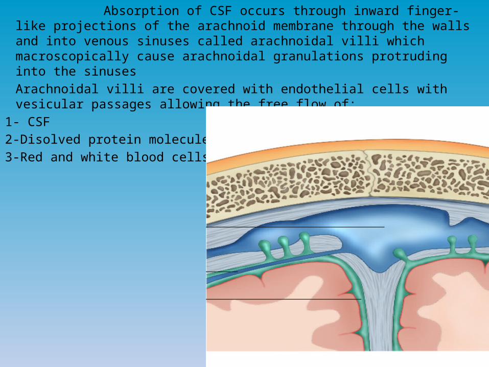
Absorption of CSF occurs through inward finger-like projections of the arachnoid membrane through the walls and into venous sinuses called arachnoidal villi which macroscopically cause arachnoidal granulations protruding into the sinuses
Arachnoidal villi are covered with endothelial cells with vesicular passages allowing the free flow of:
1- CSF2-Disolved protein molecules 3-Red and white blood cells
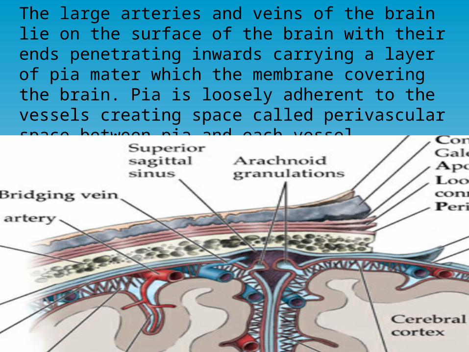
The large arteries and veins of the brain lie on the surface of the brain with their ends penetrating inwards carrying a layer of pia mater which the membrane covering the brain. Pia is loosely adherent to the vessels creating space called perivascular space between pia and each vessel.
A small amount of protein leaks out of the brain capillaries into interstitial spaces of the brain. Excess protein leaves the brain tissue flowing with fluid through perivascular spaces into subarachnoid spaces where it flows with CSF. This occurs due to absence of true lymphatics in the brain tissue to be absorbed through arachnoidal villi into large cerebral veins.
Perivascular spaces transport fluid and proteins and extraneous particulate matter out of the brain .Also, in cases of infection dead white blood cells and infection debris are transported.
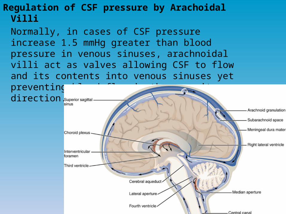
Regulation of CSF pressure by Arachoidal Villi Normally, in cases of CSF pressure
increase 1.5 mmHg greater than blood pressure in venous sinuses, arachnoidal villi act as valves allowing CSF to flow and its contents into venous sinuses yet preventing blood flow in the opposite direction.

Further increase in CSF pressure leads to wider opening of these valves, thereby preventing any rise in pressure more than few mmHg.
The pressure of CSF is increased in standing, coughing, sneezing, crying, compression of internal Jugular vein (Queckenstedt’s sign)
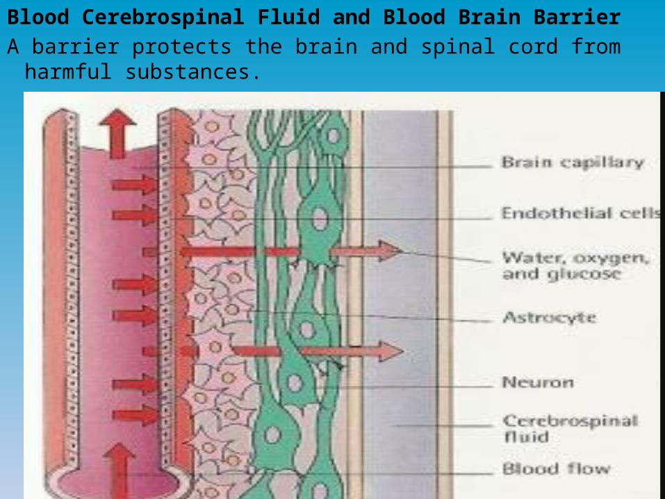
Blood Cerebrospinal Fluid and Blood Brain BarrierA barrier protects the brain and spinal cord from harmful substances.

Barrier exist both at the choroid plexus and at the tissue capillary membrane in all areas of the brain parenchyma except in some areas in the hypothalamus , pineal gland ,and area postrema , where substances infuse with greater ease in the tissue . The ease of diffusion in the areas is important because they have sensory receptors that respond to specific changes in the body fluids, such as changes in osmolarity and in glucose concentration, as well as receptors for peptide hormones that regulate thirst such
as angiotensin .

The blood-brain barrier also has specific molecules that facilitate transport of hormones such as leptin, from the blood into the hypothalamus where it binds to specific receptor that control other function such as appetite and sympathetic nervous system activity.
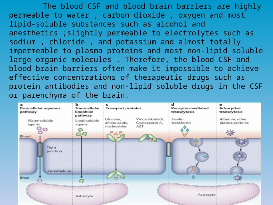
The blood CSF and blood brain barriers are highly permeable to water , carbon dioxide , oxygen and most lipid-soluble substances such as alcohol and anesthetics ;slightly permeable to electrolytes such as sodium , chloride , and potassium and almost totally impermeable to plasma proteins and most non-lipid soluble large organic molecules . Therefore, the blood CSF and blood brain barriers often make it impossible to achieve effective concentrations of therapeutic drugs such as protein antibodies and non-lipid soluble drugs in the CSF or parenchyma of the brain.
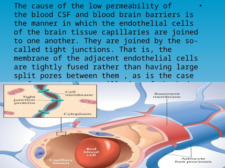
• The cause of the low permeability of the blood CSF and blood brain barriers is the manner in which the endothelial cells of the brain tissue capillaries are joined to one another. They are joined by the so-called tight junctions. That is, the membrane of the adjacent endothelial cells are tightly fused rather than having large split pores between them ,
as is the case for most other capillaries of the body .

Pathological conditions elevating CSF pressure:1-Haemorrhage or infection in the cranial vault:Where large numbers of RBCs and WBCs suddenly
appear in the CSF causing serious blockage of small absorption channels through arachnoid villi elevating pressure to 400-600 mmHg (four times normal ).
2-Large brain tumors: elevates CSF pressure by decreasing reabsorption of CSF back to blood to 500 mmHg.

High CSF pressure pathological effects:
1-Papilledema (Edema of the optic disc)
When pressure rises in the cerebrospinal fluid system, it also rises inside the optic nerve sheath .The retinal artery and vein pierce this sheath a few millimeters behind the eye and then pass alone with the optic nerve into the eye itself.
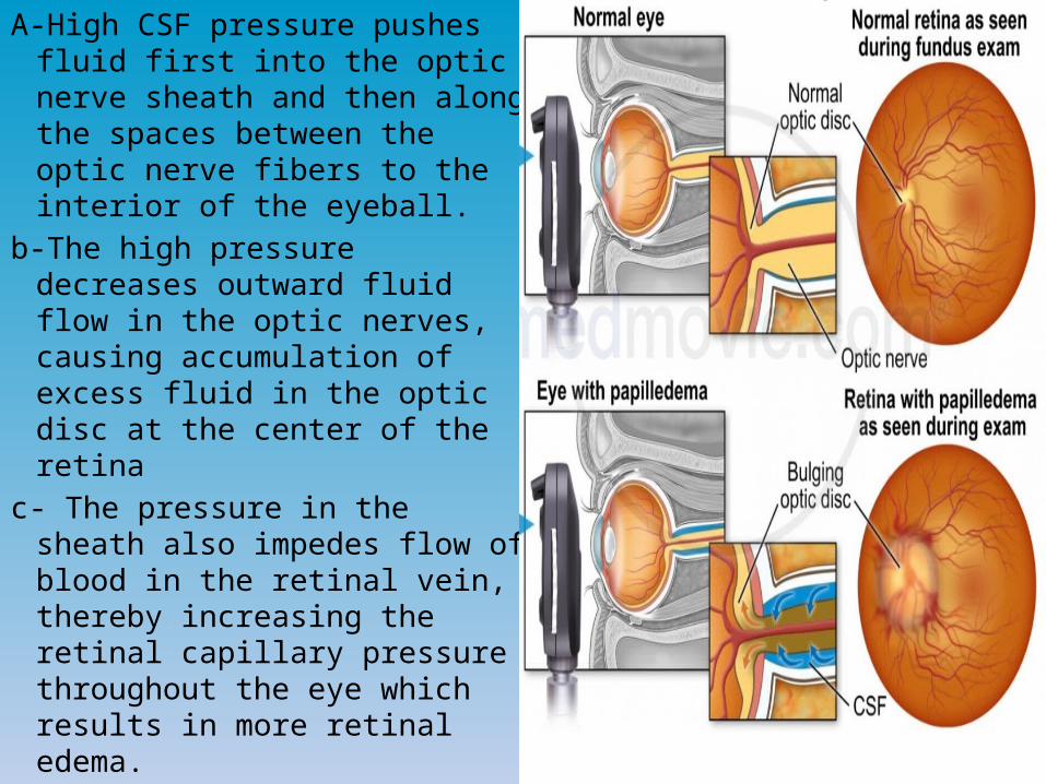
A-High CSF pressure pushes fluid first into the optic nerve sheath and then along the spaces between the optic nerve fibers to the interior of the eyeball.
b-The high pressure decreases outward fluid flow in the optic nerves, causing accumulation of excess fluid in the optic disc at the center of the retina
c- The pressure in the sheath also impedes flow of blood in the retinal vein, thereby increasing the retinal capillary pressure throughout the eye which results in more retinal edema.

2-Hydrocephalus (excess water in the cranial vault)
There are 2 types of hydrocephalus:Communicating Hydrocephalus 1.
2.Non-Communicating Hydrocephalus
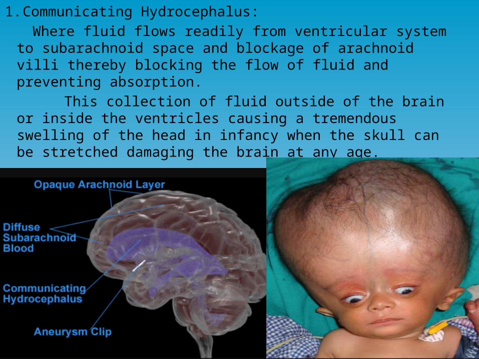
1. Communicating Hydrocephalus: Where fluid flows readily from ventricular system to
subarachnoid space and blockage of arachnoid villi thereby blocking the flow of fluid and preventing absorption.
This collection of fluid outside of the brain or inside the ventricles causing a tremendous swelling of the head in infancy when the skull can be stretched damaging the brain at any age.
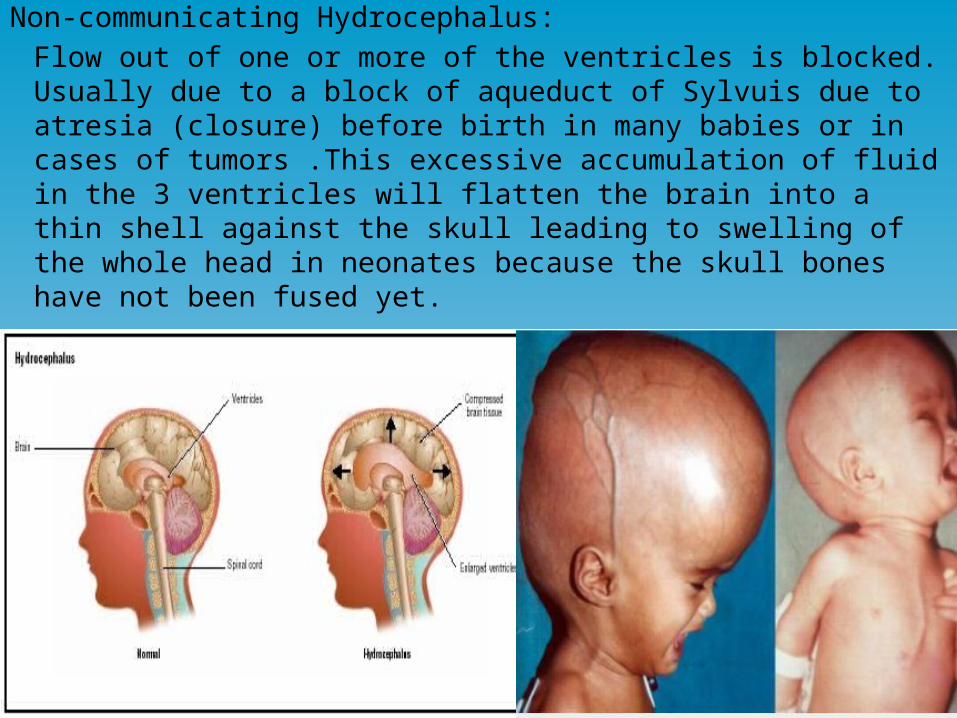
Non-communicating Hydrocephalus:Flow out of one or more of the ventricles is blocked.
Usually due to a block of aqueduct of Sylvuis due to atresia (closure) before birth in many babies or in cases of tumors .This excessive accumulation of fluid in the 3 ventricles will flatten the brain into a thin shell against the skull leading to swelling of the whole head in neonates because the skull bones have not been fused yet.
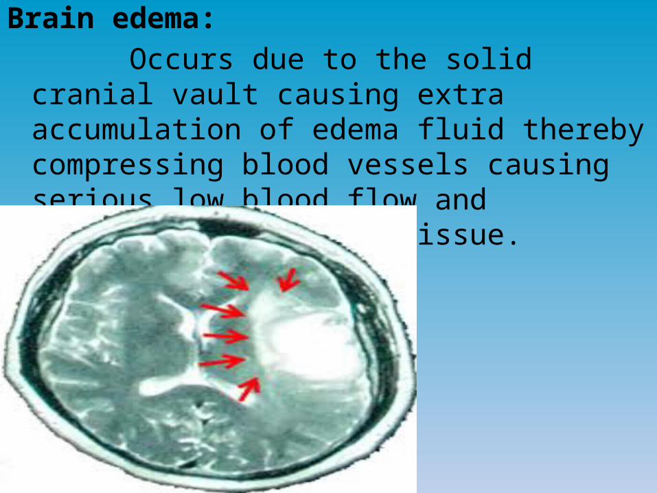
Brain edema: Occurs due to the solid cranial vault causing extra
accumulation of edema fluid thereby compressing blood vessels causing serious low blood flow and destruction of brain tissue.
Common causes:1-Increased capillary pressure 2-Damage to the capillary wall making it leaky to
the fluid 3-Serious blow to the head leading to brain
concussion

Effects of brain Edema:1-Brain Ischemia leading to arterial dilation with
further increase in capillary pressure and edema fluid thereby worsening the edema.
2-Decrease of blood flow decreasing oxygen delivery increasing the permeability of capillaries allowing more fluid leakage and turning off sodium ion pumps of neuronal tissue cells and swelling even more .

Thanks