Embed Size (px)
Citation preview
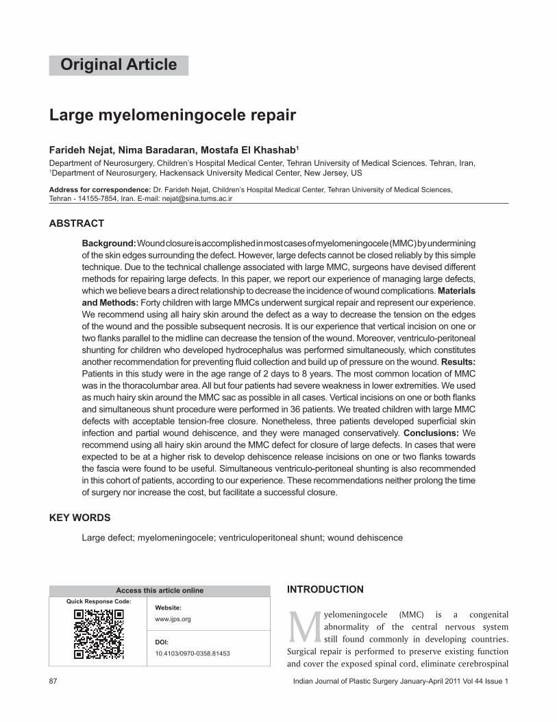
Large myelomeningocele repair
Farideh Nejat, Nima Baradaran, Mostafa El Khashab1
Department of Neurosurgery, Children’s Hospital Medical Center, Tehran University of Medical Sciences. Tehran, Iran, 1Department of Neurosurgery, Hackensack University Medical Center, New Jersey, US
Address for correspondence: Dr. Farideh Nejat, Children’s Hospital Medical Center, Tehran University of Medical Sciences, Tehran - 14155-7854, Iran. E-mail: [email protected]
ABSTRACT
Background: Wound closure is accomplished in most cases of myelomeningocele (MMC) by undermining of the skin edges surrounding the defect. However, large defects cannot be closed reliably by this simple technique. Due to the technical challenge associated with large MMC, surgeons have devised different methods for repairing large defects. In this paper, we report our experience of managing large defects, which we believe bears a direct relationship to decrease the incidence of wound complications. Materials and Methods: Forty children with large MMCs underwent surgical repair and represent our experience. We recommend using all hairy skin around the defect as a way to decrease the tension on the edges of the wound and the possible subsequent necrosis. It is our experience that vertical incision on one or two flanks parallel to the midline can decrease the tension of the wound. Moreover, ventriculo-peritoneal shunting for children who developed hydrocephalus was performed simultaneously, which constitutes another recommendation for preventing fluid collection and build up of pressure on the wound. Results: Patients in this study were in the age range of 2 days to 8 years. The most common location of MMC was in the thoracolumbar area. All but four patients had severe weakness in lower extremities. We used as much hairy skin around the MMC sac as possible in all cases. Vertical incisions on one or both flanks and simultaneous shunt procedure were performed in 36 patients. We treated children with large MMC defects with acceptable tension-free closure. Nonetheless, three patients developed superficial skin infection and partial wound dehiscence, and they were managed conservatively. Conclusions: We recommend using all hairy skin around the MMC defect for closure of large defects. In cases that were expected to be at a higher risk to develop dehiscence release incisions on one or two flanks towards the fascia were found to be useful. Simultaneous ventriculo-peritoneal shunting is also recommended in this cohort of patients, according to our experience. These recommendations neither prolong the time of surgery nor increase the cost, but facilitate a successful closure.
KEY WORDS
Large defect; myelomeningocele; ventriculoperitoneal shunt; wound dehiscence
Original Article
Access this article onlineQuick Response Code:
Website:
www.ijps.org
DOI:
10.4103/0970-0358.81453
INTRODUCTION
Myelomeningocele (MMC) is a congenital abnormality of the central nervous system still found commonly in developing countries.
Surgical repair is performed to preserve existing function and cover the exposed spinal cord, eliminate cerebrospinal
Indian Journal of Plastic Surgery January-April 2011 Vol 44 Issue 187
Nejat, et al.: Large myelomeningocele repair
fluid (CSF) leakage and prevent infection. Most of the open MMCs are small enough and for following management of the neural placode, wound closure can be accomplished by undermining of the skin and tension -free approximation in the midline. However, in 25% of the cases, the defect is large and thus is not amenable to closure by this simple technique.[1]
Large defects defined as being more than half of the width of the child’s back cannot be closed reliably by simple skin undermining. In addition to defect size, location and shape of the defect, the general status of the patient, associated kyphosis, and the condition of the surrounding tissues are other important parameters that can influence the technique and the ultimate success of the reconstruction. This, therefore, defines the appropriate method for repairing the defect.[2]
In this paper, we report our experience of with large soft tissue defect in MMCs and how we could decrease the chance of wound complications.
MATERIALS AND METHODS
In developing countries, there is no definite indication for surgery of MMC. In Iran, parents are the only support for such children with lifelong disabilities, and therefore the most accepted indication for surgery is parents’ will. After explaining the complex congenital disease, all aspects of treatment, complications and long-term problems associated with this disease, the patients are operated on according to the parents’ desire.
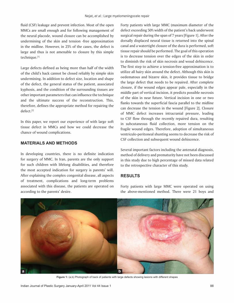
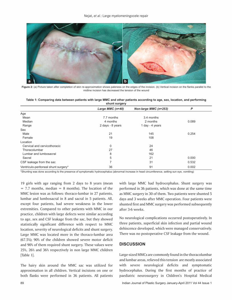
Forty patients with large MMC (maximum diameter of the defect exceeding 50% width of the patient’s back underwent surgical repair during the span of 7 years [Figure 1]. After the dorsally displaced neural tissue is returned into the spinal canal and a watertight closure of the dura is performed, soft tissue repair should be performed. The goal of this operation is to decrease tension over the edges of the skin in order to diminish the risk of skin necrosis and woud dehiscence. The first step to achieve a tension-free approximation is to utilize all hairy skin around the defect. Although this skin is oedematous and bizarre skin, it provides tissue to bridge the large defect that needs to be repaired. After complete closure, if the wound edges appear pale, especially in the middle part of vertical incision, it predicts possible necrosis of the skin in near future. Vertical incision in one or two flanks towards the superficial fascia parallel to the midline can decrease the tension in the wound [Figure 2]. Closure of MMC defect increases intracranial pressure, leading to CSF flow through the recently repaired dura, resulting in subcutaneous fluid collection, more tension on the fragile wound edges. Therefore, adoption of simultaneous ventriculo-peritoneal shunting seems to decrease the risk of CSF collection and subsequent wound dehiscence.
Several important factors including the antenatal diagnosis, method of delivery and prematurity have not been discussed in this study due to high percentage of missed data related to the retrospective character of this study.
RESULTS
Forty patients with large MMC were operated on using the above-mentioned method. There were 21 boys and
Figure 1: (a,b) Photograph of back of patients with large defects showing lesions with different shapes
a b
Indian Journal of Plastic Surgery January-April 2011 Vol 44 Issue 1 88
Nejat, et al.: Large myelomeningocele repair
19 girls with age ranging from 2 days to 8 years (mean = 7.7 months, median = 8 months). The location of the MMC lesion was as follows: thoraco-lumbar in 27 patients, lumbar and lumbosacral in 8 and sacral in 5 patients. All, except four patients, had severe weakness in the lower extremities. Compared to other patients with MMC in our practice, children with large defects were similar according to age, sex and CSF leakage from the sac, but they showed statistically significant difference with respect to MMC location, severity of neurological deficits and shunt surgery. Large MMC was located more in the thoraco-lumbar area (67.5%); 90% of the children showed severe motor deficit and 90% of them required shunt surgery. These values were 25%, 26% and 36% respectively in non large MMC children [Table 1].
The hairy skin around the MMC sac was utilized for approximation in all children. Vertical incisions on one or both flanks were performed in 36 patients. All patients
with large MMC had hydrocephalus. Shunt surgery was performed in 36 patients, which was done at the same time as MMC surgery in 30 of them. Two patients were shunted 5 days and 3 weeks after MMC operation. Four patients were shunted first and MMC surgery was performed subsequently after 3-6 weeks.
No neurological complications occurred postoperatively. In three patients, superficial skin infection and partial wound dehiscence developed, which were managed conservatively. There was no postoperative CSF leakage from the wound.
DISCUSSION
Large-sized MMCs are commonly found in the thoracolumbar and lumbar areas, relieved this tension are mostly associated with severe neurological deficits and symptomatic hydrocephalus. During the first months of practice of paediatric neurosurgery in Children’s Hospital Medical
Table 1: Comparing data between patients with large MMC and other patients according to age, sex, location, and performing shunt surgery
Large MMC (n=40) Non-large MMC (n=253) PAge
MeanMedian Range
7.7 months4 months
2 days - 8 years
3.4 months2 months
1 day - 4 years0.089
SexMaleFemale
2119
145108
0.254
LocationCervical and cervicothoracicThoracolumbarLumbar and lumbosacralSacral
02785
2446
16221 0.000
CSF leakage from the sac 7 31 0.532Ventriculo-peritoneal shunt surgery* 36 91 0.002*Shunting was done according to the presence of symptomatic hydrocephalus (abnormal increase in head circumference, setting sun eye, vomiting)
Figure 2: (a) Picture taken after completion of skin re-approximation shows paleness on the edges of the incision. (b) Vertical incision on the flanks parallel to the midline incision has decreased the tension of the wound
a b
Indian Journal of Plastic Surgery January-April 2011 Vol 44 Issue 189
Nejat, et al.: Large myelomeningocele repair
Source of Support: Nil, Conflict of Interest: None declared.
Center, in the absence of a plastic surgeon, we encountered several cases of wound dehiscence following surgery in this subset of patients. Since then, many lessons have been learned from such complicated cases for managing similar large defects in order to decrease the chance of wound problems.
Although different methods are described in the literature, closure of large MMC defects remains a challenging problem. Most of the methods described for closure of large MMCs prolong the length of surgery and anaesthesia. These methods include transposition of two skin flaps designed in an unequal Z-plasty manner, bilateral musculocutaneous flaps (based on the thoracolumbar perforators of the latissimus dorsi), en bloc medial advancement of latissimus dorsi and gluteus maximus musculocutaneous units with re-approximation in the midline and subcutaneous insertion of silicon tissue expanders.[3-6] Due to restricted dermal circulation in the newborn, extensive mobilisation of the skin is hazardous and could complicate the surgery of young infants.
We recommend some technical nuances to be employed during the surgery of large defects, which can lead to simple and efficient closure of the defects successfully without using complex surgical methods. This is particular importance to centres with restricted resources. In order to prevent wound problem in large MMC repair, we used all of the hairy skin around the MMC defect for closure (even though the colour was red or purple, and seemed to be oedematous). In cases that appeared to be at a higher risk of dehiscence after completion of wound closure and showed distinct signs of tension at the approximated wound mrgins, incision on one
or both flanks towards the fascia relieved this tension. The flank incisions could be repaired after a minimum interval of one week when the risk of dehiscence in the midline incision becomes negligible. Most lateral incisions were closed under local anaesthesia and sedation with favourable healing and aesthetics. In case of concomitant hydrocephalus, due to the ventral pressure on the wound subsequent to high dural sac pressure, simultanious ventriculo-peretoneal shunting reduced the probability of dehiscence and prevented the risk of post-operative CSF leakage from the recently repaired defect. Thus, simultaneous ventriculo-peritoneal shunting is another helpful step. These recommendations neither prolong the time of surgery nor increase the cost, thereby facilitating a successful closure.
REFERENCES
1. Erşahin Y, Yurtseven T. Delayed repair of large myelomeningoceles. Childs Nerv Syst 2004;20:427-9.
2. Akan IM, Ulusoy MG, Bilen BT, Kapucu MR. Modified bilateral advancement flap: The slide-in flap. Ann Plast Surg 1999;42: 545-8.
3. Mowatt DJ, Thomson DN, Dunaway DJ. Tissue expansion for the delayed closure of large myelomeningoceles. J Neurosurg 2005;103:544-8.
4. Ozveren MF, Erol FS, Topsakal C, Tiftikci MT, Akdemir I. The significance of the percentage of the defect size in spinal bifida cystica in determination of the surgical technique. Childs Nerv Syst 2002;18:614-20.
5. Ozçelik D, Yildiz KH, Iş M, Döşoğlu M. Soft tissue closure and plastic surgical aspects of large dorsal myelomeningocele defects (review of techniques). Neurosurg Rev 2005;28:218-25.
6. Sarifakioglu N, Bingül F, Terzioglu A, Ates L, Aslan G. Bilateral split latissimus dorsi V-Y flaps for closure of large thoracolumbar meningomyelocele defects. Br J Plast Surg 2003;56:303-6.
Indian Journal of Plastic Surgery January-April 2011 Vol 44 Issue 1 90