Embed Size (px)
Citation preview

NAVAL MEDICAL RESEARCH UNIT SAN ANTONIO TECHNICAL REPORT # TR-2012-22
COMPARISON OF NOVEL HEMOSTATIC GAUZES TO QUIKCLOT
COMBAT GAUZE IN A STANDARDIZED SWINE MODEL OF UNCONTROLLED HEMORRHAGE
Jason M. Rall, PhD1; Jennifer M. Cox2; Adam Songer, MD3; James A. Comeaux1; J. Scot Estep, DVM4; Ramon F. Cestero, MD, FACS3; James D.
Ross, PhD3
1General Dynamics Information Technology, San Antonio, TX 2Eagle Applied Sciences, L.L.C., San Antonio, TX
3Naval Medical Research Unit San Antonio, Fort Sam Houston, TX 4711th Human Performance Wing, Fort Sam Houston, TX
Naval Medical Research Unit San Antonio 3650 Chambers Pass, Bldg. 3610
Fort Sam Houston, TX 78234-6315
DISTRIBUTION A. Approved for public release; distribution is unlimited


4
Contents
ACKNOWLEDGMENTS/DISCLAIMERS ................................................................................................. 5
EXECUTIVE SUMMARY .......................................................................................................................... 6
INTRODUCTION ........................................................................................................................................ 7
Problem ..................................................................................................................................................... 7
Objective ................................................................................................................................................... 7
Background ............................................................................................................................................... 7
METHODS ................................................................................................................................................... 9
Animals ..................................................................................................................................................... 9
Surgical Procedures .................................................................................................................................. 9
Injury and Hemorrhage ............................................................................................................................. 9
Resuscitation ........................................................................................................................................... 10
Real-time Blood Collection .................................................................................................................... 11
Biochemical Analysis ............................................................................................................................. 11
Postmortem Analysis .............................................................................................................................. 11
Statistics .................................................................................................................................................. 11
RESULTS ................................................................................................................................................... 13
Pre-treatment Levels ............................................................................................................................... 13
Wound Pack Time ................................................................................................................................... 13
Hemostasis .............................................................................................................................................. 14
Blood Loss .............................................................................................................................................. 15
Resuscitation ........................................................................................................................................... 16
Coagulation ............................................................................................................................................. 16
Survival ................................................................................................................................................... 18
Morphological and Histological Assessment .......................................................................................... 19
CONCLUSIONS......................................................................................................................................... 20
REFERENCES ........................................................................................................................................... 22

5
ACKNOWLEDGMENTS
We are grateful for the technical assistance provided by Bijan Kheirabadi, Francoise Arnaud, and
Anke Scultetus. We thank the staff of our veterinary sciences group along with the pathology department
of the 711th Human Performance Wing/RHDV for their tireless and dedicated service.
DISCLAIMERS
The views expressed in this article are those of the authors and do not necessarily reflect
the official policy or position of the Department of the Navy, Department of Defense, or the U.S.
Government. Approved for public release; distribution is unlimited.
I am a military service member (government employee or contractor of the U.S.
Government). This work was prepared as part of my official duties. Title 17 U.S.C. §105
provides that ‘Copyright protection under this title is not available for any work of the United
States Government.’ Title 17 U.S.C. §101 defines a U.S. Government work as a work prepared
by a military service member or employee of the U.S. Government as part of that person’s
official duties.
The experiments reported herein were conducted in compliance with the Animal Welfare
Act and in accordance with the principles set forth in the “Guide for the Care and Use of
Laboratory Animals,” Institute of Laboratory Animals Resources, National Research Council,
National Academy Press, 1996.
This work was supported by the Defense Medical Research and Development Program
under Work Unit No. G1017.

6
EXECUTIVE SUMMARY
Problem: Uncontrolled hemorrhage is one of the leading causes of death in the battlefield. The
development, testing, and application of novel hemostatic dressings may lead to a reduction of pre-
hospital mortality through enhanced point of injury hemostatic control.
Objective: This study aimed to determine the efficacy of currently available hemostatic dressings as
compared to the current Committee for Tactical Combat Casualty Care Guidelines standard of treatment
for hemorrhage control (QuikClot Combat Gauze-QCG).
Approach: This study utilized the Department of Defense consensus swine model for uncontrolled
hemorrhage. Briefly, Yorkshire swine were anesthetized and instrumented for telemetry. Following a
femoral cut-down, a 6 mm punch injury was created in the femoral artery and free bleeding was allowed
to occur for 45 seconds. For each swine, one of five hemostatic gauzes (QCG, QuikClot Combat Gauze
XL-QCX, Celox Trauma Gauze-CTG, Celox Gauze-CEL, or ChitoGauze-HCG) was packed into the
wound site. Direct pressure (3 min) was then applied, and the animals were rapidly resuscitated to achieve
and maintain a MAP ≥ 60 mmHg for 150 minutes or until death. Animal survival, hemostasis, and blood
loss were assessed as primary endpoints.
Findings: Animals had an average weight of 36.6 ± 2.2 kg, a mean arterial pressure of 67.5 ± 8.2, and
pretreatment blood loss of 15.4 ± 3.1 ml/kg. 60% of QCG-treated animals (controls) survived through the
entire 150-minute observation period. QCX, CEL, and HCG demonstrated higher rates of survival when
compared to QCG (70%, 90%, and 70% respectively). Immediate hemostasis was achieved in 30% of
QCG applications, 80% of QCX, 70% of CEL, 60% of HCG, and 30% of CTG-treated animals. Post-
treatment blood loss varied from an average of 64 ml/kg with CTG to 29 ml/kg with CEL, but no
significant difference amongst groups was observed.
Conclusions: Novel FDA-approved hemostatic dressings exist that perform equally to the current
standard of care based on hemostasis, survival, and blood loss measured in the DoD concensus model of
swine femoral uncontrolled hemorrhage. One product, QCX was identified as outperforming the current
standard in achieving immediate hemostasis, while two products, QCX and CEL were identified as
outperforming the current standard in achieving 10-minute hemostasis. These results suggest that the
current standard for point-of-injury hemorrhage control (QCG) may need to be re-evaluated or
alternatively the standard of care expanded to include QCX, CEL, CTG and HCG.

7
INTRODUCTION
Problem Uncontrolled hemorrhage remains the most common cause of death on battlefield (1-3). The
majority of uncontrolled hemorrhage deaths are a result of injuries that are either non-compressible
(torso) or are not amenable to tourniquet (neck, groin; (1)). Many of these deaths were due to the
increased incidence of injuries sustained from the detonation of improvised explosive devices, deployed
against coalition forces in Operation Iraqi Freedom and Operation Enduring Freedom (1-3). In order to
reduce mortality from injuries resulting in uncontrolled hemorrhage, more effective means to achieve
early hemostasis must be developed and implemented. One such means, capable of mitigating
hemorrhage at point-of-injury care is hemostatic gauze.
Objective The aim of this study was to determine the efficacy of novel hemostatic gauze products as
compared to the current Committee on Tactical Combat Casualty Care (CoTCCC) standard, QuikClot
Combat Gauze (QCG; Z-Medica, Wallingford, CT) in a groin puncture model of hemorrhage in swine
(4). The model implemented in this study is an application of the United States Department of Defense
(DoD) standardized model for uncontrolled hemorrhage, described in Kheirabadi et al. 2011, based on the
recommendations of a panel of DoD medical experts who convened on June 30, 2009 (5).
Background In recent years, many new externally applied hemostatic agents have been developed that show
promise in reducing hemorrhage. These agents vary in form from gauzes and sponges to powders and
granules formulated from materials including aluminum silicates, chitosans, starches, smectite, and
proprietary formulations (6, 7). However, gauze has several aspects that make it a superior agent for
treatment of uncontrolled external hemorrhage on the battlefield. It is familiar and easily applied to self
or other casualties. Gauze is also less affected by elements such as wind or rain and is easily applied in
low-visibility conditions. Finally, gauze conforms to an injury site unlike sponges or wafers.
Currently, QuikClot Combat Gauze is the CoTCCC recommended standard hemostatic agent in
the U.S. military. QCG is a non-woven, kaolin-coated surgical gauze that has shown equal or higher
efficacy for hemorrhage control in laboratory tests when compared to other hemostatic agents including
TraumaStat (Ore-Medix, Salem, OR), Celox-D (SAM Medical, Portland, OR), and Hemcon RTS bandage
(HemCon, Portland, OR) (8-12). Kaolin is an aluminosilicate clay that activates the intrinsic coagulation

8
pathway (6, 7). QCG was observed to not produce any short term vascular damage compared to standard
gauze in an animal model (13). Finally, no adverse reactions were found during its use on the battlefield
during the Israeli Operation Cast Lead in the Gaza Strip (14).
We chose to compare four of the most promising hemostatic gauzes to QCG in a swine
uncontrolled arterial hemorrhage model. These gauzes include QuikClot Combat Gauze XL (QCX, Z-
Medica, Wallingford, CT), Celox Gauze (CEL, MedTrade Products, Crewe UK), Celox Trauma Gauze
(CTG, MedTrade Products, Crewe UK), and ChitoGauze (HCG, Hemcon, Portland, OR). QCX is similar
to QCG and only differs in that XL is 2-ply created by folding a larger piece of gauze in half during
packaging. CEL and HCG are chitosan-coated gauze dressings, while CTG is made entirely of chitosan
made flexible through its manufacturing process. As opposed to dressings that utilize kaolin, chitosan
dressings do not directly activate or stimulate the coagulation pathway, but rather promote the cross-
linking of red blood cells to form a physical barrier (6, 7). Information regarding each of the hemostatic
gauzes is summarized in Table 1.
Table 1. Characteristics of tested hemostatic gauzes Product Package Abbrev Form Size Weight Chemistry Mechanism
QuikClot Combat Gauze
QCG Z-folded gauze
3 in X 12 ft 21.4 g
Non-woven kaolin (Al-
silicate)
Activates intrinsic
coagulation
QuikClot Combat
Gauze XL
QCX Z-folded 2-ply gauze
4 in X 12 ft 49.5 g
Non-woven kaolin (Al-
silicate)
Activates intrinsic
coagulation
Celox Trauma Gauze
CTG Rolled gauze 3 in X 6 ft 19.5 g
Non-woven chitosan
fibers
Cross-links RBCs to form clot
Celox Gauze
CEL Rolled gauze 3 in X 10 ft 53.1 g
Chitosan-coated gauze
Cross-links RBCs to form clot
Hemcon ChitoGauze
HCG Z-folded gauze
3 in X 12 ft 20.1 g
Chitosan-coated gauze
Cross-links RBCs to form clot

9
METHODS All procedures involving animals were approved by Tri-Service Research Laboratory’s
Institutional Animal Care and Use Committee, Fort Sam Houston, TX. Animals were utilized in
accordance with the Guide for the Care and Use of Laboratory Animals (15).
Animals Healthy, male, Yorkshire cross-bred pigs, weighing 34-45 kg, purchased from Oak Hill Genetics
(Ewing, IL) were used in all procedures. Animals were housed on-site with enrichment and quarantined
for at least four days for acclimation prior to experimentation.
Surgical Procedures Animals were fasted for 12 hours prior to surgery, but allowed access to water ad libitum. The
animals were then sedated with 8 mg/kg Telazol (Tiletamine and Zolazepam). Buprenorphine (0.01
mg/kg IM) was administered for alleviation of pain and glycopyrrolate (0.004 mg/kg IM) to reduce
mucous secretion. Anesthesia was induced with 2-4% isoflourane in pure oxygen initially and then
decreased to 1-2% once a stable plane of anesthesia was reached. The ventilator was adjusted to maintain
an end tidal CO2 partial pressure between 38 and 42 mmHg.
The right carotid artery was cannulated via cutdown for blood sampling and invasive blood
pressure measurements. Blood pressure was continuously monitored using a Cardiocap (GE Healthcare,
Waukesha, WI). The right internal jugular was vein was cannulated for administration of resuscitation
fluids. A midline laparotomy was then performed to simulate soft tissue injury and to allow bladder
catheterization. Maintenance fluid in the form of lactated Ringer’s solution (LRS) was administered at a
rate of 5-10 ml/kg/min for a total of 10 ml/kg during surgical procedures.
Injury and Hemorrhage
Two research surgeons performed all study injuries and were blinded to the identity of the
randomly chosen test gauze until just before application. Each test gauze group consisted of ten
randomized animals. Randomization was accomplished by picking a sealed envelope that contained the
name of the test gauze by a staff member not involved in the creation of the injury or application of the
gauze. The gauze was then given to the investigator during the free bleeding portion of the experiment
immediately before application.

10
The injury procedures used in this study were developed by Kheirabadi et al. as a standardized
model for hemostatic gauze efficacy testing and has been described in detail elsewhere (5). Briefly, to
expose the femoral artery, a 10-cm incision was made in the groin above the artery. The thin overlying
adductor muscle was excised followed by careful dissection and removal of the adventitia surrounding the
artery. Finally, all small branches stemming from the artery were cauterized or suture ligated. Once all
surgical manipulations were completed and maintenance fluids were infused (lactated ringers, 10
mL/kg/hr, total 10 mL/kg).
The artery was covered
with a small piece of gauze
and bathed in 10 ml of 2%
lidocaine solution for 10
minutes to promote arterial
dilation (Figure 1). The
incision site was covered
with saline soaked gauze to
prevent drying. Following
this 10-minute stabilization period, the artery was clamped both proximally and distally using atraumatic
bulldog clamps. A 6.0-mm aortic punch (International Biophysics Corp., Austin, TX) was then used to
create an arteriotomy in the femoral artery. The clamps were then removed, and hemorrhage was allowed
to proceed unobstructed for 45 seconds, while blood was collected by suction and weighed in real time.
Next, the test article was packed quickly into the wound site along with enough cut and pre-folded Kerlix
backing to fill the cavity as determined by the applying investigator. The time taken to pack the injury
site was measured and recorded. Manual pressure was then applied for 3 minutes, followed by gentle
release. Post-injury blood was collected by vacuum suction and by pre-weighed absorbent pads for
calculation of total blood loss throughout the experiment. Hemostasis was defined as a lack of visible
blood pooling outside injury site. Immediate hemostasis was defined as hemostasis occurring within 3
minutes after compression.
Resuscitation Immediately following the 3-minute compression period, 500 ml of warmed Hextend (6%
Hetastarch, Lactated Ringer’s, 5% dextrose) was administered using a pressurized infuser bag (Ethox,
Buffalo, NY) via jugular vein catheter. Upon completion of Hextend infusion, LRS was also
administered using a pressurized infuser bag through the jugular vein catheter for resuscitation as needed
throughout the entire procedure to maintain a MAP between 60 and 65 mmHg. A maximum of 10 L of
LRS was given following the injury. Death was defined when MAP and ET CO2 fell below 20 and 15
Figure 1. Lidocaine application. Lidocaine application results in maximal dilation of target artery following surgical manipulation. Notice the increase in diameter of the post-lidocaine artery.

11
mmHg respectively and were maintained for two minutes. Animals were euthanized using Beuthasol
(Sodium Pentobarbital) after 2.5 hours or when death due to exsanguination occurred.
Real-time Blood Collection Suctioned blood was weighed with a Mettler Toledo MS6002S precision balance (Mettler
Toledo, Columbus, OH), modified with a bracket to hold a 1L suction collection bucket. Changes in
weights on the balance were recorded to file every second during the procedure and graphed for simple
visualization with custom software (BalanceChart, v1.1).
Biochemical Analysis For each animal, blood samples were was taken prior to surgical manipulation, immediately prior
to initiation of injury, then 10, 30, 60, 90, 120, and 150 minutes subsequent to the injury. Analysis
included functional coagulation (ROTEM, TEM Systems Inc, Durham, NC), CBCs using AcT Diff 2
(Beckman Coulter, Inc., Brea, CA), standard clinical coagulation panels including PT, PTT, INR,
Fibrinogen, and D-dimer using BCS XP (Siemens, Deerfield, IL), and blood gas analysis using ABL 837
Flex (Radiometer America, Westlake, OH).
Postmortem Analysis At the end of each experiment, the injured leg was moved three times in each axis to simulate
walking while looking for signs of hemorrhage. The Kerlix backing, test gauze and any pads that
captured blood were weighed to be included in blood loss calculations. Small sections (0.5 to 1.5 cm) of
the femoral artery, femoral vein, femoral nerve, and the adjacent muscle proximal to the injury site were
isolated and immediately transferred to 10% neutral buffered formalin for at least 48 hours. Tissues were
then processed into paraffin using a standard automated tissue dehydration processor, 5-7um sections
were placed on glass slides and stained with hematoxylin and eosin on a standard automated stainer. All
sections were evaluated by a board certified veterinary pathologist with experience in hemostatic bandage
research. Tissues were evaluated for injury and inflammation using a semi-quantitative scale of
0=normal, 1=minimal, 2=mild,3=moderate, 4=severe. Necropsy was performed on all animals that did
not survive the entire 150-minute observation period to determine cause of death, if present, outside of
observed exsanguination.
Statistics Differences amongst groups was considered significant when p < 0.05. Data is presented as mean
± standard deviation. Animals were excluded if their baseline MAP was < 60 mmHg or pre-treatment
blood loss < 10 mL/kg. The number of animals required in each group was determined by the likelihood
to attain hemostasis by T 10 of fluid resuscitation. Power analysis was at α = 0.05 and power of 80%.
Chi-square tests were used to determine significance amongst groups in tests with binary outcomes.

12
Analysis of variance (ANOVA) with Dunnett's multiple comparison tests were used to compare means
amongst groups. Log-rank test was used to determine significance in survival analysis. When
appropriate, data was analyzed prior to the first animal death to avoid data censoring. Data analysis was
done using Microsoft Excel 2007 (Microsoft, Redmond, WA) and SigmaPlot 12 (Systat Software, San
Jose, CA).

13
RESULTS
Pre-treatment Levels There were no statistically significant differences amongst groups with respect to pre-injury vitals
(weight, blood pressure, etc.) or hemodynamic properties (Table 2, ROTEM, blood gases, etc.). Animals
had an average weight of 36.6 ± 2.2 kg and a mean arterial pressure (MAP) of 67.5 ± 5.7. There were
also no significant differences amongst groups based on pretreatment blood loss with an average loss of
15.4 ± 3.1 ml/kg and an average rate of 11.6 ± 2.3 ml/kg/min. Hematocrit was significantly different
amongst groups by ANOVA, but Dunnett’s post-hoc analysis did not indicate any significant differences
when compared to control (QCG).
MAP, mean arterial pressure; temp, temperature; WBC, white blood cells; PT, prothrombin time; PTT, partial
thromboplastin time; QCG, QuikClot Combat Gauze; QCX, QuikClot Combat Gauze XL; CTG, Celox Trauma
Gauze; CEL, Celox Gauze; HCG, HemCon ChitoGauze
Wound Pack Time Each test gauze was packed into the injury site as rapidly as possible while still maintaining
pressure and contact with the injury site (Figure 2). The overall average time to pack was 38.8 ± 11.0
seconds with times ranging from 32.0 ± 9.2 seconds for CTG to 47.7 ± 11.3 seconds for QCX. QCG was
the second fastest packed gauze followed by HCG, CEL, and finally QCX. An ANOVA performed on
pack time revealed significant differences amongst groups (p = 0.02), but no differences compared to
QCG using Dunnet’s multiple comparison test.
Table 2. Baseline and Pretreatment Values
QCG QCX CTG CEL HCG p
Weight (kg) 36.6 ± 1.8 37.6 ± 3.0 37.0 ± 1.9 36.2 ± 2.1 35.9 ± 1.7 0.39
MAP (mmHg) 66.1 ± 7.6 64.8 ± 6.1 66.9 ± 12.2 64.0 ± 8.9 67.3 ± 5.6 0.55
Blood Loss (mL/kg) 16.2 ± 3.5 15.0 ± 3.6 16.3 ± 3.0 15.2 ± 3.0 14.4 ± 2.4 0.62
Rectal Temp (°C) 36.6 ± 1.0 36.7 ± 0.56 36.9 ± 0.83 37.0 ± 0.85 36.9 ± 0.56 0.86
Lowest MAP (mmHg) 33.4 ± 6.3 32.7 ± 6.8 29.5 ± 9.8 33.1 ± 7.0 33.0 ± 7.1 0.76
Hematocrit (%) 29.5 ± 2.1 27.2 ± 2.4 27.8 ± 2.1 30.1 ± 2.8 28.5 ± 2.8 0.04
Platelets (x103/µL) 349 ± 57 383 ± 63 311 ± 70 359 ± 48 375 ± 59 0.08
Fibrinogen (mg/dL) 208 ± 31 209 ± 22 211 ± 19 214 ± 12 216 ± 25 0.94
WBC (x103/µL) 20.0 ± 3.7 18.5 ± 4.4 18.7 ± 4.1 19.1 ± 5.9 17.7 ± 4.3 0.85
PT (sec) 11.6 ± 0.5 11.4 ± 0.5 11.3 ± 0.6 11.3 ± 0.4 11.4 ± 0.6 0.64
PTT (sec) 17.4 ± 1.0 17.5 ± 0.6 17.1 ± 1.2 17.3 ± 1.4 17.2 ± 0.8 0.94

14
Hemostasis Immediate hemostasis (no visible bleeding
from the wound during the first three minutes after
compression) ranged from 30% (3/10) of QCG-
and CTG-treated animals to 80% (8/10) of QCX
(Figure 3A). Chi-squared analysis reveals that
these differences were significant (p = 0.02).
QCG also had an additional three animals that
eventually achieved hemostasis after the immediate
hemostasis period ended, with one taking 84
minutes to achieve hemostasis. The other gauzes
had either one or two animals reach hemostasis
during the observation period except QCX. QCX,
CEL, and HCG also had incidences of re-bleeding in a wound that had previously reached hemostasis
(Figure 3B), but in only one pig in the QCX and one in the HCG group did this re-bleeding lead to the
death of the animal. Total hemostasis time was also measured (Figure 3C). This time, where there was
no visible bleeding from the wound, ranged from just over an hour for CTG (64.8 ± 72.1 minutes) to two
hours for CEL (120.5 ± 51 minutes). While a strong trend was observed in total time of hemostasis
amongst some groups, this parameter was not found to be statistically significant (p= 0.27).
Figure 2. Pack Time. The average time taken to
pack each group. Time includes both test gauze and backing. QCG, QuikClot Combat Gauze; QCX, QuikClot Combat Gauze XL; CTG, Celox Trauma Gauze; CEL, Celox Gauze; HCG, HemCon ChitoGauze
15
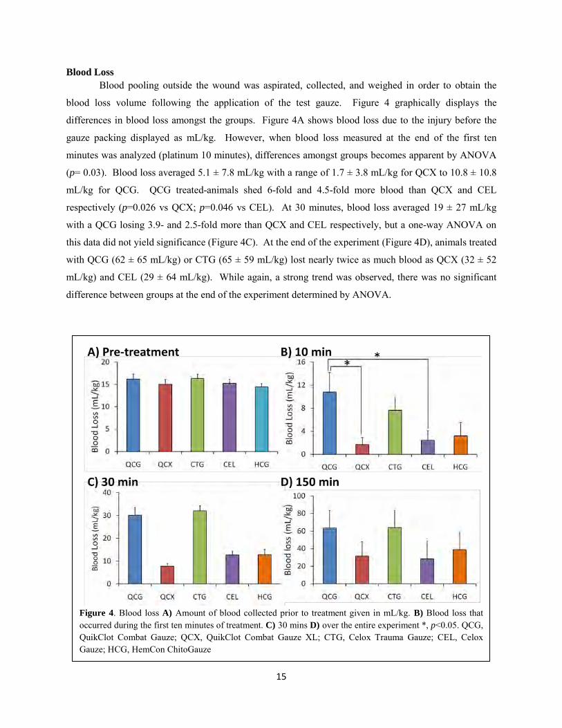
Blood Loss Blood pooling outside the wound was aspirated, collected, and weighed in order to obtain the
blood loss volume following the application of the test gauze. Figure 4 graphically displays the
differences in blood loss amongst the groups. Figure 4A shows blood loss due to the injury before the
gauze packing displayed as mL/kg. However, when blood loss measured at the end of the first ten
minutes was analyzed (platinum 10 minutes), differences amongst groups becomes apparent by ANOVA
(p= 0.03). Blood loss averaged 5.1 ± 7.8 mL/kg with a range of 1.7 ± 3.8 mL/kg for QCX to 10.8 ± 10.8
mL/kg for QCG. QCG treated-animals shed 6-fold and 4.5-fold more blood than QCX and CEL
respectively (p=0.026 vs QCX; p=0.046 vs CEL). At 30 minutes, blood loss averaged 19 ± 27 mL/kg
with a QCG losing 3.9- and 2.5-fold more than QCX and CEL respectively, but a one-way ANOVA on
this data did not yield significance (Figure 4C). At the end of the experiment (Figure 4D), animals treated
with QCG (62 ± 65 mL/kg) or CTG (65 ± 59 mL/kg) lost nearly twice as much blood as QCX (32 ± 52
mL/kg) and CEL (29 ± 64 mL/kg). While again, a strong trend was observed, there was no significant
difference between groups at the end of the experiment determined by ANOVA.
Figure 4. Blood loss A) Amount of blood collected prior to treatment given in mL/kg. B) Blood loss that occurred during the first ten minutes of treatment. C) 30 mins D) over the entire experiment *, p<0.05. QCG, QuikClot Combat Gauze; QCX, QuikClot Combat Gauze XL; CTG, Celox Trauma Gauze; CEL, Celox Gauze; HCG, HemCon ChitoGauze

16
Figure 5. Resuscitation fluids. Values shown include both Hextend and Lactated Ringer’s solution and are given as mean ± standard error. QCG, QuikClot Combat Gauze; QCX, QuikClot Combat Gauze XL; CTG, Celox Trauma Gauze; CEL, Celox Gauze; HCG, HemCon ChitoGauze
Resuscitation
In these experiments, animals were resucitated with lactated Ringer’s solution to maintain a MAP
≥ 60 mmHg. Therefore, the total amount of fluids infused can be analyzed as an indirect measure of the
success of the hemostatic agent. Fluids
infused averaged 160.2 ± 116.8 mL/kg and
ranged from 116 ± 131 for HCG to 207 ±
118 for CTG with no statistically
significant differences amongst groups
(Figure 5). Interestingly, four animals
required the full 10 L of lactated Ringer’s,
but were hemostatic and survived the
entire procedure.
Coagulation
The extent of coagulopathy is a measurable outcome of the effectiveness of each gauze (as a
function of hemostasis and the amount of resuscitation fluid delivered, Figure 6). ROTEM, prothrombin
time (PT) and partial thromboplastin time (PTT) were measured in order to determine the coagulation
state of each animal. There were no significant differences amongst the groups based on ANOVA before
or after the injury in any of the tests for coagulation. Figure 6A and B show the results of ROTEM
analysis until 60 minutes after injury. Alpha is a measure of the kinetics of clot formation, while
Maximum Clot Firmness (MCF) reflects the strength of the clot. Figure 6C and D show the results of the
prothrombin time (PT) and the partial thromboplastin time (PTT). All four tests implicate the trend that
QCX-, CEL, and HCG-treated animal’s blood performs closer to baseline than the QCG-treated animals.
However, CTG-treated animals appear to have worse coagulation parameters than the control.

17
Figure 6. Coagulation parameters. Values are shown only up to 60 minutes due to high level of animal death following 60 minute timepoint A) Alpha values obtained from ROTEM analysis indicating kinetics of clot formation. B) MCf values indicating the quality of the clot. C) Prothrombin time showing the coagulation properties of the extrinsic coagulation pathway. D) PTT illustrating the intrinsic pathway of coagulation. QCG, QuikClot Combat Gauze; QCX, QuikClot Combat Gauze XL; CTG, Celox Trauma Gauze; CEL, Celox Gauze; HCG, HemCon ChitoGauze; PT, prothrombin time; PTT, partial thromboplastin time; MCF, Maximum Clotting Firmness

18
Survival Survival varied amongst groups with 60% (6/10) of the QCG-treated animals surviving through
the entire 150 minutes of the experiment (Figure 7). CEL had the highest observed survival rate with
90% (9/10) of the animals surviving, followed by 70 % (7/10) for both QCX and HCG. CTG ranked
lowest with only half (5/10) of the treated animals surviving. However, differences amongst groups were
not significant by either log-rank test or by chi-squared analysis.
Figure 7. Kaplan-Meier analysis of surviving pigs treated with each test gauze. No significant differences were detected amongst groups (log-rank test). Inset shows survival percentage at end of the experiment.

19
Morphological and Histological Assessment
Following the completion of the experiment, the gauzes were assessed for their ability to maintain
hemostasis following a challenge of repeated leg movements by an experimenter. All gauzes tested
retained hemostasis during the leg movement challenge. However, all gauzes tested allowed for free
bleeding when gently removed from the wound site indicating the requirement for the gauze to remain in
place to be continually effective. All animals that died during the experimentation were examined by
necropsy to ensure the all deaths were due to exsanguinations and not an underlying physical condition.
No comorbidities were found any of the animals examined.
All animals were subject to histological
analysis regardless of outcome. The analysis
revealed no significant damage to any of the tissues
examined and no differences between groups. All
gauzes had some endothelial cell loss near the injury
site and minor necrosis of the muscle. There was no
apparent lesion in any of the nerve tissue examined.
However, linear foreign material was found in all
tissues in the CEL group which likely is chitosan, but
none was found inside the vessels (Figure 7). The
results are summarized in Table 4.
Table 4. Histological Evaluation Summary
QCG QCX CTG CEL HCG
Vein Mild neutrophil transmigration
Mild multifocal neutrophil
transmigration
No Significant lesions
Mild neutrophil transmigration
Moderate neutrophil
transmigration
Artery Mild,
endothelial loss at injury site
Mild endothelial loss and edema at
injury site
Endothelial loss and edema
Moderate endothelial loss
and edema
Mild endothelial loss and edema at
injury site
Nerve No Significant lesions
No Significant lesions
No Significant lesions
No Significant lesions
No Significant lesions
Muscle Mild
degeneration and necrosis
Mild degeneration and necrosis
Mild degeneration and necrosis
Mild degeneration and necrosis
Mild degeneration and necrosis
Figure 7. Representative micrograph showing foreign material on the outside of Celox gauze-treated arteries (arrowheads). Hematoxylin and eosin stained (20X).

20
CONCLUSIONS
This study compared the effectiveness of four hemostatic gauzes to the current standard
of care, QCG, using a standardized swine model of femoral arterial uncontrolled hemorrhage.
Using this model, we compared the efficacy of QCX, CEL, CTG, and HCG to QCG. These test
objects reflected the current FDA approved state of the art for hemostatic gauze technology at
the onset of this study. All test objects examined in this study performed at least as well as the
current standard of care. While some test articles excelled in specific analysis (QCX -
significantly better rate of immediate hemostasis and reduced total shed blood, CEL -
significantly reduced 10 minute shed blood) no test articles were determined to be deficient as
compared to the current standard.
Of particular note, one factor that differentiated QCX and CEL was their mass (nearly
twice the mass of other test articles, Table 1). Because QCX exhibited a higher degree of
efficacy (immediate hemostasis and 10 minute blood loss) than the traditional and smaller QCG,
it may be inferred that the performance differences observed are not necessarily due to the kaolin
content of the gauze but rather the total mass of test gauze applied. Unfortunately, this study was
not designed to address the question as to whether the differences observed were due to an
enhanced tamponade effect produced by increased gauze mass or greater quantities of active
ingredients (kaolin, chitosan). Further study may be required to address this question.
Also of note were the measured times for a test article to be fully packed into the injury
site. QCG, along with CTG, required the least amount of time while QCX and CEL required the
most (Fig 1). These differences in pack time likely result from the larger volumes of gauze
present in QCX and CEL (Table 1). Although the pack time amongst gauzes was slight (15
seconds), these differences could prove important during care under fire situations.
The investigators acknowledge that this study has some weaknesses with regard to
statistical power and in fact is largely observational. However, upon post-hoc power analysis,
we determined that in order to achieve statistical significant amongst groups, at least an
additional 15 animals per group would have been required. Therefore it was determined that the
n=10 reported here was sufficient to characterize efficacy of the test articles as compared to the
current standard. Additionally, it should be noted that the DoD standardized model for

21
uncontrolled arterial hemorrhage is, to some degree, non-physiologically relevant to
clinical/battlefield presentation of hemorrhage. The standardized model incorporates un-realistic
resuscitation strategy by design, to align gauze failure with survival outcome, and should be
interpreted in the context of testing and evaluation of gauze products in a “worst case scenario”.
The current study aimed to determine the effectiveness of five hemostatic gauzes and although a
practical way to examine these gauzes, the methodology here may not translate directly to use on
the battlefield or during emergent care. There are differences between human and swine
including blood component ratios and anatomy. Another contrived component of the model is
found in the precision of the injury, which is in stark contrast to the battlefield scenario where
one would more likely encounter a higher degree of polytrauma and hemorrhage sources not
readily amenable to gauze application. Despite these shortcomings, the work here and similar
experiments have provided valuable information as to the efficacy of modern hemostatic gauze
products.

22
REFERENCES
1. Kelly JF, Ritenour AE, McLaughlin DF, et al. Injury severity and causes of death from Operation Iraqi Freedom and Operation Enduring Freedom: 2003-2004 versus 2006. The Journal of trauma 2008;64:S21-26; discussion S26-27.
2. Holcomb J, Caruso J, McMullin N, et al. Causes of death in US Special Operations Forces in the global war on terrorism: 2001-2004. US 2007:24-37.
3. Owens BD, Kragh JF, Jr., Wenke JC, et al. Combat wounds in operation Iraqi Freedom and operation Enduring Freedom. The Journal of trauma 2008;64:295-299.
4. National Association of Emergency Medical Technicians. Prehospital Trauma Life Support, Military Edition. 2010.
5. Kheirabadi BS, Arnaud F, McCarron R, et al. Development of a standard swine hemorrhage model for efficacy assessment of topical hemostatic agents. The Journal of trauma 2011;71:S139-146.
6. Granville-Chapman J, Jacobs N, Midwinter MJ. Pre-hospital haemostatic dressings: a systematic review. Injury 2011;42:447-459.
7. Lawton G, Granville-Chapman J, Parker PJ. Novel haemostatic dressings. Journal of the Royal Army Medical Corps 2009;155:309-314.
8. Kheirabadi BS, Scherer MR, Estep JS, et al. Determination of efficacy of new hemostatic dressings in a model of extremity arterial hemorrhage in swine. The Journal of trauma 2009;67:450-459; discussion 459-460.
9. Watters JM, Van PY, Hamilton GJ, et al. Advanced hemostatic dressings are not superior to gauze for care under fire scenarios. The Journal of trauma 2011;70:1413-1419.
10. Littlejohn LF, Devlin JJ, Kircher SS, et al. Comparison of Celox-A, ChitoFlex, WoundStat, and combat gauze hemostatic agents versus standard gauze dressing in control of hemorrhage in a swine model of penetrating trauma. Acad Emerg Med 2011;18:340-350.
11. Schwartz RB, Reynolds BZ, Shiver SA, et al. Comparison of two packable hemostatic Gauze dressings in a porcine hemorrhage model. Prehosp Emerg Care 2011;15:477-482.
12. Arnaud F, Teranishi K, Okada T, et al. Comparison of Combat Gauze and TraumaStat in Two Severe Groin Injury Models. The Journal of surgical research 2011;169:92-98.
13. Kheirabadi BS, Mace JE, Terrazas IB, et al. Safety evaluation of new hemostatic agents, smectite granules, and kaolin-coated gauze in a vascular injury wound model in swine. The Journal of trauma 2010;68:269-278.
14. Ran Y, Hadad E, Daher S, et al. QuikClot Combat Gauze use for hemorrhage control in military trauma: January 2009 Israel Defense Force experience in the Gaza Strip--a preliminary report of 14 cases. Prehosp Disaster Med 2010;25:584-588.
15. Institute of Laboratory Animal Resources NRC. Guide for the Care and Use of Laboratory Animals. Washington D.C.: National Academy Press; 1996.


![[Ruaro, Antonio Francisco Umuarama] Ortopedia e Tr(BookZa.org)](https://img.pdfslide.us/doc/110x75/55cf9adc550346d033a3c33d/ruaro-antonio-francisco-umuarama-ortopedia-e-trbookzaorg.jpg)

