Embed Size (px)
Citation preview
54
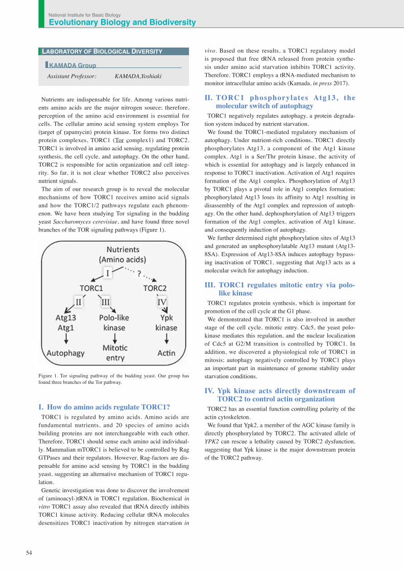
Nutrients are indispensable for life. Among various nutri-ents amino acids are the major nitrogen source; therefore, perception of the amino acid environment is essential for cells. The cellular amino acid sensing system employs Tor (target of rapamycin) protein kinase. Tor forms two distinct protein complexes, TORC1 (Tor complex1) and TORC2. TORC1 is involved in amino acid sensing, regulating protein synthesis, the cell cycle, and autophagy. On the other hand, TORC2 is responsible for actin organization and cell integ-rity. So far, it is not clear whether TORC2 also perceives nutrient signals.The aim of our research group is to reveal the molecular
mechanisms of how TORC1 receives amino acid signals and how the TORC1/2 pathways regulate each phenom-enon. We have been studying Tor signaling in the budding yeast Saccharomyces cerevisiae, and have found three novel branches of the TOR signaling pathways (Figure 1).
Figure 1. Tor signaling pathway of the budding yeast. Our group has found three branches of the Tor pathway.
I. How do amino acids regulate TORC1? TORC1 is regulated by amino acids. Amino acids are
fundamental nutrients, and 20 species of amino acids building proteins are not interchangeable with each other. Therefore, TORC1 should sense each amino acid individual-ly. Mammalian mTORC1 is believed to be controlled by Rag GTPases and their regulators. However, Rag-factors are dis-pensable for amino acid sensing by TORC1 in the budding yeast, suggesting an alternative mechanism of TORC1 regu-lation. Genetic investigation was done to discover the involvement
of (aminoacyl-)tRNA in TORC1 regulation. Biochemical in vitro TORC1 assay also revealed that tRNA directly inhibits TORC1 kinase activity. Reducing cellular tRNA molecules desensitizes TORC1 inactivation by nitrogen starvation in
vivo. Based on these results, a TORC1 regulatory model is proposed that free tRNA released from protein synthe-sis under amino acid starvation inhibits TORC1 activity. Therefore, TORC1 employs a tRNA-mediated mechanism to monitor intracellular amino acids (Kamada, in press 2017).
II. TORC1 phosphorylates Atg13, the molecular switch of autophagy
TORC1 negatively regulates autophagy, a protein degrada-tion system induced by nutrient starvation. We found the TORC1-mediated regulatory mechanism of
autophagy. Under nutrient-rich conditions, TORC1 directly phosphorylates Atg13, a component of the Atg1 kinase complex. Atg1 is a Ser/Thr protein kinase, the activity of which is essential for autophagy and is largely enhanced in response to TORC1 inactivation. Activation of Atg1 requires formation of the Atg1 complex. Phosphorylation of Atg13 by TORC1 plays a pivotal role in Atg1 complex formation; phosphorylated Atg13 loses its affinity to Atg1 resulting in disassembly of the Atg1 complex and repression of autoph-agy. On the other hand, dephosphorylation of Atg13 triggers formation of the Atg1 complex, activation of Atg1 kinase, and consequently induction of autophagy. We further determined eight phosphorylation sites of Atg13
and generated an unphosphorylatable Atg13 mutant (Atg13-8SA). Expression of Atg13-8SA induces autophagy bypass-ing inactivation of TORC1, suggesting that Atg13 acts as a molecular switch for autophagy induction.
III. TORC1 regulates mitotic entry via polo-like kinase
TORC1 regulates protein synthesis, which is important for promotion of the cell cycle at the G1 phase. We demonstrated that TORC1 is also involved in another
stage of the cell cycle, mitotic entry. Cdc5, the yeast polo-kinase mediates this regulation, and the nuclear localization of Cdc5 at G2/M transition is controlled by TORC1. In addition, we discovered a physiological role of TORC1 in mitosis; autophagy negatively controlled by TORC1 plays an important part in maintenance of genome stability under starvation conditions.
IV. Ypk kinase acts directly downstream of TORC2 to control actin organization
TORC2 has an essential function controlling polarity of the actin cytoskeleton. We found that Ypk2, a member of the AGC kinase family is
directly phosphorylated by TORC2. The activated allele of YPK2 can rescue a lethality caused by TORC2 dysfunction, suggesting that Ypk kinase is the major downstream protein of the TORC2 pathway.
LABORATORY OF BIOLOGICAL DIVERSITY
Assistant Professor: KAMADA,Yoshiaki
KAMADA Group
Evolutionary Biology and BiodiversityNational Institute for Basic Biology

55
Plant cells can induce, degenerate and differentiate their organelles to adapt to environmental changes. This flexibility of plant organelles is the basis of the strategy for environmental adaptation in plants. The aims of our research group are to clarify the molecular mech-
anisms underlying the induction, differentiation, and interaction of organelles, and to understand the integrated functions of individual plants through organelle dynamics.
I. Molecular mechanisms of peroxisome dynamics and functions in plant cells
Peroxisomes are single-membrane bounded organelles, which are ubiquitously present in eukaryotic cells, and they are involved in various biological processes such as lipid metabolism and pho-torespiration. To understand peroxisome dynamics and functions, we have been analyzing a number of Arabidopsis mutants having aberrant peroxisome morphology (apem mutants) and peroxisome unusual poisoning (peup mutants). Based on the analyses using these mutants a part of the mechanism of division, protein transport, degradation of peroxisomes, and the interactions of peroxisomes with other organelles were revealed. In addition, we found that peroxisomes are involved in the reproductive process. Therefore, peroxisome dynamics in gametes and gametophytes are currently under investigation.
II. Accumulation mechanism of seed storage oils and proteins
Plant seeds accumulate huge amounts of storage reserves such as oils, carbohydrates and proteins. Humans use these storage reserves as foods and industrial materials. Storage reserves are different among different plant seeds. Wheat, maize and rice seeds mainly accumulate starch, whereas rapeseed, pumpkin and sesame contain large amounts of oils. Soybean contains proteins as a major reserve. We are analyzing the mechanisms controlling oil and protein contents in seeds, and trying to apply our knowledge and tech-niques for increasing beneficial storage reserves (Figure 1).
III. Construction of The Plant Organelles Database 3 (PODB3)
PODB3 was built to promote a comprehensive understanding of organelle dynamics. PODB3 consists of six individual units: the electron micrograph database, the perceptive organelles database, the organelles movie database, the organellome database, the func-tional analysis database, and external links. Through these data-bases, users can obtain information on plant organelle responses to environmental stimuli of various tissues of several plant species, at different developmental stages. We expect that PODB3 will enhance the understanding of plant organelles among researchers.
Publication List: 〔Original papers〕
• Cui, S., Hayashi, Y., Otomo, M., Mano, S., Oikawa, K., Hayashi, M., and Nishimura, M. (2016). Sucrose production mediated by lipid metabolism suppresses physical interaction of peroxisomes and oil bodies during germination of Arabidopsis thaliana. J. Biol. Chem. 291, 19734-19745.
• Hosokawa, Y., Iino, T., Oikawa, K., Mano, S., Yamada, K., and Nishimura, M. (2016). Quantification of the adhesion strength between peroxisomes and chloroplasts by femtosecond laser technology. Bio-Protoc. 6, e1834.
• Kamigaki, A.*, Nito, K.*, Hikino, K., Goto-Yamada, S., Nishimura, M., Nakagawa, T., and Mano, S. (2016). Gateway vectors for simultaneous detection of multiple protein−protein interactions in plant cells using bimolecular fluorescence complementation. PLoS One 11, e0160717. (*Co-first authors)
• Kanai, M., Mano, S., Kondo, M., Hayashi, M., and Nishimura, M. (2016). Extension of oil biosynthesis during the mid-phase of seed development enhances oil content in Arabidopsis seeds. Plant Biotechnol. J. 14, 1241-1250.
• Kimori, Y., Hikino, K., Nishimura, M., and Mano, S. (2016). Quantifying morphological features of actin cytoskeletal filaments in plant cells based on mathematical morphology. J. Theor. Biol. 389, 123-131.
• Oikawa, K., Mano, S., Yamada, K., Hosokawa, Y., and Nishimura, M. (2016). Measuring the interactions between peroxisomes and chloroplasts by in situ laser analysis. Bio-Protoc. 6, e1790.
• Ueda, H., Yokota, E., Kuwata, K., Kutsuna, N., Mano, S., Shimada, T., Tamura, K., Stefano, G., Fukao, Y., Brandizzi, F., Shimmen, T., Nishimura, M., and Hara-Nishimura, I. (2016). Phosphorylation of the C-terminus of RHD3 has a critical role in homotypic ER membrane fusion in Arabidopsis. Plant Physiol. 170, 867-880.
• Watanabe, E., Mano, S., Nomoto, M., Tada, Y., Hara-Nishimura, I., Nishimura, M., and Yamada, K. (2016). HSP90 stabilizes auxin-responsive phenotypes by masking a mutation in the auxin receptor TIR1. Plant Cell Physiol. 57, 2245-2254.
〔Original paper (E-publication ahead of print)〕
• Kanai, M., Mano, S., and Nishimura, M. An efficient method for the isolation of highly purified RNA from seeds for use in quantitative transcriptome analysis. J. Vis. Exp. 2016 Aug 24.
〔Review articles〕
• Kanai, M., Mano, S., Hayashi, M., and Nishimura, M. (2016). Designing novel breeding strategies for producing high-oil crops based on the molecular understanding of triacylglycerol metabolism. In New Challenges in Seed Biology -Basic and translational research driving seed technology-, Araújo, S. and Balestrazzi, A., eds. (Croatia, InTech) pp. 137-153.
• Oikawa, K., Mano, S., Hosokawa, Y., and Nishimura, M. (2016). Analysis of physical interaction between peroxisomes and chloroplast induced by dynamic morphological changes of peroxisomes using femtosecond laser impulsive force. Plant Morphol. 28, 29-34.
LABORATORY OF BIOLOGICAL DIVERSITY
Assistant Professor: MANO, ShojiPostdoctoral Fellow: KANAI, MasatakeVisiting Scientist: WATANABE, Etsuko KAMIGAKI, AkaneTechnical Assistant: HIKINO, Kazumi KATO, Kyoko NAKAYAMA, TomomiSecretary: UEDA, Chizuru
MANO Group
Figure 1. Biosynthesis of lipids and proteins during Arabidopsis seed develop-ment and characterization of transgenic seeds overexpressing WRI1 during the middle and late phases. (A, B) Lipid biosynthesis and accumulation begins before that of seed storage proteins (SSP) during seed development. (C) A master transcription factor regulating seed oil biosynthesis, WRINKLED1 (WRI1), is expressed under the control of the FUSCA3 (FUS3) promoter, which specifically expresses during the middle phase, in the wild type (middle panel) and SSP knockout mutants (right panel). Bar: 0.5 mm.

56
LABORATORY OF BIOLOGICAL DIVERSITY
Assistant Professor: OHNO, Kaoru
OHNO Group
The aim of this laboratory is to research reproductive hormones in invertebrates, especially in echinoderms, and to analyze the mechanisms by which they work. The compari-sons of such molecules and mechanisms in various species are expected to provide insights into the evolution of repro-ductive hormone systems.
I. Gonadotropins in the starfish, Patiria pectinifera
Gonadotropins play important regulatory roles in reproduc-tion in both vertebrates and invertebrates. The vertebrate gonadotropins, LH and FSH are structurally and functionally conserved across various species, whereas no such molecule has been identified in invertebrates. The insect parsin hormones are assumed to be the physiological counterpart of LH and FSH in mammals. Some gonadotropic hormones, such as the egg development neurosecretory hormone of the mosquito, the egg-laying hormone of the sea hare, and the androgenic gland hormone of the terrestrial isopod, have been found in invertebrate species. More recently, an insulin-like peptide was reported to be responsible for the regulation of egg maturation in the mosquito, Aedes aegypti, demon-strating the involvement of insulin signaling in egg matura-tion among invertebrates.The gonad-stimulating substance (GSS) of an echinoderm,
the starfish, was the very first gonadotropin to be identified in invertebrates. GSS mediates oocyte maturation in starfish by acting on the ovary to produce the maturation-inducing hormone (MIH), 1-methyladenine, which in turn induces the maturation of the oocytes. In this sense, GSS is functionally identical to vertebrate LH, especially piscine and amphib-ian LHs, acting on the ovarian follicle cells to produce MIH to induce the final maturation or meiotic resumption of the oocyte. Considering the functional similarity that GSS shares with vertebrate LH, it is very important from an evolutionary point of view to know the chemical and molecular struc-ture of GSS. We cloned the gene encoding GSS referred to amino acid sequence of purified GSS from radial nerves of the starfish, Pateria pectinifera. Interestingly, phylogenetic analyses revealed that it belonged to the insulin/insulin-like growth factor (IGF)/relaxin superfamily and, more precisely, to the subclass of relaxin peptides (Figure 1).
Figure 1. Amino acid sequence of starfish GSS. (A) Comparison of the heterodimeric structure of starfish GSS with those of various representa-tive members of the insulin superfamily. The cysteine bridges are shown in red. (B) Coding DNA sequence and predicted amino acid sequences of GSS. Sequences of A and B chains are shown in green and blue boxes, respectively. Characters shown in red boldface indicate basic dipeptides that are the sites of proteolytic cleavage. Inverted triangle shows the deduced cleavage site of the signal peptide.
II. Search for reproductive hormones in invertebrates
In a collaborative effort with Prof. Yoshikuni’s Laboratory of Kyushu Univ. and Dr. Yamano and Dr. Awaji of the National Research Institute of Aquaculture, Fisheries Research Agency (NRIA), we are searching for reproduc-tive hormones in invertebrates; starfishes, brittle stars, sea urchins, sea cucumbers, crinoids, oyster, and shrimp. The collaborators have been able to purify physiological materi-als which induce egg maturation from nerve extracts and analyze them with a protein sequencer and a tandem mass spectrometer in the analytical center of our institute. One of them, named cubifrin, an NGIWY-amide peptide, in the sea cucumber Aposticopus japonicus, the others are in prepara-tion for publications. We have identified many neuropeptides from our EST
analysis of nerve tissues and many from RNA-seq data of the NCBI database. Especially relaxin like peptide precur-sor genes and insulin/IGF like peptide precursor genes were identified from many species. We are testing production of these neuropeptides by biological methods, e.g. bacterial systems and yeast systems, for providing to collaborators for biological assays.
Evolutionary Biology and BiodiversityNational Institute for Basic Biology

57
We have been interested in the developmental and evo-lutional aspects of the structure of mammalian brains. In a comprehensive analysis of homeobox genes expressed in the developing mouse neocortex, we isolated a novel gene Zfhx2, which encodes a transcription factor containing three homeobox domains and 18 Zn-finger motifs. Zfhx2 is highly expressed in the developing mouse brain, particularly in dif-ferentiating neurons, and continues to be expressed through-out adulthood at a low level. Two other phylogenically related genes, Zfhx3 and Zfhx4, have been identified. The former was reported to be expressed in a manner dependent on neural differentiation, and the latter is a candidate gene causing congenital bilateral isolated ptosis. Although these three genes are expressed in substantially similar patterns in the developing brain, common functional features have not been clarified. Currently we have been focusing on Zfhx2 to reveal its function and mechanisms of expression control in the developing brain.
I. Expression of Zfhx2 is negatively regulated by its own antisense RNA
We found that the antisense strand of Zfhx2 is also expressed in the mouse brain in a manner complementary to the expression of Zfhx2 mRNA (Figure 1). Although most neurons express Zfhx2 mRNA immediately after their final mitosis, several types of neuron (e.g., granule cells in the olfactory bulb and pyramidal and granule cells in the hippo-campus) express antisense RNA prior to Zfhx2 mRNA during the early phase of their differentiation. By generating a gene-targeting mouse line in which Zfhx2 sense RNA is expressed but not antisense RNA, we showed that this antisense RNA has a negative regulatory role in the expression of Zfhx2 mRNA. These observations suggest that the ZFHX2 protein might have a role in a particular step of neuronal differentia-tion, and in some types of neuron, this step might be delayed by the expression of antisense RNA.
II. ZFHX2 might play roles in controlling emotional aspects
To elucidate the function of ZFHX2, we have also gener-ated a Zfhx2-deficient mouse line. Although the produc-tion of the ZFHX2 protein is completely abolished in the homozygous mutant mice, the mice appear grossly normal and healthy. No anatomical abnormality has been observed in the mutant mouse brains so far examined. We hence sub-jected the Zfhx2-deficient mice to a comprehensive battery of behavioral tests to explore the physiological function of ZFHX2 in the nervous system. The homozygous Zfhx2 deficient mice showed several behavioral abnormalities, namely, hyperactivity (Figure 2), enhanced depression-like behaviors, and an aberrantly altered anxiety-like phenotype. These behavioral phenotypes suggest that ZFHX2 might play roles in controlling emotional aspects through the function of monoaminergic neurons where ZFHX2 is expressed.
30 60 90 1200
200400600800
10001200
Dis
tanc
e Tr
avel
ed (c
m)
Time (min)
p=0.0003
Figure 2. Locomotor activity of the Zfhx2-deficient mice. Mice were transferred into a novel environment and the distance traveled of each animal was measured for 2 hours. The Zfhx2-deficient mice (●, n=19) were significantly more active than the wild-type mice (○, n=21).
LABORATORY OF BIOLOGICAL DIVERSITY
Assistant Professor: KOMINE, Yuriko
KOMINE Group
Figure 1. Expression of Zfhx2 sense RNA (mRNA) and antisense RNA in the embryonic mouse brain. The antisense RNA was expressed where mRNA was not.

58
While genomic structures as well as their genetic informa-tion appear to stably transmit into daughter cells during cell division, and also into the next generation, they can actually vary genetically and/or epigenetically. Such variability has had a large impact on gene expression and evolution. To understand these genome dynamics in eukaryotes, especially in plants, we are characterizing the flower pigmentation of morning glories including Ipomoea nil (Japanese morning glory), I. purpurea (the common morning glory), and I. tricolor.
I. Flower pigmentation patternsThe wild type morning glories produce flowers with uni-
formly pigmented corolla, whereas a number of mutants dis-playing particular pigmentation patterns have been collected. Because flower pigmentation patterns are easily observed, the molecular mechanisms underlying these phenomena provide fine model systems for investigating genome vari-ability. The recessive mutations, duskish of I. nil and pearly-v of I.
tricolor, confer variegated flowers, and epigenetic mecha-nisms are thought to regulate their flower pigmentation. We are currently characterizing detailed molecular mechanisms of these mutations.
II. Genome sequence of the Japanese morning glory
To facilitate the studies of our group as well as all morning glory researchers, we conducted de novo genome sequencing of I. nil. We chose a standard line, Tokyo Kokei Standard, for genome sequencing, and employed shotgun sequencing using a single molecule real time sequencing system. We successfully reported an I. nil draft genome sequence with a scaffold N50 of 2.88 Mb, covering 98% of the 750 Mb genome. Scaffolds covering 91% of the genome sequence are anchored to 15 pseudo-chromosomes. From the draft genome, we could identified around 340
Tpn1 family transposons, known as the major mutagen of I. nil, and found a putative autonomous Tpn1 transposon, named TpnA1. We also identified the CONTRACTED gene located on the genetic map published in 1956. Comparative genomic analysis suggested that a whole genome duplication in Convolvulaceae, distinct from the recent whole genome duplication in Solanaceae, has occurred after the divergence of the two sister families.
III. A novel active transposon in the Japanese morning glory
InWDR1 is the multifunction transcriptional regulator of I. nil. Characterization of a medicinal herb line showing white flowers and whitish seeds revealed that the line has
the InWDR1 gene carrying an insertion of a Stowaway-like transposon, InSto1. InSto1 is the first example of an active transposon inducing spontaneous mutations other than the Tpn1 family transposons in I. nil.
IV. BioResource of morning glories NIBB is the sub-center for the National BioResource
Project (NBRP) for morning glory. In this project, we are collecting, maintaining and distributing standard lines, mutant lines for flower pigmentation, and DNA clones from EST and BAC libraries of I. nil and its related species. I. nil is one of the most popular floricultural plants in Japan, and has a 100 year history of extensive genetic studies. Our col-lections include 240 lines and 160,000 DNA clones.
a b
Figure 1. Tokyo Kokei Standard used for genome sequencing (a), and a mutant carrying contracted and star mutations (b). The CONTRACTED and STAR genes encode enzymes catalyzing brassinosteroid hormone biosynthesis.
Publication List:〔Original paper〕
• Azuma, M., Morimoto, R., Hirose, M., Morita, Y., Hoshino, A., Iida, S., Oshima, Y., Mitsuda, N., Ohme-Takagi, M., and Shiratake, K. (2016). A petal-specific InMYB1 promoter from Japanese morning glory: a useful tool for molecular breeding of floricultural crops. Plant Biotechnol. J. 14, 354-363.
• Hoshino, A., Jayakumar, V., Nitasaka, E., Toyoda, A., Noguchi, H., Itoh, T., Shin-I, T., Minakuchi, Y., Koda, Y., Nagano, A., Yasugi, M., Honjo, M., Kudoh, H., Seki, M., Kamiya, A., Shiraki, T., Carninci, P., Asamizu, E., Nishide, H., Tanaka, S., Park, K.I., Morita, Y., Yokoyama, K., Uchiyama, I., Tanaka, Y., Tabata, S., Shinozaki, K., Hayashizaki, Y., Kohara, Y., Suzuki, Y., Sugano, S., Fujiyama, A., Iida, S., and Sakakibara, Y. (2016). Genome sequence and analysis of the Japanese morning glory Ipomoea nil. Nat. Commun. 7, 13295.
• Hoshino, A., Yoneda, Y., and Kuboyama, T. (2016). A Stowaway transposon disrupts the InWDR1 gene controlling flower and seed coloration in a medicinal cultivar of the Japanese morning glory. Genes Genet. Syst. 91, 37-40.
LABORATORY OF BIOLOGICAL DIVERSITY
Assistant Professor: HOSHINO, AtsushiTechnical Assistant: NAKAMURA, Ryoko TAKEUCHI, Tomoyo ITO, Kazuyo
HOSHINO Group
Evolutionary Biology and BiodiversityNational Institute for Basic Biology

59
Although transposons occupying a large portion of the genome in various plants were once thought to be junk DNA, they play an important role in genome reorganization and evolution. Active DNA transposons are important tools for gene functional analysis. The endogenous non-autonomous transposon, nDart1-0, in rice (Oryza sativa L.) is expected to generate various transposon-insertion mutants because nDart1-0 elements tend to insert into genic regions under natural growth conditions. The transpositions of nDart1-0 were promoted by an active autonomous element, aDart1-27, on chromosome 6. By using the endogenous nDart1/aDart1-27 system in rice, a large-scale nDart-inserted mutant popu-lation was easily generated under normal field conditions, and the resulting tagged lines were free of somaclonal varia-tion. The nDart1/aDart1-27 system was introduced into a rice variety, Koshihikari, named MK-1. 3000 MK-1 plants were grown in field conditions (IPSR, Okayama Univ.) (Figure 1). The genome of all plants were isolated for identi-fying insertion sites of nDart1.
Figure 1. Mk-1 plants were grown at normal field condition. Various mutants were observed.
I. Semidominant mutation in rice The semidominant mutations produce the intermedi-
ate phenotype in individuals heterozygous for the gene concerned. The semidominant mutations were occasion-ally isolated from the MK-1, it was unclear what causes dominant mutations. Efficient selection and analysis of dominant mutants to analyze the gene functions in rice is very useful. Newly isolated, Bushy dwarf tiller2 (Bdt2), which has the valuable agronomic traits of multiple tiller-ing and dwarfism, was obtained from the MK-1 (Figure 2). Genetic analysis revealed the Bdt2 mutation was con-trolled by two genetic elements. One Bdt2 element, Bdt2a showed weak dwarf phenotype, another element, Bdt2b strengthened the effect of Bdt2a. A wild plant produces only 10 spikes or less, while Bdt2a produced 3 times as many, Bdt2 produced 10 times the ears.
Publication List:〔Original paper〕
• Gichuhi, E., Himi, E., Takahashi, H., Zhu, S., Doi, K., Tsugane, K., and Maekawa, M. (2016). Identification of QTLs for yield-related traits in RILs derived from the cross between pLIA-1 carrying Oryza longistaminata chromosome segments and Norin 18 in rice. Breed. Sci. 66, 720-733.
LABORATORY OF BIOLOGICAL DIVERSITY
Assistant Professor: TSUGANE, Kazuo
TSUGANE Group
Figure 2. Phenotype of Bushy dwarf tillers2 (Bdt2). (A) Two-month-old plants in the field (B) Segregants of Bdt2 mutants. Wild type (left), Bdt2a (middle), Bdt2(right). (C) Abnormal bract of Bdt2.

60
Chromosome condensation is a basic cellular process that ensures the faithful segregation of chromosomes in both mitosis and meiosis. This process is required not only for shrinking the length of chromosome arms, but also for resolving entanglements between sister-chromatids that are created during DNA replication. Any abnormality in this process leads to segregation errors or aneuploidy, resulting in cell lethality. Chromosome condensation is mainly achieved by condensin, a hetero-pentameric protein complex, widely conserved from yeast to humans. Despite its conservation and importance for chromosome dynamics, how condensin works is not well understood. Recent studies reveal that con-densin functions are not restricted to chromosome condensa-tion and segregation during cell divisions. It is required for diverse DNA metabolism such as genome stability, transcrip-tional regulation, and cell differentiation.Our research interest is to understand the mechanism
and regulation of chromosome condensation. We have been studying the role of condensin in the budding yeast Saccharomyces cerevisiae. Microscopic observation indi-cated the nucleolar localization of condensin. Consistent with this, the ribosomal RNA gene (rDNA) repeat is the most con-densed region in the genome during mitosis. We have found that condensin specifically binds to the RFB site located within the rDNA repeat. To date, the best characterized con-densin binding region is the rDNA repeat on the right arm of chromosome XII in budding yeast. We further discovered the multiple protein network required to recruit condensin to the RFB site.
I. Dynamic relocalization of condensin during meiosis
Our genetic screening indicated that two proteins, Csm1 and Lrs4, were required for condensin recruitment to the RFB site. Physical interactions between Csm1/Lrs4 and subunits of condensin are important for recruitment of con-densin to the RFB site. These proteins are known as com-ponents of monopolin complex that are required for faithful segregation of homologous chromosomes during meiotic division I. During meiosis I, monopolin complex re-localizes from rDNA repeat to the centromere and acts for ensuring sister-chromatid co-orientation. Re-localization of Csm1/Lrs4 proteins suggested the re-localization of condensin from rDNA repeat to centromere. As expected, chromatin-IP experiments indicated that condensin re-localizes to the centromere during meiosis I. Condensin might clamp sister-chromatids together during meiosis I.
II. Condensin-dependent chromatin foldingThe RFB site, which consists of a ~150bp DNA sequence,
is functioning as a cis-element for recruitment of condensin to chromatin in the yeast genome. If the RFB site is inserted into an ectopic chromosomal locus, condensin can associate
with the ectopic RFB site. To explore the role of condensin in chromosome organization, we have constructed a strain in which two RFB sites are inserted on an ectopic chromo-some arm with an interval of 15kb distance in the cell with complete deletion of chromosomal rDNA repeat. Using this strain, condensin-dependent chromatin interaction between two RFBs was examined by chromosome conformation capture (3C) assay. We found the condensin-dependent chro-matin interaction between the two RFB sites on the chromo-some arm. This result indicates that condensin plays a role in chromatin interaction between condensin binding sites, and this interaction leads to creation of a chromatin loop between those sites (Figure 1). It is thought that condensin-dependent chromatin folding is one of the basic molecular processes of chromosome condensation. During the cell cycle stages, the RFB - RFB interaction signal increases in metaphase and reaches its maximum level in anaphase. In addition to the RFB – RFB interaction, the chromatin interactions between internal regions of two RFBs increases in anaphase. Thus, the configuration of chromatin fiber changes from a simple loop into a complicated twisted shape as the cell cycle pro-gresses from metaphase to anaphase.
RFB
Chromatin folding
Condensin
Figure 1. A Schematic model of chromosome condensation. Condensin makes chromatin interactions between adjacent binding sites (RFB, for example). This leads to a folding of chromatin fibers between the sites, as a basic process of chromosome condensation.
LABORATORY OF BIOLOGICAL DIVERSITY
Assistant Professor: JOHZUKA, KatsukiTechnical Assistant: ISHINE Naomi
JOHZUKA Group
Evolutionary Biology and BiodiversityNational Institute for Basic Biology

61
Organogenesis is accomplished by a series of deformations of the planar cell sheet into a three-dimensional shape during embryogenesis. This drastic structural change is an integrated result of individual cell behaviors in response to spatio-tem-porally controlled mechanisms. To better understand the programs underlying organ forma-
tion, it is required to analyze individual cells’ morphology and dynamics quantitatively. However, due to the massive images generated by 4D microscopy and their ambiguity, this made it difficult to perform these analyses. To unveil organogenesis from the point of view of distinct
cell behaviors, we are developing application software that is capable of describing cell dynamics out of 4D time-lapse imaging data sets by employing image processing tech-niques.
I. 4D cell segmentation/tracking systemEpithelial morphogenesis in the developing embryo is
considered to be an essential model for collective cell migrations. Drastic cell rearrangements lead drastic struc-tural changes to build elaborate organs such as the tubular network of Drosophila trachea. We are developing a software pipeline, which automatically recognizes individual cell shapes out of 3D space and tracks them through time. This system extracts cell boundaries and reconstitutes cell shapes as a skeletonized chain of voxels spanning 3D space. This abstract form of cell visualization makes it possible to describe morphometric quantities and kinetics of cells in single-cell resolution (Figure 1). These morphometric quanti-ties allow us to perform comparative studies on shapes and behaviors precisely among several experimental conditions, to gain a better understanding of the genetic programs under-lying organogenesis. We are now applying this system to several experimental models to determine the practicality of the system.
Figure 1. Visualized apical cell surface of Drosophila embryonic epi-dermal cells. A time-lapse confocal microscopic data set of a fly embryo expressing E-cadherin-GFP was subjected to our automatic cell shape recognition system. Cell boundaries (green), center of gravity (blue) and normal vector (magenta) are indicated for each cell.
II. Particle tracking for tissue deformation analysis
Besides cell boundary extraction, we also developed a
derived algorithm for particle image velocimetry (PIV). This system is designed to measure tissue deformation even though the imaging constraints do not allow identification of individual cells out of images. This implementation detects structural characteristics, such as uneven fluorescence dis-tributed over the specimen and tracks these patterns along a time-series. Despite that the tissue was labeled with non-targeted cytoplasmic GFP, this tracking software successfully outlined developmental dynamics of Xenopus neuroectoderm (Figure 2).
Figure 2. Collective cell migration of Xenopus neuroectodermal cells visualized as optical flow along a time-series. A modified PIV method successfully tracks uneven subcellular distribution of GFP signals over time. Dr. M. Suzuki (Prof. Ueno’s laboratory at NIBB) performed the microscopy.
III. A GUI application for manual image quantification
Biologically significant image features are not always sig-nificant to computational algorithms due to their structural instability. This kind of difficulty requires human eye inspec-tion for feature extractions. A GUI (Graphical User Interface) application we developed can easily visualize 4D imaging data and has made manual feature annotations easy (Figure 3). This application is freely available at our website (https://is.cnsi.nins.jp/).
Figure 3. A lightweight/native 4D stack viewer equipped with functions for manual feature extraction.
Publication List:〔Original Paper〕
• Kato, K., Dong, B., Wada, H., Tanaka-Matakatsu, M., Yagi, Y., and Hayashi, S. (2016). Microtubule-dependent balanced cell contraction and luminal-matrix modification accelerate epithelial tube fusion. Nat. Commun. 7, 11141.
LABORATORY OF BIOLOGICAL DIVERSITY
Specially Appointed Assistant Professor: KATO, Kagayaki
KATO Group
62
Image processing methods significantly contribute to visualization of biomedical targets acquired from a variety of imaging techniques, including: wide-field optical and electron microscopy, X-ray computed tomography, magnetic resonance imaging and mammography. Quantitative inter-pretation of the deluge of complicated biomedical images, however, poses many research challenges. We have devel-oped new computational methods based on mathematical morphology for quantitative image analysis. One of the most important purposes of image processing is to derive mean-ingful information, which is expressed as image structural properties. Mathematical morphology is a nonlinear image processing method based on set theory and is useful for the extraction of the structural properties from an image. It can be used as a fundamental tool to analyze biomedical images.
Novel image segmentation method based on mathematical morphologyImage processing is a crucial step in the quantification of
biomedical imaging data. As such, it is fundamental to a wide range of biomedical imaging fields. Image process-ing derives structural features, which are then numerically quantified by image analysis. Image segmentation plays an important role in image processing. Segmentation is the par-titioning of an image into sub-regions and the extraction of a target that is to be analyzed.In this study, an image segmentation approach based on
a new type of mathematical morphology is introduced. Mathematical morphology is a methodology for extracting shape and size information from an image. It involves con-figuration of a set of nonlinear operators that act on images by using structuring elements (SE). The SE, which indicates the shape characteristics in an image, is generally a small and simple binary image. The two basic morphological operators are dilation and erosion, from which many operations can be derived.This segmentation method is based on a double mor-
phological subtraction method. Generally, morpho-logical subtraction methods may include top-hat transform, rolling-ball transform, and h-maxima transform. These methods extract target objects that are brighter than the surrounding areas. However, these methods may also extract unwanted structures which are located in the neighborhood of the target object. Thus, an identification process between the target regions and other object regions is required. Commonly, this task is difficult due to intensity and size similarities among these extracted regions.In this new type of morphological segmentation method,
h-maxima transform is applied twice to the original image. The unwanted structures that surround the target are sup-pressed in the process of target segmentation. Furthermore, this new method has no restrictions on the size and shape of
the target object that is to be segmented. This method was applied to segmentation of an abnormal
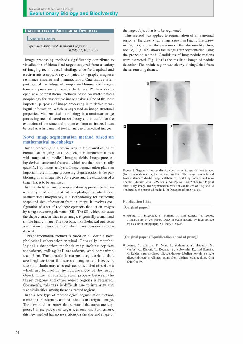
region in the chest x-ray image shown in Fig. 1. The arrow in Fig. 1(a) shows the position of the abnormality (lung nodule). Fig. 1(b) shows the image after segmentation using the proposed method. Candidates of lung nodule regions were extracted. Fig. 1(c) is the resultant image of nodule detection. The nodule region was clearly distinguished from the surrounding tissues.
Figure 1. Segmentation results for chest x-ray image: (a) test image. (b) Segmentation using the proposed method. The image was obtained from a standard digital image database of chest lung nodules and non-nodules (Shiraishi et al., ARJ Am. J. Roentgenol. 174, 2000). (a) Original chest x-ray image. (b) Segmentation result of candidates of lung nodule obtained by the proposed method. (c) Detection of lung nodule.
Publication List:〔Original paper〕
• Murata, K., Hagiwara, S., Kimori, Y., and Kaneko, Y. (2016). Ultrastructure of compacted DNA in cyanobacteria by high-voltage cryo-electron tomography. Sci. Rep. 6, 34934.
〔Original paper (E-publication ahead of print)〕
• Osanai, Y., Shimizu, T., Mori, T., Yoshimura, Y., Hatanaka, N., Nambu, A., Kimori, Y., Koyama, S., Kobayashi, K., and Ikenaka, K. Rabies virus-mediated oligodendrocyte labeling reveals a single oligodendrocyte myelinates axons from distinct brain regions. Glia 2016 Oct 19.
LABORATORY OF BIOLOGICAL DIVERSITY
Specially Appointed Assistant Professor: KIMORI, Yoshitaka
KIMORI Group
National Institute for Basic BiologyEvolutionary Biology and Biodiversity