Embed Size (px)
Citation preview

Organization of Cytoskeletal Elements and Organelles Preceding Growth Cone Emergence from an Identified Neuron In Situ Frances Lefcort* and David Bentley * Neurobiology Group and Department of Zoology, University of California, Berkeley, California 94720
Abstract. The purpose of this study was to investigate the arrangement of cytoskeletal elements and organ- elles in an identified neuron in situ at the site of emer- gence of its growth cone just before and concurrent with the onset of axonogenesis. The Til pioneer neu- rons are the first pair of afferent neurons to differenti- ate in embryonic grasshopper limbs. They arise at the distal tip of the limb bud epithelium, the daughter cells of a single precursor cell, the Pioneer Mother Cell (PMC). Using immunohistochemical markers, we characterized the organization of microtubules, centro- somes, Golgi apparatus, midbody, actin filaments, and chromatin from mitosis in the PMC through axono- genesis in the Tils. Just before and concurrent with the
onset of axonogenesis, a characteristic arrangement of tubulin, actin filaments, and Golgi apparatus is local- ized at the proximal pole of the proximal pioneer neu- ron. The growth cone of the proximal cell stereotypi- cally arises from this site. Although the distal cell's axon generally grows proximally, occasionally it arises from its distal pole; in such limbs, the axons from the sister cells extend from mirror symmetric locations on their somata. In the presence of cytochalasin D, the PMC undergoes nuclear division but not cytokinesis and although other neuronal phenotypes are expressed, axonogenesis is inhibited. Our data suggest that intrin- sic information determines the site of growth cone emergence of an identified neuron in situ.
T HE factors that determine the site, on a cell body, from which a growth cone will emerge remain unidentified. Certain intracellular components such as centrosomes
or microtubule organizing centers (MTOCs) ~ have been postulated to play a major role in the determination of cell morphology by inducing an asymmetry in the distribution of one of the principal cytoskeletal elements, the microtubules (for review see Mclntosh, 1983; Brinkley et al., 1980). In fact, for many motile cells, in response to a stimulatory sig: nal, the centrosome and Golgi apparatus (GA; which are of- ten colocalized) migrate to a site anterior to the nucleus; this reorientation precedes the migration of the cell in the direc- tion of the stimulus (for review, see Singer and Kupfer, 1986). Previous attempts to address this issue in neurons have shown that while in a specific neuroblastoma cell line the MTOC was aligned with the direction of neurite exten- sion (Spiegelman et al., 1979), in PC 12 cells centrioles were found adjacent to the nucleus and often on the same side of the developing neurite but never at the base of the neurite (Stevens et al., 1988). Both of these studies, however, fo- cused on cells in vitro and thus because the site of growth cone emergence could not be predicted, the organization of
Dr. Francis Lefcort's present address is Howard Hughes Medical Institute and Department of Physiology, University of California, San Francisco, CA 94143-0724.
1. Abbreviations used in thispaper: CD, cytochalasin D; GA, golgi appara- tus; MTOC, microtubule organizing center; PMC, Pioneer Mother Cell; Tild, Til distal sibling; Tilp, Til proximal sibling; WGA, wheat germ ag- glutinin.
these organelles was examined after axonogenesis had com- menced. Since centrosomes and GA are known to be able to migrate and reorganize on the order of minutes (Hyman and White, 1987; Singer and Kupfer, 1986), the organization of these organelles might have changed once axonogenesis had been initiated.
The embryonic grasshopper peripheral nervous system is a useful system in which to address this question because the pathway established by the earliest arising afferent neurons (the Til pioneer neurons) is extremely stereotyped and well- characterized (Bate, 1976; Ho and Goodman, 1982; Bentley and Keshishian, 1982; Candy and Bentley, 1986a,b). Since the site from which the growth cone will emerge is predict- able, we could examine the arrangement of specific intracel- lular elements at this site before and concurrent with the on- set of axonogenesis and thus investigate the role of intrinsic information in the determination of the site of growth cone emergence.
The pioneer neuron pair arises in the ectodermal epithe- lium, at the distal tip of the limb, as the progeny of an iden- tiffed ectodermal epithelial cell, the Pioneer Mother Cell (PMC; Keshishian, 1980). With the completion ofcytokine- sis, the two daughter cells migrate out of the epithelium and undergo axonogenesis. Using immunohistochemical mark- ers, we have characterized the organization of intracellular elements known to affect cell and neuronal morphology, in- cluding microtubules, centrosomes, actin microfilaments, GA, and midbody from the onset of mitosis in the PMC through axonogenesis in the daughter Tils. In addition we
© The Rockefeller University Press, 0021-9525/89/05/1737/13 $2.00 The Journal of Cell Biology, Volume 108, May 1989 1737-1749 1737
Dow
nloaded from http://rupress.org/jcb/article-pdf/108/5/1737/1058017/1737.pdf by guest on 19 February 2022

have perturbed one of the events preceding axonogenesis, cytokinesis, and examined the effect on the initiation of ax- onogenesis.
Materials and Methods
Embryos were obtained from a colony of Schistocerca americana at the University of California at Berkeley, and dissected and staged according to Bentley et al. (1979). Key developmental stages were as follows: at 27% of development, the PMC has not divided in any of the limbs; between 28 and 30%, the PMC divides in all three limbs; at 31% axonogenesis is initiated; at 35%, the pioneer axons have reached the central nervous system in all three limbs (for discussion of staging and definition of limb axes, see Caudy and Bentley, 1986a).
Chromatin Labeling Embryos (n = 139) were fixed in grasshopper saline (Bentley et al., 1979) containing 4% formaldehyde for 3-24 h at 4°C and then permeabilized in 0.1 M PBS with 0.5% Triton for 2-4 h. They then were incubated in propidium iodide (1 t~g/ml; Molecular Probes Inc., Eugene, OR) or Hoechst 33258 (0.1 t~g/ml; Sigma Chemical Co., St. Louis, MO) in 4% formalde- hyde overnight, followed by a 1-h rinse in 0.1 M Tris buffer, pH 9. Often, embryos were double labeled in conjunction with one of the other markers to be described below.
Actin Labeling Embryos (n = 13) were fixed for 1.5-16.0 h in 4% formaldehyde and then rinsed for 1-2 h in PBS with 0.5% Triton. Embryos were then incubated in 4% formaldehyde containing 0.3 ~M rhodamine-phaUoidin (Molecular Probes Inc.) for 2-16 h at 4°C and then rinsed for 1 h in Tris buffer, pH 9.
GA Labeling with Wheat Germ Agglutinin (WGA ) Embryos (n = 20) were fixed in 4 % formaldehyde in grasshopper saline for 1.5 h at 4°C and rinsed in 0.1 M PBS supplemented with i mM of MgC12, MnCI2, CaCl2, and 1% BSA and 0.25% Triton for 1.5 h. They were then incubated for 1 h in 50 #g/ml fluorescein-conjugated WGA (E. Y. Laboralories, Inc., San Mateo, CA) in the same supplemented buffer at 300C. Embryos were then rinsed for 1 h in 0.1 M PBS with several ex- changes of solution throughout the incubation, followed by a l-h incubation in 4% formaldehyde and then a final rinse in 0.1 M PBS for 0.5 h.
GA Labeling with C6-NBD-Ceramide 1 ml of grasshopper saline containing 0.1% BSA was added to 1 mg of Ca- NBD-ceramide (Molecular Probes Inc.) and the solution was sonicated until at least half of the NBD-ceramide appeared to have dissolved. Each em- bryo's (n = 3) dorsal closure was opened before a 0.5-h incubation in NBD- ceramide (final concentration '~1 nM) to facilitate diffusion of the label throughout the embryonic limbs. Embryos were then rinsed extensively in saline for 15 min and then mounted live under coverslips.
Tubulin Labeling Embryos (n = 57) were extracted for 2-3 rain in 80 mM Pipes buffer con- raining 5 mM EGTA, 1 mM MgCI2, and 0.3% Triton, and then rinsed in Pipes buffer and fixed in Pipes buffer containing 3.7 % formaldehyde for 16-24 h at 4°C. They were then rinsed for 2-4 h in 0.1 M PBS containing 2% BSA and 0.3% Triton at 30°C and incubated for 16-24 h in a 1:1,000 dilution ofa monoclonal antibody against sea urchin flagellar tubulin (alpha subunit; gift of Dr. Linda Wordeman; Asai et al., 1982) followed by a 1-h rinse in the same supplemented PBS. After a 16-24-h incubation in fluores- cein-conjugate.xi goat anti-mouse IgG (1:200), embryos were rinsed in PBS for 1-2 h and then examined.
Centrosomes and Midbody Embryos (n = 14) were extracted for 30 s in heptane/MeOH (1:1; MeOH solution contained 3% 0.5 M EGTA), and then fixed for 16-24 h in 97% MeOH plus 3% EGTA at 4°C. Embryos were then incubated for 3 h in 0.1 M PBS with 3% BSA and 0.2% Triton followed by a 16-24-h incubation in either mouse antiserum No. 32 or 40 (gift of Douglas Kellogg, Univer- sity of California, San Francisco) at 1:250 dilution. After a t-h rinse in the
supplemented PBS, embryos were incubated for 16-24 h in fluorescein- conjugated goat anti-mouse IgG (1:200) and then rinsed for 1 h in sup- plemented PBS.
Anti-HRP Antibody Embryos were fixed for 16-24 h in 4% formaldehyde, permeabilized for 1 h in 0.1 M PBS containing 0.5% Triton, and then incubated for 16-24 h in 0.02 % rabbit anti-HRP antibody (Cappel Laboratories, Inc., Cochranville, PA) which selectively labels insect neurons (Jan and Jan, 1982; Snow et al., 1987). They then rinsed for 1 h in PBS with 1% BSA and 0.5% Triton and then incubated overnight in either TRITC or FITC-conjugated goat anti- rabbit IgG (0.04%; U.S. Biochemical Corp., Cleveland, OH).
Fluorescence Microscopy All fixed embryos were mounted in 90% glycerol/10% saline containing 1.5 mg/ml of the antioxidant Hanker-Yates reagent, under glass coverslips with 40-~m wire spacers. Embryos were examined on a Zeiss Universal micro- scope equipped with fluorescence optics or on a Bio Rad-Lasersharp Confo- cal scanning laser microscope (White et al., 1987).
Timing of Developmental Events To determine the timing of successive developmental events in the genesis of the pioneer neurons, a single clutch of eggs at ,~27% of development was identified and monitored. For the following 12 h, at l-h intervals, three to six embryos were dissected from eggs belonging to the clutch and immedi- ately fixed. All of the embryos (n = 52) were then double labeled with anti-HRP antibody and Hoechst 33258, and the extent of development of the PMC or daughter pioneer neurons was recorded in each limb. We deter- mined the average elapsed time, from the enlargement of the PMC, when each successive developmental event occurred (see Fig. 3).
Assessment of Growth Cone Orientation Embryos at 31-32 % of development were labeled with the anti-HRP anti- body and examined on a compound microscope. Using an ocular microme- ter, the angle of the emerging growth cone with respect to the cleavage plane between the two sister pioneer somata was scored (n = 34 cell pairs). Using camera lucida, the cleavage plane was aligned on a polar grid, and the orien- tation of the growth cone traced on the same grid. We also assessed the angle of orientation of the nascent growth cone with respect to the limb axis (n = 35 limbs). At the tip, the limb axis was defined as the inner surface of the dorsal side of the epithelium.
Assessment of Initial Filopodial Disposition Using camera lucida, the filopodia emanating from proximal pioneer so- mata (n = 20 cells) were drawn. All of the drawings were then transcribed onto a single polar diagram. The point of origin on the somata from which each filopodium emerged was aligned on the polar grid and the filopodium was then traced along that radius away from the soma. The filopodia were classed according to their length to one of two categories: those less than half cell diameter and those between half and one cell diameter. None of the filopodia extended beyond one cell diameter.
Co~emM Embryos in which the pioneer neurons had just initiated axonogenesis (31-32% of development) were exposed to a 2-3-h pulse of colcemid (10 ~g/ml; Sigma Chemical Co.) in supplemented RPMI (Lefcort and Bentley, 1987) and then immediately fxed in 4 % formaldehyde. Embryos were then frozen sectioned on an IEC Minitome at 12 ttm. After a 15-rain postfix, the limb sections, adhered to subbed slides (2% gelatin), were indirectly la- beled with anti-HRP, antitubulin and Hoechst 33258. An exposure to col- cemid at this concentration and duration was sufficient to depolymerize the majority of microtubules in embryonic limbs.
Cytochalasin D (CD) Embryos in which the PMC had not completed its division in most of the limbs (28 % of development) had their dorsal closures opened and were cul- tured for 24 h in supplemented RPMI with 0.05 t~g/ml CD. This period of incubation was sufficient for the extension of axons of at least 2 cell di- ameters in the cultured control embryos of the same age. Because the CD
The Journal of Cell Biology, Volume 108, 1989 1738
Dow
nloaded from http://rupress.org/jcb/article-pdf/108/5/1737/1058017/1737.pdf by guest on 19 February 2022
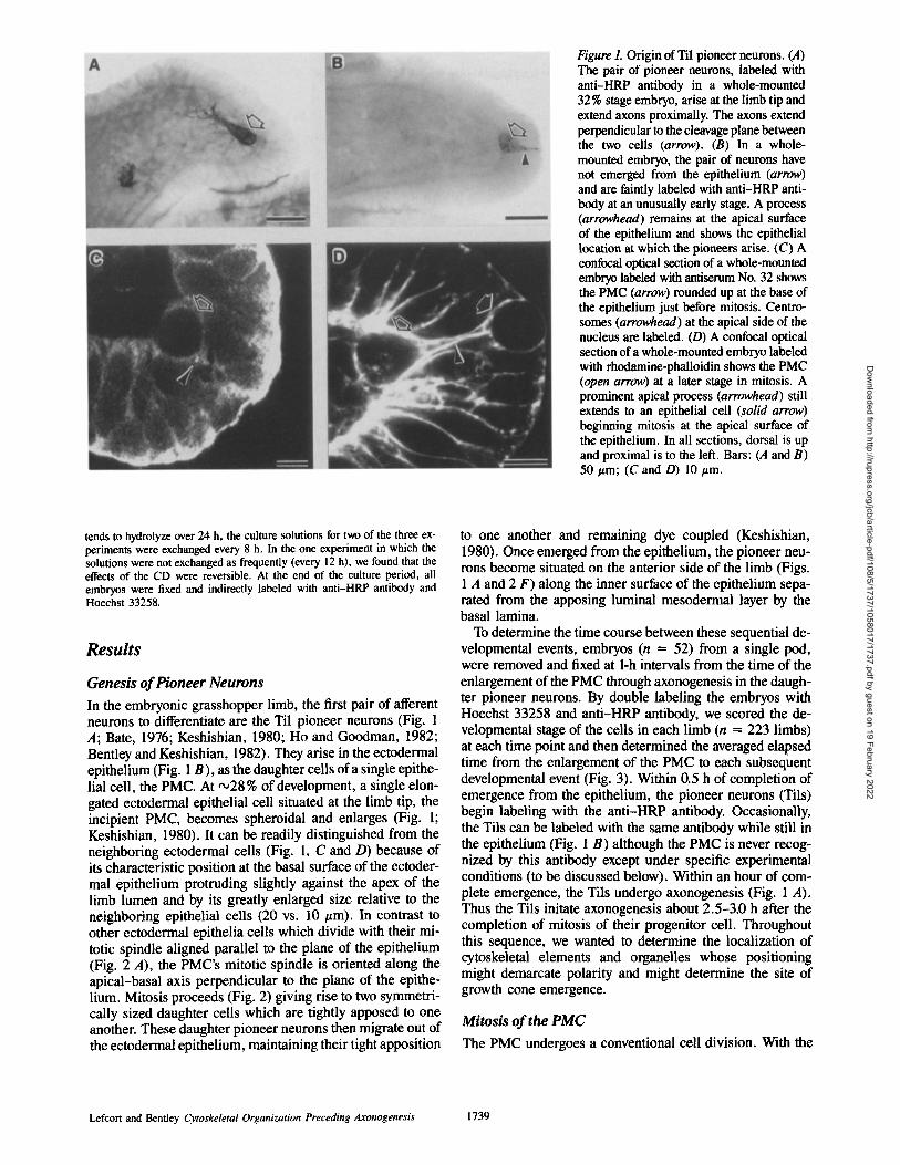
Figure 1. Origin of Til pioneer neurons. (A) The pair of pioneer neurons, labeled with anti-HRP antibody in a whole-mounted 32% stage embryo, arise at the limb tip and extend axons proximally. The axons extend perpendicular to the cleavage plane between the two cells (arrow). (B) In a whole- mounted embryo, the pair of neurons have not emerged from the epithelium (arrow) and are faintly labeled with anti-HRP anti- body at an unusually early stage. A process (arrowhead) remains at the apical surface of the epithelium and shows the epithelial location at which the pioneers arise. (C) A confocal optical section of a whole-mounted embryo labeled with antiserum No. 32 shows the PMC (arrow) rounded up at the base of the epithelium just before mitosis. Centro- somes (arrowhead) at the apical side of the nucleus are labeled. (D) A confocal optical section of a whole-mounted embryo labeled with rhodamine-phalloidin shows the PMC (open arrow) at a later stage in mitosis. A prominent apical process (arrowhead) still extends to an epithelial cell (solid arrow) beginning mitosis at the apical surface of the epithelium. In all sections, dorsal is up and proximal is to the left. Bars: (A and B) 50 t~m; (C and D) 10 #m.
tends to hydrolyze over 24 h, the culture solutions for two of the three ex- periments were exchanged every 8 h. In the one experiment in which the solutions were not exchanged as frequently (every 12 h), we found that the effects of the CD were reversible. At the end of the culture period, all embryos were fixed and indirectly labeled with anti-HRP antibody and Hoechst 33258.
Results
Genesis of Pioneer Neurons
In the embryonic grasshopper limb, the first pair of afferent neurons to differentiate are the Til pioneer neurons (Fig. 1 A; Bate, 1976; Keshishian, 1980; Ho and Goodman, 1982; Bentley and Keshishian, 1982). They arise in the ectodermal epithelium (Fig. 1 B), as the daughter cells of a single epithe- lial cell, the PMC. At ,,o28 % of development, a single elon- gated ectodermal epithelial cell situated at the limb tip, the incipient PMC, becomes spheroidal and enlarges (Fig. 1; Keshishian, 1980). It can be readily distinguished from the neighboring ectodermal cells (Fig. 1, C and D) because of its characteristic position at the basal surface of the ectoder- mal epithelium protruding slightly against the apex of the limb lumen and by its greatly enlarged size relative to the neighboring epithelial cells (20 vs. 10 #m). In contrast to other ectodermal epithelia cells which divide with their mi- totic spindle aligned parallel to the plane of the epithelium (Fig. 2 A), the PMC's mitotic spindle is oriented along the apical-basal axis perpendicular to the plane of the epithe- lium. Mitosis proceeds (Fig. 2) giving rise to two symmetri- cally sized daughter cells which are tightly apposed to one another. These daughter pioneer neurons then migrate out of the ectodermal epithelium, maintaining their tight apposition
to one another and remaining dye coupled (Keshishian, 1980). Once emerged from the epithelium, the pioneer neu- rons become situated on the anterior side of the limb (Figs. 1 A and 2 F) along the inner surface of the epithelium sepa- rated from the apposing luminal mesodermal layer by the basal lamina.
To determine the time course between these sequential de- velopmental events, embryos (n = 52) from a single pod, were removed and fixed at 1-h intervals from the time of the enlargement of the PMC through axonogenesis in the daugh- ter pioneer neurons. By double labeling the embryos with Hoechst 33258 and anti-HRP antibody, we scored the de- velopmental stage of the cells in each limb (n = 223 limbs) at each time point and then determined the averaged elapsed time from the enlargement of the PMC to each subsequent developmental event (Fig. 3). Within 0.5 h of completion of emergence from the epithelium, the pioneer neurons (Tils) begin labeling with the anti-HRP antibody. Occasionally, the Tils can be labeled with the same antibody while still in the epithelium (Fig. 1 B) although the PMC is never recog- nized by this antibody except under specific experimental conditions (to be discussed below). Within an hour of com- plete emergence, the Tils undergo axonogenesis (Fig. 1 A), Thus the Tils initate axonogenesis about 2.5-3.0 h after the completion of mitosis of their progenitor cell. Throughout this sequence, we wanted to determine the localization of cytoskeletal elements and organelles whose positioning might demarcate polarity and might determine the site of growth cone emergence.
Mitosis of the PMC
The PMC undergoes a conventional cell division. With the
Lefcort and Bentley Cytoskeletal Organization Preceding Axonogenesis 1739
Dow
nloaded from http://rupress.org/jcb/article-pdf/108/5/1737/1058017/1737.pdf by guest on 19 February 2022

Figure 2. Mitosis of the PMC. Panels show confocal optical sections of whole-mounted embryos labeled with propidium iodide to visualize chromatin. Open arrows indicate the PMC or distal daughter cell and point proximally along the limb axis. Solid arrows indicate the prox- imal daughter cell. (A) Enlarged PMC just before entering prophase. Note the dividing epithelial cell at the apical surface whose mitotic spindle is oriented parallel to the plane of the epithelium. (B) PMC in prophase. (C) PMC in metaphase. (D) PMC in anaphase. The position of the sets of chromosomes indicates the apical-basal orientation of the mitotic spindle. (E) PMC in telophase. (F) After PMC cytokinesis, the daughter pioneer neurons have emerged from the epithelium and are situated along its inner surface. Bars, 10 #m.
. c v
G) E
"o
CL
O) o1
,<
1 0 '
9 '
8 '
7 -
6 -
5 -
4 -
3 -
2 -
I
0
2
c
"7, E o ® I:L c r -r
o
0 1
a: o .-r _ ¢,
Figure 3. The time course of PMC mitosis and events in early differentiation of the pioneer neurons. The sequence of develop- mental events is indicated on the x-axis. The average time from rounding up of the PMC to each event is shown (see Materials and Methods; emerging, partial emergence of the pioneer neurons from the epithelium; luminal, completion of this process).
antitubulin antibody, a dense array of spindle fibers are ob- served which are most prominent during anaphase (Fig. 4 A) and begin to depolymerize during telophase. Just before the onset of its division, the PMC's centrosomes are located on the apical side of its nucleus (Fig. 1 C), at the base of a pro- cess which extends to the apical surface of the epithelium. During mitosis, using the antisera Nos. 32 and 40, centro- somes could be identified at each spindle pole (Fig. 4, B and C); these antisera also recognize the midbody (Fig. 4 B). As the PMC enlarges, with rhodamine-phalloidin, a cortical array of actin is observed distributed relatively uniformly around the cell. At anaphase, actin becomes concentrated along the invaginating cleavage furrow. By the end of telo- phase, all that is left of the constriction ring is a bright spot at the cleavage plane, presumably now associated with the midbody (n = 19 limbs; Fig. 4, J and K).
Thus, by the end of mitosis, in the absence of any internal reorganization or cell-cell rearrangement, the cell pole of the PMC which was adjacent to the basal lamina would be situated at the proximal pole of the Til proximal daughter cell, while the cell pole of the PMC which was closest to the apical surface of the epithelium would be located in the distal pole of the Til distal daughter cell. Hereafter, we will refer to the nascent pioneer neurons as the Til proximal sibling (Tilp) and the Til distal sibling (Til~).
Cellular Organization during the Period between Cytokinesis and Axonogenesis
A prominent feature of cellular organization in this period is the localization of several intraceUular elements at the cell pole from which axonogenesis is initiated in Tilp. One of
The Journal of Cell Biology, Volume 108, 1989 1740
Dow
nloaded from http://rupress.org/jcb/article-pdf/108/5/1737/1058017/1737.pdf by guest on 19 February 2022
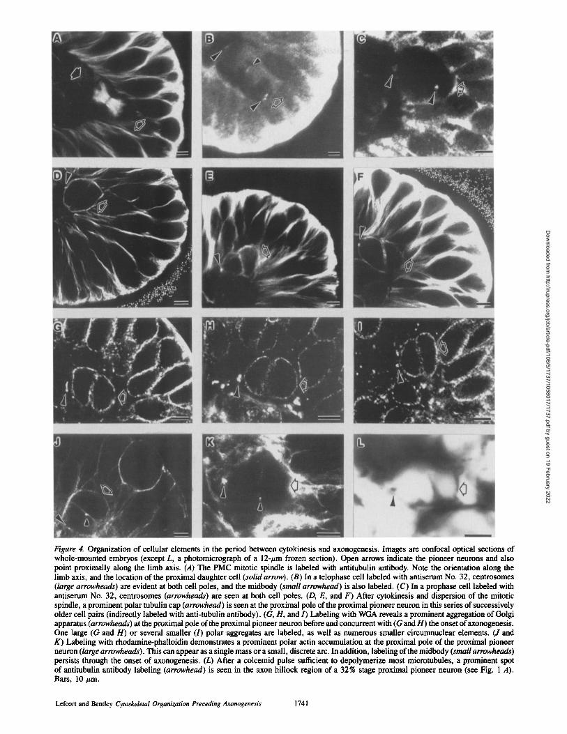
Figure 4. Organization of cellular elements in the period between cytokinesis and axonogenesis. Images are confocal optical sections of whole-mounted embryos (except L, a photomicrograph of a 12-#m frozen section). Open arrows indicate the pioneer neurons and also point proximally along the limb axis. (A) The PMC mitotic spindle is labeled with antitubulin antibody. Note the orientation along the limb axis, and the location of the proximal daughter cell (solid arrow). (B) In a telophase cell labeled with antiserum No. 32, centrosomes (large arrowheads) are evident at both cell poles, and the midbody (small arrowhead) is also labeled. (C) In a prophase cell labeled with antiserum No. 32, centrosomes (arrowheads) are seen at both cell poles. (D, E, and F) After cytokinesis and dispersion of the mitotic spindle, a prominent polar tubulin cap (arrowhead) is seen at the proximal pole of the proximal pioneer neuron in this series of successively older cell pairs (indirectly labeled with anti-tubulin antibody). (G, H, and I) Labeling with WGA reveals a prominent aggregation of Golgi apparatus (arrowheads) at the proximal pole of the proximal pioneer neuron before and concurrent with (G and H) the onset of axonogenesis. One large (G and H) or several smaller (I) polar aggregates are labeled, as well as numerous smaller circumnuclear elements. (J and K) Labeling with rhodamine-phalloidin demonstrates a prominent polar actin accumulation at the proximal pole of the proximal pioneer neuron (large arrowheads). This can appear as a single mass or a small, discrete arc. In addition, labeling of the midbody (small arrowheads) persists through the onset of axonogenesis. (L) After a colcemid pulse sufficient to depolymerize most microtubules, a prominent spot of antitubulin antibody labeling (arrowhead) is seen in the axon hillock region of a 32% stage proximal pioneer neuron (see Fig. 1 A). Bars, 10 #m.
Lefcort and Bentley Cytoskeletal Organization Preceding Axonogenesis 1741
Dow
nloaded from http://rupress.org/jcb/article-pdf/108/5/1737/1058017/1737.pdf by guest on 19 February 2022

\ \
J /
/ J
Figure 5. The disposition of filopodia which are extended from the proximal pioneer neuron before the first morphological indication of protrusion of the growth cone. The polar plot indicates the emer- gence sites of 137 filopodia from 20 neurons. The two length classes indicate filopodia greater or less than a half cell diameter in length (no filopodia were greater than one cell diameter in length). Distal filopodia are obscured by the distal pioneer neuron (broken line) and are not plotted. The asterisk indicates the proximal pole of the proximal cell and the typical site of growth cone emergence.
these elements is a tubulin-containing polar cap. With the completion of cytokinesis, the cell perimeter is brightly la- beled with antitubulin antibody (Fig. 4, D-F). Once the cells begin their exodus from the epithelium, a discrete focus of tubulin labeling becomes prominent at the proximal pole of the Tilp (n = 26 limbs; Fig. 4, D-F). It persists and is de- tectable as the Tilp alters its morphology at its proximal pole just before the onset of axonogenesis (Fig. 4, E and F). This tubulin-containing cap may be a microtubule organizing center (MTOC); since there is very little cytoplasm in these cells relative to the extremely large size of the nucleus, there is no obvious cell center from which a network of microtu- bules radiate. However, the band of microtubules which wraps around the cell often appears to be in contact with this tubulin cap. The cap is not labeled by the anticentrosome an- tisera (Nos. 32 and 40); however, these antisera cease label- ing at cytokinesis and don't label centrosomes in interphase ceils.
A second prominent polar feature during this time perio~ is the GA. As the daughter neurons migrate out of the epithe- lium, the GA, as visualized with both WGA and NBD-cer- amide appears to be distributed circumnuclearly. However, just before and concurrent with the onset of axonogenesis, an aggregation of staining (with both probes) is localized at the proximal face of the Tilp's nucleus (Fig. 4, G-l). The phenotypes observed are either a discrete aggregation at the proximal pole (n = 21 limbs; Fig. 4, G and H) or a more distributed series of clusters surrounding the proximal/dor-
sal quadrant of the nucleus (n = 12 limbs; Fig. 4 I). In addi- tion, the cells retain circumnuclear labeling.
A third polar feature is a concentration of actin microfila- ments during this time period. As the ceils migrate out of the epithelium a focus of actin labeling is evident at the proximal pole of the Tilp (n = 11 limbs; Fig 4, J and K). This actin focus usually consists of a single aggregation (Fig. 4, J and K), but may be dispersed into discrete clusters just anterior to the nucleus around the proximal pole of Tilp. In addi- tion, phalloidin reveals that the midbody persists through this time period (Fig. 4, J and K).
The pioneer daughter cells begin to extend short radial filopodia soon after cytokinesis and well before the onset of growth cone protrusion. Using camera lucida, we deter- mined the site of origin and relative length of these initial filopodia (n = 137) in 20 proximal pioneer neurons. We found that filopodia were widely distributed around the cir- cumference of the somata (Fig. 5), and their distribution did not predict the site of growth cone emergence. Once the pro- trusion of the growth cone begins, longer filopodia are often extended proximally from the proximal pole region (Caudy and Bentley, 1986b).
Axonogenesis
About 3 h after cytokinesis, protrusion of the axonal growth cone commences from the proximal side of the proximal pi- oneer neuron. The organization of tubulin, GA, and actin in- dicates that the morphological cell pole of this cell is derived from the orientation of the mitotic spindle of the PMC. If the Tilp's axon emerges from this polar site, then one would predict that the growth cone should reliably emerge perpen- dicular to the PMC's cleavage plane. To determine the site of growth cone emergence from the Tilp with respect to the cleavage plane (as defined by the apposition plane between the sister pioneer neurons), embryos of 31-32 % of develop- ment were fixed and labeled with anti-HRP antibody. The orientation of the growth cone of Tilp with respect to the cleavage plane was scored for 34 cell pairs (Fig. 6). In the majority of cases, the growth cone emerged within 10 ° of a line perpendicular to the cleavage plane.
As the nascent growth cone protrudes from the proximal
Figure 6. The sites of growth cone emergence from 34 proximal pi- oneer neurons. Position on the cell circumference (inset) is indi- cated in degrees (bin width is 20°). Most growth cones emerge per- pendicular (*) to the former cleavage plane (inset, 270°--90 ° line) between the sibling neurons.
The Journal of Cell Biology, Volume 108. 1989 1742
Dow
nloaded from http://rupress.org/jcb/article-pdf/108/5/1737/1058017/1737.pdf by guest on 19 February 2022

Figure 7. Arrangement of cytoskeletal ele- ments during axonogenesis. Images are confocal optical sections of whole-mounted embryos. Open arrows indicate pioneer neurons and point proximally along the limb axis. (A) At the onset of axonogenesis, rhodamine-phalloidin labeling shows abun- dant actin microfilaments in the nascent axon hillock. The midbody (small arrow- head) also labels. (B) After the growth cone extends away from the soma, actin microfilaments (labeled with rhodamine- phalloidin) are confined to the cortex of the axon hillock (arrowhead). In these 32.5 % stage cells, labeling of the midbody (small arrowhead) still persists. (C) As the na- scent growth cone emerges, microtubules, labeled with antitubulin antibody, extend into it (arrowhead) and accumulate at its base. (D and E) After the growth cone mi- grates away from the soma, the nascent axon is solidly packed with microtubules which spread out at its base (arrowhead) to fill the proximal face of the cell (indirect labeling with antitubulin antibody). Small arrowhead indicates aster of dividing meso- dermal cell. (F) The dense bundle of axonal microtubules terminates in the growth cone (large arrowhead, left) but small bundles of microtubules extend from the growth cone into newly forming branches (small arrow- head). Note, this axon is extending from the distal pioneer (large arrowhead, right), whose distal process is also filled with microtubules. Bars: (A-D) 5/xm; (E and F) 10 t~m.
pole, the polar actin concentration observed before axono- genesis develops into dense labeling within the axon hillock (Fig. 7 A). As axonogenesis proceeds, actin labeling within the hillock is replaced by cortical labeling of the axon (and soma; Fig. 7 B). Actin labeling of the midbody is still evi- dent at the onset of growth cone emergence (Fig. 7 A) and, surprisingly, persists well into the period of axonogenesis (Fig. 7 B; Sanger et al., 1985). Midbody labeling is seen as late as the 33 % stage of axonogenesis.
At the onset of axonogenesis, microtubules begin to appear in the axon hillock (Fig. 7 C) at the proximal pole of the cell. At this stage, microtubules could be nucleated off of the discrete tubulin focus situated there. But, once further axon outgrowth has occurred, the axon hillock region becomes densely packed with microtubules that do not appear to be nucleated from a single focus. Instead, their ends are ob- served funneling broadly out of the proximal portion of the cell, perhaps off of the proximal face of the nucleus (Fig. 7, D-F). The entire length of the axon is solidly packed with microtubules (Fig. 7, E and F). These extend into the base of the growth cone (Fig. 7 F) as well as into the distally directed apical dendrite that is often extended by these cells
(Fig. 7 F). Microtubules also extend into branches that are emerging from the growth cone (Fig. 7 F).
A method commonly used to localize centrosomes or MTOCs in the absence of a histochemical marker, is to depolymerize the majority of microtubules and then label with antitubulin antibodies. When embryos at 31-32 % of de- velopment were exposed to a pulse of colcemid (10 #g/ml) for 2-3 h and then fixed and triple labeled with antitubulin antibody, anti-HRP antibody, and Hoechst 33258, a spot similar in size and location to the tubulin cap observed in the preaxonogenesis limbs was observed in the axon hillock of the proximal pioneer neuron at the proximal face of the nu- cleus (n = 2 embryos; Fig. 4 L). Several nascent neurons in the CNS also contained a tubulin staining focus in their axon hillocks after the colcemid pulse. In older embryos (32-33%) examined, no discrete spots were observed any- where around the pioneer somata after the colcemid pulse (n = 8). Thus it is likely that those dots correspond to MTOCs or centrosomes and that they might disappear once axonogenesis is well underway. Once a well-established axon has been extended, only the circumnuclear GA staining is evident. The discrete aggregates observed earlier (Fig. 4,
Lefcort and Bentley Cytoskeletal Organization Preceding Axonogenesis 1743
Dow
nloaded from http://rupress.org/jcb/article-pdf/108/5/1737/1058017/1737.pdf by guest on 19 February 2022

Figure 8. Mirror symmetry in process outgrowth from sibling pi- oneer neurons. Images are from anti-HRP antibody labeled, whole-mounted embryos. (A) Shortly after PMC cleavage (arrow, cleavage plane), these daughter cells extend mirror image filopodia from opposite poles. (B) In a 34% stage limb, two pioneers extend mirror image axons. (C) A rare phenotype, in which both the proximal and distal pioneers extend distally directed axons. Bars, 25 ~m.
G-l) have disassociated, although occasionally some small- er dispersed spots are observed in the axon hillock (n = 2).
Axonogenesis in the Distal Pioneer Neuron
The preceding description has focused upon the proximal daughter neuron. If the pioneer neurons are inherently polar- ized as a result of the orientation of their mother cell's mitotic spindle, then the two pioneer axons should in fact be mirror symmetric. This symmetry predicts that the distal daughter neuron's axon should extend distally. Instead, as stated previ- ously, the pattern of emergence of the pioneer axons is highly stereotyped with both axons growing proximally down the
limb (e.g., Fig. 1 A). However, occasionally, aberrant axon outgrowth is observed. These aberrancies are of two pheno- types only: either the distal cell's axon grows out straight dis- tally, while the proximal cell's axon grows out in the normal direction (straight proximally), often resulting in mirror symmetric outgrowth (Fig. 8, A and B), or both cells send their axons straight distally (Fig. 8 C). Thus in both cells, the pioneer axons emerge from one of two polarized points that are 180 ° apart.
Since the Tile's growth cone does occasionally emerge from its distal pole (Bentley and Candy, 1983), we looked for intracellular correlates of distal growth cone emergence. We found one case of a distal staining actin focus (Fig. 9 A) and five limbs with a distal staining tubulin cap (Fig. 9 C) in the distal cell. In addition we saw two limbs that had an accumulation of GA at the distal pole of Tild. The paucity of these phenotypes corresponds to the infrequency of distal growth cone emergence.
Labeling at the proximal pole of the distal cell was difficult to resolve due to an abundance of tubulin, actin, and WGA labeling near the cleavage plane. However, we did observe a few limbs in which there was a focus of tubulin staining (n = 4 limbs) and WGA labeling (n = 3 limbs) in the prox- imal/dorsal quadrant of the Tild, where the Tild'S axon often emerges (e.g., Fig. 7 F). These proximal/dorsal located tubulin dots in the distal cell were generally smaller in di- ameter than those described in the proximal pole of the prox- imal cell.
Although the PMC divides symmetrically, the two daugh- ter cells are not always equal in all respects: Tilp often ap- pears to mature faster than Til~. Occasionally we find cell pairs in which the distal cell's chromatin has not fully dis- persed nor its spindle completely broken down, while both events have been completed in the proximal cell (Fig. 9, B and D). In addition, often we find newly emerged or emerg- ing cell pairs in which the proximal cell is more brightly la- beled with the anti-HRP antibody, a marker of neuronal differentiation, than is the distal cell (data not shown).
Orientation of the Pioneer Axons W~th Respect To the Proximal-Distal Limb Axis
PMCs are located very close to the limb tip when they initiate mitosis (Fig. 1 B and Fig. 10). The combination of this loca- tion, the apical-basal orientation of the PMC mitotic spin- dle, and the emergence of the growth cone from the proximal cell pole should confer strong directionality on the orienta- tion of the nascent axon within the limb bud (Fig. 10). To assess this directionality, embryos at 31-32 % of development were fixed and labeled with anti-HRP antibody. The angle of orientation of their axons with respect to the dorsal epithe- lium was determined. In the 35 limbs examined, 86% of the axon pairs emerged with an orientation within l0 ° of parallel to the limb axis (Fig. 11).
Cytokinesis and Axonogenesis
Cytokinesis and axonogenesis both involve polarized flow of cortical actin (Bray and White, 1988; Forscher and Smith, 1988). To explore the possibility that these sequential events may be interrelated in this system, we sought to determine whether axonogenesis would be initiated in the absence of cytokinesis. Embryos at 28% of development, just before
The Journal of Cell Biology, Volume 108, 1989 1744
Dow
nloaded from http://rupress.org/jcb/article-pdf/108/5/1737/1058017/1737.pdf by guest on 19 February 2022

Figure 9. Cytoskeletal organization and delayed maturation in the distal pioneer neuron. Confocal optical sections of whole- mounted limbs (arrows, cleavage plane be- tween the pioneers). (A) Rhodamine-phal- loidin labeling shows an actin microfilament accumulation (arrowhead) at the distal pole of the distal cell. (B) Antitubulin antibody reveals that the spindle microtubules are still present in the distal cell after the spin- dle of the proximal cell is fully disassem- bled. This indicates that maturation of the distal cell can lag that of the proximal cell. (C) Antitubulin antibody shows a tubulin cap at the distal pole of the distal cell (ar- rowhead). (D) The same cells as in B, which were double-labeled with propidium iodide to view the chromatin, (arrowhead points to same location as in B). Chromatin is more dispersed in the proximal cell, indicating more advanced maturation. Bars, 5 #m.
cytokinesis of the PMC in the majority of the limbs, were placed into culture medium containing CD (0.05 ttg/ml) for 24 h. To insure that the culture period was long enough to permit axonogenesis, some embryos from the same pod and of the same age were cultured in normal media in the absence of CD. At the end of the culture period, embryos were fixed and double labeled with anti-HRP antibody and Hoechst 33258 (Fig. 12). Of limbs in which the PMC underwent karyokinesis, in 100% of the control limbs the PMC com- pleted cytokinesis, whereas cytokinesis was completed in only 31% of the experimental limbs (Table I). Axonogenesis occurred in 73 % of the experimental limbs in which the PMC underwent cytokinesis. However, axonogenesis oc- curred in only 8 % of the experimental limbs in which the PMC failed to complete cytokinesis although it did undergo karyokinesis. Although in ,030% of the experimental limbs some outgrowth was observed, these processes were gener- ally short (less than or equal to 1 cell length) branches, with- out indication of a growth cone. When limbs in which the pioneer neurons have already initiated axonogenesis are cul- tured in the presence of this concentration of CD (and higher; e.g., 0.1 #g/ml), further axon extension is not in- hibited (Bentley and Toroian-Raymond, 1986).
Other features of differentiation of the pioneer neurons proceeded normally in the absence of cytokinesis and axono- genesis: (a) cells that had undergone karyokinesis but not cytokinesis always bound the anti-HRP antibody (Table I; the PMC never expresses this binding site in normal em- bryos); (b) 80% of the PMCs which underwent karyokinesis, but not cytokinesis emerged from the epithelium, a process the PMC never normally undergoes. Interestingly, in 28% of the limbs exposed to CD, an extra round of nuclear division occurred such that extra pioneer neurons or extra nuclei in- side the undivided PMC were observed (Fig. 12, A-C). The effects of the CD were at least partially reversible; in limbs in which the CD medium was not exchanged as frequently (every 12 h instead of every 8 h), limbs with four separate
pioneer neurons, all recognized by anti-HRP antibody, were observed. In such limbs, the extra pioneer neurons degener- ated as their nuclei contained condensed chromatin; in addi- tion, no more than two axons were observed emerging in these limbs.
Discussion The object of this study was to investigate the arrangement of cytoskeletal elements and organeUes at the site of and preceding the organization of a growth cone in a neuron in situ. To this end, we selected an identified neuron, (the Til, proximal sibling) whose place and date of birth is known, and where the site of growth cone emergence is highly pre- dictable. We fixed embryos at successive developmental stages, from the enlargement of the PMC through axonogen- esis of the Tils and examined the external morphology as well as actin microfilaments, microtubules, centrosomes, mid- body, GA, and chromatin. Our results show that emergence of the growth cone, from the cell pole, is preceded by a spa- tially and temporally specific arrangement of intracellular elements (Fig. 13).
PMC Mitosis
The Tilp arises from a precursor cell which is located at the limb tip. At ,028 % of development, the PMC rounds up at the basal surface of the epithelium, begins to enlarge, and un- dergoes mitosis. This mitosis is conventional, as indicated, for example, by the polar localization of centrosomes (Fig. 4, B and C), array of spindle microtubules (Fig. 4 A), and localization of actin filaments in the developing cleavage fur- row. It is, however, distinguished from mitosis in other epi- thelial cells in that the PMC's spindle is oriented perpendicu- lar to the epithelium and at the basal surface, rather than parallel to the plane of the epithelium and at the apical sur- face (Figs. 1 D and 2 D vs. Fig. 2 A). As a result, at the com- pletion of mitosis, one of the centrosomes is at the proximal
Lefcort and Bentley Cytoskeletal Organization Preceding Axonogenesis 1745
Dow
nloaded from http://rupress.org/jcb/article-pdf/108/5/1737/1058017/1737.pdf by guest on 19 February 2022
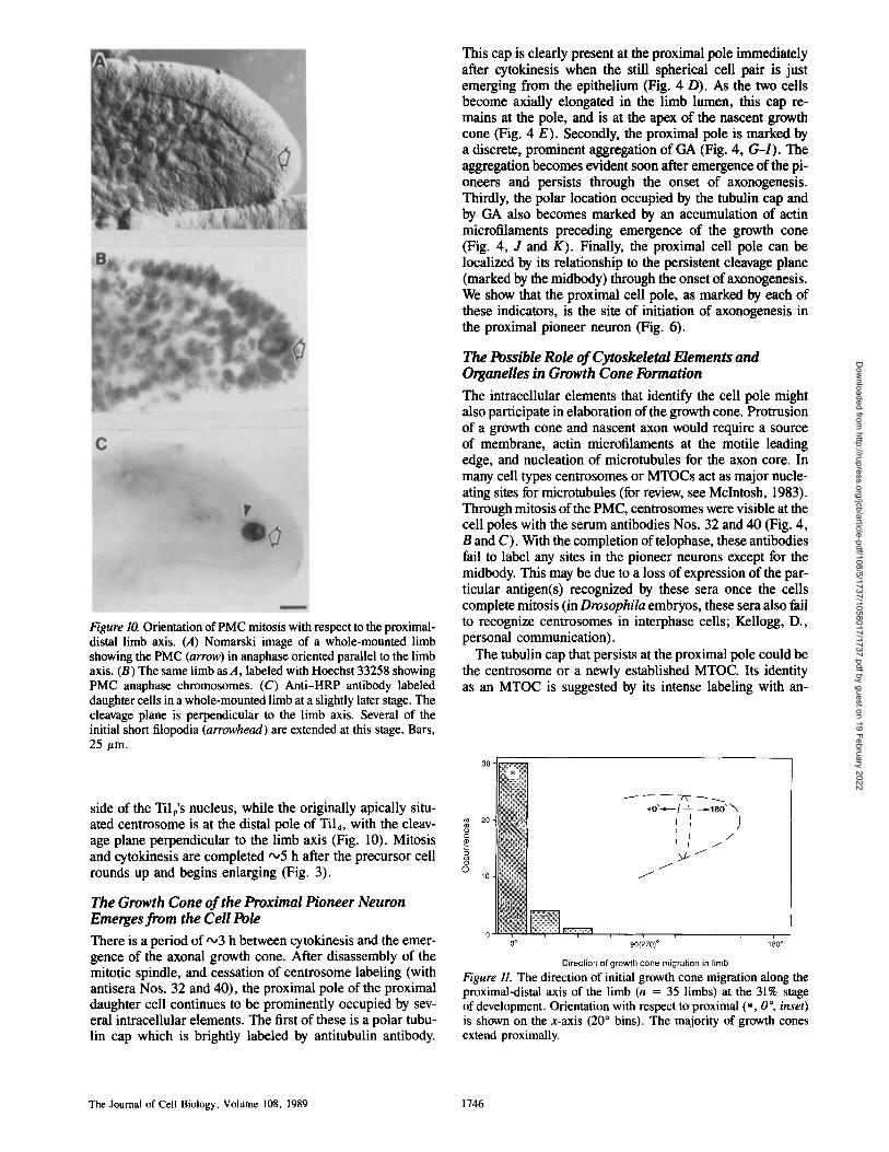
Figure 10. Orientation of PMC mitosis with respect to the proximal- distal limb axis. (A) Nomarski image of a whole-mounted limb showing the PMC (arrow) in anaphase oriented parallel to the limb axis. (B) The same limb as A, labeled with Hoechst 33258 showing PMC anaphase chromosomes. (C) Anti-HRP antibody labeled daughter cells in a whole-mounted limb at a slightly later stage. The cleavage plane is perpendicular to the limb axis. Several of the initial short filopodia (arrowhead) are extended at this stage. Bars, 25 #m.
side of the Tilp's nucleus, while the originally apically situ- ated centrosome is at the distal pole of Tild, with the cleav- age plane perpendicular to the limb axis (Fig. 10). Mitosis and cytokinesis are completed '~5 h after the precursor cell rounds up and begins enlarging (Fig. 3).
The Growth Cone of the Proximal Pioneer Neuron Emerges from the Cell Pole
There is a period of •3 h between cytokinesis and the emer- gence of the axonal growth cone. After disassembly of the mitotic spindle, and cessation of centrosome labeling (with antisera Nos. 32 and 40), the proximal pole of the proximal daughter cell continues to be prominently occupied by sev- eral intracellular elements. The first of these is a polar tubu- lin cap which is brightly labeled by antitubulin antibody.
This cap is clearly present at the proximal pole immediately after cytokinesis when the still spherical cell pair is just emerging from the epithelium (Fig. 4 D). As the two cells become axially elongated in the limb lumen, this cap re- mains at the pole, and is at the apex of the nascent growth cone (Fig. 4 E). Secondly, the proximal pole is marked by a discrete, prominent aggregation of GA (Fig. 4, G-l). The aggregation becomes evident soon after emergence of the pi- oneers and persists through the onset of axonogenesis. Thirdly, the polar location occupied by the tubulin cap and by GA also becomes marked by an accumulation of actin microfilaments preceding emergence of the growth cone (Fig. 4, J and K). Finally, the proximal cell pole can be localized by its relationship to the persistent cleavage plane (marked by the midbody) through the onset of axonogenesis. We show that the proximal cell pole, as marked by each of these indicators, is the site of initiation of axonogenesis in the proximal pioneer neuron (Fig. 6).
The Possible Role of Cytoskeletal Elements and OrganeUes in Growth Cone Formation
The intracellular elements that identify the cell pole might also participate in elaboration of the growth cone. Protrusion of a growth cone and nascent axon would require a source of membrane, actin microfilaments at the motile leading edge, and nucleation of microtubules for the axon core. In many cell types centrosomes or MTOCs act as major nucle- ating sites for microtubules (for review, see Mclntosh, 1983). Through mitosis of the PMC, centrosomes were visible at the cell poles with the serum antibodies Nos. 32 and 40 (Fig. 4, B and C). With the completion of telophase, these antibodies fail to label any sites in the pioneer neurons except for the midbody. This may be due to a loss of expression of the par- ticular antigen(s) recognized by these sera once the cells complete mitosis (in Drosophila embryos, these sera also fail to recognize centrosomes in interphase cells; Kellogg, D., personal communication).
The tubulin cap that persists at the proximal pole could be the centrosome or a newly established MTOC. Its identity as an MTOC is suggested by its intense labeling with an-
3 0
2 0 .
10 -
* O*-.t,---- / ' .180° ~
I j . / I i /
/ /
0 ,
o o 90('270)0 i~o o
Direction of growth cone migration in limb
Figure 11. The direction of initial growth cone migration along the proximal-distal axis of the l imb (n = 35 limbs) at the 31% stage of development. Orientation with respect to proximal (*, 0 °, inset) is shown on the x-axis (20 ° bins). The majority of growth cones extend proximally.
The Journal of Cell Biology, Volume 108, 1989 1746
Dow
nloaded from http://rupress.org/jcb/article-pdf/108/5/1737/1058017/1737.pdf by guest on 19 February 2022

Figure 12. Effect of CD on PMC differentiation. Fluorescence pho- tomicrographs from a whole-mounted embryo that, starting just be- fore PMC mitosis, was cultured for 24 h in medium with 0.05 /.tg/ml CD, and then double-labeled with Hoechst 33258 and (in- directly) anti-HRP antibody. (A) The PMC has emerged from the epithelium and acquired anti-HRP binding sites, but has not under- gone cytokinesis or initiated axogenesis. (B) Enlargement ofA. The cell has undergone two rounds of karyokinesis, and contains four nuclei (arrowheads). (C) Viewing the cell under Hoechst 33258 optics confirms the presence of four nuclei (arrowheads). Bars: (A) 25 /~m; (B and C) 15 p.m.
titubulin antibody and the persistence of labeling at this loca- tion after colcemid-induced depolymerization of microtu- bules in nascent axons (Fig. 4 L). The MTOCs (in this case, centrosome) in interphase cardiac myocytes have the same phenotype: they appear as a bright spot adjacent to the nu- cleus (Kronebusch and Singer, 1987). In certain lines of differentiated neuroblastoma cells, an MTOC is also located near the nucleus, aligned in the direction of the extended neurite (Spiegelman et al., 1979). In PC 12 cells which have recently undergone axonogenesis, it has been shown that centrioles are not characteristically located at the base of the
axon hillock, although they typically reside on the same face of the nucleus from which the axon arises (Stevens et al., 1988). In these cells, it appears that the axonal microtubules are not nucleated from a single MTOC; instead, microtu- bules fan out at the base of the axon as if they were extruding from it. This microtubule organization is very similar to what we see in the axon hillock region of more mature pi- oneer axons (Fig. 7, D-F). But in pioneer neurons, the tubu- lin cap is characteristically present in the axon hillock at the onset of axonogenesis. Therefore the location of this putative MTOC does not exclude its possible participation in the nucleation of growth cone microtubules. This tubulin focus could not be identified in cells at a slightly later stage (>32%) and may be transient. In Drosophila epithelium, microtubule nucleation can proceed independently of the centrosome (Tucker et al., 1986).
A second localized feature in this period is the GA. In most animal cells that have been examined, the GA is situated ad- jacent to the MTOC (for review, see Singer and Kupfer, 1986). In several types of motile cells it has been demon- strated that in response to a polar, migratory signal, the MTOC and GA coordinately relocalize to the side of the cell closest to the signal. This organization may be crucial in facilitating directional movement since it allows for polar- ized insertion of membrane via vesicles budded off from the adjacent GA to the leading edge of the motile cell or growth cone. With both markers used to identify GA in pioneer neu- rons, WGA and NBD-ceramide, we found that as the cells migrated out from the epithelium, the GA appeared to be dis- tributed circumnuclearly. However, immediately before and concurrent with the onset of axonogenesis, a discrete aggre- gation of GA was evident at the proximal pole (Fig. 4, G-I) and/or proximal face (Fig. 4 I) of the Tilp. Therefore GA may participate in the promotion of growth cone extrusion at this site. Once axonogenesis is well underway, however, the discrete aggregates of GA observed earlier are no longer evident at the proximal face of the nucleus.
Finally, a third feature of this polar region is a concentra- tion of actin microfilaments. Just before the initiation of ax- onogenesis, actin labeling accumulates in one or a few bright foci at the proximal cell pole (Fig. 4, J and K). Presumably these microfilaments participate in establishing the motile leading edge of the growth cone (Bray and White, 1988; Forscher and Smith, 1988).
Table L Effects of Cytochalasin D on Differentiation of the PMC
Cytokinesis completed Cytokinesis blocked
Acquisition of Emergence Acquisition of Emergence anti-HRP from anti-HRP from
binding epithelium Axonogenesis binding epithelium Axonogenesis
Control (n = 44)
CD (n = 36)
% % % % % %
100 100 80 - - -
100 100 73 I00 80 8
* All limbs were examined after 24-h culture period at 30 5:1 °C in either normal medium (see Materials and Methods) or in the presence of 0.05 #g/ml CD. PMCs scored in Table I were those which underwent karyokinesis. There were 47 control limbs of which 44 underwent karyokinesis, and 42 experimental limbs of which 36 underwent karyokinesis. Of this set of 36, cytokinesis was completed in I I and blocked in 25.
Lefcort and Bentley Cytosketetal Organization Preceding Axonogenesis 1747
Dow
nloaded from http://rupress.org/jcb/article-pdf/108/5/1737/1058017/1737.pdf by guest on 19 February 2022

Figure 13. Summary diagram depicting the time course for the ar- rangement of organelles and cytoskeletal elements described in the text associated with the development of the pioneer neurons, begin- ning with the onset of mitosis in the PMC.
Axonogenesis in the Distal Til Sib
The PMC undergoes a symmetric division creating two ap- parently mirror image cells. If these cells are truly mirror symmetric, and the site of growth cone initiation is at the former spindle pole, then the growth cone from the distal Til sibling should emerge from the distal face of the cell. Occa- sionally this emergence pattern is observed (Fig. 8). We also occasionally observed the intracellular elements which mark the pole of the proximal cell at the distal pole of the distal cell (Fig. 9, A and C). Therefore, at times an element of mir- ror symmetry is revealed in the initiation of axonogenesis from these sister neurons. Albrecht-Bueller (1977) showed that pairs of clonally related 3T3 mouse fibroblasts could es- tablish mirror symmetric pathways when grown on a uniform substrate in the absence of contact with neighboring cells. He proposed that this mirror symmetric behavior was the mani- festation of a mirror symmetric intracellular organization whose structure was determined at mitosis. Solomon (1981) proposed a similar mechanism to account for the morpholog- ical similarity between mitotically related neuroblastoma cells.
Over 99% of Tile's do not extend axons distally in situ, but rather, they extend proximally, usually fasciculated with the proximal Til axon. This suggests that normally some kind of intracellular reorganization occurs in the distal cell before axonogenesis perhaps as the result of a rotation of in- ternal elements (Hyman and White, 1987) as is known to oc- cur in early embryogenesis in C. elegans. Alternatively, a secondary axonogenesis site could be constructed. Thus al- though the pioneers occasionally establish mirror symmetric pathways, this pattern of axonogenesis in situ appears to be generally superceded by other factors. Impinging factors,
such as various extrinsic cues may constrain or override the intrinsic determinants of neuronal morphology (Solomon, 1981; Kirschner and Mitchison, 1986; Dotti et al., 1988; La- sek, 1988).
Why would the distal cell reorganize rather than its prox- imal sibling? The proximal cell often matures faster (Fig. 9, B and D) and acquires neuronal characteristics earlier than the distal cell. Some aspect of differentiation of the proximal cell may promote growth cone extension from the proximal face of the distal cell. Since the PMC's mitotic spindle is oriented along the apical-basal axis within the epithelium, the proximal pole of the Trip is adjacent to the basal lamina while the distal pole of the Til~ is closer to the apical sur- face of the epithelium. This configuration could potentially result in the exposure of the daughter cells to cues which differ qualitatively or quantitatively, resulting in an asym- metry between the sister cells.
Cytokinesis and Axonogenesis
In these neurons, the onset of axonogenesis occurs soon after cytokinesis. An interesting possibility is that some aspect of cytokinesis may promote or facilitate axonogenesis. Cytoki- nesis, cell locomotion, and growth cone extension are all ac- companied by waves of polarized cortical flow of actin mi- crofilaments (Bray and White, 1988; Forscher and Smith, 1988). The microfilament movement would be in the same direction during both cytokinesis and Trip growth cone ex- tension: from the pole toward the midbody. The polarized flow of actin filaments initiated in cytokinesis may facilitate the emergence of a growth cone from the cell pole. When cytokinesis is blocked with CD, the initiation of axonogene- sis is inhibited (Fig. 12; Table I). This effect is not due simply to disruption of actin microfilaments at the leading edge of the growth cone because axon extension proceeds readily in the presence of this concentration (and higher) of CD once axonogenesis has commenced (Marsh and Letourneau, 1984; Bentley and Toroian-Raymond, 1986). The appearance of a succession of neuronal phenotypes, including emergence from the epithelium and expression of anti-HRP antibody binding sites, suggests that the blockage of axonogenesis is also not caused by a cytotoxic effect of the CD.
Extrinsic and Intrinsic Contributions to Growth Cone Orientation
The pioneer growth cones, once extended, respond to a dis- crete set of extrinsic cues including the cell bodies of other neurons, segment boundaries, and a cue(s) residing in the epithelium and/or its basal lamina which appears to vary axi- ally (Caudy and Bentley, 1986a,b). We have previously dem- onstrated (Lefcort and Bentley, 1987) that the factors which induce the proximally directed emergence of the pioneer growth cones reside either in the epithelium and/or its basal lamina or are intrinsic constituents of the pioneer neurons themselves. In the study presented here, we provide evidence supporting a role for intrinsic information in the determina- tion of the site (and hence direction) of growth cone emer- gence. We show here that the consequence of PMC position, PMC mitotic spindle orientation, and growth cone emer- gence from the proximal cell pole is to initiate proximally directed outgrowth from the Til proximal sibling, which is an afferent neuron whose axon eventually contacts the prox-
The Journal of Cell Biology, Volume 108, 1989 1748
Dow
nloaded from http://rupress.org/jcb/article-pdf/108/5/1737/1058017/1737.pdf by guest on 19 February 2022

imally located CNS (Bate, 1976; Ho and Goodman, 1982; Bentley and Keshishian, 1982).
By studying an identified neuron in situ, whose site of growth cone emergence is known, we were able to examine the organization of intracellular elements known to affect cell polarity at time points before and concurrent with the onset of axonogenesis. Our data cannot speak to a causal rela- tionship between the site of growth cone emergence and the organization of cytoskeletal elements and organelles which precedes the emergence of the Tilp growth cone. But, the location of these markers does reliably predict the site of growth cone emergence from the Tilp. We conclude, there- fore, that the orientation of the PMC's mitotic spindle ap- paratus confers the morphological polarity of the proximal pioneer neuron which may then determine the functional polarity of that neuron.
We thank Douglas Kellogg and Dr. Linda Wordeman for providing us with anticentrosome sera and the antitubulin antibody, respectively; Dr. R. Rivas for help with the Golgi apparatus staining; Dr. Tim Mitchison for use of his confocal microscope and for discussions on this study; Dr. Janet Duerr for technical help with the confocal microscope; Andrew Maniotis for useful discussions; Maureen Condic for the photographs in Fig. 8, B and C; and Dr. David Weisblat for criticism of the manuscript.
Support was provided by National Institutes of Health (2T32-GM07379), the Elizabeth Roboz Einstein Fellowship for Developmental Neurosci- ence to F. Lefcort, by National Institutes of Health Jacob Javits Award (NS09074-17), and March of Dimes Birth Defects Foundation Grant 1-1089 to D. Bentley.
Received for publication 11 October 1988 and in revised form 4 January 1989.
References
Albrecht-Buehler, G. 1977. Daughter 3T3 cells: are they mirror images of each other? J. Cell Biol. 72:595-603.
Asai, D. J., C. J. Brokaw, W. C. Thompson, and L. Wilson. 1982. Two differ- ent monoclonal antibodies to tubulin inhibit the bending of reactivated sea urchin spermatozoa. Cell Motil. 2:599-614.
Bate, C. M. 1976. Pioneer neurons in an insect embryo. Nature (Loud.). 260:54-56.
Bentley, D., and H. Keshishian. 1982. Pathfinding by peripheral pioneer neu- rons in grasshoppers. Science (Wash. DC). 218:1082-1088.
Bentley, D., and M. Caudy. 1983. Navigational substrates for peripheral pi- oneer growth cones: limb-axis polarity cues, limb-segment boundaries, and guidepost neurons. CoM Spring Harbor Syrup. Quant. Biol. 48:573-585.
Bentley, D., and A. Toroian-Raymond. 1986. Disoriented pathfinding by pi- oneer neurone growth cones deprived of filopodia by cytochalasin treatment. Nature (Loud.). 323:712-715.
Bentley, D., H. Keshishian, M. Shankland, and A. Toroian-Raymond. 1979. Quantitative staging of embryonic development of the grasshopper, Schisto- cerca nitens. J. Embryol. Exp. Morphol. 54:47-74.
Bray, D., and J. G. White. 1988. Cortical flow in animal cells. Science (WasK DC). 239:883-887.
Brinkley, B. R., S. M. Cox, and S. H. Fistel. 1980. Organizing centers for cell
processes. In Neurosci. Res. Program Bull. 19:108-124. Candy, M., and D. Bentley. 1986a. Pioneer growth cone morphologies reveal
proximal increases in substrate affinity within leg segments of grasshopper embryos. J. Neurosci. 6:364-379.
Caudy, M., and D. Bentley. 1986b. Pioneer growth cone steering along a series of neuronal and non-neuronal cues of different affinities..J. Neurosci. 6: 1781-1795.
Dotti, C. G., C. A. Sullivan, and G. A. Banker. 1988. The establishment of polarity by hippocampal neurons in culture. J. Neurosci. 8:1454-1468.
Forscher, P., and S. J. Smith. 1988. Actions of cytochalasins on the organiza- tion of actin filaments and microtubules in a neuronal growth cone. J. Cell Biol. 107:1505-1516.
Ho, R. K., and C. S. Goodman. 1982. Peripheral pathways are pioneered by an array of central and peripheral neurones in grasshopper embryos. Nature (Lond.). 297:404-406.
Hyman, A. A., and J. G. White. 1987. Determination of cell division axes in the early embryogenesis of Caenorhabditis elegans. J. Cell Biol. 105: 2123-2135.
Jan, L. Y., and Y. N. Jan. 1982. Antibodies to horseradish peroxidase as specific neuronal markers in Drosophila and grasshopper embryos. Proc. Natl. Acad. Sci. USA. 79:2700-2704.
Keshishian, H. 1980. The origin and morphogenesis of pioneer neurons in the grasshopper metathoracic leg. Dev. Biol. 80:388-397.
Kirschner, M., and T. Mitchison. 1986. Beyond self-assembly: from microtu- bules to morphogenesis. Cell. 45:329-342.
Kronebusch, P. J., and S. J. Singer. 1987. The microtubule organizing complex and the golgi apparatus are co-localized around the entire nuclear envelope of interphase cardiac myocytes. J. Cell Sci. 88:25-34.
Lasek, R. J. 1988. Studying the intrinsic determinants of neuronal form and function. In Intrinsic Determinants of Neuronal Form and Function. Alan R. Liss, Inc., New York. 3-58.
Lefcort, F., and D. Bentley. 1987. Pathfinding by pioneer neurons in isolated, opened and mesoderm-free limb buds of embryonic grasshoppers. Dev. Biol. 119:466-480.
Marsh, L., and P. C. Letourneau. 1984. Growth of neurites without filopodial or lamellipodial activity in the presence of cytochalasin B. J. Cell Biol. 99:2041-2047.
Mclntosh, J. R. 1983. The centrosome as an organizer of the cytoskeleton. Mod. Cell Biol. 2:115-142.
Sanger, J. M., M. B. Pochapin, and J. W. Sanger. 1985. Midbody sealing after cytokinesis in embryos of the sea urchin Arabacia punctulata. Cell Tissue Res. 240:287-292.
Singer, S. J., and A. Kupfer. 1986. The directed migration of eukaryotic cells. Annu. Rev. Cell Biol. 2:337-365.
Snow, P. M., N. H. Patel, A. L. Harrelson, and C. S. Goodman. 1987. Neural specific carbohydrate moiety shared by many surface glycoproteins in Dro- sophila and grasshopper embryos. J. Neurosci. 7:4137-4144.
Solomon, F. 1981. Specification of cell morphology by endogenous deter- minants. J. Cell Biol. 90:547-553.
Spiegelman, B. M., M. A. Lopata, and M. W. Kirschner. 1979. Aggregation of microtubule initiation sites preceding neurite outgrowth in mouse neuro- blastoma cells. Cell. 16:253-263.
Stevens, J. K., J. Trogadis, and J. R. Jacobs. 1988. Development and control of axial neurite form: a serial electron microscopic analysis. In Intrinsic De- terminants of Neuronal Form and Function. Alan R. Liss, Inc., New York. 115-145.
Tucker, J. B., M. J. Milner, D. A. Cuttle, J. W. Muir, D. A. Forrest, and M. J. Spencer. 1986. Centrosomal microtubule-organizing centres and a switch in the control of protofilament number for cell surface-associated microtubules during Drosophila wing morphogenesis. Fur. J. Cell Biol. 41:279-289.
White, J. G., W. B. Amos, and M. Fordham. 1987. An evaluation of confocal versus conventional imaging of biological ~tructures by fluorescence light microscopy_ Z Cell Biol. 105:41-48.
Lefcorl and Bentley Cytoskeletal Organization Preceding Axonogenesis 1749
Dow
nloaded from http://rupress.org/jcb/article-pdf/108/5/1737/1058017/1737.pdf by guest on 19 February 2022