Embed Size (px)
Citation preview
1
NANOSTRUCTURES AND PATTERNING IN
INDIAN PARROT Psittacula krameri.
A THESIS SUBMITTED IN FULFILLMENT OFTHE DEGREE OF
Master In Life Science
By PRIYADARSHANI SUCHISMITA SETHY
413LS2050
Under The Supervision of
Dr. MONALISA MISRA
DEPARTMENT OF LIFE SCIENCE
NATIONAL INSTITUTE OF TECHNOLOGY
ROURKELA-769008, ORISSA,2015
2
CERTIFICATE
This is to certify that, Priyadarshani Suchismita Sethy (Roll number:
413LS2050), a final year student of M.sc (2 years), batch 2013-2015, of this
institute, has successfully submitted the project titled “NANOSTRUCTURES
AND PATTERNING IN INDIAN PARROT Psittacula krameri” to
NATIONAL INSTITUTE OF TECHNOLOGY ROURKELA under my
supervision. I believe that the thesis fulfill part of the requirements for the award of
Master of Science in LIFE SCINCES. The results embodied in the thesis have not
been submitted for the award of any other degree.
Dr .MONALISA MISHRA Associate Professor
Department of Life Science National Institute of Technology, Rourkela, Orissa,India
3
DECLARATION
I hereby declare that the thesis entitled “NANOSTRUCTURES AND PATTERNING IN
INDIAN PARROT Psittacula krameri.” submitted to Department of Life Science, National
Institute of Technology, Rourkela for the partial fulfillment of the requirements for the degree of
master of science in life science is an original piece of research work done by me under the
guidance of Dr. Monalisa Mishra, Assistant Professor, Department of Life Science, National
Institute of Technology, Rourkela. No part of this work has been done by any other research
person and has not been submitted for any other purpose.
PRIYADARSHANI SUCHISMITA SETHY
413LS2050

4
ACKNOWLEDGEMENT
I take the privilege to express my utmost gratitude to my guide Dr. Monalisa Mishra, Assistant
Professor, Department of Life Science, National Institute of Technology, Rourkela for her idea,
excellent guidance, care, patience and for providing me with every facility to complete my
dissertation.
I am thankful to National Institute of Technology, Rourkela, for providing all equipment I
required in the course of this project in order to complete it.
I convey my deepest gratitude to Mr. Debabrat Sabat (PhD Scholar), Department of Life
Science, National Institute of Technology, Rourkela, for his care, co-operation, patience and
timely advice in each and every step of my work.
I would also like to thank all my classmates and my lab mates for their help and support during
my work. Last but not the least, I express my heartiest devotion to my beloved parents for their
ethical and moral support, love and blessings which helped in the successful completion of my
dissertation.
I bow my head before the Almighty for his blessings on me.
5
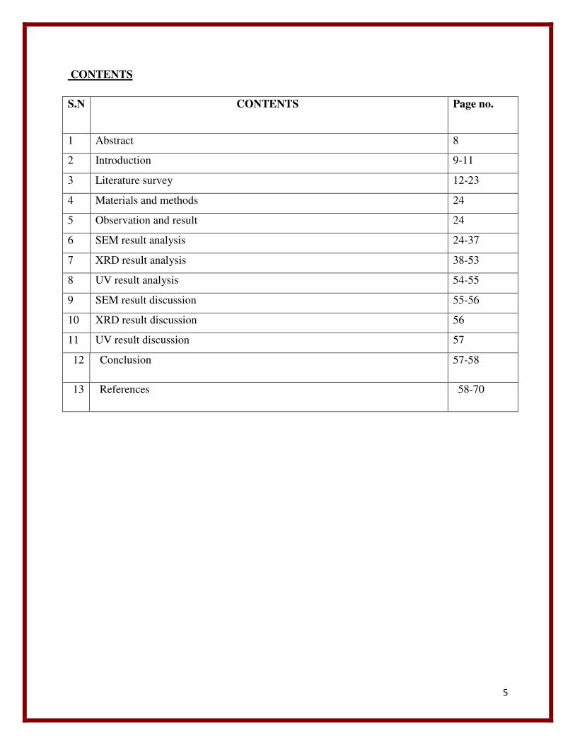
CONTENTS
S.N CONTENTS Page no.
1 Abstract 8
2 Introduction 9-11
3 Literature survey 12-23
4 Materials and methods 24
5 Observation and result 24
6 SEM result analysis 24-37
7 XRD result analysis 38-53
8 UV result analysis 54-55
9 SEM result discussion 55-56
10 XRD result discussion 56
11 UV result discussion 57
12 Conclusion 57-58
13 References 58-70
6
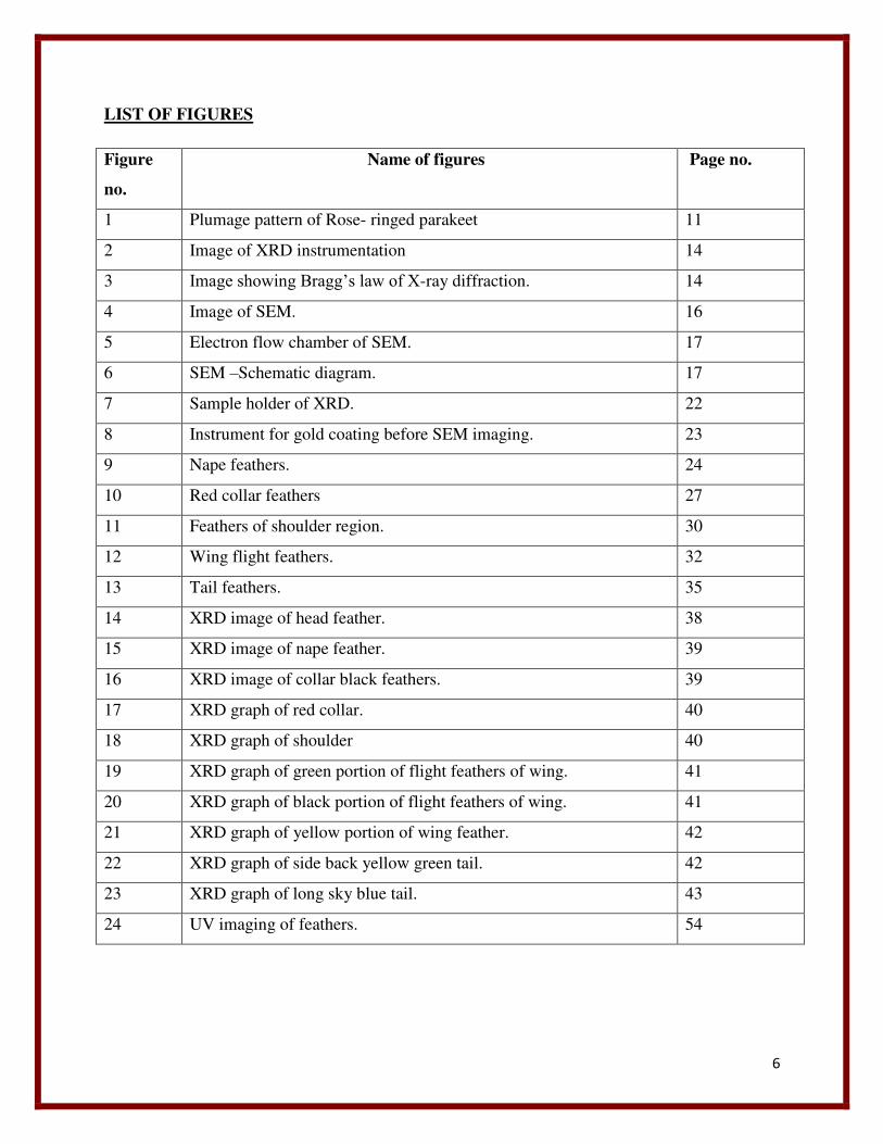
LIST OF FIGURES
Figure
no.
Name of figures Page no.
1 Plumage pattern of Rose- ringed parakeet 11
2 Image of XRD instrumentation 14
3 Image showing Bragg’s law of X-ray diffraction. 14
4 Image of SEM. 16
5 Electron flow chamber of SEM. 17
6 SEM –Schematic diagram. 17
7 Sample holder of XRD. 22
8 Instrument for gold coating before SEM imaging. 23
9 Nape feathers. 24
10 Red collar feathers 27
11 Feathers of shoulder region. 30
12 Wing flight feathers. 32
13 Tail feathers. 35
14 XRD image of head feather. 38
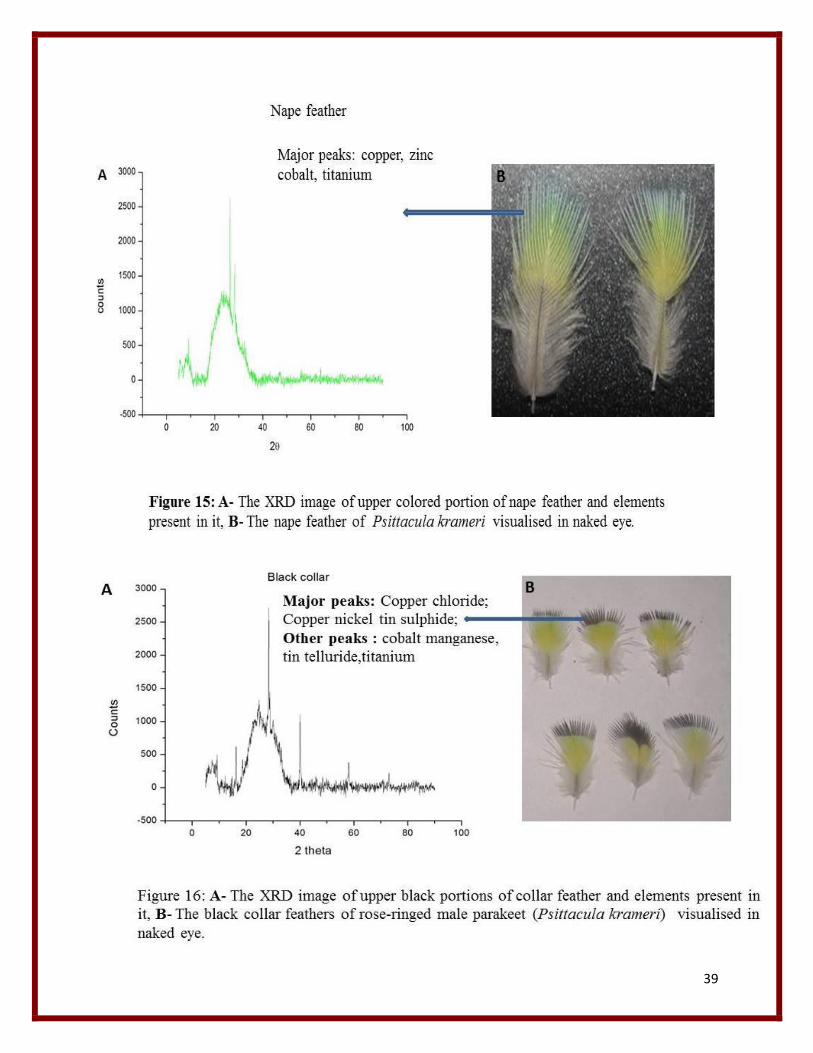
15 XRD image of nape feather. 39
16 XRD image of collar black feathers. 39
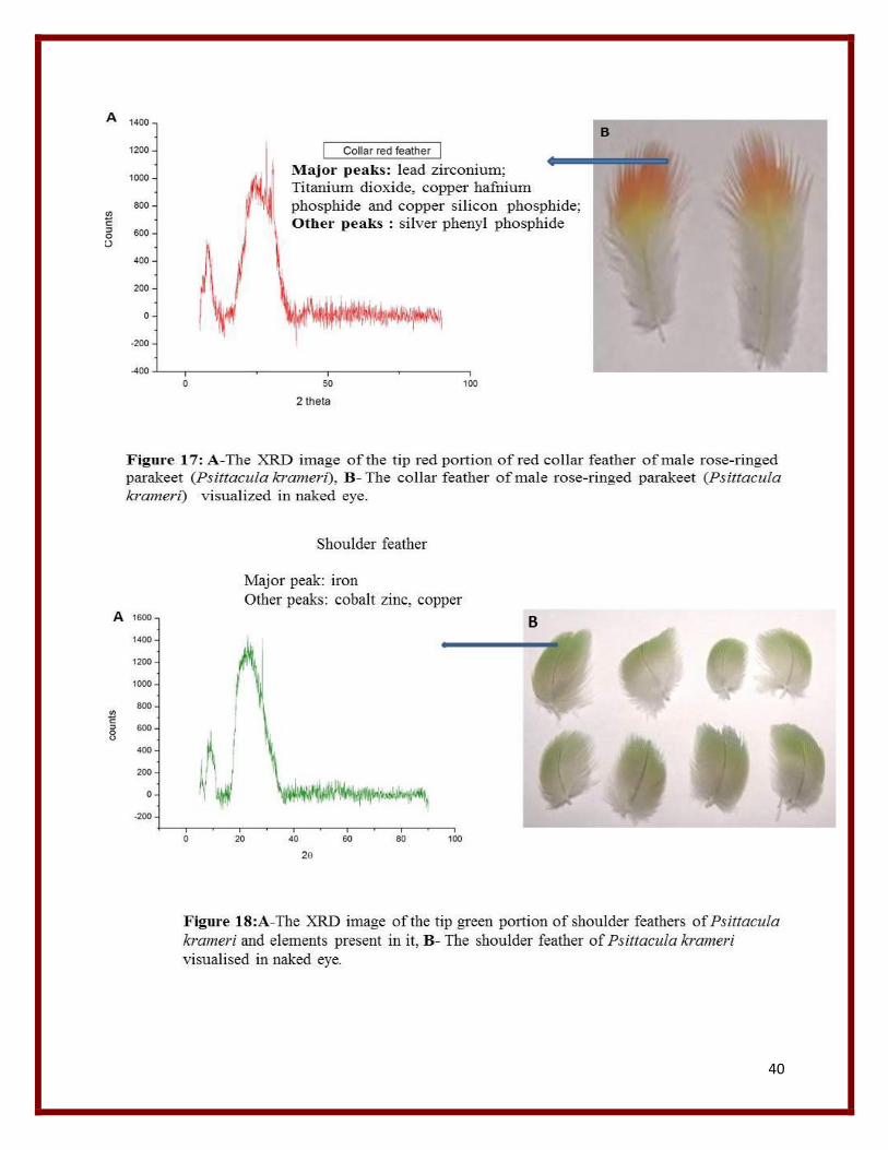
17 XRD graph of red collar. 40
18 XRD graph of shoulder 40
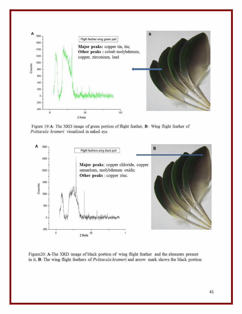
19 XRD graph of green portion of flight feathers of wing. 41
20 XRD graph of black portion of flight feathers of wing. 41
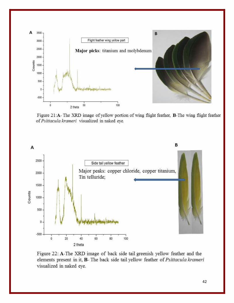
21 XRD graph of yellow portion of wing feather. 42
22 XRD graph of side back yellow green tail. 42
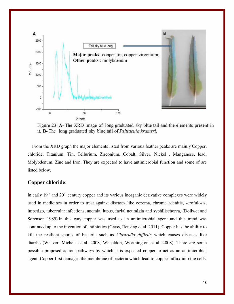
23 XRD graph of long sky blue tail. 43
24 UV imaging of feathers. 54
7
ABBREVIATIONS
XRD: X-ray diffraction
UV: Ultra violet
SEM: Scanning Electron Microscope
NPS: Nanoparticles
ROS: Reactive Oxygen Species
NM: Nanomedicines
µM: Micrometer
nm: nanometer
8
ABSTRACT:
The Indian parakeet (Psittacula krameri) belongs to the order Psittaciformes family Psittacidae.
The male has a red ring in the neck collar region hence called rose ringed parakeet while the
female lack it, showing sexual dimorphism. Parrots attract because of their phenomenal feather
coloration which is because of iridescent and non-iridescent phenomenon of light. They show
different color when they are viewed from different angle and the phenomena is called
iridescent. It has been reported that the coloration is due to the microstructures that are present in
barb. To some extend these microstructures are responsible for UV reflectance property of these
feathers. It has been reported that the coloration is also due to nanostructures present on parrot’s
feather and as well as the structure and pattern formation of different feather components of
parrot. Here the XRD analyses of the feathers were done and the elements were detected using
the EDX software, and some of their possible functions were predicted. Apart from this UV
reflectance of various feathers were examined, where some show UV reflectance and some lack
it. The possible function of UV reflectance is predicted to be useful in mate selection. Most
importantly to understand pattern formation of various feather components the SEM was used.
Key words: Parakeet, Scanning electron microscopy (SEM), X-ray diffraction (XRD),
Nanoparticles (NP), Nanomedicines (NM), Iridescent, UV reflectance.
9
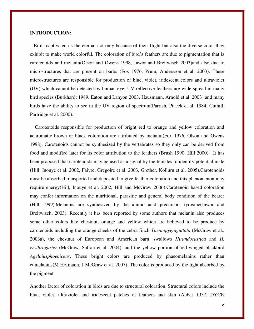
INTRODUCTION:
Birds captivated us the eternal not only because of their flight but also the diverse color they
exhibit to make world colorful. The coloration of bird’s feathers are due to pigmentation that is
carotenoids and melanin(Olson and Owens 1998, Jawor and Breitwisch 2003)and also due to
microstructures that are present on barbs (Fox 1976, Prum, Andersson et al. 2003). These
microstructures are responsible for production of blue, violet, iridescent colors and ultraviolet
(UV) which cannot be detected by human eye. UV reflective feathers are wide spread in many
bird species (Burkhardt 1989, Eaton and Lanyon 2003, Hausmann, Arnold et al. 2003) and many
birds have the ability to see in the UV region of spectrum(Parrish, Ptacek et al. 1984, Cuthill,
Partridge et al. 2000).
Carotenoids responsible for production of bright red to orange and yellow coloration and
achromatic brown or black coloration are attributed by melanin(Fox 1976, Olson and Owens
1998). Carotenoids cannot be synthesized by the vertebrates so they only can be derived from
food and modified later for its color attribution to the feathers (Brush 1990, Hill 2000). It has
been proposed that carotenoids may be used as a signal by the females to identify potential male
(Hill, Inouye et al. 2002, Faivre, Grégoire et al. 2003, Grether, Kolluru et al. 2005).Carotenoids
must be absorbed transported and deposited to give feather coloration and this phenomenon may
require energy(Hill, Inouye et al. 2002, Hill and McGraw 2006).Carotenoid based coloration
may confer information on the nutritional, parasitic and general body condition of the bearer
(Hill 1999).Melanins are synthesized by the amino acid precursors tyrosine(Jawor and
Breitwisch, 2003). Recently it has been reported by some authors that melanin also produces
some other colors like chestnut, orange and yellow which are believed to be produce by
carotenoids including the orange cheeks of the zebra finch Taeniopygiaguttata (McGraw et al.,
2003a), the chestnut of European and American barn iswallows Hirundorustica and H.
erythrogaster (McGraw, Safran et al. 2004), and the yellow portion of red-winged blackbird
Agelaiusphoeniceus. These bright colors are produced by phaeomelanins rather than
eumelanins(M Hofmann, J McGraw et al. 2007). The color is produced by the light absorbed by
the pigment.
Another factor of coloration in birds are due to structural coloration. Structural colors include the
blue, violet, ultraviolet and iridescent patches of feathers and skin (Auber 1957, DYCK
10
1985).This structural coloration occur due to scattering of coherent light by the nanostructure
arrangement of the various feather components like keratin melanin and air in barbs and
barbules. The structural coloration produce due to refractive index variation of all those feathers
(Prum 2006). However it has been reported that the structural coloration are due to the melanin
containing organelles called melanosomes they are formed during the course of development
(Maia, Brasileiro et al. 2012), to form hexagonal closed packed structures (Durrer, 1977).
Brighter and more contrasting saturated colors can be produced by increasing the contrast
refractive index, increasing the relative amount of elements of low refractive index (keratin) by
adding space between melanosomes, with potentially decreasing order (Torquato, Truskett et al.
2000). This pattern of coloration is called iridescent that is different colors are observed when
viewed from different angles (Newton) and this phenomena is very common in birds (Yoshioka
and Kinoshita 2002, Eliason and Shawkey 2012). Another subtype of structural coloration is
non-iridescent. Non-iridescent colors generally remain similar in appearance regardless of
viewing geometry or angle of observation (Newton 1952). Barbule curvature influences the
orientation of the melanosomes with respect to the viewer which is responsible for the
production of structural coloration (Wilts, Michielsen et al. 2012).
However there are some exceptions found in case of parrot. They show a wide variety of
coloration that is from red to yellow in their feathers, but they don’t derive their coloration from
carotenoids (Hudon and Brush 1992). They derive their coloration from a specific class of
biochromes called psittacofulvins (Krukenberg 1882). However psittacofulvins show similar
characterstics as of the carotenoids like they have similar light reflectance based on their C=C
bond (Veronelli, Zerbi et al. 1995), similarity in solubility (Hudon and Brush1992) and follow an
equal mechanism to incorporate into feathers for exhibiting color. As they are lipids it is believed
that they get into the follicles from blood stream by passive diffusion (Lucas and Stettenheim
1972). The physiological and anatomical origin of psittacofulvins are not known (Stradi, Pini et
al. 2001). Why parrots use psittacofulvin instead of carotenoids is an open question. Although
parrots have a capability to accumulate carotenoids in a significant amount still they fail to
incorporate it into feathers for coloration (McGraw and Nogare 2004). These pigments are also
not obtained from diet as the blood shows the absence of psittacofulvins (McGraw and Nogare
2004). Hence like other birds parrots don’t show a change in color in the disturbance of nutrition
(Nemésio 2001). Their absence in blood also shows that they are synthesized locally but not
11
centrally (McGraw and Nogare 2004). It is expected that they might be synthesized at the mature
follicles of growing feathers (McGraw and Nogare 2004), as melanins are synthesized within the
feather tract (Ralph 1969), and carotenoids are synthesized at the feather follicle (Stradi 1998).
Parrots may be the only class of aves who synthesize their own pigments (McGraw 2005) and
they show composite coloration, both pigmentation and structural coloration.. Here we made a
study on the structure, structural arrangement and pattern formation by different feather
components of parrot, taken from different parts of its body and there by predicted some of their
possible functions. Here we will study regarding the Indian rose ringed parakeet Psittacula
krameri of Class-Aves, Order-Psittaciformes, Superfamily- Psittacoidea, Family- Psittaculidae,
Sub family- Psittaculinae, Tribe- Psittaculini, Genus- Psittacula, Species-Krameri.
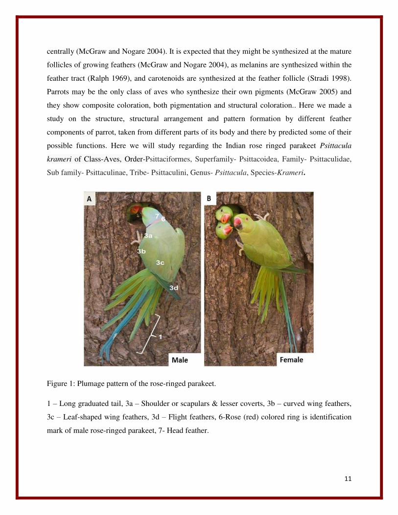
Figure 1: Plumage pattern of the rose-ringed parakeet.
1 – Long graduated tail, 3a – Shoulder or scapulars & lesser coverts, 3b – curved wing feathers,
3c – Leaf-shaped wing feathers, 3d – Flight feathers, 6-Rose (red) colored ring is identification
mark of male rose-ringed parakeet, 7- Head feather.
12
LITERATURE SURVEY:
PRINCIPLE OF XRD:
In order to understand the function of various biological structures it is important to study their
structure as well as the structural interactions. Although a number of high resolution microscopes
have been developed the only method that currently yields better result of biological structure at
atomic level of resolution is X-ray diffraction from a single crystal. This technique involve three
distinct steps, i) growing a crystal ii) collecting X-ray diffraction pattern from the crystals iii)
constructing and refining a structural model from the crystals to fit the X-ray diffraction pattern.
A molecular structure solved to atomic resolution means the position of each atom can be
distinguished from those of all atom in three dimensional space without applying any
assumptions regarding the structure of molecule. The closest distance between the two atoms in
space is the length of covalent bond between them. Since the approximate distance between a
covalent bond is 0.12nm we need to see two atoms as distinct particles. There are theoretical and
practical limitations to resolve a structure to this fine level. First the system has its atoms of its
molecules held rigidly and second each molecule or group of molecules in the system must have
identical confirmation. We must now find a radiation source that allows us to see two atoms
separated by a radius 0.12nm. The limit of resolution (LR) of any optical method is defined by
the wavelength lambda of the incident radiation.
LR= �/2
This is because an extension of Heisenberg’s principle that results from treating light as a wave
states that the position of a particle cannot be fixed to better than about half the wavelength of
the radiation used to examine that particle. If we require the resolution of the technique to be
0.12nm to resolve the atoms of a molecule, the wavelength of the light required to be 0.24nm.
This falls into the X-ray range of electromagnetic spectrum. As x-rays cannot be focused to
obtain the image as we get by light microscope, so we have to depend upon the constructive and
destructive inference we obtain depending upon the scattering of radiation by the regular and
repeating lattice of the crystals to determine the structure. The energy of a quantum of X-ray
radiation is equivalent to 8000eV which is approximately equal to the energy of the all electrons
present on its orbit. This equivalence of the energy leads to interaction so that the electrons of an
13
atom are primarily responsible for the scattering of X-rays. The numbers of electrons present in a
given volume or space determines how strongly an atom scatters x-rays. The interference of the
scattered x-rays leads to the general phenomenon of diffraction.
How x-ray diffraction is used to study the structures:
All electromagnetic radiations have dual nature of matter, both particulate as well as wave
nature of matter. In x-ray scattering we treat all electromagnetic radiation as waves. Scattering
simply refers to the ability of an object to change the direction of waves. An object placed in the
path of a light from a point of source cannot cast a sharp shadow because of scattering from its
edges. The origin of scattering of light can be best understood from Huygens’s principle that
“every point along a wave front can be considered to be the origin of a new wave front. The
velocity of this new wave front can be considered to be same as the original one. Objects placed
on the path of a wave front act as a point of propagation of new waves. The entirely new wave is
called scattered wave. If we place two objects A and B on the path of a wave front each of the
two objects will propagate a new wave front having identical wavelength and velocities. At some
position in space the wave propagating from point A will reinforce the scattered wave from point
B through constructive interference through if the two scattered waves are in phase. If the
amplitude of the wave A reduces the amplitude of the wave B then it is said waves are out of
phase hence called destructive interference. This is called diffraction, the sum of the two waves
propagated from point A and B result in an amplitude that is relatively depend on the positions
of A and B and where new wave fronts are being observed. This is how the diffraction of x-ray
solves the structure of molecules from single crystal.
14
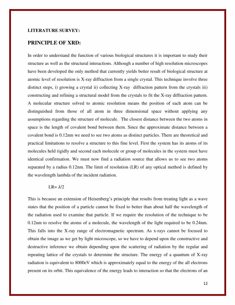
Figure2: Image of XRD instrument. A-The X-ray instrument, B- the X-ray beam generation
chamber.
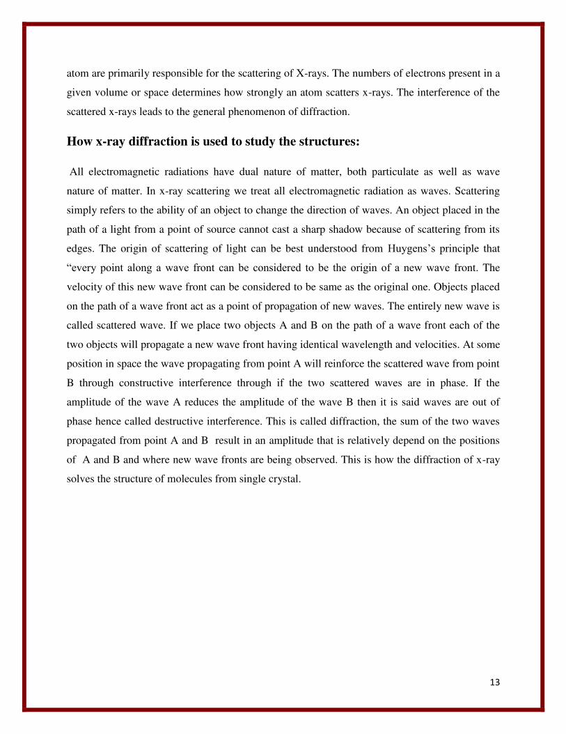
Bragg’s law of x-ray diffraction:
Figure3: Parallel lattice planes A and B showing the constructive interference of a front wave of
parallel X-ray beam, 1and 2 are the incident radiations incidence at an angle of θ while 1’ and 2’
are the reflected radiations at an angle of θ.
15
W.L.Bragg developed a relationship to understand how diffraction relates to the relative
positions of point objects in shape. To define Bragg’s law of diffraction, lets assume lattice
points on the crystal as parallel planes. Stacking a set of reflecting planes at regularly spaced
intervals d creates a simple model of a one dimensional crystal. In this model a wave of x-rays
with a wavelength λ is incident on the reflecting planes at an angle θ. The wave scattered from
this plane is at an identical angle θ. But the question is which value of θ will result in
constructive and destructive interference. As the distance between the parallel planes is d so the
individual paths of the scattered light are parallel. Because we have a large no. of planes we
observe constructive interference only when the reflected waves are perfectly in phase. This only
occurs when the difference in the length of the path of the incident and reflected waves of each
plane that is path difference (PD) is equal to some integer n of the wavelength of the incident x-
rays.
PD= nλ
This path is related to the distance separating the reflecting planes by the relationship
½ PD =dsinθ
=2d sin θ= nλ
=2sinθ=nλ/d
Larger spacing units in crystal result in smaller diffraction angle.
This principle of x-ray diffraction explains how diffractions through crystal determine the
structure of molecules.
16
PRINCIPLE OF SEM
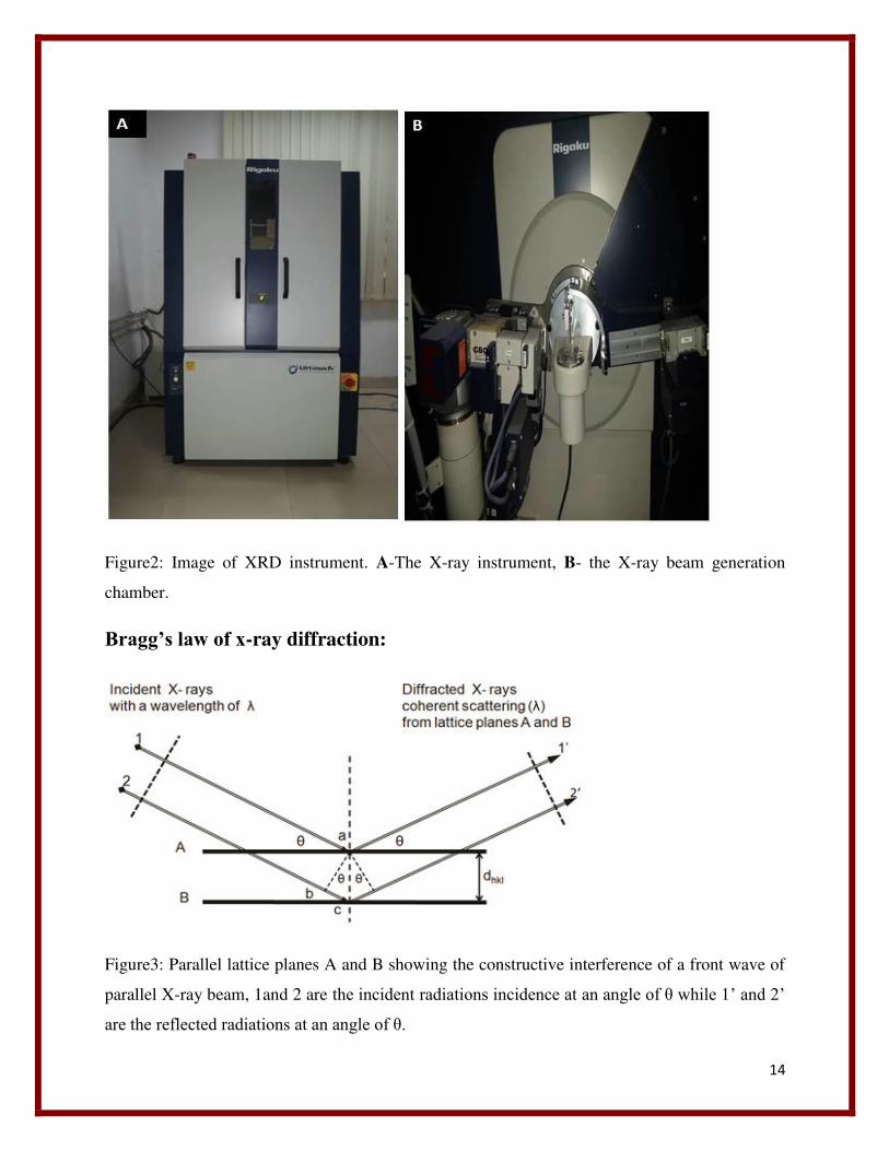
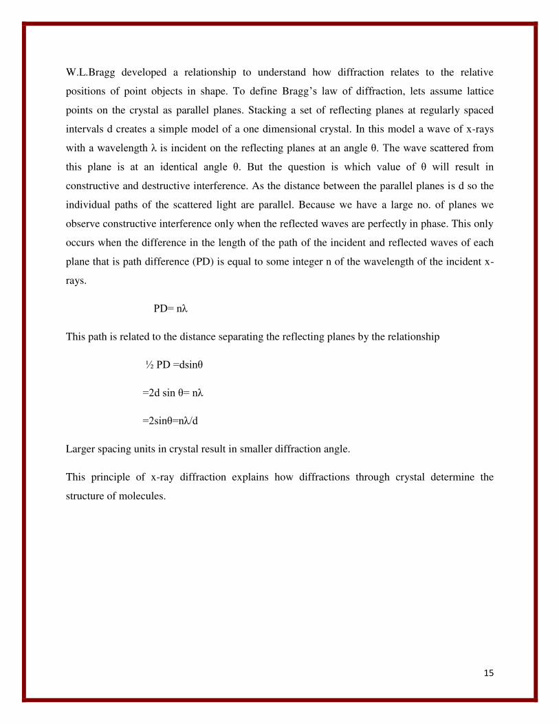
Figure4: The image of Scanning Electron Microscope
SEM stands for scanning electron microscope, is an instrument that scan the surface of the
specimen,by a beam of electron, instead of light and that reflect to form an image. Now a days it
is largely used in physical sciences and biological sciences for detecting the structures. SEM
works by the coordinated action of seven primary operational systems. Vacuum, beam
generation, beam manipulation, beam interaction, detection, signaling processing, and display
record. The following components are present in a scanning electron microscope:
17
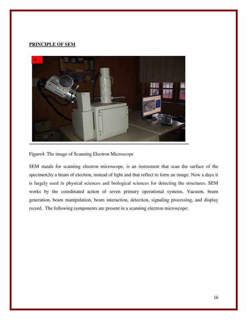
Figure5: Image showing electron flow chamber of SEM.
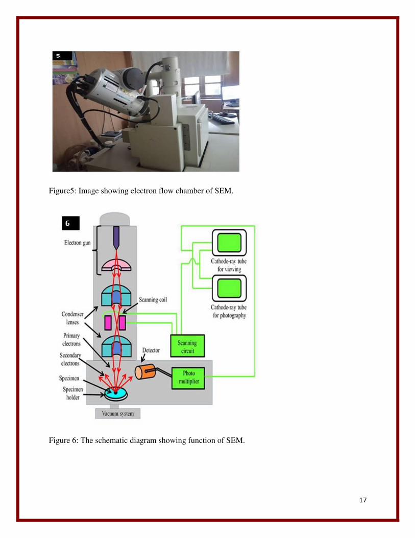
Figure 6: The schematic diagram showing function of SEM.
18
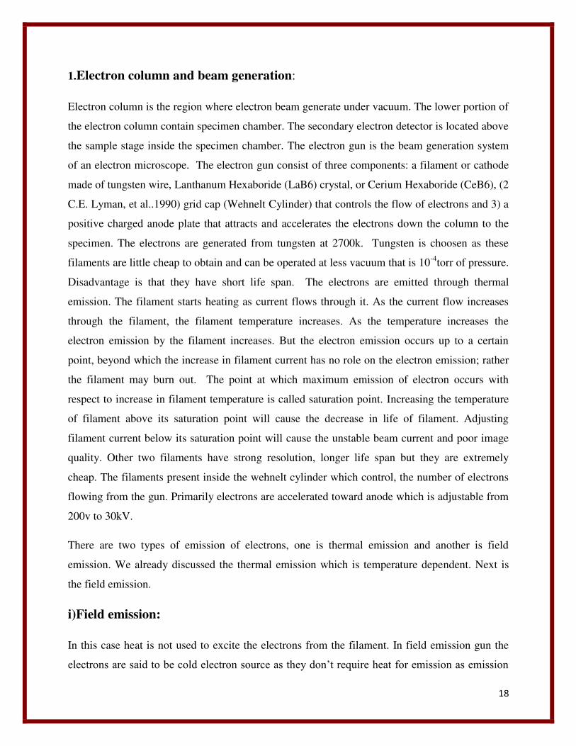
1.Electron column and beam generation:
Electron column is the region where electron beam generate under vacuum. The lower portion of
the electron column contain specimen chamber. The secondary electron detector is located above
the sample stage inside the specimen chamber. The electron gun is the beam generation system
of an electron microscope. The electron gun consist of three components: a filament or cathode
made of tungsten wire, Lanthanum Hexaboride (LaB6) crystal, or Cerium Hexaboride (CeB6), (2
C.E. Lyman, et al..1990) grid cap (Wehnelt Cylinder) that controls the flow of electrons and 3) a
positive charged anode plate that attracts and accelerates the electrons down the column to the
specimen. The electrons are generated from tungsten at 2700k. Tungsten is choosen as these
filaments are little cheap to obtain and can be operated at less vacuum that is 10-4torr of pressure.
Disadvantage is that they have short life span. The electrons are emitted through thermal
emission. The filament starts heating as current flows through it. As the current flow increases
through the filament, the filament temperature increases. As the temperature increases the
electron emission by the filament increases. But the electron emission occurs up to a certain
point, beyond which the increase in filament current has no role on the electron emission; rather
the filament may burn out. The point at which maximum emission of electron occurs with
respect to increase in filament temperature is called saturation point. Increasing the temperature
of filament above its saturation point will cause the decrease in life of filament. Adjusting
filament current below its saturation point will cause the unstable beam current and poor image
quality. Other two filaments have strong resolution, longer life span but they are extremely
cheap. The filaments present inside the wehnelt cylinder which control, the number of electrons
flowing from the gun. Primarily electrons are accelerated toward anode which is adjustable from
200v to 30kV.
There are two types of emission of electrons, one is thermal emission and another is field
emission. We already discussed the thermal emission which is temperature dependent. Next is
the field emission.
i)Field emission:
In this case heat is not used to excite the electrons from the filament. In field emission gun the
electrons are said to be cold electron source as they don’t require heat for emission as emission
19
in this case occur by strong magnetic field. The filament consists of a fine pointed tungsten wire
(ideally one atom across at the tip), kept at a high negative charge along with the rest of the
cathode. The anode is kept at a positive charge. The large difference in potential difference on a
small point pulls the electrons out of the filament. The advantages to this system are that the
emission is cool, the emitted beam is much smaller in diameter (better resolution), and filament
life is generally longer. The disadvantages are a much higher vacuum (approximately 10-7Torr)
require and a cleaner microscope (special pumps not oil diffusion) is needed for operation. Any
debris sticking on the fine tip of the filament will cause a reduction in emission. Debris will
routinely stick to the filament. Thus the microscope needs to be cleaned out routinely to maintain
a clean gun. Another disadvantage of field emission is long acquisition times during X-ray
analysis. Field emission gun will not generate many X-rays since low beam currents and the
small beam diameters are used.
ii) Condenser lenses and electron beam manipulation:
Two condenser lenses are there. After the beam passed through the anode it again travel through
two condenser lenses in order to converge or narrow down the beams , so that it will pass
through the focal point of lense. Along with the accelerating current, the lenses are used for
determining the intensity of the electron beam when it strikes to the specimen ( Postek, et
al.1980).
iii) Apertures:
Depending upon the kinds of aperture can be used in the electron column. The aperture located
below the scanning coil determines the diameter or spot size of the beam at the specimen. The
spot size determines the resolution of the image. Lesser the diameter of the spot size more will be
the resolution.
iv) Scanning system:
The image is formed when the electron beam scan the surface of specimen. There are two coils
present in the objective lense for scanning and raster formation. One is rastering coil and other is
deflecting coil. Deflecting coils are present to deflect the electron beam such a way in a multiple
direction that it will focus on the scanning spot. The deflecting coils produce a uniform magnetic
20
field for the uniform movement of electron field. The rastering coil causes the beam to scan over
a square area on the sample surface.
v) Specimen chamber:
Specimen chamber and its control are located at the lower part of the column. The secondary
electrons from the specimen that is the electrons are obtained on specimen by coating are
attracted to the detector by a positive charge.
2.Vacuum system:
High vacuum is needed to maintain the electron column in order to control the electron beam
falling on the specimen. A high vacuum is maintained at a pressure of atleast of 5x10⁻⁵torr. High
vacuum is required for the reasons like, first when the current passes through the filament cause
the increase of temperature around 2700k.(2 C.E. Lyman, et al..1990). At this temperature and in
the presence of air hot tungsten filament will burn out because of oxidation. The column optics
need a clean and dust free environment in order to operate. The dust particle or air inside the
column may block the path of electron beam before it reaches to specimen chamber (Postek, et
al.,1980).
3.Electron beam and specimen interaction :
To get a good resolution an electron source is required instead of light source which can bring
resolution up to 25A0. In SEM image can be visualized by two kinds of electron flow, secondary
and back scattered electrons. These two kinds of electrons produced from the surface of
specimen, but under electron beam they are result of two different interactions. Secondary
electrons are a result of the inelastic collision and scattering of incident electrons with specimen
electrons. They are generally characterized by possessing energies of less than 50 eV (Postek, et
al.,1980). They are used to reveal the surface structure of a material with a resolution of ~10 nm
or better (Postek et al.,1980). There are several types of signals are generated from the
interaction of electron beam and specimen. One among them is X-ray, the only other signal
which is used typically in SEM. The x-ray signal is the result of interactions between free
electrons and positive electron holes that are generated within the material. The x-ray signal can
originate from further down into the surface of the specimen surface and allows for
21
determination of elemental composition through EDS (energy dispersive x-ray spectroscopy)
analysis of characteristic x-ray signals.
4.Accelerating Voltage:
The voltage can be adjusted between 200-30kV. Higher voltages (15- 30kv) generally give high
resolution at high magnifications although; this can damage the specimen very quickly if it is not
highly conductive.
5.Working distance:
Working distance and spot size will greatly influence the image quality. Generally a working
distance of 10mm should be used and will allow for a good depth of field while maintaining
good resolution. Working distance can be reduced in case of low accelerating voltage in order to
get good resolution.
6.Spot size:
Spot size basically restricts the beam current and thereby cause for brightness and contrastness of
image. Smaller spot sizes require higher brightness and contrast levels thus there can be a limits
when using a small spot size. Typically smaller spot sizes allow for higher resolution and a
greater depth of field.
MATERIALS AND METHODS:
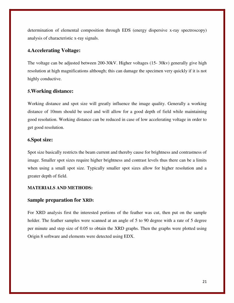
Sample preparation for XRD:
For XRD analysis first the interested portions of the feather was cut, then put on the sample
holder. The feather samples were scanned at an angle of 5 to 90 degree with a rate of 5 degree
per minute and step size of 0.05 to obtain the XRD graphs. Then the graphs were plotted using
Origin 8 software and elements were detected using EDX.
22
Figure 7: Sample holder of XRD
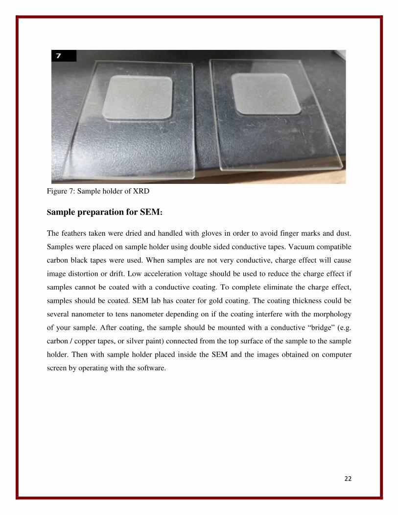
Sample preparation for SEM:
The feathers taken were dried and handled with gloves in order to avoid finger marks and dust.
Samples were placed on sample holder using double sided conductive tapes. Vacuum compatible
carbon black tapes were used. When samples are not very conductive, charge effect will cause
image distortion or drift. Low acceleration voltage should be used to reduce the charge effect if
samples cannot be coated with a conductive coating. To complete eliminate the charge effect,
samples should be coated. SEM lab has coater for gold coating. The coating thickness could be
several nanometer to tens nanometer depending on if the coating interfere with the morphology
of your sample. After coating, the sample should be mounted with a conductive “bridge” (e.g.
carbon / copper tapes, or silver paint) connected from the top surface of the sample to the sample
holder. Then with sample holder placed inside the SEM and the images obtained on computer
screen by operating with the software.
23
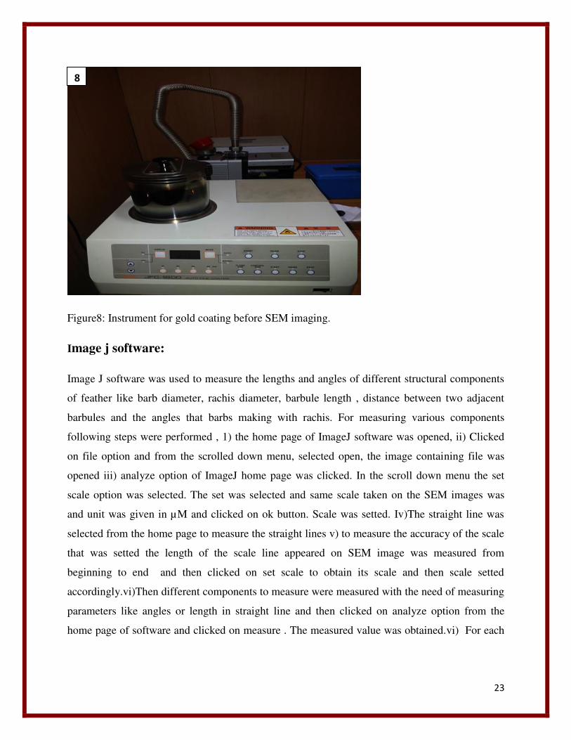
Figure8: Instrument for gold coating before SEM imaging.
Image j software:
Image J software was used to measure the lengths and angles of different structural components
of feather like barb diameter, rachis diameter, barbule length , distance between two adjacent
barbules and the angles that barbs making with rachis. For measuring various components
following steps were performed , 1) the home page of ImageJ software was opened, ii) Clicked
on file option and from the scrolled down menu, selected open, the image containing file was
opened iii) analyze option of ImageJ home page was clicked. In the scroll down menu the set
scale option was selected. The set was selected and same scale taken on the SEM images was
and unit was given in µM and clicked on ok button. Scale was setted. Iv)The straight line was
selected from the home page to measure the straight lines v) to measure the accuracy of the scale
that was setted the length of the scale line appeared on SEM image was measured from
beginning to end and then clicked on set scale to obtain its scale and then scale setted
accordingly.vi)Then different components to measure were measured with the need of measuring
parameters like angles or length in straight line and then clicked on analyze option from the
home page of software and clicked on measure . The measured value was obtained.vi) For each
8
24
measurement 10 readings were taken and the average and standard deviation calculated to obtain
the measurement of particular component.
OBSERVATION AND RESULT
Nape feather:
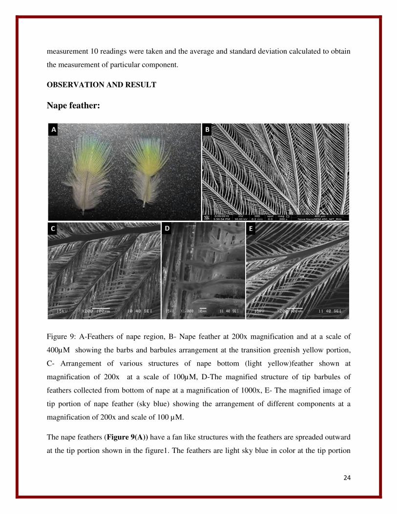
Figure 9: A-Feathers of nape region, B- Nape feather at 200x magnification and at a scale of
400µM showing the barbs and barbules arrangement at the transition greenish yellow portion,
C- Arrangement of various structures of nape bottom (light yellow)feather shown at
magnification of 200x at a scale of 100µM, D-The magnified structure of tip barbules of
feathers collected from bottom of nape at a magnification of 1000x, E- The magnified image of
tip portion of nape feather (sky blue) showing the arrangement of different components at a
magnification of 200x and scale of 100 µM.
The nape feathers (Figure 9(A)) have a fan like structures with the feathers are spreaded outward
at the tip portion shown in the figure1. The feathers are light sky blue in color at the tip portion
25
followed by a light yellowish green transition toward the bottom , the next portion present after
this transition region toward the bottom is yellow in color , then the region present next to it
toward extreme bottom is grayish white in color. In general half of the portion, these feathers
toward the tip are colored while half of the portions toward bottom are grayish white in color.
The SEM images of these feathers were taken at an electron flow of 10.00KV. at a spot
size of 6.0 with different magnifications at different scales too in order to study the arrangement
of various components of feathers, which are done by nature at a nanoscale.
Unlike all feathers it (Figure 9(B)) consists of a central shaft or rachis. The lower portion of
the shaft or rachis is free from barbs or barbules and is called quillis. In the upper portion of
feather the rachis gets thinner, tapered and branched toward the end. The rachis gives rise to
barbs laterally and they are present parallel to each other by forming an angle with the rachis.
Then barbs give rise to barbules, which are also arising laterally from the barbs. This gives the
feather an appearance of branches of a coconut tree. The barbules of each barb are arranged in
such a way that whole, together they look like veins of a leaf to naked eye. When this feather is
visualized under SEM (200x) the nice structural arrangement of the barbs and barbules are
visible Barbules of each barb generally form a pattern with its adjacent barbules present on both
left and right side, by interlocking with each other. Each barb contains barbules on its both left
and right side in a symmetric fashion. In each barb containing barbules, one row of barbule
present on one side are exposed while other side is present beneath the adjacent barbules of just
nearby barb. When we observed the arrangement of barbs and barbules on both side of the rachis
an opposite pattern is viewed. On the left side of the rachis the barbules present on left of each
barb are covered by its adjacent right barbules of the barb present just left to it. Hence the left
barbules of one barb are interlocked beneath the right barbules of another barb just present left to
it. But when we viewed on right side of the rachis we observed the just opposite pattern of
arrangement of the barbules of each barb, that the left barbules are exposed while the barbules
present on the right side of barb are present beneath the barbules of the barb just present right
adjacent to it.
At the bottom of nape feather (Figure 9 (C)) a different arrangement pattern is observed, the
structures of barbule components are also found to be different in these regions. The barbules
which are exposed are flattened in shape from its base and elongated to a distance of 118.16±
26
10.09µM then tapered toward the end where the distance is calculated to be 66.36 ±11.63 µM
.The diameter of rachis at this point is calculated to be 27.10 ± 3.82µM. Distance between two
individual barbules is 13.80 ± 1.78µM. The diameter or thickness of the barbs present at nape
bottom region is 6.72 ±1.18 µM. The angle that barbs at bottom nape region are making with the
rachis is 42.42 ± 1.10µM. The distance between two barbs at nape bottom region of feather was
calculated to be 304.08 ± 6.52 µM.
The structure and arrangement of barbules at higher magnification of 1000x and at a scale of
10µM was studied in (Figure 9(D)).In some of the barbules, at tip portion they became branched
and in higher magnification it looks like an outgrowth from the branch. But this tip branching of
barbules are not symmetric or regular. It varies from barbules to barbules. The flattened barbules
are present obliquely and interlocked with each other. Very small air gaps are present in between
two barbules viewed at 1000x magnification at a scale of 10µM.
The tip portion of nape feather which looks very light sky blue was studied at a magnification of
200x with a scale length of 100µM in the (Figure 9 (E)).The tip of nape feather contains a
different type of barbules which are thin, long and similar as the middle nape feathers. The rachis
at the top little thickened branched to give three barbs. The rachis along its entire length
excluding the quillis portion lined with barbules. At the tip portion the barbules of barbs are
interlocked with the immediate barbules that are arises from the rachis itself. At the sharp tip
portion the barbules of each branched barb formed by the end branching of rachis are highly
zipped with each other. The diameter of the tip portion of the rachis was measured to be 29.78 ±
7.36 µM. The diameter or thickness of the barbs at the tip portion of nape feather was found to
be 19.86 ±3.50 µM. The diameter of rachis at the tip branching portion is found to be 30.61 ±
1.13µM. The distance between two barbules at nape tip feathers is found to be 14.99 ±2.36µM.
At the extreme end point of rachis at the nape tip region, the rachis is branched to give two barbs.
These two barbs are arise from a same point of rachis. The diameter of these two barbs were
calculated to be 14.78 ±3.78µM
27
Red collar feather:
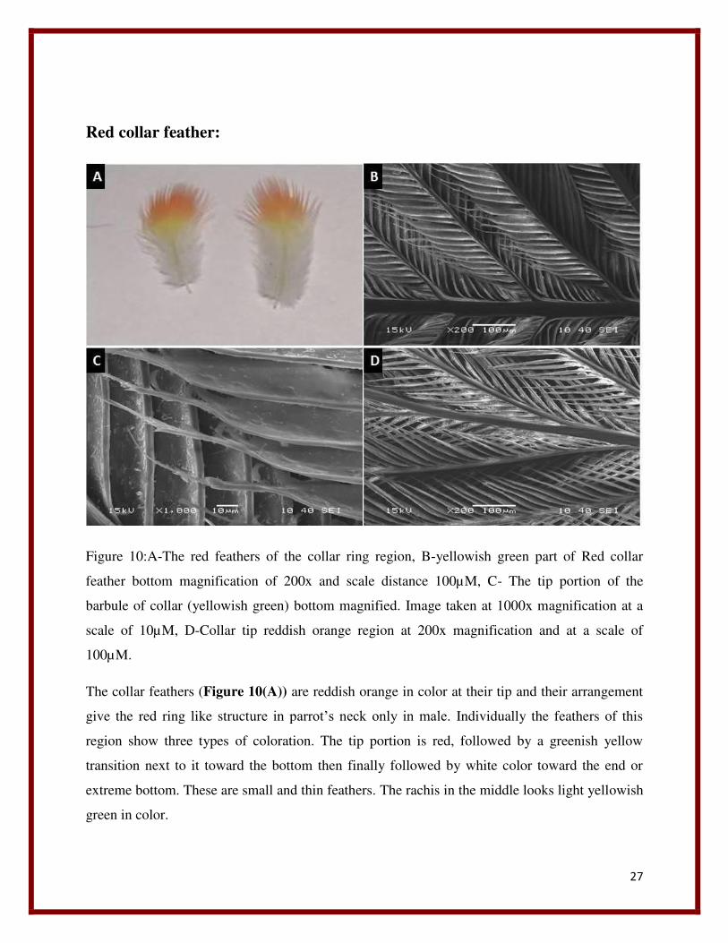
Figure 10:A-The red feathers of the collar ring region, B-yellowish green part of Red collar
feather bottom magnification of 200x and scale distance 100µM, C- The tip portion of the
barbule of collar (yellowish green) bottom magnified. Image taken at 1000x magnification at a
scale of 10µM, D-Collar tip reddish orange region at 200x magnification and at a scale of
100µM.
The collar feathers (Figure 10(A)) are reddish orange in color at their tip and their arrangement
give the red ring like structure in parrot’s neck only in male. Individually the feathers of this
region show three types of coloration. The tip portion is red, followed by a greenish yellow
transition next to it toward the bottom then finally followed by white color toward the end or
extreme bottom. These are small and thin feathers. The rachis in the middle looks light yellowish
green in color.
28
The feathers at collar region are red. The SEM image of the red feathers at collar region is taken
at 200x magnification and at a scale of 100µM with a spot size of 6.0.
The diameter of the rachis at collar bottom (Figure 10(B)) was calculated to be 35.21±
2.49µM. From the rachis barbs are arise from both sides alternate to each other. The diameter of
barbs were measured and calculated to be 9.21±2.70 µM. The barbules are flattened in shape
from the beginning that is from the base, then at the terminal end these are narrowed to form a
thread like thin structure. Hence it can be said the barbules have two parts one is anterior wide
and flattened part and second posterior terminal thread like part. These thread like structures in
some barbules are extended to reach the adjacent barb present near to it. The length up to which
the flattened portions of barbules are extended measured and calculated to be 128.94±9.51µM.
The length of the tapered thread like part of the barbule were measured and calculated to be
79.28±12.48µM. The exposed body surface width of the exposed barbules were measured and
calculated to be 16.58±3.06µM. The distance between two adjacent barbs in these feathers was
measured and calculated to be 216.74±6.30µM. In this feather the barbules form a uniform
arrangement by overlapping on the barbules of adjacent barb. The barbules which are exposed
visible entirely with all its structures and the barbules which are present underneath expose the
base portion only , while the remaining portions are covered by the nearby barbules.
The thread like tip portion of the barbules are magnified to 1000x at a scale of 10µM
(Figure 10 (C)). It was observed that the point from where the thread like elongated tapered
structures arises from the flattened barbules , are associated with another outgrowth like structure
along one side which can be visualized as a bifurcation. The elongated thread like tapered end
and barbules are highly irregular and gives rise to many outgrowths like structures along its
entire length and pointed toward end. It can be observed from the above image the flattened end
of barbules is bifurcated at one end to give the elongated thread like structure while the other
side is placed at its position as such, and tapered to become pointed.
At the tip portion of collar feather the rachis is more branched (Figure 10 (D)). The diameter of
the rachis was measured and calculated to be 30.96±3.05µM. At the tip of the collar the rachis is
highly branched to give many barbs which form a canopy like structure at the top. Toward the tip
of the collar feather the rachis becomes thinner and the diameter can be measured and calculated
as 20.57±2.34µM. The diameter of the barbs measured and calculated to be 11.04±1.76 µM. The
29
barbules present at this region are observed to have a different structure, than the one present at
bottom. These are thinner and less flattened than the structures found at collar bottom region. As
observed from the image, barbules of this region become thinner from base to the tip gradually in
a uniform way and pointed at the end. In this image it can be observed that there present some
spaces between each adjacent barbules at top although they are not uniform. The distance
between the barbules at tip measured and calculated to be 16.38±3.85 µM. At the tip the barbules
were observed to show a highly interlocked structure, with the barbules at one side of barbs are
exposed and the other side was falling beneath the barbules of adjacent barb.
SEM ANALYSIS OF SHOULDER FEATHER:
The feathers from the shoulder region were collected (Figure 11 (A)). These are the tiny and
thinnest feather of the parrots. They look grayish green in color. The tip portion is light faded
green. Then it is followed by a light grey coloration toward the base. The feathers are wide,
curved with a blade like structure. Even though all these feathers are collected from the same
part of body, still their pattern of coloration and their shape varies from each other which can be
visualized from the original feather images of figure 11(A). All these feathers are curved from
their lateral dorsal side. Some feathers are curved bulged out laterally from right edge and some
are from left edge and some are uniform along its both edges.
30
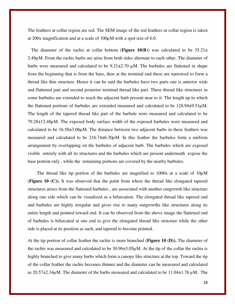
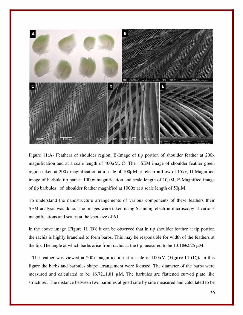
Figure 11:A- Feathers of shoulder region, B-Image of tip portion of shoulder feather at 200x
magnification and at a scale length of 400µM, C- The SEM image of shoulder feather green
region taken at 200x magnification at a scale of 100µM at electron flow of 15kv, D-Magnified
image of barbule tip part at 1000x magnification and scale length of 10µM, E-Magnified image
of tip barbules of shoulder feather magnified at 1000x at a scale length of 50µM.
To understand the nanostructure arrangements of various components of these feathers their
SEM analysis was done. The images were taken using Scanning electron microscopy at various
magnifications and scales at the spot size of 6.0.
In the above image (Figure 11 (B)) it can be observed that in tip shoulder feather at tip portion
the rachis is highly branched to form barbs. This may be responsible for width of the feathers at
the tip. The angle at which barbs arise from rachis at the tip measured to be 13.18±2.25 µM.
The feather was viewed at 200x magnification at a scale of 100µM (Figure 11 (C)). In this
figure the barbs and barbules shape arrangement were focused. The diameter of the barbs were
measured and calculated to be 16.72±1.81 µM. The barbules are flattened curved plate like
structures. The distance between two barbules aligned side by side measured and calculated to be
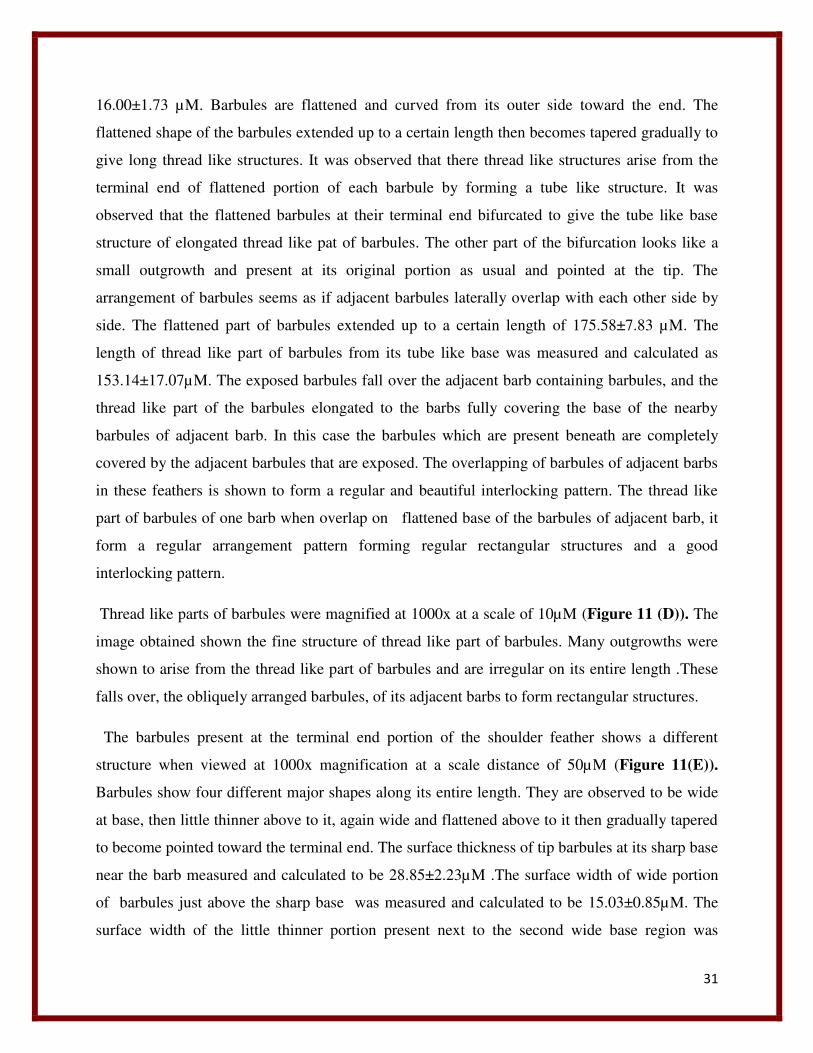
31
16.00±1.73 µM. Barbules are flattened and curved from its outer side toward the end. The
flattened shape of the barbules extended up to a certain length then becomes tapered gradually to
give long thread like structures. It was observed that there thread like structures arise from the
terminal end of flattened portion of each barbule by forming a tube like structure. It was
observed that the flattened barbules at their terminal end bifurcated to give the tube like base
structure of elongated thread like pat of barbules. The other part of the bifurcation looks like a
small outgrowth and present at its original portion as usual and pointed at the tip. The
arrangement of barbules seems as if adjacent barbules laterally overlap with each other side by
side. The flattened part of barbules extended up to a certain length of 175.58±7.83 µM. The
length of thread like part of barbules from its tube like base was measured and calculated as
153.14±17.07µM. The exposed barbules fall over the adjacent barb containing barbules, and the
thread like part of the barbules elongated to the barbs fully covering the base of the nearby
barbules of adjacent barb. In this case the barbules which are present beneath are completely
covered by the adjacent barbules that are exposed. The overlapping of barbules of adjacent barbs
in these feathers is shown to form a regular and beautiful interlocking pattern. The thread like
part of barbules of one barb when overlap on flattened base of the barbules of adjacent barb, it
form a regular arrangement pattern forming regular rectangular structures and a good
interlocking pattern.
Thread like parts of barbules were magnified at 1000x at a scale of 10µM (Figure 11 (D)). The
image obtained shown the fine structure of thread like part of barbules. Many outgrowths were
shown to arise from the thread like part of barbules and are irregular on its entire length .These
falls over, the obliquely arranged barbules, of its adjacent barbs to form rectangular structures.
The barbules present at the terminal end portion of the shoulder feather shows a different
structure when viewed at 1000x magnification at a scale distance of 50µM (Figure 11(E)).
Barbules show four different major shapes along its entire length. They are observed to be wide
at base, then little thinner above to it, again wide and flattened above to it then gradually tapered
to become pointed toward the terminal end. The surface thickness of tip barbules at its sharp base
near the barb measured and calculated to be 28.85±2.23µM .The surface width of wide portion
of barbules just above the sharp base was measured and calculated to be 15.03±0.85µM. The
surface width of the little thinner portion present next to the second wide base region was
32
measured and calculated to be 10.07±1.99µM. The surface width of wide portion of barbule just
present above the thinner portion was measured and calculated to be 16.29±1µM. The surface
width of the region fourth measured wide segment of barbule was measured and calculated to be
11.12±1.77µM. The surface width of the tube like base of thread like part of barbule was
measured and calculated to be 4.60±1.44µM. The body width of thread like part of the barbules
measured and calculated to be 2.76±0.31 µM. In the image the open space between two barbules
clearly can be observed at the base. The distance between two barbules or the open space present
between them measured and calculated to be 10.97±1.68µM.
WING FEATHERS:
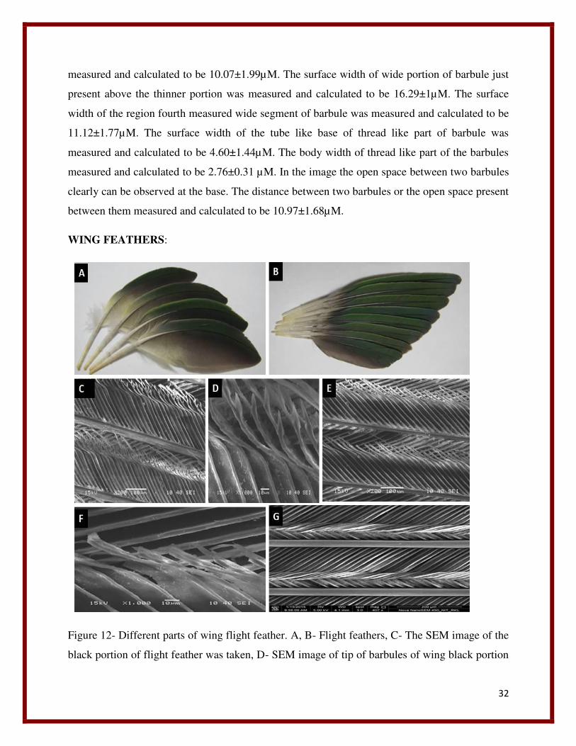
Figure 12- Different parts of wing flight feather. A, B- Flight feathers, C- The SEM image of the
black portion of flight feather was taken, D- SEM image of tip of barbules of wing black portion
33
at 1000x magnification and at a scale length of 10µM, E- SEM imaging of wing yellow portion
at 200x magnification and a scale length of 100µM, F- SEM image of tip portion of barbules of
wing yellow feathers were taken at 1000x magnification at a scale of 10µM, G- SEM image of
wing green region of flight feathers at a spot size of 3.0 at 400x magnification and at a scale
length of 200µM.
Wing feathers (Figure12(A), (B))are the strongest feathers and play a major role in bird’s flight.
These feathers exhibit three kinds of coloration, green, yellow and black. They show a vein
asymmetry due to the unsymmetrical placing of the rachis. The rachis is placed asymmetrically
to give a small vein upper one exhibiting green color and a larger vein lower portion showing
yellow and black colorations. The quillis is thick downward showing its strangeness. It
resembles like leaf in its shape.
The SEM imaging of different portions of this feather that is the yellow, black and green were
done to understand the structure and pattern of arrangement of various components.
Wing black:
The black portion of the flight feather was imaged to unravel the arrangement of these feather
components. The SEM image was taken at 200x magnification at a scale of 100µM and at an
electron flow of 15KV (Figure 12 (C)). The barbules of this region are different from the
barbules of different feathers. The average diameter of the rachis was measured and calculated to
be 32.86±2.00µM. The barbules are long parallel scale like structures with thin thread like
terminal ends. The terminal thread like ends of barbules are long and highly branched giving
appearance of flower or leaf buds. The distance between two adjacent barbules measured and
calculated as 12.39±2.50µM. In this case the flattened portions of barbules are parallel and
uniform throughout without any laterally curved surface. The length of the flattened plate like
portion of barbules were measured and calculated to be 183.01±3.77µM. As shown in the above
image (Figure 12(C)) the terminal thread like end of barbules show unique variation in their
length as well as structure. Some of the barbule terminals are longer and some are smaller,
hence shows a variation. The average length of smaller length thread like end of barbules were
measured and calculated to be 91.15±10µM. The average length of longer barbules were
measured and calculated to be 156.15±71.30µM. A huge variation in length was observed. The
34
barbules were imaged at 1000x magnification and a scale length of 10µM (Figure 12 (D)).The
image shows heading toward the tip portion the barbules are folded. The tip portions of barbules
are highly branched and resemble some flower bud or leaf bud like structure.
Wing Yellow:
The SEM imaging of the wing yellow portion was done to understand the arrangement of various
structural components of this part of feather (Figure 12 (E)).The diameter of barbs of this feather
were measured and calculated to be 17.71±1.70µM approximately. The structure of barbules of
this region also shows a different shape than the wing black region. The distance between two
barbules measured and calculated to be 14.26±1.90µM. The barbules are parallel and scale like
in shape. The length of flattened portion of barbules measured and calculated to be
207.95±6.76µM. The length of terminal part of the barbules measured and calculated to be
85.66±24.12µM. The terminal barbules of this region show high variation in their length.
The above image (Figure 12 (F)) shows the arrangement of barbules at its tip portion. Toward
the tip the barbules are folded, remain thick and are not pointed like other feathers. The barbules
in this figure are unbranched.
Wing green:
The SEM imaging of the green region of the wing feathers were done to unravel their structural
components and arrangement (Figure 12 (G)) at a spot size of 3.0 at 400 x magnifications and at
a scale length of 200µM. This region shows a very different and unique shape of various
structural components and their pattern formation. The barbs are parallel to each other and the
diameter was measured and calculated to be 36.72±2.02µM. The barbules are long structures
with two parts, the proximal flattened parallel plate structures and the distal thin part curved with
certain angle, overlap on the adjacent barbules. The tip parts of barbules are remaining in an
elevated condition instead of overlapping. The length of flattened parallel part of barbule
measured and calculated to be 186.93±3.53µM. The length of terminal thin portion of barbules
were measured and calculated to be 118.81±24.64µM. The ends of barbules seem to be overlap
on the barbules of adjacent barb in a symmetrical fashion. The length of exposed base portions of
35
underneath barbules were measured and calculated to be 70.37±6.17µM. The distance between
two barbules from their bases were measured and calculated to be 15.39±2.40µM.
TAIL FEATHER:
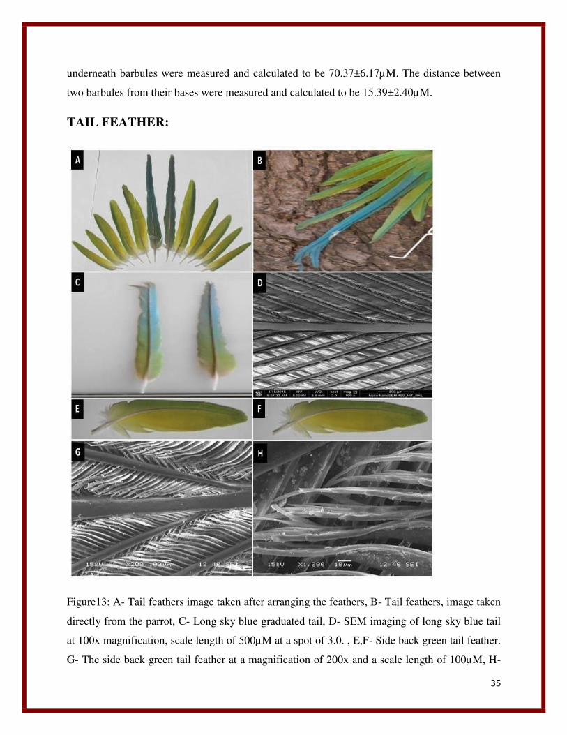
Figure13: A- Tail feathers image taken after arranging the feathers, B- Tail feathers, image taken
directly from the parrot, C- Long sky blue graduated tail, D- SEM imaging of long sky blue tail
at 100x magnification, scale length of 500µM at a spot of 3.0. , E,F- Side back green tail feather.
G- The side back green tail feather at a magnification of 200x and a scale length of 100µM, H-
36
The structure of barbules of side back tail green feather at a magnification of 1000x at a scale of
10µM.
Tail is made up of 10 feathers two long sky blue feathers and 8 side light green feathers (Figure
13 (A)). The long sky blue tail feathers are the longest feathers present in pair one above the
other at tail region. These feathers are present at the middle and surrounded by 10 side feathers
from both sides, 5 feathers from left and 5 feathers from right. All the feathers present both at
right and left are arranged in an ascending way in a gradually increasing size toward the middle
large sky blue feather. The long sky blue feather is bifurcated at the end. It has a long rachis. In
the above images it can be seen all the feathers show a different color pattern when they are
viewed through different angles. The side back feathers which appear green in the (figure 13
(A))exhibit a different coloration pattern of yellowish green vein at one side and yellowish green
and sky blue combination at other vein along the rachis in (figure 13(B)). The SEM analysis of
this long sky blue feather was done to understand the arrangement of the various structural
components of this feather.
Long Sky Blue feather:
The SEM imaging of the long sky blue feather (Figure 13(C)) was done to unravel its structural
components and their arrangement. The SEM imaging of the long sky blue tail observed to
consist of a middle long rachis (Figure 13(D)). The diameter of rachis was measured and
calculated to be 136.52±14.60µM. Barbs arise laterally from the barbules and are parallel to each
other. The diameter of the barbs were measured and calculated to be 38.65±3.09µM. The barbs
at their bases from origin are thin and thicken gradually on elongation. The distance between two
barbs from base were measured and calculated to be 594.36±36.05µM. The distance between
two barbs when they are parallel to each other was measured and calculated to be
198.11±7.57µM. The average angle that the barb is making with the barbules were measured and
calculated to be 20.50±0.68 degree.
Tail side back green feathers:
The SEM imaging of the side back tail feather surrounding the long sky blue tail was done.
There are eight side back green tail feathers surrounding the long sky blue graduated feather
37
from its both sides (Figure 13 (E,F)). The image was taken at an electron flow of 15kV at a
magnification of 200x and a scale length of 100µM (Figure 13 (G)). The structure and
arrangement of various tail components of this feather were viewed. It has a middle rachis which
is thick at the region where it gives rise to the barbs. The rachis of this feather shows a unique
feature that in between two barbs, it becomes thinner in its diameter toward the middle. The
diameter of the end thicker portion of rachis were measured and calculated to be 60.56±4.18µM
when viewed at 200x magnification. The diameter of the thin middle portion of rachis were
measured and calculated to be 48.15±2.28µM. The barbs are arise alternatively from the rachis
along each side and the arrangement is such that one lie just above the other one along the
alternate sides. The barbs are thin at base that is at the point of origin and gradually becomes
thick toward the tip as they elongate and after elongating a certain length they become constant
toward the tip. The diameter of the barbs at the base were measured and calculated to be
13.84±2.11µM. The diameter of the middle transition portion of barb that is the region between
thicker posterior portion and anterior thinner base were measured and calculated to be
26.00±1.00µM. The diameter of the posterior thicker portion of barb were measured and
calculated to be 32.38±2.44µM. The distance between two barbules were measured and
calculated to be 15.14±2.24µM. The barbules are flattened, elongated to a length of
143.55±5.92µM and further elongate to a length of 72.05±7.72µM and fold toward the end
instead of giving thread like structure.
When the structure of tip portion of barbules of side back feathers were imaged at 1000x
magnification at a scale length of 15kV unique structures were observed (Figure 13 (H))The tip
portion of the barbules of side back green feathers are folded structures and at the tip the
barbules become branched to give finger like structures. Each barbule divides to give 6 finger
like projections at the tip. The structure of barbules at this region shows a unique pattern than
rest of feathers.
38
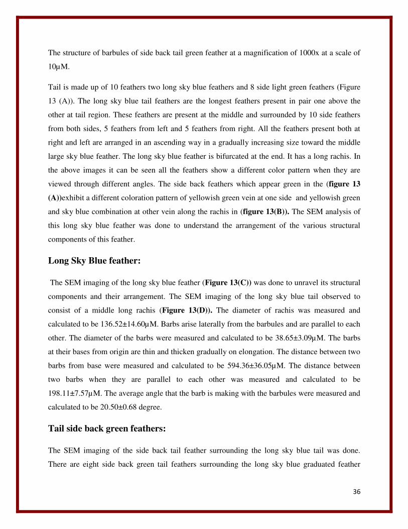
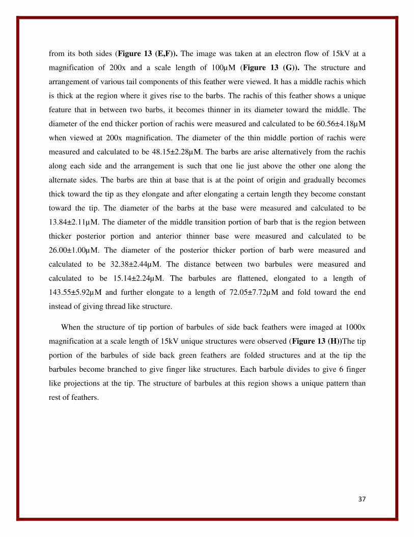
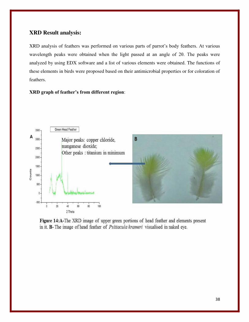
XRD Result analysis:
XRD analysis of feathers was performed on various parts of parrot’s body feathers. At various
wavelength peaks were obtained when the light passed at an angle of 2θ. The peaks were
analyzed by using EDX software and a list of various elements were obtained. The functions of
these elements in birds were proposed based on their antimicrobial properties or for coloration of
feathers.
XRD graph of feather’s from different region:
39
40
41
42
43
From the XRD graph the major elements listed from various feather peaks are mainly Copper,
chloride, Titanium, Tin, Tellurium, Zirconium, Cobalt, Silver, Nickel , Manganese, lead,
Molybdenum, Zinc and Iron. They are expected to have antimicrobial function and some of are
listed below.
Copper chloride:
In early 19th and 20th century copper and its various inorganic derivative complexes were widely
used in medicines in order to treat against diseases like eczema, chronic adenitis, scrofulosis,
impetigo, tubercular infections, anemia, lupus, facial neuralgia and syphilischorea, (Dollwet and
Sorenson 1985).In this way copper was used as an antimicrobial agent and this trend was
continued up to the invention of antibiotics (Grass, Rensing et al. 2011). Copper has the ability to
kill the resilient spores of bacteria such as Clostridia difficile which causes diseases like
diarrhea(Weaver, Michels et al. 2008, Wheeldon, Worthington et al. 2008). There are some
possible proposed action pathways by which it is expected copper to act as an antimicrobial
agent. Copper first damages the membrane of bacteria which lead to copper influx into the cells,
44
cause oxidative damage, fragment DNA and subsequently cell death(Warnes, Green et al. 2010,
Santo, Lam et al. 2011).
Copper homeostatic system:
Some microbes like pseudomonas aeruginosa have genes like CnR encode for copper responsive
regulator and CinA encode for azurin like protein which is involved in copper resistance
(Elguindi, Wagner et al. 2009). Microbes like Enterococcus hiraehas a gene called CopB which
encodes for copper export pump. In E.Coli genes are present like CueO encodes periplasmic
copper oxidase, cus encodes periplasmic copper efflux system and copA encodes cytoplasmic
copper extrusion pump that are involved in copper homeostatic system (Elguindi, Wagner et al.
2009, Santo, Lam et al. 2011). The microbes with a copper resistance don’t get survive from the
contact killing by copper , resist to the copper system for a prolonged period of time but finally
they die(Grass, Rensing et al. 2011). Non of any of bacteria completely resistant to contact
killing has been found yet, because during cell death the plasmid DNAs are completely killed in
cell death by this mechanism and it leaves no resistant determinants to transfer into the
organisms(Warnes, Green et al. 2010). Contact killing is very rapid and it blocks the cell division
on copper surface (Grass, Rensing et al. 2011).
Other possible mechanism by which copper shows antimicrobial properties is by generating
ROS (Reactive oxygen species) which cause oxidative stress. ROS is produced by change in
redox states between different oxidation states of copper like Cu(0), Cu(I), and Cu(II) . The
absence of oxygen don’t inhibit contact killing by Cu but in E.Coli double the time of
killing(Santo, Taudte et al. 2008). Copper act as an antimicrobial agent by following different
mechanism like, generation of ROS, cause oxidative damage of DNA, damage cell wall and iron
sulfur containing enzymes. Cu2+ binds to these structures and that alter their cellular functions
which ultimately leads to death of cell (Ohsumi, Kitamoto et al. 1988, Yates, Brook et al. 2008,
Quaranta, Krans et al. 2011, Santo, Lam et al. 2011, Warnes and Keevil 2011).Copper interact
with the sulfhydryl group of enzymes lead to formation of oxygen reactive radicals that cause
protein oxidation and cleave DNA and RNA and damage the plasma membrane by lipid
peroxidation (Sunada, Watanabe et al. 2003, Warnes and Keevil 2011).
45
As SEM analysis of feathers revealed the presence nanostructures, it is expected that various
elements are involved in the formation of those nanostructures. Now a day the resistance of
microbes toward antibiotics and various antimicrobial agents are increasing. Mostly there is no
microbe present in both human and animal which don’t show resistance to antimicrobial agent
(Frye, Zweig et al. 2002). So the use of nanoparticles has given an utmost importance to fight
against these microbes as well as antibiotic resistance pathogenic microbes. The antimicrobial
properties of CuNP collected from different papers are shown to exhibit biocidal activity against
bacteria such as Staphylococcus aureus(Jokar, Rahman et al. 2012), Escherichia coli(Zapata,
Tamayo et al. 2011)Klebsiella pneumonia(Vimala, Mohan et al. 2010), Pseudomonas
aeruginosa(Perkas, Amirian et al. 2007),Enterobacter cloacae(Kim, Lee et al. 2007), Salmonella
typhimurium (Cárdenas,et al.,2009) and L. monocytogenes(Cioffi, Torsi et al. 2005).The possible
mechanism of action CuNP morphological and structural changes that bring in bacterial cells ,
by attacking the respiratory chain and may be by forming regions of low molecular weight
within the bacteria (Sondi and Salopek-Sondi 2004, Morones, Elechiguerra et al. 2005).CuNP
perform antimicrobial activity by release of Cu2+,that may cause the disruption of the plasmatic
membrane of bacteria, enabling their entry into the bacteria and alteration of the bacteria
enzymatic functions (Gunawan, Teoh et al. 2011, Xiu, Ma et al. 2011).
Chloride:
Chloride in its elemental form called chlorine, known to be the most common disinfectant, is a
moderate oxidizing agent interacts with various components of cells (White 1992). The action
of chlorine is proposed to damage the cell membrane of the bacterial cells (Kim, Pitts et al.
2008). Chlorine kills the microbes by disrupting the cell wall where it chlorinate the lipid protein
substance present in the cell wall and form toxic chloro compounds (Venkobachar, Iyengar et al.
1977, Haas and Engelbrecht 1980). This causes the leakage of the cell wall and the
macromolecules present in the cell releases out (Kim, Pitts et al. 2008).
Manganese Dioxide:
Manganese is found in the active site of many enzymes and plays an important role in the
functionality of the enzyme (Larson and Pecoraro 1992). Mn forms complex by combining
with various ligands and show anticancer (Li et al., 2010;Chen et al., 2010) , antibacterial
46
(Dorkov, Pantcheva et al. 2008) and antifungal actions (Singh et al.,2010 ; Mohamed et al., 2010
). The Mn complexes react with the plasmid DNA and degrade it. Mn (II) is present as a cofactor
in many enzymes. The antimicrobial activity of Mn is based on the basis of overtone concept of
cell permeability. Mn element is more active in its antimicrobial properties when it is in the
complex form. This increased activity of the element in its complexes can be explained on the
basis of overtone and chelation theory (Islam, Farooque et al. 2002).According to overtone’s
concept of cell permeability the cell membrane favors the passage of only lipid soluble materials,
called liposolubility is an important factor for membrane permeability that regulate antimicrobial
activity on chelation with other ligand during complex formation. During chelation the polarity
of metal ion is reduced to a greater extent due to overlapping of donor group of ligand orbital and
partial sharing of the positive charge of the metal ion. Then this overlapping results in increases
the delocalization of pi electrons over the whole ring and increases the liophilicity of the
complex. The increased liophilicities of complex allow easy diffusion into lipid membranes of
organisms and facilities as blockage of metal binding sites in enzyme (Gudasi, Vadavi et al.
2005).
Titanium:
Titanium in in its oxides form i.e. (TiO2) shows antimicrobial properties and also in its
nanoform (TiO2NP) of size less than 25nm. Nanomaterials act as antimicrobial agent by
hindering the action of cellular enzyme and inactivate them. Nanoparticles have ability to bind
the electron donating group like carboxylate, amides, indoles, hydroxylates and thiols etc. NP
penetrates the bacterial wall by forming pores on cell walls lead to increased permeability and
cell death. NP act against microbes which develop resistance against the drugs. Metal oxides of
NP exhibit excellent biocidal and biostatic action against gram positive and gram negative
bacteria (Cuéllar-Cruz, Vega-González et al. 2012).The antimicrobial mechanism of metal
oxides can be explained as the basis of charge difference that is present on the metal oxides and
the charge on the microbial cells. It is proposed that the microbial cells contain negative charge
and the metal oxides possess a positive charge as a result causes an electromagnetic attraction
between the microorganisms and metal oxides that lead to oxidization and ultimate death of
microorganisms (Li, Zhang et al. 2012). TiO2 is also involved in the formation of pigment
(Baan, Straif et al. 2006). Hence it also can be proposed that it might be responsible for
47
exhibiting certain color in parrot. As TiO2 NP are used in sunscreen for protection against sun
rays (Trouiller, Reliene et al. 2009), it also can be assumed that it might be playing some
protective role in bird’s feather against sun rays. TiO2 NP when exposed to nonlethal UV
produce hydroxyl radicals and superoxide ions , which is shown to be highly effective in
inhibiting Staphylococcus Aureus (Shah et al. 2008) and also degrade organic materials. In vivo
studies show the major pathogenic mechanism initiated by TiO2NP produce inflammatory
response. The above functions of TiO2 NP can be predicted in case of feathers of Psitacula
krameri.
Nickel:
Nickel toxicity is given an importance as it is related with cancer in human beings (Denkhaus
and Salnikow 2002, Das, Das et al. 2008). Microbes have a nickel homeostasis system to resist
nickel toxicity. The exact mechanisms of nickel toxicity in microbes are unknown even though
their effects on higher organisms are well demonstrated (BAEICII and Stotzky 1983). However
from some of in vitro studies of nickel toxicity, some action mechanism are hypothesized like
1) nickel replaces the vital metal from metalloproteins, 2) nickel binds to catalytic sites of non-
metal enzymes, 3) nickel binds the allosteric site of an enzyme to inhibit it and 4) nickel leads to
oxidative stress and strain that can affect proteins, DNA, or lipids(Macomber and Hausinger
2011).
Ni-NPs have antimicrobial activity, however very few studies have done on this (Pang, Lu et al.
2009, Kumar, Rani et al. 2010). Ni-NPs shows a bacteriostatic property against S. aureus, E. coli
and S. mutans (Argueta-Figueroa, Morales-Luckie et al. 2014). Other possible mechanism by
which nickel act is, its ions penetrate in microbial cells and more preferably inactivate their
enzymes leading to inhibit their cellular metabolic functions resulting ultimate death(Chohan
2000). Some of nickel complexes exhibit an antibacterial activity against gram positive bacteria
(Popova, Smith et al. 2012). Different authors report, nickel play most of its antimicrobial
properties when it is complexed with certain ligands and in other synthetic complexes.
In the collar region the presence of Ni as one of major element is expected to exhibit
antimicrobial properties as discussed above, in order to keep bird free from microbial infection
diseases
48
Tin:
Tin in its nanoparticle form in SnO2 NP shown to have antimicrobial properties. These nano
particles get adsorb on the bacterial cell and undergo dehydrogenation, due to respiration process
which occurs at the cell membrane of bacteria. This reaction inactivate the bacterial enzymes by
generating hydrogen peroxide that causes oxidation of bacterial cells and cause death (Awwad,
Salem et al. 2012).The Because, Escherichia coli was not having cell wall it get affected more
easily than S.aureus with cell wall (Kamaraj, Vennila et al. 2014). But the exact functions of Tin
present in parrots are unknown. The above functions may be predicted.
Cobalt:
Cobalt is present in the active center of vitamin B12, (Bernhardt, Bozoglián et al. 2005) an
important element found in certain cobalt-dependent proteins and in its complexes it act as
hydrolytic agents for DNA cleavage (Hadjiliadis and Sletten 2009) and some have antitumor–
anti-proliferative, antimicrobial, antifungal, and antiviral activity (Eshkourfu, Čobeljić et al.
2011).
Cobalt(III) in its complexes functions as an antiviral agent (Chang, Simmers et al. 2010). It is
used widely in medicines for example cobalt complex CTC-96 was effective in the treatment of
epithelial herpetic keratitis, identified as one of the major disease causing blindness in industrial
nations (Asbellet al.,1998).The action mechanism is that CTC-96 inhibits membrane fusion
events hence preventing virus entry. There by CTC-96 obstruct plaque formation by VSV
(vesicular stomatitis virus) and VZV (varicella-zoster virus) (Delehanty, Bongard et al. 2008).
CTC-96 also act against adenovirus (Epstein, Pashinsky et al. 2006).Co(III ) in its coordination
complexes act as an antibacterial agent. The possible action mechanism of these complexes can
be explained on the basis of, binding to DNA of microbes to cause DNA damage (Kumar, 2008).
For example some complexes like bis(ethylenediamine)cobalt(III) has screened to perform
antibacterial actions against some Gram positive and Gram negative bacteria like E. coli, E. coli
HB101, Salmonella typhimurium, Proteus vulgaris, P. aeruginosa, S. aureus, S. faecalis, B.
subtilis (Nagababu, Latha et al. 2006).
49
Although the exact functions of the Cobalt and its complexes present in feather of parrots are
unknown, it may be proposed they might be having antimicrobial activity against different
microbes as discussed above.
Tellurium:
Tellurium very rarely found in its native state, it is found in the form of telluride of gold and in
combination with other elements (Cunha, Gouvea et al. 2009).In case of parrot’s feather we
identified in the form of tin telluride. Already we discussed regarding the antimicrobial activity
of tin. Tellurium is a metalloid and very few studies have been made on this. From these few
studies it has been proposed to possess a very interesting antimicrobial activity. A novel enzyme
called tellurite reductase has been purified and identified which can be used for the generation of
tellurium nanostructures that has an interesting and eco-friendly antimicrobial activity which was
proposed to replace AgNP or antibiotic therapy in near future (Plugin and coworkers, 2014,
American Society for Microbiology,7061–7070 ).
Tellurium (Te) compounds have been used as antimicrobial agents in the treatment of
infectious diseases (e.g., leprosy, tuberculosis, dermatitis) (Turner, Borghese et al. 2012).
Recently, many synthetic organo-tellurium compounds have been developed for inhibition of
bacteria growth (Daniel-Hoffmann, Sredni et al. 2012). For example, nontoxic immune
modulator ammonium trichloro (dioxyethylene-O,́)- tellurate (AS101) is used to inhibit
cysteine proteases and modulate the redox state of glutathione (Daniel-Hoffmann et al.,2012).
Tellurite (TeO32−) ions have also been used to inhibit the growth of many microorganisms,
particularly penicillin-resistant bacteria (Valdivia-González, Pérez-Donoso et al. 2012).The
released TeO32− ions from Te NMs have been shown effective to kill E. coli.31.
The mechanism of action of these Nano medicines proposed to progress through release of Te
ions. Tellurium in combination with other nanostructures like Ag and Au are used in Nano
medicines. The possible mechanism of action of these Nano medicines are believed to have
enzyme like catalytic activity and can generate ROS (Wei and Wang 2013). However the
possible biological and chemical mechanism of generation of ROS from released Te-related ions
are yet to reveal (Molina-Quiroz, Muñoz-Villagrán et al. 2012, Molina-Quiroz, Loyola et al.
2013). The possible mechanism for action of ROS to inhibit bacterial growth can be explained by

50
following ways like through lipid peroxidation and reaction with membrane proteins, DNA, or
metabolic enzymes (Lin, Shih et al. 2012).
Some other organometallic compounds of tellurium such as AS101 shown to work as an
antimicrobial agent by damaging and altering the Na+-K+ pump on cell membrane and has been
experimented in E. cloacae. The results showed an increase in influx of Na+ and Cl- destabilizes
the membrane stability. The cell membrane instability was observed, shown by increase in Mg.
A decrease in the level of phosphorus indicated loss of ATP and damage to DNA. It also damage
cell by causing perforation in cell wall (Daniel-Hoffmann, Sredni et al. 2012).
Due to these antimicrobial properties of Tellurium it is used in drug development. The exact
function of Tellurium in parrot’s feathers are unknown, but keeping in view the above functions
of Tellurium, the same function may be expected in bird’s feathers.
Lead:
Studies have shown that long-term heavy metal contamination of soils has harmful effects on soil
microbial activity, especially microbial respiration (Doelman and Haanstra 1984).Exposure of
microbes to heavy metal for a long term duration alter their enzymatic activity, short term
exposure reduces the microbial activity (Doelman and Haanstra 1979) .
Pb(II) toxicity occurs as a result of changes in the conformation of nucleic acids and proteins,
inhibition of enzyme action, disruption of membrane functioning and oxidative phosphorylation,
as well as change in osmotic balance (Bruins et al., 2000; Vallee & Ulmer,1972). Pb(II) also
shows a stronger affinity for thiol and oxygen groups than essential metals such as calcium and
zinc (Bruins et al., 2000). In spite of the high toxicity of Pb, many micro-organisms have
developed mechanisms that facilitate them to resist and survive Pb exposure (Jarosławiecka and
Piotrowska-Seget 2014).
Although the expected roles of lead in case of parrot’s feather are unknown, based on the
antimicrobial properties we may predict some of the above.
51
Zirconium:
Zirconium in its nanoparticle form that is in the form of ZrO2NP is resistant against the microbes
like Staphylococcus aureus, Escherichia coli and Pseudomonas aeruginosa (Li and Coleman
2014).Zirconium in its complexes like zirconium(IV) porphyrin experimented to show
antimicrobial activity against some bacterial strains, namely, Bacillus subtilis, Micrococcus
luteus, Staphylococcus aureus, Pseudomonas fluorescens, and Escherichia coli forming zone of
inhibition (Bajju, Devi et al. 2013).Very few works have been carried out regarding the
antimicrobial properties of Zirconium. The exact mechanism of Zirconium antimicrobial activity
is unknown and yet to reveal.
The roles of Zirconium in case of parrot’s feathers are unknown, but from the few available
information regarding its antimicrobial properties, the same in case of feathers may be predicted.
Silver:
Silver is an antimicrobial agent perform action in its ionic form, non-oxidative in nature. But in
a cell it inactivates enzymes by binding to the –SH group (Lin et al., 1996). Silver interacts with
the ribosome, inhibit the expression of enzyme and other protein responsible for ATP production
(Yamanaka, Hara et al. 2005). Silver also interact with the respiratory chain and inhibit the
oxidation of glucose , glycerol, fumarate (Bragg and Rainnie 1974). Silver forms a complex
with DNA that is Ag-DNA complex, which is expected to cause antimicrobial activity (Arakawa,
Neault et al. 2001). AgNP exhibit wide range of antimicrobial activity. AgNP gets adhered on
the surface of bacteria, damage the cell wall, when the size of nanoparticle is smaller than 10nm
it penetrate into bacteria (Morones, Elechiguerra et al. 2005, El Badawy, Silva et al. 2010).
AgNP function as an antimicrobial agent by forming Ag+ and ROS which is the major source of
toxicity (Hwang, Lee et al. 2008). Silver, may be because of its antimicrobial properties both in
its ionic and complex form and hence it is expected its presence in feathers of parrot may be
playing the same role.
Molybdenum:
The action of Mo as an antimicrobial agent has shown non-specific mechanism. It is believed
the antimicrobial property is based on the formation of an acidic surface that inhibits cell growth
52
and proliferation. It is active against a broad range of both Gram positive and Gram negative
bacteria, and does not produce bacterial resistance to the action of antibiotics. The acidic surface
prevents the growth and proliferation of microbial cells and biofilm formation. The acidity
mechanism can be explained by the diffusion of hydronium ions through the cell membrane.
This results in the imbalance of pH equilibrium, which affects both enzyme proteins and the
transport system of cell (Plumridge, Hesse et al. 2004). This also degrade the DNA helix
(Schüller, Mamnun et al. 2004). In order to adjust this disturbed pH equilibrium mechanism
energy required for the cell, that cause in further weakening of cell (Ra and Parks 2007). As the
energy used against the physiological gradient. The acidic surface prevents microbes from
adhering to the surface and forming bio film. But the microbes should have a direct contact with
the acidic surface for antimicrobial activity (Zollfrank, Gutbrod et al. 2012).
Zinc:
In earlier studies it has been proposed that zinc play an important role in melanin synthesis in
birds for imparting their feather coloration. They act upon the enzyme tyrosinase that plays a
major role in the synthesis of melanin from amino acid precursors (McGraw 2003). Zinc is
involved in both eumelanin and reddish brown color imparting pheomelanin synthesis (Niecke,
Rothlaender et al. 2003). Zinc in its ionic, oxide and nanoparticle form has antibacterial
properties (Söderberg, Sunzel et al. 1990). ZnO particles have been demonstrated to show
antimicrobial properties in a wide range against both Gram positive and Gram negative bacteria
like Escherichia coli, Salmonella, Listeria monocytogenes, and Staphylococcus aureus (Jones,
Ray et al. 2008, Liu, He et al. 2009). The exact mechanism of antibacterial function of ZnO are
exactly not cleared but, it has been proposed that it may act by disrupting cell membrane activity
(Brayner, Ferrari-Iliou et al. 2006). Zinc destabilizes the cell membrane and alters by increasing
the cell membrane permeability hence affect the membrane function (Stanić, Dimitrijević et al.
2010).Some other possible mechanism proposed are production of ROS (Reactive Oxygen
Species), Hydrogen peroxide (H2O2), is a strong oxidizing agent that is harmful for bacterial cell
(Sawai 2003, Jones, Ray et al. 2008). Another possible mechanism of action of Zinc has been
proposed that it interacts with the nucleic acid and make the respiratory enzymes nonfunctional
(Fang, Chen et al. 2006).It has been demonstrated that these oxidizing species like ROS, OH-,
H2O2, and O2- are produced by UV- Visible light and those function in antimicrobial properties.
53
The hydroxyl radicals and super-oxides remain on the upper surface of bacterial cell whereas
peroxide penetrate into the bacterial cell and induce antimicrobial properties (Padmavathy and
Vijayaraghavan 2008). ZnO is used in cosmetics to protect against sunlight. It may be predicted
that it might be playing some role in protecting Psittacularis from sunlight.
The above function of Zinc may be proposed in case of its presence in Psittacularis feathers.
However the exact functions of its presence are unknown and more researches are required to be
carried out.
Iron:
Iron plays an important role in Psittacularis feathers in imparting color. It is involved in synthesis
of melanin like Zinc (McGraw 2003). Iron in forms of Porphyrin(With 1957, With 1978)and Iron
Oxide (Negro, Margalida et al. 1999) can give colors to bird’s feathers. The Iron nanoparticles
may be involved in the antimicrobial action in bird’s feather by following the mechanisms like
generation of reactive oxygen species (ROS), release of harmful and toxic ions, oxidative
damage through catalysis, and disturbance of the ion cell membrane transport activity by altering
permeability and lipid peroxidation or surfactant properties. ROS is considered as being the main
elementary chemical process in nano-toxicology which can lead to secondary processes that can
ultimately cause cell damage and even cell death. Moreover, ROS is one of the main factors
involved in inflammatory processes (Singh, Jenkins et al. 2010).
Hence the above functions of Iron may be expected in case of Parrot’s feathers although the
exact functions are unknown. However more researches need to be carried out in this field.
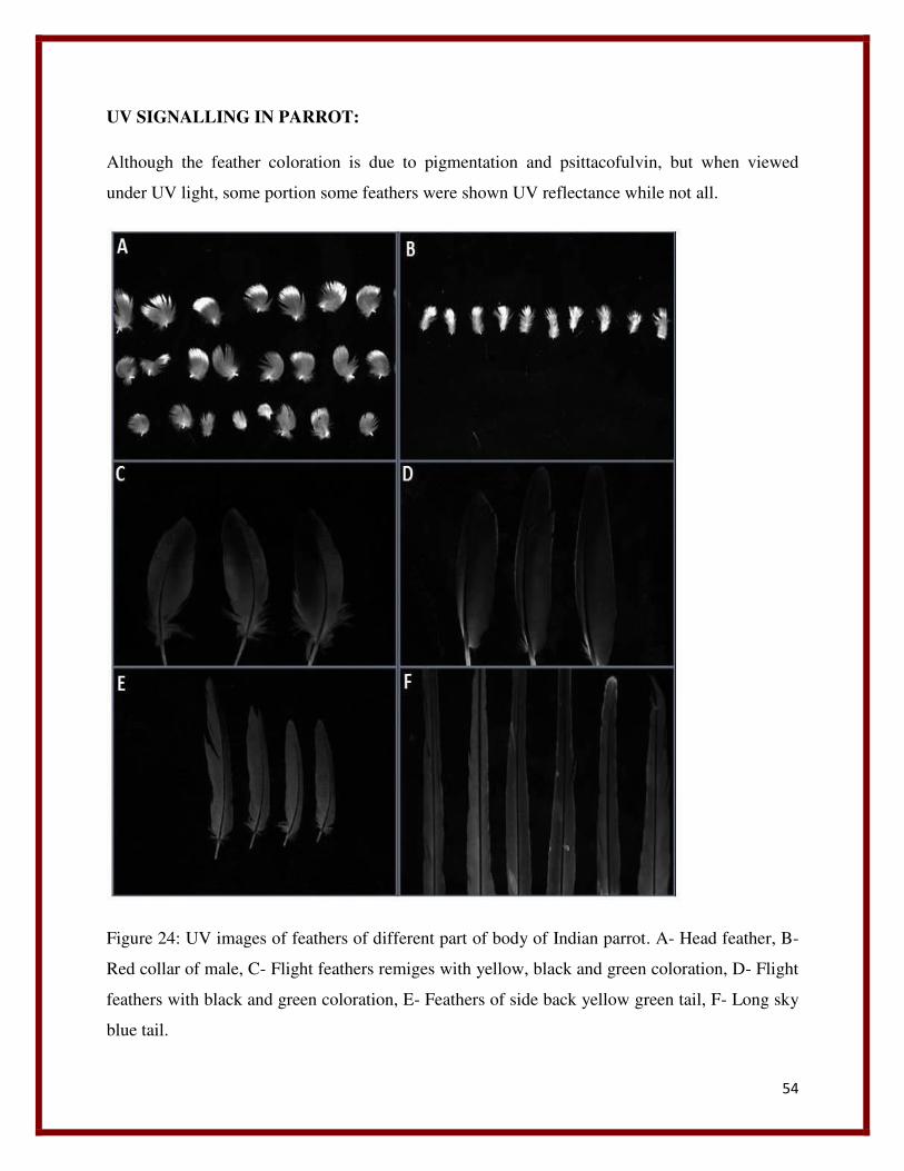
54
UV SIGNALLING IN PARROT:
Although the feather coloration is due to pigmentation and psittacofulvin, but when viewed
under UV light, some portion some feathers were shown UV reflectance while not all.
Figure 24: UV images of feathers of different part of body of Indian parrot. A- Head feather, B-
Red collar of male, C- Flight feathers remiges with yellow, black and green coloration, D- Flight
feathers with black and green coloration, E- Feathers of side back yellow green tail, F- Long sky
blue tail.
55
The head feathers, flight feathers, tail feathers and collar feathers of male were viewed under UV
light. Only the feathers head region, and red collar of male shown UV reflectance. The exposed
tip portions of head feathers were shown UV reflectance but not the entire feather. Apart from tip
portion although UV reflectance is observed in some other portions of head feather but more
significant is at tip regions. In case of collar red feather of male UV reflectance was observed in
the entire feather.
DISCUSSION:
SEM result discussion:
This is expected that these arrangements of the barbules are responsible for giving a
structural integrity and regularity to the feathers and make it resemble as a leaf to the naked eye.
It also can be proposed that this pattern of arrangement result for imparting coloration of to the
feathers due to Tindall effect and reflection of light through these barbules, which is the physical
basis of coloration in parrots feather or structural basis of coloration. The structural basis of
coloration is produced due to the interaction between the coherent light and the nanostructures
present in the feathers (Fox 1976). The structural basis of coloration can be classified into two
sub-catagories called iridescent and non- iridescent. Iridescent color change in appearance when
visualized through different angle and iridescent colors exhibit no change with respect to the
change of geometry Newton). Both the structural colors are produced by similar coherent
scattering of light by feather components but they differ by the composition and arrangement of
their light scattering elements (Prum 2006). Non iridescent colors are produced due to the
nanostructures present in barb. The orderly arrangement of keratin and the air in between the
barbs responsible for the exhibition of non-iridescent color like blue, violet, and ultraviolet
colors (Prum, Andersson et al. 2003, Shawkey, Estes et al. 2003, Doucet, Shawkey et al. 2004),
Iridescent colors produced due to the structure and arrangement of barbules (Prum 2006). In case
of barbules the constructive scattering of coherent light takes place between keratin, melanin and
air present alternatively with a variable refractive index (Doucet, Mennill et al. 2005). The
variation in the nanostructures that are present in barbules responsible for various colors .It also
can be proposed that this interlocking pattern of arrangement of the barbules help the bird to
prevent water flow to reach its body surface hence making feathers impermeable to water. This
56
may prevent the bird to get less affected during rain, and also get rid of microbial infections up to
some extent. This interlocking pattern of arrangement of barbules also can be responsible for
making the feathers more insulated by closing the air spaces on it which again prevent the bird
from cold.
In each feathers we obtained different measurements for different structural components. The
shape and different components and their arrangement also vary from feather to feather. This
variations may result in exhibiting the phenomena of iridescent of light. This is the reason why
one can observe various color when observed through different angles. The same is observed in
case of feathers of parrots due to its different structural build up as well as arrangement of
various components. The tail feathers are expected to show iridescent coloration in a higher
degree.
Though in some cases we observed some similarities in their structural components like
barbs, barbules and their arrangement, but the structures and arrangement of the terminal end of
barbules varies. It is expected this variations in structural buildup of barbules are responsible for
exhibiting the phenomena of refraction , Tindall effect as well as scattering of light, as in a same
feather we observed various length , structures and arrangement of barbule terminals.
XRD result discussion:
The elements obtained from the XRD data analysis are predicted to play an antimicrobial
activity or play role in coloration of feathers. These elements both in their ionic form as well as
in their complexes may be playing both of the roles. Some transition elements are also obtained
in XRD peaks like Cobalt, Nickel, and Manganese etc., which have a chemical property of
forming coordination complexes with some ligands. An important property of these coordinating
complexes are to show color , which may be contributing for color exhibition in feathers up to
some extent, although the primary factors of color formation are pigmentation and Tindall effect
,the physical basis of color formation. Very few works have been carried out in this field so the
microbial action mechanism of many elements is unknown. The exact role of these elements and
their complexes in feathers are some of questions need to be answered.
57
Discussion of UV result:
It has been reported some microstructures are present in barb (Fox 1976, Prum, Andersson et al.
2003) of some feathers responsible for UV reflectance, along with the blue violet and other
iridescent colors (Korsten, Vedder et al. 2007). It has been found that feathers of many bird
species show UV reflectance (Burkhardt 1989, Eaton and Lanyon 2003), many species have the
ability to detect the UV region of spectrum (Parrish, Ptacek et al. 1984, Cuthill, Partridge et al.
2000), which normal human eye can’t do. Several studies have shown that UV reflectance play a
major role in mate selection. Female give preference to high UV reflectance male species than
low reflectance (Andersson and Andersson 1998, Johnsen, Andersson et al. 1998).
UV reflectance may be a good medium of social signaling for mate selection because of its short
wavelength for which it can be a signaling medium over shorter distance and it may be distorted
over longer distance due to scattering by particles (Lythgoe 1979, Andersson 1996). Some
authors has been suggested that UV may be a secret avian communication channel which many
potential mammals cannot read (Guilford and Harvey 1998). As chlorophyll absorbs UV light,
plants may provide a contrasting background to UV reflective feathers which may make UV a
good signaling medium in avian community (Andersson and Andersson 1998). It has been
suggested that bird’s feather are sensitive to UV wavelength than other wavelength (Burkhardt
and Maier 1989), may be because of their sensitive vision toward UV wavelength to which they
are pre-exposed which may help them to search their food (Church, Bennett et al. 1998). UV
signals are iridescent which may help bird’s to help in their courtship selection with an accuracy
and greater potency.
Conclusion:
Very few experiments are conducted about Indian parrot. Depending upon the information that
are present about feathers of other birds we tried to propose some information regarding the
nanostructures present in feathers and their role in coloration in parrot. By observing the
different structures and arrangement of barbules the expected function of barbules were proposed
to involve in iridescent and non-iridescent coloration. The SEM imaging magnified the various
feather components out of their nanostructure form. However these varied structures and pattern
formation of these feather components are expected to responsible for the different color
58
exhibition by different feathers. This is expected because, although to naked eye these feather
components almost seems similar to each other but their SEM imaging structures particularly the
structure and arrangement of barbules to pattern formation are different from each other as
observed from all those images. From the XRD analysis data the elements present in the feather
microstructures were detected. Although the exact functions of these elements in parrot’s feather
are unknown, some of the antimicrobial properties of them were expected. The elements detected
from the XRD predicted to present in its nano form and might be playing many more roles. The
element detection reveals many elements which are present in nature and are not studied yet. In
order to study those, the laboratory synthesis is required. The study of these structures in
laboratory may discover some other amazing dimensions of these elements in their nano form.
From UV imaging of the various feathers the function was predicted to be helpful in courtship or
mate selection or in social signaling. The red collar feathers of the male shows UV reflectance
which may be helpful for female for male selection. The UV reflectance of other feathers like
head feather may be helpful in potent male selection. The phenomenon of scattering of coherent
light was discussed in order to understand iridescent and non -iridescent coloration and
pigmentation. However further researches are required to unravel the story behind this
phenomenal feather coloration of nature.
REFERENCES:
Auber, L. (1957). THE STRUCTURES PRODUCING “NON‐ IRIDESCENT” BLUE COLOUR IN BIRD FEATHERS. Proceedings of the Zoological Society of London, Wiley Online Library.
Brush, A. H. (1990). "Metabolism of carotenoid pigments in birds." The FASEB Journal4(12): 2969-2977.
Burkhardt, D. (1989). "UV vision: a bird's eye view of feathers." Journal of Comparative Physiology A164(6): 787-796.
Cuthill, I. C., et al. (2000). "Ultraviolet vision in birds." Advances in the Study of Behavior29: 159-214.
59
Eaton, M. D. and S. M. Lanyon (2003). "The ubiquity of avian ultraviolet plumage reflectance." Proceedings of the Royal Society of London. Series B: Biological Sciences270(1525): 1721-1726.
Fox, D. L. (1976). Animal Biochromes and Structural Colours: Physical, Chemical, Distribution & Physiological Features of Coloured Bodies in the Animal World, University of California Press.
Hausmann, F., et al. (2003). "Ultraviolet signals in birds are special." Proceedings of the Royal Society of London B: Biological Sciences270(1510): 61-67.
Hill, G. E. (1999). "Is There an Immunological Cost to Carotenoid‐ Based Ornamental Coloration?" The American Naturalist154(5): 589-595.
Hill, G. E. (2000). "Energetic constraints on expression of carotenoid― based plumage coloration." Journal of Avian Biology31(4): 559-566.
Hill, G. E., et al. (2002). "Dietary carotenoids predict plumage coloration in wild house finches." Proceedings of the Royal Society of London B: Biological Sciences269(1496): 1119-1124.
Jawor, J. M. and R. Breitwisch (2003). "Melanin ornaments, honesty, and sexual selection." The Auk120(2): 249-265.
M Hofmann, C., et al. (2007). "Melanin coloration in New World orioles I: carotenoid masking and pigment dichromatism in the orchard oriole complex." Journal of Avian Biology38(2): 163-171.
McGraw, K. J., et al. (2004). "European barn swallows use melanin pigments to color their feathers brown." Behavioral Ecology15(5): 889-891.
Olson, V. r. A. and I. P. F. Owens (1998). "Costly sexual signals: are carotenoids rare, risky or required?" Trends in Ecology & Evolution13(12): 510-514.
Parrish, J. W., et al. (1984). "The detection of near-ultraviolet light by nonmigratory and migratory birds." The Auk: 53-58.
Prum, R. O., et al. (2003). "Coherent scattering of ultraviolet light by avian feather barbs." The Auk120(1): 163-170.
60
Andersson, S. (1996). "Bright ultraviolet colouration in the Asian whistling-thrushes (Myiophonus spp.)." Proceedings of the Royal Society of London. Series B: Biological Sciences 263(1372): 843-848.
Andersson, S. and M. Andersson (1998). "Ultraviolet sexual dimorphism and assortative mating in blue tits." Proceedings of the Royal Society of London. Series B: Biological Sciences 265(1395): 445-450.
Arakawa, H., et al. (2001). "Silver (I) complexes with DNA and RNA studied by Fourier transform infrared spectroscopy and capillary electrophoresis." Biophysical Journal 81(3): 1580-1587.
Argueta-Figueroa, L., et al. (2014). "Synthesis, characterization and antibacterial activity of copper, nickel and bimetallic Cu–Ni nanoparticles for potential use in dental materials." Progress in Natural Science: Materials International 24(4): 321-328.
Auber, L. (1957). THE STRUCTURES PRODUCING “NON‐ IRIDESCENT” BLUE COLOUR IN BIRD FEATHERS. Proceedings of the Zoological Society of London, Wiley Online Library.
Awwad, A. M., et al. (2012). "Biosynthesis of silver nanoparticles using Olea europaea leaves extract and its antibacterial activity." Nanoscience and Nanotechnology 2(6): 164-170.
Baan, R., et al. (2006). "Carcinogenicity of carbon black, titanium dioxide, and talc." The lancet oncology 7(4): 295-296.
BAEICII, H. and G. Stotzky (1983). "Toxicity of nickel to microbes: environmental aspects." Advances in applied microbiology 29: 195.
Bajju, G. D., et al. (2013). "Synthesis, spectroscopic, and biological studies on new zirconium (IV) porphyrins with axial ligand." Bioinorganic chemistry and applications 2013.
Bernhardt, P. V., et al. (2005). "Molecular mixed-valence cyanide bridged Co III–Fe II complexes." Coordination chemistry reviews 249(17): 1902-1916.
Bragg, P. and D. Rainnie (1974). "The effect of silver ions on the respiratory chain of Escherichia coli." Canadian journal of microbiology 20(6): 883-889.
Brayner, R., et al. (2006). "Toxicological impact studies based on Escherichia coli bacteria in ultrafine ZnO nanoparticles colloidal medium." Nano Letters 6(4): 866-870.
Brush, A. H. (1990). "Metabolism of carotenoid pigments in birds." The FASEB Journal 4(12): 2969-2977.
61
Burkhardt, D. (1989). "UV vision: a bird's eye view of feathers." Journal of Comparative Physiology A 164(6): 787-796.
Burkhardt, D. and E. Maier (1989). "The spectral sensitivity of a passerine bird is highest in the UV." Naturwissenschaften 76(2): 82-83.
Chang, E. L., et al. (2010). "Cobalt complexes as antiviral and antibacterial agents." Pharmaceuticals 3(6): 1711-1728.
Chohan, Z. H. (2000). "SYMMETRIC 1, 1-DIMETHYLFERROCENE-DERIVED AMINO ACIDS: THEIR SYNTHESIS, CHARACTERIZATION, LIGATIONAL AND BIOLOGICAL PROPERTIES WITH Cu (II), Co (II) and Ni (II) IONS." Metal Based Drugs 7: 177-184.
Church, S. C., et al. (1998). "Ultraviolet cues affect the foraging behaviour of blue tits." Proceedings of the Royal Society of London. Series B: Biological Sciences 265(1405): 1509-1514.
Cioffi, N., et al. (2005). "Copper nanoparticle/polymer composites with antifungal and bacteriostatic properties." Chemistry of Materials 17(21): 5255-5262.
Cuéllar-Cruz, M., et al. (2012). "The effect of biomaterials and antifungals on biofilm formation by Candida species: a review." European journal of clinical microbiology & infectious diseases 31(10): 2513-2527.
Cunha, R. L., et al. (2009). "A glimpse on biological activities of tellurium compounds." Anais da Academia Brasileira de Ciências 81(3): 393-407.
Cuthill, I. C., et al. (2000). "Ultraviolet vision in birds." Advances in the Study of Behavior 29: 159-214.
Cuthill, I. C., et al. (2000). "Ultraviolet vision in birds." Advances in the Study of Behavior 29: 159-214.
Daniel-Hoffmann, M., et al. (2012). "Bactericidal activity of the organo-tellurium compound AS101 against Enterobacter cloacae." Journal of Antimicrobial Chemotherapy: dks185.
Das, K., et al. (2008). "Nickel, its adverse health effects & oxidative stress." Indian Journal of Medical Research 128(4): 412.
Delehanty, J. B., et al. (2008). "Antiviral properties of cobalt (III)-complexes." Bioorganic & medicinal chemistry 16(2): 830-837.
62
Denkhaus, E. and K. Salnikow (2002). "Nickel essentiality, toxicity, and carcinogenicity." Critical reviews in oncology/hematology 42(1): 35-56.
Doelman, P. and L. Haanstra (1979). "Effects of lead on the soil bacterial microflora." Soil Biology and Biochemistry 11(5): 487-491.
Doelman, P. and L. Haanstra (1984). "Short-term and long-term effects of cadmium, chromium, copper, nickel, lead and zinc on soil microbial respiration in relation to abiotic soil factors." Plant and soil 79(3): 317-327.
Dollwet, H. and J. Sorenson (1985). "Historic uses of copper compounds in medicine." Trace elements in Medicine 2(2): 80-87.
Dorkov, P., et al. (2008). "Synthesis, structure and antimicrobial activity of manganese (II) and cobalt (II) complexes of the polyether ionophore antibiotic Sodium Monensin A." Journal of inorganic biochemistry 102(1): 26-32.
Doucet, S., et al. (2004). "Concordant evolution of plumage colour, feather microstructure and a melanocortin receptor gene between mainland and island populations of a fairy–wren." Proceedings of the Royal Society of London B: Biological Sciences 271(1549): 1663-1670.
Doucet, S. M., et al. (2005). "Achromatic plumage reflectance predicts reproductive success in male black-capped chickadees." Behavioral Ecology 16(1): 218-222.
DYCK, J. (1985). "The Evolution of Feathers*." Zoologica Scripta 14(2): 137-154.
Eaton, M. D. and S. M. Lanyon (2003). "The ubiquity of avian ultraviolet plumage reflectance." Proceedings of the Royal Society of London. Series B: Biological Sciences 270(1525): 1721-1726.
El Badawy, A. M., et al. (2010). "Surface charge-dependent toxicity of silver nanoparticles." Environmental science & technology 45(1): 283-287.
Elguindi, J., et al. (2009). "Genes involved in copper resistance influence survival of Pseudomonas aeruginosa on copper surfaces." Journal of Applied Microbiology 106(5): 1448-1455.
Eliason, C. M. and M. D. Shawkey (2012). "A photonic heterostructure produces diverse iridescent colours in duck wing patches." Journal of The Royal Society Interface 9(74): 2279-2289.
Epstein, S. P., et al. (2006). "Efficacy of topical cobalt chelate CTC-96 against adenovirus in a cell culture model and against adenovirus keratoconjunctivitis in a rabbit model." BMC ophthalmology 6(1): 22.
63
Eshkourfu, R., et al. (2011). "Synthesis, characterization, cytotoxic activity and DNA binding properties of the novel dinuclear cobalt (III) complex with the condensation product of 2-acetylpyridine and malonic acid dihydrazide." Journal of inorganic biochemistry 105(9): 1196-1203.
Faivre, B., et al. (2003). "Immune activation rapidly mirrored in a secondary sexual trait." Science 300(5616): 103-103.
Fang, M., et al. (2006). "Antibacterial activities of inorganic agents on six bacteria associated with oral infections by two susceptibility tests." International Journal of Antimicrobial Agents 27(6): 513-517.
Fox, D. L. (1976). Animal Biochromes and Structural Colours: Physical, Chemical, Distribution & Physiological Features of Coloured Bodies in the Animal World, University of California Press.
Frye, D. M., et al. (2002). "An outbreak of febrile gastroenteritis associated with delicatessen meat contaminated with Listeria monocytogenes." Clinical infectious diseases 35(8): 943-949.
Grass, G., et al. (2011). "Metallic copper as an antimicrobial surface." Applied and environmental microbiology 77(5): 1541-1547.
Grether, G. F., et al. (2005). "Carotenoid availability affects the development of a colour-based mate preference and the sensory bias to which it is genetically linked." Proceedings of the Royal Society B: Biological Sciences 272(1577): 2181-2188.
Gudasi, K. B., et al. (2005). "Five-coordinate cobalt (II), nickel (II) and zinc (II) complexes derived from 2-pyridine-2-yl-3-(pyridine-2-carboxylideneamino)-1, 2-dihydroquinazolin-4 (3H)-one. The crystal structure of the cobalt (II) complex." Transition metal chemistry 30(6): 661-668.
Guilford, T. and P. H. Harvey (1998). "Ornithology: The purple patch." Nature 392(6679): 867-869.
Gunawan, C., et al. (2011). "Cytotoxic origin of copper (II) oxide nanoparticles: comparative studies with micron-sized particles, leachate, and metal salts." ACS nano 5(9): 7214-7225.
Haas, C. N. and R. S. Engelbrecht (1980). "Physiological alterations of vegetative microorganisms resulting form chlorination." Journal (Water Pollution Control Federation): 1976-1989.
Hadjiliadis, N. D. and E. Sletten (2009). Metal complex-DNA interactions, Wiley Online Library.
Hausmann, F., et al. (2003). "Ultraviolet signals in birds are special." Proceedings of the Royal Society of London B: Biological Sciences 270(1510): 61-67.
64
Hill, G. E. (1999). "Is There an Immunological Cost to Carotenoid‐ Based Ornamental Coloration?" The American Naturalist 154(5): 589-595.
Hill, G. E. (2000). "Energetic constraints on expression of carotenoid― based plumage coloration." Journal of Avian Biology 31(4): 559-566.
Hill, G. E., et al. (2002). "Dietary carotenoids predict plumage coloration in wild house finches." Proceedings of the Royal Society of London B: Biological Sciences 269(1496): 1119-1124.
Hill, G. E. and K. J. McGraw (2006). Bird coloration: mechanisms and measurements, Harvard University Press.
Hudon, J. and A. H. Brush (1992). "[29] Identification of carotenoid pigments in birds." Methods in Enzymology 213: 312-321.
Hwang, E. T., et al. (2008). "Analysis of the Toxic Mode of Action of Silver Nanoparticles Using Stress‐ Specific Bioluminescent Bacteria." Small 4(6): 746-750.
Islam, M. S., et al. (2002). "Antimicrobial and toxicological studies of mixed ligand transition metal complexes of Schiff bases." OnLine Journal of Biological Sciences (Pakistan).
Jarosławiecka, A. and Z. Piotrowska-Seget (2014). "Lead resistance in micro-organisms." Microbiology 160(Pt 1): 12-25.
Jawor, J. M. and R. Breitwisch (2003). "Melanin ornaments, honesty, and sexual selection." The Auk 120(2): 249-265.
Johnsen, A., et al. (1998). "Ultraviolet plumage ornamentation affects social mate choice and sperm competition in bluethroats (Aves: Luscinia s. svecica): a field experiment." Proceedings of the Royal Society of London. Series B: Biological Sciences 265(1403): 1313-1318.
Jokar, M., et al. (2012). "Melt production and antimicrobial efficiency of low-density polyethylene (LDPE)-silver nanocomposite film." Food and bioprocess technology 5(2): 719-728.
Jones, N., et al. (2008). "Antibacterial activity of ZnO nanoparticle suspensions on a broad spectrum of microorganisms." FEMS microbiology letters 279(1): 71-76.
Kamaraj, P., et al. (2014). "BIOLOGICAL ACTIVITIES OF TIN OXIDE NANOPARTICLES SYNTHESIZED USING PLANT EXTRACT."
65
Kim, J., et al. (2008). "Comparison of the antimicrobial effects of chlorine, silver ion, and tobramycin on biofilm." Antimicrobial agents and chemotherapy 52(4): 1446-1453.
Kim, Y. H., et al. (2007). "Synthesis and characterization of antibacterial Ag-SiO2 nanocomposite." The Journal of Physical Chemistry C 111(9): 3629-3635.
Korsten, P., et al. (2007). "Absence of status signalling by structurally based ultraviolet plumage in wintering blue tits (Cyanistes caeruleus)." Behavioral Ecology and Sociobiology 61(12): 1933-1943.
Kumar, H., et al. (2010). "Reverse micellar synthesis, characterization & antibacterial study of nickel nanoparticles." Advances in Control, Chemical Engineering, Civil Engineering and Mechanical Engineering: 88-94.
Larson, E. and V. Pecoraro (1992). "Introduction to manganese enzymes." Manganese Redox Enzymes: 1-28.
Li, Q. and N. J. Coleman (2014). "Hydration kinetics, ion-release and antimicrobial properties of white Portland cement blended with zirconium oxide nanoparticles." Dental materials journal 33(6): 805-810.
Li, Y., et al. (2012). "Mechanism of photogenerated reactive oxygen species and correlation with the antibacterial properties of engineered metal-oxide nanoparticles." ACS nano 6(6): 5164-5173.
Lin, Z. H., et al. (2012). "Preparation of photocatalytic Au–Ag2Te nanomaterials." Chemistry-A European Journal 18(39): 12330-12336.
Liu, Y., et al. (2009). "Antibacterial activities of zinc oxide nanoparticles against Escherichia coli O157: H7." Journal of Applied Microbiology 107(4): 1193-1201.
Lucas, A. and P. Stettenheim (1972). "Avian Anatomy-Integument. Agricultural Handbook. 362: Agricultural Research Services." US Department of Agriculture, Washington, DC.
Lythgoe, J. N. (1979). Ecology of vision, Clarendon Press; Oxford University Press.
M Hofmann, C., et al. (2007). "Melanin coloration in New World orioles I: carotenoid masking and pigment dichromatism in the orchard oriole complex." Journal of Avian Biology 38(2): 163-171.
Macomber, L. and R. P. Hausinger (2011). "Mechanisms of nickel toxicity in microorganisms." Metallomics 3(11): 1153-1162.
66
Maia, R., et al. (2012). "Social environment affects acquisition and color of structural nuptial plumage in a sexually dimorphic tropical passerine." PloS one 7(10): e47501.
McGraw, K. and M. Nogare (2004). "Carotenoid pigments and the selectivity of psittacofulvin-based coloration systems in parrots." Comparative Biochemistry and Physiology Part B: Biochemistry and Molecular Biology 138(3): 229-233.
McGraw, K. J. (2003). "Melanins, metals, and mate quality." Oikos: 402-406.
McGraw, K. J. (2005). "The antioxidant function of many animal pigments: are there consistent health benefits of sexually selected colourants?" Animal Behaviour 69(4): 757-764.
McGraw, K. J., et al. (2004). "European barn swallows use melanin pigments to color their feathers brown." Behavioral Ecology 15(5): 889-891.
Molina-Quiroz, R. C., et al. (2013). "DNA, cell wall and general oxidative damage underlie the tellurite/cefotaxime synergistic effect in Escherichia coli." PloS one 8(11): e79499.
Molina-Quiroz, R. C., et al. (2012). "Enhancing the antibiotic antibacterial effect by sub lethal tellurite concentrations: tellurite and cefotaxime act synergistically in Escherichia coli." PloS one 7(4): e35452.
Morones, J. R., et al. (2005). "The bactericidal effect of silver nanoparticles." Nanotechnology 16(10): 2346.
Nagababu, P., et al. (2006). "Studies on antimicrobial activity of cobalt (III) ethylenediamine complexes." Canadian journal of microbiology 52(12): 1247-1254.
Negro, J. J., et al. (1999). "The function of the cosmetic coloration of bearded vultures: when art imitates life." Animal Behaviour 58(5): F14-F17.
Nemésio, A. (2001). "Colour production and evolution in parrots." Int. J. Ornithol 4: 75-102.
Newton, I. Dover Publications; Mineola, NY: 1704, Opticks.
Newton, I. (1952). "1730." Opticks, or a Treatise of the Reflections, Refractions, Inflections and Colours of Light: 378.
Niecke, M., et al. (2003). "Why do melanin ornaments signal individual quality? Insights from metal element analysis of barn owl feathers." Oecologia 137(1): 153-158.
67
Ohsumi, Y., et al. (1988). "Changes induced in the permeability barrier of the yeast plasma membrane by cupric ion." Journal of Bacteriology 170(6): 2676-2682.
Olson, V. r. A. and I. P. F. Owens (1998). "Costly sexual signals: are carotenoids rare, risky or required?" Trends in Ecology & Evolution 13(12): 510-514.
Padmavathy, N. and R. Vijayaraghavan (2008). "Enhanced bioactivity of ZnO nanoparticles—an antimicrobial study." Science and Technology of Advanced Materials 9(3): 035004.
Pang, H., et al. (2009). "Facile synthesis of nickel oxide nanotubes and their antibacterial, electrochemical and magnetic properties." Chem. Commun.(48): 7542-7544.
Parrish, J. W., et al. (1984). "The detection of near-ultraviolet light by nonmigratory and migratory birds." The Auk: 53-58.
Perkas, N., et al. (2007). "Ultrasound‐ assisted coating of nylon 6, 6 with silver nanoparticles and its antibacterial activity." Journal of applied polymer science 104(3): 1423-1430.
Plumridge, A., et al. (2004). "The weak acid preservative sorbic acid inhibits conidial germination and mycelial growth of Aspergillus niger through intracellular acidification." Applied and environmental microbiology 70(6): 3506-3511.
Popova, L., et al. (2012). "Immunodominance of antigenic site B over site A of hemagglutinin of recent H3N2 influenza viruses." PloS one 7(7): e41895.
Prum, R. (2006). Anatomy, physics, and evolution of avian structural colors. Á In: Hill, GE and McGraw, KJ (eds). Bird coloration, volume 1: mechanisms and measurements, Harvard University Press, Cambridge, pp. 295Á355.
Prum, R. O. (2006). "Anatomy, physics, and evolution of structural colors." Bird coloration 1: 295-353.
Prum, R. O., et al. (2003). "Coherent scattering of ultraviolet light by avian feather barbs." The Auk 120(1): 163-170.
Quaranta, D., et al. (2011). "Mechanisms of contact-mediated killing of yeast cells on dry metallic copper surfaces." Applied and environmental microbiology 77(2): 416-426.
Ra, H.-J. and W. C. Parks (2007). "Control of matrix metalloproteinase catalytic activity." Matrix biology 26(8): 587-596.
68
Ralph, C. L. (1969). "The control of color in birds." American Zoologist 9(2): 521-530.
Santo, C. E., et al. (2011). "Bacterial killing by dry metallic copper surfaces." Applied and environmental microbiology 77(3): 794-802.
Santo, C. E., et al. (2008). "Contribution of copper ion resistance to survival of Escherichia coli on metallic copper surfaces." Applied and environmental microbiology 74(4): 977-986.
Sawai, J. (2003). "Quantitative evaluation of antibacterial activities of metallic oxide powders (ZnO, MgO and CaO) by conductimetric assay." Journal of Microbiological Methods 54(2): 177-182.
Schüller, C., et al. (2004). "Global phenotypic analysis and transcriptional profiling defines the weak acid stress response regulon in Saccharomyces cerevisiae." Molecular biology of the cell 15(2): 706-720.
Shawkey, M. D., et al. (2003). "Nanostructure predicts intraspecific variation in ultraviolet–blue plumage colour." Proceedings of the Royal Society of London B: Biological Sciences 270(1523): 1455-1460.
Singh, N., et al. (2010). "Potential toxicity of superparamagnetic iron oxide nanoparticles (SPION)." Nano reviews 1.
Söderberg, T. A., et al. (1990). "Antibacterial effect of zinc oxide in vitro." Scandinavian Journal of Plastic and Reconstructive Surgery and Hand Surgery 24(3): 193-197.
Sondi, I. and B. Salopek-Sondi (2004). "Silver nanoparticles as antimicrobial agent: a case study on E. coli as a model for Gram-negative bacteria." Journal of colloid and interface science 275(1): 177-182.
Stanić, V., et al. (2010). "Synthesis, characterization and antimicrobial activity of copper and zinc-doped hydroxyapatite nanopowders." Applied Surface Science 256(20): 6083-6089.
Stradi, R. (1998). The colour of flight: carotenoids in bird plumage, Solei Gruppo Editoriale Informatico.
Stradi, R., et al. (2001). "The chemical structure of the pigments in Ara macao plumage." Comparative Biochemistry and Physiology Part B: Biochemistry and Molecular Biology 130(1): 57-63.
Sunada, K., et al. (2003). "Bactericidal activity of copper-deposited TiO2 thin film under weak UV light illumination." Environmental science & technology 37(20): 4785-4789.
Torquato, S., et al. (2000). "Is random close packing of spheres well defined?" Physical review letters 84(10): 2064.
69
Trouiller, B., et al. (2009). "Titanium dioxide nanoparticles induce DNA damage and genetic instability in vivo in mice." Cancer research 69(22): 8784-8789.
Turner, R. J., et al. (2012). "Microbial processing of tellurium as a tool in biotechnology." Biotechnology advances 30(5): 954-963.
Valdivia-González, M., et al. (2012). "Effect of tellurite-mediated oxidative stress on the Escherichia coli glycolytic pathway." Biometals 25(2): 451-458.
Venkobachar, C., et al. (1977). "Mechanism of disinfection: effect of chlorine on cell membrane functions." Water Research 11(8): 727-729.
Veronelli, M., et al. (1995). "In situ resonance Raman spectra of carotenoids in bird's feathers." Journal of Raman Spectroscopy 26(8‐ 9): 683-692.
Vimala, K., et al. (2010). "Fabrication of porous chitosan films impregnated with silver nanoparticles: a facile approach for superior antibacterial application." Colloids and Surfaces B: Biointerfaces 76(1): 248-258.
Warnes, S., et al. (2010). "Biocidal efficacy of copper alloys against pathogenic enterococci involves degradation of genomic and plasmid DNAs." Applied and environmental microbiology 76(16): 5390-5401.
Warnes, S. and C. Keevil (2011). "Mechanism of copper surface toxicity in vancomycin-resistant enterococci following wet or dry surface contact." Applied and environmental microbiology 77(17): 6049-6059.
Weaver, L., et al. (2008). "Survival of Clostridium difficile on copper and steel: futuristic options for hospital hygiene." Journal of Hospital Infection 68(2): 145-151.
Wei, H. and E. Wang (2013). "Nanomaterials with enzyme-like characteristics (nanozymes): next-generation artificial enzymes." Chemical Society Reviews 42(14): 6060-6093.
Wheeldon, L., et al. (2008). "Antimicrobial efficacy of copper surfaces against spores and vegetative cells of Clostridium difficile: the germination theory." Journal of Antimicrobial Chemotherapy 62(3): 522-525.
Wilts, B. D., et al. (2012). "Brilliant camouflage: photonic crystals in the diamond weevil, Entimus imperialis." Proceedings of the Royal Society B: Biological Sciences: rspb20112651.
With, T. (1957). "Pure Unequivocal Uroporphyrin III Simplified Method of Preparation from Turaco Feathers." Scandinavian Journal of Clinical & Laboratory Investigation 9(4): 398-401.
70
With, T. K. (1978). "On porphyrins in feathers of owls and bustards." International Journal of Biochemistry 9(12): 893-895.
Xiu, Z.-M., et al. (2011). "Differential effect of common ligands and molecular oxygen on antimicrobial activity of silver nanoparticles versus silver ions." Environmental science & technology 45(20): 9003-9008.
Yamanaka, M., et al. (2005). "Bactericidal actions of a silver ion solution on Escherichia coli, studied by energy-filtering transmission electron microscopy and proteomic analysis." Applied and environmental microbiology 71(11): 7589-7593.
Yates, H., et al. (2008). "Photo-induced self-cleaning and biocidal behaviour of titania and copper oxide multilayers." Journal of Photochemistry and Photobiology A: Chemistry 197(2): 197-205.
Yoshioka, S. and S. Kinoshita (2002). "Effect of macroscopic structure in iridescent color of the peacock feathers." FORMA-TOKYO- 17(2): 169-181.
Zapata, P. A., et al. (2011). "Nanocomposites based on polyethylene and nanosilver particles produced by metallocenic “in situ” polymerization: synthesis, characterization, and antimicrobial behavior." European Polymer Journal 47(8): 1541-1549.
Zollfrank, C., et al. (2012). "Antimicrobial activity of transition metal acid MoO 3 prevents microbial growth on material surfaces." Materials Science and Engineering: C 32(1): 47-54.