Embed Size (px)
Citation preview
REV I EW
B IOMED ICAL ENG INEER ING
1Division of Oral Biology and Medicine, University of California, Los Angeles (UCLA) Schoolof Dentistry, Los Angeles, CA 90095, USA. 2Department of Bioengineering, UCLA Schoolof Engineering and Applied Science, Los Angeles, CA 90095, USA. 3The Jane and JerryWeintraub Center for Reconstructive Biotechnology, UCLA School of Dentistry, LosAngeles, CA 90095, USA. 4California NanoSystems Institute, UCLA, Los Angeles, CA 90095,USA. 5Jonsson Comprehensive Cancer Center, UCLA, Los Angeles, CA 90095, USA. 6BRIMBiotechnology Inc., Taipei 11560, Taiwan, R.O.C. 7Cancer Science Institute of Singapore,Yong Loo Lin School of Medicine, National University of Singapore, Singapore 117599,Singapore. 8Department of Pharmacology, Yong Loo Lin School of Medicine, NationalUniversity of Singapore, Singapore 177599, Singapore. 9National University Cancer Insti-tute, Singapore, Singapore 119082, Singapore.*Corresponding author. E-mail: [email protected] (D. H.); [email protected] (E. K.-H. C.)
Ho, Wang, Chow Sci. Adv. 2015;1:e1500439 21 August 2015
2015 © The Authors, some rights reserved;
exclusive licensee American Association for
the Advancement of Science. Distributed
under a Creative Commons Attribution
NonCommercial License 4.0 (CC BY-NC).
10.1126/sciadv.1500439
Nanodiamonds: The intersection ofnanotechnology, drug development, andpersonalized medicine
Dean Ho,1,2,3,4,5* Chung-Huei Katherine Wang,6 Edward Kai-Hua Chow7,8,9*http://advancD
ownloaded from
The implementation of nanomedicine in cellular, preclinical, and clinical studies has led to exciting advances rangingfrom fundamental to translational, particularly in the field of cancer. Many of the current barriers in cancer treat-ment are being successfully addressed using nanotechnology-modified compounds. These barriers include drugresistance leading to suboptimal intratumoral retention, poor circulation times resulting in decreased efficacy,and off-target toxicity, among others. The first clinical nanomedicine advances to overcome these issues were basedon monotherapy, where small-molecule and nucleic acid delivery demonstrated substantial improvements overunmodified drug administration. Recent preclinical studies have shown that combination nanotherapies, composedof either multiple classes of nanomaterials or a single nanoplatform functionalized with several therapeutic agents,can image and treat tumors with improved efficacy over single-compound delivery. Among the many promisingnanomaterials that are being developed, nanodiamonds have received increasing attention because of the uniquechemical-mechanical properties on their faceted surfaces. More recently, nanodiamond-based drug delivery hasbeen included in the rational and systematic design of optimal therapeutic combinations using an implicitly de-riskeddrug development platform technology, termed Phenotypic Personalized Medicine–Drug Development (PPM-DD).The application of PPM-DD to rapidly identify globally optimized drug combinations successfully addressed a per-vasive challenge confronting all aspects of drug development, both nano and non-nano. This review will examinevarious nanomaterials and the use of PPM-DD to optimize the efficacy and safety of current and future cancertreatment. How this platform can accelerate combinatorial nanomedicine and the broader pharmaceutical industrytoward unprecedented clinical impact will also be discussed.
es.
on July 9, 2018sciencemag.org/
INTRODUCTION
Over the past several decades, substantial progress toward improvingcancer treatment outcomes has been made because of advances in drugcompounds that can improve tumor-targeting efficacy and safety. Abroad spectrum of nanomaterials has been used for pioneering cancertreatment studies that have been validated from in vitro to in vivo toclinical trials. These include poly(lactic-co-glycolic acid) (PLGA), metal-lic nanoparticles, carbon nanotubes (CNTs), polymer-based materials,and lipid-based materials, among many others (1–12). Clinical trials usingPLGA-docetaxel nanoparticles have demonstrated efficacy in tonsillarcancer therapy, first-in-human trials using small interfering RNA (siRNA)–cyclodextrin compounds resulted in clinically validated RNA interfer-ence, and a clinical trial for gold nanoshell–based photothermal ablationtherapy against head and neck cancer has recently been completed (3, 13).
Among the nanomaterials that are being developed for clinical ther-apeutic applications, carbon-based nanomaterials are being increasinglystudied as drug delivery and bioimaging agents. Carbon-based nano-materials evaluated for biomedical applications include CNTs, graphene,
fullerenes, carbon nanospheres, and carbon dots, among others. Studieshave shown that these carbon-based nanomaterials can be easily func-tionalized to deliver a wide range of therapeutics and are well toleratedin acute toxicity studies (14–18). Additionally, a number of these carbon-based nanomaterials have intrinsic properties that can be harnessed inimaging applications (19–21). Detonation nanodiamonds (DND) andfluorescent nanodiamonds (FNDs), in particular, have piqued interestin the biomedical community because of their several favorable prop-erties (22–35). For example, NDs have unique faceted surfaces, and theirimportance in biological and medical applications was initially eluci-dated on the basis of the seminal work by Barnard and colleagues(22, 36–39). Additionally, facet-specific electrostatics have played a rolein coordinating water molecules around the ND surface. This led to re-markably high relaxivity values being observed after the conjugation ofgadolinium(III) to ND particles (40). At values approaching 60 mM−1 s−1,which are one order of magnitude greater than clinical standards,ND-gadolinium(III) complexes produced the highest ever reported per-gadolinium values. These relaxivity measurements, attributed to watercoordination around the ND facets, imply that a marked decrease ingadolinium dosing can be used in the clinic. In addition to this particularlyunique approach to magnetic resonance imaging using NDs, other bio-medical applications of NDs that have been previously explored includeorthopedic engineering (41), the synthesis of contact lenses (42), single-cellmagnetometry (43), toxicity studies in worms and rodents (44), cancerstem cell targeting (45), and targeted preclinical breast cancer therapy (46).
Given the significant costs associated with new drug development,it is becoming increasingly important to engineer nanomedicine thera-pies where the therapeutic and nanomaterial carriers are optimallysuited for the intended indication. More specifically, stable drug loading,
1 of 14

REV I EW
on July 9, 2018http://advances.sciencem
ag.org/D
ownloaded from
sustained drug elution, reduced off-target toxicity, enhanced efficacyover the clinical standard and other nanoparticle-drug formulations,scalable drug-nanomaterial integration, and confirmation of materialsafety are among the many criteria for continued development towardclinical implementation. More recently, multifunctional drug deliveryusing single nanoparticle platforms has been demonstrated. Examplesinclude aptamer-based targeting coupled with small-molecule deliveryas well as co-delivery of siRNA and small molecules to simultaneouslydown-regulate drug transporters that mediate resistance and mediatecell death (1, 47, 48). Layer-by-layer deposition of multiple drugs ontoa single nanoparticle for breast cancer therapy has also been demon-strated (49). Adenosine triphosphate (ATP)–triggered therapeutic re-lease and other hybrid delivery approaches have also been shown to bemore effective in improving cancer therapy over conventional approaches(50, 51). These and other breakthroughs in nanomedicine have madethe need for combination therapy, or the ability to concurrently ad-dress multiple tumor proliferation mechanisms, clearly evident (52).Combination therapy represents a powerful standard of care, and ifnanomedicine can markedly improve monotherapy over the adminis-tration of drugs alone, it is apparent that combination nanotherapy canfurther improve on what is currently being used in the clinic.
As the utility of nanomedicine in the clinical setting is becomingmore apparent, new challenges pertaining to globally optimizing treat-ment have arisen. Conventional approaches to formulating unmodi-fied drug combinations are based on additive design. This concept usesthe initial combination of maximum tolerated doses (MTDs) for eachdrug and then adjusting each dose using a scaling factor to minimizetoxicity while mediating an expected high level of efficacy. Given thenearly infinite number of combinations that are possible when a three-drug combination is being designed, additive design precludes combi-nation therapy optimization. This is a long-standing challenge that hasconfronted the pharmaceutical industry and will undoubtedly need to beaddressed by the nanomedicine community as well. As powerfulgenomics-based precision medicine approaches are being developedto potentially enable the design of tailored therapies, nanotechnology-modified drug development may be able to take advantage of patient ge-netics to improve treatment outcomes. In addition to genomics-basedprecision medicine, a recent example of mechanism-independent phe-notypic optimization of combination therapy has been demonstrated.This approach systematically created ND-modified and unmodified drugcombinations. The lead combinations developed using this novel approachmediated marked enhancements in efficacy and safety compared to ran-domly formulated combinations in multiple breast cancer models (53).Moreover, because this process was based on experimental data and notmodeling, the optimized drug combinations were implicitly validated.
This review will first examine some of the promising advances thathave been made with respect to ND-based applications in biology andmedicine. In highlighting the potential of NDs as translationally rele-vant platforms for drug delivery and imaging, this review will also ex-amine new multidisciplinary opportunities to systematically optimizecombinatorial therapy. This will collectively have an impact on bothnano and non-nano drug development to ensure that the most effec-tive medicines possible are being translated into the clinic.
UNIQUE SURFACES OF NDs
NDs have several unique properties that make them a promising na-nomaterial for biomedical applications. These include unique electro-
Ho, Wang, Chow Sci. Adv. 2015;1:e1500439 21 August 2015
static properties, a chemically inert core, and a tunable surface. TheND surface can be modified with a wide variety of functional groupsto control interaction with water molecules as well as biologically rel-evant conjugates. In particular, the unique truncated octahedral shapeof DNDs influences facet-specific surface electrostatic potentials (Fig. 1)and the anisotropic distribution of functional groups, such as carboxylgroups. These properties mediate the formation of favorable DNDaggregate sizes and drug adsorption capacity (36, 38). Depending onthe shape and structure of DNDs, the frequency of (111) and (100)surfaces will vary and along with it the overall surface electrostatic po-tentials. For a typical truncated octahedral DND used for drug deliv-ery and imaging applications, the (100) and (100)/(111) edges exhibitstrong positive potential. The graphitized (111) surfaces exhibit eitherstrong negative potentials or a more neutral potential because of a slightasymmetry of the truncated octahedral DNDs. These unique facet- andshape-dependent electrostatic properties result in favorable DNDaggregate sizes through the interaction of negatively charged (111)−
facets with neutral (111)0 or neutral (110)0 facets. In initial preclinicalstudies, this unique property of ordered ND self-aggregation was shownto contribute substantially to the improved efficacy of drug-resistanttumor therapy (37). This served as a vital foundation for the experimental
Fig. 1. Unique electrostatic properties of NDs. Analysis of the surfaceelectrostatic potential of truncated octahedral NDs reveals that there is
a strong relationship between the shape of the ND facet surfaces andelectrostatic potential. (100) surfaces, as well as the (100)/(111) edges, ex-hibit strong positive potential, whereas graphitized (111) surfaces exhibitstrong negative potentials. Reproduced from A. S. Barnard, M. Sternberg,Crystallinity and surface electrostatics of diamond nanocrystals. J. Mater.Chem. 17, 4811 (2007), with permission from The Royal Society ofChemistry.2 of 14
REV I EW
observation of DND aggregates, particularly the DND-anthracyclinecomplexes for cancer therapy. Of note, the aggregate sizes (~80 nmin diameter) were shown to be critically important for improved tu-mor therapy. Specifically, the limited clearance effects of the reticulo-endothelial system on the DND clusters resulted in a 10-fold increasein circulatory half-life and markedly improved intratumoral drug re-tention because of this aggregation (54, 55). Therefore, favorable DNDaggregate sizes combined with high adsorption capacity allow for ef-ficient drug loading while maintaining a suitable ND-drug complexsize for effective passive targeted therapy. This ultimately results in in-creased efficacy and safety of ND-based cancer therapy approaches (55).
Dow
nloade
ND-BASED IMAGING
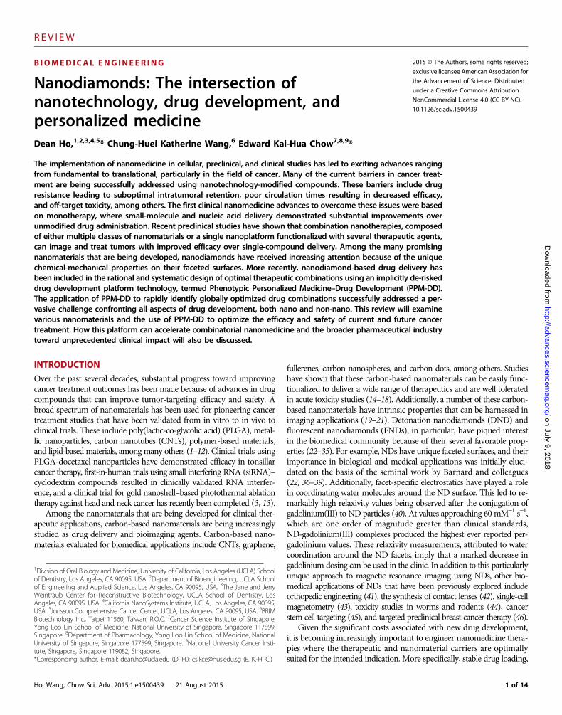
NDs, both DND and FND, are also being widely used for imagingapplications. Each class of diamond has unique surface or structuralfeatures that markedly improve their performance as imaging agentscompared to clinical and nanoparticle standards (Fig. 2) (56–59). Inaddition to the improvements in magnetic resonance imaging mentionedin the introduction, a recent breakthrough using FNDs pertained to
Ho, Wang, Chow Sci. Adv. 2015;1:e1500439 21 August 2015
http://d from
the sustained labeling of lung stem cells (LSCs) to track their engraft-ment and regenerative potential after lung tissue injury in a murine mod-el (60). LSCs are important mediators of epithelial tissue regenerationin vivo as well as regulators of lung tissue homeostasis. Tracking LSCs,however, has been difficult because of the photobleaching and toxicityobserved with conventional agents, which can impede the differentia-tion capabilities or viability of the LSCs. A recent study by Wu et al.has demonstrated stable tracking of LSC with fluorescent NDs, confirm-ing LSC localization to the terminal bronchioles after transplantation(Fig. 2B). The NDs were excited by green-yellow light, and the inte-gration of negatively charged nitrogen-vacancy centers resulted in stablefar-red emission at a 15-ns lifetime. Because conventional agents havefluorescent lifetimes in the range of 1 to 4 ns, ND fluorescence couldbe easily differentiated from tissue autofluorescence using fluorescencelifetime imaging microscopy (FLIM). LSCs were screened for the CD54and CD157 markers to ensure their capacity for differentiation, andfurther studies confirmed that the cells were from a hematopoietic lin-eage. Fluorescent NDs incubated with CD45−CD54+CD157+ cellswere readily endocytosed with no apparent exocytosis. After tail-veininjection of the ND-containing LSCs, their engraftment and differen-tiation capabilities were unimpaired, resulting in improved localizationand epithelial regeneration at the sites of lung injury compared to salinecontrol. This was an important advance because of the sustained LSCmonitoring enabled by the photostability and biocompatibility of thefluorescent NDs.
on July 9, 2018advances.sciencem
ag.org/
ND-BASED DRUG DELIVERY
ND drug delivery has received significant attention because of the fac-ile nature of functionalizing their surfaces with drug compounds, par-ticularly anthracyclines. The anthracycline class of compounds, whichinclude doxorubicin, epirubicin, and daunorubicin, among others, arepotent DNA intercalating agents that are used in most chemotherapyregimens. Although anthracyclines have effective anticancer activity,they are also extremely toxic. They induce myelosuppression (whichis the dose-limiting side effect of chemotherapy), mediate cardiotox-icity that can result in heart failure, can lead to superinfections, andmay markedly increase the risk of developing acute myelogenous leu-kemia (61). Early studies successfully formulated ND-doxorubicincompounds (NDX) through physisorption, enabling potent drug bind-ing and subsequent release without the need to chemically modify thedrug itself (62, 63). The NDX compound was subsequently validated ina broad array of cancer models that ranged from in vitro through pre-clinical in vivo models. Most notably, given that the problem of drugresistance accounts for greater than 90% of tumor treatment failure inmetastatic cancer, NDX was tested against two highly drug-resistantcancer models: the 4T1 breast cancer model and the LT2-M liver can-cer model (54). These tumors are known to express ABC (ATP-bindingcasette) transporter proteins that mediate drug efflux, markedly reducingthe efficacy of chemotherapy with unmodified drugs. This lack of drugretention also results in elevated levels of toxicity. The initial stages ofNDX preclinical validation involved the administration of NDs aloneto confirm that they were well tolerated in murine models. High NDlevels (20 mg) resulted in no apparent increase in serum alanine amino-transferase (ALT) levels, an indicator that the NDs do not cause livertoxicity. In addition, these same dosages did not cause an increase in seruminterleukin-6 levels, demonstrating an absence of systemic toxicity as
Fig. 2. Imaging applications of FND fluorescent NDs. (A) C. elegans fedwith dextran-coated fluorescent NDs. Reprinted (adapted) with permission
from N. Mohan, C.-S. Chen, H.-H. Hsieh, Y.-C. Wu, H.-C. Chang, In vivo imagingand toxicity assessments of fluorescent nanodiamonds in Caenorhabditiselegans. Nano-Lett. 10, 3692 (2010/09/08, 2010). Copyright 2010 AmericanChemical Society. (B) Engraftment of fluorescent ND-labeled LSCs in a lunginjury mouse model. Adapted with permission from Macmillan PublishersLtd.: T.-J. Wu et al., Tracking the engraftment and regenerative capabil-ities of transplanted LSCs using fluorescent NDs. Nat. Nanotechnol. 8, 682(09//print, 2013), copyright 2013.3 of 14

REV I EW
on July 9, 2018http://advances.sciencem
ag.org/D
ownloaded from
well. After the initial validation of ND biocompatibility and intra-cellular retention, verapamil blocking assays were performed, which con-firmed that the NDX, compared to unmodified doxorubicin (Dox),was retained longer in 4T1, LT2-M, Huh7, and MDA-MB-231 breastcancer cells. Pharmacokinetic analysis of NDX revealed an observedfirst phase half-life of 8.43 hours for NDX compared to 0.83 hours forDox alone. Drug efficacy studies demonstrated a clear decrease in tu-mor size with NDX administration compared to free Dox administra-tion. In 4T1 tumors, NDX administration (100 mg equivalence) againresulted in markedly reduced tumor sizes compared to the adminis-tration of Dox alone. Of note, the administration of Dox alone at 100 mgshowed virtually no efficacy, with tumor sizes on the order of thoseobserved with saline control treatment. When the Dox dosage wasincreased to 200 mg, all of the mice experienced early mortality. WhenNDX at 200-mg Dox equivalence was administered, all of the mice sur-vived the entire duration of the study, with the tumors being the smallestamong all of the test conditions observed. This confirmed that the NDXplatform improved therapeutic efficacy against highly drug-resistanttumors and also markedly enhanced drug tolerance, all without theneed to chemically modify Dox. Furthermore, the intravenous admin-istration of NDX resulted in no apparent myelosuppression, whereasDox alone resulted in a substantial decrease in white blood cell count.This finding confirmed the existence of a potent ND-Dox interactionsuch that premature drug elution did not take place even after systemicinjection.
Whereas the NDX compound represented a passive form of Doxdelivery, actively targeted ND drug delivery has also been demonstrated.Antibodies against the epidermal growth factor receptor (EGFR) wereconjugated to fluorescently labeled NDs with bifunctional cross-linkersfor subsequent targeting. Introducing epidermal growth factor (EGF)as a control to block targeting confirmed the improved specificity of deliv-ery in the EGFR-overexpressing MDA-MB-231 breast cancer cell linecompared to the MCF7 breast cancer cell line, which does not over-express EGFR (64). Preclinical validation of EGFR targeting was dem-onstrated with a liposome-encapsulated ND-epirubicin complex. Inthis iteration of a targeted ND drug delivery complex, the EGFR anti-bodies were conjugated onto the surface of the liposome, which in turnwas used to encapsulate the ND-epirubicin compounds. In mice withMDA-MB-231 tumors, the targeted ND complexes mediated completetumor regression to the point where they were no longer detectable.The administration of epirubicin alone at 150 mg resulted in earlymortality, whereas EGFR-targeted ND delivery of epirubicin at thesame dosage resulted in complete animal survival and tumor regres-sion (Fig. 3A) (46).
The properties of ND delivery of anthracyclines that allow ND-anthracycline complexes to overcome ABC transporter–mediated drugresistance also lend NDs as a suitable drug delivery platform for ef-fectively treating cancer stem cells (CSCs) (45, 65). Chemoresistance,including ABC transporter–mediated resistance, is often linked to CSCsand is a major mechanism by which these tumor-initiating cells escapetraditional therapy and contribute to recurrence (66–68). This is par-ticularly true for hepatic cancers where chemoresistant and metastaticCSCs have been identified and isolated by expression of these ABC trans-porter proteins (69–71). Overexpression of ABC transporter proteinsis clinically linked to poorer drug response, including to epirubicin, inhepatic cancers (72, 73). Delivery of epirubicin by NDs was demon-strated to overcome this mechanism of resistance in CSCs and more ef-fectively kill CSCs compared to epirubicin alone (Fig. 3, B and C) (45).
Ho, Wang, Chow Sci. Adv. 2015;1:e1500439 21 August 2015
Treatment with epirubicin alone resulted in a positive selection of he-patic CSCs and in respectively 8.13- and 3.88-fold increases in vitroand in vivo in the frequency of tumor-initiating CSCs among tumorcells that survived drug treatment. In contrast, similar treatment withND-epirubicin resulted in respectively 3.4- and 5.46-fold decreases invitro and in vivo in the frequency of tumor-initiating CSCs amongremaining tumor cells after ND-drug treatment. This translated intodecreased tumor colony formations in vitro as well as a lack of sec-ondary tumor formation in vivo, demonstrating effective eliminationof key tumor-initiating CSCs after ND-epirubicin treatment.
Although ND-based drug delivery against cancer remains one ofthe most developed biomedical applications, tissue engineering andantimicrobial applications are also promising fields in which NDs mayalso have a therapeutic role (74–85). Thin-film nanocrystalline diamond(NCD) surfaces were functionalized with growth factors, such as bonemorphogenetic protein-2 (BMP-2), through physisorption to promotelocalized bone formation (86). BMP-2–functionalized hydrophilic NCDsurfaces were able to promote osteogenic induction in human stromalcells in vitro. In vivo studies with BMP-2–functionalized NCD-coatedimplants in sheep revealed long-term retention of BMP-2 at the site ofimplantation compared to control implants. This translated into greaterbone formation around the BMP-2–functionalized NCD-coated im-plant by 4 weeks after implantation. The addition of NDs to copolymerscaffolds can also increase the hydrophilicity of the scaffold to pro-mote the attachment, proliferation, and differentiation of bone marrowstromal cells in vitro and new bone formation in vivo (87). Furtherfunctionalization with physisorption of BMP-2 to NDs in copolymerscaffolds promoted de novo bone formation in models of mandibulardefects in vivo, demonstrating the potential of integrating NDs intotissue-engineering disease applications (41).
The versatility of ND surface functionalization and the anisotropicdistribution of charges on the ND surface also lend the ND platformto antimicrobial applications. NDs can be functionalized with saccharidesto detect and capture bacteria to effectively diagnose and treat infec-tions (88). Additionally, NDs can be partially oxidized to mediate potentantimicrobial activity against both Gram-negative and Gram-positivebacteria (89). The antimicrobial activity is likely mediated by boththe delivery of reactive oxygen species to bacteria cellular componentsand the alteration of bacterial surfaces by anisotropic distribution ofcharges of bacteria-interacting NDs. These studies, in addition to thoseaddressing ND drug delivery in cancer, demonstrate that NDs are apromising nanomaterial for a wide array of biomedical applications.
ND BIODISTRIBUTION AND TOXICITY
As NDs progress toward clinical translation, an increasing body ofwork has explored their biodistribution and biocompatibility proper-ties in vitro and in vivo (90, 91). Dextran- and bovine serum albumin–functionalized FND tracking in the Caenorhabditis elegans model hasbeen used to characterize their safety and excretion mechanisms inliving organisms (Fig. 2A) (44). Observation of ND consumption ormicroinjection and resulting stress response and reproductive functionassessed acute and long-term tolerance in these C. elegans preclinicalmodels. The nuclear translocation of the DAF-16 transcription factorserved as a stress readout. No apparent toxicity was observed after NDconsumption, and gonadal injection resulted in FND presence in wormoffspring.
4 of 14

REV I EW
on July 9, 2018http://advances.sciencem
ag.org/D
ownloaded from
Biodistribution studies in mice intravenously injected with fluores-cent dye–labeled NDs revealed initial accumulation of NDs in the lung,spleen, liver, and kidneys. Rapid clearance was observed from the lungfollowed by more gradual clearance of NDs from the spleen, liver, andkidney over a 10-day period. A strong fluorescently labeled ND signalvisible from the bladder suggested efficient excretion of NDs (54). Bio-distribution studies with DNDs radiolabeled with 18F radionuclide andanalyzed in mice and rats by positron emission tomography (PET)confirm these results. Radiolabeled NDs were detected primarily in thelung and urine and, to a lesser degree, in the liver and spleen 2 hoursafter administration (92). Biodistribution studies with other carbon-based nanoparticles reveal similarities as well as differences in organaccumulation and excretion of these nanoparticles. Similar to fluores-cently labeled NDs, fluorescent carbon dots accumulated mostly in the
Ho, Wang, Chow Sci. Adv. 2015;1:e1500439 21 August 2015
mouse bladder, kidney, and liver 4 hours after intravenous injection(21). Radiolabeled graphene oxide also primarily accumulated in themouse liver and spleen after intraperitoneal injections but was unableto be excreted from the body, as evidenced by minimal signal in the kid-ney. Graphene oxide particles were also detected in mouse livers 30 daysafter intraperitoneal injection (93). Whereas CNTs have been observedto be capable of being excreted and even observed by electron micros-copy in the urine of treated mice, a comparison study of radiolabeledNDs and CNTs revealed biodistribution differences. CNTs were pri-marily observed in the lung, whereas NDs were quickly cleared fromthe lung and found in the liver and spleen (94, 95). Further studies arebeing conducted to address this observation and to determine the im-pact of this long-term retention of nanocarbons in the lungs on gran-uloma formation and chronic pulmonary toxicity (96).
Fig. 3. ND-anthracycline drug delivery in cancer. (A) EGFR-targeted delivery of ND-epirubicin (anti-EGFR-NDLP-Epi) against breast cancer cellsdemonstrated increased efficacy compared to untargeted ND-epirubicin (NDLP-Epi) and unmodified epirubicin (Epi) while retaining the increased
safety that results from ND conjugation of epirubicin. Reprinted with permission from WILEY. (B) Treatment of hepatic tumor–bearing mice with ND-epirubicin (EPND) efficiently killed hepatic CSCs and prevented secondary tumor formation seen after treatment with unmodified epiruibicin (Epi).(C) A schematic model of ND-epirubicin complex formation and aggregation. Reprinted (adapted) with permission from X. Wang et al., Epirubicin-adsorbed nanodiamonds kill chemoresistant hepatic cancer stem cells. ACS Nano 8, 12151 (2014/12/23, 2014). Copyright 2014 American ChemicalSociety.5 of 14
REV I EW
on July 9, 2018http://advances.sciencem
ag.org/D
ownloaded from
Additional studies have sought to examine the cellular mechanismsthat are activated after ND exposure to provide deeper insight into thedose-dependent tolerance of NDs at the cellular and preclinical levels.Several of these studies have demonstrated that the NDs are welltolerated even at high dosages. Although prior work has been con-ducted to monitor potential hematotoxicity, comprehensive in vivo ser-um toxicity panels in another study resulted in no apparent changes inserum markers (46, 97, 98). This study and others serve as importantindicators that the NDs are well tolerated at multiple dosages in a widevariety of cell lines and a diverse range of animal models.
More recently, a study has been conducted on the cellular compat-ibility of DNDs, FND NDs, NDs with surface amine groups, and NDsphysisorbed with daunorubicin, an anthracycline chemotherapy (99).HeLa cervical cancer cells and HepG2 liver cancer cells were selectedbecause of their prevalence as toxicity and drug efficacy testing plat-forms. After their incubation with the ND subtypes, the cells were ex-amined for indications of cell death, including onset of apoptosis, metabolicstates, reduction in drug toxicity from ND sequestering effects, andgene expression profiles.
To assess the biocompatibility of the ND subtypes being investi-gated, a broad range of assays was conducted. The caspase-3/7 assaywas used to measure the potential onset of apoptosis. Cell metabolismwas examined using an XTT (2,3-bis[2-methoxy-4-nitro-5-sulfophenyl]-2H-tetrazolium-5-carboxanilide inner salt) assay, indications of cellulartoxicity were assessed using a lactate dehydrogenase assay, and gene ex-pression profiles were evaluated through quantitative real-time poly-merase chain reaction. Key findings from this study showed that highdoses (250 mg/ml) of all ND subtypes did not have a negative impacton viability in either cell line. Transcriptional regulation studies dem-onstrated that incubation of HepG2 cells with NDs at a dose of 25 mg/mldid not result in significant changes in gene expression levels of Ki-67,Bax, and c-Myc genes. This indicates the absence of apoptotic and anti-proliferative effects or a cellular stress response. Overall, this repre-sented among the most comprehensive studies of ND safety to date.
Recently, comparative in vitro studies have also been conductedwith graphene, CNTs, and NDs to understand the similarities and dif-ferences in nanocarbon toxicity (100). Whereas CNTs and grapheneexhibited similar rates of toxicity with increasing carbon concentra-tion, ND administration appeared to show less toxicity. To further un-derstand the mechanism of nanocarbon toxicity, liposomal leakagestudies and toxicogenomic analysis were conducted. The effect of dif-ferent nanocarbons on liposomal leakage was explored to determine ifmembrane damage was a possible explanation for any nanocarbon-related toxicity. NDs, CNTs, and graphene could all adsorb onto thesurface of liposomes without disrupting the lipid bilayer, suggestingthat membrane disruption is not a contributing mechanism to thelimited toxicity observed with nanocarbons. Toxicogenomic analysisof nanotitanium dioxide, carbon black, CNTs, and fullerenes in bacte-ria, yeast, and human cells revealed structure-specific mechanisms oftoxicity among nanomaterials, as well as other nanocarbons (101). Al-though both CNTs and fullerenes failed to induce oxidative damage asobserved in nanomaterials such as nanotitanium dioxide, they wereboth capable of inducing DNA double-stranded breaks (DSBs) in eu-karyotes. However, the specific mechanisms of DSBs remain unclearbecause differences in activation of pathway-specific DSB repair geneswere found between the two nanocarbons. These studies give an initialunderstanding of ND and nanocarbon toxicity to continue on a path-way toward clinical implementation and first-in-human use, and com-
Ho, Wang, Chow Sci. Adv. 2015;1:e1500439 21 August 2015
prehensive nonhuman primate studies of ND toxicity are currentlyunder way.
TRANSLATION OF NANOMEDICINE THROUGHCOMBINATION THERAPY
For all therapeutics moving from bench to bedside, including NDsand nanomedicine, additional development beyond cellular and ani-mal models of efficacy and toxicity is needed. As these therapeutics areabsorbed into drug development pipelines, they will invariably be in-tegrated into combination therapies. This strategy of combinatorial med-icine has been recognized by the industry as being essential in variousdisease areas (for example, pulmonary artery hypertension, cardiovas-cular disease, diabetes, arthritis, chronic obstructive pulmonary disease,HIV, tuberculosis) and especially oncology (102–110). How these com-binations can be rationally designed so that safety and efficacy aremaximized is still a major challenge, and current strategies have onlycontributed to the increasing cost of new drug development. The in-efficiencies in developing and validating suitable combinations lie notonly in the empirical clinical testing of these combinations in the clinicbut also in the time and resources spent in the clinic. Examples of theway these trials are conducted provide important insight into how op-timization of combination therapy can be improved.
For clinical trials conducted and listed on ClinicalTrials.gov from2008 to 2013, 25.6% of oncology trials contained combinations, com-pared to only 6.9% of non-oncology trials (110). Within each diseasearea, viral diseases had the next highest percentage of combination trialsconducted after oncology at 22.3%, followed by digestive diseases (18.6%),cardiovascular diseases (8.5%), pathological conditions (5.7%), andneurological diseases (5.4%). Because most of the combination trials wereperformed in oncology, the following clinical examples will displayhow empirical testing of therapeutic combinations is conducted in thisdisease area. In many instances, including the following clinical exam-ples, the MTD of the drugs as single agents is often directly used incombinations. This is done without using other methods to determinethe best dosing of each drug before use in a particular combination.This empirical approach can lead to poor results due to compoundedtoxic side effects of the individual therapeutics, unpredictability of othercomplications, and/or less than optimal efficacies due to the combina-tion of the drugs.
A recent phase 1 study (NCT01400451) looked at the combinationof ipilimumab, a monoclonal antibody targeted against CTLA-4, andvemurafanib, a BRAF inhibitor targeting the V600E mutation (111).Both therapeutics are approved for single use in melanoma. Becausetheir inhibition pathways are different, the use of both in combinationwas a natural progression. In this study, both therapeutics were used atthe MTD, which resulted in dose-limiting toxicities (DLTs) that un-fortunately led to early termination of the study. In another immuno-therapy study, a phase 1 study of ipilimumab combined with anotherV600E BRAF inhibitor, dabrafenib, and mitogen-activated or extra-cellular signal–regulated protein kinase kinase inhibitor, trametinib,was also terminated early because of grade 4 intestinal perforation intwo of the seven patients and grade 3 dose-limiting colitis (112). Thedoublet combination of ipilimumab with the BRAF inhibitor dabrafenibdid not show any DLTs, and an expansion cohort was in the process ofbeing enrolled. These first two examples show that direct combina-tion of therapeutics at their MTD without any initial studies looking
6 of 14

REV I EW
on July 9, 2018http://advances.sciencem
ag.org/D
ownloaded from
at appropriate dosing for the combination may result in toxicity, ulti-mately preventing the use of the combination. In addition, the secondexample shows that combinations using molecules that may target thesame mutation and pathway may not have the same types of toxic sideeffects, demonstrating that the differences in combinations using similarclasses of therapeutics need to be monitored. In an infectious diseaseand oncology example using a traditional oncology phase 1 3 + 3 dose es-calation design, non–ritonavir-based HAART (highly active antiretroviraltherapy) with standard sunitinib therapy (50 mg/day) (treatment arm 1)was compared with the combination of sunitinib in HIV-positive patientsreceiving ritonavir-based HAART (treatment arm 2) (NCT00890747)(111). Patients in treatment arm 1 tolerated treatment without any ob-served DLT. Treatment arm 2 had DLT at a sunitinib dose of 37.5 mg,with three of five patients having grade 3 neutropenia. This showed thatpatients on ritonavir could combine the use of sunitinib with ritonavair-based HAART, but that it should be given at a lower dose of sunitinibwhen used in combination.
These three examples, and many more, demonstrate how clinicaltrials for combination therapy are often conducted either at the MTDfor each therapeutic or with patients being dosed empirically withoutclear guidelines. Although nanomedicines have yet to be used in manycombination therapy trials, they will inevitably join the other types oftherapeutic classes in use for combination. Therefore, whereas the cur-rent strategy of determining combinatorial drug use has resulted in manycombination therapies used to date, it is clearly not the most efficientmethod and can still be significantly optimized. On the basis of thetrend of increasing use of combination therapy in nanomedicine andin broader drug development, as well as the challenges that are facedin determining optimized combination therapies to use, a new paradigmusing systematically designed drug combinations needs to be identified.This would be a much needed tool that the pharmaceutical industry isready to embrace in their efforts to define new combinations that mayhelp their product lifecycle management, or “evergreening,” an impor-tant part of many companies.
The concept of evergreening is a widely used approach in the phar-maceutical industry to retain patent protection and rent-earning rightson protected compounds with imminent expiry dates (113–115). Thisconcept includes formulating new drug combinations containingsoon-to-be generic compounds to create new patents that may extendthe financial lifetime of the drug. Whereas this process may result inintended or unexpected improvements to the safety and/or efficacy oftreatment, there is debate about whether these are measures that the drugmanufacturers are purposefully taking to prevent generic drug makersfrom producing these compounds. If so, this may ultimately lead torestricted competition and limitations in the availability of lower-priced medicines to the general population, causing controversy overthe concept of suboptimal evergreening.
The application of nanotechnology to novel formulation and deliv-ery has become a highly active area of evergreening strategies. The capa-city for nanotechnology to modify properties such as pharmacokinetics,oral bioavailability, drug toxicity and efficacy, and others may result insubstantial growth in nanomedicine development. This is particularlytrue as patent cliffs approach for some of the most profitable medicinesin the world. Challenges from both patent-holding and genericscompanies have been raised in an effort to either promote competitionin the pharmaceutical industry, on the one hand, or potentially suppressgeneric entry on the other. Regardless of the continued controversy sur-rounding the practice of evergreening, a new challenge that has arisen
Ho, Wang, Chow Sci. Adv. 2015;1:e1500439 21 August 2015
concerning both sides of the debate involves the need to truly optimizenew combinations, both nano and non-nano. This will be necessary tosuccessfully address the issues of maximizing efficacy and safety forthe good of public health, as well as meeting the increasing thresholdsof patentability. To address this challenge, a key advance at the inter-section of nanotechnology and engineering optimization has openeddoors to simultaneously optimizing and de-risking the drug develop-ment pipeline using phenotype to drive the rational design of combi-nation therapy.
PERSONALIZING AND OPTIMIZING NANOMEDICINEDRUG DEVELOPMENT
Innovative advances in functionalizing nanoparticles combined withmultiple classes of therapeutic agents have improved efficacy over mono-therapy with nanomedicine. However, the process of globally optimizingcombination therapies has thus far been challenging, if not impossible.Dosing levels of the drugs in combination are a major factor in deter-mining the efficacy and toxicity of therapy. Hence, there is a nearlyinfinite number of possible drug dose combinations that can be designedwhen conventional screening or predictive approaches are used. Emerg-ing strategies are continually being explored with regard to integratingseveral therapies with a single class of nanoparticle carriers or the useof several different classes of nanoparticle carriers to mediate combi-natorial nanomedicine (49, 50, 52). These strategies have shown thatthe delivery of multiple compounds using nanoparticles has resultedin early indications of improved efficacy and toxicity. Therefore, aplatform technology that is applicable to all types of nanoparticles andis capable of rationally and systematically optimizing these approachestoward globally optimized safety and efficacy across the in vitro, in vivo,and translational stages of drug development would represent a majoradvance.
High-throughput screening is a valuable in vitro approach that canuse brute force to identify drug combinations that enable the mostfavorable outcome from those that have been tested. Limitations arise,however, when attempts to simultaneously optimize multiple outcomes,including several safety and efficacy parameters, are made. Aside froma limitless number of combinations that would need to be tested, pri-mary sample testing is likely to be ruled out because of inadequatesample availability. Other efforts to develop optimal drug administra-tion conditions have included the use of pharmacokinetic modeling,median-effect methods to assess drug synergism and antagonism,prediction-based genomic modeling, and mechanism-based systemsbiology approaches (116–118). However, the use of these approachesto design drug combinations can result in limitations on the maximumnumber of drugs that can be used within the combination, mixturesthat are rendered ineffective because of resistance, and the inabilityto optimize on the basis of undruggable mechanistic data. All of theseapproaches are also subject to significant risks during the developmentof both nanotechnology-modified and unmodified drugs. The inabilityto definitively determine optimal drug dose ratios during each stage oftesting and development coupled with the confounding aspects of themechanisms used for drug design commonly result in clinical trialfailure.
Recently, Phenotypic Personalized Medicine–Drug Development(PPM-DD) has been developed as a mechanism-independent and model-less platform that uses experimental data to formulate phenotype-based
7 of 14
REV I EW
on July 9, 2018http://advances.sciencem
ag.org/D
ownloaded from
drug response landscapes (119–122). As such, mechanistic propertiessuch as signaling pathway behavior, drug-drug interactions, pharmaco-kinetics, and heterogeneity are innately accounted for using the PPM-DDapproach. It is important to note that PPM-DD does not require theuse of feedback control, predictive algorithms and modeling, or a phar-macogenetics platform. Instead, it uses experimental data to formulatephenotypic maps to systematically and rapidly identify optimal drugcombinations during each stage of the drug development roadmapthat ranges from in vitro through in vivo and to translational stages.More specifically, the in vitro stage is used to broadly explore the sys-tematic formulation of novel and optimized initial drug combinationsand to narrow down the drugs and lead combinations of initial interest.Subsequent in vivo validation is conducted to reoptimize the drug doseratios at the preclinical level. Lead combinations can then be furtheroptimized in the translational setting. The ability to properly determineoptimal drug dose ratios from discovery and preclinical validationthrough translation can provide a definitive pathway toward achievingpopulation response rates that will far supersede those that are currentlyobserved with conventionally designed drug combinations.
The first version of PPM-DD was termed Feedback System Control.I(FSC.I). This system used an iterative search process that previouslyused a search/feedback algorithm to guide experimental validation ofcombinations to rapidly find a combination that performed optimallyboth in vitro and in vivo, even from prohibitively large pools of pos-sible combinations (119, 123). The term Feedback System Control is aremnant of the first version of the platform, and subsequent iterationswere no longer based on feedback. Therefore, the recent developmentof PPM-DD [previously referred to as Feedback System Control.II(FSC.II)] resulted in an experimentally driven optimization platformthat inherently accounts for all mechanistic components of disease (forexample, cellular signaling networks, patient heterogeneity, genomicaberrations) to formulate drug combinations that culminate in an op-timal phenotypic output (53, 124).
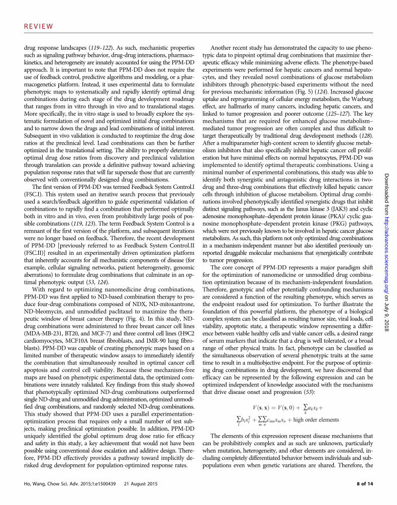
With regard to optimizing nanomedicine drug combinations,PPM-DD was first applied to ND-based combination therapy to pro-duce four-drug combinations composed of NDX, ND-mitoxantrone,ND-bleomycin, and unmodified paclitaxel to maximize the thera-peutic window of breast cancer therapy (Fig. 4). In this study, ND-drug combinations were administered to three breast cancer cell lines(MDA-MB-231, BT20, and MCF-7) and three control cell lines (H9C2cardiomyocytes, MCF10A breast fibroblasts, and IMR-90 lung fibro-blasts). PPM-DD was capable of creating phenotypic maps based on alimited number of therapeutic window assays to immediately identifythe combination that simultaneously resulted in optimal cancer cellapoptosis and control cell viability. Because these mechanism-freemaps are based on phenotypic experimental data, the optimized com-binations were innately validated. Key findings from this study showedthat phenotypically optimized ND-drug combinations outperformedsingle ND-drug and unmodified drug administration, optimized unmodi-fied drug combinations, and randomly selected ND-drug combinations.This study showed that PPM-DD uses a parallel experimentation-optimization process that requires only a small number of test sub-jects, making preclinical optimization possible. In addition, PPM-DDuniquely identified the global optimum drug dose ratio for efficacyand safety in this study, a key achievement that would not have beenpossible using conventional dose escalation and additive design. There-fore, PPM-DD effectively provides a pathway toward implicitly de-risked drug development for population-optimized response rates.
Ho, Wang, Chow Sci. Adv. 2015;1:e1500439 21 August 2015
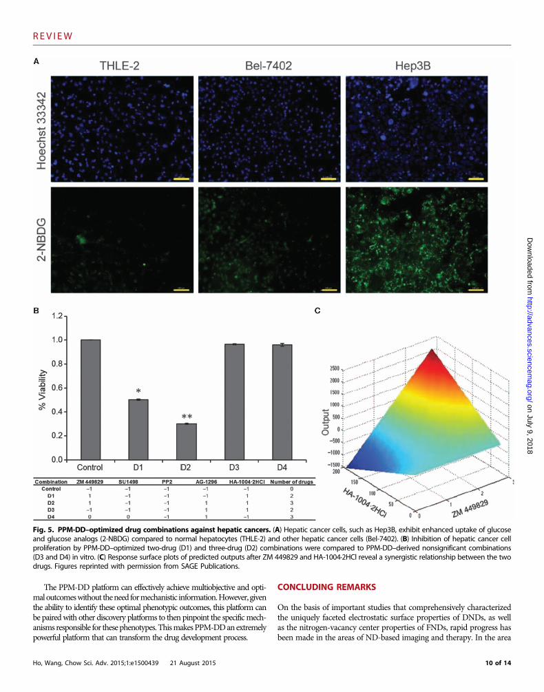
Another recent study has demonstrated the capacity to use pheno-typic data to pinpoint optimal drug combinations that maximize ther-apeutic efficacy while minimizing adverse effects. The phenotype-basedexperiments were performed for hepatic cancers and normal hepato-cytes, and they revealed novel combinations of glucose metabolisminhibitors through phenotypic-based experiments without the needfor previous mechanistic information (Fig. 5) (124). Increased glucoseuptake and reprogramming of cellular energy metabolism, the Warburgeffect, are hallmarks of many cancers, including hepatic cancers, andlinked to tumor progression and poorer outcome (125–127). The keymechanisms that are required for enhanced glucose metabolism–mediated tumor progression are often complex and thus difficult totarget therapeutically by traditional drug development methods (128).After a multiparameter high-content screen to identify glucose metab-olism inhibitors that also specifically inhibit hepatic cancer cell prolif-eration but have minimal effects on normal hepatocytes, PPM-DD wasimplemented to identify optimal therapeutic combinations. Using aminimal number of experimental combinations, this study was able toidentify both synergistic and antagonistic drug interactions in two-drug and three-drug combinations that effectively killed hepatic cancercells through inhibition of glucose metabolism. Optimal drug combi-nations involved phenotypically identified synergistic drugs that inhibitdistinct signaling pathways, such as the Janus kinase 3 (JAK3) and cyclicadenosine monophosphate–dependent protein kinase (PKA)/ cyclic gua-nosine monophosphate–dependent protein kinase (PKG) pathways,which were not previously known to be involved in hepatic cancer glucosemetabolism. As such, this platform not only optimized drug combinationsin a mechanism-independent manner but also identified previously un-reported druggable molecular mechanisms that synergistically contributeto tumor progression.
The core concept of PPM-DD represents a major paradigm shiftfor the optimization of nanomedicine or unmodified drug combina-tion optimization because of its mechanism-independent foundation.Therefore, genotypic and other potentially confounding mechanismsare considered a function of the resulting phenotype, which serves asthe endpoint readout used for optimization. To further illustrate thefoundation of this powerful platform, the phenotype of a biologicalcomplex system can be classified as resulting tumor size, viral loads, cellviability, apoptotic state, a therapeutic window representing a differ-ence between viable healthy cells and viable cancer cells, a desired rangeof serum markers that indicate that a drug is well tolerated, or a broadrange of other physical traits. In fact, phenotype can be classified asthe simultaneous observation of several phenotypic traits at the sametime to result in a multiobjective endpoint. For the purpose of optimiz-ing drug combinations in drug development, we have discovered thatefficacy can be represented by the following expression and can beoptimized independent of knowledge associated with the mechanismsthat drive disease onset and progression (53):
V ðs; xÞ ¼ V ðs; 0Þ þ ∑kakxkþ
∑lblx
2l þ∑
m∑ncmnxmxn þ high order elements
The elements of this expression represent disease mechanisms thatcan be prohibitively complex and as such are unknown, particularlywhen mutation, heterogeneity, and other elements are considered, in-cluding completely differentiated behavior between individuals and sub-populations even when genetic variations are shared. Therefore, the
8 of 14
REV I EW
on July 9, 2018http://advances.sciencem
ag.org/D
ownloaded from
overall treatment outcome can be represented by the difference in efficacybefore and after treatment. It is important to note that the resultingquadratic algebraic sequence is a function of the doses only and is hencemechanism-free. Unprecedented capabilities in optimizing combi-natorial drug development can then be achieved through facile sam-pling of various dose combinations to rapidly identify the algebraicseries coefficients, resulting in the most potent drug dose combinationaccording to phenotype only. Figures 4C and 5D harness this quadrat-ic algebraic equation to provide a global analysis of the drug-drug in-teraction map in a wide dose range. This map visually demonstrates
Ho, Wang, Chow Sci. Adv. 2015;1:e1500439 21 August 2015
that dose dependence in drug design can have a profound impact ondrug synergism and antagonism. A systematic combination therapydevelopment platform such as the PPM-DD approach can rationallypinpoint the specific drug dose ratios that result in globally optimaltreatment outcomes, not just the best outcome for a specific sampleset. The number or types of drugs within the combination do not limitthis approach. Therefore, PPM-DD can develop combinations con-taining multiple nanoformulated therapies and unmodified therapiesand is not confined to conventional triplet or doublet therapy formu-lation (53, 55, 119, 120, 123, 124, 129–131).
Fig. 4. PPM-DD–optimized ND-drug combinations. (A) A schematic model of the PPM experimental framework. Dox, doxorubicin; Bleo, bleomycin;Mtx, mitoxantrone; Pac, paclitaxel. (B) PPM-derived optimal ND-drug combinations (NDC) outperform a random sampling of NDCs in effective therapeutic
windows of treatment of cancer cells compared to control cells. Reprinted (adapted) with permission from H. Wang et al., Mechanism-independentoptimization of combinatorial nanodiamond and unmodified drug delivery using a phenotypically driven platform technology. ACS Nano (2015/02/17, 2015).Copyright 2014 American Chemical Society.9 of 14
REV I EW
on July 9, 2018http://advances.sciencem
ag.org/D
ownloaded from
The PPM-DD platform can effectively achieve multiobjective and opti-maloutcomeswithout theneed formechanistic information.However, giventhe ability to identify these optimal phenotypic outcomes, this platform canbe pairedwith other discovery platforms to then pinpoint the specificmech-anisms responsible for thesephenotypes.ThismakesPPM-DDanextremelypowerful platform that can transform the drug development process.
Ho, Wang, Chow Sci. Adv. 2015;1:e1500439 21 August 2015
CONCLUDING REMARKS
On the basis of important studies that comprehensively characterizedthe uniquely faceted electrostatic surface properties of DNDs, as wellas the nitrogen-vacancy center properties of FNDs, rapid progress hasbeen made in the areas of ND-based imaging and therapy. In the area
Fig. 5. PPM-DD–optimized drug combinations against hepatic cancers. (A) Hepatic cancer cells, such as Hep3B, exhibit enhanced uptake of glucoseand glucose analogs (2-NBDG) compared to normal hepatocytes (THLE-2) and other hepatic cancer cells (Bel-7402). (B) Inhibition of hepatic cancer cell
proliferation by PPM-DD–optimized two-drug (D1) and three-drug (D2) combinations were compared to PPM-DD–derived nonsignificant combinations(D3 and D4) in vitro. (C) Response surface plots of predicted outputs after ZM 449829 and HA-1004·2HCl reveal a synergistic relationship between the twodrugs. Figures reprinted with permission from SAGE Publications.10 of 14
REV I EW
http://advancesD
ownloaded from
of cancer therapy, passive and actively targeted ND-anthracyclinecomplexes have proven to be scalable platforms for hard-to-treatcancers that increase the efficacy and safety of chemotherapy. ND-basedimaging agents enabling preclinical tracking of LSC engraftment andmarkedly increasing per-gadolinium relaxivity provide a strong foun-dation for continued development for both basic and translational ap-plications. As more delivery platforms within the nanomedicine fieldare clinically validated, their role in transforming the pharmaceuticalindustry will become more defined. Monotherapy mediated by nano-medicine vehicles has already resulted in improved efficacy and safetyover clinical standards in recent human trials. Combination therapy isanother area where nanotechnology is poised to have an impact on pa-tient care in an important way. However, this also raises challenges ofhow these combinations can be rationally designed, given the enormouslimitations associated with identifying proper drug dose parametersfrom an infinite parameter space.
To circumvent the limitations of conventional combinatorial de-sign approaches, a paradigm-shifting platform that uses phenotype tosystematically identify globally optimized drug combinations was uti-lized to formulate ND-based and unmodified drug combinations. Theserationally developed therapies substantially outperformed randomlysampled drug combinations with respect to efficacy and safety. Further-more, the use of experimental data to formulate phenotypic responsemaps innately validated the lead combinations. Combining nanoma-terials with specific drug compounds using engineering optimizationplatforms can truly optimize drug dose combinations for defined in-dications. This will lead to unprecedented advances in patient treat-ment outcomes against the most serious diseases of our time.
on July 9, 2018.sciencem
ag.org/
OUTLINE OF UNRESOLVED QUESTIONS
The field of nanomedicine has given rise to a collection of promisingnanomaterial platforms. As nanomedicine-modified monotherapiescontinue to move into the clinic following important initial findingsfrom first-in-human studies, the next frontier will involve the clinicalimplementation of combination nanotherapies. The inefficiency of doseescalation– or additive design–based formulation of combination ther-apies is a challenge that has persistently confronted the broader phar-maceutical industry. It is evident that the nanomedicine field will needto address this barrier, particularly as nanotechnology drug deliveryand imaging agents increase in complexity. Nanomedicines are nowbeing designed to simultaneously carry several classes of payloads, ordifferent classes of nanomaterials are being co-delivered as a combina-tion. This review used the ND platform to illustrate specific examples,such as magnetic resonance imaging and cancer therapy, where NDsimmensely outperform conventional modalities. A recent advance atthe multidisciplinary interface of engineering systems identificationand ND drug delivery resulted in the demonstration that ND-drugcombinations could be effectively optimized for multiple parametersin a mechanism-independent fashion. This work simultaneously ad-dressed the challenges of optimal drug discovery and the use of nano-medicine to even further boost efficacy and safety. This reviewaddressed the following pervasive challenges and breakthroughsin drug development:• Nano-based monotherapy implementation in the clinic has made im-
portant advances in improving treatment outcomes. Nanotechnology-based modification of drugs is also becoming increasingly prevalent
Ho, Wang, Chow Sci. Adv. 2015;1:e1500439 21 August 2015
as the pharmaceutical industry looks for ways to innovate existingdrugs. Combination therapy represents the next stage of nanomedi-cine implementation.
• As the costs of drug development continue to climb, a strategy topinpoint which nanomaterial platforms are best suited for specificdrug and imaging compounds and indications must be developed.
• NDs have emerged as promising materials for imaging and therapy.Their specific clinical role will depend on continued toxicity and ef-ficacy studies, but initial studies in magnetic resonance imaging andanthracycline delivery are promising.
• Combination therapy is currently designed using additive formula-tion. This makes it virtually impossible to optimize therapy, whichhas a negative impact on public health. When simultaneously ad-dressing the prohibitively large number of possible drug combina-tions using current methods and requiring that the efficacy and safetyare both optimal, the parameter space is simply too large.
• The emergence of PPM-DD, previously referred to as the FSC.IItechnology, has now made it possible to design globally optimal drugcombinations, even with multiobjective criteria, using nanothera-peutics and non-nano therapeutics. PPM-DD is capable of optimiz-ing combination therapy design at each stage of development. Thisimplicitly de-risks the drug development process because the glob-ally optimal drug dose ratios are identified from an empirically con-structed phenotypic map.
• The demonstration of PPM-DD-based optimization in ND combi-nation therapy optimization resulted in globally maximal cancercell death and minimal healthy cell death. This was all accom-plished in a mechanism-independent fashion using a small sampleof phenotypic assays. This signified a major advance for nano-enhancedcombination therapy.
REFERENCES AND NOTES1. X. Xu, K. Xie, X. Q. Zhang, E. M. Pridgen, G. Y. Park, D. S. Cui, J. Shi, J. Wu, P. W. Kantoff, S. J. Lippard,
R. Langer, G. C. Walker, O. C. Farokhzad, Enhancing tumor cell response to chemotherapythrough nanoparticle-mediated codelivery of siRNA and cisplatin prodrug. Proc. Natl. Acad.Sci. U.S.A. 110, 18638–18643 (2013).
2. N. A. Peppas, J. Z. Hilt, A. Khademhosseini, R. Langer, Hydrogels in biology and medicine:From molecular principles to bionanotechnology. Adv. Mater. 18, 1345–1360 (2006).
3. J. Hrkach, D. Von Hoff, M. M. Ali, E. Andrianova, J. Auer, T. Campbell, D. De Witt, M. Figa,M. Figueiredo, A. Horhota, S. Low, K. McDonnell, E. Peeke, B. Retnarajan, A. Sabnis, E. Schnipper,J. J. Song, Y. H. Song, J. Summa, D. Tompsett, G. Troiano, T. Van Geen Hoven, J. Wright,P. LoRusso, P. W. Kantoff, N. H. Bander, C. Sweeney, O. C. Farokhzad, R. Langer, S. Zale,Preclinical development and clinical translation of a PSMA-targeted docetaxel nanoparticlewith a differentiated pharmacological profile. Sci. Transl. Med. 4, 128ra139 (2012).
4. T. Dvir, M. Bauer, A. Schroeder, J. H. Tsui, D. G. Anderson, R. Langer, R. Liao, D. S. Kohane,Nanoparticles targeting the infarcted heart. Nano Lett. 11, 4411–4414 (2011).
5. X. Zhang, M. D. Do, K. Dean, P. Hoobin, I. M. Burgar, Wheat-gluten-based natural polymernanoparticle composites. Biomacromolecules 8, 345–353 (2007).
6. M. M. Abdel-Mottaleb, D. Neumann, A. Lamprecht, Lipid nanocapsules for dermalapplication: A comparative study of lipid-based versus polymer-based nanocarriers.Eur. J. Pharm. Biopharm. 79, 36–42 (2011).
7. S. A. Jensen, E. S. Day, C. H. Ko, L. A. Hurley, J. P. Luciano, F. M. Kouri, T. J. Merkel, A. J. Luthi,P. C. Patel, J. I. Cutler, W. L. Daniel, A. W. Scott, M. W. Rotz, T. J. Meade, D. A. Giljohann, C. A. Mirkin,A. H. Stegh, Spherical nucleic acid nanoparticle conjugates as an RNAi-based therapy forglioblastoma. Sci. Transl. Med. 5, 209ra152 (2013).
8. X.-Q. Zhang, X. Xu, R. Lam, D. Giljohann, D. Ho, C. A. Mirkin, Strategy for increasing drugsolubility and efficacy through covalent attachment to polyvalent DNA–nanoparticleconjugates. ACS Nano. 5, 6962–6970 (2011).
9. A. Bianco, K. Kostarelos, M. Prato, Applications of carbon nanotubes in drug delivery. Curr.Opin. Chem. Biol. 9, 674–679 (2005).
10. Z. Liu, K. Chen, C. Davis, S. Sherlock, Q. Cao, X. Chen, H. Dai, Drug delivery with carbonnanotubes for in vivo cancer treatment. Cancer Res. 68, 6652–6660 (2008).
11 of 14
REV I EW
on July 9, 2018http://advances.sciencem
ag.org/D
ownloaded from
11. A. K. Patri, I. J. Majoros, J. R. Baker Jr., Dendritic polymer macromolecular carriers for drugdelivery. Curr. Opin. Chem. Biol. 6, 466–471 (2002).
12. A. M. Gobin, M. H. Lee, N. J. Halas, W. D. James, R. A. Drezek, J. L. West, Near-infraredresonant nanoshells for combined optical imaging and photothermal cancer therapy.Nano Lett. 7, 1929–1934 (2007).
13. M. E. Davis, J. E. Zuckerman, C. H. J. Choi, D. Seligson, A. Tolcher, C. A. Alabi, Y. Yen, J. D. Heidel,A. Ribas, Evidence of RNAi in humans from systemically administered siRNA via targetednanoparticles. Nature 464, 1067–1070 (2010).
14. C. Wang, J. Li, C. Amatore, Y. Chen, H. Jiang, X. M. Wang, Gold nanoclusters and graphenenanocomposites for drug delivery and imaging of cancer cells. Angew. Chem. Int. Ed. Engl.50, 11644–11648 (2011).
15. R. P. Feazell, N. Nakayama-Ratchford, H. Dai, S. J. Lippard, Soluble single-walled carbonnanotubes as longboat delivery systems for platinum(IV) anticancer drug design. J. Am.Chem. Soc. 129, 8438–8439 (2007).
16. N. W. Kam, M. O’Connell, J. A. Wisdom, H. Dai, Carbon nanotubes as multifunctionalbiological transporters and near-infrared agents for selective cancer cell destruction.Proc. Natl. Acad. Sci. U.S.A. 102, 11600–11605 (2005).
17. L. Wang, Q. Sun, X. Wang, T. Wen, J. J. Yin, P. Wang, R. Bai, X. Q. Zhang, L. H. Zhang, A. H. Lu,C. Chen, Using hollow carbon nanospheres as a light-induced free radical generator toovercome chemotherapy resistance. J. Am. Chem. Soc. 137, 1947–1955 (2015).
18. P. Chaudhuri, A. Paraskar, S. Soni, R. A. Mashelkar, S. Sengupta, Fullerenol–cytotoxic conju-gates for cancer chemotherapy. ACS Nano. 3, 2505–2514 (2009).
19. W. Li, Z. Zhang, B. Kong, S. Feng, J. Wang, L. Wang, J. Yang, F. Zhang, P. Wu, D. Zhao,Simple and green synthesis of nitrogen-doped photoluminescent carbonaceous nano-spheres for bioimaging. Angew. Chem. Int. Ed. Engl. 52, 8151–8155 (2013).
20. Q. Liu, B. Guo, Z. Rao, B. Zhang, J. R. Gong, Strong two-photon-induced fluorescence fromphotostable, biocompatible nitrogen-doped graphene quantum dots for cellular anddeep-tissue imaging. Nano Lett. 13, 2436–2441 (2013).
21. S. T. Yang, L. Cao, P. G. Luo, F. Lu, X. Wang, H. Wang, M. J. Meziani, Y. Liu, G. Qi, Y. P. Sun,Carbon dots for optical imaging in vivo. J. Am. Chem. Soc. 131, 11308–11309 (2009).
22. A. S. Barnard, Self-assemblyin nanodiamond agglutinates. J. Mater. Chem. 18, 4038–4041(2008).
23. A. S. Barnard, Diamond standard in diagnostics: Nanodiamond biolabels make their mark.Analyst 134, 1751–1764 (2009).
24. A. Adnan, R. Lam, H. Chen, J. Lee, D. J. Schaffer, A. S. Barnard, G. C. Schatz, D. Ho, W. K. Liu,Atomistic simulation and measurement of pH dependent cancer therapeutic interactionswith nanodiamond carrier. Mol. Pharm. 8, 368–374 (2010).
25. Y. Y. Hui, L.-J. Su, O. Y. Chen, Y.-T. Chen, T.-M. Liu, H.-C. Chang, Wide-field imaging andflow cytometric analysis of cancer cells in blood by fluorescent nanodiamond labelingand time gating. Sci. Rep. 4, 5574 (2014).
26. Y. Y. Hui, C.-L. Cheng, H.-C. Chang, Nanodiamonds for optical bioimaging. J. Phys. D Appl.Phys. 43, 374021 (2010).
27. L. P. McGuinness, Y. Yan, A. Stacey, D. A. Simpson, L. T. Hall, D. Maclaurin, S. Prawer, P. Mulvaney,J. Wrachtrup, F. Caruso, R. E. Scholten, L. C. Hollenberg, Quantum measurement and orientationtracking of fluorescent nanodiamonds inside living cells. Nat. Nanotechnol. 6, 358–363 (2011).
28. J. Tisler, G. Balasubramanian, B. Naydenov, R. Kolesov, B. Grotz, R. Reuter, J.-P. Boudou,P. A. Curmi, M. Sennour, A. Thorel, M. Börsch, K. Aulenbacher, R. Erdmann, P. R. Hemmer,F. Jelezko, J. Wrachtrup, Fluorescence and spin properties of defects in single digit nano-diamonds. ACS Nano 3, 1959–1965 (2009).
29. O. Faklaris, V. Joshi, T. Irinopoulou, P. Tauc, M. Sennour, H. Girard, C. Gesset, J.-C. Arnault,A. Thorel, J.-P. Boudou, P. A. Curmi, F. Treussart, Photoluminescent diamond nanoparticles forcell labeling: Study of the uptake mechanism in mammalian cells. ACS Nano 3, 3955–3962(2009).
30. A. Alhaddad, C. Durieu, G. Dantelle, E. Le Cam, C. Malvy, F. Treussart, J.-R. Bertrand, Influenceof the internalization pathway on the efficacy of siRNA delivery by cationic fluorescentnanodiamonds in the Ewing sarcoma cell model. PLOS One 7, e52207 (2012).
31. Y. Liang, M. Ozawa, A. Krueger, A general procedure to functionalize agglomerating nano-particles demonstrated on nanodiamond. ACS Nano 3, 2288–2296 (2009).
32. S. Heyer, W. Janssen, S. Turner, Y.-G. Lu, W. S. Yeap, J. Verbeeck, K. Haenen, A. Krueger,Toward deep blue nano hope diamonds: Heavily boron-doped diamond nanoparticles.ACS Nano 8, 5757–5764 (2014).
33. V. N. Mochalin, O. Shenderova, D. Ho, Y. Gogotsi, The properties and applications of nano-diamonds. Nat. Nanotechnol. 7, 11–23 (2012).
34. H. A. Girard, T. Petit, S. Perruchas, T. Gacoin, C. Gesset, J. C. Arnault, P. Bergonzo, Surfaceproperties of hydrogenated nanodiamonds: A chemical investigation. Phys. Chem. Chem.Phys. 13, 11517–11523 (2011).
35. W. Dexters, E. Bourgeois, M. Nesladek, J. D’Haen, E. Goovaerts, K. Haenen, Molecular orien-tation of lead phthalocyanine on (100) oriented single crystal diamond surfaces. Phys. Chem.Chem. Phys. 17, 9619–9623 (2015).
36. A. S. Barnard, M. Sternberg, Crystallinity and surface electrostatics of diamond nanocrystals.J. Mater. Chem. 17, 4811–4819 (2007).
Ho, Wang, Chow Sci. Adv. 2015;1:e1500439 21 August 2015
37. L.-Y. Chang, E. Osawa, A. S. Barnard, Confirmation of the electrostatic self-assembly ofnanodiamonds. Nanoscale 3, 958–962 (2011).
38. L. Lai, A. S. Barnard, Anisotropic adsorption and distribution of immobilized carboxyl onnanodiamond. Nanoscale 6, 14185–14189 (2014).
39. A. S. Barnard, M. C. Per, Size and shape dependent deprotonation potential and protonaffinity of nanodiamond. Nanotechnology 25, 445702 (2014).
40. L. M. Manus, D. J. Mastarone, E. A. Waters, X.-Q. Zhang, E. A. Schultz-Sikma, K. W. MacRenaris,D. Ho, T. J. Meade, GD(III)-nanodiamond conjugates for MRI contrast enhancement. NanoLett. 10, 484–489 (2010).
41. S. Suliman, Z. Xing, X. Wu, Y. Xue, T. O. Pedersen, Y. Sun, A. P. Døskeland, J. Nickel, T. Waag,H. Lygre, A. Finne-Wistrand, D. Steinmüller-Nethl, A. Krueger, K. Mustafa, Release and bio-activity of bone morphogenetic protein-2 are affected by scaffold binding techniques invitro and in vivo. J. Control. Release 197, 148–157 (2015).
42. H.-J. Kim, K. Zhang, L. Moore, D. Ho, Diamond nanogel-embedded contact lenses mediatelysozyme-dependent therapeutic release. ACS Nano 8, 2998–3005 (2014).
43. G. Balasubramanian, I. Y. Chan, R. Kolesov, M. Al-Hmoud, J. Tisler, C. Shin, C. Kim, A. Wojcik,P. R. Hemmer, A. Krueger, T. Hanke, A. Leitenstorfer, R. Bratschitsch, F. Jelezko, J. Wrachtrup,Nanoscale imaging magnetometry with diamond spins under ambient conditions. Nature455, 648–651 (2008).
44. N. Mohan, C.-S. Chen, H.-H. Hsieh, Y.-C. Wu, H.-C. Chang, In vivo imaging and toxicityassessments of fluorescent nanodiamonds in Caenorhabditis elegans. Nano Lett. 10,3692–3699 (2010).
45. X. Wang, X. C. Low, W. Hou, L. N. Abdullah, T. B. Toh, M. Mohd Abdul Rashid, D. Ho, E. K.-H. Chow,Epirubicin-adsorbed nanodiamonds kill chemoresistant hepatic cancer stem cells. ACS Nano8, 12151–12166 (2014).
46. L. Moore, E. K.-H. Chow, E. Osawa, J. M. Bishop, D. Ho, Diamond-lipid hybrids enhancechemotherapeutic tolerance and mediate tumor regression. Adv. Mater. 25, 3532–3541 (2013).
47. H. Meng, W. X. Mai, H. Zhang, M. Xue, T. Xia, S. Lin, X. Wang, Y. Zhao, Z. Ji, J. I. Zink, A. E. Nel,Codelivery of an optimal drug/siRNA combination using mesoporous silica nanoparticles toovercome drug resistance in breast cancer in vitro and in vivo. ACS Nano 7, 994–1005 (2013).
48. H. Meng, M. Xue, T. Xia, Z. Ji, D. Y. Tarn, J. I. Zink, A. E. Nel, Use of size and a copolymerdesign feature to improve the biodistribution and the enhanced permeability and retentioneffect of doxorubicin-loaded mesoporous silica nanoparticles in a murine xenograft tumormodel. ACS Nano 5, 4131–4144 (2011).
49. Z. J. Deng, S. W. Morton, E. Ben-Akiva, E. C. Dreaden, K. E. Shopsowitz, P. T. Hammond,Layer-by-layer nanoparticles for systemic codelivery of an anticancer drug and siRNA forpotential triple-negative breast cancer treatment. ACS Nano 7, 9571–9584 (2013).
50. T. Jiang, R. Mo, A. Bellotti, J. Zhou, Z. Gu, Gel–liposome-mediated co-delivery of anticancermembrane-associated proteins and small-molecule drugs for enhanced therapeutic efficacy.Adv. Funct. Mater. 24, 2295–2304 (2014).
51. R. Mo, T. Jiang, R. DiSanto, W. Tai, Z. Gu, ATP-triggered anticancer drug delivery. Nat. Commun.5, 3364 (2014).
52. G. von Maltzahn, J.-H. Park, K. Y. Lin, N. Singh, C. Schwöppe, R. Mesters, W. E. Berdel, E. Ruoslahti,M. J. Sailor, S. N. Bhatia, Nanoparticles that communicate in vivo to amplify tumour targeting.Nat. Mater. 10, 545–552 (2011).
53. H. Wang, D.-K. Lee, K.-Y. Chen, J.-Y. Chen, K. Zhang, A. Silva, C.-M. Ho, D. Ho, Mechanism-independent optimization of combinatorial nanodiamond and unmodified drug deliveryusing a phenotypically driven platform technology. ACS Nano 9, 3332–3344 (2015).
54. E. K. Chow, X.-Q. Zhang, M. Chen, R. Lam, E. Robinson, H. Huang, D. Schaffer, E. Osawa,A. Goga, D. Ho, Nanodiamond therapeutic delivery agents mediate enhanced chemo-resistant tumor treatment. Sci. Transl. Med. 3, 73ra21 (2011).
55. E. K. Chow, D. Ho, Cancer nanomedicine: From drug delivery to imaging. Sci. Transl. Med.5, 216rv4 (2013).
56. T. A. Dolenko, S. A. Burikov, A. M. Vervald, I. I. Vlasov, S. A. Dolenko, K. A. Laptinskiy, J. M. Rosenholm,O. A. Shenderova, Optical imaging of fluorescent carbon biomarkers using artificial neuralnetworks. J. Biomed. Opt. 19, 117007 (2014).
57. V. Vaijayanthimala, D. K. Lee, S. V. Kim, A. Yen, N. Tsai, D. Ho, H. C. Chang, O. Shenderova,Nanodiamond-mediated drug delivery and imaging: Challenges and opportunities. ExpertOpin. Drug. Deliv. 12, 735–749 (2015).
58. D. A. Simpson, A. J. Thompson, M. Kowarsky, N. F. Zeeshan, M. S. Barson, L. T. Hall, Y. Yan,S. Kaufmann, B. C. Johnson, T. Ohshima, F. Caruso, R. E. Scholten, R. B. Saint, M. J. Murray,L. C. Hollenberg, In vivo imaging and tracking of individual nanodiamonds in drosophilamelanogaster embryos. Biomed. Opt. Express 5, 1250–1261 (2014).
59. T. Chen, F. Lu, A. M. Streets, P. Fei, J. Quan, Y. Huang, Optical imaging of non-fluorescentnanodiamonds in live cells using transient absorption microscopy. Nanoscale 5, 4701–4705(2013).
60. T.-J. Wu, Y.-K. Tzeng, W.-W. Chang, C.-A. Cheng, Y. Kuo, C.-H. Chien, H.-C. Chang, J. Yu,Tracking the engraftment and regenerative capabilities of transplanted lung stem cellsusing fluorescent nanodiamonds. Nat. Nanotechnol. 8, 682–689 (2013).
61. R. J. Cersosimo, W. K. Hong, Epirubicin: A review of the pharmacology, clinical activity,and adverse effects of an adriamycin analogue. J. Clin. Oncol. 4, 425–439 (1986).
12 of 14
REV I EW
on July 9, 2018http://advances.sciencem
ag.org/D
ownloaded from
62. H. Huang, E. Pierstorff, E. Osawa, D. Ho, Active nanodiamond hydrogels for chemo-therapeutic delivery. Nano Lett. 7, 3305–3314 (2007).
63. R. Lam, M. Chen, E. Pierstorff, H. Huang, E. Osawa, D. Ho, Nanodiamond-embedded microfilmdevices for localized chemotherapeutic elution. ACS Nano 2, 2095–2102 (2008).
64. X.-Q. Zhang, R. Lam, X. Xu, E. K. Chow, H.-J. Kim, D. Ho, Multimodal nanodiamond drugdelivery carriers for selective targeting, imaging, and enhanced chemotherapeutic efficacy.Adv. Mater. 23, 4770–4775 (2011).
65. T. B. Toh, D. K. Lee, W. Hou, L. N. Abdullah, J. Nguyen, D. Ho, E. K. Chow, Nanodiamond-mitoxantrone complexes enhance drug retention in chemoresistant breast cancer cells.Mol. Pharm. 11, 2683–2691 (2014).
66. T. Kondo, T. Setoguchi, T. Taga, Persistence of a small subpopulation of cancer stem-likecells in the C6 glioma cell line. Proc. Natl. Acad. Sci. U.S.A. 101, 781–786 (2004).
67. P. P. Szotek, R. Pieretti-Vanmarcke, P. T. Masiakos, D. M. Dinulescu, D. Connolly, R. Foster,D. Dombkowski, F. Preffer, D. T. Maclaughlin, P. K. Donahoe, Ovarian cancer side popu-lation defines cells with stem cell-like characteristics and Mullerian Inhibiting Substanceresponsiveness. Proc. Natl. Acad. Sci. U.S.A. 103, 11154–11159 (2006).
68. E. K. Chow, Implication of cancer stem cells in cancer drug development and drug delivery.J. Lab. Autom. 18, 6–11 (2013).
69. T. Chiba, K. Kita, Y. W. Zheng, O. Yokosuka, H. Saisho, A. Iwama, H. Nakauchi, H. Taniguchi,Side population purified from hepatocellular carcinoma cells harbors cancer stem cell–likeproperties. Hepatology 44, 240–251 (2006).
70. G. M. Shi, Y. Xu, J. Fan, J. Zhou, X. R. Yang, S. J. Qiu, Y. Liao, W. Z. Wu, Y. Ji, A. W. Ke, Z. B. Ding,Y. Z. He, B. Wu, G. H. Yang, W. Z. Qin, W. Zhang, J. Zhu, Z. H. Min, Z. Q. Wu, Identification ofside population cells in human hepatocellular carcinoma cell lines with stepwise metastaticpotentials. J. Cancer Res. Clin. Oncol. 134, 1155–1163 (2008).
71. E. K. Chow, L. L. Fan, X. Chen, J. M. Bishop, Oncogene-specific formation of chemo-resistant murine hepatic cancer stem cells. Hepatology 56, 1331–1341 (2012).
72. Z. Sun, Z. Zhao, G. Li, S. Dong, Z. Huang, L. Ye, H. Liang, J. Qu, X. Ai, W. Zhang, X. Chen,Relevance of two genes in the multidrug resistance of hepatocellular carcinoma: In vivoand clinical studies. Tumori 96, 90–96 (2010).
73. I. O. Ng, C. L. Liu, S. T. Fan, M. Ng, Expression of P-glycoprotein in hepatocellular carcinoma.A determinant of chemotherapy response. Am. J. Clin. Pathol. 113, 355–363 (2000).
74. Z. Zhang, B. Niu, J. Chen, X. He, X. Bao, J. Zhu, H. Yu, Y. Li, The use of lipid-coated nano-diamond to improve bioavailability and efficacy of sorafenib in resisting metastasis ofgastric cancer. Biomaterials 35, 4565–4572 (2014).
75. G. Xi, E. Robinson, B. Mania-Farnell, E. F. Vanin, K. W. Shim, T. Takao, E. V. Allender, C. S. Mayanil,M. B. Soares, D. Ho, T. Tomita, Convection-enhanced delivery of nanodiamond drug deliveryplatforms for intracranial tumor treatment. Nanomedicine 10, 381–391 (2014).
76. J. Xiao, X. Duan, Q. Yin, Z. Zhang, H. Yu, Y. Li, Nanodiamonds-mediated doxorubicin nu-clear delivery to inhibit lung metastasis of breast cancer. Biomaterials 34, 9648–9656(2013).
77. A. D. Salaam, P. Hwang, R. McIntosh, H. N. Green, H. W. Jun, D. Dean, Nanodiamond-DGEApeptide conjugates for enhanced delivery of doxorubicin to prostate cancer. Beilstein J.Nanotechnol. 5, 937–945 (2014).
78. J. Slegerova, M. Hajek, I. Rehor, F. Sedlak, J. Stursa, M. Hruby, P. Cigler, Designing thenanobiointerface of fluorescent nanodiamonds: Highly selective targeting of gliomacancer cells. Nanoscale 7, 415–420 (2015).
79. I. Rehor, K. L. Lee, K. Chen, M. Hajek, J. Havlik, J. Lokajova, M. Masat, J. Slegerova, S. Shukla,H. Heidari, S. Bals, N. F. Steinmetz, P. Cigler, Plasmonic nanodiamonds: Targeted core–shell type nanoparticles for cancer cell thermoablation. Adv. Healthc. Mater. 4, 460–468(2015).
80. T. Zhang, H. Cui, C. Y. Fang, K. Cheng, X. Yang, H. C. Chang, M. L. Forrest, Targeted nanodia-monds as phenotype-specific photoacoustic contrast agents for breast cancer. Nanomedicine10, 573–587 (2015).
81. Q. Zhang, V. N. Mochalin, I. Neitzel, I. Y. Knoke, J. Han, C. A. Klug, J. G. Zhou, P. I. Lelkes,Y. Gogotsi, Fluorescent PLLA-nanodiamond composites for bone tissue engineering.Biomaterials 32, 87–94 (2011).
82. Q. Zhang, V. N. Mochalin, I. Neitzel, K. Hazeli, J. Niu, A. Kontsos, J. G. Zhou, P. I. Lelkes, Y. Gogotsi,Mechanical properties and biomineralization of multifunctional nanodiamond-PLLA compo-sites for bone tissue engineering. Biomaterials 33, 5067–5075 (2012).
83. A. Alhaddad, M. P. Adam, J. Botsoa, G. Dantelle, S. Perruchas, T. Gacoin, C. Mansuy, S. Lavielle,C. Malvy, F. Treussart, J. R. Bertrand, Nanodiamond as a vector for siRNA delivery to Ewingsarcoma cells. Small 7, 3087–3095 (2011).
84. L. Grausova, L. Bacakova, A. Kromka, S. Potocky, M. Vanecek, M. Nesladek, V. Lisa, Nano-diamond as promising material for bone tissue engineering. J. Nanosci. Nanotechnol. 9,3524–3534 (2009).
85. M. Khanal, F. Larsonneur, V. Raks, A. Barras, J. S. Baumann, F. A. Martin, R. Boukherroub,J. M. Ghigo, C. Ortiz Mellet, V. Zaitsev, J. M. Garcia Fernandez, C. Beloin, A. Siriwardena,S. Szunerits, Inhibition of type 1 fimbriae-mediated Escherichia coli adhesion and biofilmformation by trimeric cluster thiomannosides conjugated to diamond nanoparticles.Nanoscale 7, 2325–2335 (2015).
Ho, Wang, Chow Sci. Adv. 2015;1:e1500439 21 August 2015
86. F. R. Kloss, R. Gassner, J. Preiner, A. Ebner, K. Larsson, O. Hachl, T. Tuli, M. Rasse, D. Moser,K. Laimer, E. A. Nickel, G. Laschober, R. Brunauer, G. Klima, P. Hinterdorfer, D. Steinmuller-Nethl,G. Lepperdinger, The role of oxygen termination of nanocrystalline diamond on immobilisationof BMP-2 and subsequent bone formation. Biomaterials 29, 2433–2442 (2008).
87. Z. Xing, T. O. Pedersen, X. Wu, Y. Xue, Y. Sun, A. Finne-Wistrand, F. R. Kloss, T. Waag, A. Krueger,D. Steinmuller-Nethl, K. Mustafa, Biological effects of functionalizing copolymer scaffolds withnanodiamond particles. Tissue Eng. Part A 19, 1783–1791 (2013).
88. M. Hartmann, P. Betz, Y. Sun, S. N. Gorb, T. K. Lindhorst, A. Krueger, Saccharide-modifiednanodiamond conjugates for the efficient detection and removal of pathogenic bacteria.Chemistry 18, 6485–6492 (2012).
89. J. Wehling, R. Dringen, R. N. Zare, M. Maas, K. Rezwan, Bactericidal activity of partiallyoxidized nanodiamonds. ACS Nano 8, 6475–6483 (2014).
90. V. Paget, J. A. Sergent, R. Grall, S. Altmeyer-Morel, H. A. Girard, T. Petit, C. Gesset, M. Mermoux,P. Bergonzo, J. C. Arnault, S. Chevillard, Carboxylated nanodiamonds are neither cytotoxic norgenotoxic on liver, kidney, intestine and lung human cell lines. Nanotoxicology 8, 46–56(2014).
91. Y. A. Huang, C. W. Kao, K. K. Liu, H. S. Huang, M. H. Chiang, C. R. Soo, H. C. Chang, T. W. Chiu,J. I. Chao, E. Hwang, The effect of fluorescent nanodiamonds on neuronal survival andmorphogenesis. Sci. Rep. 4, 6919 (2014).
92. S. Rojas, J. D. Gispert, R. Martin, S. Abad, C. Menchon, D. Pareto, V. M. Victor, M. Alvaro,H. Garcia, J. R. Herance, Biodistribution of amino-functionalized diamond nanoparticles.In vivo studies based on 18F radionuclide emission. ACS Nano 5, 5552–5559 (2011).
93. K. Yang, H. Gong, X. Shi, J. Wan, Y. Zhang, Z. Liu, In vivo biodistribution and toxicology offunctionalized nano-graphene oxide in mice after oral and intraperitoneal administration.Biomaterials 34, 2787–2795 (2013).
94. Q. Wei, L. Zhan, B. Juanjuan, W. Jing, W. Jianjun, S. Taoli, G. Yi’an, W. Wangsuo, Biodistributionof co-exposure to multi-walled carbon nanotubes and nanodiamonds in mice. Nanoscale Res. Lett.7, 473 (2012).
95. R. Singh, D. Pantarotto, L. Lacerda, G. Pastorin, C. Klumpp, M. Prato, A. Bianco, K. Kostarelos,Tissue biodistribution and blood clearance rates of intravenously administered carbon nano-tube radiotracers. Proc. Natl. Acad. Sci. U.S.A. 103, 3357–3362 (2006).
96. C. A. Poland, R. Duffin, I. Kinloch, A. Maynard, W. A. Wallace, A. Seaton, V. Stone, S. Brown,W. Macnee, K. Donaldson, Carbon nanotubes introduced into the abdominal cavity of miceshow asbestos-like pathogenicity in a pilot study. Nat. Nanotechnol. 3, 423–428 (2008).
97. S. Kumari, M. K. Singh, S. K. Singh, J. J. A. Grácio, D. Dash, Nanodiamonds activate bloodplatelets and induce thromboembolism. Nanomedicine 9, 427–440 (2013).
98. Y. Xing, W. Xiong, L. Zhu, E. O ̅sawa, S. Hussin, L. Dai, DNA damage in embryonic stem cellscaused by nanodiamonds. ACS Nano 5, 2376–2384 (2011).
99. L. Moore, V. Grobarova, H. Shen, H. B. Man, J. Micova, M. Ledvina, J. Stursa, M. Nesladek,A. Fiserova, D. Ho, Comprehensive interrogation of the cellular response to fluorescent,detonation and functionalized nanodiamonds. Nanoscale 6, 11712–11721 (2014).
100. X. Y. Zhang, W. B. Hu, J. Li, L. Tao, Y. Wei, A comparative study of cellular uptake andcytotoxicity of multi-walled carbon nanotubes, graphene oxide, and nanodiamond. Tox. Res.1, 62–68 (2012).
101. J. Lan, N. Gou, C. Gao, M. He, A. Z. Gu, Comparative and mechanistic genotoxicity assess-ment of nanomaterials via a quantitative toxicogenomics approach across multiple species.Environ. Sci. Technol. 48, 12937–12945 (2014).
102. M. S. Buckley, R. L. Staib, L. M. Wicks, Combination therapy in the management of pulmonaryarterial hypertension. Int. J. Clin. Pract. Suppl. 179, 13–23 (2013).
103. R. Smith, T. McCready, S. Yusuf, Combination therapy to prevent cardiovascular disease:Slow progress. JAMA 309, 1595–1596 (2013).
104. G. Derosa, S. Sibilla, Optimizing combination treatment in the management of type 2diabetes. Vasc. Health Risk Manag. 3, 665–671 (2007).
105. J. Dale, N. Alcorn, H. Capell, R. Madhok, Combination therapy for rheumatoid arthritis:Methotrexate and sulfasalazine together or with other DMARDs. Nat. Clin. Pract. Rheumatol.3, 450–458 (2007).
106. D. P. Tashkin, G. T. Ferguson, Combination bronchodilator therapy in the management ofchronic obstructive pulmonary disease. Respir. Res. 14, 49 (2013).
107. C. P. Passaes, A. Saez-Cirion, HIV cure research: Advances and prospects. Virology 454-455,340–352 (2014).
108. D. Mitchison, G. Davies, The chemotherapy of tuberculosis: Past, present and future. Int. J.Tuberc. Lung Dis. 16, 724–732 (2012).
109. M. L. Maitland, C. Hudoba, K. L. Snider, M. J. Ratain, Analysis of the yield of phase II combi-nation therapy trials in medical oncology. Clin. Cancer Res. 16, 5296–5302 (2010).
110. M. Wu, M. Sirota, A. J. Butte, B. Chen, Characteristics of drug combination therapy inoncology by analyzing clinical trial data on ClinicalTrials.gov. Pac. Symp. Biocomput. 20,68–79 (2015).
111. A. Ackerman, O. Klein, D. F. McDermott, W. Wang, N. Ibrahim, D. P. Lawrence, A. Gunturi,K. T. Flaherty, F. S. Hodi, R. Kefford, A. M. Menzies, M. B. Atkins, G. V. Long, R. J. Sullivan,Outcomes of patients with metastatic melanoma treated with immunotherapy prior to orafter BRAF inhibitors. Cancer 120, 1695–1701 (2014).
13 of 14
REV I EW
http://advances.sciencema
Dow
nloaded from
112. J. D. Wolchok, paper presented at the AACR Annual Meeting, Philadelphia, PA, 18 to 22 April 2015.113. R. Collier, Drug patents: The evergreening problem. CMAJ 185, E385–E386 (2013).114. C. S. Hemphill, B. N. Sampat, Evergreening, patent challenges, and effective market life in
pharmaceuticals. J. Health Econ. 31, 327–339 (2012).115. R. G. Frank, The ongoing regulation of generic drugs. N Engl. J Med. 357, 1993–1996 (2007).116. T.-C. Chou, Drug combination studies and their synergy quantification using the Chou-Talalay
method. Cancer Res. 70, 440–446 (2010).117. T.-C. Chou, P. Talalay, Quantitative analysis of dose-effect relationships: The combined
effects of multiple drugs or enzyme inhibitors. Adv. Enzyme Regul. 22, 27–55 (1984).118. L. Hood, J. R. Heath, M. E. Phelps, B. Lin, Systems biology and new technologies enable
predictive and preventative medicine. Science 306, 640–643 (2004).119. P. K. Wong, F. Yu, A. Shahangian, G. Cheng, R. Sun, C.-M. Ho, Closed-loop control of
cellular functions using combinatory drugs guided by a stochastic search algorithm. Proc.Natl. Acad. Sci. U.S.A. 105, 5105–5110 (2008).
120. H. Tsutsui, B. Valamehr, A. Hindoyan, R. Qiao, X. Ding, S. Guo, O. N. Witte, X. Liu, C.-M. Ho,H. Wu, An optimized small molecule inhibitor cocktail supports long-term maintenanceof human embryonic stem cells. Nat. Commun. 2, 167 (2011).
121. I. Al-Shyoukh, F. Yu, J. Feng, K. Yan, S. Dubinett, C.-M. Ho, J. S. Shamma, R. Sun, Systematicquantitative characterization of cellular responses induced by multiple signals. BMC Syst. Biol.5, 88 (2011).
122. H. Yu, W. L. Zhang, X. Ding, K. Y. Zheng, C.-M. Ho, K. W. Tsim, Y.-K. Lee, Optimizing com-binations of flavonoids deriving from astragali radix in activating the regulatory elementof erythropoietin by a feedback system control scheme. Evid. Based Complement. Alternat.Med. 2013, 541436 (2013).
123. A. Weiss, X. Ding, J. van Beijnum, I. Wong, T. Wong, R. Berndsen, O. Dormond, M. Dallinga,L. Shen, R. Schlingemann, R. Pili, C.-M. Ho, P. Dyson, H. van den Bergh, A. Griffioen,P. Nowak-Sliwinska, Rapid optimization of drug combinations for the optimal angiostatictreatment of cancer. Angiogenesis 18, 233–244 (2015).
124. M. B. Mohd Abdul Rashid, T. B. Toh, A. Silva, L. Nurrul Abdullah, C.-M. Ho, D. Ho, E. K.-H. Chow,Identification and optimization of combinatorial glucose metabolism inhibitors in hepato-cellular carcinomas. J. Lab. Autom. 20, 423–437 (2015).
125. T. Amann, U. Maegdefrau, A. Hartmann, A. Agaimy, J. Marienhagen, T. S. Weiss, O. Stoeltzing,C. Warnecke, J. Scholmerich, P. J. Oefner, M. Kreutz, A. K. Bosserhoff, C. Hellerbrand, GLUT1expression is increased in hepatocellular carcinoma and promotes tumorigenesis. Am. J.Pathol. 174, 1544–1552 (2009).
126. O. Warburg, On the origin of cancer cells. Science 123, 309–314 (1956).127. M. Younes, R. W. Brown, M. Stephenson, M. Gondo, P. T. Cagle, Overexpression of glut1
and glut3 in stage I nonsmall cell lung carcinoma is associated with poor survival. Cancer80, 1046–1051 (1997).
Ho, Wang, Chow Sci. Adv. 2015;1:e1500439 21 August 2015
128. M. O. Yuneva, T. W. Fan, T. D. Allen, R. M. Higashi, D. V. Ferraris, T. Tsukamoto, J. M. Mates,F. J. Alonso, C. Wang, Y. Seo, X. Chen, J. M. Bishop, The metabolic profile of tumors de-pends on both the responsible genetic lesion and tissue type. Cell Metab. 15, 157–170(2012).
129. X. Ding, D. J. Sanchez, A. Shahangian, I. Al-Shyoukh, G. Cheng, C.-M. Ho, Cascade searchfor HSV-1 combinatorial drugs with high antiviral efficacy and low toxicity. Int. J. Nanomed. 7,2281 (2012).
130. X. Ding, H. Xu, C. Hopper, J. Yang, C. M. Ho, Use of fractional factorial designs in antiviraldrug studies. Qual. Reliab. Eng. Int. 29, 299–304 (2013).
131. Y. Honda, X. Ding, F. Mussano, A. Wiberg, C.-M. Ho, I. Nishimura, Guiding the osteogenicfate of mouse and human mesenchymal stem cells through feedback system control. Sci.Rep. 3, 3420 (2013).
Acknowledgments: We acknowledge all of the studies that were not discussed in this reviewbecause of space limitations. Funding: D.H. gratefully acknowledges support from the Nation-al Science Foundation CAREER Award (CMMI-1350197); Center for Scalable and IntegratedNanoManufacturing (DMI-0327077); National Science Foundation Awards CMMI-0856492,DMR-1343991, and IIA-1444100; V Foundation for Cancer Research Scholars Award; WallaceH. Coulter Foundation Translational Research Award; National Cancer Institute grant U54CA151880(the content is solely the responsibility of the authors and does not necessarily represent theofficial views of the National Cancer Institute or the NIH); Society for Laboratory Automationand Screening Endowed Fellowship; Beckman Coulter Life Sciences; and American Academyof Implant Dentistry Research Foundation under grant number 20150460. E.K.-H.C. gratefullyacknowledges support from the National Research Foundation Cancer Science Institute ofSingapore RCE Main Grant, National Medical Research Council (NMRC CBRGNIG NMRC/BNIG/2012/2013), and Ministry of Education Academic Research Fund (MOE AcRF Tier 1 T1-2012Oct-11 and Seed Fund Grant T1-BSRG 2014-05). This work is funded by the NCIS Yong Siew YoonResearch Grant through donations from the Yong Loo Lin Trust. Author contributions: D.H.,C-H.K.W., and E.K.-H.C. wrote the paper. Competing Interests: D.H. and E.K.-H.C. are inventorsof pending patents pertaining to ND drug delivery and imaging, as well as the PPM and Feed-back System Control (FSC) platforms.
Submitted 7 April 2015Accepted 20 July 2015Published 21 August 201510.1126/sciadv.1500439
Citation: D. Ho, C.-H. K. Wang, E. K.-H. Chow, Nanodiamonds: The intersection ofnanotechnology, drug development, and personalized medicine. Sci. Adv. 1, e1500439 (2015).
g.o
14 of 14
on July 9, 2018rg/
medicineNanodiamonds: The intersection of nanotechnology, drug development, and personalized
Dean Ho, Chung-Huei Katherine Wang and Edward Kai-Hua Chow
DOI: 10.1126/sciadv.1500439 (7), e1500439.1Sci Adv
ARTICLE TOOLS http://advances.sciencemag.org/content/1/7/e1500439
REFERENCES
http://advances.sciencemag.org/content/1/7/e1500439#BIBLThis article cites 130 articles, 16 of which you can access for free
PERMISSIONS http://www.sciencemag.org/help/reprints-and-permissions
Terms of ServiceUse of this article is subject to the
registered trademark of AAAS.is aScience Advances Association for the Advancement of Science. No claim to original U.S. Government Works. The title
York Avenue NW, Washington, DC 20005. 2017 © The Authors, some rights reserved; exclusive licensee American (ISSN 2375-2548) is published by the American Association for the Advancement of Science, 1200 NewScience Advances
on July 9, 2018http://advances.sciencem
ag.org/D
ownloaded from


![Research Article Mechanistic Features of Nanodiamonds in ...downloads.hindawi.com/journals/tswj/2014/326427.pdfshape, and particle size distribution of nanodiamonds [ ,]. Nanodiamonds](https://img.pdfslide.us/doc/110x75/60d652e9c66bff789f39432c/research-article-mechanistic-features-of-nanodiamonds-in-shape-and-particle.jpg)