Embed Size (px)
Citation preview
Myers’ Psychology for AP*
David G. Myers
*AP is a trademark registered and/or owned by the College Board, which was not involved in the production of, and does not endorse, this product.
PowerPoint Presentation Slides
by Kent Korek
Germantown High SchoolWorth Publishers, © 2010
Unit 3B:
Biological Bases of Behavior:
The Brain
Unit Overview
• The Tools of Discovery: Having Our Head
Examined
• Older Brain Structures
• The Cerebral Cortex
• Our Divided Brain
• Right-Left Differences in the
Intact Brain
• The Brain and Consciousness
Click on the any of the above hyperlinks to go to that section in the presentation.
The Tools of Discovery:
Having Our Head Examined
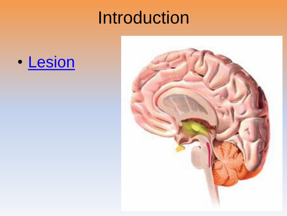
Introduction
• Lesion
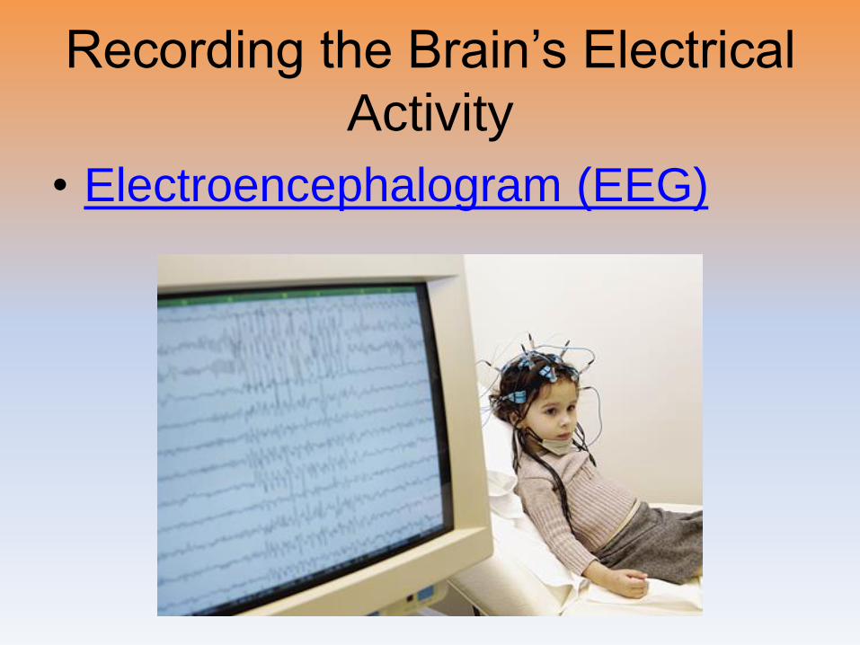
Recording the Brain’s Electrical
Activity
• Electroencephalogram (EEG)
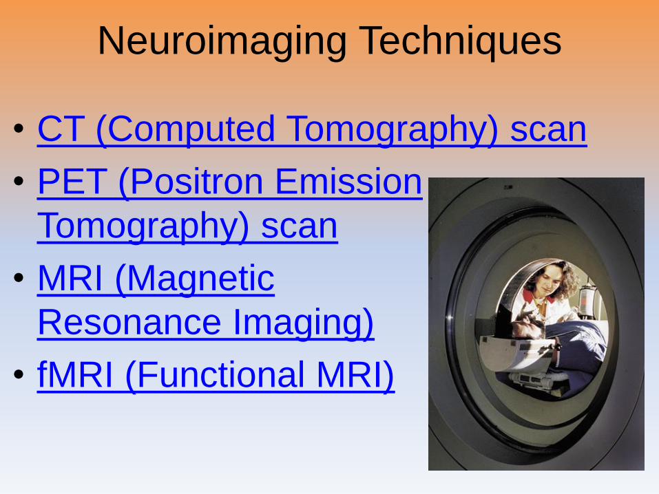
Neuroimaging Techniques
• CT (Computed Tomography) scan
• PET (Positron Emission
Tomography) scan
• MRI (Magnetic
Resonance Imaging)
• fMRI (Functional MRI)
Older Brain Structures
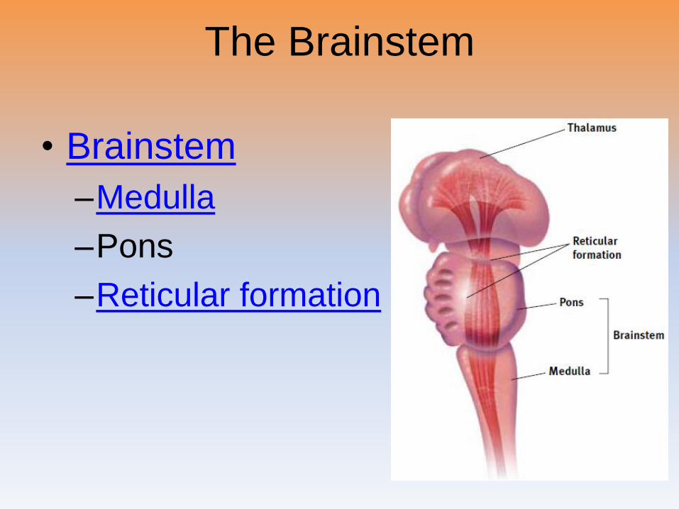
The Brainstem
• Brainstem
–Medulla
–Pons
–Reticular formation
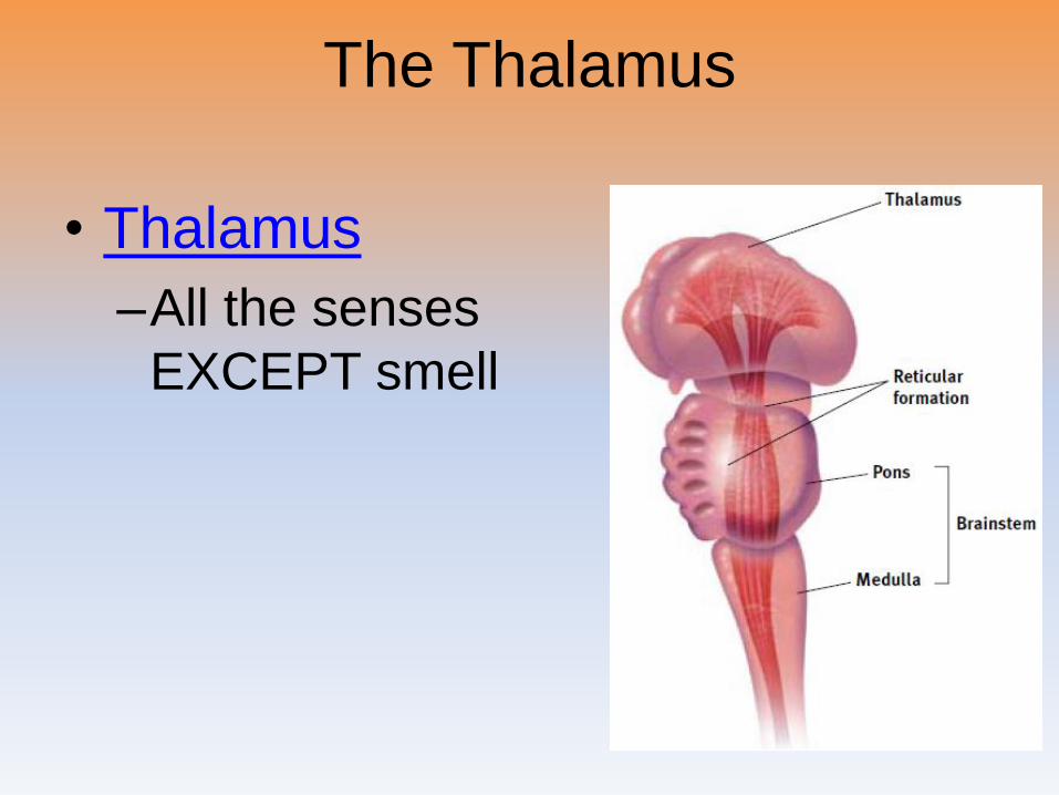
The Thalamus
• Thalamus
–All the senses
EXCEPT smell
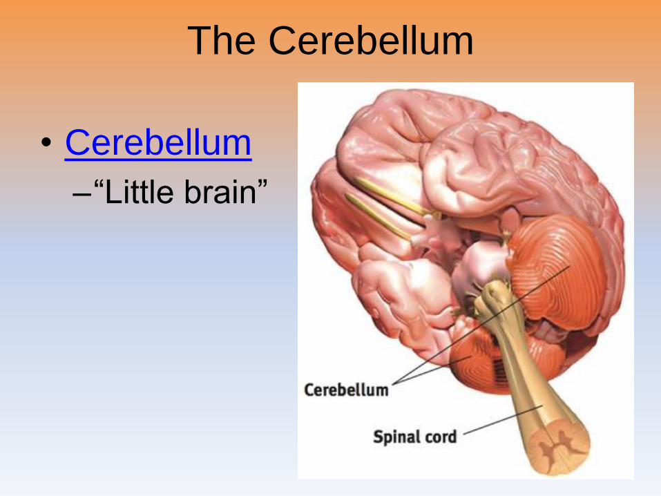
The Cerebellum
• Cerebellum
–“Little brain”
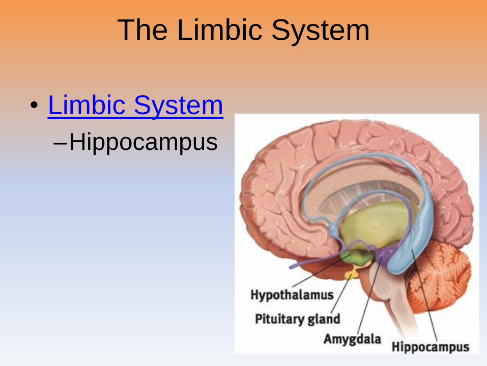
The Limbic System
• Limbic System
–Hippocampus
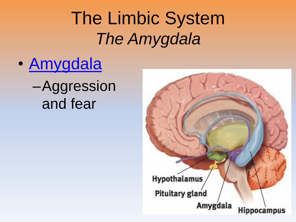
The Limbic SystemThe Amygdala
• Amygdala
–Aggression
and fear
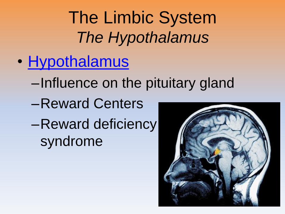
The Limbic SystemThe Hypothalamus
• Hypothalamus
–Influence on the pituitary gland
–Reward Centers
–Reward deficiency
syndrome
The Cerebral Cortex
Introduction
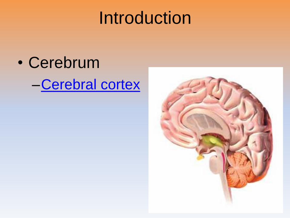
• Cerebrum
–Cerebral cortex
Structure of the Cortex
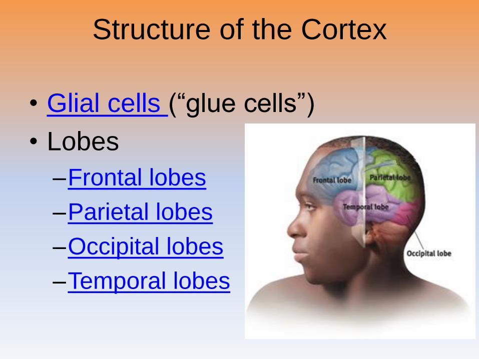
• Glial cells (“glue cells”)
• Lobes
–Frontal lobes
–Parietal lobes
–Occipital lobes
–Temporal lobes
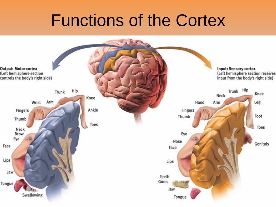
Functions of the CortexMotor Functions
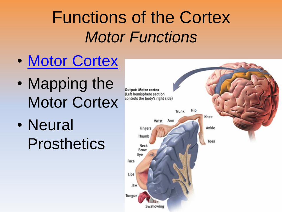
• Motor Cortex
• Mapping the
Motor Cortex
• Neural
Prosthetics
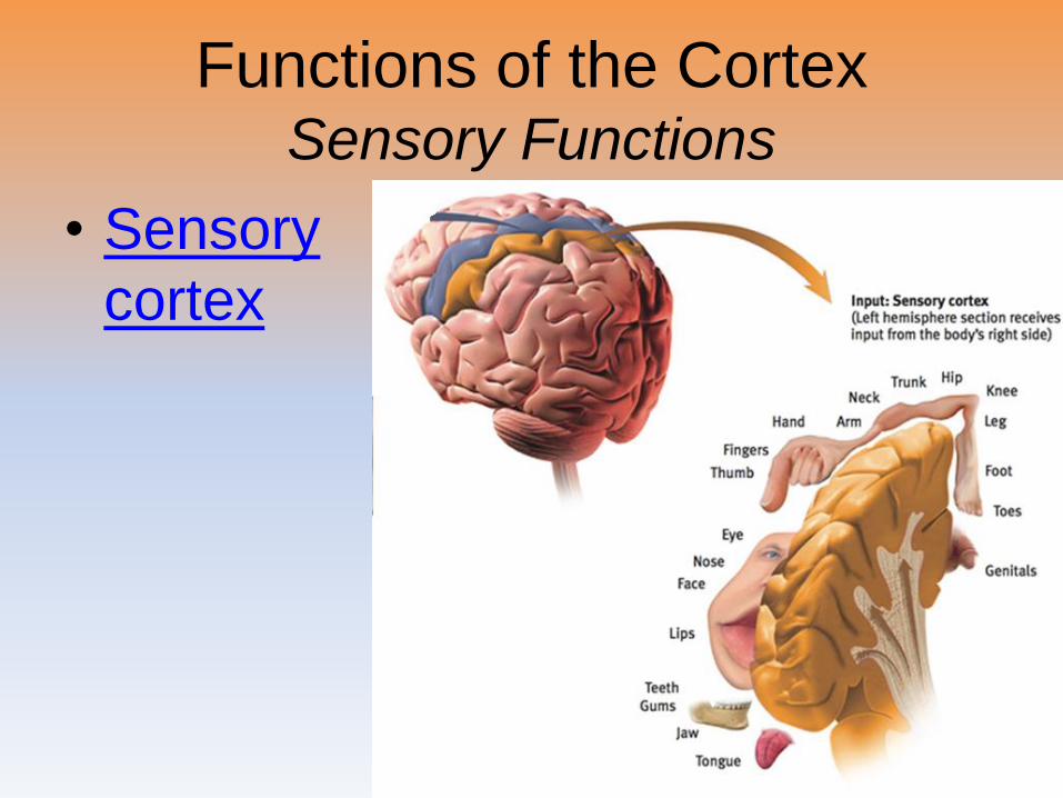
Functions of the CortexSensory Functions
• Sensory
cortex
Functions of the Cortex
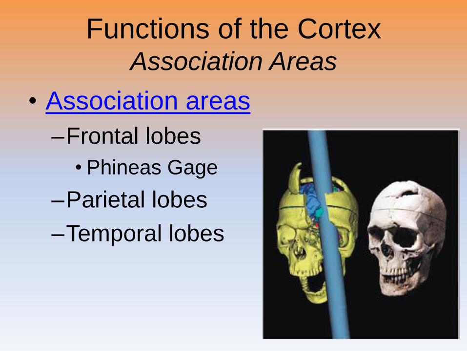
Functions of the CortexAssociation Areas
• Association areas
–Frontal lobes
• Phineas Gage
–Parietal lobes
–Temporal lobes
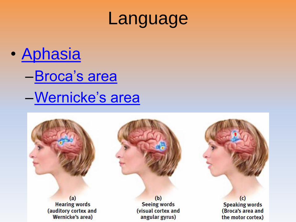
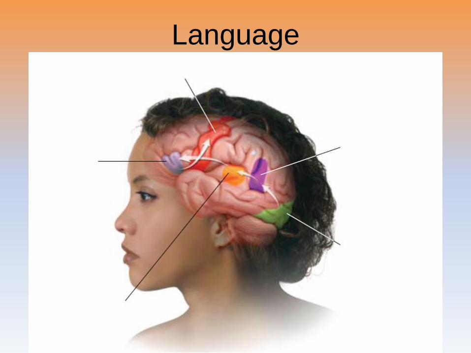
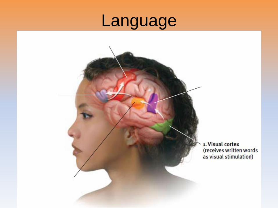
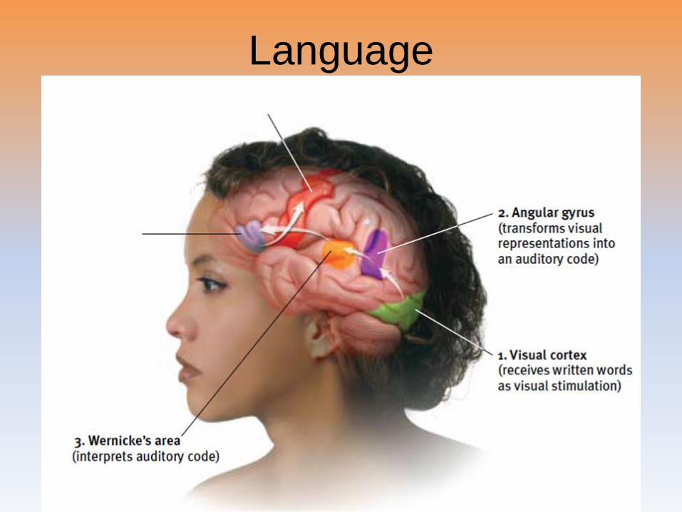
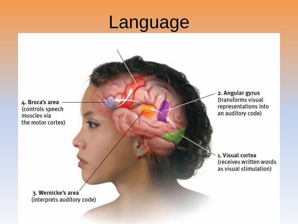
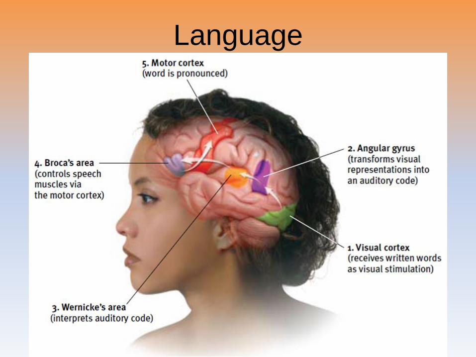
Language
• Aphasia
–Broca’s area
–Wernicke’s area
Language
Language

Language
Language
Language
Language
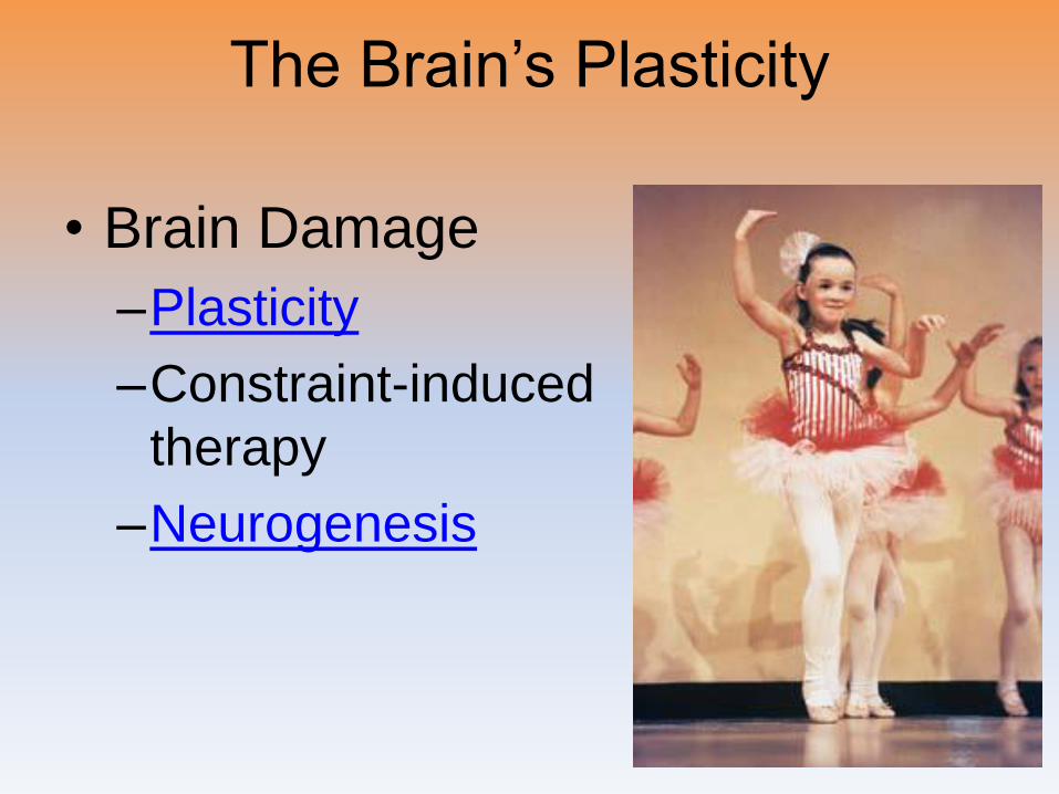
The Brain’s Plasticity
• Brain Damage
–Plasticity
–Constraint-induced
therapy
–Neurogenesis
Our Divided Brain
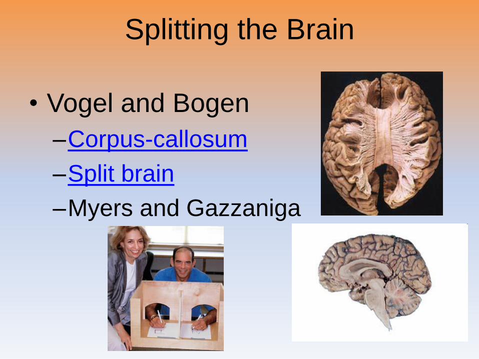
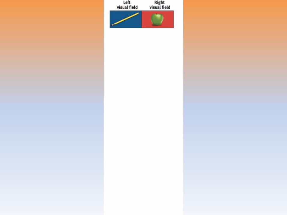
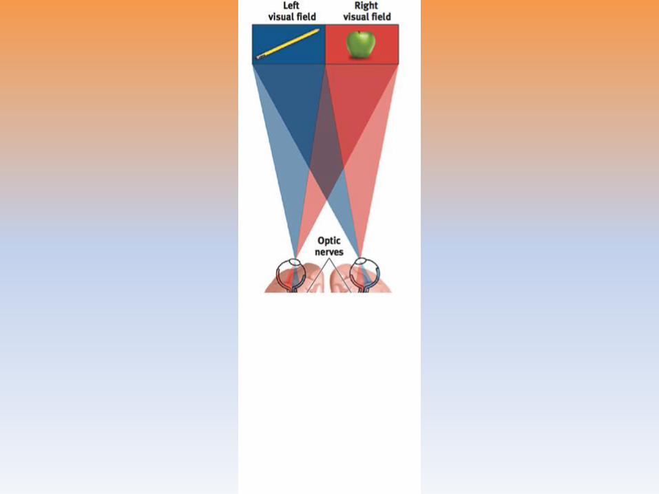
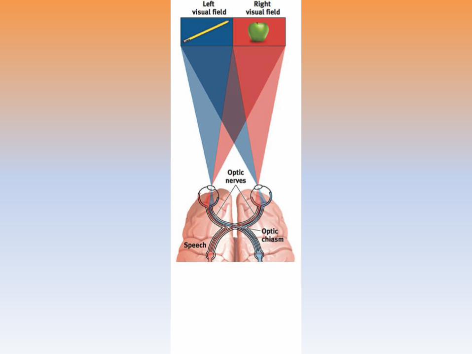
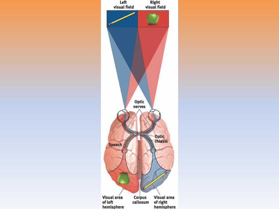
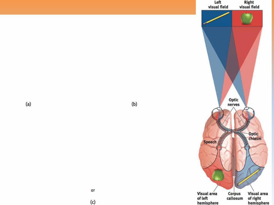
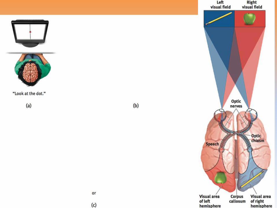
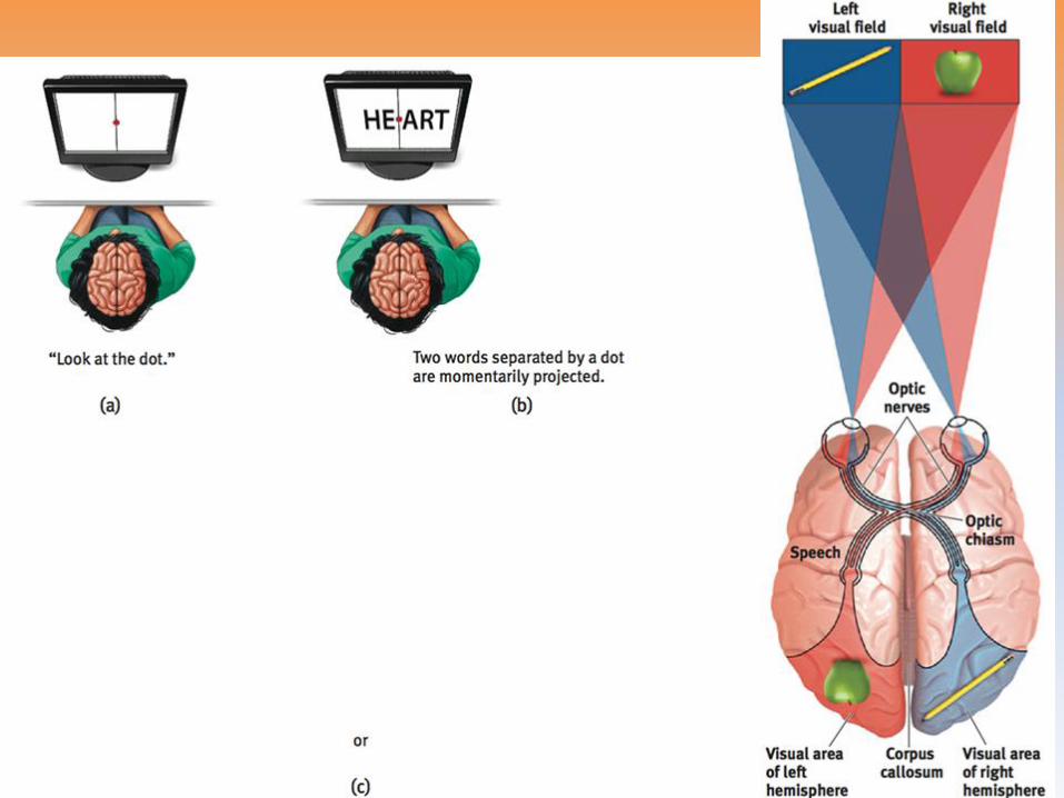
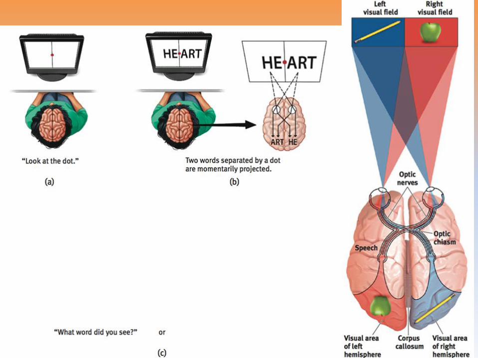
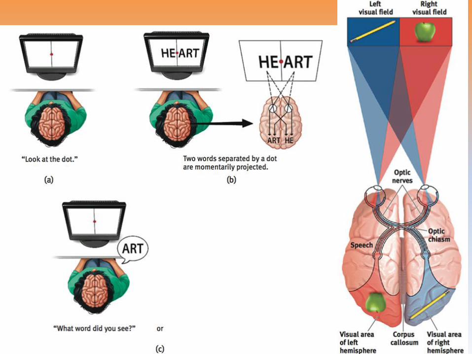
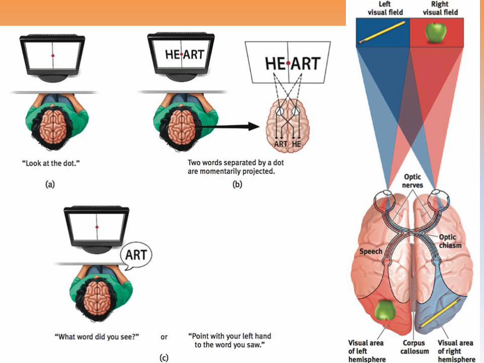
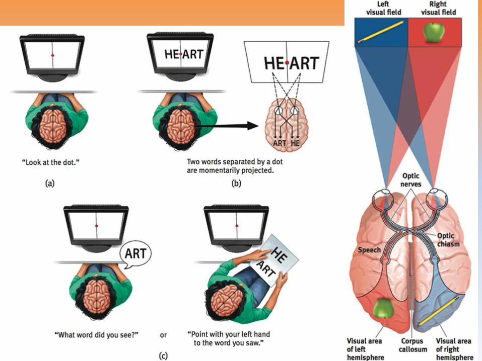
Splitting the Brain
• Vogel and Bogen
–Corpus-callosum
–Split brain
–Myers and Gazzaniga

Right-Left Differences in the
Intact Brain
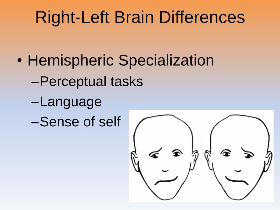
Right-Left Brain Differences
• Hemispheric Specialization
–Perceptual tasks
–Language
–Sense of self
The Brain and Consciousness
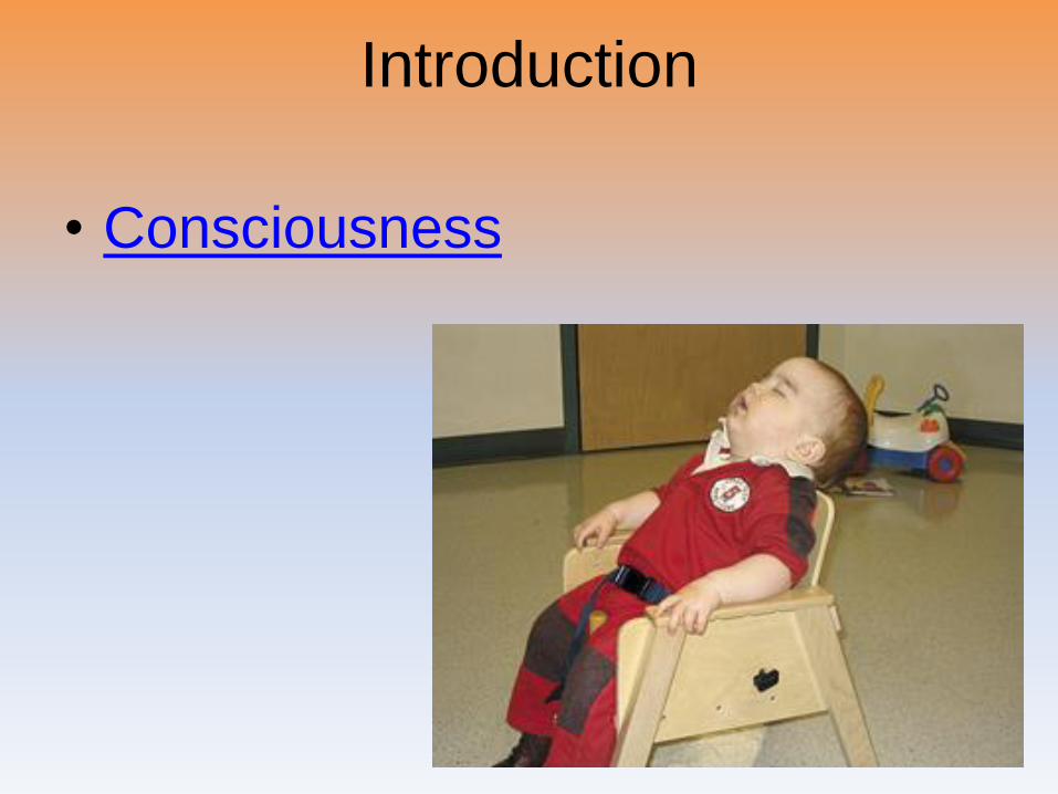
Introduction
• Consciousness
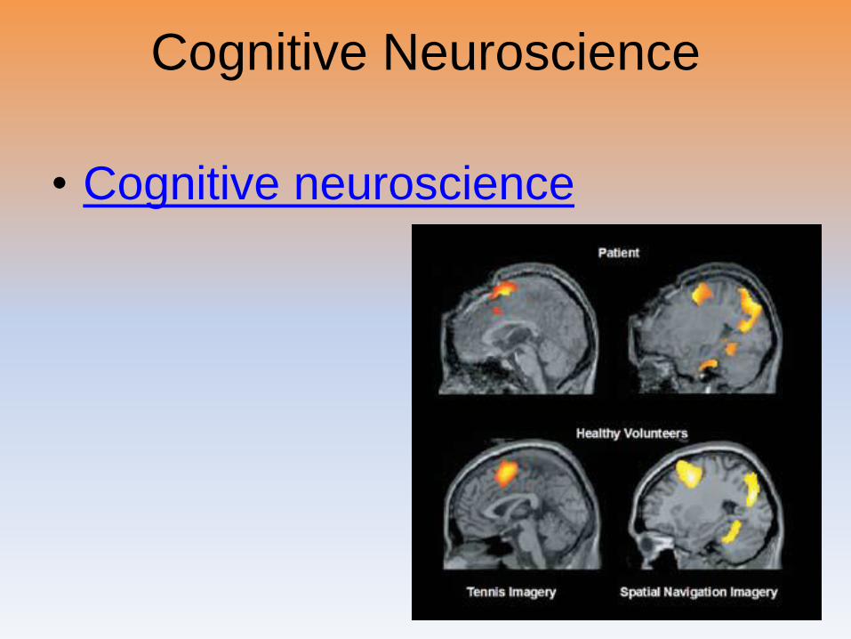
Cognitive Neuroscience
• Cognitive neuroscience
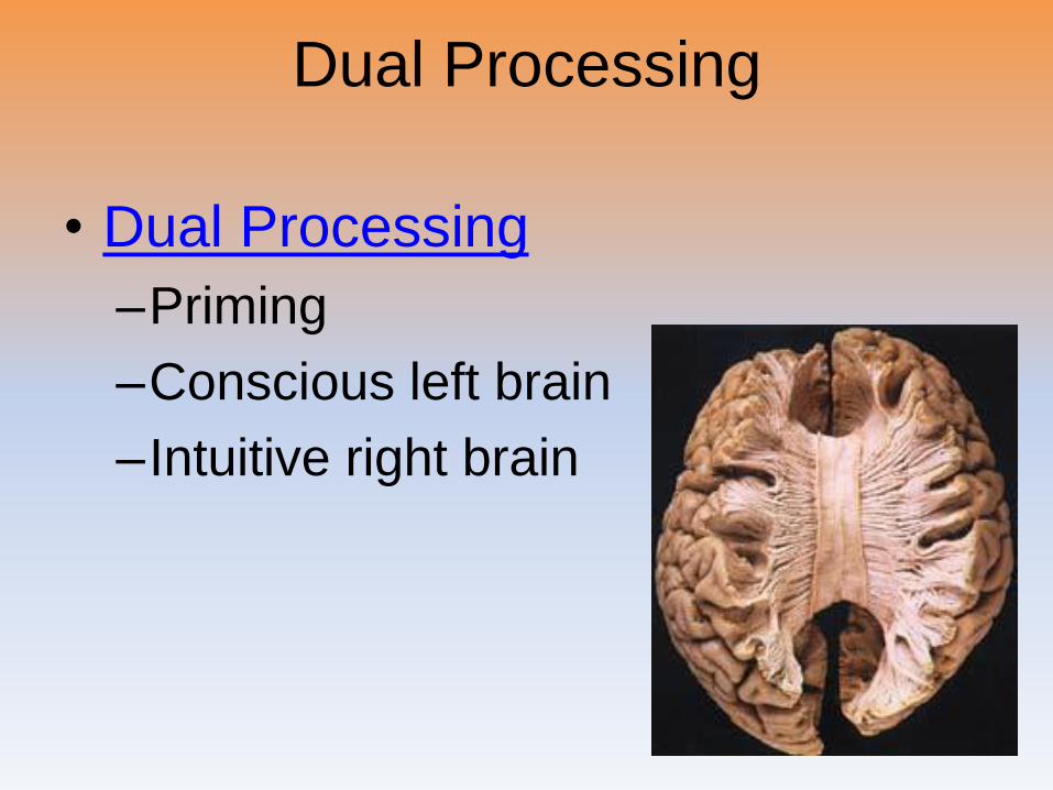
Dual Processing
• Dual Processing
–Priming
–Conscious left brain
–Intuitive right brain
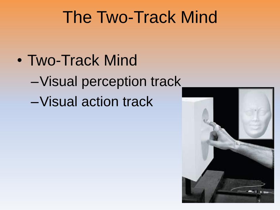
The Two-Track Mind
• Two-Track Mind
–Visual perception track
–Visual action track
The End
Teacher Information• Types of Files
– This presentation has been saved as a “basic” Powerpoint file. While this file format placed a few limitations on the presentation, it insured the file would be compatible with the many versions of Powerpoint teachers use. To add functionality to the presentation, teachers may want to save the file for their specific version of Powerpoint.
• Animation– Once again, to insure compatibility with all versions of Powerpoint, none of the
slides are animated. To increase student interest, it is suggested teachers animate the slides wherever possible.
• Adding slides to this presentation– Teachers are encouraged to adapt this presentation to their personal teaching
style. To help keep a sense of continuity, blank slides which can be copied and pasted to a specific location in the presentation follow this “Teacher Information” section.
Teacher Information• Hyperlink Slides - This presentation contain two types of hyperlinks. Hyperlinks
can be identified by the text being underlined and a different color (usually purple).
– Unit subsections hyperlinks: Immediately after the unit title slide, a page (slide #3) can be found listing all of the unit’s subsections. While in slide show mode, clicking on any of these hyperlinks will take the user directly to the beginning of that subsection. This allows teachers quick access to each subsection.
– Bold print term hyperlinks: Every bold print term from the unit is included in this presentation as a hyperlink. While in slide show mode, clicking on any of the hyperlinks will take the user to a slide containing the formal definition of the term. Clicking on the “arrow” in the bottom left corner of the definition slide will take the user back to the original point in the presentation.
These hyperlinks were included for teachers who want students to see or copy down the exact definition as stated in the text. Most teachers prefer the definitions not be included to prevent students from only “copying down what is on the screen” and not actively listening to the presentation.
For teachers who continually use the Bold Print Term Hyperlinks option, please contact the author using the email address on the next slide to learn a technique to expedite the returning to the original point in the presentation.
Teacher Information• Continuity slides
– Throughout this presentation there are slides, usually of graphics or tables, that build on one another. These are included for three purposes.
• By presenting information in small chunks, students will find it easier to process and remember the concepts.
• By continually changing slides, students will stay interested in the presentation.
• To facilitate class discussion and critical thinking. Students should be encouraged to think about “what might come next” in the series of slides.
• Please feel free to contact me at [email protected] with any questions, concerns, suggestions, etc. regarding these presentations. Kent Korek
Germantown High School
Germantown, WI 53022
262-253-3400
Division title (green print)
subdivision title (blue print)
• xxx
–xxx
–xxx
Division title (green print)
subdivision title (blue print)
Use this slide to add a table, chart, clip art, picture, diagram, or video clip. Delete
this box when finished
Definition Slide
= add definition here
Definition
Slides
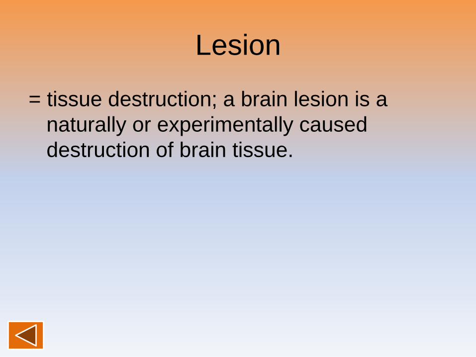
Lesion
= tissue destruction; a brain lesion is a
naturally or experimentally caused
destruction of brain tissue.
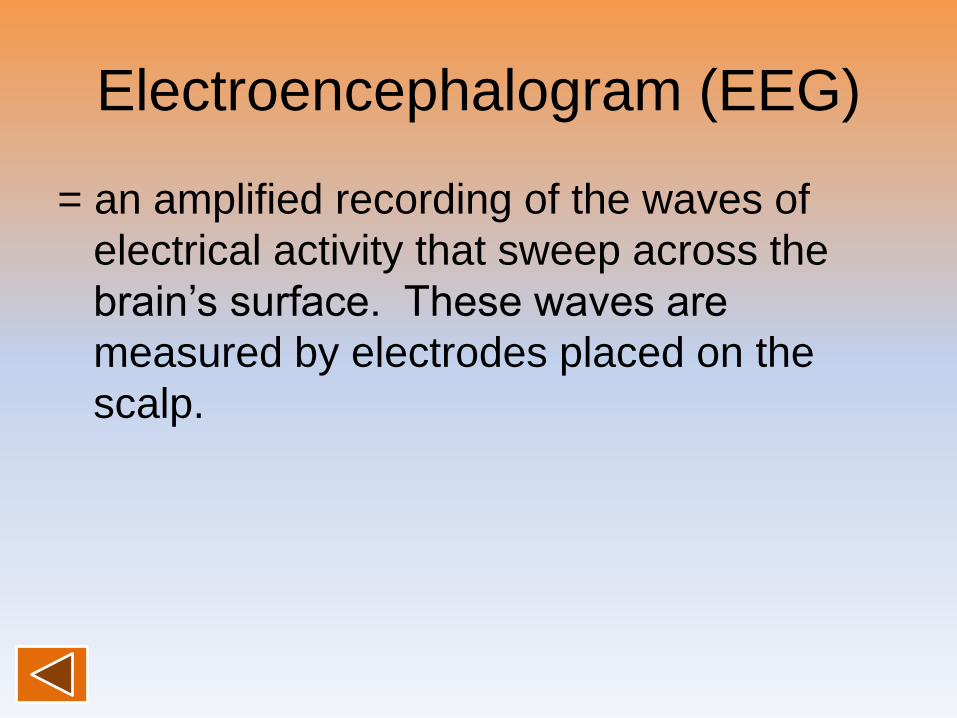
Electroencephalogram (EEG)
= an amplified recording of the waves of
electrical activity that sweep across the
brain’s surface. These waves are
measured by electrodes placed on the
scalp.
CT (computed tomography)
Scan= a series of X-ray photographs taken from
different angles and combined by
computer into a composite representation
of a slice through the body.
• Also called CAT scan.
PET (positron emission
tomography) Scan= a visual display of brain activity that
detects where a radioactive form of
glucose goes while the brain performs a
given task.
MRI (magnetic resonance
imaging)= a technique that uses magnetic fields and
radio waves to produce computer-
generated images of soft tissue. MRI
scans show brain anatomy.
fMRI (functional MRI)
= a technique for revealing bloodflow and,
therefore, brain activity by comparing
successive MRI scans. fMRI scans show
brain function.
Brainstem
= the oldest part of the central core of the
brain, beginning where the spinal cord
swells as it enters the skull; the brainstem
is responsible for automatic survival
functions.
Medulla
= the base of the brainstem; controls
heartbeat and breathing.
Reticular Formation
= a nerve network in the brainstem that
plays an important role in controlling
arousal.
Thalamus
= the brain’s sensory switchboard, located
on top of the brainstem; it directs
messages to the sensory receiving areas
in the cortex and transmits replies to the
cerebellum and medulla.
Cerebellum
= the “little brain” at the rear of the
brainstem; functions include processing
sensory input and coordinating movement
output and balance.
Limbic System
= doughnut-shaped neural system (including
the hippocampus, amygdala, and
hypothalamus) located below the cerebral
hemispheres; associated with emotions
and drives.
Amygdala
= two lima bean-sized neural clusters in the
limbic system; linked to emotion.
Hypothalamus
= a neural structure lying below (hypo) the
thalamus; it directs several maintenance
activities (eating, drinking, body
temperature), helps govern the endocrine
system via the pituitary gland, and is
linked to emotion and reward.
Cerebral Cortex
= the intricate fabric of interconnected neural
cells covering the cerebral hemispheres;
the body’s ultimate control and
information-processing center.
Glial Cells
= cells in the nervous system that support,
nourish, and protect neurons.

Frontal Lobes
= portion of the cerebral cortex lying just
behind the forehead; involved in speaking
and muscle movements and in making
plans and judgments.
Parietal Lobes
= portion of the cerebral cortex lying at the
top of the head and toward the rear;
receives sensory input for touch and body
position.
Occipital Lobes
= portion of the cerebral cortex lying at the
back of the head; includes areas that
receive information from the visual fields.
Temporal Lobes
= portion of the cerebral cortex lying roughly
above the ears; includes the auditory
areas, each receiving information primarily
from the opposite ear.
Motor Cortex
= an area at the rear of the frontal lobes that
controls voluntary movements.

Sensory Cortex
= area at the front of the parietal lobes that
registers and processes body touch and
movement sensations.
Association Areas
= areas of the cerebral cortex that are not
involved in primary motor or sensory
functions; rather, they are involved in
higher mental functions such as learning,
remembering, thinking, and speaking.
Aphasia
= impairment of language, usually caused by
left hemisphere damage either to Broca’s
area (impairing speaking) or to Wernicke’s
area (impairing understanding).
Broca’s Area
= controls language expression that directs
the muscle movements involved in
speech.
Wernicke’s Area
= controls language reception – a brain area
involved in language comprehension and
expression; usually in the left temporal
lobe.
Plasticity
= the brain’s ability to change, especially
during childhood, by reorganizing after
damage or by building new pathways
based on experience.
Neurogenesis
= the formation of new neurons.
Corpus Callosum
= the large band of neural fibers connecting
the two brain hemispheres and carrying
messages between them.
Split Brain
= a condition resulting from surgery that
isolates the brain’s two hemispheres by
cutting the fibers (mainly those of the
corpus callosum) connecting them.
Consciousness
= our awareness of ourselves and our
environment.
Cognitive Neuroscience
= the interdisciplinary study of the brain
activity linked with cognition (including
perception, thinking, memory and
language).
Dual Processing
=the principle that information is often
simultaneously processed on separate
conscious and unconscious tracks.