Embed Size (px)
Citation preview
8/10/2019 My Fourth Seminar - Anshu
http://slidepdf.com/reader/full/my-fourth-seminar-anshu 1/26
GINGIVAL TISSUE MANAGEMENT
CONTENTS:
Introduction
Periodontal aspects of restorative dentistry
Normal periodontium
Need for gingival tissue management
Gingival retraction
-Definition
-Requirements
Techniques of gingival retraction
-classifications
Methods of gingival retraction-
I. NON SURGICAL
1. Mechanical
2. Chemomechanical
II. SURGICAL
1. Rotary curettage
2. Electrosurgery
Armamentarium for gingival displacement
Techniques for gingival displacement
Recent Advances in Gingival retraction
References
Conclusion
Submitted by:
Dr. Anshuman Khaitan
Post graduate student
Department of conservative dentistry and endodontics
College of Dental Sciences, Davanagere
8/10/2019 My Fourth Seminar - Anshu
http://slidepdf.com/reader/full/my-fourth-seminar-anshu 2/26
Gingival Tissue Management
INTRODUCTION:
High quality restorative dental procedures & gingival tissue management go hand in
hand. An objective of restorative dental procedures is the placement of dental
materials to restore teeth to proper form and function. The form and function must be
in harmony with the periodontium for a restoration to become an integral component
of total oral complex. The purpose of this seminar is to blend the microgingival
retraction methods with the principles of restorative dentistry to establish a sound
biologic approach.
PERIODONTAL ASPECTS OF RESTORATIVE DENTISTRY
-Microbial plaque is undoubtedly the primary etiological agent in periodontal disease.
Never the less, other factors do contribute to gingival inflammation and subsequent
loss of periodontal attachment.
-The production of plaque is not as important as the retention of plaque on and around
the tooth surfaces. This retention increases manifolds in case of calculus deposits,
poor margins of the restorations and perhaps the mere presence of dental restorations.
- The outer surface of a restoration is of significance from periodontal aspects.
- Therefore it is mandatory to have thorough knowledge of anatomy and morphology
of the tooth to be restored and also the techniques leading to the restoration of proper
cavosurface margins.
- The operative dentist should always take care of the health of the periodontium
during all restorative procedures.
Normal Periodontium
Before elaborating the restorative implications on gingival tissue & other
periodontal tissues let us have a brief knowledge of the normal
periodontium.
Dentogingival Unit
8/10/2019 My Fourth Seminar - Anshu
http://slidepdf.com/reader/full/my-fourth-seminar-anshu 3/26
Gingival Tissue Management
The dentogingival unit and its epithelial and connective tissue covering can be studied
as follows.
Gingiva
It is composed of free gingiva & attached gingiva
Attached gingiva is bound to the cementum and the underlying bone by means
of supra alveolar connective tissue and lamina propria.
If restorative procedure are to invade the gingival crevice, approximately 5
mm depth of gingiva is involved out of which 2mm is free gingival and 3mm
is attached gingiva.
A second dimension of gingival tissue to be evaluated is its thickness.
Along with the vertical dimension, the thickness of the gingiva to tolerate
intra-crevicular restorative procedures must also be taken into account.
Gingival sulcus
Crevice is lined with crevicular epithelium
It extends from the free gingival margin to the junctional epithelium
Two parameters are important
- Depth 2.00 mm &
- Circumference
Depth:
Excessive crevicular depth pathognomonic of periodontal disease &
restorative procedure should be avoided in such cases
A minimum of 1.5-2 mm of depth is essential for preparing intracrevicular
margins in a tooth
In case the depth is less & clinician tries to place the margins of restoration
apical to crevicular depth, there are chances of permanent damage to the
junctional epithelium and the underlying connective tissue.
Circumference:
In healthy gingival there is hardly any space separating the epithelial lining of
crevice from the tooth surface.
8/10/2019 My Fourth Seminar - Anshu
http://slidepdf.com/reader/full/my-fourth-seminar-anshu 4/26
Gingival Tissue Management
Care must be taken to avoid distension of crevicular epithelial lining.
Quantitative Violation: excessive material is being placed within the crevice
Qualitative Violation: poor adaptation/ roughness of the margins that leads to
both mechanical irritation and harbours microbial flora.
Biologic width:
Definition:
It is defined as “Combined dimension of the supra alveolar gingival
connective tissue and the junctional epithelium”.
Average measurement for the epithelial attachment and C/T attachment have
been found to be 0.97mm and 1.07mm is required.
A restoration that impinges upon biologic width will result in progressive
periodontal disease.
According to Carranza:
Biologic width is defined as the physiologic dimension of the junctional
epithelium & c/t attachment.
- Relatively constant at 2mm
It has been theorized that infringement on the biologic width by the
placement of a restoration within its zone may result in gingival
inflammation and subsequently packet formation and bone loss.
Encroachment into this space is prevalent amongst restorative dentists as
they attempt to place a margin subgingival rather than intracrevicular.
Placement of the restorative margin upto 0.5mm into the sulcus helps in the
maintenance of the biologic width.
Need for soft tissue management
• Restore and maintain health, functional comfort and aesthetic appearance.
• Placement of proper cervical finish lines.
•
Helps in making accurate impressions.
8/10/2019 My Fourth Seminar - Anshu
http://slidepdf.com/reader/full/my-fourth-seminar-anshu 5/26
Gingival Tissue Management
• Helps in blending of the restoration with the unprepared tooth surface.
• To enhance access and to prevent damage to the soft tissue during cavity
preparation.
• While cementation it helps in easy removal of cement without tissue damage.
DEFINITION OF GTM:
“The procedure of temporary eversion or resection of gingiva away from the tooth
surface or deepening of gingival sulcus to expose the cervical portion of tooth in order
to have proper marginal finish of the restoration or by establishing a good cervical
cavosurface margin to the tooth preparation”
Gingival retraction:
Acc to GPT 8: “The deflection of the marginal gingiva away from a tooth.”
Gingival retraction is the process of exposing the margins of the prepared teeth.
Requirements:
• The gingival tissues must be healthy and free of inflammation.
• Cervical margins should be placed in the appropriate position.
– The optimum position of the margins is 0.5 mm from the healthy free
gingival margins or 3-4 mm from the crest of the alveolar bone.
– Quality provisional restoration
CLASSIFICATION:
a.) Barkmier W.W. and Williams H.W (1978)
1. Non-Surgical Methods
Rubber dam and clamps
Retraction cords
impregnated and
8/10/2019 My Fourth Seminar - Anshu
http://slidepdf.com/reader/full/my-fourth-seminar-anshu 6/26
Gingival Tissue Management
non-impregnated
Retraction rings
Copper bands
2. Surgical Methods
i.
Gingivectomy and Gingivoplasty
ii. Periodontal flap procedures
iii. Electrosurgery
iv.
Rotary Gingival Curettage
b.) Thompson M.J.(1959)
1. Conventional
2. Radical
c.) B.W.Benson et al (1986)
1. Mechanical method
2. Mechanico-chemical method
3. Rotary gingival curettage
4. Electrosurgical methods.
MECHANICAL METHODS
• Among the first to be developed
• Involve the physical displacement of the gingival tissue by placement of
materials within the gingival sulcus
• The various materials used :
A. Heavy weight Rubber dams.
B.
Copper bands.
C. Aluminum shell.
8/10/2019 My Fourth Seminar - Anshu
http://slidepdf.com/reader/full/my-fourth-seminar-anshu 7/26
Gingival Tissue Management
D. Retraction rings
E. Mechanical Pack of Zinc oxide eugenol.
F.
Rolled cotton or synthetic cord.
A. Heavy Weight Rubber Dam
• The retraction is produced when the heavy weight rubber dam compresses the
tissue.
• According to Gilmore , it can be called “Gum compression” rather than gum
retraction.
Indications
• Limited number of teeth in one quadrant are being restored and in
situations in which preparation do not have to be extended very far sub-
gingivally.
Contraindications
• Should not be used with polyvinyl siloxane impression material
Advantages
• Control of seepage and hemorrhage.
• Ease of application
Disadvantages
•
Full arch models cannot be made.
• Cannot be used in Severe cervical extension preparations.
B. Copper Band
•
It serves as a means of carrying the impression material as well as a
mechanism for displacing the gingiva.
8/10/2019 My Fourth Seminar - Anshu
http://slidepdf.com/reader/full/my-fourth-seminar-anshu 8/26
Gingival Tissue Management
• One end of the tube is festooned, or trimmed to follow the profile of the
gingival finish line, which in turn often follows the contours of the gingival
margin.
•
Impression compound and elastomeric materials are used. Band is seated
securely into its position & pressure is applied on the compound directly.
Chilling of the impression can be done with cold water.
• Copper bands are especially useful for situations in which several teeth have
been prepared.
• The use of copper bands can cause incisional injuries of gingival tissues but
recession followed by their use is minimal, ranging from 0.1mm in healthy
adolescent to 0.3mm in general clinical population.
C. Aluminum Shell
• Aluminum shell of correct size is selected, trimmed to conform to the gingival
contours and the margins are smoothened.
• It is filled with compound or gutta percha and placed on the tooth under the
occlusal pressure.
•
The excess material from gingival end will displace the free gingiva.
D. Retraction Ring (Epipak Rings)
Dilatation of the sulcus via a retraction ring; the marginal gingiva is displaced
laterally and apically by a largely non-traumatic insertion of the ring using a
suitable retraction thread plugger, thus creating a barrier between the
preparation instrument and the periodontium which prevents an invasion into
the zone of the biological width as far as possible.
E. Rolled Cotton Or Synthetic Cord
• This is the method of choice as the availability is not a problem and the
application is exceedingly easy.
•
Plain cotton thread, Unwaxed Floss, Cotton synthetic cord, untreated surgicalsilk, and elastic retraction rings.
8/10/2019 My Fourth Seminar - Anshu
http://slidepdf.com/reader/full/my-fourth-seminar-anshu 9/26
Gingival Tissue Management
• Wet or dry.
• Thinner grade cord is used around the anterior teeth and thicker one around
the posterior teeth.
• Three varieties of cords are generally available.
•
Loose twisted
• Braided
• Knitted
• Braided and knitted variety does not separate when they are pushed into the
sulcus and so they are easier to use.
• Newer cords have fine copper wire within them to keep their shape & stay put
in the crevice.
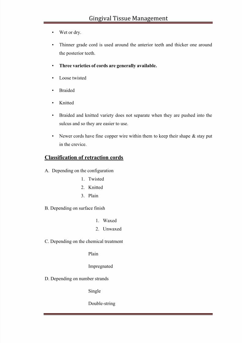
Classification of retraction cords
A. Depending on the configuration
1.
Twisted2. Knitted
3.
Plain
B. Depending on surface finish
1. Waxed
2. Unwaxed
C. Depending on the chemical treatment
Plain
Impregnated
D. Depending on number strands
Single
Double-string
8/10/2019 My Fourth Seminar - Anshu
http://slidepdf.com/reader/full/my-fourth-seminar-anshu 10/26
Gingival Tissue Management
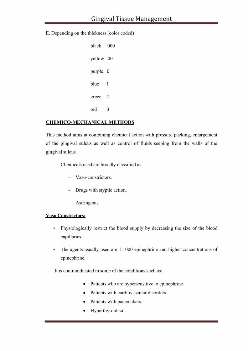
E. Depending on the thickness (color coded)
black 000
yellow 00
purple 0
blue 1
green 2
red 3
CHEMICO-MECHANICAL METHODS
This method aims at combining chemical action with pressure packing, enlargement
of the gingival sulcus as well as control of fluids seeping from the walls of the
gingival sulcus.
Chemicals used are broadly classified as:
–
Vaso-constrictors.
– Drugs with styptic action.
– Astringents.
Vaso Constrictors:
•
Physiologically restrict the blood supply by decreasing the size of the blood
capillaries.
•
The agents usually used are 1:1000 epinephrine and higher concentrations of
epinephrine.
It is contraindicated in some of the conditions such as:
Patients who are hypersensitive to epinephrine.
Patients with cardiovascular disorders.
Patients with pacemakers.
Hyperthyroidism.
8/10/2019 My Fourth Seminar - Anshu
http://slidepdf.com/reader/full/my-fourth-seminar-anshu 11/26
Gingival Tissue Management
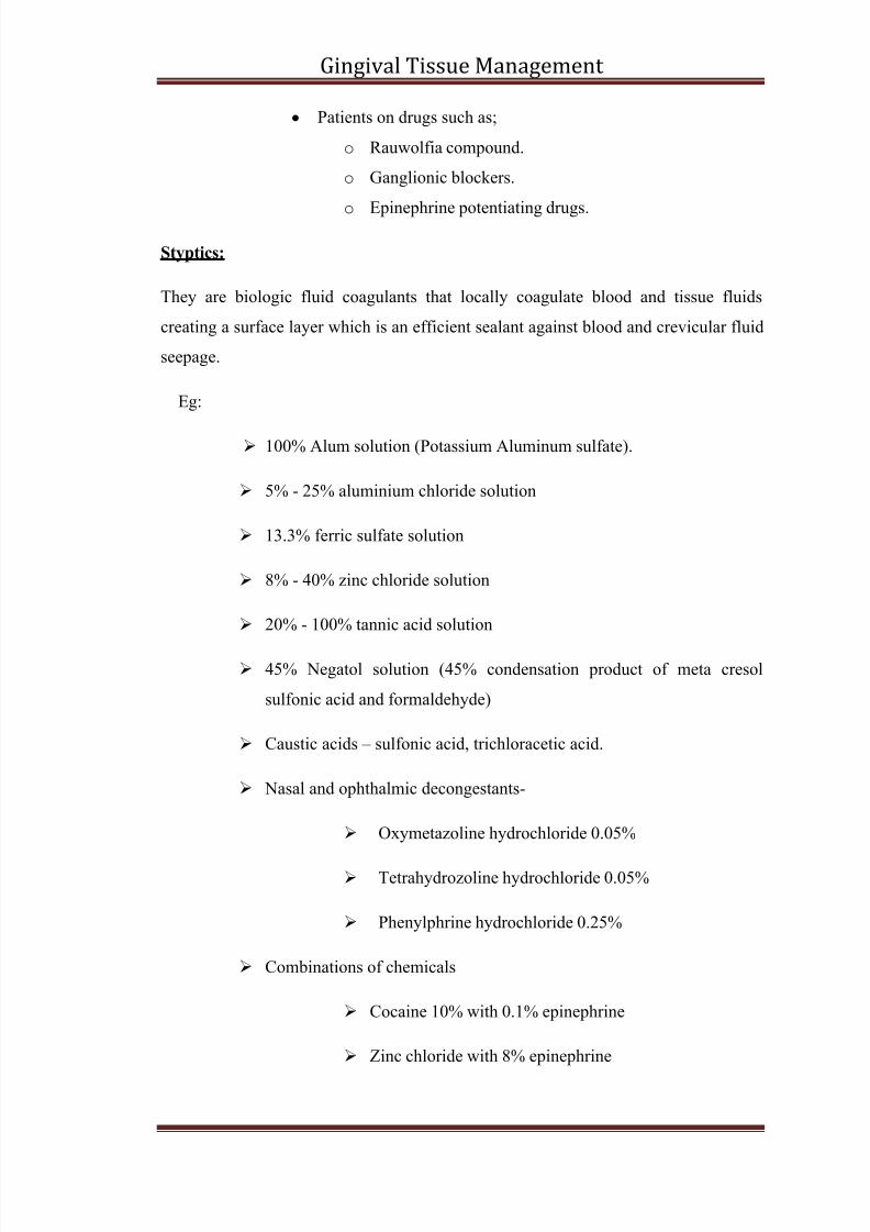
Patients on drugs such as;
o Rauwolfia compound.
o Ganglionic blockers.
o
Epinephrine potentiating drugs.
Styptics:
They are biologic fluid coagulants that locally coagulate blood and tissue fluids
creating a surface layer which is an efficient sealant against blood and crevicular fluid
seepage.
Eg:
100% Alum solution (Potassium Aluminum sulfate).
5% - 25% aluminium chloride solution
13.3% ferric sulfate solution
8% - 40% zinc chloride solution
20% - 100% tannic acid solution
45% Negatol solution (45% condensation product of meta cresol
sulfonic acid and formaldehyde)
Caustic acids – sulfonic acid, trichloracetic acid.
Nasal and ophthalmic decongestants-
Oxymetazoline hydrochloride 0.05%
Tetrahydrozoline hydrochloride 0.05%
Phenylphrine hydrochloride 0.25%
Combinations of chemicals
Cocaine 10% with 0.1% epinephrine
Zinc chloride with 8% epinephrine
8/10/2019 My Fourth Seminar - Anshu
http://slidepdf.com/reader/full/my-fourth-seminar-anshu 12/26
Gingival Tissue Management
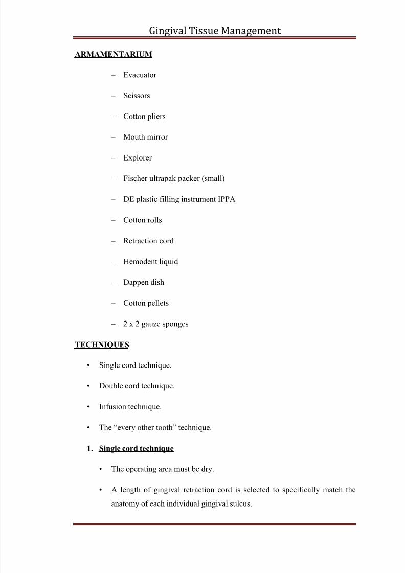
ARMAMENTARIUM
– Evacuator
–
Scissors
– Cotton pliers
– Mouth mirror
– Explorer
–
Fischer ultrapak packer (small)
–
DE plastic filling instrument IPPA
– Cotton rolls
– Retraction cord
– Hemodent liquid
–
Dappen dish
–
Cotton pellets
– 2 x 2 gauze sponges
TECHNIQUES
• Single cord technique.
• Double cord technique.
•
Infusion technique.
• The “every other tooth” technique.
1. Single cord technique
•
The operating area must be dry.
• A length of gingival retraction cord is selected to specifically match the
anatomy of each individual gingival sulcus.
8/10/2019 My Fourth Seminar - Anshu
http://slidepdf.com/reader/full/my-fourth-seminar-anshu 13/26
Gingival Tissue Management
• Retraction cord should be moistened by dipping it in buffered 25%
Aluminum Chloride solution in a dappen dish.
• Form the cord into a „U‟ and loop it around the prepared tooth.
• Hold the cord between the thumb and forefinger and apply slight tension
in an apical direction.
• Gently slip the cord between the tooth and gingiva in the mesial inter-
proximal area with a cord packing instrument.
• Once the cord has been tucked in on the mesial, use the instrument to
lightly secure it in the distal inter-proximal area.
• Proceed to the lingual surface and begin working from the mesio-lingual
corner around to the disto-lingual corner.
• The tip of the instrument should be inclined slightly towards the area
where the cord has already been placed; i.e. the mesial.
•
If the tip of the instrument is away then the cord may be displaced and
pulled out.
• Gently press apically on the cord with the instrument directing the tip
slightly towards the tooth.
• Continue packing the cord around the facial surface, overlapping the cord
in the mesial inter-proximal area.
• Pack all but the last 2 mm or 3 mm of cord should be left.
•
This tag can be grasped for easy removal.
• After the cord is in place, the tooth preparation is carefully inspected to
ascertain that the entire cervical margin can clearly be visualized and that
there is no soft tissue impediment to easy injection of the impression
material to capture all of the cervical margin detail.
• Wait 8 to 10 minutes before removing the cord and making the impression.
8/10/2019 My Fourth Seminar - Anshu
http://slidepdf.com/reader/full/my-fourth-seminar-anshu 14/26
Gingival Tissue Management
• The cord needs time to effect adequate lateral displacement, and the
medicament needs time to create hemostasis and crevicular fluid control.
2. Double Cord Technique
• The double cord technique is routinely used when making impressions of
multiple prepared teeth and when making impressions when tissue health
is compromised.
• Some clinicians use this technique routinely for all impressions.
• A small-diameter cord is placed in the sulcus.
•
The ends of this cord should be cut so that they exactly abut against
one another in the sulcus.
• A second cord, soaked in the hemostatic agent of choice, is placed in the
sulcus above the small-diameter cord.
• The diameter of the second cord should be the largest diameter that can
readily be placed in the sulcus.
•
After waiting 8 to 10 minutes after placement of the large cord, the second
cord is soaked in water and removed.
• The preparation(s) are dried, and the impression is made with the primary
cord in place.
•
After successfully making the impression, the small cord is soaked in
water and removed from the sulcus.
3. Infusion Technique
• Dan E Fisher in 1981 introduced a new concept for hemostasis known as
the infusion technique.
• The infusion technique for gingival displacement uses a significantly
different approach from the single or double cord techniques.
8/10/2019 My Fourth Seminar - Anshu
http://slidepdf.com/reader/full/my-fourth-seminar-anshu 15/26
Gingival Tissue Management
• After careful preparation of the cervical margins in an intra-crevicular
position, hemorrhage is controlled using a specifically designed Dento-
infusor TM with a ferric sulfate medicament.
•
Two concentrations of ferric sulfate, 15% and 20% are available.
• The infusor is used with a burnishing motion in the sulcus and is carried
circumferentially 3600 around the sulcus.
• The medicament is extruded from the syringe/infusor as the instrument is
manipulated around the gingival sulcus.
•
When hemostasis is verified, a knitted retraction cord is soaked in the
ferric sulfate solution and packed into the sulcus.
•
Technique recommended the cord be in place 1 to 3 minutes.
• The cord is removed, the sulcus is rinsed with water, and the impression is
made.
•
When using ferric sulfate materials patient should be warned that the
tissues may be temporarily darkened. The tissues take on a blue- blackappearance that usually disappears in a few days.
4. The “Every other tooth technique”
When making impressions of anterior tooth preparations it is critical that
no damage is done to the gingival tissues that may result in recession.
In teeth with root proximity, placing retraction cord simultaneously around
all prepared teeth may result in strangulation of the gingival papilla and
eventual loss of the papilla.
This creates unaesthetic black triangles in the gingival embrasures.
This undesirable outcome can be prevented with “every other tooth”
technique. This can be used with single/ double cord technique.
Retraction cord is placed around the most distal prepared tooth. No cord is
placed around the prepared tooth mesial to this tooth.
Retraction procedures are completed on alternate teeth.
8/10/2019 My Fourth Seminar - Anshu
http://slidepdf.com/reader/full/my-fourth-seminar-anshu 16/26
Gingival Tissue Management
For ex: teeth #5 through# 12 are prepared, cords will be placed around
teeth #5,#7, #9, #11
The impression is made; gingival displacement is accomplished on teeth
#6, #8, #10 & # 12 and a second impression is made.
A subsequent pick up impression allows fabrication of a master cast with
dies for all eight prepared teeth.
SURGICAL METHODS
1. Rotary Curettage
•
It was described by Amsterdam
in 1954, and subsequently modified by
Ingraham.
•
Rotary curettage is a “troughing” technique, also called as “gingettage”,
done to produce limited removal of epithelial tissue in the sulcus.
• The technique is used with the subgingival placement of restoration
margins.
Procedure:
•
It is usually done simultaneously along with finish line preparation.
• A torpedo diamond of 150 to 180 grit is used to extend the finish line
apically.
• Bur should be extended into the gingival sulcus to remove a portion
of the sulcular epithelium.
•
Cord impregnated with Aluminum Chloride or Alum is gently placed to
control hemorrhage.
• The cord is removed after 4 to 8 minutes, and the sulcus is thoroughly
irrigated with water.
• This technique is well suited for use with reversible hydrocolloid.
Disadvantages:
8/10/2019 My Fourth Seminar - Anshu
http://slidepdf.com/reader/full/my-fourth-seminar-anshu 17/26
Gingival Tissue Management
• There is poor tactile sensation when using diamond points on the sulcular
walls, which can produce deepening of the sulcus.
• The technique also has the potential for destruction of the periodontium if
used incorrectly, making this method harmful, that is probably best used
by experienced dentists.
2. Electrosurgery
-There are situations in which it may not be feasible or desirable to manage the
gingival tissues with retraction cord.
- When areas of inflammation and granulation tissues are present caused either
due to caries or overhanging restorations, it may be necessary to place the
finish line of the preparation near the epithelial attachment and it is impossible
to retract the gingival tissues sufficiently to get an adequate impression.
- In these cases it may be necessary to use some other means like
elctrosurgery.
-The use of electrosurgery has been recommended for enlargement of the
gingival sulcus & control of haemorrhage to facilitate impression making.
-It has also been recommended for removal of irritated tissue that has
proliferated over the preparation finish lines & it is commonly used for that
purpose.
History:
D‟ Arsonval, explained in 1891 that electricity at high frequency will pass
through a body without producing a shock, instead produced an increase in
temperature of the tissue. This discovery was used as the basis for the eventual
development of ELECTROSURGERY.
Mechanism of action:
Principle:
“Intentional passage of high frequency waveforms or the currents through thetissues of the body to achieve a controllable effect.”
8/10/2019 My Fourth Seminar - Anshu
http://slidepdf.com/reader/full/my-fourth-seminar-anshu 18/26
Gingival Tissue Management
• When these waveforms pass through it, intense intracellular heat is
produced within the tissues contacted by active electrode tip.
• This heat volatizes the cells and as the electrode is guided through the
tissue, it leaves a path of cell destruction in the path of the incision.
• By varying the mode of this current, the clinician can use electro-surgical
unit for cutting or coagulation of soft tissues.
• The use of electro-surgery has been recommended for enlargement of the
gingival sulcus and control of hemorrhage to facilitate impression making.
Electro-surgery Unit:
• It is a high frequency oscillator or a radio-transmitter that uses either a
vacuum tube or a transistor to deliver high – frequency electrical current at
atleast 1.0 MHZ.
• It generates heat in a way that is similar to a microwave oven or a
diathermy machine producing heat in muscle tissue for physical therapy.
•
Electro-surgery has been also called as surgical diathermy.
• Electro-surgery produces a controlled tissue destruction to achieve a
surgical result.
• Current flows from a small cutting electrode that produces a high current
density and a rapid temperature rise at the point of contact with tissue.
• Five commonly used electro-surgical electrodes.
•
Coagulating electrode
• Diamond loop
• Round loop
• Small straight electrode
• Small loop
•
Electro-surgical currents are used for…….
8/10/2019 My Fourth Seminar - Anshu
http://slidepdf.com/reader/full/my-fourth-seminar-anshu 19/26
Gingival Tissue Management
• Electro-section or incision
• Coagulation
•
Fulguration
• Desiccation
Advantages:
• Excellent vision of margins.
•
Immediate hemostasis.
•
Predictable healing of the tissues.
• Improved accuracy of the impression by providing more bulk of material
at the margins.
• Decreased chair time and stress for the dentist and the patient.
• Ability to remove irregular or excess tissue around the teeth.
• Minimal postoperative discomfort for the patient.
• Decreased cross infection.
Disadvantages:
• Unpleasant odour
• Slight loss of crestal bone
•
Burn mark on root surface
• Not suitable for thin gingiva
Contraindications:
• Patients with cardiac pace makers.
• Should not be used in conjunction with flammable gases and also the use
of topical anesthetics such as ethyl chloride.
8/10/2019 My Fourth Seminar - Anshu
http://slidepdf.com/reader/full/my-fourth-seminar-anshu 20/26
Gingival Tissue Management
• Patients with expected abnormal healing process such as diabetes mellitus,
and blood dyscrasias.
• Irradiated patients.
• Patients with collagen disturbances.
Precautions:
• Tooth and adjacent area are to be properly isolated with only minimal
moisture content.
• Use only fully, rectified, un-damped, filtered current with the minimum
energy output required for the desired purpose.
• Only shallow part of the sulcular epithelium should be involved,
the crest of the free gingiva should not be involved in the cutting line of
the electrode.
• For coagulation, specially shaped bulky electrodes are used with a partially
rectified, partially damped output from the apparatus.
•
The tooth metallic restorations should not be touched. This can create a
short circuit.
• The attached gingiva or periodontal ligament should never be approached.
The separation that may occur will be permanent.
•
The debris from the electrodes should be cleaned using alcohol soaked
gauze.
Procedure:
• The working electrode must be clean.
• Cutting electrode must be applied with very light pressure and should be
guided, not pushed through the tissue.
• To prevent lateral penetration of heat into tissue with subsequent injury,
the electrode should be kept moving and strokes should not be repeatedimmediately.
8/10/2019 My Fourth Seminar - Anshu
http://slidepdf.com/reader/full/my-fourth-seminar-anshu 21/26
Gingival Tissue Management
• At least 5 seconds of gap should be given before repeating the stroke.
• For a proper technique, the following are important:
1. Proper power setting
2. Quick movement of the electrode
3. Adequate time interval between strokes
• The electrode should be parallel to the long axis of the tooth so that the
tissue is removed from the inner wall of the sulcus.
• The whole tooth should be encompassed in four separate portions: facial,
mesial, lingual and distal.
• A cotton pellet dipped in hydrogen peroxide is used to clean debris from
the sulcus.
• The tissue healing is rapid, the „subgingival trough‟ heals in 5 – 7 days.
Other applications of Electrosurgery:
•
Removal of edentulous cuff of tissue.
•
Crown lengthening.
• Exposure of sound tooth structure.
• Excessive tissue removal.
• Hypertrophied/ malpositioned papilla correction.
RECENT ADVANCES
• Gingifoam.
• ExpasylTM
.
• Affinis/Magic foam cord.
• Merocel.
• Gel-cord.
8/10/2019 My Fourth Seminar - Anshu
http://slidepdf.com/reader/full/my-fourth-seminar-anshu 22/26
Gingival Tissue Management
• Stay-put retraction cord.
• Gingipak.
• Lasers.
Gingifoam
•
Principle: Dilation of the gingival sulcus by expansion.
• Gingifoam is a silicone elastomer that vulcanizes at room temperature; it is
composed of two components.
–
poly-dimethyl siloxane base.
– Catalyst based on Tin.
Gingifoam has the characteristic of increasing its volume by four times after
its polymerization.
It is totally free of irritant qualities and the ability to absorb liquids render the
material particularly useful for insertion into the gingival margins
ExpasylTm
• Is an innovative system for access to the gingival margin.
• It contains a paste that opens the sulcus by physically displacing the tissues
and leaving the field dry, ready for impression making or cementation.
• The paste has to be placed in sulcus for 2 minutes and rinsed.
Compend Contin Educ Dent. 2002 Jan;23(1 Suppl):13-7;18-9.
Affinis/Magic FoamCord
• Unique expanding silicone „foam‟ for sulcus enlargement without cord or
instrumentation.
• Simple, non invasive, technique gives excellent patient acceptability.
•
Sulcus enlarged quickly to give a perfect “margin” and impression.
8/10/2019 My Fourth Seminar - Anshu
http://slidepdf.com/reader/full/my-fourth-seminar-anshu 23/26
Gingival Tissue Management
• Easy application with conventional dispenser.
Merocel
A tissue retraction or displacement sponge comprising a rigid dry absorbentshaped sponge. The sponge when hydrated substantially expands thus
displacing the tissue.
The main advantage of Merocel retraction material is that it is capable of
innocuously expanding the gingival sulcus.
Merocel was evaluated in a clinical trial with 10 selected abutments, each
selected abutment required an anterior single unit crown.
This preliminary study suggested that a Merocel strip was a predictable
retraction material in conjunction with impression procedures.
Gel-cord Technique
• Unique 25% Aluminum Sulfate Gel.
•
Aluminum Sulfate has proven to be a successful hemostatic agent with
reduced tissue trauma and no adverse reactions to impression materials.
Advantages:
•
Stays where you place it - will not run or dilute like liquid astringents.
• Reduces tissue trauma. No tissue necrosis or blackening of tissue.
• No adverse reaction to impression materials.
• Blue in color for easy visibility and placement.
•
Makes initial cord packing easier by providing lubrication when packing
cord, allowing the cord to glide into the sulcus
Procedure:
•
Pre-filled Disposable Syringe
• Application of the gel
8/10/2019 My Fourth Seminar - Anshu
http://slidepdf.com/reader/full/my-fourth-seminar-anshu 24/26
Gingival Tissue Management
• Placement of the retraction cord
• Completed impression
Stay put retraction cord:
•
It is a revolutionary cord.
• Stay – put is a unique combination of softly braided retraction cord and
ultra fine copper filaments.
•
When the stay – put cord is shaped, it remains in shape and does not
deform.
Advantages
•
Can be easily adapted.
• Can be preformed.
• Does not lift in the sulcus.
• No overlapping required.
Gingi – Pak Tm
retraction materials:
Includes:
•
Kutter Kap®,
• Original Retraction Cords.
•
Soft-Twist cords, &
• Z-Twist weave.
Kutter Kap®
•
Gingi-Pak's patented packaging design includes the Kutter Kap on every bottle
of retraction cord.
•
The Kutter Kap cuts the cord without the need for scissors and automatically
holds and stores the cord to prevent cross-contamination.
8/10/2019 My Fourth Seminar - Anshu
http://slidepdf.com/reader/full/my-fourth-seminar-anshu 25/26
Gingival Tissue Management
• It is a time-saving, ergonomic feature.
Original Cords - Adjustable and Easy to Pack
•
Gingi-Pak Original 2-Ply and Crown-Pak ® 4-Ply retraction cords were the first
impregnated dry pack cords produced for the dental profession.
•
The 2-ply and 4-ply cords are loosely wound and the strands are easily
separated, twisted or combined to make it readily adaptable for use in the
gingival sulcus, regardless of the sulcus size and is suitable for all techniques,
including the 2-cord technique.
Soft-Twist Cords - 3 Easy-to-Pack-Sizes
• Gingi-Pak Soft-Twist cord is firmly twisted for easy packing, yet absorbent
and adaptable to the sulcus.
• Made of 100% cotton, the cord stays positioned and will not pop out of the
sulcus.
•
Soft-Twist cords, offered in 3 sizes, are suitable for all techniques including
the 2-cord technique.
Z-Twist Weave - Easy to Pack and See
• Z – twist weave is a 4th Generation, state of the art retraction material.
• Its unique braided configuration helps in excellent handling of the 100%
cotton cord.
• The tight weave resists the penetration even by the smallest packing
instrument.
Just arrived !!!
Lasers
• Soft tissue reduction with lasers in the field of dentistry has been subjected to
intense scrutinization in recent years.
•
Types of soft tissue Lasers used in dentistry are
8/10/2019 My Fourth Seminar - Anshu
http://slidepdf.com/reader/full/my-fourth-seminar-anshu 26/26
Gingival Tissue Management
– Co2
– Nd – YAG (Neodymium-Yittrium-Aluminium-Garnet).
–
Argon
• Lasers work through Photo-ablation and Produce Completely blood – free
incisions followed by rapid, Pain – free healing with no underlying
inflammation.
• The laser technique is a little slower than using a scalpel but produces a very
controlled tissue removal free of hemorrhage and pain. Healing is rapid and
uneventful.
CONCLUSION:
Gingival displacement is an important procedure for fabricating indirect
restorations. Several techniques have proven to be relatively predictable, safe, and
efficacious. No scientific evidence has established the superiority of one technique
over the other, so the choice of technique depends on the presenting clinical
situation & operator preference.
REFERENCES
1. Principles & practice of operative dentistry- 3 rd ed: Gerald A Charbeneau
2. Periodontal therapy-Vol -1: clinical approaches &evidence of success: Myron
Nevis, James T Melloning
3. Text Book of Operative Dentistry- 2nd Ed- Vimal K.Sikri
4. Fundamentals of Fixed prosthodontics- 3rd Ed: Herbert T.Shillinburg, Sumiya
Hobo.
5. Current concepts of gingival displacement, DCNA vol-48 (2):433-444
6. www.google.com
7. www.pubmed.com