Embed Size (px)
Citation preview
Developmental Biology 367 (2012) 55–65
Contents lists available at SciVerse ScienceDirect
Developmental Biology
0012-16
http://d
n Corr
E-m
journal homepage: www.elsevier.com/locate/developmentalbiology
Mutual repression between Gbx2 and Otx2 in sensory placodes reveals ageneral mechanism for ectodermal patterning
Ben Steventon a,b, Roberto Mayor b, Andrea Streit a,n
a Department of Craniofacial Development, King’s College London, Guy’s Campus, Tower Wing Floor 27, London SE1 9RT, UKb Department of Cell and Developmental Biology, University College London, Gower Street, London WC1E 6BT, UK
a r t i c l e i n f o
Article history:
Received 12 March 2012
Received in revised form
13 April 2012
Accepted 17 April 2012Available online 28 April 2012
Keywords:
Chick
Cranial ganglia
Ear
Eye
Fate map
Placodes
Trigeminal
Xenopus
06 & 2012 Elsevier Inc.
x.doi.org/10.1016/j.ydbio.2012.04.025
esponding author. Fax: þ44 20 7188 1674.
ail address: [email protected] (A. Streit)
Open access under CC BY
a b s t r a c t
In the vertebrate head, central and peripheral components of the sensory nervous system have different
embryonic origins, the neural plate and sensory placodes. This raises the question of how they develop
in register to form functional sense organs and sensory circuits. Here we show that mutual repression
between the homeobox transcription factors Gbx2 and Otx2 patterns the placode territory by
influencing regional identity and by segregating inner ear and trigeminal progenitors. Activation of
Otx2 targets is necessary for anterior olfactory, lens and trigeminal character, while Gbx2 function is
required for the formation of the posterior otic placode. Thus, like in the neural plate antagonistic
interaction between Otx2 and Gbx2 establishes positional information thus providing a general
mechanism for rostro-caudal patterning of the ectoderm. Our findings support the idea that the Otx/
Gbx boundary has an ancient evolutionary origin to which different modules were recruited to specify
cells of different fates.
& 2012 Elsevier Inc.Open access under CC BY license.
Introduction
In the vertebrate head, placodes give rise to crucial parts of thesensory nervous system including the olfactory epithelium, thelens, the inner ear and the sensory neurons of the cranial ganglia(Baker and Bronner-Fraser, 2001; Streit, 2007; Schlosser, 2010).They form at discrete positions outside of the central nervoussystem, with which they build complete sense organs and sensorycircuits. How are central and peripheral components aligned?Here we explore the possibility that a common molecularmechanism allocates anterior-posterior positional informationacross the entire ectoderm.
At neurula stages, placode precursors occupy a unique territory,the pre-placodal region (PPR), where cells of different placodalfates are interspersed (Kozlowski et al., 1997; Streit, 2002;Bhattacharyya et al., 2004; Xu et al., 2008; Pieper et al., 2011);their anterior-posterior identity is not fully specified (Henry andGrainger, 1987; Gallagher et al., 1996; Grainger et al., 1997; Bakeret al., 1999; Groves and Bronner-Fraser, 2000; Baker and Bronner-Fraser, 2000; Bhattacharyya et al., 2004; Bailey et al., 2006;Bhattacharyya and Bronner-Fraser, 2008). Although some placodeinducing signals have been identified (McCabe and Bronner-Fraser,
.
license.
2009; Ladher et al., 2010; Schlosser, 2010), additional cell intrinsicmechanisms must exist that determine the interpretation of suchsignals and mediate PPR subdivision. In the neural tube, mutualrepression between pairs of transcription factors establishesboundaries to segregate cells of different fates (Broccoli, et al.,1999; Millet, et al., 1999; Katahira, et al., 2000; Li and Joyner, 2001;Kobayashi et al., 2000; Nakamura and Watanabe, 2005). One of thebest-studied interactions is that between Otx2 and Gbx2. WhileGbx2 is first detected within the posterior neuroectoderm, Otx2becomes restricted anteriorly (Simeone et al., 1992, 1993; vonBubnoff et al., 1996; Tour et al., 2001). Both factors mutuallyrepress each other to form a sharp border (Millet et al., 1999;Katahira et al., 2000; Tour et al., 2002a; Glavic et al., 2002) and thisinteraction establishes the midbrain-hindbrain boundary (MHB)(Wassarman et al., 1997; Acampora et al., 1995, 1997, 1998; Rhinnet al., 1998; Broccoli, et al., 1999; Li et al., 2005). However, in theabsence of Otx2 or Gbx2 function MHB specific genes remainexpressed, but are mislocalized. These observations suggest thatthe Otx2/Gbx2 interface is primarily important for positioning theMHB (Li and Joyner, 2001; Raible and Brand, 2004).
Does a similar mechanism establish regional identity withinthe PPR? Some neural plate border derivatives depend on Gbx2and Otx2 function. Gbx2 is required for neural crest cell formationand transcripts are also found in the PPR (Li et al., 2009). In mice,Gbx2 is necessary for otic vesicle morphogenesis after placodeformation (Lin et al., 2005). Anteriorly, Otx2 cooperates with
B. Steventon et al. / Developmental Biology 367 (2012) 55–6556
Notch signaling to promote lens fate (Ogino et al., 2008), whileloss of Otx2 function in mice results in lens, olfactory and innerear defects (Acampora et al., 1995). However, due to the severehead phenotypes in these mutants it is difficult to assess thespecific requirement of Otx2 in placode formation.
Here, we test the hypothesis that Otx2 and Gbx2 provide a cellintrinsic mechanism to establish anterior-posterior positional infor-mation in sensory placode progenitors. We show that they mutuallyrepress each other to form a boundary between prospective otic andtrigeminal placodes and mediate cell segregation within the PPR.While Gbx2 is required for otic specification, Otx2 is necessary forthe specification of the olfactory, lens and trigeminal placodes. Thus,Otx2 and Gbx2 provide a global mechanism for patterning of theembryonic ectoderm and ensure the coordinated development ofthe central and peripheral nervous system in the head.
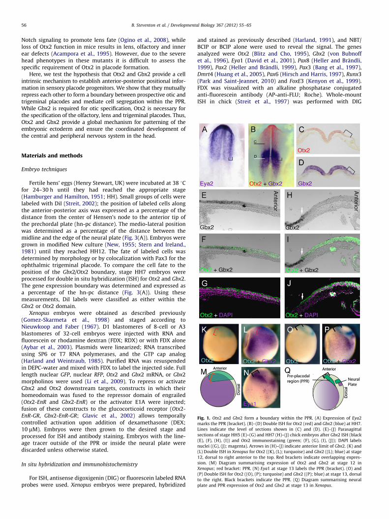
Fig. 1. Otx2 and Gbx2 form a boundary within the PPR. (A) Expression of Eya2
marks the PPR (bracket). (B)–(D) Double ISH for Otx2 (red) and Gbx2 (blue) at HH7.
Lines indicate the level of sections shown in (C) and (D). (E)–(J) Parasagittal
sections of stage HH5 (E)–(G) and HH7 (H)–(J) chick embryos after Gbx2 ISH (black
(E), (F), (H), (I)) and Otx2 immunostaining (green; (F), (G), (I), (J)); DAPI labels
nuclei ((G), (J); magenta). Arrows in (H)–(J) indicate anterior limit of Gbx2. (K) and
(L) Double ISH in Xenopus for Otx2 ((K), (L); turquoise) and Gbx2 ((L); blue) at stage
12, dorsal to right anterior to the top. Red brackets indicate overlapping expres-
sion. (M) Diagram summarising expression of Otx2 and Gbx2 at stage 12 in
Xenopus; red bracket: PPR. (N) Eya1 at stage 13 labels the PPR (bracket). (O) and
(P) Double ISH for Otx2 ((O), (P); turquoise) and Gbx2 ((P); blue) at stage 13, dorsal
to the right. Black brackets indicate the PPR. (Q) Diagram summarising neural
plate and PPR expression of Otx2 and Gbx2 at stage 13 in Xenopus.
Materials and methods
Embryo techniques
Fertile hens’ eggs (Henry Stewart, UK) were incubated at 38 1Cfor 24–30 h until they had reached the appropriate stage(Hamburger and Hamilton, 1951; HH). Small groups of cells werelabeled with DiI (Streit, 2002); the position of labeled cells alongthe anterior-posterior axis was expressed as a percentage of thedistance from the center of Hensen’s node to the anterior tip ofthe prechordal plate (hn-pc distance). The medio-lateral positionwas determined as a percentage of the distance between themidline and the edge of the neural plate (Fig. 3(A)). Embryos weregrown in modified New culture (New, 1955; Stern and Ireland.,1981) until they reached HH12. The fate of labeled cells wasdetermined by morphology or by colocalization with Pax3 for theophthalmic trigeminal placode. To compare the cell fate to theposition of the Gbx2/Otx2 boundary, stage HH7 embryos wereprocessed for double in situ hybridization (ISH) for Otx2 and Gbx2.The gene expression boundary was determined and expressed asa percentage of the hn-pc distance (Fig. 3(A)). Using thesemeasurements, DiI labels were classified as either within theGbx2 or Otx2 domain.
Xenopus embryos were obtained as described previously(Gomez-Skarmeta et al., 1998) and staged according toNieuwkoop and Faber (1967). D1 blastomeres of 8-cell or A3blastomeres of 32-cell embryos were injected with RNA andfluorescein or rhodamine dextran (FDX; RDX) or with FDX alone(Aybar et al., 2003). Plasmids were linearized; RNA transcribedusing SP6 or T7 RNA polymerases, and the GTP cap analog(Harland and Weintraub, 1985). Purified RNA was resuspendedin DEPC-water and mixed with FDX to label the injected side. Fulllength nuclear GFP, nuclear RFP, Otx2 and Gbx2 mRNA, or Gbx2morpholinos were used (Li et al., 2009). To repress or activateGbx2 and Otx2 downstream targets, constructs in which theirhomeodomain was fused to the repressor domain of engrailed(Otx2-EnR and Gbx2-EnR) or the activator E1A were injected;fusion of these constructs to the glucocorticoid receptor (Otx2-
EnR-GR, Gbx2-EnR-GR; Glavic et al., 2002) allows temporallycontrolled activation upon addition of dexamethasone (DEX;10 mM). Embryos were then grown to the desired stage andprocessed for ISH and antibody staining. Embryos with the line-age tracer outside of the PPR or inside the neural plate werediscarded unless otherwise stated.
In situ hybridization and immunohistochemistry
For ISH, antisense digoxigenin (DIG) or fluorescein labeled RNAprobes were used. Xenopus embryos were prepared, hybridized
and stained as previously described (Harland, 1991), and NBT/BCIP or BCIP alone were used to reveal the signal. The genesanalyzed were Otx2 (Blitz and Cho, 1995), Gbx2 (von Bubnoffet al., 1996), Eya1 (David et al., 2001), Pax8 (Heller and Brandli,1999), Pax2 (Heller and Brandli, 1999), Pax3 (Bang et al., 1997),Dmrt4 (Huang et al., 2005), Pax6 (Hirsch and Harris, 1997), Runx3(Park and Saint-Jeannet, 2010) and FoxE3 (Kenyon et al., 1999).FDX was visualized with an alkaline phosphatase conjugatedanti-fluorescein antibody (AP-anti-FLU; Roche). Whole-mountISH in chick (Streit et al., 1997) was performed with DIG
B. Steventon et al. / Developmental Biology 367 (2012) 55–65 57
labeled anti-sense probes for Otx2 (Bally-Cuif et al., 1995) andfluorescein-labeled probes for Gbx2 (Shamim and Mason, 1998).For double ISH, embryos were hybridized with both probesfollowed by consecutive antibody staining with alkaline phos-phatase coupled-anti-DIG and anti-fluorescein antibodies (Roche)using fast red and NBT/BCIP for color development, respectively.Immunostaining on cryosections was performed (Bailey et al.,2006) using antibodies against Otx2 (Abcam; 1:50) and Pax3(Developmental Hybridoma Bank; 1:10) and appropriate second-ary antibodies (Invitrogen; 1:1000). Sections were imaged on aLeica TCS SP5 confocal microscope.
Results
Otx2 and Gbx2 form a boundary within the sensory placode territory
In the neural tube Otx2 and Gbx2 expression initially overlapsbut then resolves to form a boundary at the MHB (Simeone et al.,1993; von Bubnoff et al., 1996; Millet et al., 1999; Tour et al.,2002a; Glavic et al., 2002). In chick Eya2 expression identifies thePPR (Fig. 1(A); McLarren et al., 2003; Streit, 2007). While Otx2 andGbx2 expression overlap in this territory at HH5 (Fig. 1(E)–(G)),both domains abut later (Fig. 1(B)–(D), (H)–(J)) with Gbx2restricted to the posterior PPR. A similar boundary is observedin the Xenopus PPR (Fig. 1(K)–(P)). At stage 11.5, Otx2 encom-passes both the anterior neural plate and its border (Fig. 1(K)),while Gbx2 is present posteriorly but overlapping with Otx2(Fig. 1(L) and (M); red bracket). At neural plate stages, Eya1demarcates the PPR (Fig. 1(N); black bracket); Otx2 and Gbx2expression has resolved into a neural plate domain dorsally and aPPR domain laterally (Fig. 1(O) and (P); black bracket). In bothregions, Otx2 and Gbx2 expression does not overlap (Fig. 1(Q)).Thus, like in the neural plate, Gbx2 and Otx2 form a geneexpression boundary within the PPR in Xenopus and chick.
Otx2 and Gbx2 segregate otic and trigeminal fates
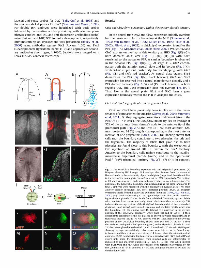
Otx2 and Gbx2 have previously been implicated in the main-tenance of compartment boundaries (Zervas et al., 2004; Sunmonuet al., 2011). Do they segregate progenitors of different fates in thePPR? At HH 7 in chick, the Otx2/Gbx2 boundary lies on average at35% of the distance from Hensen’s node to the anterior tip of theprechordal plate (Fig. 2(A) and (A’); 3577%; most anterior: 42%,most posterior: 24.5%) roughly corresponding to the most anteriorlocation of otic progenitors (Streit, 2002). DiI labeling shows thatcells near the boundary contribute to two placodes: the otic andthe trigeminal. The majority of labels that gave rise to bothplacodes are found close to this boundary, with the exception oftwo injections at around 20% i.e., within the Gbx2 territory.Anterior to the boundary cells mainly contribute to the maxillo-mandibular trigeminal placode (mmV) and to the ophthalmicPax3þ (opV) trigeminal territory (Fig. 2(B), (F)–(H)). In contrast,
Fig. 2. The Otx2/Gbx2 boundary separates otic and trigeminal precursors. (A)
Diagram showing HH 7 stage chick embryo: the distance from the center of
Hensen’s node to the anterior tip of prechordal plate (hn-pc) and from the midline
to the edge of the neural plate (ml-np) were set to 100%, respectively. The position
of DiI label was measured and expressed as percentage of each distance. (A0). The
position of the Gbx2/Otx2 boundary was measured using the same landmarks. In
total 9 embryos were measured with the boundary on average at 3577%; most
anterior position measured: 42%, most posterior position: 24.5%. (B) Diagram
combining labels from this study and published fate maps (Streit, 2002; Xu et al.,
2008); gray: labels contributing to the trigeminal placode; blue: labels contribut-
ing to the otic placode. Circles: labels from published fate maps; squares: labels
with dual fate from the current study; stars: labels from the current study. 35%
indicates the average position of the Otx2/Gbx2 boundary (dotted line)7standard
deviation (small arrow); note: mixed trigeminal and otic fates mostly locate near
this boundary. (C) HH7 embryo with DiI labeled cells posterior to the average
position of the Otx2/Gbx2 boundary (white line). (D) and (E) At HH12 their
descendants contribute to the otic placode as shown in whole mount (D) and in
transverse sections (E) and (F). HH7 embryo with DiI label anterior to the average
position of the Otx2/Gbx2 boundary (black line). (G) and (H) At HH11 their
descendants overlap with Pax3 protein (green) in the trigeminal placode. In total,
21 labels were placed into the Otx2þ and 12 into the Gbx2þ domain. (I) Diagram
showing the experimental design: blastomeres were injected at the 64-cell stage
in Xenopus and their position scored at stage 14. Arrows show the orientation of all
embryos. (J)–(L) Neighboring blastomeres were injected with nGFP and nRFP and
grown until stage 14. Descendants from injected cells are intermingled as
indicated by red and green outlines in L (100%, n¼10). (M)–(O) When injected
with nGFP/Otx2 and nRFP/Gbx2 descendants from adjacent blastomeres do not
mix (boundary in 79% of embryos, n¼14). Red and green outlines in O show the
distribution of cells.
B. Steventon et al. / Developmental Biology 367 (2012) 55–6558
posterior to the Otx2/Gbx2 border, cells mainly localize to the oticplacode (Fig. 2(B)–(D)), but are occasionally found in the mmV(Fig. 2(B); Xu et al., 2008). However, except the two injectionsmentioned above their original location lies well within the mostposterior position of the Otx2/Gbx2 boundary measured suggest-ing that these cells may indeed arise from the Gbx2 territory. It istherefore possible that due to differences in individual embryos,fate maps overestimate cell mixing (see Pieper et al., 2011 fordiscussion). In general, our current findings agree with previouslypublished fate maps (Fig. 2(B); Streit, 2002; Xu et al., 2008). Thus,although no strict cell fate segregation is observed, the vastmajority of trigeminal precursors come from the Otx2 region, whileotic cells largely arise from Gbx2þ cells suggesting that in the PPRthe Otx2/Gbx2 boundary roughly separates trigeminal and oticterritories.
Can Otx2 and Gbx2 sort cells in the PPR? Using Xenopus wecompared the behavior of control-injected cells with those carry-ing exogenous Gbx2 and Otx2. Descendents of the A3 blastomere,which gives rise to placodes, were injected at the 64-cell stagewith mRNA encoding nuclear-GFP and nuclear-RFP alone or incombination with Otx2 and Gbx2, respectively (Fig. 2(I)). Signifi-cant overlap between GFP and RFP expressing cells is observed atstage 14 in controls (Fig. 2(J)–(L)). In contrast, cells in the PPR andfuture epidermis overexpressing Gbx2 form a boundary with cellsexpressing exogenous Otx2 with only some cells intermingling(Fig. 2(M)–(O)). Together these results show that Otx2 and Gbx2control cell sorting and are part of the molecular mechanism thatsegregates sensory progenitors to different placodes.
The Otx2/Gbx2 boundary in the PPR forms by a cross-repressive
mechanism
In the neural plate, Otx2 and Gbx2 mutually repress each othertranscriptionally to form a sharp boundary (Millet et al., 1999;Katahira et al., 2000; Tour et al., 2002a; Glavic et al., 2002). Toconfirm this we injected Otx2 or Gbx2 mRNA into the D1blastomere at the 8-cell stage targeting the neural plate and itsborder. As expected Otx2 misexpression shifts the MHB markerEn-1 posteriorly (Fig. 3(A)), whereas misexpression of Gbx2 leadsto an anterior shift (Fig. 3(C); see also: Tour et al., 2002b). Incontrast, when Otx2 or Gbx2 mRNA is targeted to the PPR (A3blastomere injection at 32-cell stage) changes in neural En-1expression are rarely observed (Fig. 3(B) and (D)). This approachtherefore allows us to analyze the role of these transcriptionfactors specifically in placode progenitors, without interferingwith neural patterning.
In the PPR, misexpression of Otx2 mRNA results in a loss ofGbx2 expression (Fig. 3(E)). To ask whether Otx2 acts as atranscriptional repressor in this context we used a constitutiverepressor form Otx2-EnR. Like full-length Otx2, misexpression ofOtx2-EnR leads to Gbx2 reduction in the PPR (Fig. 3(F)). Misex-pression of Gbx2 (Fig. 3(G) and (H)) or the constitutive repressorGbx2-EnR (Fig. 3(K) and (L)) reduces Otx2 expression in the PPR,while Gbx2 morpholino knock-down (Li et al., 2009) expands itsexpression (Fig. 3(I) and (J)). Together, these results demonstratethat in the PPR Otx2 and Gbx2 act as transcriptional repressorsand mutually repress each other suggesting that this interactiongenerates the Gbx2/Otx2 expression boundary.
Dual Gbx2 function in otic placode specification
The otic placode forms within the Gbx2þ territory; is Gbx2required for its specification? Gbx2 knock-down by splice- andtranslation-blocking morpholinos prevents the expression of the oticmarkers Pax8 and Pax2 (Fig. 4(A) and (B)). When analyzed at laterstages, the size of the otic vesicle in embryos injected with both
morpholinos is severely reduced (Fig. 4(D) – (F)). Although Gbx2 isnormally expressed prior to the pre-placodal marker Eya1 (Li et al.,2009), Eya1 expression is unaffected in Gbx2 morphants (Fig. 4(C)).Thus, Gbx2 is required for otic, but not for PPR specification.
Is this function of Gbx2 simply due to its Otx2-repressingactivity or does it also regulate otic-specific genes? To test this weused the inducible Gbx2-EnR-GR, which constitutively repressesall Gbx2 targets including Otx2 (Fig. 3(K) and (L)). When thisconstruct is activated at the beginning of gastrulation (stage 10),the earliest expression of Pax8 (stage 13) and Pax2 (stage 16) isreduced (Fig. 4(G)–(I)); Pax8 remains absent when embryos aregrown to stage 18 (Fig. 4(J)) similar to Gbx2 morphants. Incontrast, without activation (Fig. 4(K)) no effect is observed. Thus,even in the absence of Otx2, Gbx2-EnR prevents the expression ofotic genes suggesting that the loss of otic markers in Gbx2morphants is not solely a consequence of ectopic Otx2 expression.To assess whether Gbx2 function is required after initial oticspecification, we activated Gbx2-EnR-GR later at neural platestages (Fig. 4(L)): otic genes continue to be expressed normallysuggesting that Gbx2 function is not required for the maintenanceof otic fate.
Finally, we tested whether Gbx2 is sufficient to impart oticcharacter to cells in the anterior PPR. Gbx2 misexpression does notlead to expansion or ectopic expression of Pax8 (Fig. 4(M)) or Pax2(Fig. 4(N)), nor does it affect the general PPR marker Eya1 (Fig. 4(O)).However, ectopic Gbx2 expression does repress anterior cell fates asdemonstrated by the loss of the olfactory marker Dmrt4 (Huanget al., 2005; Fig. 4(P)) and the lens marker FoxE3 (Fig. 4(Q)). Thus,while Gbx2 is not sufficient to impart otic identity to non-otic cells,it plays a dual role during otic specification: it restricts Otx2 (whichotherwise inhibits posterior fate; see below) and provides a positiveinput for otic specifiers. However, once induced maintenance of oticidentity is independent of Gbx2 function.
Otx2 is required for trigeminal placode specification
Future trigeminal cells initially lie within the Otx2 domain(Fig. 2(H) and (B)). Is the activation of Otx2 target genes requiredfor trigeminal cell specification? Targeting the PPR with RNAencoding the constitutive repressor form Otx2-EnR leads to a lossof Runx3 labeling trigeminal/profundal precursors (Park and Saint-Jeannet, 2010; Fig. 5(A)) at stage 23, while co-injection with fulllength Otx2 mRNA restores its expression (Fig. 5(B)). To analyzewhether activation of Otx2 targets is required for the profundaland trigeminal placode (opV and mmV in amniotes), Runx3expression was assessed at stage 28 when both can be distin-guished: after Otx2-EnR injection both placodes are absent(Fig. 5(C) and (D)). Our results suggest that Otx2 and Gbx2 separateprospective otic and trigeminal territories (Fig. 2(B)) predictingthat profundal and trigeminal specification should be independentof Gbx2. Indeed, injection of Gbx2 morpholinos (Fig. 5(E) and (F))or of Gbx2-EnR (Fig. 5(G) and (H)) does not alter Runx3 expressionto the same extent as Otx2-EnR, and Pax3 expression is normalafter Gbx2 knock down (Supplementary Fig. 1D).
To test when the activation of Otx2 target genes is required fortrigeminal development, we used inducible Otx2-EnR-GR. In theabsence of DEX, profundal Pax3 is normal (Fig. 5(I)); activation atgastrulation stage (stage 10) reduces Pax3 (Fig. 5(J)), while activationat stage 14 has no effect (Fig. 5(K)). Otx2 or Otx2E1A (not shown)expression in the trigeminal territory has no effect on Pax3 expres-sion (Supplementary Fig. 1A); thus the constitutive repressor form ofOtx2 does not mimic misexpression of wild type Otx2 suggestingthat Otx2 acts as a transcriptional activator in trigeminal precursors.In addition, ectopic Otx2 expression in the posterior PPR is notsufficient to expand Pax3 transcripts (Supplementary Fig. 1A). Thus,
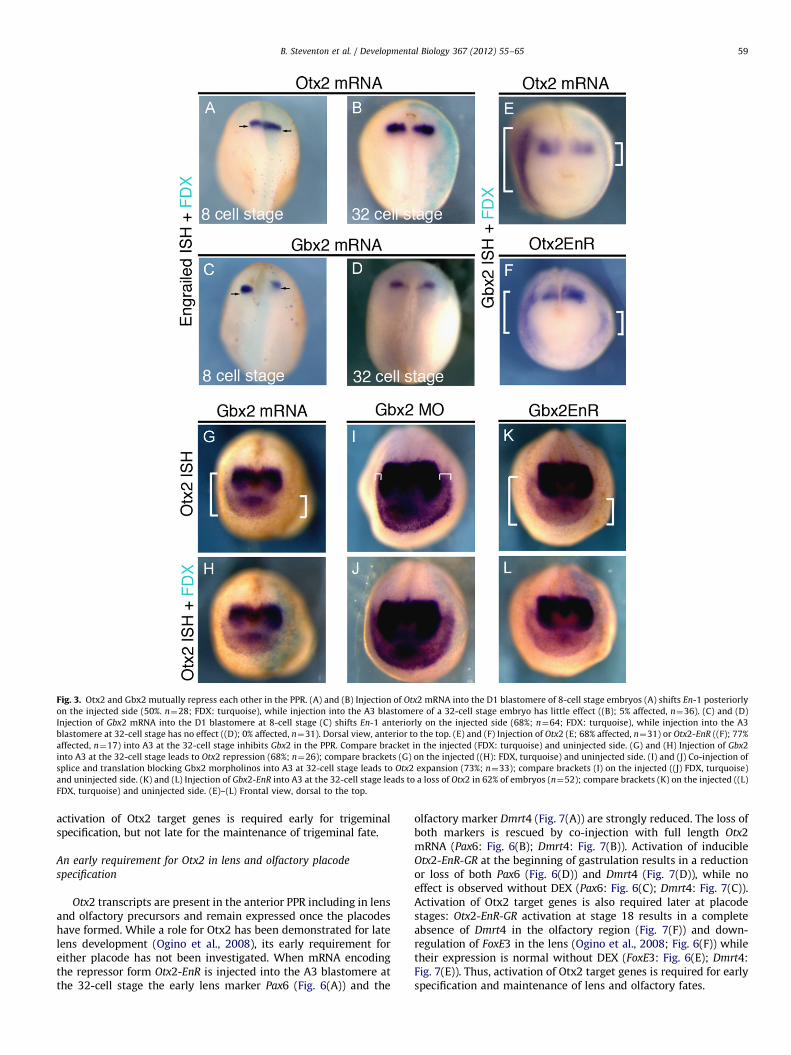
Fig. 3. Otx2 and Gbx2 mutually repress each other in the PPR. (A) and (B) Injection of Otx2 mRNA into the D1 blastomere of 8-cell stage embryos (A) shifts En-1 posteriorly
on the injected side (50%. n¼28; FDX: turquoise), while injection into the A3 blastomere of a 32-cell stage embryo has little effect ((B); 5% affected, n¼36). (C) and (D)
Injection of Gbx2 mRNA into the D1 blastomere at 8-cell stage (C) shifts En-1 anteriorly on the injected side (68%; n¼64; FDX: turquoise), while injection into the A3
blastomere at 32-cell stage has no effect ((D); 0% affected, n¼31). Dorsal view, anterior to the top. (E) and (F) Injection of Otx2 (E; 68% affected, n¼31) or Otx2-EnR ((F); 77%
affected, n¼17) into A3 at the 32-cell stage inhibits Gbx2 in the PPR. Compare bracket in the injected (FDX: turquoise) and uninjected side. (G) and (H) Injection of Gbx2
into A3 at the 32-cell stage leads to Otx2 repression (68%; n¼26); compare brackets (G) on the injected ((H): FDX, turquoise) and uninjected side. (I) and (J) Co-injection of
splice and translation blocking Gbx2 morpholinos into A3 at 32-cell stage leads to Otx2 expansion (73%; n¼33); compare brackets (I) on the injected ((J) FDX, turquoise)
and uninjected side. (K) and (L) Injection of Gbx2-EnR into A3 at the 32-cell stage leads to a loss of Otx2 in 62% of embryos (n¼52); compare brackets (K) on the injected ((L)
FDX, turquoise) and uninjected side. (E)–(L) Frontal view, dorsal to the top.
B. Steventon et al. / Developmental Biology 367 (2012) 55–65 59
activation of Otx2 target genes is required early for trigeminalspecification, but not late for the maintenance of trigeminal fate.
An early requirement for Otx2 in lens and olfactory placode
specification
Otx2 transcripts are present in the anterior PPR including in lensand olfactory precursors and remain expressed once the placodeshave formed. While a role for Otx2 has been demonstrated for latelens development (Ogino et al., 2008), its early requirement foreither placode has not been investigated. When mRNA encodingthe repressor form Otx2-EnR is injected into the A3 blastomere atthe 32-cell stage the early lens marker Pax6 (Fig. 6(A)) and the
olfactory marker Dmrt4 (Fig. 7(A)) are strongly reduced. The loss ofboth markers is rescued by co-injection with full length Otx2mRNA (Pax6: Fig. 6(B); Dmrt4: Fig. 7(B)). Activation of inducibleOtx2-EnR-GR at the beginning of gastrulation results in a reductionor loss of both Pax6 (Fig. 6(D)) and Dmrt4 (Fig. 7(D)), while noeffect is observed without DEX (Pax6: Fig. 6(C); Dmrt4: Fig. 7(C)).Activation of Otx2 target genes is also required later at placodestages: Otx2-EnR-GR activation at stage 18 results in a completeabsence of Dmrt4 in the olfactory region (Fig. 7(F)) and down-regulation of FoxE3 in the lens (Ogino et al., 2008; Fig. 6(F)) whiletheir expression is normal without DEX (FoxE3: Fig. 6(E); Dmrt4:Fig. 7(E)). Thus, activation of Otx2 target genes is required for earlyspecification and maintenance of lens and olfactory fates.
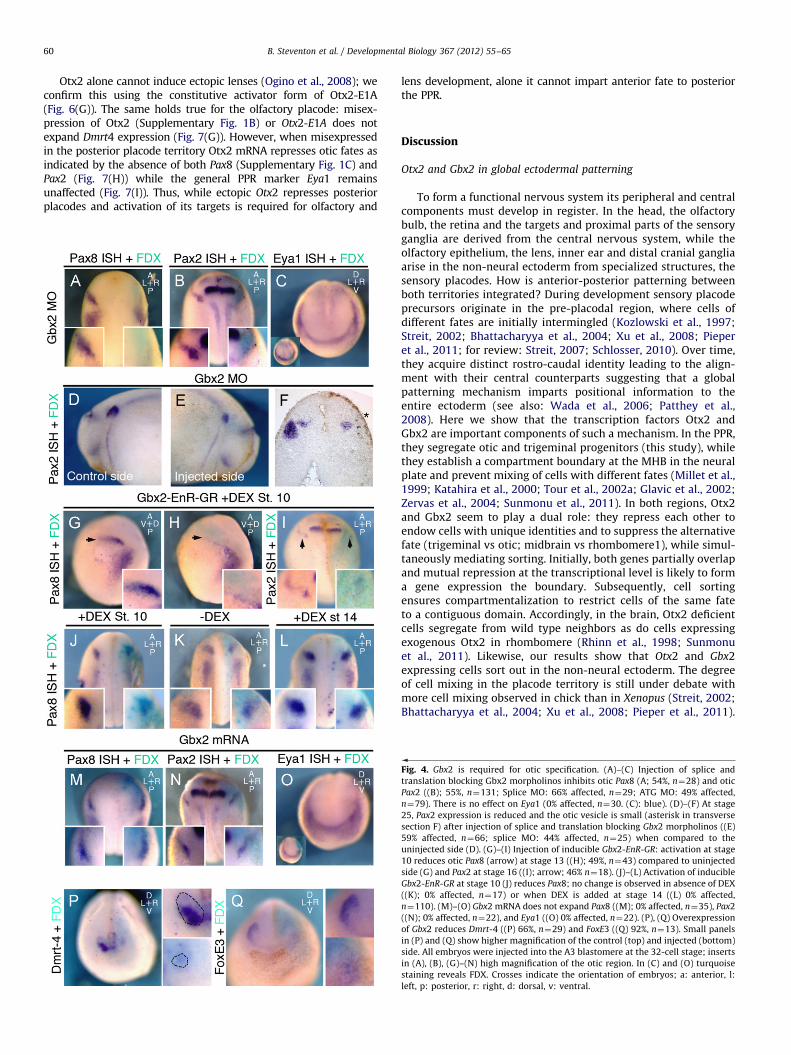
B. Steventon et al. / Developmental Biology 367 (2012) 55–6560
Otx2 alone cannot induce ectopic lenses (Ogino et al., 2008); weconfirm this using the constitutive activator form of Otx2-E1A(Fig. 6(G)). The same holds true for the olfactory placode: misex-pression of Otx2 (Supplementary Fig. 1B) or Otx2-E1A does notexpand Dmrt4 expression (Fig. 7(G)). However, when misexpressedin the posterior placode territory Otx2 mRNA represses otic fates asindicated by the absence of both Pax8 (Supplementary Fig. 1C) andPax2 (Fig. 7(H)) while the general PPR marker Eya1 remainsunaffected (Fig. 7(I)). Thus, while ectopic Otx2 represses posteriorplacodes and activation of its targets is required for olfactory and
lens development, alone it cannot impart anterior fate to posteriorthe PPR.
Discussion
Otx2 and Gbx2 in global ectodermal patterning
To form a functional nervous system its peripheral and centralcomponents must develop in register. In the head, the olfactorybulb, the retina and the targets and proximal parts of the sensoryganglia are derived from the central nervous system, while theolfactory epithelium, the lens, inner ear and distal cranial gangliaarise in the non-neural ectoderm from specialized structures, thesensory placodes. How is anterior-posterior patterning betweenboth territories integrated? During development sensory placodeprecursors originate in the pre-placodal region, where cells ofdifferent fates are initially intermingled (Kozlowski et al., 1997;Streit, 2002; Bhattacharyya et al., 2004; Xu et al., 2008; Pieperet al., 2011; for review: Streit, 2007; Schlosser, 2010). Over time,they acquire distinct rostro-caudal identity leading to the align-ment with their central counterparts suggesting that a globalpatterning mechanism imparts positional information to theentire ectoderm (see also: Wada et al., 2006; Patthey et al.,2008). Here we show that the transcription factors Otx2 andGbx2 are important components of such a mechanism. In the PPR,they segregate otic and trigeminal progenitors (this study), whilethey establish a compartment boundary at the MHB in the neuralplate and prevent mixing of cells with different fates (Millet et al.,1999; Katahira et al., 2000; Tour et al., 2002a; Glavic et al., 2002;Zervas et al., 2004; Sunmonu et al., 2011). In both regions, Otx2and Gbx2 seem to play a dual role: they repress each other toendow cells with unique identities and to suppress the alternativefate (trigeminal vs otic; midbrain vs rhombomere1), while simul-taneously mediating sorting. Initially, both genes partially overlapand mutual repression at the transcriptional level is likely to forma gene expression the boundary. Subsequently, cell sortingensures compartmentalization to restrict cells of the same fateto a contiguous domain. Accordingly, in the brain, Otx2 deficientcells segregate from wild type neighbors as do cells expressingexogenous Otx2 in rhombomere (Rhinn et al., 1998; Sunmonuet al., 2011). Likewise, our results show that Otx2 and Gbx2expressing cells sort out in the non-neural ectoderm. The degreeof cell mixing in the placode territory is still under debate withmore cell mixing observed in chick than in Xenopus (Streit, 2002;Bhattacharyya et al., 2004; Xu et al., 2008; Pieper et al., 2011).
Fig. 4. Gbx2 is required for otic specification. (A)–(C) Injection of splice and
translation blocking Gbx2 morpholinos inhibits otic Pax8 (A; 54%, n¼28) and otic
Pax2 ((B); 55%, n¼131; Splice MO: 66% affected, n¼29; ATG MO: 49% affected,
n¼79). There is no effect on Eya1 (0% affected, n¼30. (C): blue). (D)–(F) At stage
25, Pax2 expression is reduced and the otic vesicle is small (asterisk in transverse
section F) after injection of splice and translation blocking Gbx2 morpholinos ((E)
59% affected, n¼66; splice MO: 44% affected, n¼25) when compared to the
uninjected side (D). (G)–(I) Injection of inducible Gbx2-EnR-GR: activation at stage
10 reduces otic Pax8 (arrow) at stage 13 ((H); 49%, n¼43) compared to uninjected
side (G) and Pax2 at stage 16 ((I); arrow; 46% n¼18). (J)–(L) Activation of inducible
Gbx2-EnR-GR at stage 10 (J) reduces Pax8; no change is observed in absence of DEX
((K); 0% affected, n¼17) or when DEX is added at stage 14 ((L) 0% affected,
n¼110). (M)–(O) Gbx2 mRNA does not expand Pax8 ((M); 0% affected, n¼35), Pax2
((N); 0% affected, n¼22), and Eya1 ((O) 0% affected, n¼22). (P), (Q) Overexpression
of Gbx2 reduces Dmrt-4 ((P) 66%, n¼29) and FoxE3 ((Q) 92%, n¼13). Small panels
in (P) and (Q) show higher magnification of the control (top) and injected (bottom)
side. All embryos were injected into the A3 blastomere at the 32-cell stage; inserts
in (A), (B), (G)–(N) high magnification of the otic region. In (C) and (O) turquoise
staining reveals FDX. Crosses indicate the orientation of embryos; a: anterior, l:
left, p: posterior, r: right, d: dorsal, v: ventral.
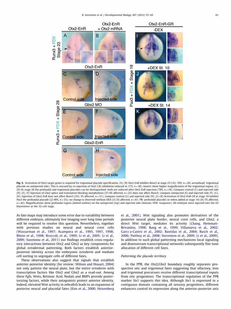
Fig. 5. Activation of Otx2 target genes is required for trigeminal placode specification. (A), (B) Otx2-EnR inhibits Runx3 at stage 23 ((A); 59%, n¼26; arrowhead: trigeminal
placode on uninjected side). This is rescued by co-injection of Otx2 ((B) inhibition reduced to 17%, n¼36). Inserts show higher magnification of the trigeminal region. (C),
(D) At stage 28 the profundal and trigeminal placodes can be distinguished; both are reduced after Otx2-EnR injection (78%, n¼18). Compare control (C) and injected side
(D). (E), (F) Injection of Gbx2 splice and translation blocking morpholinos ((F) 0% affected, n¼25) does not affect Runx3; compare uninjected (E) and injected side (F). (G),
(H). Injection of Gbx2-EnR does not affect Runx3 ((H); 3% affected, n¼31); compare control (G) and injected side (H). (I)–(K) Activation of Otx2-EnR-GR at stage 10 inhibits
Pax3 the profundal placode ((J) 49%, n¼33); no change is observed without DEX ((I) 5% affected, n¼61; PR: profundal placode) or when added at stage 14 ((K) 5% affected,
n¼42). Magnifications show profundal region (dotted outline) on the uninjected (top) and injected side (bottom; FDX: turquoise). All embryos were injected into the A3
blastomere at the 32-cell stage.
B. Steventon et al. / Developmental Biology 367 (2012) 55–65 61
As fate maps may introduce some error due to variability betweendifferent embryos, ultimately live imaging over long time periodswill be required to resolve this question. Nevertheless, togetherwith previous studies on neural and neural crest cells(Wassarman et al., 1997; Acampora et al., 1995, 1997, 1998;Rhinn et al., 1998; Broccoli, et al., 1999; Li et al., 2005; Li et al.,2009; Sunmonu et al., 2011) our findings establish cross-regula-tory interactions between Otx2 and Gbx2 as key components forglobal ectodermal patterning. Both factors establish anterior-posterior identity across the embryonic ectoderm and mediatecell sorting to segregate cells of different fates.
These observations also suggest that signals that establishanterior-posterior identity (for review: Wilson and Houart, 2004)not only pattern the neural plate, but the entire ectoderm withtranscription factors like Otx2 and Gbx2 as a read-out. Amongthese Fgfs, Wnts, Retinoic Acid, Nodals and BMPs provide poster-iorizing factors, while their antagonists protect anterior identity.Indeed, elevated Wnt activity in zebrafish leads to an expansion ofposterior neural and placodal fates (Kim et al., 2000; Heisenberg
et al., 2001). Wnt signaling also promotes derivatives of theposterior neural plate border, neural crest cells, and Gbx2, adirect Wnt target, mediates its activity (Chang, Hemmati-Brivanlou, 1998; Bang et al., 1999; Villanueva et al., 2002;Garcı!a-Castro et al., 2002; Bastidas et al., 2004; Basch et al.,2006; Patthey et al., 2008; Steventon et al., 2009; Li et al., 2009).In addition to such global patterning mechanisms local signalingand downstream transcriptional networks subsequently fine tuneallocation of different cell fates.
Patterning the placode territory
In the PPR, the Otx2/Gbx2 boundary roughly separates pro-spective otic and trigeminal fates suggesting that olfactory, lensand trigeminal precursors receive different transcriptional inputsfrom otic progenitors. The transcriptional regulation of the PPRmarker Six1 supports this idea. Although Six1 is expressed in acontiguous domain containing all sensory progenitors, differentenhancers control its expression along the anterior-posterior axis
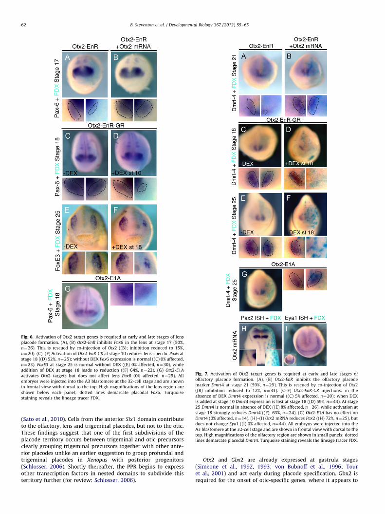
Fig. 6. Activation of Otx2 target genes is required at early and late stages of lens
placode formation. (A), (B) Otx2-EnR inhibits Pax6 in the lens at stage 17 (50%,
n¼26). This is rescued by co-injection of Otx2 ((B); inhibition reduced to 15%,
n¼20). (C)–(F) Activation of Otx2-EnR-GR at stage 10 reduces lens-specific Pax6 at
stage 18 ((D) 52%, n¼25); without DEX Pax6 expression is normal ((C) 0% affected,
n¼23). FoxE3 at stage 25 is normal without DEX ((E) 0% affected, n¼30), while
addition of DEX at stage 18 leads to reduction ((F) 64%, n¼22). (G) Otx2-E1A
activates Otx2 targets but does not affect lens Pax6 (0% affected, n¼25). All
embryos were injected into the A3 blastomere at the 32-cell stage and are shown
in frontal view with dorsal to the top. High magnifications of the lens region are
shown below each panel; dotted lines demarcate placodal Pax6. Turquoise
staining reveals the lineage tracer FDX.
Fig. 7. Activation of Otx2 target genes is required at early and late stages of
olfactory placode formation. (A), (B) Otx2-EnR inhibits the olfactory placode
marker Dmrt4 at stage 21 (59%, n¼29). This is rescued by co-injection of Otx2
((B) inhibition reduced to 12%, n¼33). (C–F) Otx2-EnR-GR injections: in the
absence of DEX Dmrt4 expression is normal ((C) 5% affected, n¼20); when DEX
is added at stage 10 Dmrt4 expression is lost at stage 18 ((D) 59%, n¼44). At stage
25 Dmrt4 is normal in absence of DEX ((E) 8% affected, n¼26), while activation at
stage 18 strongly reduces Dmrt4 ((F); 63%, n¼24). (G) Otx2-E1A has no effect on
Dmrt4 (0% affected, n¼14). (H)–(I) Otx2 mRNA reduces Pax2 ((H) 72%, n¼25), but
does not change Eya1 ((I) 0% affected, n¼44). All embryos were injected into the
A3 blastomere at the 32-cell stage and are shown in frontal view with dorsal to the
top. High magnifications of the olfactory region are shown in small panels; dotted
lines demarcate placodal Dmrt4. Turquoise staining reveals the lineage tracer FDX.
B. Steventon et al. / Developmental Biology 367 (2012) 55–6562
(Sato et al., 2010). Cells from the anterior Six1 domain contributeto the olfactory, lens and trigeminal placodes, but not to the otic.These findings suggest that one of the first subdivisions of theplacode territory occurs between trigeminal and otic precursorsclearly grouping trigeminal precursors together with other ante-rior placodes unlike an earlier suggestion to group profundal andtrigeminal placodes in Xenopus with posterior progenitors(Schlosser, 2006). Shortly thereafter, the PPR begins to expressother transcription factors in nested domains to subdivide thisterritory further (for review: Schlosser, 2006).
Otx2 and Gbx2 are already expressed at gastrula stages(Simeone et al., 1992, 1993; von Bubnoff et al., 1996; Touret al., 2001) and act early during placode specification. Gbx2 isrequired for the onset of otic-specific genes, where it appears to
B. Steventon et al. / Developmental Biology 367 (2012) 55–65 63
act as transcriptional activator: the constitutive repressor Gbx2-EnR mimics MO-mediated knock-down. This is in contrast to itsearlier role as repressor during boundary formation (see above)suggesting that the availability of cofactors determines the finaloutcome as observed for other homeobox factors (Brugmannet al., 2004; Anderson et al., 2012). After initial specification, oticdevelopment is Gbx2 independent, although it is later involved inear morphogenesis (Lin et al., 2005). The lack of an early earphenotype in Gbx2 mutant mice (Lin et al., 2005) is likely due tofunctional redundancy with Gbx1. In contrast, Otx2 is necessaryfor both formation and maintenance of lens and olfactory identity(Ogino et al., 2008; this work) consistent with its continuedexpression in both placodes. In the trigeminal placode, Otx2 isdownregulated shortly after its specification probably due torepression by Pax3 (unpublished observations), which also inhi-bits Pax6 in this territory (Wakamatsu, 2011). Like Gbx2, Otx2switches from a transcriptional repressor at early stages to anactivator later. In summary, like in the neural plate, in the PPROtx2 and Gbx2 are among the earliest factors that subdivide acontiguous territory along the anterior-posterior axis.
Although Otx2 and Gbx2 are required for early placodespecification, neither factor alone is sufficient to endow cells withnew regional character or to induce ectopic placodes. This appearsto differ considerably from their activity in the neural tube, whereectopic expression of either factor respecifies anterior-posterioridentity (Glavic et al., 2002; Tour et al., 2002a). However, hereOtx2 and Gbx2 mainly function to position the MHB (Li andJoyner, 2001; Raible and Brand, 2004), an organizer region thatitself patterns the brain. Thus, changes in regional identity arelikely to be a consequence of MHB induction. Whether a similarorganizing center forms at the Otx2/Gbx2 boundary in the PPRremains to be established, however, so far our results argueagainst this notion. The finding that neither Gbx2 nor Otx2 issufficient to induce ectopic placodes suggests that additionalfactors cooperate to control the expression of placode-specificdownstream targets. This is indeed the case in the lens, whereOtx2 directly binds to the lens-specific FoxE3 enhancer andtogether with intracellular effectors of Notch signaling activatesits transcription (Ogino et al., 2008).
Evolutionary conservation of anterior-posterior patterning by
Otx2 and Gbx2
The development of cranial sensory placodes and neural crest isconsidered to be a key step in the evolution of the vertebrate head(Northcutt and Gans, 1983). Like in vertebrates Gbx and Otx form aboundary within the Amphioxus ectoderm (Williams and Holland,1996, 1998; Castro et al., 2006; Benito-Gutierrez, 2006) raising thequestion whether, at an early stage of their evolution, neural crestand placodes co-opted an already existing gene expression bound-ary to position themselves along the anterior-posterior axis. DespiteGbx2/Otx2 apposition in Amphioxus, MHB specific genes such as En,Wnt1, FGF8/17/18 and Pax2/5/8 are not restricted to this boundary(Holland et al., 1997, 2000; Meulemans and Bronner-Fraser, 2007),indicating that MHB organizer genes were recruited to the Otx/Gbxborder in early vertebrates (Castro et al., 2006; Holland and Short,2008; Holland, 2009). A Gbx/Otx boundary appears to have beenpresent in the early bilaterian ancestor as Unpg/Gbx and Otd/Otx alsonegatively regulate one another to form a boundary that positionsEn and Pax2/5/8 in Drosophila (Hirth et al., 2003). In addition, Gbx2and Otx2 form a boundary in the annelid Platynereis dumerilii thatcorresponds to a band of En expression (Arendt et al., 2001;Steinmetz et al., 2007, 2011). Together these findings raise thepossibility that Otx2 and Gbx2 form an ancient boundary of geneexpression responsible for anterior-posterior patterning of both theneural plate and neural plate border. However, this boundary has
been utilized differently in each territory: to position an organizingregion at the MHB, and to specify placodal fates in the PPR.
Acknowledgments
We would like to thank Claudio Stern and Jeremy Green forcritical comments on the manuscript, Ivor Mason, Thomas Holle-mann, Andre Brandli, Milan Jamrich and Jean-Pierre Saint-Jeannetfor cDNA clones and Susmitha Rao and Ewa Kolano for excellenttechnical support. This work was funded by a Wellcome Trustproject grant to AS and RM, and by a Medical Research Councilgrant to RM.
Appendix A. Supporting information
Supplementary data associated with this article can be found inthe online version at http://dx.doi.org/10.1016/j.ydbio.2012.04.025.
References
Acampora, D., Mazan, S., Lallemand, Y., Avantaggiato, V., Maury, M., Simeone, A.,Brulet, P., 1995. Forebrain and midbrain regions are deleted in Otx2 �/�mutants due to a defective anterior neuroectoderm specification duringgastrulation. Development 121, 3279–3290.
Acampora, D., Avantaggiato, V., Tuorto, F., Briata, P., Corte, G., Simeone, A., 1998.Visceral endoderm-restricted translation of Otx1 mediates recovery of Otx2requirements for specification of anterior neural plate and normal gastrula-tion. Development 125, 5091–5104.
Acampora, D., Avantaggiato, V., Tuorto, F., Simeone, A., 1997. Genetic control ofbrain morphogenesis through Otx gene dosage requirement. Development124, 3639–3650.
Arendt, D., Technau, U., Wittbrodt, J., 2001. Evolution of the bilaterian larvalforegut. Nature 409, 81–85.
Anderson, A.M., Weasner, B.M., Weasner, B.P., Kumar, J.P., 2012. Dual transcrip-tional activities of SIX proteins define their roles in normal and ectopic eyedevelopment. Development 139, 991–1000.
Aybar, M.J., Nieto, M.A., Mayor, R., 2003. Snail precedes slug in the genetic cascaderequired for the specification and migration of the Xenopus neural crest.Development 130, 483–494.
Bailey, A.P., Bhattacharyya, S., Bronner-Fraser, M., Streit, A., 2006. Lens specifica-tion is the ground state of all sensory placodes, from which FGF promotesolfactory identity. Dev. Cell 11, 505–517.
Baker, C.V., Stark, M.R., Marcelle, C., Bronner-Fraser, M., 1999. Competence,specification and induction of Pax-3 in the trigeminal placode. Development126, 147–156.
Baker, C.V., Bronner-Fraser, M., 2000. Establishing neuronal identity in vertebrateneurogenic placodes. Development 127, 3045–3056.
Baker, C.V.H., Bronner-Fraser, M., 2001. Vertebrate Cranial Placodes I. EmbryonicInduction. Dev. Biol. 232, 1–61.
Bally-Cuif, L., Gulisano, M., Broccoli, V., Bonicelli, E., 1995. C-otx2 is expressed intwo different phases of gastrulation and is sensitive to retinoic acid treatmentin chick embryo. Mech. Dev. 49, 49–63.
Bang, A.G., Papalopulu, N., Kintner, C., Goulding, M.D., 1997. Expression of Pax-3 isinitiated in the early neural plate by posteriorizing signals produced by theorganizer and by posterior non-axial mesoderm. Development 124,2075–2085.
Bang, A.G., Papalopulu, N., Goulding, M.D., Kintner, C., 1999. Expression of Pax-3 inthe lateral neural plate is dependent on a Wnt-mediated signal from posteriornonaxial mesoderm. Dev. Biol. 212, 366–380.
Benito-Gutierrez, �E., 2006. A gene catalogue of the amphioxus nervous system. Int.J. Biol. Sci. 2, 149–160.
Basch, M., Bronner-Fraser, M., Garcı!a-Castro, M., 2006. Specification of the neuralcrest occurs during gastrulation and requires Pax7. Nature 41, 218–222.
Bastidas, F., De Calisto, J., Mayor, R., 2004. Identification of neural crest compe-tence territory: role of Wnt signaling. Dev. Dyn. 229, 109–117.
Bhattacharyya, S., Bailey, A.P., Bronner-Fraser, M., Streit, A., 2004. Segregation oflens and olfactory precursors from a common territory: cell sorting andreciprocity of Dlx5 and Pax6 expression. Dev. Biol. 271, 403–414.
Bhattacharyya, S., Bronner-Fraser, M., 2008. Competence, specification and com-mitment to an olfactory placode fate. Development 135, 4165–4177.
Blitz, I.L., Cho, K.W., 1995. Anterior neurectoderm is progressively induced duringgastrulation: the role of the Xenopus homeobox gene orthodenticle. Develop-ment 121, 993–1004.
Broccoli, V., Boncinelli, E., Wurst, W., 1999. The caudal limit of Otx2 expressionpositions the isthmic organizer. Nature 401, 164–168.
B. Steventon et al. / Developmental Biology 367 (2012) 55–6564
Brugmann, S.A., Pandur, P.D., Kenyon, K.L., Pignoni, F., Moody, S.A., 2004. Six1promotes a placodal fate within the lateral neurogenic ectoderm by function-ing as both a transcriptional activator and repressor. Development 131,5871–5881.
Castro, L.F.C., Rasmussen, S.L.K., Holland, P.W.H., Holland, N.D., Holland, L.Z., 2006.A Gbx homeobox gene in amphioxus: Insights into ancestry of the ANTP classand evolution of the midbrain/hindbrain boundary. Dev.Biol. 295, 40–51.
Chang, C., Hemmati-Brivanlou, A., 1998. Neural crest induction by Xwnt7B inXenopus. Dev. Biol. 194, 129–134.
David, R., Ahrens, K., Wedlich, D., Schlosser, G., 2001. Xenopus Eya1 demarcates allneurogenic placodes as well as migrating hypaxial muscle precursors. Mech.Dev. 103, 189–192.
Gallagher, B.C., Henry, J.J., Grainger, R.M., 1996. Inductive processes leading toinner ear formation during Xenopus development. Dev. Biol. 175, 95–107.
Garcı!a-Castro, Marcelle, C., Bronner-Fraser, M., 2002. Ectodermal Wnt functionsas a neural crest inducer. Science 297, 848–851.
Glavic, A., Gomez-Skarmeta, J.L., Mayor, R., 2002. The homeoprotein Xiro1 isrequired for midbrain-hindbrain boundary formation. Development 129,1609–1621.
Gomez-Skarmeta, J.L., Glavic, A., de la Calle-Mustienes, E., Modolell, J., Mayor, R.,1998. Xiro, a Xenopus homolog of Drosophila Iroquois complex genes, controlsdevelopment at the neural plate. EMBO J. 17, 181–190.
Grainger, R.M., Mannion, J.E., Cook, T.L., Zygar, C.A., 1997. Defining intermediatestages in cell determination: acquisition of a lens-forming bias in headectoderm during lens determination. Dev. Genet. 20, 246–257.
Groves, A.K., Bronner-Fraser, M., 2000. Competence, specification and commit-ment in otic placode induction. Development 127, 3489–3499.
Hamburger, V., Hamilton, H.L., 1951. A series of normal stages in the developmentof the chick embryo. J. Morph. 88, 49–92.
Harland, R.M., Weintraub, H., 1985. Translation of mRNA injected into Xenopusoocytes is specifically inhibited by antisense RNA. Cell Biol. 10, 1511–1514.
Harland, R.M., 1991. In situ hybridization: An improved whole-mount method forXenopus embryos. Methods Cell Biol. 36, 685–695.
Heisenberg, C.-P., Houart, C., Take-uchi, M., Rauch, G., Young, N., Coutinho, P.,Masai, I., Caneparo, L., Concha, M.L., Geisler, R., et al., 2001. A mutation in theGsk3b-binding domain of zebrafish masterblind/axin1 leads to a fate trans-formation of telencephalon and eyes to diencephalon. Genes. Dev. 15,1427–1434.
Heller, N., Brandli, A.W., 1999. Xenopus Pax-2/5/8 orthologues: Novel insights intoPax gene evolution and identification of Pax-8 as the earliest marker for oticand pronephric cell lineages. Dev. Genet. 24, 208–219.
Henry, J.J., Grainger, R.M., 1987. Inductive interactions in the spatial and temporalrestriction of lens-forming potential in embryonic ectoderm of Xenopus laevis.Dev. Biol. 124, 200–214.
Hirth, F., Kammermeier, L., Frei, E., Walldorf, U., Noll, M., Reichert, H., 2003. Anurbilaterian origin of the tripartite brain: developmental genetici nsights fromDrosophila. Development 130. 2365-237.
Hirsch, N., Harris, W.A., 1997. Xenopus Pax-6 and retinal development. J. Neurobiol.32, 45–61.
Holland, L.Z., Kene, M., Williams, N.A., Holland, N.D., 1997. Sequence andembryonic expression of the amphioxus engrailed gene (AmphiEn): themetameric pattern of transcription resembles that of its segment-polarityhomolog in Drosophila. Development 124, 1723–1732.
Holland, L.Z., Holland, N.D., Schubert, M., 2000. Developmental expression ofAmphiWnt1, an amphioxus gene in the Wnt1/wingless subfamily. Dev. Genes.Evol. 210, 522–524.
Holland, L.Z., Short, S., 2008. Gene duplication, co-option and recruitment duringthe origin of the vertebrate brain from the invertebrate chordate brain. BrainBehav. Evol. 72, 91–105.
Holland, L.Z., 2009. Chordate roots of the vertebrate nervous system: expandingthe molecular toolkit. Nat. Rev. Neurosci. 10, 736–746.
Huang, X., Hong, C.-S., O’Donnell, M., Saint-Jeannet, J.-P., 2005. The doublesex-related gene, XDmrt4, is required for neurogenesis in the olfactory system.Proc. Nat. Acad. Sci. U.S.A. 102, 11349–11354.
Katahira, T., Sato, T., Sugiyama, S., Okafuji, T., Araki, I., Funahashi, J.-I., Nakamura,H., 2000. Interaction between Otx2 and Gbx2 defines the organizing center forthe optic tectum. Mech. Dev. 91, 43–52.
Kenyon, K.L., Moody, S.A., Jamrich, M., 1999. A novel fork head gene mediates earlysteps during Xenopus lens formation. Development 126, 5107–5116.
Kim, C.-H., Oda, T., Itoh, M., Jiang, D., Artinger, K.B., Chandrasekharappa, S.C.,Driever, W., Chitnis, A.B., 2000. Repressor activity of Headless/Tcf3 is essentialfor vertebrate head formation. Nature 407, 913–916.
Kobayashi, M., Osanai, H., Kawakami, K., Yamamoto, M., 2000. Expression of threezebrafish Six4 genes in the cranial sensory placodes and the developingsomites. Mech. Dev. 98, 151–155.
Kozlowski, D.J., Murakami, T., Ho, R.K., Weinberg, E.S., 1997. Regional cell move-ment and tissue patterning in the zebrafish embryo revealed by fate mappingwith caged fluorescein. Biochem. Cell Biol. 75, 551–562.
Ladher, R.K., O’Neill, P., Begbie, J., 2010. From shared lineage to distinct functions:the development of the inner ear and epibranchial placodes. Development137, 1777–1785.
Li, J.Y.H., Joyner, A.L., 2001. Otx2 and Gbx2 are required for refinement and notinduction of mid-hindbrain gene expression. Development 128, 4979–4991.
Li, J.Y.H., Lao, Z., Joyner, A.L., 2005. New regulatory interactions and cellularresponses in the isthmic organizer region revealed by altering Gbx2 expres-sion. Development 132, 1971–1981.
Li, B., Kuriyama, S., Moreno, M., Mayor, R., 2009. The posteriorizing gene Gbx2 is adirect target of Wnt signalling and the earliest factor in neural crest induction.Development 136, 3267–3278.
Lin, Z., Cantos, R., Patente, M., Wu, D.K., 2005. Gbx2 is required for themorphogenesis of the mouse inner ear: a downstream candidate of hindbrainsignaling. Development 132, 2309–2318.
McCabe, K.L., Bronner-Fraser, M., 2009. Molecular and tissue interactions govern-ing induction of cranial ectodermal placodes. Dev. Biol. 332, 189–195.
McLarren, K.W., Litsiou, A., Streit, A., 2003. DLX5 positions the neural crest andpre-placodal region at the border of the neural plate. Dev. Biol. 259, 34–47.
Meulemans, D., Bronner-Fraser, M., 2007. Insights from Amphioxus into theevolution of vertebrate cartilage. PLoS ONE 2, e787.
Millet, S., Campbell, K., Epstein, D.J., Losos, K., Harris, E., Joyner, A.L., 1999. A rolefor Gbx2 in repression of Otx2 and positioning the mid/hindbrain organizer.Nature 401, 161–164.
Nakamura, H., Watanabe, Y., 2005. Isthmus organizer and regionalization of themesencephalon and metencephalon. Int. J. Dev. Biol. 49, 231–235.
New, D.A.T., 1955. A new technique for the cultivation of the chick embryo in vitro.J. Embryol. Exp. Morphol. 3, 326–331.
Nieuwkoop, P.D., Faber, J., 1967. Normal table of Xenopus laevis, 2nd edn. Daudin,North Holland, Amsterdam.
Northcutt, R.G., Gans, C., 1983. The genesis of neural crest and epidermal placodes:are interpretation of vertebrate origins. Science 158, 1–28.
Ogino, H., Fisher, M., Grainger, R.M., 2008. Convergence of a head-field selectorOtx2 and Notch signaling: a mechanism for lens specification. Development135, 249–258.
Park, B.-Y., Saint-Jeannet, J.-P., 2010. Expression analysis of Runx3 and other Runxfamily members during Xenopus development. Gene Expression Pattern 10,159–166.
Patthey, C.D., Gunhaga, L., Edlund, T., 2008. Early development of the central andperipheral nervous system is coordinated byWnt and BMP signals. PLoS ONE 3,e1625.
Pieper, M., Eagleson, G.W., Wosniok, W., Schlosser, G., 2011. Origin and segrega-tion of cranial placodes in Xenopus laevis. Dev. Biol. 360, 257–275.
Raible, F., Brand, M., 2004. Divide et impera - the midbrain-hindbrain boundaryand its organizer. Trends Neurosci. 27, 727–734.
Rhinn, M., Dierich, A., Shawlot, W., Behringer, R.R., Le Meur, M., Ang, S.L., 1998.Sequential roles for Otx2 in visceral endoderm and neuroectoderm for forebrainand midbrain induction and specification. Development 125, 845–856.
Sato, S., Ikeda, K., Shioi, G., Ochi, H., Ogino, H., Yajima, H., Kawakami, K., 2010.Conserved expression of mouse Six1 in the pre-placodal region (PPR) andidentification of an enhancer for the rostral PPR. Dev. Biol. 344, 158–171.
Schlosser, G., 2006. Induction and specification of cranial placodes. Dev. Biol. 294,303–351.
Schlosser, G., 2010. Making Senses: Development of vertebrate cranial placodes.Int. Rev. Cell Mol. Biol. 283, 129–234.
Shamim, H., Mason, I., 1998. Expression of Gbx-2 during early development of thechick embryo. Mech. Dev. 76, 157–159.
Simeone, A., Acampora, D., Gulisano, M., Stornaiuolo, A., Boncinelli, E., 1992.Nested expression domains of four homeobox genes in developing rostralbrain. Nature 358, 687–690.
Simeone, A., Acampora, D., Mallamaci, A., Stornaiuolo, A., D’Apice, M.R., Nigro, V.,Boncinelli, E., 1993. A vertebrate gene related to orthodenticle contains ahomeodomain of the bicoid class and demarcates anterior neuroectoderm inthe gastrulating mouse embryo. EMBO J. 12, 2735–2747.
Steinmetz, P.R.H., Zelada-Gonzales, F., Burgtorf, C., Wittbrodt, J., Arendt, D., 2007.Polychaete trunk neuroectoderm converges and extends by mediolateral cellintercalation. Proc. Nat. Acad. Sci. 104, 2727–2732.
Steinmetz, P.R.H., Kostyuchenko, R.P., Fischer, A., Arendt, D., 2011. The segmentalpattern of Otx, Gbx, and Hox genes in the annelid Platynereis dumerilii. Evol.Dev. 13, 72–79.
Stern, C.D., Ireland., G.W., 1981. An integrated experimental study of endodermformation in avian embryos. Anat. Embryol. 163, 245–263.
Steventon, B., Araya, C., Linker, C., Kuriyama, S., Mayor, R., 2009. Differentialrequirements of BMP and Wnt signalling during gastrulation and neurulationdefine two steps in neural crest induction. Development 136, 771–779.
Streit, A., 2002. Extensive cell movements accompany formation of the oticplacode. Dev. Biol. 249, 237–254.
Streit, A., 2007. The preplacodal region: an ectodermal domain with multipotentialprogenitors that contribute to sense organs and cranial sensory ganglia. Int. J.Dev. Biol. 51, 447–461.
Streit, A., Sockanathan, S., Perez, L., Rex, M., Scotting, P.J., Sharpe, P.T., Lovell-Badge,R., Stern, C.D., 1997. Preventing the loss of competence for neural induction:HGF/SF, L5 and Sox-2. Development 124, 1191–1202.
Sunmonu, N.A., Li, K., Guo, Q., Li, J.Y.H., 2011. Gbx2 and Fgf8 are sequentiallyrequired for formation of the midbrain-hindbrain compartment boundary.Development 138, 725–734.
Tour, E., Pillemer, G., Gruenbaum, Y., Fainsod, A., 2001. The two Xenopus Gbx2genes exhibit similar, but not identical expression patterns and can affect headformation. FEBS Lett. 507, 205–209.
Tour, E., Pillemer, G., Gruenbaum, Y., Fainsod, A., 2002a. Gbx2 interacts with Otx2and patterns the anterior-posterior axis during gastrulation in Xenopus. Mech.Dev. 112, 141–151.
Tour, E., Pillemer, G., Gruenbaum, Y., Fainsod, A., 2002b. Otx2 can activate the isthmicorganizer genetic network in the Xenopus embryo. Mech. Dev. 110, 3–13.

B. Steventon et al. / Developmental Biology 367 (2012) 55–65 65
Villanueva, S., Glavic, A., Ruiz, P., Mayor, R., 2002. Posteriorization by FGF, Wnt, andretinoic acid is required for neural crest induction. Dev. Biol. 241, 289–301.
von Bubnoff, A., Schmidt, J.E., Kimelman, D., 1996. The Xenopus laevis homeoboxgene Xgbx-2 is an early marker of anteroposterior patterning in the ectoderm.Mech. Dev. 54, 149–160.
Wada, H., Escriva, H., Zhang, S., Laudet, V., 2006. Conserved RARE localization inamphioxus Hox clusters and implications for hox code evolution in thevertebrate neural crest. Dev. Dyn. 235, 1522–1531.
Wakamatsu, Y., 2011. Mutual repression between Pax3 and Pax6 is involved in thepositioning of ophthalmic trigeminal placode in avian embryo. Dev. GrowthDiffer. 53, 994–1003.
Wassarman, K.M., Lewandoski, M., Campbell, K., Joyner, A.L., Rubenstein, J.L.,Martinez, S., Martin, G.R., 1997. Specification of the anterior hindbrain and
establishment of a normal mid/hindbrain organizer is dependent on Gbx2
gene function. Development 124, 2923–2934.Williams, N.A., Holland, P.W., 1998. Gene and domain duplication in the chordate
Otx gene family: insights from amphioxus Otx. Mol. Biol. Evol. 15, 600–607.Williams, N., Holland, P., 1996. Old head on young shoulders. Nature 383, 490.Wilson, S.W., Houart, C., 2004. Early steps in the development of the forebrain.
Dev. Cell 6, 167–181.Xu, H., Dude, C.M., Baker, C.V.H., 2008. Fine-grained fate maps for the ophthalmic
and maxillomandibular trigeminal placodes in the chick embryo. Dev. Biol.
317, 174–186.Zervas, M., Millet, S., Ahn, S., Joyner, A.L., 2004. Cell behaviors and genetic lineages
of the mesencephalon and rhombomere 1. Neuron 43, 345–357.