Embed Size (px)
Citation preview
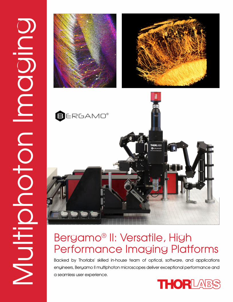
Backed by Thorlabs' skilled in-house team of optical, software, and applications
engineers, Bergamo II multiphoton microscopes deliver exceptional performance and
a seamless user experience.
Bergamo® II: Versatile, High Performance Imaging Platforms
Mu
ltip
ho
ton
Ima
gin
g
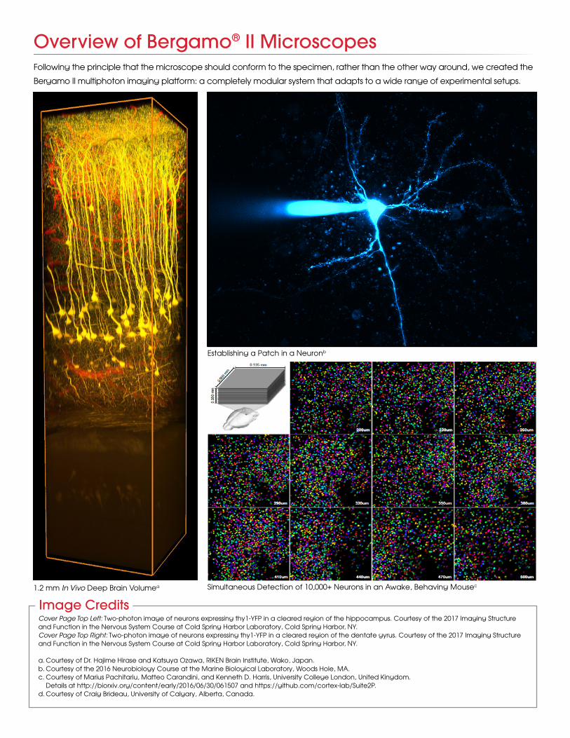
Overview of Bergamo® II Microscopes
Simultaneous Detection of 10,000+ Neurons in an Awake, Behaving Mousec
Following the principle that the microscope should conform to the specimen, rather than the other way around, we created the
Bergamo II multiphoton imaging platform: a completely modular system that adapts to a wide range of experimental setups.
1.2 mm In Vivo Deep Brain Volumea
Establishing a Patch in a Neuronb
Image CreditsCover Page Top Left: Two-photon image of neurons expressing thy1-YFP in a cleared region of the hippocampus. Courtesy of the 2017 Imaging Structure and Function in the Nervous System Course at Cold Spring Harbor Laboratory, Cold Spring Harbor, NY.Cover Page Top Right: Two-photon image of neurons expressing thy1-YFP in a cleared region of the dentate gyrus. Courtesy of the 2017 Imaging Structure and Function in the Nervous System Course at Cold Spring Harbor Laboratory, Cold Spring Harbor, NY.
a. Courtesy of Dr. Hajime Hirase and Katsuya Ozawa, RIKEN Brain Institute, Wako, Japan.b. Courtesy of the 2016 Neurobiology Course at the Marine Biological Laboratory, Woods Hole, MA.c. Courtesy of Marius Pachitariu, Matteo Carandini, and Kenneth D. Harris, University College London, United Kingdom. Details at http://biorxiv.org/content/early/2016/06/30/061507 and https://github.com/cortex-lab/Suite2P.d. Courtesy of Craig Brideau, University of Calgary, Alberta, Canada.
Applicationsu Structural Neurobiology
u Neurological Disorders
u Neural Development and Plasticity
u Neurogenetics
u Functional and Molecular Imaging
u Synapses and Circuits
u Ion Channels, Transporters, and Neurotransmitter
Reporters
u Cell Biology of Neurons, Muscles, and Glia
u Drug Discovery
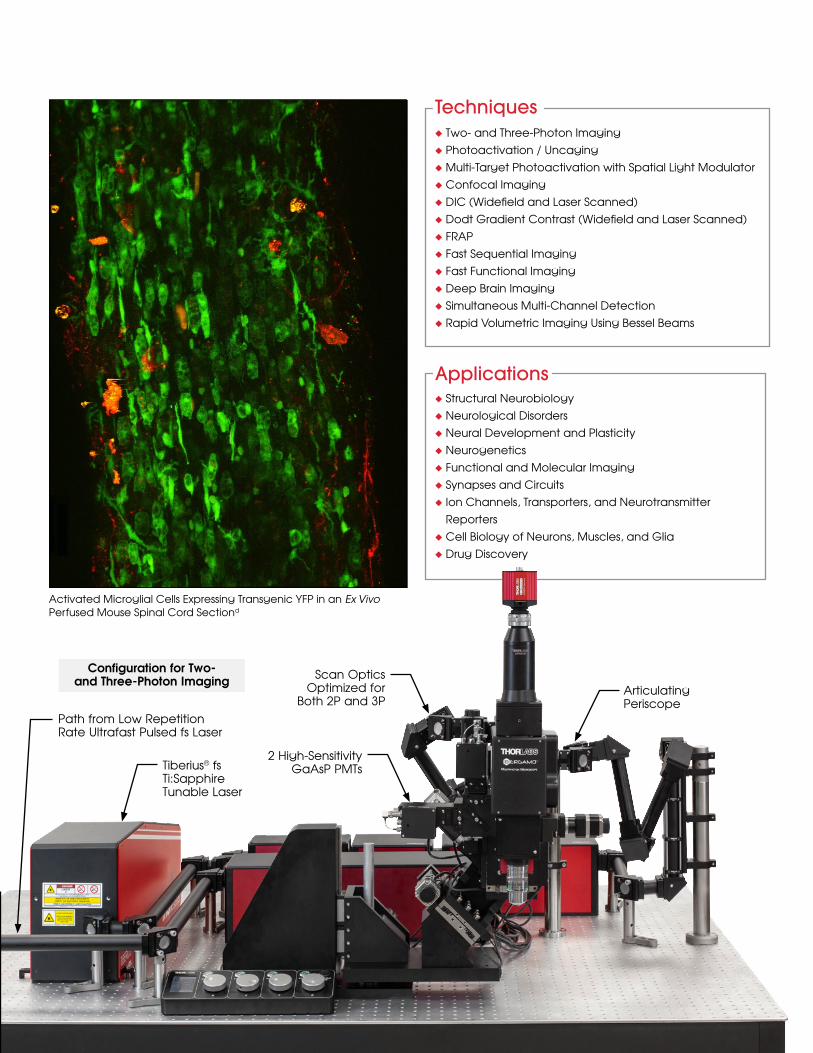
Tiberius® fs Ti:SapphireTunable Laser
Path from Low RepetitionRate Ultrafast Pulsed fs Laser
2 High-Sensitivity GaAsP PMTs
Scan Optics Optimized for
Both 2P and 3PArticulating Periscope
Configuration for Two- and Three-Photon Imaging
Activated Microglial Cells Expressing Transgenic YFP in an Ex Vivo Perfused Mouse Spinal Cord Sectiond
Techniquesu Two- and Three-Photon Imaging
u Photoactivation / Uncaging
u Multi-Target Photoactivation with Spatial Light Modulator
u Confocal Imaging
u DIC (Widefield and Laser Scanned)
u Dodt Gradient Contrast (Widefield and Laser Scanned)
u FRAP
u Fast Sequential Imaging
u Fast Functional Imaging
u Deep Brain Imaging
u Simultaneous Multi-Channel Detection
u Rapid Volumetric Imaging Using Bessel Beams
Other Highlighted Features & UpgradesOur Bergamo® II microscopes are modular systems that can be customized in the design process to meet the exact
needs of the experiment. The highlighted features below reflect our focus on developing cutting-edge capabilities
without compromising usability.
Three-Photon ImagingWe have developed scan path optics for the 800 - 1800 nm range in response to requests
from our collaborators in the field. Extending deeper into the infrared than the existing
choices, these scan optics open the door to three-photon techniques, which provide
reduced background scatter for greater sensitivity in deep tissue imaging.
This wavelength range joins 450 - 1100 nm and 680 - 1300 nm as one of the three super
broadband options available for newly ordered Bergamo II microscopes.
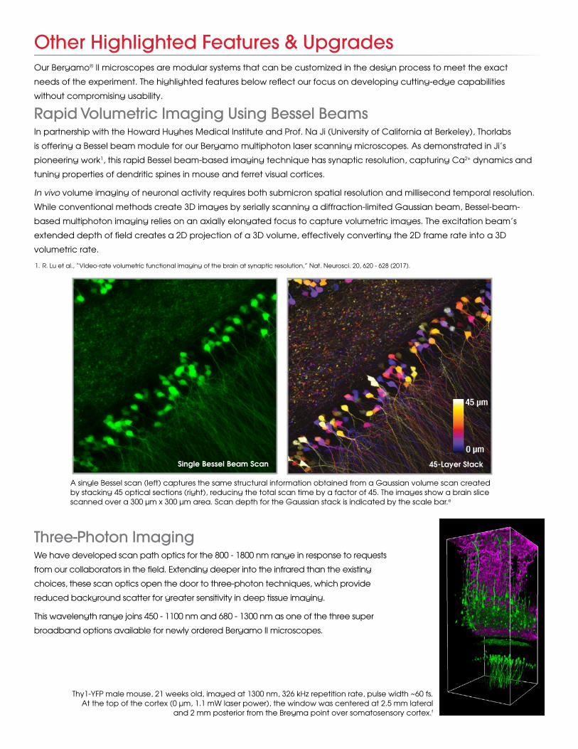
Rapid Volumetric Imaging Using Bessel BeamsIn partnership with the Howard Hughes Medical Institute and Prof. Na Ji (University of California at Berkeley), Thorlabs
is offering a Bessel beam module for our Bergamo multiphoton laser scanning microscopes. As demonstrated in Ji’s
pioneering work1, this rapid Bessel beam-based imaging technique has synaptic resolution, capturing Ca2+ dynamics and
tuning properties of dendritic spines in mouse and ferret visual cortices.
In vivo volume imaging of neuronal activity requires both submicron spatial resolution and millisecond temporal resolution.
While conventional methods create 3D images by serially scanning a diffraction-limited Gaussian beam, Bessel-beam-
based multiphoton imaging relies on an axially elongated focus to capture volumetric images. The excitation beam’s
extended depth of field creates a 2D projection of a 3D volume, effectively converting the 2D frame rate into a 3D
volumetric rate.
A single Bessel scan (left) captures the same structural information obtained from a Gaussian volume scan created by stacking 45 optical sections (right), reducing the total scan time by a factor of 45. The images show a brain slice scanned over a 300 μm x 300 μm area. Scan depth for the Gaussian stack is indicated by the scale bar.e
Single Bessel Beam Scan 45-Layer Stack
1. R. Lu et al., “Video-rate volumetric functional imaging of the brain at synaptic resolution,” Nat. Neurosci. 20, 620 - 628 (2017).
Thy1-YFP male mouse, 21 weeks old, imaged at 1300 nm, 326 kHz repetition rate, pulse width ~60 fs. At the top of the cortex (0 μm, 1.1 mW laser power), the window was centered at 2.5 mm lateral
and 2 mm posterior from the Bregma point over somatosensory cortex.f
Spatial Light Modulator for Simultaneous Multi-Site Activation
Additional Unintentionally
Activated Neurons
Excitation Beam
Excitation Beam
TargetNeuron
Target Neuron
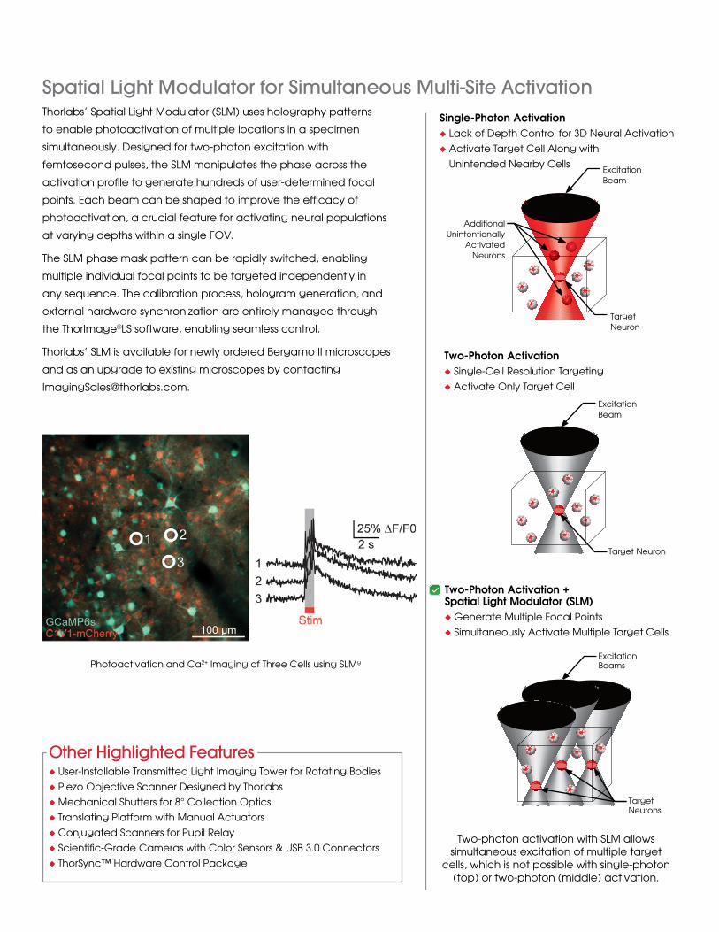
Two-photon activation with SLM allows simultaneous excitation of multiple target
cells, which is not possible with single-photon (top) or two-photon (middle) activation.
Excitation Beams
Target Neurons
u User-Installable Transmitted Light Imaging Tower for Rotating Bodies
u Piezo Objective Scanner Designed by Thorlabs
u Mechanical Shutters for 8° Collection Optics
u Translating Platform with Manual Actuators
u Conjugated Scanners for Pupil Relay
u Scientific-Grade Cameras with Color Sensors & USB 3.0 Connectors
u ThorSync™ Hardware Control Package
Single-Photon Activationu Lack of Depth Control for 3D Neural Activation
u Activate Target Cell Along with
Unintended Nearby Cells
Two-Photon Activationu Single-Cell Resolution Targeting
u Activate Only Target Cell
Two-Photon Activation + Spatial Light Modulator (SLM)u Generate Multiple Focal Points
u Simultaneously Activate Multiple Target Cells
Other Highlighted Features
Photoactivation and Ca2+ Imaging of Three Cells using SLMg
Thorlabs’ Spatial Light Modulator (SLM) uses holography patterns
to enable photoactivation of multiple locations in a specimen
simultaneously. Designed for two-photon excitation with
femtosecond pulses, the SLM manipulates the phase across the
activation profile to generate hundreds of user-determined focal
points. Each beam can be shaped to improve the efficacy of
photoactivation, a crucial feature for activating neural populations
at varying depths within a single FOV.
The SLM phase mask pattern can be rapidly switched, enabling
multiple individual focal points to be targeted independently in
any sequence. The calibration process, hologram generation, and
external hardware synchronization are entirely managed through
the ThorImage®LS software, enabling seamless control.
Thorlabs’ SLM is available for newly ordered Bergamo II microscopes
and as an upgrade to existing microscopes by contacting
✓
Two Microscope Body Choices
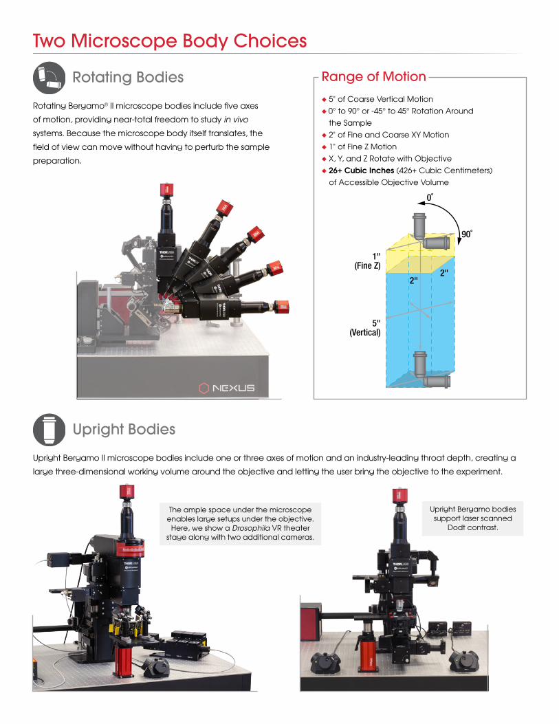
Rotating Bodies
Upright Bodies
Rotating Bergamo® II microscope bodies include five axes
of motion, providing near-total freedom to study in vivo
systems. Because the microscope body itself translates, the
field of view can move without having to perturb the sample
preparation.
Upright Bergamo II microscope bodies include one or three axes of motion and an industry-leading throat depth, creating a
large three-dimensional working volume around the objective and letting the user bring the objective to the experiment.
Range of Motion
u 5" of Coarse Vertical Motion
u 0° to 90° or -45° to 45° Rotation Around
the Sample
u 2" of Fine and Coarse XY Motion
u 1" of Fine Z Motion
u X, Y, and Z Rotate with Objective
u 26+ Cubic Inches (426+ Cubic Centimeters)
of Accessible Objective Volume
0˚
5"(Vertical)
1"(Fine Z)
2"2"
90˚
The ample space under the microscope enables large setups under the objective.
Here, we show a Drosophila VR theater stage along with two additional cameras.
Upright Bergamo bodies support laser scanned
Dodt contrast.
Versatile Photodetection Systems
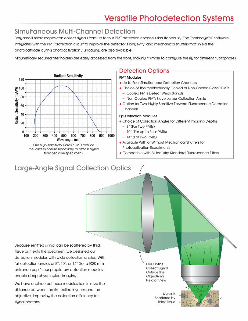
Bergamo II microscopes can collect signals from up to four PMT detection channels simultaneously. The ThorImage®LS software
integrates with the PMT protection circuit to improve the detector’s longevity, and mechanical shutters that shield the
photocathode during photoactivation / uncaging are also available.
Magnetically secured filter holders are easily accessed from the front, making it simple to configure the rig for different fluorophores.
Detection OptionsPMT Modulesu Up to Four Simultaneous Detection Channels
u Choice of Thermoelectrically Cooled or Non-Cooled GaAsP PMTs
– Cooled PMTs Detect Weak Signals
– Non-Cooled PMTs have Larger Collection Angle
u Option for Two Highly Sensitive Forward Fluorescence Detection
Channels
Epi-Detection Modulesu Choice of Collection Angles for Different Imaging Depths
– 8° (For Two PMTs)
– 10° (For up to Four PMTs)
– 14° (For Two PMTs)
u Available With or Without Mechanical Shutters for
Photoactivation Experiments
u Compatible with All Industry-Standard Fluorescence Filters
Radiant Sensitivity
100 200 300 400 500 600 700 800 900 1000
120
100
80
60
40
20
0
Wavelength (nm)
Radi
ant S
ensi
tivity
(mA/
W)
Our high-sensitivity GaAsP PMTs reduce Our high-sensitivity GaAsP PMTs reduce the laser exposure necessary to obtain signal the laser exposure necessary to obtain signal
from sensitive specimens.from sensitive specimens.
Simultaneous Multi-Channel Detection
Large-Angle Signal Collection Optics
Our Optics Collect Signal Outside the Objective’s Field of View
Signal is Scattered by
Thick Tissue
Because emitted signal can be scattered by thick
tissue as it exits the specimen, we designed our
detection modules with wide collection angles. With
full collection angles of 8°, 10°, or 14° (for a Ø20 mm
entrance pupil), our proprietary detection modules
enable deep physiological imaging.
We have engineered these modules to minimize the
distance between the first collecting lens and the
objective, improving the collection efficiency for
signal photons.
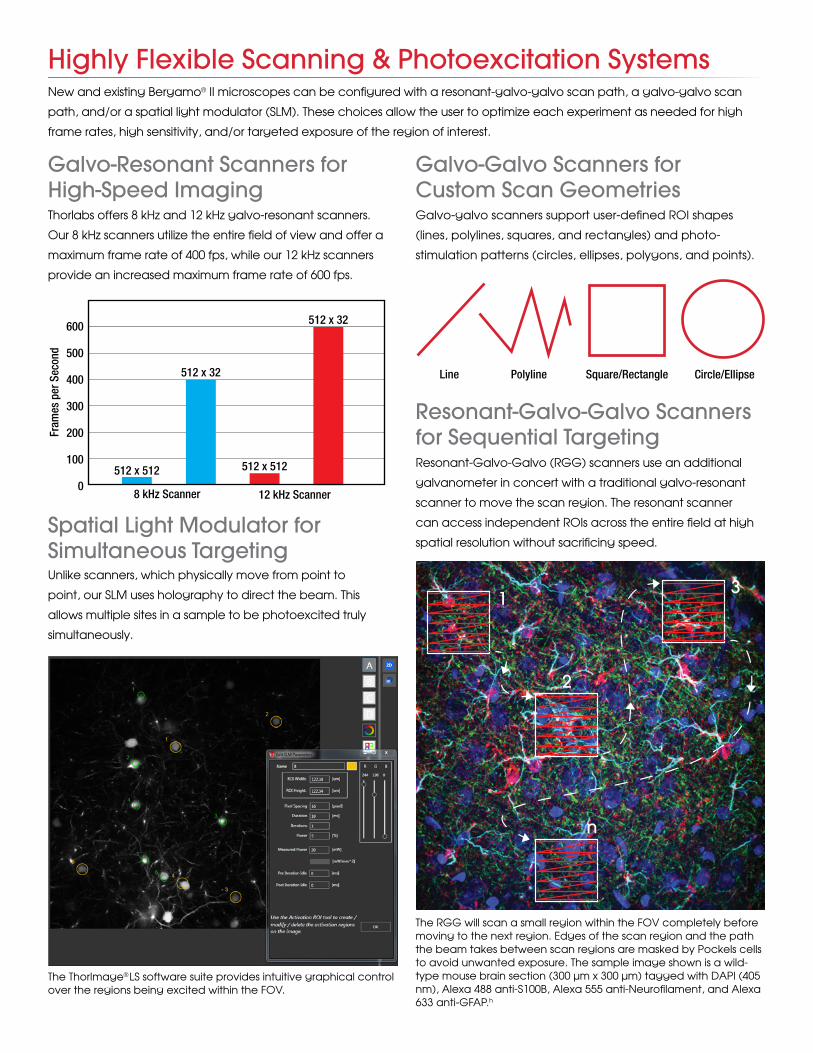
Highly Flexible Scanning & Photoexcitation Systems
Galvo-Resonant Scanners for High-Speed Imaging
Spatial Light Modulator for Simultaneous Targeting
Resonant-Galvo-Galvo Scanners for Sequential TargetingResonant-Galvo-Galvo (RGG) scanners use an additional
galvanometer in concert with a traditional galvo-resonant
scanner to move the scan region. The resonant scanner
can access independent ROIs across the entire field at high
spatial resolution without sacrificing speed.
Galvo-Galvo Scanners for Custom Scan Geometries
Thorlabs offers 8 kHz and 12 kHz galvo-resonant scanners.
Our 8 kHz scanners utilize the entire field of view and offer a
maximum frame rate of 400 fps, while our 12 kHz scanners
provide an increased maximum frame rate of 600 fps.
Unlike scanners, which physically move from point to
point, our SLM uses holography to direct the beam. This
allows multiple sites in a sample to be photoexcited truly
simultaneously.
Galvo-galvo scanners support user-defined ROI shapes
(lines, polylines, squares, and rectangles) and photo-
stimulation patterns (circles, ellipses, polygons, and points).
The ThorImageThe ThorImage®®LS software suite provides intuitive graphical control LS software suite provides intuitive graphical control over the regions being excited within the FOV.over the regions being excited within the FOV.
Square/Rectangle Circle/Ellipse Line Polyline
600
500
400
300
200
100
0
Fram
es p
er S
econ
d
8 kHz Scanner 12 kHz Scanner
512 x 512
512 x 32
512 x 32
512 x 512
New and existing Bergamo® II microscopes can be configured with a resonant-galvo-galvo scan path, a galvo-galvo scan
path, and/or a spatial light modulator (SLM). These choices allow the user to optimize each experiment as needed for high
frame rates, high sensitivity, and/or targeted exposure of the region of interest.
The RGG will scan a small region within the FOV completely before moving to the next region. Edges of the scan region and the path the beam takes between scan regions are masked by Pockels cells to avoid unwanted exposure. The sample image shown is a wild-type mouse brain section (300 μm x 300 μm) tagged with DAPI (405 nm), Alexa 488 anti-S100B, Alexa 555 anti-Neurofilament, and Alexa 633 anti-GFAP.h
1
2
3
n
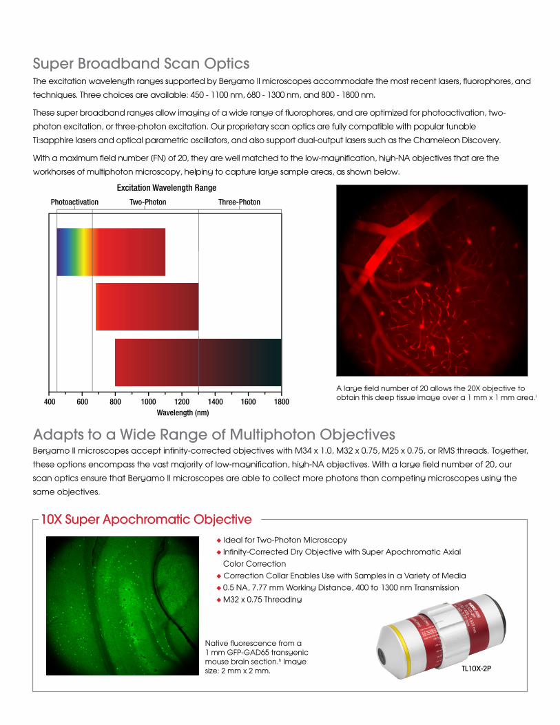
Super Broadband Scan OpticsThe excitation wavelength ranges supported by Bergamo II microscopes accommodate the most recent lasers, fluorophores, and
techniques. Three choices are available: 450 - 1100 nm, 680 - 1300 nm, and 800 - 1800 nm.
These super broadband ranges allow imaging of a wide range of fluorophores, and are optimized for photoactivation, two-
photon excitation, or three-photon excitation. Our proprietary scan optics are fully compatible with popular tunable
Ti:sapphire lasers and optical parametric oscillators, and also support dual-output lasers such as the Chameleon Discovery.
With a maximum field number (FN) of 20, they are well matched to the low-magnification, high-NA objectives that are the
workhorses of multiphoton microscopy, helping to capture large sample areas, as shown below.
A large field number of 20 allows the 20X objective to obtain this deep tissue image over a 1 mm x 1 mm area.i
Excitation Wavelength Range
Wavelength (nm)400 600 800 1000 1200 1400 1600 1800
Photoactivation Two-Photon Three-Photon
Bergamo II microscopes accept infinity-corrected objectives with M34 x 1.0, M32 x 0.75, M25 x 0.75, or RMS threads. Together,
these options encompass the vast majority of low-magnification, high-NA objectives. With a large field number of 20, our
scan optics ensure that Bergamo II microscopes are able to collect more photons than competing microscopes using the
same objectives.
Adapts to a Wide Range of Multiphoton Objectives
u Ideal for Two-Photon Microscopy
u Infinity-Corrected Dry Objective with Super Apochromatic Axial
Color Correction
u Correction Collar Enables Use with Samples in a Variety of Media
u 0.5 NA, 7.77 mm Working Distance, 400 to 1300 nm Transmission
u M32 x 0.75 Threading
10X Super Apochromatic Objective
TL10X-2P
Native fluorescence from a 1 mm GFP-GAD65 transgenic mouse brain section.h Image size: 2 mm x 2 mm.
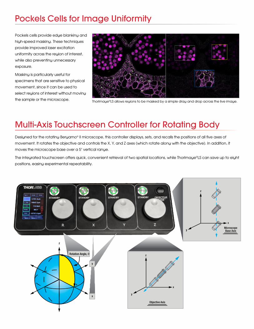
Pockels Cells for Image Uniformity
Designed for the rotating Bergamo® II microscope, this controller displays, sets, and recalls the positions of all five axes of
movement. It rotates the objective and controls the X, Y, and Z axes (which rotate along with the objective). In addition, it
moves the microscope base over a 5" vertical range.
The integrated touchscreen offers quick, convenient retrieval of two spatial locations, while ThorImage®LS can save up to eight
positions, easing experimental repeatability.
Multi-Axis Touchscreen Controller for Rotating Body
Rotation Angle, θ
+
MicroscopeBase Axis
x
y
z
y
z
x
y
z
x
Objective Axis
Pockels cells provide edge blanking and
high-speed masking. These techniques
provide improved laser excitation
uniformity across the region of interest,
while also preventing unnecessary
exposure.
Masking is particularly useful for
specimens that are sensitive to physical
movement, since it can be used to
select regions of interest without moving
the sample or the microscope. ThorImage®LS allows regions to be masked by a simple drag and drop across the live image.
Rotation Angle, θ
+
MicroscopeBase Axis
x
y
z
y
z
x
y
z
x
Objective Axis
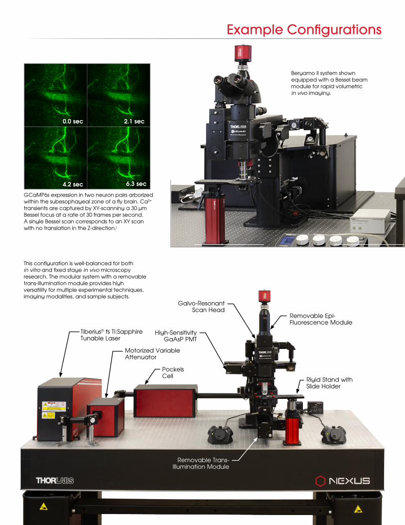
GCaMP6s expression in two neuron pairs arborizedwithin the subesophageal zone of a fly brain. Ca2+
transients are captured by XY-scanning a 30 μmBessel focus at a rate of 30 frames per second.A single Bessel scan corresponds to an XY scanwith no translation in the Z-direction.j
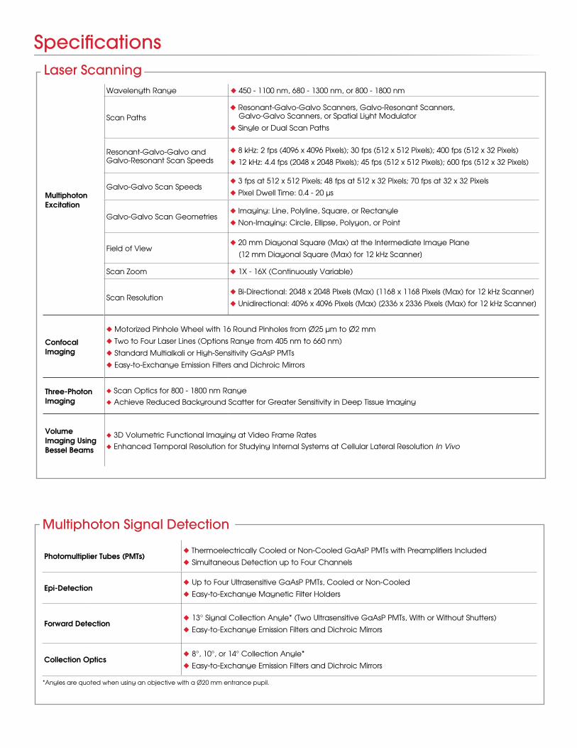
Bergamo II system shown equipped with a Bessel beam module for rapid volumetricin vivo imaging.
FPO0.0 sec
4.2 sec 6.3 sec
2.1 sec
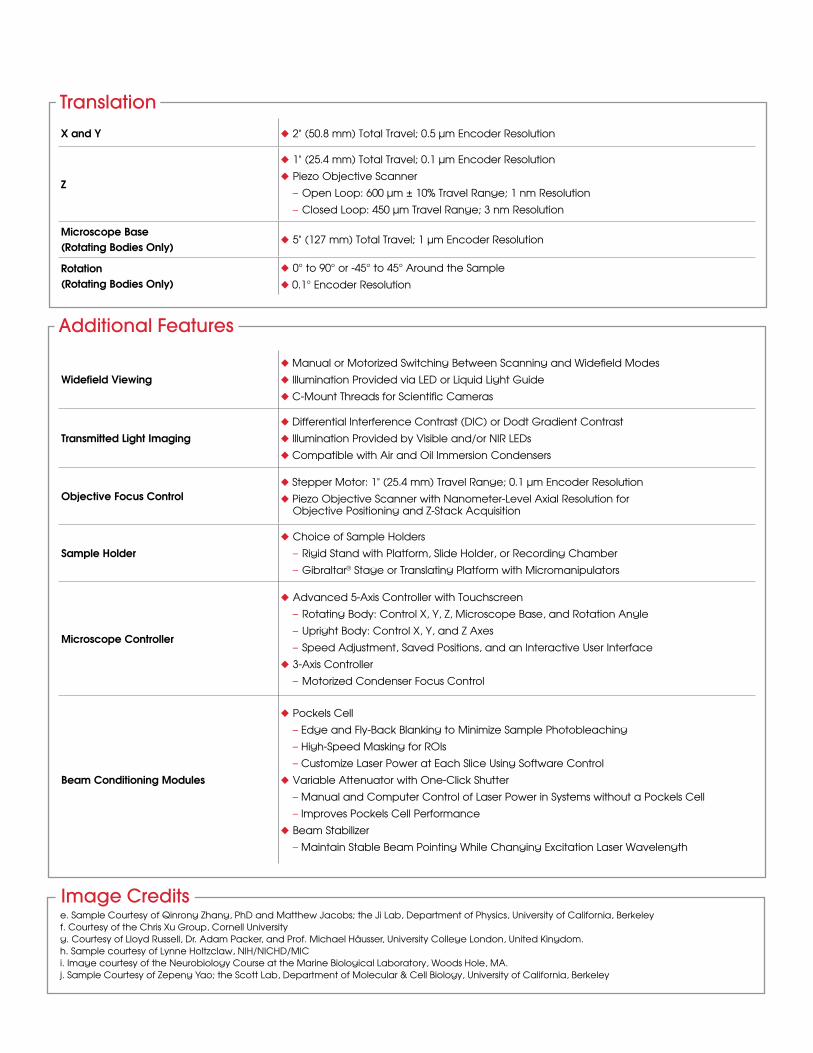
Removable Epi-Fluorescence Module
Galvo-Resonant Scan Head
High-Sensitivity GaAsP PMT
Rigid Stand with Slide Holder
Motorized Variable Attenuator
Pockels Cell
Tiberius® fs Ti:Sapphire Tunable Laser
This configuration is well-balanced for both in vitro and fixed stage in vivo microscopy research. The modular system with a removable trans-illumination module provides high versatility for multiple experimental techniques, imaging modalities, and sample subjects.
Removable Trans-Illumination Module
Example Configurations
Photomultiplier Tubes (PMTs)u Thermoelectrically Cooled or Non-Cooled GaAsP PMTs with Preamplifiers Included
u Simultaneous Detection up to Four Channels
Epi-Detectionu Up to Four Ultrasensitive GaAsP PMTs, Cooled or Non-Cooled
u Easy-to-Exchange Magnetic Filter Holders
Forward Detectionu 13° Signal Collection Angle* (Two Ultrasensitive GaAsP PMTs, With or Without Shutters)
u Easy-to-Exchange Emission Filters and Dichroic Mirrors
Collection Opticsu 8°, 10°, or 14° Collection Angle*
u Easy-to-Exchange Emission Filters and Dichroic Mirrors
Multiphoton Signal Detection
Specifications
*Angles are quoted when using an objective with a Ø20 mm entrance pupil.
Multiphoton Excitation
Wavelength Range u 450 - 1100 nm, 680 - 1300 nm, or 800 - 1800 nm
Scan Pathsu Resonant-Galvo-Galvo Scanners, Galvo-Resonant Scanners, Galvo-Galvo Scanners, or Spatial Light Modulator
u Single or Dual Scan Paths
Resonant-Galvo-Galvo and Galvo-Resonant Scan Speeds
u 8 kHz: 2 fps (4096 x 4096 Pixels); 30 fps (512 x 512 Pixels); 400 fps (512 x 32 Pixels)
u 12 kHz: 4.4 fps (2048 x 2048 Pixels); 45 fps (512 x 512 Pixels); 600 fps (512 x 32 Pixels)
Galvo-Galvo Scan Speedsu 3 fps at 512 x 512 Pixels; 48 fps at 512 x 32 Pixels; 70 fps at 32 x 32 Pixels
u Pixel Dwell Time: 0.4 - 20 μs
Galvo-Galvo Scan Geometriesu Imaging: Line, Polyline, Square, or Rectangle
u Non-Imaging: Circle, Ellipse, Polygon, or Point
Field of Viewu 20 mm Diagonal Square (Max) at the Intermediate Image Plane
[12 mm Diagonal Square (Max) for 12 kHz Scanner]
Scan Zoom u 1X - 16X (Continuously Variable)
Scan Resolutionu Bi-Directional: 2048 x 2048 Pixels (Max) [1168 x 1168 Pixels (Max) for 12 kHz Scanner]
u Unidirectional: 4096 x 4096 Pixels (Max) [2336 x 2336 Pixels (Max) for 12 kHz Scanner]
Confocal Imaging
u Motorized Pinhole Wheel with 16 Round Pinholes from Ø25 μm to Ø2 mm
u Two to Four Laser Lines (Options Range from 405 nm to 660 nm)
u Standard Multialkali or High-Sensitivity GaAsP PMTs
u Easy-to-Exchange Emission Filters and Dichroic Mirrors
Three-Photon Imaging
u Scan Optics for 800 - 1800 nm Rangeu Achieve Reduced Background Scatter for Greater Sensitivity in Deep Tissue Imaging
Volume Imaging Using Bessel Beams
u 3D Volumetric Functional Imaging at Video Frame Ratesu Enhanced Temporal Resolution for Studying Internal Systems at Cellular Lateral Resolution In Vivo
Laser Scanning
Widefield Viewingu Manual or Motorized Switching Between Scanning and Widefield Modes
u Illumination Provided via LED or Liquid Light Guide
u C-Mount Threads for Scientific Cameras
Transmitted Light Imagingu Differential Interference Contrast (DIC) or Dodt Gradient Contrast
u Illumination Provided by Visible and/or NIR LEDs
u Compatible with Air and Oil Immersion Condensers
Objective Focus Controlu Stepper Motor: 1" (25.4 mm) Travel Range; 0.1 μm Encoder Resolution
u Piezo Objective Scanner with Nanometer-Level Axial Resolution for Objective Positioning and Z-Stack Acquisition
Sample Holderu Choice of Sample Holders
– Rigid Stand with Platform, Slide Holder, or Recording Chamber
– Gibraltar® Stage or Translating Platform with Micromanipulators
Microscope Controller
u Advanced 5-Axis Controller with Touchscreen
– Rotating Body: Control X, Y, Z, Microscope Base, and Rotation Angle
– Upright Body: Control X, Y, and Z Axes
– Speed Adjustment, Saved Positions, and an Interactive User Interface
u 3-Axis Controller
– Motorized Condenser Focus Control
Beam Conditioning Modules
u Pockels Cell
– Edge and Fly-Back Blanking to Minimize Sample Photobleaching
– High-Speed Masking for ROIs
– Customize Laser Power at Each Slice Using Software Control
u Variable Attenuator with One-Click Shutter
– Manual and Computer Control of Laser Power in Systems without a Pockels Cell
– Improves Pockels Cell Performance
u Beam Stabilizer
– Maintain Stable Beam Pointing While Changing Excitation Laser Wavelength
Additional Features
X and Y u 2" (50.8 mm) Total Travel; 0.5 μm Encoder Resolution
Z
u 1" (25.4 mm) Total Travel; 0.1 μm Encoder Resolution
u Piezo Objective Scanner
– Open Loop: 600 μm ± 10% Travel Range; 1 nm Resolution
– Closed Loop: 450 μm Travel Range; 3 nm Resolution
Microscope Base(Rotating Bodies Only)
u 5" (127 mm) Total Travel; 1 μm Encoder Resolution
Rotation(Rotating Bodies Only)
u 0° to 90° or -45° to 45° Around the Sample
u 0.1° Encoder Resolution
Translation
Image Creditse. Sample Courtesy of Qinrong Zhang, PhD and Matthew Jacobs; the Ji Lab, Department of Physics, University of California, Berkeley f. Courtesy of the Chris Xu Group, Cornell Universityg. Courtesy of Lloyd Russell, Dr. Adam Packer, and Prof. Michael Häusser, University College London, United Kingdom.h. Sample courtesy of Lynne Holtzclaw, NIH/NICHD/MICi. Image courtesy of the Neurobiology Course at the Marine Biological Laboratory, Woods Hole, MA.j. Sample Courtesy of Zepeng Yao; the Scott Lab, Department of Molecular & Cell Biology, University of California, Berkeley
Recent Publications Using Thorlabs' Imaging Systems
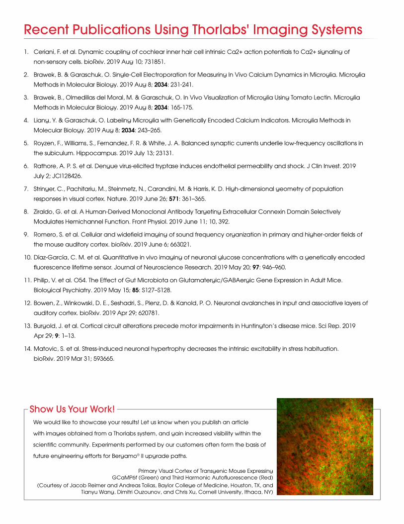
Show Us Your Work!We would like to showcase your results! Let us know when you publish an article
with images obtained from a Thorlabs system, and gain increased visibility within the
scientific community. Experiments performed by our customers often form the basis of
future engineering efforts for Bergamo® II upgrade paths.
1. Ceriani, F. et al. Dynamic coupling of cochlear inner hair cell intrinsic Ca2+ action potentials to Ca2+ signaling of
non-sensory cells. bioRxiv. 2019 Aug 10; 731851.
2. Brawek, B. & Garaschuk, O. Single-Cell Electroporation for Measuring In Vivo Calcium Dynamics in Microglia. Microglia
Methods in Molecular Biology. 2019 Aug 8; 2034: 231-241.
3. Brawek, B., Olmedillas del Moral, M. & Garaschuk, O. In Vivo Visualization of Microglia Using Tomato Lectin. Microglia
Methods in Molecular Biology. 2019 Aug 8; 2034: 165-175.
4. Liang, Y. & Garaschuk, O. Labeling Microglia with Genetically Encoded Calcium Indicators. Microglia Methods in
Molecular Biology. 2019 Aug 8; 2034: 243–265.
5. Royzen, F., Williams, S., Fernandez, F. R. & White, J. A. Balanced synaptic currents underlie low-frequency oscillations in
the subiculum. Hippocampus. 2019 July 13; 23131.
6. Rathore, A. P. S. et al. Dengue virus-elicited tryptase induces endothelial permeability and shock. J Clin Invest. 2019
July 2; JCI128426.
7. Stringer, C., Pachitariu, M., Steinmetz, N., Carandini, M. & Harris, K. D. High-dimensional geometry of population
responses in visual cortex. Nature. 2019 June 26; 571: 361–365.
8. Ziraldo, G. et al. A Human-Derived Monoclonal Antibody Targeting Extracellular Connexin Domain Selectively
Modulates Hemichannel Function. Front Physiol. 2019 June 11; 10, 392.
9. Romero, S. et al. Cellular and widefield imaging of sound frequency organization in primary and higher-order fields of
the mouse auditory cortex. bioRxiv. 2019 June 6; 663021.
10. Díaz-García, C. M. et al. Quantitative in vivo imaging of neuronal glucose concentrations with a genetically encoded
fluorescence lifetime sensor. Journal of Neuroscience Research. 2019 May 20; 97: 946–960.
11. Philip, V. et al. O54. The Effect of Gut Microbiota on Glutamatergic/GABAergic Gene Expression in Adult Mice.
Biological Psychiatry. 2019 May 15; 85: S127–S128.
12. Bowen, Z., Winkowski, D. E., Seshadri, S., Plenz, D. & Kanold, P. O. Neuronal avalanches in input and associative layers of
auditory cortex. bioRxiv. 2019 Apr 29; 620781.
13. Burgold, J. et al. Cortical circuit alterations precede motor impairments in Huntington’s disease mice. Sci Rep. 2019
Apr 29; 9: 1–13.
14. Matovic, S. et al. Stress-induced neuronal hypertrophy decreases the intrinsic excitability in stress habituation.
bioRxiv. 2019 Mar 31; 593665.
Primary Visual Cortex of Transgenic Mouse Expressing GCaMP6f (Green) and Third Harmonic Autofluorescence (Red)
(Courtesy of Jacob Reimer and Andreas Tolias, Baylor College of Medicine, Houston, TX, and Tianyu Wang, Dimitri Ouzounov, and Chris Xu, Cornell University, Ithaca, NY)
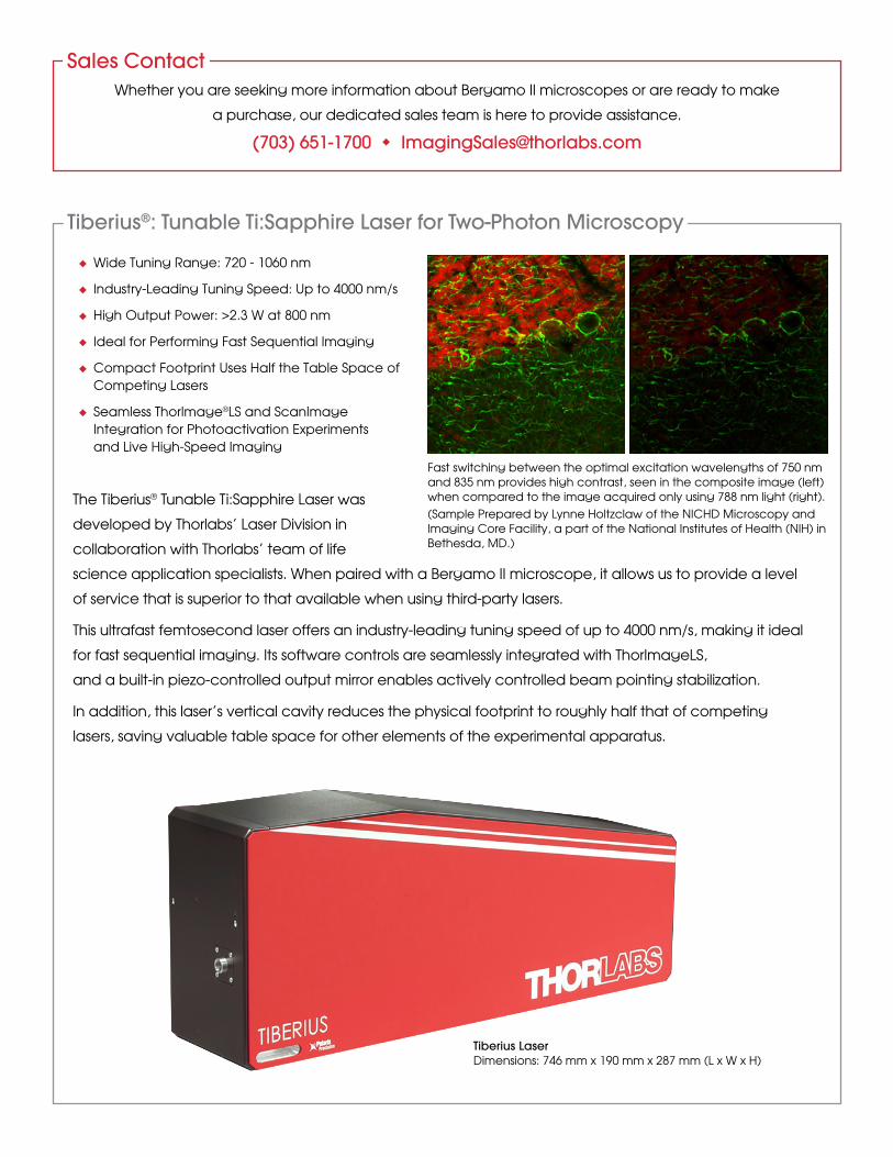
The Tiberius® Tunable Ti:Sapphire Laser was
developed by Thorlabs’ Laser Division in
collaboration with Thorlabs’ team of life
science application specialists. When paired with a Bergamo II microscope, it allows us to provide a level
of service that is superior to that available when using third-party lasers.
This ultrafast femtosecond laser offers an industry-leading tuning speed of up to 4000 nm/s, making it ideal
for fast sequential imaging. Its software controls are seamlessly integrated with ThorImageLS,
and a built-in piezo-controlled output mirror enables actively controlled beam pointing stabilization.
In addition, this laser’s vertical cavity reduces the physical footprint to roughly half that of competing
lasers, saving valuable table space for other elements of the experimental apparatus.
Whether you are seeking more information about Bergamo II microscopes or are ready to make
a purchase, our dedicated sales team is here to provide assistance.
(703) 651-1700 u [email protected]
Sales Contact
Tiberius®: Tunable Ti:Sapphire Laser for Two-Photon Microscopy
Fast switching between the optimal excitation wavelengths of 750 nm and 835 nm provides high contrast, seen in the composite image (left) when compared to the image acquired only using 788 nm light (right).(Sample Prepared by Lynne Holtzclaw of the NICHD Microscopy and Imaging Core Facility, a part of the National Institutes of Health (NIH) in Bethesda, MD.)
Tiberius LaserDimensions: 746 mm x 190 mm x 287 mm (L x W x H)
u Wide Tuning Range: 720 - 1060 nm
u Industry-Leading Tuning Speed: Up to 4000 nm/s
u High Output Power: >2.3 W at 800 nm
u Ideal for Performing Fast Sequential Imaging
u Compact Footprint Uses Half the Table Space of Competing Lasers
u Seamless ThorImage®LS and ScanImage Integration for Photoactivation Experiments and Live High-Speed Imaging

Worldwide Support
56 Sparta Avenue • Newton, New Jersey 07860Sales: 973.300.3000 • Fax: 973.300.3600 • www.thorlabs.com
Thorlabs Ltd.Phone: +44 (0)1353 654440 Fax: +44 (0)1353 654444 Email: [email protected]
Thorlabs CanadaPhone: 1-973-300-3000 Fax: 1-973-300-3600 Email: [email protected]
Thorlabs Vytran DivisionMorganville, New Jersey Phone: 1-973-300-3000 Email: [email protected]
Thorlabs Ultrafast OptoelectronicsAnn Arbor, Michigan Phone: 1-973-300-3000 Email: [email protected]
Thorlabs JapanPhone: +81-3-6915-7701Fax: +81-3-6915-7716Email: [email protected]
Thorlabs Elliptec GmbHPhone: +44 (0)1353 654440 Fax: +44 (0)1353 654444 Email: [email protected]
Thorlabs GmbH / Thorlabs LübeckPhone: +49 (0) 8131 5956-0 Fax: + 49(0) 8131 5956-99Email: [email protected]
Thorlabs Sweden ABPhone: +46 31 733 30 00 Fax: +46 31 703 40 45 Email: [email protected]
Thorlabs Vytran EuropePhone: +44 (0) 1392-445777 Email: [email protected]
Thorlabs China Ltd.Phone: +86 (0)21-60561122 Fax: +86 (0)21-32513480 Email: [email protected]
Thorlabs Ltda, BrazilPhone: +55 (21) 2018 6490 Email: [email protected]
Thorlabs, Inc.Newton, New Jersey Phone: 1-973-300-3000 Fax: 1-973-300-3600 Email: [email protected] www.thorlabs.com
Thorlabs Quantum Electronics (TQE)Jessup, Maryland Catalog Phone: 973-300-3000 OEM Phone: 240-456-7100 Email: [email protected]
Thorlabs Scientific Imaging (TSI)Austin, Texas Phone: 1-973-300-3000 Email: [email protected]
Thorlabs SAS FrancePhone: +33 (0) 970 444 844 Fax: +33 (0) 825 744 800 Email: [email protected]
Thorlabs Imaging Systems Sterling, Virginia Phone: 1-703-651-1700 Email: [email protected]
V21-1


![Biochimica et Biophysica Acta - CORE · fluorescence imaging with pioneering works by Skala et al. [10–12], Gratton et al.[15–20], and others. Multiphoton and FLIM imaging makeit](https://img.pdfslide.us/doc/110x75/5e65076e698709731d2158f6/biochimica-et-biophysica-acta-core-iuorescence-imaging-with-pioneering-works.jpg)


![Multiphoton imaging to identify grana, stroma thylakoid ...cellstudio.org/.../BMCPlant2014_Multiphoton-grana.pdf · and grana in plant [11,16-27]. Inside a chloroplast, both starch](https://img.pdfslide.us/doc/110x75/5fb8149a7316217d4776dc5c/multiphoton-imaging-to-identify-grana-stroma-thylakoid-and-grana-in-plant-1116-27.jpg)