Embed Size (px)
Citation preview

Multiphoton Autofluorescence and Second-HarmonicGeneration Imaging of the Ex Vivo Porcine Eye
Shu-Wen Teng,1 Hsin-Yuan Tan,2,3 Ju-Li Peng,4 Huei-Hsing Lin,4 Ki Hean Kim,5 Wen Lo,1
Yen Sun,1 Wei-Chou Lin,6 Sung-Jan Lin,3,7 Shiou-Hwa Jee,7,8 Peter T. C. So,5 andChen-Yuan Dong1
PURPOSE. The purpose of this work was to demonstrate the useof the combined imaging modality of multiphoton autofluores-cence and second-harmonic generation (SHG) microscopy inobtaining spectrally resolved morphologic features of the cor-nea, limbus, conjunctiva, and sclera in whole, ex vivo porcineeyes.
METHODS. The 780-nm output of a femtosecond, titanium-sap-phire laser was used to induce broadband autofluorescence(435–700 nm) and SHG (390 nm) from various regions of thesurface of ex vivo porcine eyes. A water-immersion objectivewas used for convenient imaging of the curved surface of theeye.
RESULTS. Multiphoton autofluorescence was useful in identify-ing cellular structures of the different domains of the ocularsurface, and the SHG signal can be used to resolve collagenorganization within the cornea stroma and sclera of ex vivoporcine eyes.
CONCLUSIONS. Multiphoton autofluorescence and SHG micros-copy have been demonstrated to be an effective technique forresolving, respectively, the cellular and collagen structureswithin the ocular surface of ex vivo porcine eyes. SHG imagingresolved the difference in structural orientations between cor-neal and sclera collagen fibers. Specifically, the corneal colla-gen is organized in a depth-dependent fashion, whereas thescleral collagen is randomly packed. Because this techniquedoes not require histologic preparation procedures, it has thepotential to be applied for in vivo studies with minimal distur-bance to the eye. (Invest Ophthalmol Vis Sci. 2006;47:1216–1224) DOI:10.1167/iovs.04-1520
The ability to acquire structural information is of key signif-icance in the physiological studies and disease diagnosis of
the eye. Various structures within the ocular surface are ofstructural and functional significance. Morphologically, it isknown that the cornea is covered with epithelium followed bystroma, which is composed mainly of collagen fibers. At theboundary of the cornea and sclera lies the limbus, whichcontains stem cells responsible for maintaining the cornealepithelium. The outer edge of the ocular surface is composedof sclera, which has a composition similar to the cornea in thatit contains collagen fibers, and is covered with conjunctiva.However, unlike corneal collagen fibers, scleral collagen is notwell organized into the striation pattern responsible for thecornea transparency.1 For many ophthalmic applications, theability for physiological studies and disease diagnosis of the eyehinges on the ability to visualize the morphologic features ofthe ocular surface.
As in many areas of biology and medicine, ophthalmologyhas depended heavily on traditional histologic procedures inprobing eye morphology. Although these procedures areinvaluable in providing detailed structural information onthe eye at the tissue and cellular levels, there are severaldrawbacks. First, the morphologic artifacts associated withthe fixation, labeling, and processing procedures used inhistology are always of concern. In addition, histologic pro-cessing represents the end of specimen vitality. Observingphysiological dynamics under in vivo conditions is impossi-ble after fixation of the sample. With advances in opticalimaging technology, the limitations imposed by histologicprocedures are now being overcome. For example, opticalcoherence tomography (OCT), an imaging technique thatdepends on the interference of light reflected from layerswithin the eye has been demonstrated to be useful forophthalmic imaging.2– 4 In addition, confocal microscopyhas been useful for in vivo ophthalmologic observationswith applications in disease diagnosis.5,6
A third promising imaging modality is multiphoton micros-copy. Based on the nonlinear excitation of fluorescent mole-cules or the induction of harmonic generation (HG), multipho-ton microscopy has been widely applied to different areas ofthe life sciences, including neurobiology, development biol-ogy, oncology, transdermal delivery, immunology, hepatology,and deep-tissue imaging.7–20 In addition, NADP(H) autofluores-cence and collagen second-harmonic generation (SHG) havebeen applied to cornea imaging.21,22 However, to the best ofour knowledge, multiphoton imaging has been applied neitherto explore cornea structures at a large geometric scale, nor tothe investigation of other key features of the ocular surface.
Several advantages enable multiphoton microscopy to be apromising imaging modality in ophthalmology. First, the highphoton flux needed to induce nonlinear optical response fromthe specimen limits sample excitation to the focal spot. Be-cause most fluorescence or harmonic generating signals areexcluded from off-focal volume, optical imaging obtained by
From the 1Microscopic Biophysics Laboratory, Department ofPhysics, the 3Institute of Biomedical Engineering, College of Medicineand College of Engineering, and the 4Department of Life Science,National Taiwan University, Taipai, Taiwan; the Departments of 6Pa-thology, and 7Dermatology, National Taiwan University Hospital, Tai-pei Taiwan; the 8Department of Dermatology, College of Medicine,National Taiwan University Hospital, Taipei, Taiwan; the 2Departmentof Ophthalmology, Chang Gung Memorial Hospital, Linko, Taiwan; andthe 5Department of Mechanical Engineering, Massachusetts Institute ofTechnology, Cambridge, Massachusetts.
Supported by Grant NSC 93-3112-B-002-033 from the NationalScience Council, Taiwan.
Submitted for publication December 25, 2004; revised March 16and April 24, 2005; accepted January 11, 2006.
Disclosure: S.-W. Teng, None; H.-Y. Tan, None; J.-L. Peng,None; H.-H. Lin, None; K.H. Kim, None; W. Lo, None; Y. Sun, None;W.-C. Lin, None; S.-J. Lin, None; S.-H. Jee, None; P.T.C. So, None;C.-Y. Dong, None
The publication costs of this article were defrayed in part by pagecharge payment. This article must therefore be marked “advertise-ment” in accordance with 18 U.S.C. §1734 solely to indicate this fact.
Corresponding author: Chen-Yuan Dong, Microscopic BiophysicsLaboratory, Department of Physics, National Taiwan University, Taipei106, Taiwan; [email protected].
Investigative Ophthalmology & Visual Science, March 2006, Vol. 47, No. 31216 Copyright © Association for Research in Vision and Ophthalmology
Downloaded From: http://iovs.arvojournals.org/pdfaccess.ashx?url=/data/journals/iovs/933440/ on 05/23/2018
scanning the pointlike multiphoton volume can generate im-ages with excellent axial depth description and high contrast.Furthermore, the pointlike excitation volume limits specimenphotodamage to the focus. This feature enables sample longev-ity to be achieved. Finally, the near-infrared excitation wave-lengths used in multiphoton microscopy are absorbed andscattered less by the tissues than is the ultraviolet or visibleradiation used in standard single-photon excitation micros-copy. As a result, multiphoton microscopy can be used toachieve in-depth bioimaging without invasive histologic proce-dures.7,8
In ophthalmology, a high-contrast, high-resolution, andlarge-area imaging technique with the potential of in vivoapplications is of immense value in physiological studies anddisease diagnosis. Although multiphoton imaging remains to bewithin the realm of surface tissue imaging, this approach canbe readily applied to the ocular surface for obtaining imageswith subcellular resolution. To begin with, changes to thecorneal structures are important in vision correction proce-dures. In laser in situ keratomileusis (LASIK) or conductivekeratoplasty, the ability to monitor structural change of corneacollagen is vital in assessing the success of these proce-dures.23–26 Although histologic procedures are available tomonitor changes to the cornea, the transparent nature of thecornea prevents its internal structures to be analyzed easily.27
Although x-ray diffraction techniques have added valuable in-formation to the organization of collagen fibers in the cor-nea,28–31 a direct visualization technique to obtain structuralinformation about the cornea in intact eyes would be of im-mense value in clinical evaluation of corneal states, and mul-tiphoton collagen imaging offers such possibilities.20–22 Inaddition, the limbus, the junctional structure separating thetransparent cornea and opaque sclera, is important in main-taining corneal epithelial tissues. Structurally, the limbus con-tains stem cells that differentiate into corneal epithelium. Forophthalmologists, the ability to monitor the physiologicalstates within the limbus without histologic fixation procedureshas potential application in tissue engineering.32,33 Specifi-cally, if multiphoton microscopy can be used to image thelimbal epithelium, the spectral separation of cellular autofluo-rescence from the SHG signal of the surrounding cornea col-lagen fibers will enable high-contrast imaging of the limbus tobe achieved. Finally, because the conjunctiva and sclera are
respectively organized into connective tissues of the outeredges of the eye, one can anticipate multiphoton autofluores-cence and SHG imaging to be useful in spectrally resolving thestructures of the sclera from the cornea. In vivo imaging of thesclera is invaluable in assessing tissue states in clinical settings.For example, thermal treatment of the sclera has been used fortreatment of ocular melanoma.34 However, the ideal scenariowould be the eradication of the tumor mass without damagingthe surrounding connective tissues. Because the SHG signal issensitive to the thermal denaturation of collagen fibers, a po-tential application of SHG imaging is to assess the degree ofthermal damage to the sclera without histologic procedures.35
Finally, the change of autofluorescence in diabetic patients anddiseases such as corneal and conjunctival tumors are potentialimaging targets of multiphoton microscopy.36–38
MATERIALS AND METHODS
Multiphoton Microscopy Instrumentation
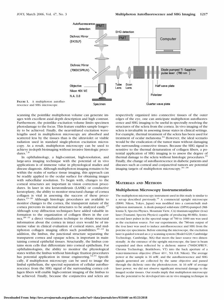
The multiphoton microscopy instrument used in this study is similar toa set-up described previously.15 A commercial upright microscope(E800; Nikon, Tokyo, Japan) was modified into a custom-built mul-tiphoton instrument. A diode-pumped solid-state (DPSS)-pumped (Mil-lennia X; Spectra Physics, Mountain View, CA) titanium-sapphire (ti-sa)laser (Tsunami; Spectra Physics) capable of producing 80-MHz, femto-second laser pulses in the spectral range of 700 to 1000 nm was usedas the excitation source. For our experiments, the 780-nm output ofthe ti-sa laser was used to induce autofluorescence and SHG from theporcine eye specimens. Before entering the microscope, the excitationlaser is guided toward an x–y scanning system (Model 6220; CambridgeTechnology, Cambridge, MA) that steers the laser source two dimen-sionally. At the entrance of the upright microscope, the laser is beamexpanded and then reflected by a dichroic mirror (700DCSPRUV;Chroma Technology, Brattleboro, VT) into the back aperture of awater-immersion objective (Fluor 40�, NA 0.8; Nikon). The averagepower at the sample is 16 mW, and the autofluorescence and SHGsignals generated are collected by the same objective and passedthrough the dichroic onto the photodetectors. At 16 mW of averagelaser power, we did not observe significant structural damage to theimaged ocular tissues. Our results imply that multiphoton microscopyhas the potential to be developed into an in vivo imaging technique in
FIGURE 1. A multiphoton autofluo-rescence and SHG microscope.
IOVS, March 2006, Vol. 47, No. 3 Multiphoton Autofluorescence and SHG Imaging 1217
Downloaded From: http://iovs.arvojournals.org/pdfaccess.ashx?url=/data/journals/iovs/933440/ on 05/23/2018
ophthalmology. Before reaching the detectors, the autofluorescenceand SHG signals are separated by a secondary dichroic mirror(435DCSX; Chroma Technology). The SHG signal centered at 390 nmis reflected by the secondary dichroic and further filtered with aband-pass filter (HQ390/20; Chroma Technology), whereas the longerwavelength autofluorescence passes through the dichroic mirror and abroad band-pass filter (E435LP�E700SP; Chroma Technology) beforebeing detected. Both the autofluorescence and SHG signals are de-tected with single-photon-counting photomultiplier tubes (R7400P;Hamamatsu, Hamamatsu City, Japan). For large-area scans of the eye
specimens, a two-dimensional stage scanning system (H101; PriorScientific Instruments, Cambridge, UK) was used for specimen trans-lation after each x–y scan of the eye sample. The overlapping mul-tiphoton images acquired in this fashion can then be assembled into alarge area, high-resolution tissue map of the eye tissue. An instrumentdiagram of our experimental apparatus is shown in Figure 1.
Acquisition and Processing of Porcine EyesThe porcine eyes used in the study were obtained so that they wouldbe as fresh as possible. The pigs were killed at approximately 4 AM,
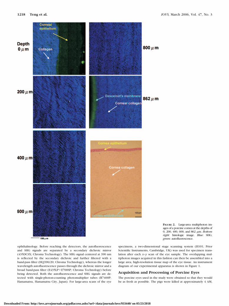
FIGURE 2. Large-area multiphoton im-ages of a porcine cornea at the depths of0, 200, 400, 600, and 862 �m. Bottomright: histologic image. Blue: SHG;green: autofluorescence.
1218 Teng et al. IOVS, March 2006, Vol. 47, No. 3
Downloaded From: http://iovs.arvojournals.org/pdfaccess.ashx?url=/data/journals/iovs/933440/ on 05/23/2018
and the eyes were obtained from the local market at approximately 8AM. Shortly after their acquisition, the porcine eyes were taken to ourlaboratory. With the extraocular tissue removed, the eyes were thenimmersed in PBS buffer. For viewing purposes, we placed the eyespecimen in an empty coverslip container filled with PBS buffer, andwe selected an optically clear porcine eye for multiphoton imaging.The sample was then placed on the microscope stage for viewing. Weselected the main regions of the eye exterior for viewing: cornea,limbus, conjunctiva, and sclera. Both localized three-dimensional im-ages and large area scans at different depths were acquired.
We made attempts to acquire the porcine eyes at the earliestpossible times. However, the specimens acquired 4 hours after deathare not fresh. In an earlier study, it was shown that human donorcorneas stored in MEM at 31°C under tissue bank conditions undergoendothelial damage over a period of weeks with complete necrosisover 6 weeks.39 Nonetheless, in our study, cornea clouding cannot beneglected in all cases, and we selectively chose the porcine eyes withoptically clear corneas for imaging purposes. Because our goal is todemonstrate the capability of multiphoton imaging in acquiring spec-trally resolved morphologic information on selected ocular tissues, webelieve that our approach is acceptable.
After multiphoton imaging was completed, the porcine eyes wereplaced in fixation medium composed of formalin-PBS (1:9 vol/vol). Theeye samples were then dehydrated and embedded in paraffin. Thinsections (approximately 5 �m in thickness) of the cornea, limbus,conjunctiva, and sclera were then prepared and labeled with hema-toxylin and eosin (HE).
RESULTS
Cornea
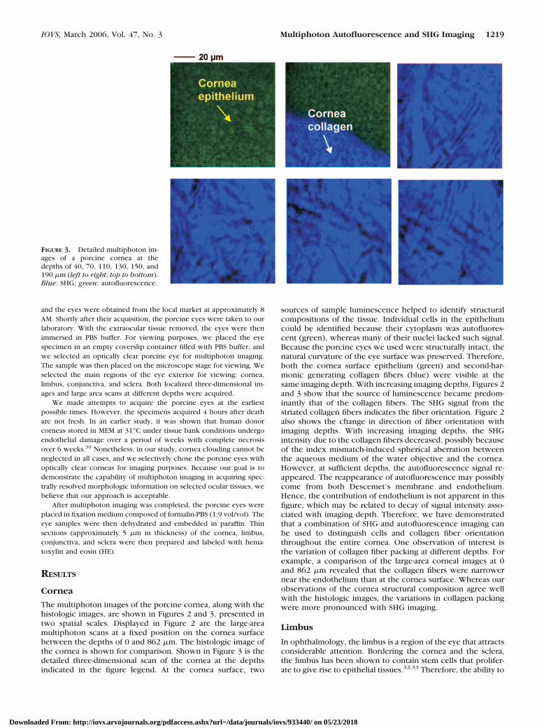
The multiphoton images of the porcine cornea, along with thehistologic images, are shown in Figures 2 and 3, presented intwo spatial scales. Displayed in Figure 2 are the large-areamultiphoton scans at a fixed position on the cornea surfacebetween the depths of 0 and 862 �m. The histologic image ofthe cornea is shown for comparison. Shown in Figure 3 is thedetailed three-dimensional scan of the cornea at the depthsindicated in the figure legend. At the cornea surface, two
sources of sample luminescence helped to identify structuralcompositions of the tissue. Individual cells in the epitheliumcould be identified because their cytoplasm was autofluores-cent (green), whereas many of their nuclei lacked such signal.Because the porcine eyes we used were structurally intact, thenatural curvature of the eye surface was preserved. Therefore,both the cornea surface epithelium (green) and second-har-monic generating collagen fibers (blue) were visible at thesame imaging depth. With increasing imaging depths, Figures 2and 3 show that the source of luminescence became predom-inantly that of the collagen fibers. The SHG signal from thestriated collagen fibers indicates the fiber orientation. Figure 2also shows the change in direction of fiber orientation withimaging depths. With increasing imaging depths, the SHGintensity due to the collagen fibers decreased, possibly becauseof the index mismatch-induced spherical aberration betweenthe aqueous medium of the water objective and the cornea.However, at sufficient depths, the autofluorescence signal re-appeared. The reappearance of autofluorescence may possiblycome from both Descemet’s membrane and endothelium.Hence, the contribution of endothelium is not apparent in thisfigure, which may be related to decay of signal intensity asso-ciated with imaging depth. Therefore, we have demonstratedthat a combination of SHG and autofluorescence imaging canbe used to distinguish cells and collagen fiber orientationthroughout the entire cornea. One observation of interest isthe variation of collagen fiber packing at different depths. Forexample, a comparison of the large-area corneal images at 0and 862 �m revealed that the collagen fibers were narrowernear the endothelium than at the cornea surface. Whereas ourobservations of the cornea structural composition agree wellwith the histologic images, the variations in collagen packingwere more pronounced with SHG imaging.
Limbus
In ophthalmology, the limbus is a region of the eye that attractsconsiderable attention. Bordering the cornea and the sclera,the limbus has been shown to contain stem cells that prolifer-ate to give rise to epithelial tissues.32,33 Therefore, the ability to
FIGURE 3. Detailed multiphoton im-ages of a porcine cornea at thedepths of 40, 70, 110, 130, 150, and190 �m (left to right; top to bottom).Blue: SHG; green: autofluorescence.
IOVS, March 2006, Vol. 47, No. 3 Multiphoton Autofluorescence and SHG Imaging 1219
Downloaded From: http://iovs.arvojournals.org/pdfaccess.ashx?url=/data/journals/iovs/933440/ on 05/23/2018
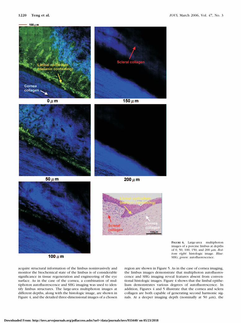
acquire structural information of the limbus noninvasively andmonitor the biochemical state of the limbus is of considerablesignificance in tissue regeneration and engineering of the eyesurface. As in the case of the cornea, a combination of mul-tiphoton autofluorescence and SHG imaging was used to iden-tify limbus structures. The large-area multiphoton images atdifferent depths, along with the histologic image, are shown inFigure 4, and the detailed three-dimensional images of a chosen
region are shown in Figure 5. As in the case of cornea imaging,the limbus images demonstrate that multiphoton autofluores-cence and SHG imaging reveal features absent from conven-tional histologic images. Figure 4 shows that the limbal epithe-lium demonstrates various degrees of autofluorescence. Inaddition, Figures 4 and 5 illustrate that the cornea and scleracollagen are both capable of generating second harmonic sig-nals. At a deeper imaging depth (nominally at 50 �m), the
FIGURE 4. Large-area multiphotonimages of a porcine limbus at depthsof 0, 50, 100, 150, and 200 �m. Bot-tom right: histologic image. Blue:SHG; green: autofluorescence.
1220 Teng et al. IOVS, March 2006, Vol. 47, No. 3
Downloaded From: http://iovs.arvojournals.org/pdfaccess.ashx?url=/data/journals/iovs/933440/ on 05/23/2018
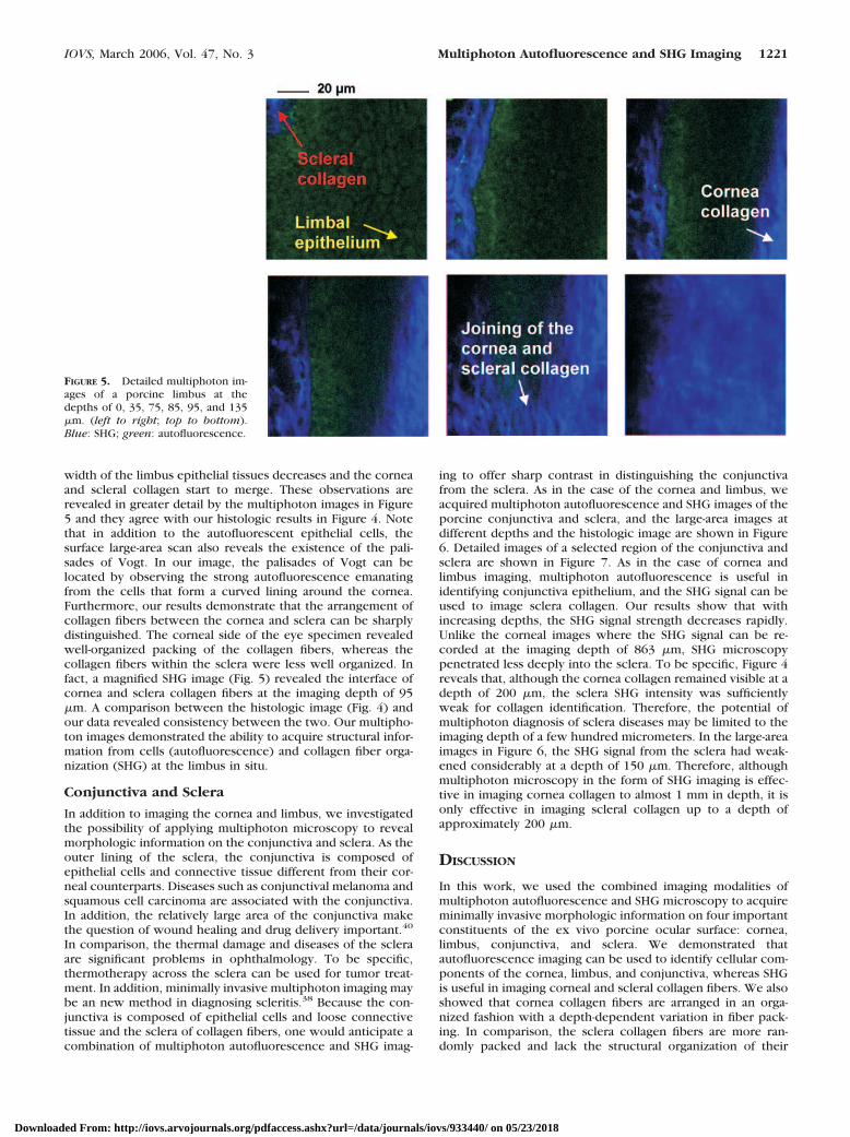
width of the limbus epithelial tissues decreases and the corneaand scleral collagen start to merge. These observations arerevealed in greater detail by the multiphoton images in Figure5 and they agree with our histologic results in Figure 4. Notethat in addition to the autofluorescent epithelial cells, thesurface large-area scan also reveals the existence of the pali-sades of Vogt. In our image, the palisades of Vogt can belocated by observing the strong autofluorescence emanatingfrom the cells that form a curved lining around the cornea.Furthermore, our results demonstrate that the arrangement ofcollagen fibers between the cornea and sclera can be sharplydistinguished. The corneal side of the eye specimen revealedwell-organized packing of the collagen fibers, whereas thecollagen fibers within the sclera were less well organized. Infact, a magnified SHG image (Fig. 5) revealed the interface ofcornea and sclera collagen fibers at the imaging depth of 95�m. A comparison between the histologic image (Fig. 4) andour data revealed consistency between the two. Our multipho-ton images demonstrated the ability to acquire structural infor-mation from cells (autofluorescence) and collagen fiber orga-nization (SHG) at the limbus in situ.
Conjunctiva and Sclera
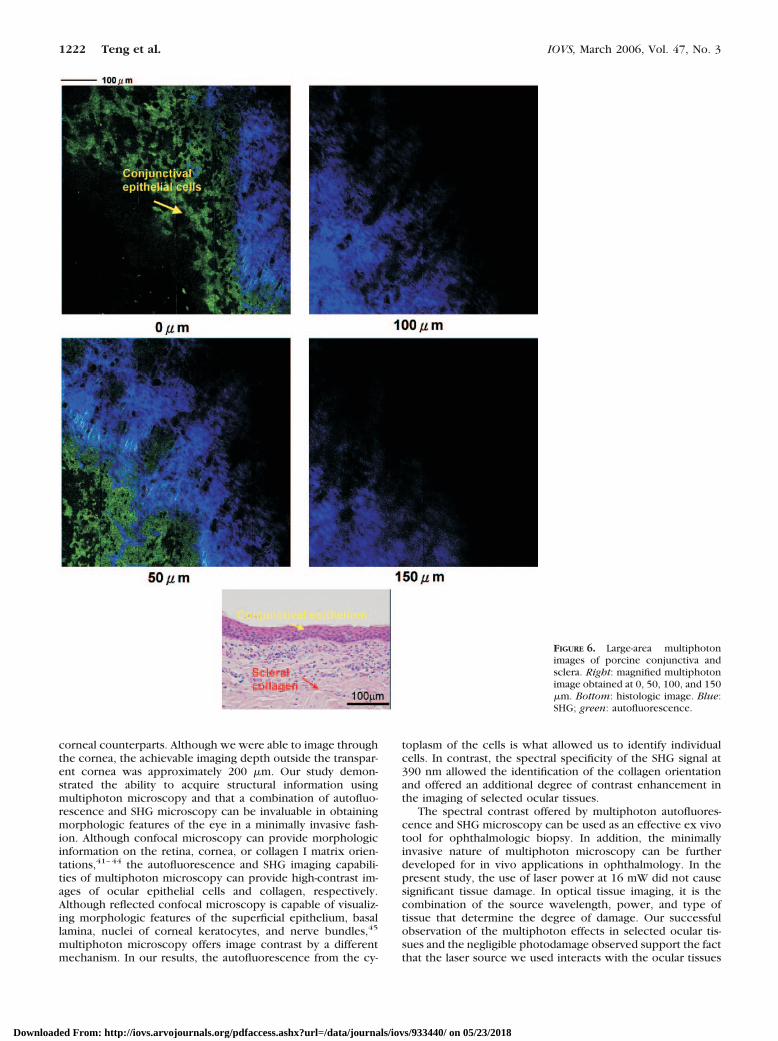
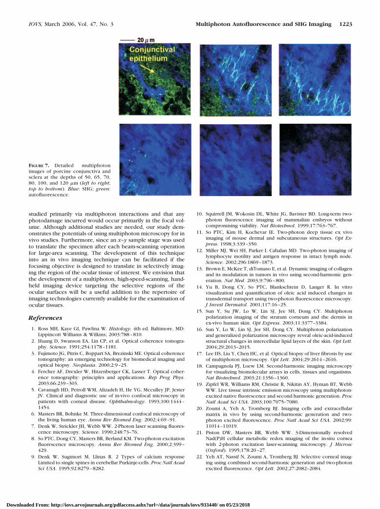
In addition to imaging the cornea and limbus, we investigatedthe possibility of applying multiphoton microscopy to revealmorphologic information on the conjunctiva and sclera. As theouter lining of the sclera, the conjunctiva is composed ofepithelial cells and connective tissue different from their cor-neal counterparts. Diseases such as conjunctival melanoma andsquamous cell carcinoma are associated with the conjunctiva.In addition, the relatively large area of the conjunctiva makethe question of wound healing and drug delivery important.40
In comparison, the thermal damage and diseases of the scleraare significant problems in ophthalmology. To be specific,thermotherapy across the sclera can be used for tumor treat-ment. In addition, minimally invasive multiphoton imaging maybe an new method in diagnosing scleritis.38 Because the con-junctiva is composed of epithelial cells and loose connectivetissue and the sclera of collagen fibers, one would anticipate acombination of multiphoton autofluorescence and SHG imag-
ing to offer sharp contrast in distinguishing the conjunctivafrom the sclera. As in the case of the cornea and limbus, weacquired multiphoton autofluorescence and SHG images of theporcine conjunctiva and sclera, and the large-area images atdifferent depths and the histologic image are shown in Figure6. Detailed images of a selected region of the conjunctiva andsclera are shown in Figure 7. As in the case of cornea andlimbus imaging, multiphoton autofluorescence is useful inidentifying conjunctiva epithelium, and the SHG signal can beused to image sclera collagen. Our results show that withincreasing depths, the SHG signal strength decreases rapidly.Unlike the corneal images where the SHG signal can be re-corded at the imaging depth of 863 �m, SHG microscopypenetrated less deeply into the sclera. To be specific, Figure 4reveals that, although the cornea collagen remained visible at adepth of 200 �m, the sclera SHG intensity was sufficientlyweak for collagen identification. Therefore, the potential ofmultiphoton diagnosis of sclera diseases may be limited to theimaging depth of a few hundred micrometers. In the large-areaimages in Figure 6, the SHG signal from the sclera had weak-ened considerably at a depth of 150 �m. Therefore, althoughmultiphoton microscopy in the form of SHG imaging is effec-tive in imaging cornea collagen to almost 1 mm in depth, it isonly effective in imaging scleral collagen up to a depth ofapproximately 200 �m.
DISCUSSION
In this work, we used the combined imaging modalities ofmultiphoton autofluorescence and SHG microscopy to acquireminimally invasive morphologic information on four importantconstituents of the ex vivo porcine ocular surface: cornea,limbus, conjunctiva, and sclera. We demonstrated thatautofluorescence imaging can be used to identify cellular com-ponents of the cornea, limbus, and conjunctiva, whereas SHGis useful in imaging corneal and scleral collagen fibers. We alsoshowed that cornea collagen fibers are arranged in an orga-nized fashion with a depth-dependent variation in fiber pack-ing. In comparison, the sclera collagen fibers are more ran-domly packed and lack the structural organization of their
FIGURE 5. Detailed multiphoton im-ages of a porcine limbus at thedepths of 0, 35, 75, 85, 95, and 135�m. (left to right; top to bottom).Blue: SHG; green: autofluorescence.
IOVS, March 2006, Vol. 47, No. 3 Multiphoton Autofluorescence and SHG Imaging 1221
Downloaded From: http://iovs.arvojournals.org/pdfaccess.ashx?url=/data/journals/iovs/933440/ on 05/23/2018
corneal counterparts. Although we were able to image throughthe cornea, the achievable imaging depth outside the transpar-ent cornea was approximately 200 �m. Our study demon-strated the ability to acquire structural information usingmultiphoton microscopy and that a combination of autofluo-rescence and SHG microscopy can be invaluable in obtainingmorphologic features of the eye in a minimally invasive fash-ion. Although confocal microscopy can provide morphologicinformation on the retina, cornea, or collagen I matrix orien-tations,41–44 the autofluorescence and SHG imaging capabili-ties of multiphoton microscopy can provide high-contrast im-ages of ocular epithelial cells and collagen, respectively.Although reflected confocal microscopy is capable of visualiz-ing morphologic features of the superficial epithelium, basallamina, nuclei of corneal keratocytes, and nerve bundles,45
multiphoton microscopy offers image contrast by a differentmechanism. In our results, the autofluorescence from the cy-
toplasm of the cells is what allowed us to identify individualcells. In contrast, the spectral specificity of the SHG signal at390 nm allowed the identification of the collagen orientationand offered an additional degree of contrast enhancement inthe imaging of selected ocular tissues.
The spectral contrast offered by multiphoton autofluores-cence and SHG microscopy can be used as an effective ex vivotool for ophthalmologic biopsy. In addition, the minimallyinvasive nature of multiphoton microscopy can be furtherdeveloped for in vivo applications in ophthalmology. In thepresent study, the use of laser power at 16 mW did not causesignificant tissue damage. In optical tissue imaging, it is thecombination of the source wavelength, power, and type oftissue that determine the degree of damage. Our successfulobservation of the multiphoton effects in selected ocular tis-sues and the negligible photodamage observed support the factthat the laser source we used interacts with the ocular tissues
FIGURE 6. Large-area multiphotonimages of porcine conjunctiva andsclera. Right: magnified multiphotonimage obtained at 0, 50, 100, and 150�m. Bottom: histologic image. Blue:SHG; green: autofluorescence.
1222 Teng et al. IOVS, March 2006, Vol. 47, No. 3
Downloaded From: http://iovs.arvojournals.org/pdfaccess.ashx?url=/data/journals/iovs/933440/ on 05/23/2018
studied primarily via multiphoton interactions and that anyphotodamage incurred would occur primarily in the focal vol-ume. Although additional studies are needed, our study dem-onstrates the potentials of using multiphoton microscopy for invivo studies. Furthermore, since an x–y sample stage was usedto translate the specimen after each beam-scanning operationfor large-area scanning. The development of this techniqueinto an in vivo imaging technique can be facilitated if thefocusing objective is designed to translate in selectively imag-ing the region of the ocular tissue of interest. We envision thatthe development of a multiphoton, high-speed-scanning, hand-held imaging device targeting the selective regions of theocular surfaces will be a useful addition to the repertoire ofimaging technologies currently available for the examination ofocular tissues.
References
1. Ross MH, Kaye GI, Pawlina W. Histology. 4th ed. Baltimore, MD:Lippincott Williams & Wilkins; 2003:788–810.
2. Huang D, Swanson EA, Lin CP, et al. Optical coherence tomogra-phy. Science. 1991;254:1178–1181.
3. Fujimoto JG, Pitris C, Boppart SA, Brezinski ME. Optical coherencetomography: an emerging technology for biomedical imaging andoptical biopsy. Neoplasia. 2000;2:9–25.
4. Fercher AF, Drexler W, Hitzenberger CK, Lasser T. Optical coher-ence tomography: principles and applications. Rep Prog Phys.2003;66:239–303.
5. Cavanagh HD, Petroll WM, Alizadeh H, He YG, Mcculley JP, JesterJV. Clinical and diagnostic use of in-vivo confocal microscopy inpatients with corneal disease. Ophthalmology. 1993;100:1444–1454.
6. Masters BR, Bohnke M. Three-dimensional confocal microscopy ofthe living human eye. Annu Rev Biomed Eng. 2002;4:69–91.
7. Denk W, Strickler JH, Webb WW. 2-Photon laser scanning fluores-cence microscopy. Science. 1990;248:73–76.
8. So PTC, Dong CY, Masters BR, Berland KM. Two-photon excitationfluorescence microscopy. Annu Rev Biomed Eng. 2000;2:399–429.
9. Denk W, Sugimori M, Llinas R. 2 Types of calcium responseLimited to single spines in cerebellar Purkinje-cells. Proc Natl AcadSci USA. 1995;92:8279–8282.
10. Squirrell JM, Wokosin DL, White JG, Bavister BD. Long-term two-photon fluorescence imaging of mammalian embryos withoutcompromising viability. Nat Biotechnol. 1999;17:763–767.
11. So PTC, Kim H, Kochevar IE. Two-photon deep tissue ex vivoimaging of mouse dermal and subcutaneous structures. Opt Ex-press. 1998;3:339–350.
12. Miller MJ, Wei SH, Parker I, Cahalan MD. Two-photon imaging oflymphocyte motility and antigen response in intact lymph node.Science. 2002;296:1869–1873.
13. Brown E, McKee T, diTomaso E, et al. Dynamic imaging of collagenand its modulation in tumors in vivo using second-harmonic gen-eration. Nat Med. 2003;9:796–800.
14. Yu B, Dong CY, So PTC, Blankschtein D, Langer R. In vitrovisualization and quantification of oleic acid induced changes intransdermal transport using two-photon fluorescence microscopy.J Invest Dermatol. 2001;117:16–25.
15. Sun Y, Su JW, Lo W, Lin SJ, Jee SH, Dong CY. Multiphotonpolarization imaging of the stratum corneum and the dermis inex-vivo human skin. Opt Express. 2003;11:3377–3384.
16. Sun Y, Lo W, Lin SJ, Jee SH, Dong CY. Multiphoton polarizationand generalized polarization microscopy reveal oleic-acid-inducedstructural changes in intercellular lipid layers of the skin. Opt Lett.2004;29:2013–2015.
17. Lee HS, Liu Y, Chen HC, et al. Optical biopsy of liver fibrosis by useof multiphoton microscopy. Opt Lett. 2004;29:2614–2616.
18. Campagnola PJ, Loew LM. Second-harmonic imaging microscopyfor visualizing biomolecular arrays in cells, tissues and organisms.Nat Biotechnol. 2003;21:1356–1360.
19. Zipfel WR, Williams RM, Christie R, Nikitin AY, Hyman BT, WebbWW. Live tissue intrinsic emission microscopy using multiphoton-excited native fluorescence and second harmonic generation. ProcNatl Acad Sci USA. 2003;100:7075–7080.
20. Zoumi A, Yeh A, Tromberg BJ. Imaging cells and extracellularmatrix in vivo by using second-harmonic generation and two-photon excited fluorescence. Proc Natl Acad Sci USA. 2002;99:11014–11019.
21. Piston DW, Masters BR, Webb WW. 3-Dimensionally resolvedNad(P)H cellular metabolic redox imaging of the in-situ corneawith 2-photon excitation laser-scanning microscopy. J Microsc(Oxford). 1995;178:20–27.
22. Yeh AT, Nassif N, Zoumi A, Tromberg BJ. Selective corneal imag-ing using combined second-harmonic generation and two-photonexcited fluorescence. Opt Lett. 2002;27:2082–2084.
FIGURE 7. Detailed multiphotonimages of porcine conjunctiva andsclera at the depths of 50, 65, 70,80, 100, and 120 �m (left to right;top to bottom). Blue: SHG; green:autofluorescence.
IOVS, March 2006, Vol. 47, No. 3 Multiphoton Autofluorescence and SHG Imaging 1223
Downloaded From: http://iovs.arvojournals.org/pdfaccess.ashx?url=/data/journals/iovs/933440/ on 05/23/2018
23. McDonald MB, Hersh PS, Manche EE, Maloney RK, Davidorf J,Sabry M. Conductive keratoplasty for the correction of low tomoderate hyperopia: US clinical trial 1-year results on 355 eyes.Ophthalmology. 2002;109:1978–1989.
24. Brinkmann R, Radt B, Flamm C, Kampmeier J, Koop N, BirngruberR. Influence of temperature and time on thermally induced forcesin corneal collagen and the effect on laser thermokeratoplasty. JCataract Refr Surg. 2000;26:744–754.
25. Koch DD, Kohnen T, Anderson JA, et al. Histologic changes andwound healing response following 10-pulse noncontact holmium:YAG laser thermal keratoplasty. J Refract Surg. 1996;12:623–634.
26. Kohnen T, Husain SE, Koch DD. Corneal topographic changesafter noncontact holmium: YAG laser thermal keratoplasty to cor-rect hyperopia. J Cataract Refract Surg. 1996;22:427–435.
27. AsiyoVogel MN, Brinkmann R, Notbohm H, Eggers R, Lu-batschowski H, Laqua H. Histologic analysis of thermal effects oflaser thermokeratoplasty and corneal ablation using sirius-red po-larization microscopy. J Cataract Refract Surg. 1997;23:515–526.
28. Aghamohammadzadeh H, Newton RH, Meek KM. X-ray scatteringused to map the preferred collagen orientation in the humancornea and limbus. Structure. 2004;12:249–256.
29. Boote C, Dennis S, Meek K. Spatial mapping of collagen fibrilorganisation in primate cornea: an X-ray diffraction investigation. JStruct Biol. 2004;146:359–367.
30. Meek KM, Boote C. The organization of collagen in the cornealstroma. Exp Eye Res. 2004;78:503–512.
31. Muller LJ, Pels E, Schurmans LRHM, Vrensen GFJM. A new three-dimensional model of the organization of proteoglycans and col-lagen fibrils in the human corneal stroma. Exp Eye Res. 2004;78:493–501.
32. Dua HS, Azuara-Blanco A. Limbal stem cells of the corneal epithe-lium. Surv Ophthalmol. 2000;44:415–425.
33. Wolosin JM, Xiong XL, Schutte M, Stegman Z, Tieng A. Stem cellsand differentiation stages in the limbo-corneal epithelium. ProgRetin Eye Res. 2000;19:223–255.
34. Rem AI, Oosterhuis JA, Journee-De Korver HG, Van den Berg TJTP,Keunen JEE. Temperature dependence of thermal damage to thesclera: exploring the heat tolerance of the sclera for transscleralthermotherapy. Exp Eye Res. 2001;72:153–162.
35. Lin SJ, Sun Y, Lo W, Jee SH, Dong CY. Monitoring the thermallyinduced structural transitions of collagen using second harmonicgeneration microscopy Opt Lett. 2005;30:622–624.
36. Van Schaik HJ, Alkemade C, Swart W, Van Best JA Autofluores-cence of the diabetic and healthy human cornea in vivo at differentexcitation wavelengths. Exp Eye Res. 1999;68:1–8.
37. Shields CL, Shields JA. Tumors of the conjunctiva and cornea. SurvOphthalmol. 2004;49:3–24.
38. Watson PG, Young RD. Scleral structure, organisation and disease:a review. Exp Eye Res. 2004;78:609–623.
39. Redbrake C, Salla S, Frantz A. Changes in human donor corneaspreserved for longer than 4 weeks. Cornea. 1998;17:62–65.
40. Tasman W, Jaeger EA. Duane’s Clinical Ophthalmology on CD-ROM 2004. Baltimore: Lippincott Williams & Wilkins; 2004.
41. Guan K, Hudson C, Flanagan JG. Comparison of Heidelberg RetinaTomograph II and retinal thickness analyzer in the assessment ofdiabetic macular edema. Invest Ophthalmol Vis Sci. 2004;45:610–616.
42. Leduc C, Dupas B, Ott-Benoist AC, Baudouin C. Advantages of thein vivo HRT2 corneal confocal microscope for investigation of theocular surface epithelium. J Fr Ophtalmol. 2004;27:978–986.
43. Kovoor TA, Kim AS, McCulley JP, et al. Evaluation of the cornealeffects of topical ophthalmic fluoroquinolones using in vivo con-focal microscopy. Eye Contact Lens. 2004;30:90–94.
44. Petroll WM, Cavanagh HW, Jester JV. Dynamic three-dimensionalvisualization of collagen matrix remodeling and cytoskeletal orga-nization in living corneal fibroblasts. Scanning. 2004;26:1–10.
45. Jester JV, Petroll WM, Cavanagh HD. Corneal stromal woundhealing in refractive surgery: the role of myofibroblasts. Prog RetinEye Res. 1999;18:311–356.
1224 Teng et al. IOVS, March 2006, Vol. 47, No. 3
Downloaded From: http://iovs.arvojournals.org/pdfaccess.ashx?url=/data/journals/iovs/933440/ on 05/23/2018