Embed Size (px)
Citation preview
Journal ofthe Korean Radiologica l Society 1996 : 35(5) : 667- 672
MR Manifestations of Vertebral Artery Injuries in Cervical Spine Trauma'
Jeong SikYu, M.D. , TaeSubChung, M .D. , Young Soo Kim, M.D.2
Yong Eun Cho, M.D .2, Byung Chul Kang , M.D., Dong Ik Kim, M.D .
Purpose: To assess the diagnostic efficacy of magnetic resonance (MR) imaging in the detection of a vertebra I artery i njury occurri ng from major cervical spinetrauma .
Materials and Methods : Conventional MR findings of 63 patients and 63 control subjects were compared to detect a possible change in the vertebral arteries resulted from trauma. Plain films, CT and clinical records were also reviewed to correlatethedegree of cervical spine injurywith vascularchange.
Results: Nine cases of absent flow signals in vessel lumen were observed in eight patients and one was observed in the control group. Patients more frequently demonstrated other abnormalities such as intraluminallinearsignals (n = 3) or focal luminal narrowing (n=9) but there was no statistical significance. There was a close relationship between degree of cord damage and occlusion of the vertebral artery.
Conclusion : Conventional MR imaging is useful in the detection of vertebral arteryocclusion resulting from cervical spinetrauma.
Index Words : Arteries , vertebral Spine, injur ies Spine , MR
Acute fracture-dislocation of the cervical spine has long been known to be an important causative event of vertebral artery injury (1). However , in the early stage of admission , it is not easy to clearly recogn ize this injury by physical examination of an unstable patientwith injuries such as cord contusion and vertebral fracture , with or without head traum a. I n the I ater period of stabil ization , the possibility of vessel injury would not be suggested until a certain sign of severe vertebrobasilar insufficiency developed. Thus , further radiological investigation for possible vessel damage might not be undertaken. Recently , magnetic resonance(MR) imaging has been generally applied to cervical spine trauma patients for a detailed evaluation of spinal cord injuries. Concomitant or incidental soft tissue changes have been found and easily outlined , and suspected butclin-
'DepartmentofD iagnostic Radiology, Yonsei UniversityColl ege 01 Medicine 2Department olNeurosurgery, Yonsei University Coll ege 01 Medicine Received June28, 1996 ; Accepted August28,1996 Address reprint requeststo :Jeong Sik Yu, M.D., Department olRadiology, Yong Dong Severance Hospital, n 146-92, Dokok-Dong, Kangnam-Ku , Seou l, 135-270, Korea Tel. 82-2-3450-3515, Fax. 82-2-562-5472
ically occult vertebral artery changes can be revealed by these MR images(2, 3). In the literature, we were able to find only a few MR imag ing reports concerning this vessel problem (3 -8).
This study was undertaken to assess the efficacy of routine MR imaging in the detection of vertebral artery injuries through a review of MR images in major nonpenetrating cervical spine trauma patients.
MATERIALS and METHODS
From December 1993 to October 1995, MR imaging was used at our institute to examine sixty-three cervical spine trauma patients(with fracture, dislocation or subluxation injury) for in itial diagnosis or follow up evaluation of clinically suggested cervical cord injury. The study population included 41 male and 22 female patients , aged 20 -59 years(mean , 36.7).
In all patients , MR images were routinely obtained on a 1.0T imager(Magnetom 42sp ; Siemens, Erlangen , Germany) from one to 110 days(mean , 27 days) after injury. Neck su rface coils were routinely used. AII MR imaging exam ination was performed with a 128 -256 X
- 667 -
Journal ofthe Korean Radiological Society 1996 ; 35(5 ) : 667- 672
256 matrix and 5-mm section thickness and a 0.2mm intersl ice gap. In all patients , spin echo T1-weighted sagittal and gradient-recalled echo(GRE) sagittal and axial images were obtained. Our current MR imaging protocol incl udes a T1-weighted sequence of 550/15 (repetition time [TR] msec/echo time [TE] msec) , and a gradient echo [GRE] sequence of 510/12 with 50
0
flip angle. MR angiography was not performed initially in all patients. AII studies were assessed by two radiologists for the following diagnostic points of vertebral artery damage : [1] absent or poorly visual ized flow-related signal ; [2] an abruptly narrowed flow signal more than 50% of short diameter with a deformed contour rather than the usual round shape in axial section ; [3] abnormal traversing linear signal in axial views ; [4] abnormal location of vertebral artery flow signal outside the osteofibrous tunnel. Because the GRE sequence employed in this study allows a bright signal for fast flowing blood on axial section , the detailed evaluation of these vessels was limited to just V2 segments in the axial views , with sagiUal views for reference. This was to minimize the possibility of a false-positive interpret-
ation related to inherent artifacts of gradient echo MR imaging including slow flowing blood during the diastolic phase or the angulation of flow direction in V1 or V3 segments. For a positive reading , focal vascular lesions had to be within two vertebral levels of the major extravascular injury site.
For comparison , another 63 MR images ofthe control group (aged 25 -63 years , mean 44.8) of non-traumatic, herniated cervical disk patients were at the same time randomly sampled and reviewed. The results of positive findings and paUern interpretation of vertebral artery injuries were recorded after consensus.
One to fourteen days(mean 5 days) after initial MRI , additional MR angiography(MRA) (n =6) and digital subtraction angiography(DSA) (n=3) were performed on patients with positive MR findings to confirm vessel injury. Two-dimensional time-of-flight(TRITE/ FA=25/9/ 35
0
) MRA studies were obtained on a 1.5T imager (Magnetom VISION ; Siemens, Erlangen , Germany). Initial plain radiographic films(n=63) and computed tomographic images(n =59) were reviewed for the assessment of surrounding bony structures. The medical
Fig. 1. Bilateral vertebral artery occlusion in 45-year-old man with quadriplegia due to a C5-C6 fracture and dislocation injury
a
b c
a. Axial gradient echo MR image(51 0/12/50 0) obtained at C-5 demonstrates the ab
sence offlow-related signal usually expected for vertebral arteries(arrows) b. Right frontal oblique view, two-dimensional time-of-flight MR angiogram reveals nonvisualization of left vertebral' artery of proximal V2 segment ; distal V2 and V3 segments(open arrows) are reconstituted by collaterals from muscular arteries(arrowheads). The proximal V2 segment of right vertebral artery was not visualized also in other projections(not shown). c. Anteroposterior view of right vertebral angiogram definitively shows proximal occlusion of right vertebral artery(arrows) with collateral reconstitution of distal segments supplied by muscular branches of ascending cervical artery(open arrows) d. Axial cranial CT scan shows large infarction(arrows) in left superior cerebellar artery territory suggesting the relation to the vertebrobasilar arterial flow insuf ficiency
d
- 668
Jeong Sik Yu, et al : MR Manifestations ofVertebral Artery Injuries in Cervical Spine Trauma
records of these patients were also reviewed to search for clinical signs including the degree of cord injury. Initial prereduction subluxation or dislocation plain films were available for only 24 patients , however, so quadriplegia or paraplegia were clinically considered to be signs of severe cervical spine trauma. The chi-square test was used to elucidate the relationship between the degree of trauma and the vascular injury.
RESULTS
Twenty-three abnormal findings were identified in the 126 vertebral arteri es of 63 pati ents an d ei 9 ht i n the control group. Two unilaterally tiny vertebral foramina in each group were observed through the subsequent CT reviews but were not included in statistical analysis (Table 1). Nine vertebral arteries of eight patients and one of one in the control group showed absence or poor blood flow on initial reviews of MR images(Fig. 1,
2). Patients more frequently demonstrated other abnormalities such as intralum inallinear signals(n =3) or focalluminal narrowing(n =9) but there was no statistical significance(Fig. 3 , 4). Subsequent MRA and DSA were helpful in confirming the complete occlusion of vertebral arteries in five patients with no flow signals in six vertebral arteries(including one patient with bilateral occlusion) (Fig. 1 , 2). In one patient with absent flow signal in the unilateral vertebral artery , MRA showed an intact flow signal in a 14-day-delayed scan , which suggested a spontaneous resolution of thrombosis during the 14-day interval.
On admission , all patients complained of non-specific or posterior neck pain , and 29 also complained of headache. Sixteen patients experienced loss of consciousness during the acute stage ; in eight of these patients , the vertebral arteries on MR images were abnormal. A brain CT was obtained for evaluation of head injuries in 15 patients(including six who showed abnor-
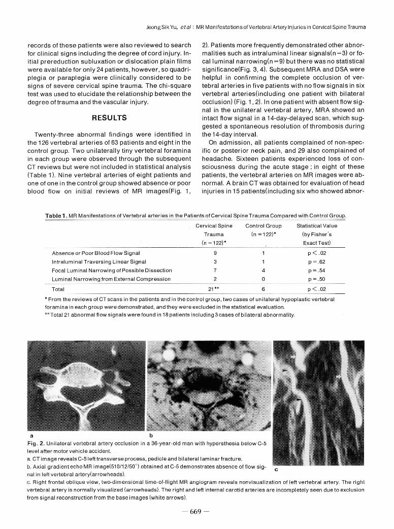
Table 1. MR Manifestations of Vertebral arteries in the Patients of Cervical Spine Trauma Compared with Control Group
Absence or Poor Blood Flow Signal
Intraluminal Traversing Li near Signal
Focal Luminal Narrowing of Possible Dissection
Luminal Narrowing from External Compression
Total
Cervical Spine Control Group
Trauma (n = 122)*
(n = 122)*
9
3
7
2
21 **
4
o 6
Statistical Value
(by Fisher ’s
ExactTest)
p < .02
P =.62
p =.54
P =.50
p < .02
* From the reviews of CT scans in the patients and in the control group, two cases of unilateral hypoplastic vertebral
foramina in each group were demonstrated, and they were excluded in the statistical evaluation **Total 21 abnormal flow signals were found in 18 patients including 3 cases of bilateral abnormality
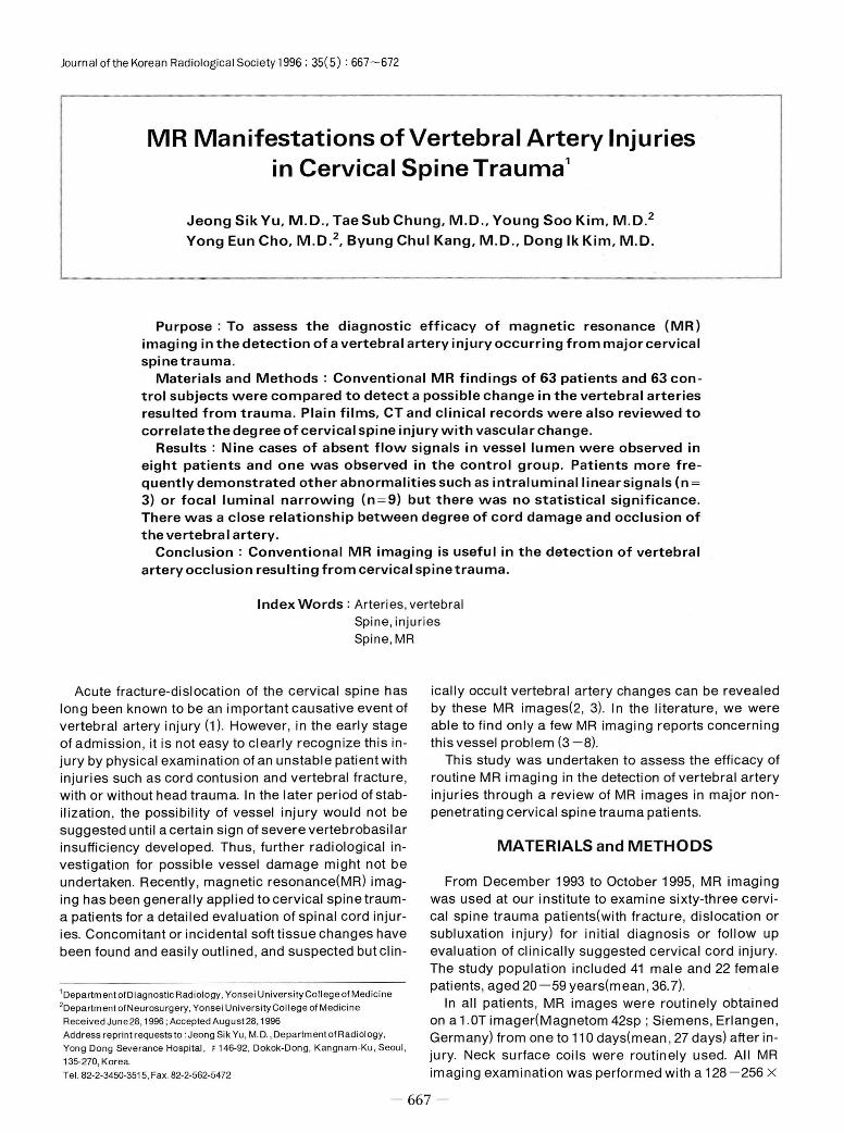
a Fig . 2. Unilateral vertebral artery occlusion in a 36-year-old man with hypersthesia below C-5 level after motor vehicle accident a. CT image reveals C-5left transverse process , pedicle and bilaterallaminar fracture. b. Axial gradient echo MR image(51 0/12/50
0) obtained at C-5 demonstrates absence of flow sig- ~
nal in left vertebral artery(arrowheads) c. Right frontal oblique view, two-dimensional time-of-flight MR angiogram reveals nonvisualization of left vertebral artery. The right vertebral artery is normally visualized (arrowheads). The right and left internal carotid arteries are incompletely seen due to exclusion from signal reconstruction from the base images (white arrows)
669
Journal ofthe Korean Radiological Society 1996 : 35(5) : 667- 672
mal vertebral arteries on MR images) , but ischemic changes in the vertebrobasilar artery territory were visualized in only two patients with occluded vertebral arteries
Compared with seven patients of the 43 with normal vessels(16 %), eight patients of the 18 with abnormal vertebral arteries(42 %) had quadriplegia or paraplegia. These differences were not statistically significant(p= . 06; chi-square test). However, with occluded vertebral artery flow only(n =9 in eight patients) , a significant correl ation was found between the presence of a severe injury(six of eight patients with absent flow signal versus nine of 53 other patients) and the presence of an occlusion(p < .02; chi-square test). The incidence of left vertebral artery involvement(n =14) exceeded that of the ri ght(n = 7) and three cases showed bilateral vessel changes. Only one case showed a total bilateral absence of vertebral arterial flow signal (Fig. 1). We were able to find cranial nerve signs in two patients, and this suggested Walienberg syndrome, resulting from vertebrobasiliar insufficiencies ; we found
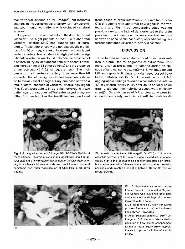
Fig. 3. Axial gradient echo MR image(51 0/12/50 0) 01 a C4-5level
reveals linear, traversing , low signal suggesting intimal Ilap(ar rowhead) in the flow related enhancement olthe left vertebral artery in a 45-year-old man who showed skull Iracture, epidural hematoma and Iracture/dislocation 01 C5-6 Irom a lall-down trauma
a b
three cases of brain infarction in six available brain CTs of patients with abnormal flow signal in the vertebral artery (Fig. 1) , but comparative study was not possible due to the lack of data oriented to the brain problem. In addition , our patients medical records showed no specific clinical history of predisposing factors for spontaneous vertebral artery dissection
DISCUSSION
Due to their unique anatomic location in the osteofibrous tunnel , the V2 segments of extracranial vertebral arteries are subject to damage during an episode of cervical spine trauma(9 -11). MR imaging and MR angiog raphic findings of a damaged vessel have been well-described(5-8). A recent report of MR angiographic findings revealed a considerable rate(24 %) of vertebral artery injury after acute cervical spine trauma, although the majority of cases were clinicaliy silent(3). Only six cases of MR angiography were included in our study , and this is insufficient data for di-
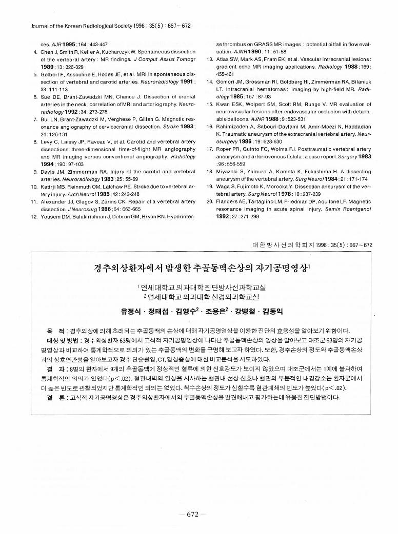
Fig. 4. Axial gradient echo MR image(51 0/12/50 0) at C-3 reveals
eccentric narrowing 01 Ilow related signal by medial inhomogeneously high signal suggesting dissection hematoma or thrombosis(arrowheads) in a 26-year-old man who sustained posterior neck pain and revealed subluxation between C4 and C5 Irom vehicular trauma.
670
Fig. 5. Expelled left vertebral artery Irom an osteolibrous tunnel. A 33-yearold woman who sustained neck pain and numbness in all linger tips lollowing a vehicular trauma a. CT image reveals C-6 left transverse process Iracture(arrow) and subluxation between C-6 and C-7. b. Axial gradient echo(510/12/50 0
) MR image at C-6, demonstrates anterior deviation 01 Ilow related enhancement lor left vertebral artery(arrow) approx-imates just posterior to the left carotid artery

JeongSikYu. etal: MR Manifestations ofVertebral Artery Injuries in Cervical Spine Trauma
rect comparison with that of the previous report. In our study , however , statistically reliable vessel occlusions were found in only 8/61 cases(13 %) , which was a lower proportion than that found in MR angiographic study (7/37 , 19 %) by Friedman et al. (3). The low incidence of vessel occlusion in our study might be partially related to the long time interval(mean 27 days) between MR imaging and the traumatic event, as well as to the potential pitfall of GRE sequence in the differentiation of subacute thrombi from flowing blood(12). In many cases in our study, the observed findings of vascular lesions might be only vestigiallesions. Moreover , the poor signal intensity difference between a thrombus and the inherent magnetization susceptibility effect of the GRE sequence(13 -15) caused difficulty in the interpretation of mild degree narrowing of a vertebral artery and no statistically significant difference between patients and the control group might be resulted(Table 1).
Initial interpretation of MR imaging , which revealed the complete absence of one vertebral artery flow in 11 vertebral arteries in ten patients , led us to consider another possi bi 1 ity : severe hypopl asi a or apl asi a of the unilateral vertebral artery should also be considered in these cases. In CT reviews of both patients and the control group, we thus excluded the cases having definite , small unilateral transverse foramina(Table 1). Six cases of occlusion were verified by subsequent MR angiography and/or DSA studies(Fig. 1, 2)
The development of aneurysms or arteriovenous fistulas from vertebral artery injury. although rare, have been reported with increasing frequency in angiographic studies(16. 17). However we could not find such a case in GRE sequence and that might be partially related to the inherent difficulty in detecting a small abnormal flow signal in the GRE sequences previously discussed.
The visualization in angiography of a double lumen or intimal flap is considered by some to be pathognomonic (18, 19). We have suggested that this might be also applicable to MR images and found cases which on axial image directly among the patients(n =3) and the control group(n=1 ), showed intimal flap and false lumen. These cases had intraluminally traversing , linear signals in continuous multiple axial views with preserved blood flow in the false lumen(Fig. 3). We could not, however , confirm them angiograp
and definitive imaging modality in the diagnosis of these vessel lesions. but it cannot be justified for the screening of asymptomatic patients. In spite of the inherent dephasing and technical problems, MR angiography can be used with a high degree of reliability to show vascular narrowing or occlusion(3 , 8, 20). In statistical analysis comparing patients with the control group, the only reliable finding on axial MR images was absent or poor flow signal in the entire length ofthe vessel(Table 1).
This study has, in fact, severallimitations. A limited number of injuries were confirmed by the generally accepted gold standard of conventional angiography; the scanning time interval were , however, long enough (mean 27 days) for the stabilization of arterial dissection , and we were unable to recommend further practical angiographic confirmation or follow-up study for patients showing subtle changes with no vessel oriented problems. Another limitation is that most ofthe sagittal images of our patients were not helpf비 in the interpretation of vascular problem due to their discrepant plane of section regardless of the course of vertebral arter-ies. Thus , the GRE image of the axial plane was the only available image in the majority of our cases.
In patient’s records we found several signs such as neck pain , headache, transient loss of consciousness and cord injury symptoms but in only two patients did clinical signs suggest a vessel problem. It is possible that the real posterior fossa sign , at least that relating to TIA in the acute stage of trauma, might be overlooked by the first examiner due to the general symptoms of head trauma and later due to restricted clinical expression while the patient is absolutely stabilized in bed. In spite of uncertain vessel related clinical signs , we found that patients with quadriplegia or quadriparesis , indicating more severe trauma to the bony framework , were significantly more likely to have arterial occlusions
In conclusion , on conventional MR imaging of major cervical spine trauma patients , we found a number of cases of damage to vertebral arteries; the incidence of this was significantly related tothe severity ofthe initial trauma. In spite of vague clinical signs of vessel injuries, MR studies (including available MR angiography) must be applied as soon as possible to patients at risk , in order to recognize a possible vertebral artery injury.
REFERENCES
1. Simeone FA, Goldberg m. Thrombosis 01 the vertebral artery Irom hyperextension injuryolthe neck. JNeurosurg 1968; 29: 540-544
2. Mirvis SE , Geisler FH , Jelinek JJ, etal. Acute cervical spinetrauma evaluation with 1.5-T MR imaging. Radiology 1988 ; 166 : 807-816
3. Friedman 0, Flanders A, Thomas C, Millar W. Vertebral artery injury after acute cervical spine trauma : rate 01 occurrence as detected by MR angiography and asessment 01 clinical consequences. AJR 1995; 164 : 443-447
m
Journal ofthe Korean Radiological Society 1996; 35(5) : 667 - 672
ces. AJR 1995 ; 164 : 443-447 se thrombus on GRASS MR images : potential pitfal1 in flow eval-
4. Chen J, Smith R, Keller A, Kucharczyk W. Spontaneous dissection uation. AJNR 1990 ; 11 : 51-58
。f the vertebral artery: MR findings. J Comput Assist Tomogr 13. Atlas SW, Mark AS , Fram EK , et al. Vascular intracraniallesions
1989 ; 13: 326-329 gradient echo MR imaging applications. Radiology 1988 ; 169
5. Gelbert F, Assouline E, Hodes JE, et al. MRI in spontaneous dis- 455-461 sectioll of vertebral and carotid arteries. Neuroradiology 1991 ; 14. Gomori JM , Grossman RI , Goldberg HI , Zimmerman RA, Bilaniuk
33:111-113 LT. Intracranial hematomas: imaging by high-field MR. Radi-
6. Sue DE, Brant-Zawadzki MN , Chance J. Dissection of cranial ology 1985 ; 157 : 87-93
arteries in the neck : correlation ofMRI and arteriography. Neuro- 15. Kwan ESK , Wolpert SM , Scott RM , Runge V. MR evaluation of
radiology 1992 ; 34: 273-278 neurovascular lesions after endovascular occlusion with detach
7. Bui LN, Brant-Zawadzki M, Verghese P, Gillan G. Magnetic res- ablebal1 oons. AJNR 1988 ; 9: 523-531
onance angiography of cervicocranial dissection. Stroke 1993 ; 16. Rahimizadeh A , Sabouri-Daylami M, Amir-Moezi N, Haddadian
24 : 126-131 K. Traumatic aneurysm of the extracranial vertebral artery. Neur-
8. Levy C, Laissy JP , Raveau V, et al. Carotid and vertebral artery osurgery 1986 ; 19: 628-630
dissections: three-dimensional time-of-flight MR angiography 17. Roper PR , Guinto FC , Wolma FJ. Posttraumatic vertebral artery
and MR imaging versus conventional angiography. Radiology aneurysm and arteriovenous fistula : a case repor t. Surgery 1983
1994 ; 190 : 97-103 ; 96 : 556-559 9. Davis JM , Zimmerman RA. Injury of the carotid and vertebral 18. Miyazaki S, Yamura A, Kamata K, Fukushima H. A dissecting
arteries. Neuroradiology 1983 ; 25: 55-69 aneurysm ofthe vertebral artery. Surg Neuro/1984 ; 21 : 171-174
10. Katirji MB , Reinmuth OM, Latchaw RE. Stroke due tovertebral ar- 19. Waga S, Fujimoto K, Morooka Y. Dissection aneurysm of the ver
tery injury. ArchNeuro/1985 ; 42: 242-248 tebral artery. Surg Neuro/1978 ; 10 : 237-239 11. Alexander JJ, Glagov S, Zarins CK. Repair of a vertebral artery 20. Flanders AE , Tartaglino LM , Friedman DP, Aquilone LF. Magnetic
dissection. J Neurosurg 1986 ;64: 663-665 resonance imaging in acute spinal injury. Semin Roentgenol
12. Yousem DM , Balakirishnan J, Debrun GM , Bryan RN. Hyperinten- 1992 ; 27: 271-298
대 한 방 사 선 의 학 회 지 1996 ; 35(5) : 667 - 672
경추외상환자에서 발생한추골동맥손상의 자기공명 영상I
1 연세대학교의과대학진단방사선과학교실
2연세대학교의과대학신경외과학교실
유정식 · 정태섭 · 김영수2 • 조용은2 • 강병철 · 김동의
목 적 : 경추외상에의해초래되는추골동맥의손상에대해자기공명영상을이용한진단의효용성을알아보기위함이다.
대상및방법 : 경추외상환자 63명에서고식적자기공명영상에나타난추골동맥손상의앙상을알아보고대조군63명의자기공
명영상과 비교하여 통계학적으로 의의가 있는 추골동맥의 변화를 규명해 보고자 하였다. 또한, 경추손상의 정도와 추글동맥손상
과의 상호연관성을알아보고자경추단순촬영, CT,임상증상에 대한비교분석을시도하였다.
결 과 8명의 환자에서 9개의 추골동맥에 정상적인 혈류에 의한신호강도가보이지 않았으며 대조군에서는 1예에 불과하여
통계학적인 의의가 있었다(p < .02). 혈관내벽의 열상을 시사하는 혈관내 선상 신호나 혈관의 부분적인 내경감소는 환자군에서
더높은빈도로관찰되었지만통계학적인의의는없었다.척수손상의정도가심할수록혈관폐쇄의빈도가높았다(p < .02).
결 론 : 고식적 자기공명영상은경추외상환자에서의 추골동맥손상을발견해내고평가하는데유용한진단방법이다.
” “