Embed Size (px)
Citation preview

Mosby items and derived items © 2007, 2003 by Mosby, Inc. Slide 1
Chapter 25Chapter 25Anatomy of the Digestive SystemAnatomy of the Digestive System

Mosby items and derived items © 2007, 2003 by Mosby, Inc. Slide 2
Overview of the Digestive System Overview of the Digestive System
Role of the digestive systemRole of the digestive system
Prepares food for absorption and use by all the Prepares food for absorption and use by all the cells of the bodycells of the body
Food material not absorbed becomes feces that is Food material not absorbed becomes feces that is eliminatedeliminated
Digestion depends on both endocrine and Digestion depends on both endocrine and exocrine secretions and the controlled movement exocrine secretions and the controlled movement of ingested food materials through the of ingested food materials through the gastrointestinal (GI) tractgastrointestinal (GI) tract
Mosby items and derived items © 2007, 2003 by Mosby, Inc. Slide 3

Mosby items and derived items © 2007, 2003 by Mosby, Inc. Slide 4
Overview of the Digestive SystemOverview of the Digestive System
Organization of the digestive systemOrganization of the digestive system
Organs of digestionOrgans of digestion
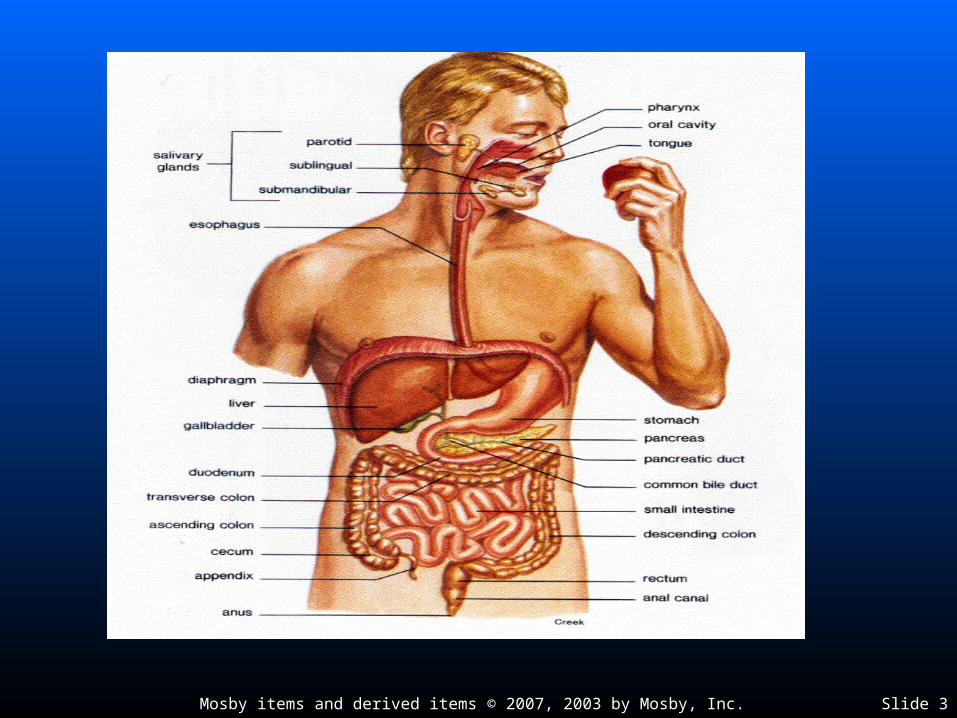
• Main organs of the digestive system form the GI tract that Main organs of the digestive system form the GI tract that extends through the abdominopelvic cavityextends through the abdominopelvic cavity
• Ingested food material passing through the lumen of the GI Ingested food material passing through the lumen of the GI tract is outside the internal environment of the bodytract is outside the internal environment of the body
Wall of the GI tractWall of the GI tract
• Layers—GI tract is made of four layers of tissues: mucosa, Layers—GI tract is made of four layers of tissues: mucosa, submucosa, muscularis, and serosasubmucosa, muscularis, and serosa
• Modifications of layers—layers of the GI tract have various Modifications of layers—layers of the GI tract have various modifications to enable it to perform various functionsmodifications to enable it to perform various functions
Mosby items and derived items © 2007, 2003 by Mosby, Inc. Slide 5
MouthMouth
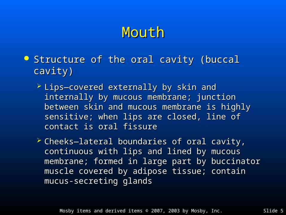
Structure of the oral cavity (buccal cavity)Structure of the oral cavity (buccal cavity)
Lips—covered externally by skin and internally by Lips—covered externally by skin and internally by mucous membrane; junction between skin and mucous membrane; junction between skin and mucous membrane is highly sensitive; when lips mucous membrane is highly sensitive; when lips are closed, line of contact is oral fissureare closed, line of contact is oral fissure
Cheeks—lateral boundaries of oral cavity, Cheeks—lateral boundaries of oral cavity, continuous with lips and lined by mucous continuous with lips and lined by mucous membrane; formed in large part by buccinator membrane; formed in large part by buccinator muscle covered by adipose tissue; contain muscle covered by adipose tissue; contain mucus-secreting glandsmucus-secreting glands

Mosby items and derived items © 2007, 2003 by Mosby, Inc. Slide 6
MouthMouth Structure of the oral cavity (buccal cavity) (cont.)Structure of the oral cavity (buccal cavity) (cont.)
Hard and soft palatesHard and soft palates
• Hard palate consists of portions of four bones: two maxillae and two Hard palate consists of portions of four bones: two maxillae and two palatinespalatines
• Soft palate forms partition between the mouth and the nasopharynx Soft palate forms partition between the mouth and the nasopharynx and is made of muscle arranged in an archand is made of muscle arranged in an arch
• Suspended from midpoint of posterior border of the arch is the uvulaSuspended from midpoint of posterior border of the arch is the uvula
Tongue—solid mass of skeletal muscle covered by a mucous Tongue—solid mass of skeletal muscle covered by a mucous membrane; extremely maneuverable (Figure 25-4)membrane; extremely maneuverable (Figure 25-4)
• Important for mastication and deglutitionImportant for mastication and deglutition
• Has three parts: root, tip, and bodyHas three parts: root, tip, and body
• Papillae located on dorsal surface of tonguePapillae located on dorsal surface of tongue
• Lingual frenulum anchors tongue to floor of mouthLingual frenulum anchors tongue to floor of mouth
Mosby items and derived items © 2007, 2003 by Mosby, Inc. Slide 7
MouthMouth
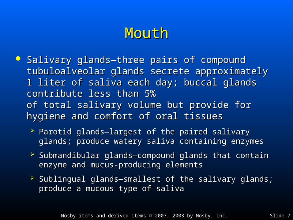
Salivary glands—three pairs of compound tubuloalveolar Salivary glands—three pairs of compound tubuloalveolar glands secrete approximately 1 liter of saliva each day; glands secrete approximately 1 liter of saliva each day; buccal glands contribute less than 5% buccal glands contribute less than 5% of total salivary volume but provide for hygiene and of total salivary volume but provide for hygiene and comfort of oral tissuescomfort of oral tissues
Parotid glands—largest of the paired salivary glands; produce Parotid glands—largest of the paired salivary glands; produce watery saliva containing enzymeswatery saliva containing enzymes
Submandibular glands—compound glands that contain enzyme Submandibular glands—compound glands that contain enzyme and mucus-producing elementsand mucus-producing elements
Sublingual glands—smallest of the salivary glands; produce a Sublingual glands—smallest of the salivary glands; produce a mucous type of salivamucous type of saliva
Mosby items and derived items © 2007, 2003 by Mosby, Inc. Slide 8
MouthMouth
Teeth—organs of masticationTeeth—organs of mastication
Typical toothTypical tooth
• Crown—exposed portion of a tooth, covered by enamel; ideally Crown—exposed portion of a tooth, covered by enamel; ideally suited to withstand abrasion during masticationsuited to withstand abrasion during mastication
• Neck—narrow portion that joins the crown to the root; surrounded Neck—narrow portion that joins the crown to the root; surrounded by the gingivaeby the gingivae
• Root fits into socket of alveolar process and is suspended by Root fits into socket of alveolar process and is suspended by fibrous periodontal membranefibrous periodontal membrane
• Outer shell contains two additional tissues: dentin and cementumOuter shell contains two additional tissues: dentin and cementum
Dentin makes up the greatest portion of the tooth shell; at crown, Dentin makes up the greatest portion of the tooth shell; at crown, covered by enamel, and at neck and root, covered by cementumcovered by enamel, and at neck and root, covered by cementum
Pulp cavity—located in dentin, contains connective tissue, blood, Pulp cavity—located in dentin, contains connective tissue, blood, and lymphatic vessels and sensory nervesand lymphatic vessels and sensory nerves

Mosby items and derived items © 2007, 2003 by Mosby, Inc. Slide 9
Mosby items and derived items © 2007, 2003 by Mosby, Inc. Slide 10
MouthMouth
Teeth (cont.)Teeth (cont.)
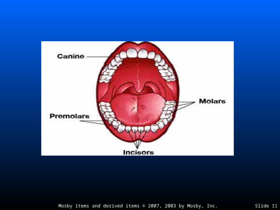
Types of teethTypes of teeth
• Deciduous teeth—20 baby teeth, which appear early in lifeDeciduous teeth—20 baby teeth, which appear early in life
• Permanent teeth—32 teeth, which replace the deciduous teethPermanent teeth—32 teeth, which replace the deciduous teeth
Mosby items and derived items © 2007, 2003 by Mosby, Inc. Slide 11
Mosby items and derived items © 2007, 2003 by Mosby, Inc. Slide 12
PharynxPharynx
Tube through which a bolus passes when Tube through which a bolus passes when moved from the mouth to the esophagus by moved from the mouth to the esophagus by the process of deglutitionthe process of deglutition

Mosby items and derived items © 2007, 2003 by Mosby, Inc. Slide 13
Mosby items and derived items © 2007, 2003 by Mosby, Inc. Slide 14
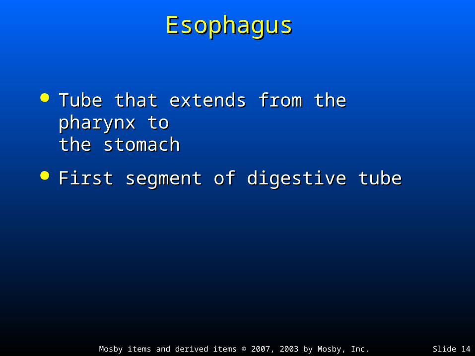
Esophagus Esophagus
Tube that extends from the pharynx to Tube that extends from the pharynx to the stomachthe stomach
First segment of digestive tubeFirst segment of digestive tube

Mosby items and derived items © 2007, 2003 by Mosby, Inc. Slide 15

Mosby items and derived items © 2007, 2003 by Mosby, Inc. Slide 16
StomachStomach
Size and position of the stomachSize and position of the stomach
Size varies according to factors such as gender and amount of Size varies according to factors such as gender and amount of distentiondistention
• When no food is in stomach, it is about the size of a large sausageWhen no food is in stomach, it is about the size of a large sausage
• In adults, capacity ranges from 1.0 to 1.5 litersIn adults, capacity ranges from 1.0 to 1.5 liters
Stomach location: upper part of abdominal cavity under liver and Stomach location: upper part of abdominal cavity under liver and diaphragmdiaphragm
Divisions of the stomachDivisions of the stomach
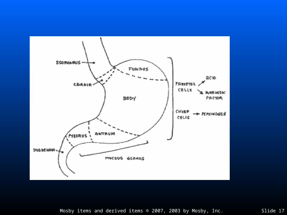
Fundus—enlarged portion to the left and above opening of Fundus—enlarged portion to the left and above opening of esophagus into stomachesophagus into stomach
Body—central portion of stomachBody—central portion of stomach
Pylorus—lower part of stomachPylorus—lower part of stomach
Mosby items and derived items © 2007, 2003 by Mosby, Inc. Slide 17

Mosby items and derived items © 2007, 2003 by Mosby, Inc. Slide 18
StomachStomach
Curves of the stomachCurves of the stomach
Lesser curvature—upper right curve of stomachLesser curvature—upper right curve of stomach
Greater curvature—lower left curve of stomachGreater curvature—lower left curve of stomach
Sphincter muscles—circular fibers arranged so that Sphincter muscles—circular fibers arranged so that there is an opening in the center when relaxed and there is an opening in the center when relaxed and no opening when contractedno opening when contracted
Lower esophageal sphincter (LES) or cardiac sphincter Lower esophageal sphincter (LES) or cardiac sphincter controls opening of esophagus into stomachcontrols opening of esophagus into stomach
Pyloric sphincter controls outlet of pyloric portion of stomach Pyloric sphincter controls outlet of pyloric portion of stomach into duodenuminto duodenum

Mosby items and derived items © 2007, 2003 by Mosby, Inc. Slide 19
StomachStomach Stomach wallStomach wall
Gastric mucosaGastric mucosa• Epithelial lining has rugae marked by gastric pits Epithelial lining has rugae marked by gastric pits
Gastric glands—found below level of the pits; secrete most Gastric glands—found below level of the pits; secrete most of gastric juiceof gastric juice
• Chief cells—secretory cells found in gastric glands; secrete the Chief cells—secretory cells found in gastric glands; secrete the enzymes of gastric juiceenzymes of gastric juice
• Parietal cells—secretory cells found in gastric glands; secrete Parietal cells—secretory cells found in gastric glands; secrete hydrochloric acid; thought to produce intrinsic factor needed for vitamin hydrochloric acid; thought to produce intrinsic factor needed for vitamin B12 absorptionB12 absorption
• Endocrine cells—secrete gastrin and ghrelinEndocrine cells—secrete gastrin and ghrelin Gastric muscularis—thick layer of muscle with three distinct Gastric muscularis—thick layer of muscle with three distinct
sublayers of smooth muscle tissue arranged in a crisscrossing sublayers of smooth muscle tissue arranged in a crisscrossing pattern; this pattern allows stomach to contract strongly at pattern; this pattern allows stomach to contract strongly at many anglesmany angles

Mosby items and derived items © 2007, 2003 by Mosby, Inc. Slide 20
StomachStomach Functions of the stomachFunctions of the stomach
Reservoir for food until it is partially digested and Reservoir for food until it is partially digested and moved further along GI tractmoved further along GI tract
Secretes gastric juice to aid in digestion of foodSecretes gastric juice to aid in digestion of food Breaks food into small particles and mixes them Breaks food into small particles and mixes them
with gastric juicewith gastric juice Secretes intrinsic factorSecretes intrinsic factor Limited absorptionLimited absorption Produces gastrin and ghrelinProduces gastrin and ghrelin Helps protect body from pathogenic bacteria Helps protect body from pathogenic bacteria
swallowed with foodswallowed with food

Mosby items and derived items © 2007, 2003 by Mosby, Inc. Slide 21
Small IntestineSmall Intestine
Size and position of the small intestine—tube Size and position of the small intestine—tube approximately 2.5 cm in diameter and 6 m in approximately 2.5 cm in diameter and 6 m in length; coiled loops fill most of abdominal length; coiled loops fill most of abdominal cavitycavity
Divisions of the small intestineDivisions of the small intestine
Duodenum—uppermost division; approximately Duodenum—uppermost division; approximately 25 cm long, shaped roughly like the letter C25 cm long, shaped roughly like the letter C
Jejunum—approximately 2.5 m longJejunum—approximately 2.5 m long
Ileum—approximately 3.5 m longIleum—approximately 3.5 m long

Mosby items and derived items © 2007, 2003 by Mosby, Inc. Slide 22

Mosby items and derived items © 2007, 2003 by Mosby, Inc. Slide 23
Small IntestineSmall Intestine
Wall of the small intestineWall of the small intestine
Intestinal lining has plicae with villiIntestinal lining has plicae with villi
Villi—important modifications of mucosal layerVilli—important modifications of mucosal layer
• Each villus contains an arteriole, venule, and lactealEach villus contains an arteriole, venule, and lacteal
• Covered by a brush border made up of 1,700 ultrafine Covered by a brush border made up of 1,700 ultrafine microvilli per cellmicrovilli per cell
• Villi and microvilli increase surface area of small intestine Villi and microvilli increase surface area of small intestine hundreds of timeshundreds of times

Mosby items and derived items © 2007, 2003 by Mosby, Inc. Slide 24

Mosby items and derived items © 2007, 2003 by Mosby, Inc. Slide 25
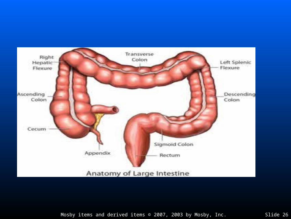
Large IntestineLarge Intestine Size of the large intestine—average diameter, 6 cm; Size of the large intestine—average diameter, 6 cm;
length, approximately 1.5 to 1.8 mlength, approximately 1.5 to 1.8 m Divisions of the large intestineDivisions of the large intestine
Cecum—first 5 to 8 cm of large intestine, blind pouch located in Cecum—first 5 to 8 cm of large intestine, blind pouch located in lower right quadrant of abdomenlower right quadrant of abdomen
ColonColon• Ascending colon—vertical position on right side of abdomen; Ascending colon—vertical position on right side of abdomen;
ileocecal valve prevents material passing from large intestine into ileocecal valve prevents material passing from large intestine into ileumileum
• Transverse colon passes horizontally across abdomen, above small Transverse colon passes horizontally across abdomen, above small intestine; extends from hepatic flexure to splenic flexureintestine; extends from hepatic flexure to splenic flexure
• Descending colon—vertical position on left side of abdomenDescending colon—vertical position on left side of abdomen
• Sigmoid colon joins descending colon to rectumSigmoid colon joins descending colon to rectum
• Rectum—last 7 or 8 inches of intestinal tube; terminal inch is anal Rectum—last 7 or 8 inches of intestinal tube; terminal inch is anal canal with opening called the anus (Figure 25-17)canal with opening called the anus (Figure 25-17)
Mosby items and derived items © 2007, 2003 by Mosby, Inc. Slide 26

Mosby items and derived items © 2007, 2003 by Mosby, Inc. Slide 27
Large IntestineLarge Intestine
Wall of the large intestine (Figure 25-19)Wall of the large intestine (Figure 25-19)
Intestinal mucous glands produce lubricating Intestinal mucous glands produce lubricating mucus that coats feces as they are formedmucus that coats feces as they are formed
Uneven distribution of fibers in the muscle coatUneven distribution of fibers in the muscle coat
Mosby items and derived items © 2007, 2003 by Mosby, Inc. Slide 28
Vermiform Appendix Vermiform Appendix
Accessory organ of digestive system; 8 to 10 Accessory organ of digestive system; 8 to 10 cm in length; communicates with cecumcm in length; communicates with cecum
Mosby items and derived items © 2007, 2003 by Mosby, Inc. Slide 29
Peritoneum Peritoneum
Large, continuous sheet of serous membraneLarge, continuous sheet of serous membrane
Made up of parietal and visceral layersMade up of parietal and visceral layers
Mesentery—projection of parietal peritoneum; Mesentery—projection of parietal peritoneum; allows free movement of each coil of the allows free movement of each coil of the intestine and helps prevent strangulation of intestine and helps prevent strangulation of the long tubethe long tube
Transverse mesocolon—extension of Transverse mesocolon—extension of peritoneum that supports transverse colonperitoneum that supports transverse colon
Mosby items and derived items © 2007, 2003 by Mosby, Inc. Slide 30
LiverLiver
Location and size of the liver—largest gland in body, Location and size of the liver—largest gland in body, weighs approximately 1.5 kg; lies under diaphragm; weighs approximately 1.5 kg; lies under diaphragm; occupies most of right hypochondrium and part of occupies most of right hypochondrium and part of epigastriumepigastrium
Liver lobes and lobules—two lobes separated by Liver lobes and lobules—two lobes separated by falciform ligamentfalciform ligament
Left lobe—forms about one sixth of liverLeft lobe—forms about one sixth of liver
Right lobe—forms about five sixths of liver; divides into right Right lobe—forms about five sixths of liver; divides into right lobe proper, caudate lobe, and quadrate lobelobe proper, caudate lobe, and quadrate lobe
Hepatic lobules—anatomical units of liver; small branch of Hepatic lobules—anatomical units of liver; small branch of hepatic vein extends through the center of each lobulehepatic vein extends through the center of each lobule
Mosby items and derived items © 2007, 2003 by Mosby, Inc. Slide 31
Mosby items and derived items © 2007, 2003 by Mosby, Inc. Slide 32
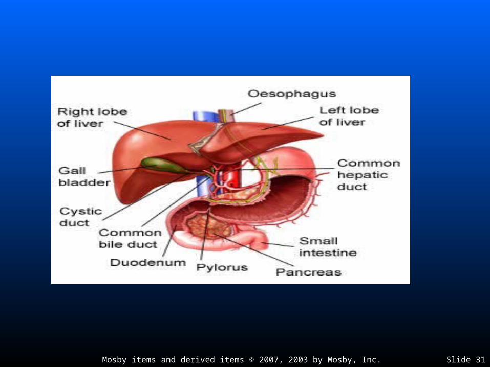
LiverLiver
Bile ductsBile ducts
Small bile ducts form right and left hepatic ductsSmall bile ducts form right and left hepatic ducts
Right and left hepatic ducts immediately join to Right and left hepatic ducts immediately join to form one hepatic ductform one hepatic duct
Hepatic duct merges with cystic duct to form Hepatic duct merges with cystic duct to form common bile duct, which opens into duodenumcommon bile duct, which opens into duodenum

Mosby items and derived items © 2007, 2003 by Mosby, Inc. Slide 33
LiverLiver
Functions of the liverFunctions of the liver
Detoxification by liver cells—ingested toxic substances and Detoxification by liver cells—ingested toxic substances and toxic substances formed in intestines may be changed to toxic substances formed in intestines may be changed to nontoxic substancesnontoxic substances
Bile secretion by liver—bile salts are formed in liver from Bile secretion by liver—bile salts are formed in liver from cholesterol and are the most essential part of bile; liver cells cholesterol and are the most essential part of bile; liver cells secrete approximately 1 pint of bile per daysecrete approximately 1 pint of bile per day
Liver metabolism carries out numerous important steps in the Liver metabolism carries out numerous important steps in the metabolizing of proteins, fats, and carbohydratesmetabolizing of proteins, fats, and carbohydrates
Storage of substances such as iron and some vitaminsStorage of substances such as iron and some vitamins
Production of important plasma proteinsProduction of important plasma proteins

Mosby items and derived items © 2007, 2003 by Mosby, Inc. Slide 34
GallbladderGallbladder
Size and location of the gallbladder—pear-shaped sac Size and location of the gallbladder—pear-shaped sac from 7 to 10 cm long and 3 cm wide at its broadest point; from 7 to 10 cm long and 3 cm wide at its broadest point; holds 30 to 50 ml of bile; lies on undersurface of liverholds 30 to 50 ml of bile; lies on undersurface of liver
Structure of gallbladder—serous, muscular, and mucous Structure of gallbladder—serous, muscular, and mucous layers compose the gallbladder wall; mucosal lining has layers compose the gallbladder wall; mucosal lining has rugaerugae
Functions of gallbladder:Functions of gallbladder:
Storage of bileStorage of bile
Concentration of bile fivefold to tenfoldConcentration of bile fivefold to tenfold
Ejection of the concentrated bile into duodenumEjection of the concentrated bile into duodenum

Mosby items and derived items © 2007, 2003 by Mosby, Inc. Slide 35
Pancreas Pancreas
Size and location of the pancreas—grayish pink–colored Size and location of the pancreas—grayish pink–colored gland; 12 to 15 cm long; weighs approximately 60 g; runs gland; 12 to 15 cm long; weighs approximately 60 g; runs from duodenum and behind stomach to spleenfrom duodenum and behind stomach to spleen
Structure of the pancreas—composed of endocrine and Structure of the pancreas—composed of endocrine and exocrine glandular tissueexocrine glandular tissue
Exocrine portion makes up majority of pancreas; has a Exocrine portion makes up majority of pancreas; has a compound acinar arrangement; tiny ducts unite to form main compound acinar arrangement; tiny ducts unite to form main pancreatic duct, which empties into duodenumpancreatic duct, which empties into duodenum
Endocrine portion—embedded between exocrine units; called Endocrine portion—embedded between exocrine units; called pancreatic islets; constitute only 2% of total mass of pancreas; pancreatic islets; constitute only 2% of total mass of pancreas; made up of alpha cells and beta cells; pass secretions into made up of alpha cells and beta cells; pass secretions into capillariescapillaries
Mosby items and derived items © 2007, 2003 by Mosby, Inc. Slide 36
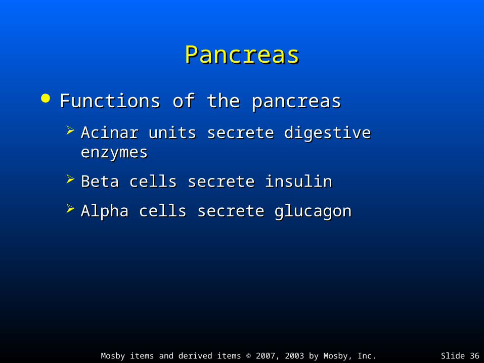
PancreasPancreas
Functions of the pancreasFunctions of the pancreas
Acinar units secrete digestive enzymesAcinar units secrete digestive enzymes
Beta cells secrete insulinBeta cells secrete insulin
Alpha cells secrete glucagonAlpha cells secrete glucagon

Mosby items and derived items © 2007, 2003 by Mosby, Inc. Slide 37
Cycle of Life: Digestive SystemCycle of Life: Digestive System
Changes in digestive function and structure are age-relatedChanges in digestive function and structure are age-related
Result in diseases or pathological conditionsResult in diseases or pathological conditions
May occur in any segment of intestinal tractMay occur in any segment of intestinal tract
Changes involve accessory organs: teeth, salivary glands, liver, Changes involve accessory organs: teeth, salivary glands, liver, gallbladder, and pancreasgallbladder, and pancreas
Infants—immature intestinal mucosa; intact proteins can Infants—immature intestinal mucosa; intact proteins can pass through epithelial cells lining the tract and trigger pass through epithelial cells lining the tract and trigger allergic responseallergic response
Lactose intolerance affects infants who lack the enzyme Lactose intolerance affects infants who lack the enzyme lactaselactase

Mosby items and derived items © 2007, 2003 by Mosby, Inc. Slide 38
Cycle of Life: Digestive SystemCycle of Life: Digestive System
Young age—mumps common in children; Young age—mumps common in children; appendicitis more common in adolescents appendicitis more common in adolescents and then decreases with advancing ageand then decreases with advancing age
Middle age—ulcers and gallbladder disease Middle age—ulcers and gallbladder disease commoncommon
Old age—decreased digestive fluids, slowing Old age—decreased digestive fluids, slowing of peristalsis, and reduced physical activity of peristalsis, and reduced physical activity lead to constipation and diverticulosislead to constipation and diverticulosis