Embed Size (px)
Citation preview
~ 40 ~
Journal of Entomology and Zoology Studies 2013;1 (4): 40-46 ISSN 2320-7078 JEZS 2013;1 (4): 40-46 © 2013 AkiNik Publications Received: 1-07-2013 Accepted: 21-07-2013 Nurcan Özyurt Department of Biology, Faculty of Science, Gazi University, 06500 Teknikokullar, Ankara, Turkey. Selami Candan Department of Biology, Faculty of Science, Gazi University, 06500 Teknikokullar, Ankara, Turkey. Zekiye Suludere Department of Biology, Faculty of Science, Gazi University, 06500 Teknikokullar, Ankara, Turkey. Damla Amutkan Department of Biology, Faculty of Science, Gazi University, 06500 Teknikokullar, Ankara, Turkey. Correspondence: Selami Candan Department of Biology, Faculty of Science, Gazi University, 06500 Teknikokullar, Ankara, Turkey. E-Mail: [email protected]
Morphology and Histology of the Male Reproductive
System in Graphosoma lineatum (Heteroptera: Pentatomidae) Based on Optical and Scanning
Electron Microscopy Nurcan Özyurt, Selami Candan, Zekiye Suludere, Damla Amutkan
ABSTRACT
The male reproductive system of Graphosoma lineatum (Linnaeus 1758) is studied morphologically and histologically using both light and scanning electron microscopes (SEM). On the basis of histological sections and whole morfological studies, the following elements in the structure of the male reproductive system of G. lineatum have been examined: the structure of a pair of testis containing the testes follicles and the stage of spermatogenesis, the connection of testes and vas deferens; the position and the histological structure the of a pair of vas deferens which is the tube down which the sperm travels, the structure of seminal vesicle which is where the sperm is stored prior to mating which supply seminal fluid for additional volume and to nourish the sperm before-during their journey, the bulbus ejaculatorius, a pair of ectodermal sacs the ductus ejaculatorius as well as the structure of aedeagus.
Keywords: Male reproductive system, spermatogonium, spermatozoa, light and scanning electron microscope (SEM).
1. Introduction
Adults and nymphs of Graphosoma lineatum (L.) (Heteroptera: Pentatomidae) shows harmful effection on the plants, which belong to Umbelliferae family, by sticking to absorb the flowers, peduncles and seeds of the plants in their umbrella-shaped flower clusters. They can make significant harms, when they are in high population, in Western Anatolia Region of Turkey, especially in areas anise is grown [19,20]. Despite the importance of Pentatomidae as biological control agents, few reports exist on the internal morphology of the reproductive tract of these organisms, especially with a reference to male reproductive system. The male reproductive morphology of Nezara viridula (Linnaeus, 1758) [26, 28], Perillus bioculatus (Fabricius) [1], Podisus nigrispinus (Dallas) [16,29] has been studied. However, the number of studies on the structural traits of male reproductive system is very limited. Due to lack of morfological and histological studies on the reproductive tract of Pentatomidae, the reproductive morphology and histology of of G. lineatum is investigated in present study. The male reproductive tract of Heteropteran insects typically consists of a pair of testes, a pair of vas deferens, a pair of seminal vesicles, a complex bulbus ejaculatorius, a pair of ectodermal sacs, a ductus ejaculatorius, accessory glands [1, 5, 6, 11, 16, 23, 26]. The main function of the male reproductive system is the production and storage of spermatozoa and their transport in a viable stage state into the reproductive tract of the female. Histologically, the male reproductive system consists of paired testes which each containing a series of testicular tubes or follicles (in which spermatozoa are produced) with different length among genera and species [5, 9, 16, 23]. The male reproductive organs of the Pentatomidae are in general, more complex than those of other families of the Heteroptera. This complexity is apparent in the structure of the bulbus ejaculatorius and in the presence of often elaborate accessory glands in addition to accessory glands [26]. Most insects present a number of accessory glands, which open into the vas deferens or the ductus ejaculatorius [1, 5, 7, 11, 16, 21].
~ 41 ~
Journal of Entomology and Zoology Studies
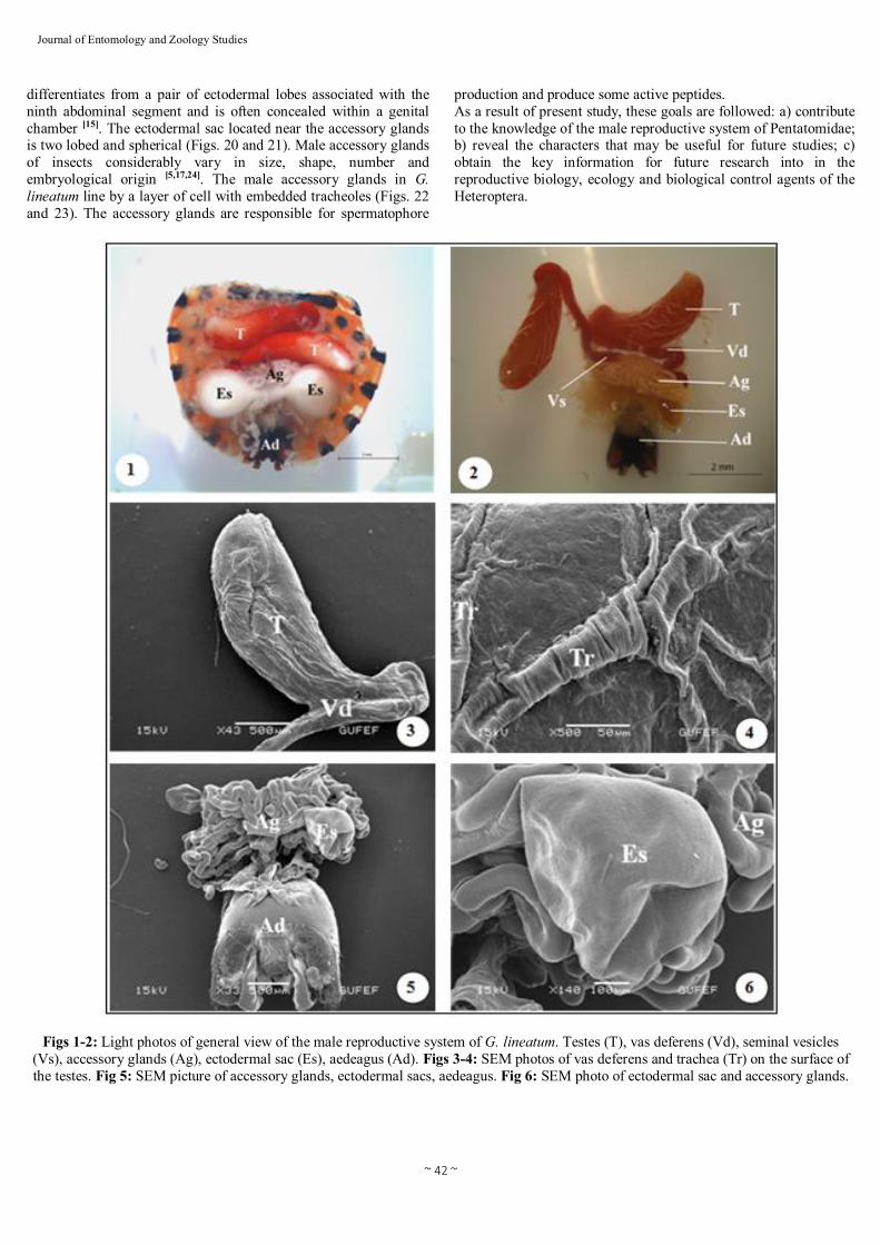
The acccessory glands of male insects can be classified into two types according to their mesodermal or ectodermal derivation. Almost all that have been studied are mesodermal in origin, while little is known about those that have an ectodermal origin. Secretions of the male accessory glands from the spermatophore contribute to the seminal fluid which nourishes the spermatozoa during transport to the female [5, 6, 26]. 2. Materials and methods 2.1. Gross anatomy analysis Adult males of G. lineatum were collected in june-july 2008, from Kırşehir, Turkey. Adult males were killed with ethyl acetate fumes and dissected in 70% ethyl alcohol under a stereomicroscope. For the morphological analysis, the dorsal cuticle was first removed from the prothorax to the last full-sized abdominal segments. Subsequently, the epidermis and the digestive system were removed. Two insect pins were inserted laterally through the last full-sized sternite to spread apart the abdomen and to expose the base of the reproductive system attached to the genital segments. The gross morphology of the reproductive systems of the males were examined and photographed with a Leica EZ4D stereomicroscope. 2.2. Light Microscopy For the histological analysis, the reproductive systems of fifteen males were fixed in bouin’s for 24 h. After that, the tissues were washed, dehyrated in a grade series of ethanol solutions (70%, 80%, 90%, 100%) and finaly embedded in paraffin. Paraffin sections were cut into 6-7 µm thick slides and stained with Hemotoxylin and Eosin for light microscopic examined. The sections were viewed and photographed by using Olympus BX51 microscope. 2.3. Scanning Electron Microscopy For the scanning electron microscopy specimens, cleaned and dried with Critical Point Drying (Polaron CPD 7501 Critical Point Dryer), then specimens were mounted by double sided tape on SEM stubs and coated with gold in a Polaron SC 502 sputter coater, then examined with JEOL JSM 6060 LV scanning electron microscope at accelerating voltage 10-15 kV. Photos were taken. 3. Results and Discussion 3.1. Morphology In this study, the male reproductive morphology and histology of G. lineatum is described. The male reproductive organs of the G. lineatum are comprised of paired testis, a pair of vas deferens, a pair of seminal vesicles, a complex bulbus and a ductus ejaculatorius, a pair of ectodermal sacs and accessory glands (Figs. 1 and 2). The results support the existence of differences as regards location, size, shape and colour of parts in G. lineatum male reproductive system when compared to other Heteroptera. However, despite such differences, that general morphological and histological pattern is very similar for the entire Pentatomidae [1, 2, 4,
5, 7, 9, 10, 12, 13, 14, 18, 21, 24, 25, 26, 27, 29, 30]. The testes of the G. lineatum are elongate ovoid in shape and red in colour (Figs. 1 and 2), lie on either side of the digestive tract, which lined by a layer of cells with embedded tracheoles (Figs. 3 and 4).The paired testes are connected to the vas deferens (through which the sperm travels) (Fig. 3). Vas deferens are also red pigmented, long ducts and extend posteriorly from the testes to the seminal vesicles (where the
sperms is stored prior to mating) (Figs. 2 and 3). Each vas deferens emerges as a very narrow duct. The seminal vesicles are not clearly differentiated from the rest of the duct (Fig. 2). The seminal vesicles insert into the anterior medial portion of the bulbus ejaculatorius covered by irregularly shaped accessory glands (Fig. 5). The male accessory glands in G. lineatum are balloon shaped and milk white in color (Figs. 5 and 6). The ectodermal sacs lie on the either side of the accessory glands (Figs. 5 and 6). The bulbus ejaculatorius continuouses with that of the ductus ejaculatorius. The ductus ejaculatorius of G. lineatum connects to the aedeagus, located medially with a cuticular cover, demonstrating ectodermic origin (Fig. 5). 3.2. Histology The male reproductive system of G. lineatum consists of paired testes are responsible for the production of sperm, which is transfered to the females during copulation. The testes are ovoid in form, consisting of five testes tubules which enter to the vas deferens (Fig. 7). The number of follicles varies widely between species and presents taxonomic importance [1, 7, 9, 16, 22, 26, 29]. The male reproductive system of G. lineatum comprises of paired testes consisting of five sperm tubes. In Nezara viridula, Podisus nigrispinus, Dolycoris baccarum (Heteroptera: Pentatomidae) and Aphelocheirus aestivalis (Heteroptera: Aphelocheiridae) have been observed 4-6 sperm tubes [16, 24, 25, 26]. There are three development zones (growth, maturation, differentiation zone) within the testicular follicles of G. lineatum. The growth zone, where groups of spermatogonia become separated from the germarium and form the spherical clusters. These groups of cells become enclosed by several cells which form the wall of the sperm cyst. Spermatogonia increase and allowing the occurrence of mitosis and differentiation into spermatocytes (Figs. 8 and 9). The maturation zone, where two meiotic divisions occur and these cells become spermatids (Figs. 10 and 11). The differentiation zone, where spermatids enlarge and change their shapes which lead to forming spermatozoa (Figs. 12 and 13). The spermatozoa are grouped together in bundles and then are liberated (Figs. 14 and 15). Mechanisms of spermatogenesis in G. lineatum, including sperm differentiation, are rather similar to other Heteroptera [3, 5, 7, 8, 13, 16, 24, 27, 29]. The spermatozoa migrate to the vas deferens, pass into the the vas deferens, and are transferred to the seminal vesicle (Fig. 16). In G. lineatum, the heads of the spermatozoa are embedded in the epithelial lining of the seminal vesicles and their tails extend posteriorly in a spiral into the lumen (Fig. 17). There is no outstanding difference between the histology of the vas deferens and seminal vesicles (Fig. 16). The walls of these ducts consist of an inner layer of epithelial cells and surround by a network of muscle fibers extending in various directions (Fig. 17). Similar structures in ducts were observed in vas deferens and seminal vesicles of D. baccarum [24]. As other Heteroptera, the seminal vesicle of G. lineatum opens into the bulbus ejaculatorius [24]. The bulbus ejaculatorius is long, fusiform with a very complex construction (Fig. 18). It is partly surrounded by an epithelium (here termed investing epithelium) which is continuous with the ductus ejaculatorius. The walls of bulbus ejaculatorius are limited by muscles and running concentric to the lumen of the ductus ejaculatorius (Fig. 19). The ductus ejaculatorius transports sperm and secretions of the a lot of accessory glands. The ductus ejaculatorius, continuous by the aedeagus, is ectodermal in origin and covered by cuticle. It
~ 42 ~
Journal of Entomology and Zoology Studies
differentiates from a pair of ectodermal lobes associated with the ninth abdominal segment and is often concealed within a genital chamber [15]. The ectodermal sac located near the accessory glands is two lobed and spherical (Figs. 20 and 21). Male accessory glands of insects considerably vary in size, shape, number and embryological origin [5,17,24]. The male accessory glands in G. lineatum line by a layer of cell with embedded tracheoles (Figs. 22 and 23). The accessory glands are responsible for spermatophore
production and produce some active peptides. As a result of present study, these goals are followed: a) contribute to the knowledge of the male reproductive system of Pentatomidae; b) reveal the characters that may be useful for future studies; c) obtain the key information for future research into in the reproductive biology, ecology and biological control agents of the Heteroptera.
Figs 1-2: Light photos of general view of the male reproductive system of G. lineatum. Testes (T), vas deferens (Vd), seminal vesicles (Vs), accessory glands (Ag), ectodermal sac (Es), aedeagus (Ad). Figs 3-4: SEM photos of vas deferens and trachea (Tr) on the surface of the testes. Fig 5: SEM picture of accessory glands, ectodermal sacs, aedeagus. Fig 6: SEM photo of ectodermal sac and accessory glands.
~ 43 ~
Journal of Entomology and Zoology Studies
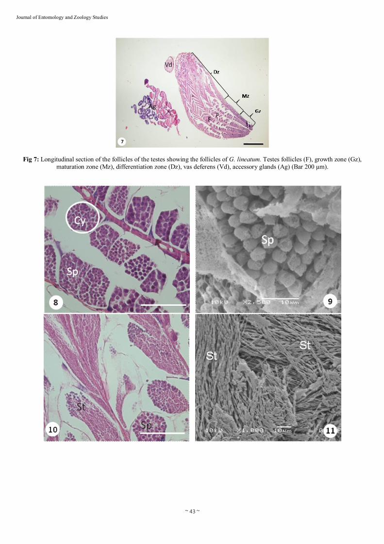
Fig 7: Longitudinal section of the follicles of the testes showing the follicles of G. lineatum. Testes follicles (F), growth zone (Gz), maturation zone (Mz), differentiation zone (Dz), vas deferens (Vd), accessory glands (Ag) (Bar 200 µm).
~ 44 ~
Journal of Entomology and Zoology Studies
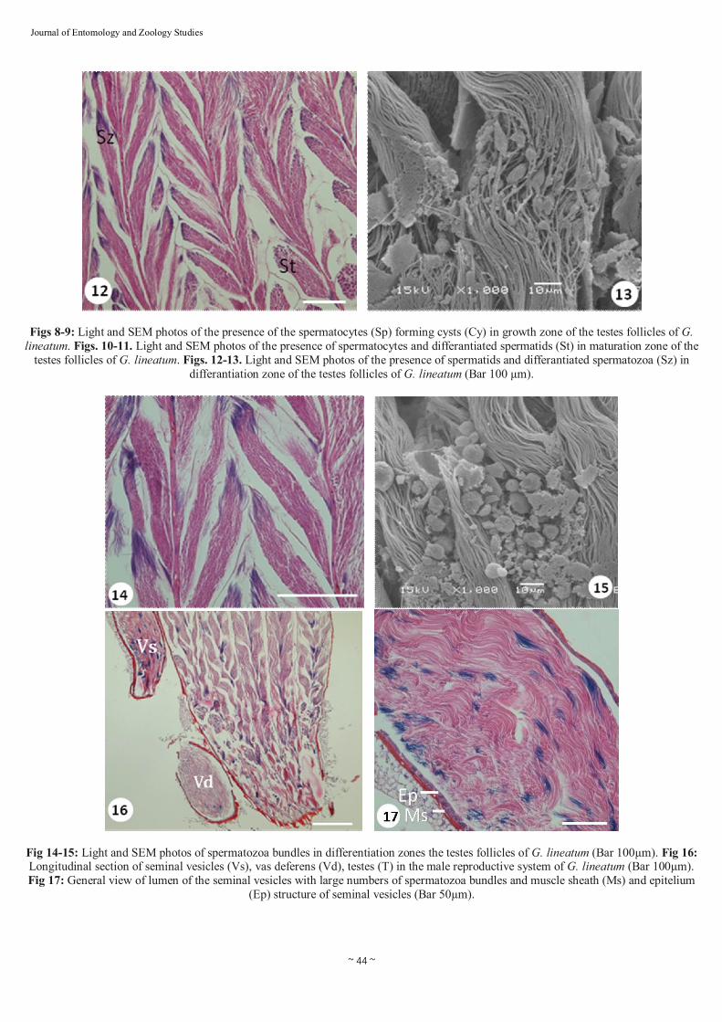
Figs 8-9: Light and SEM photos of the presence of the spermatocytes (Sp) forming cysts (Cy) in growth zone of the testes follicles of G. lineatum. Figs. 10-11. Light and SEM photos of the presence of spermatocytes and differantiated spermatids (St) in maturation zone of the
testes follicles of G. lineatum. Figs. 12-13. Light and SEM photos of the presence of spermatids and differantiated spermatozoa (Sz) in differantiation zone of the testes follicles of G. lineatum (Bar 100 µm).
Fig 14-15: Light and SEM photos of spermatozoa bundles in differentiation zones the testes follicles of G. lineatum (Bar 100µm). Fig 16: Longitudinal section of seminal vesicles (Vs), vas deferens (Vd), testes (T) in the male reproductive system of G. lineatum (Bar 100µm). Fig 17: General view of lumen of the seminal vesicles with large numbers of spermatozoa bundles and muscle sheath (Ms) and epitelium
(Ep) structure of seminal vesicles (Bar 50µm).
~ 45 ~
Journal of Entomology and Zoology Studies
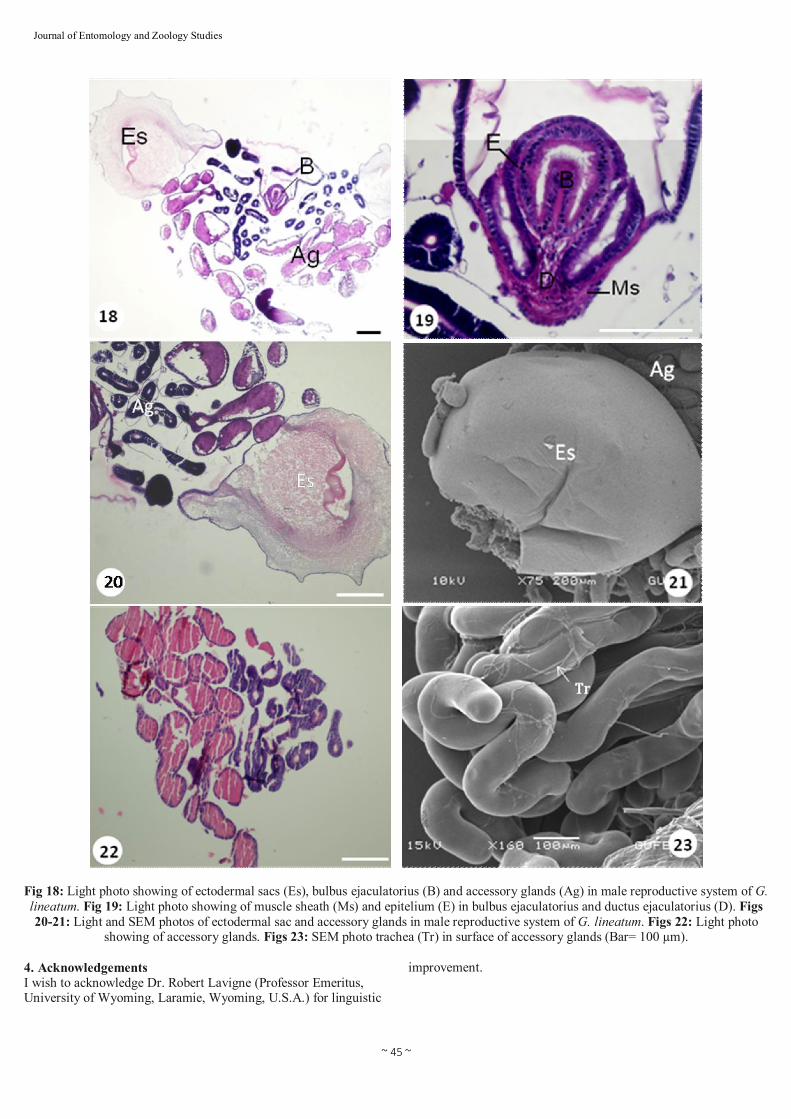
Fig 18: Light photo showing of ectodermal sacs (Es), bulbus ejaculatorius (B) and accessory glands (Ag) in male reproductive system of G. lineatum. Fig 19: Light photo showing of muscle sheath (Ms) and epitelium (E) in bulbus ejaculatorius and ductus ejaculatorius (D). Figs 20-21: Light and SEM photos of ectodermal sac and accessory glands in male reproductive system of G. lineatum. Figs 22: Light photo
showing of accessory glands. Figs 23: SEM photo trachea (Tr) in surface of accessory glands (Bar= 100 µm).
4. Acknowledgements I wish to acknowledge Dr. Robert Lavigne (Professor Emeritus, University of Wyoming, Laramie, Wyoming, U.S.A.) for linguistic
improvement.
~ 46 ~
Journal of Entomology and Zoology Studies
5. Reference: 1. Adams TS. Morphology of the internal reproductive system of the
male and female two-spotted stink bug, Perillus bioculatus (F.) (Heteroptera: Pentatomidae) and the transfer of products during mating. Invertebr Reprod Dev 2001; 39(1):45-53.
2. Bonhag PF, Wick JR. The fuctional anatomy of the male and female reproductive systems of the milkweed bug, Oncopeltus fasciatus (Dallas). J Morphol 1953; 93:177-284.
3. Bowen RH. Studies on insect spermatogenesis. II. The components of the spermatid and their role in the formation of sperm in Hemiptera. J Morphol 1922; 37:79-193.
4. Bushrow ES, Fuller CL, Cowan DP, Byrd CA. Anatomy of the male reproductive system and sperm morphology in the caterpillar-hunting wasp Ancistrocerus antilope (Hymenoptera, Vespidae). Invertebr Biol 2006; 125:354-362.
5. Chapman RF. The insects: Structure & Function. Cambridge University Press, Cambridge, UK, 1998.
6. Davey KG, Krieger FL. Variations during the gonotropic cycle in the titer of the myotrophic ovulation hormone and the response of the ovarian muscles in Rhodnius prolixus. Gen Comp Endocr 1985; 59: 452-457.
7. Davis NT. The morphology and functional anatomy of the male and female reproductive systems of Cimex lectularius L. (Heteroptera, Cimicidae). Ann Entomol Soc of Am 1955; 49:466–493.
8. Engelmann F. The Physiology of Insect Reproduction. Pergamon Press, New York, 1970.
9. Gonçalves TCM, Lent H, Almeida JR. Estudo anatômico e morfométrico dos foliculos testiculares de algumas espécies de Triatominae (Hemiptera: Reduviidae). Mem Inst Oswaldo Cruz 1987; 82:543-550.
10. Grasse PP. Termitologia: Anatomie, Physiologie, Reproduction des Termites. Masson, Paris, 1982.
11. Happ GM. Maturation of the male reproductive system and its endocrine regulation. Annu Rev Entomol 1992; 37:303–320.
12. Jahnke SM, Redaelli LR, Diefenbach LMG. Internal reproductive organs of Cosmoclopius nigroannulatus (Hemiptera: Reduviidae). Braz J Biol 2006; 66:509-512.
13. Karakaya G, Özyurt N, Candan S, Suludere Z. Structure of the male reproductive system in Coreus marginatus (Linneaus, 1758) (Hemiptera: Coreidae). Turk Entomol Derg 2006; 36(2):193-204.
14. Kartal V, Zeybekoğlu U. Türkiye faunası için yeni üç Diplocolenus Rib. (Hom., Auch., Cicadellidae, Deltocephalinae) türü üzerine taksonomik bir araştırma. Turk Entomol Derg 1997; 21:291-294.
15. Klowden MJ. Physiological Systems in Insects. Academic Press, San Diego, 2007.
16. Lemos WP, Serrao JE, Ramalho FS, Cola-Zanuncio JC, Lacerda MC. Effect of diet on male reproductive tract of Podisus nigrispinus (Dallas) (Heteroptera: Pentatomidae). Braz J Biol 2006; 65(1):91-96.
17. Leopold RA. The role of male accessory glands in insect reproduction. Annu Rev Entomol 1976; 21:199–221.
18. Leston D. Notes on the Ethiopian Pentatomoidea (Hemiptera). 14. An Acanthosomid from Angola, with remarks upon the status and morphology of Acanthosomidae Stål. Publicações Culturais da Companhia de Diamantes de Angola, 1953; 16:123-132.
19. Lodos N, Önder F, Pehlivan E, Atalay R. Ege ve Marmara Bölgesi’nin Zararlı Böcek Faunasının Tespiti Üzerinde Çalışmalar (Curculionidae, Scarabaeidae, (Coleoptera); Pentatomidae, Lygaeidae, Miridae (Heteroptera). T.C. Gıda-Tarım ve Hayvancılık Bakanlığı Zirai Mücadele ve Zirai Karantina Genel Müdürlüğü, Ankara, 1978.
20. Lodos N, Önder F, Pehlivan E, Atalay R, Erkin E, Karsavuran Y et al. Faunistic Studies on Pentatomoidea (Plataspidae, Acanthosomatidae, Cydnidae, Scutelleridae, Pentatomidae) of Western Black Sea, Central Anatolia and Mediterranean Regions of Turkey. Ege Üniversitesi Basımevi, İzmir, 1998.
21. Matsuda R. Morphology and Evolution of the Insect Abdomen. Pergamon Press, Oxford, 1976.
22. Nation JL. Insect physiology and biochemistry. CRC Press, London, 2002.
23. Nijhout HF. Insect Hormone. Princeton, University Press, Princeton, New Jersey, 1994.
24. Ozyurt N, Candan S, Suludere Z. The morphology and histology of the male reproductive system in Dolycoris baccarum Linnaeus 1758 (Heteroptera: Pentatomidae). Light and scanning electron micoscope studies. Micron 2013; 44:101-106.
25. Papáček M, Soldán T. Structure and development of the reproductive system in Aphelocheirus aestivalis (Hemiptera: Heteroptera: Nepomorpha: Aphelocheiridae). Acta Ent Mus Nat Pra 2008; 48(2): 299-318.
26. Pendergrast JG. The male reproductive organs of Nezara viridula with a preliminary account of their development (Heteroptera; Pentatomidae). T Roy Soc NZ 1956; 84(1):139-146.
27. Pires EM, Ferreira PSF, Guedes RNC, Serrao JE. Morphology of the phytophagus bug Platyscytus decempunctatus (Carvalho) (Heteroptera: Miridae). Neotrop Entomol 2007; 36(4):510-513.
28. Ramamurty PS. Histological studies of the internal organs of reproduction in Nezara viridula Fabr. Pentatomidae: Heteroptera, Insecta. Zool Anz 1969; 183:119-139.
29. Rodrigues ARS, Serrao JE, Teixeira VW, Torres JB, Teixeira AA. Spermatogenesis, changes in reproductive structures, and time constraint associated with insemi22 in Podisus nigrispinus. J Insect Physiol 2008; 54(1):1543-1551.
30. Wieczorek K. Structure of the male reproductive system of Anoecia corni Fabricius, 1775 (Hemiptera, Aphidoidea), a representative of the family Anoeciidae. Acta Zool 2008; 89:163-167.