Embed Size (px)
Citation preview
HistologyLecture 21 Male Reproductive System

Male Reproductive System
Male reproductive system is consists of
Testes
Genital ducts Intratesticularextratesticular
Accessory glands Seminal vesicles prostate gland
bulbourethral glands (of Cowper) Copulatory organ
Penis

TestisReproductive functions (produce sperm)
Endocrine functions (produce testosterone)
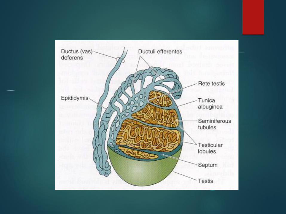
Tunica vaginalis is a peritoneal outpouching that form a serous cavity that partially surrounds testis
Dense irregular connective tissue capsule: tunica albuginea with fibrous incomplete septa divide testis into about 250 lobules
Deep to tunica albuginea is a layer of loose highly vascular CT known as tunica vasculosa
Each lobule has 1-4 seminiferous tubules with surrounding loose connective tissue, vessels and interstitial cells of Leydig
Sperm produced by seminiferous tubule
Testosterone produced by Leydig cells

Seminiferous Tubule
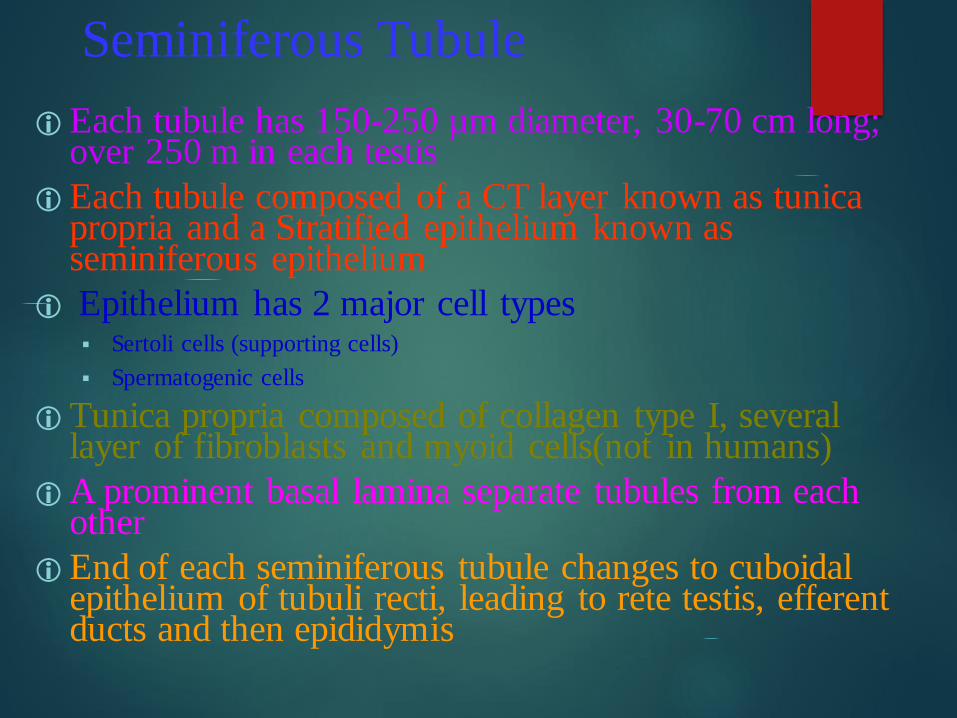
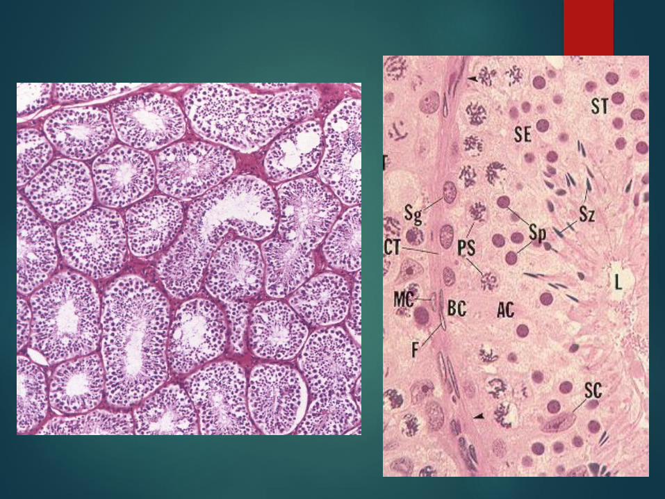
Each tubule has 150-250 µm diameter, 30-70 cm long; over 250 m in each testis
Each tubule composed of a CT layer known as tunica propria and a Stratified epithelium known as seminiferous epithelium
Epithelium has 2 major cell types Sertoli cells (supporting cells)
Spermatogenic cells
Tunica propria composed of collagen type I, several layer of fibroblasts and myoid cells(not in humans)
A prominent basal lamina separate tubules from each other
End of each seminiferous tubule changes to cuboidal epithelium of tubuli recti, leading to rete testis, efferent ducts and then epididymis
Spermatogenic Cells
4-8 layers of cells dividing by mitosis and meiosis to produce spermatozoa
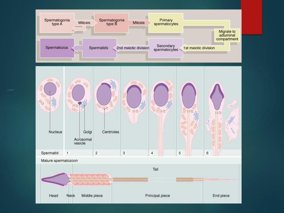
3 phases of spermatogenesis
spermatocytogenesis: spermatogonia divide by mitosis giving rise to primary
spermatocytes
Meiosis: primary spermatocytes go through meiotic divisions giving rise to
haploid spermatids
spermiogenesis: spermatids differentiate into spermatozoa

Spermatogonia Spermatogonia are small and diploid cells,12 µm
diameter that lie on basal lamina
Spermatogonia are three categories consist of:
Type A spermatogonia continue dividing; flattened oval nucleus light and dark
types )))))Dark Type A: are reserve cells when
divide form additional dark type A and pale type A )))))Pale type
A: have abundant euchromatin, divide and form additional pale type A and type B
spermatogonia
Type B spermatogonia undergo meiosis; round nucleus, differentiate into primary
spermatocytes
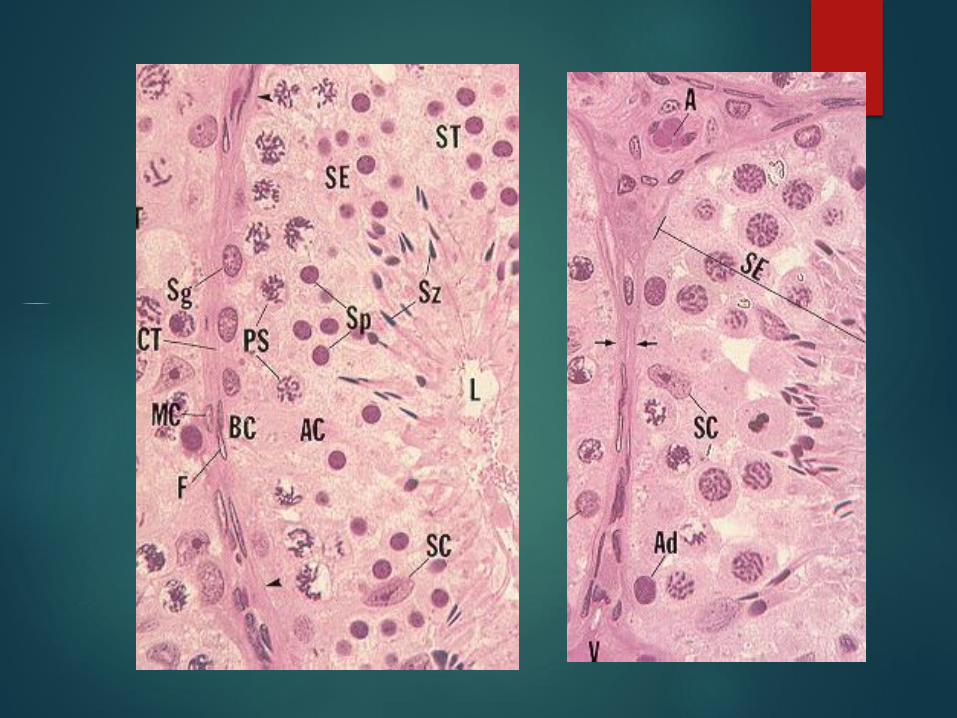
Spermatocytes & Spermatids
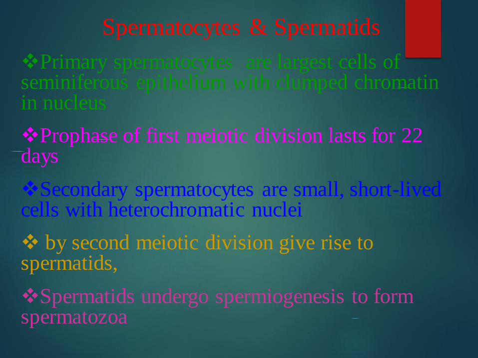
Primary spermatocytes are largest cells of seminiferous epithelium with clumped chromatin in nucleus
Prophase of first meiotic division lasts for 22 days
Secondary spermatocytes are small, short-lived cells with heterochromatic nuclei
by second meiotic division give rise to spermatids,
Spermatids undergo spermiogenesis to form spermatozoa
Spermatid
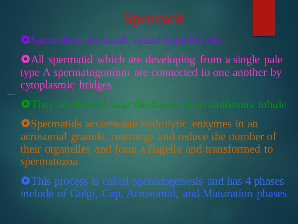
Spermatids are small, round haploid cells
All spermatid which are developing from a single pale type A spermatogonium are connected to one another by cytoplasmic bridges
They are located near the lumen of seminiferous tubule
Spermatids accumulate hydrolytic enzymes in an acrosomal granule, rearrange and reduce the number of their organelles and form a flagella and transformed to spermatozoa
This process is called spermiogenesis and has 4 phases include of Golgi, Cap, Acrosomal, and Maturation phases
Timing of Spermatogenesis
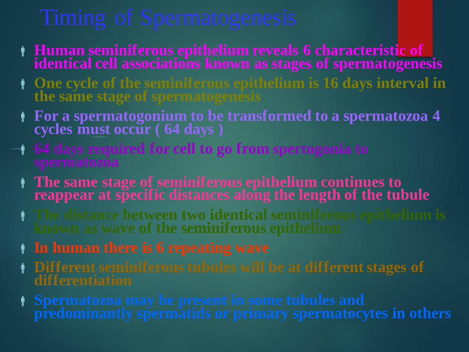
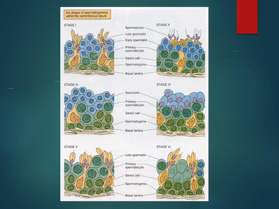
Human seminiferous epithelium reveals 6 characteristic of identical cell associations known as stages of spermatogenesis
One cycle of the seminiferous epithelium is 16 days interval in the same stage of spermatogenesis
For a spermatogonium to be transformed to a spermatozoa 4 cycles must occur ( 64 days )
64 days required for cell to go from spertogonia to spermatozoa
The same stage of seminiferous epithelium continues to reappear at specific distances along the length of the tubule
The distance between two identical seminiferous epithelium is known as wave of the seminiferous epithelium
In human there is 6 repeating wave
Different seminiferous tubules will be at different stages of differentiation
Spermatozoa may be present in some tubules and predominantly spermatids or primary spermatocytes in others
Sertoli Cells Elongated columnar supporting cells with base at basal
lamina and apex near lumen of seminiferous tubule
Lateral complex infoldings surround spermatogonia and spermatocytes
Lateral cell membranes of adjacent sertoli cells form occluding junction with each other that establish Blood Testis Barrier
Tight junctions subdivide lumen into two compartments basal and adluminal
Nucleus basally located, euchromatic and with distinct nucleolus
Cytoplasm houses inclusion products known as crystalloids of Charcot-Böttcher
Functions of Sertoli Cells Support and nourish cells of seminiferous tubule
Establishment of blood testis barrier and protecting new developing gametes from immune system
No vasculature inside tubule so sertoli cells transport nutrients to lumenal region
Phagocytose cast-off cytoplasmic droplets from spermatids
Secrete fructose rich fluid into lumen that helps sperm flow in genital ducts
Produce androgen binding protein (ABP)
Produce inhibin which decreases FSH release by pituitary, slowing spermatogenesis
Syntesis of antimüllerian hormone during embryogenesis
Synthesis and secretion of testicular transferrin that conveys iron to gametes
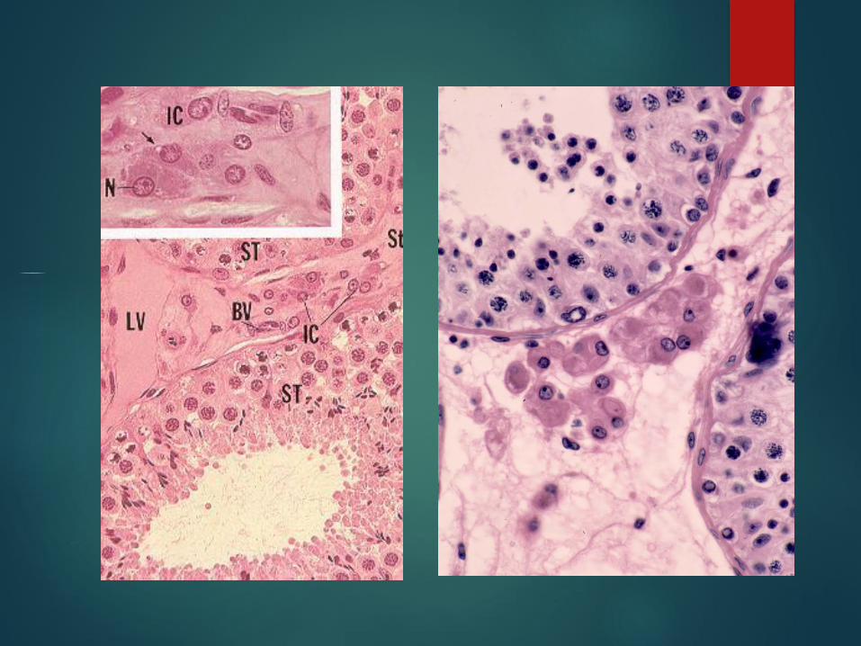
Interstitial Tissue
Between seminiferous tubules are vessels, nerves,
fibroblasts and other connective tissue cells which is
collectively called tunica vasculosa
After puberty interstitial cells of Leydig differentiate
and produce testosterone
Leydig cells are eosinophilic and have lots of SER and
mitochondria with tubular cristae, lipid droplets and
other characteristic of a steroid producing cell
The cytoplasm contains crystallized proteins known as
crystals of Reinke
Intratesticular Genital Ducts
Intratesticular duct system connect seminiferous tubules to epididymis
These ducts are:
Tubuli recti (straight tubules)
Rete testis
Efferent ducts
Tubuli Recti
Short segment of duct at the end of seminiferous tubules
Lined by Sertoli cells that change to simple cuboidal epithelial cells at its
second half near rete testis
Cuboidal cells have short stubby microvilli and a single flagellum
High glycogen amount in some species
Connective tissue sheath support the tubuli
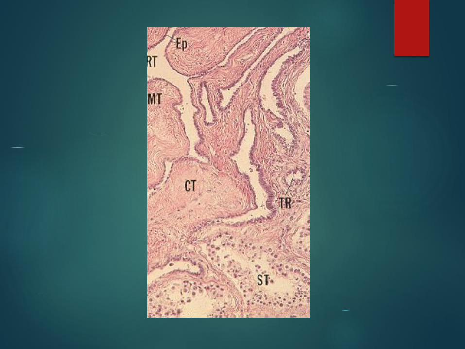
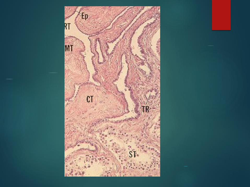
Rete Testis
Branched network of channels lined with simple cuboidal
epithelium
Some cells ciliated and have a single flagellum
Surrounded by connective tissue of mediastinum
Efferent Ducts 10 – 20 short tubule that pierce tunica albugina
Tubules with nonciliated cuboidal cells alternating
with region of ciliated columnar cells giving a
scalloped look to epithelium
Nonciliated cells absorb fluids secreted by
seminiferous tubule cells by endocytosis
Beating of cilia helps sperm movement
Thin loose CT surrounded by a layer of smooth
muscle cells that are circularly arrayed
Lead to epididymis

Excretory Genital Ducts
Excretory or extratesticular ducts are:
Ductus epididymis
Vas deferens
Ejaculatory duct

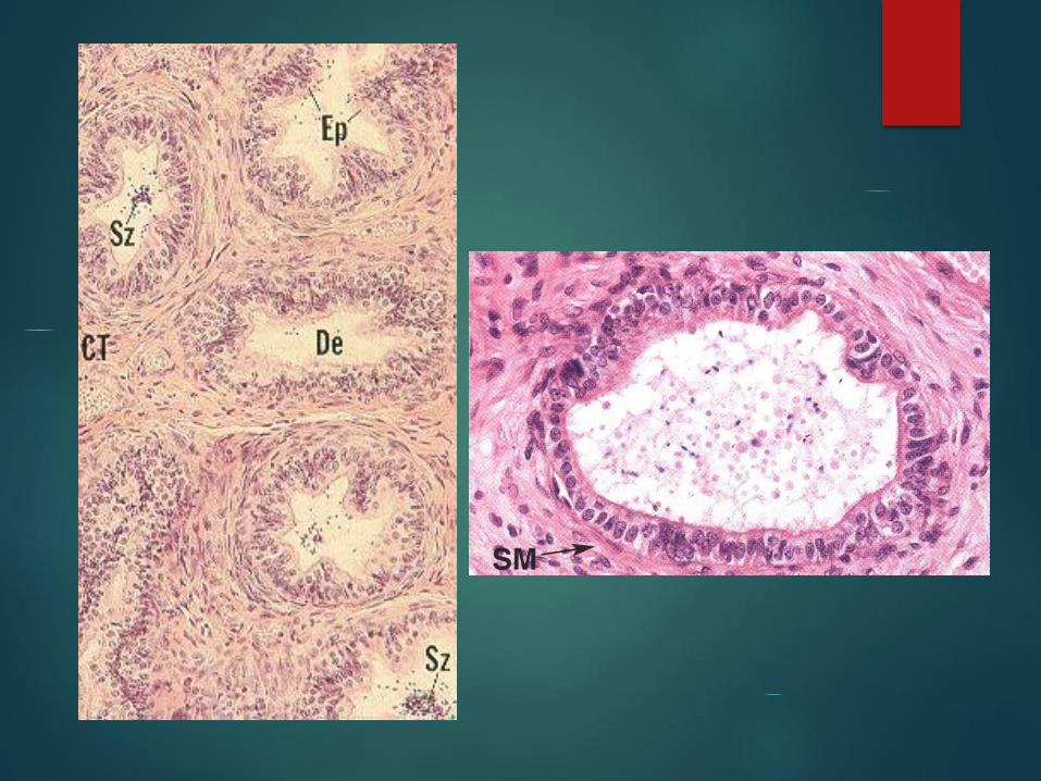
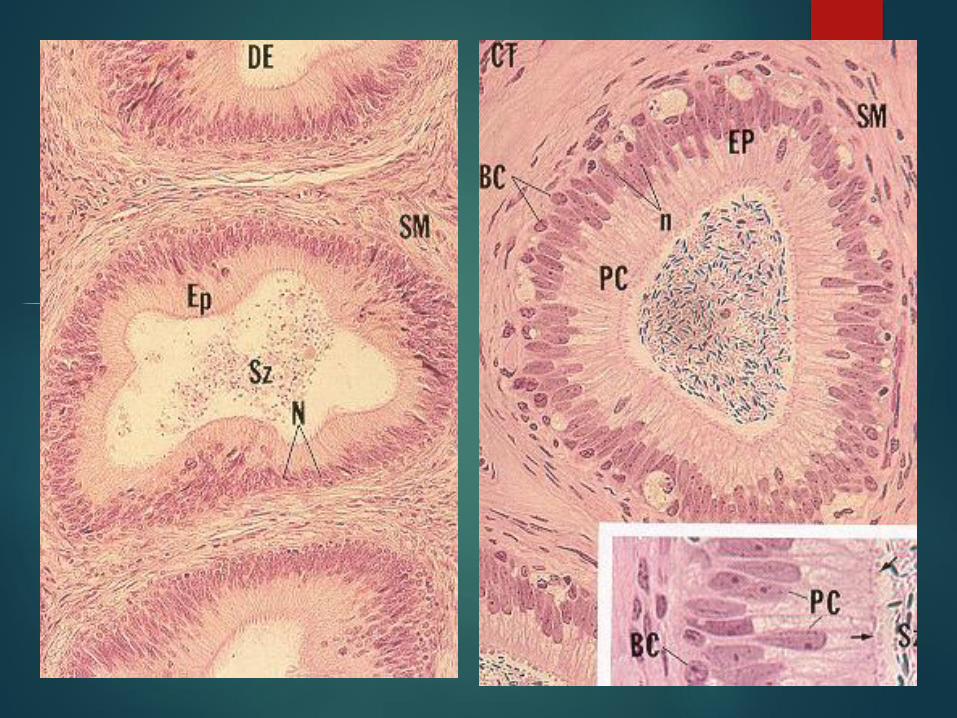
Ductus Epididymis Highly coiled tube 4-6 m long
Pseudostratified columnar epithelium with stereocilia
Rounded basal cells or stem cell with dense nuclei
Columnar principal cells with pale nuclei and stereocilia
Columnar principal cells have many stereocilia, pinocytotic and coated vesicles, RER, and well developed golgi apparatus
Thin connective tissue layer with underlying circularly arrayed smooth muscle
Secretions preserve sperm capacitation
Phagocytosis of residual bodies and resorbtion of luminal fluid
Smooth muscle contractions for peristalsis to move sperm through the tubule
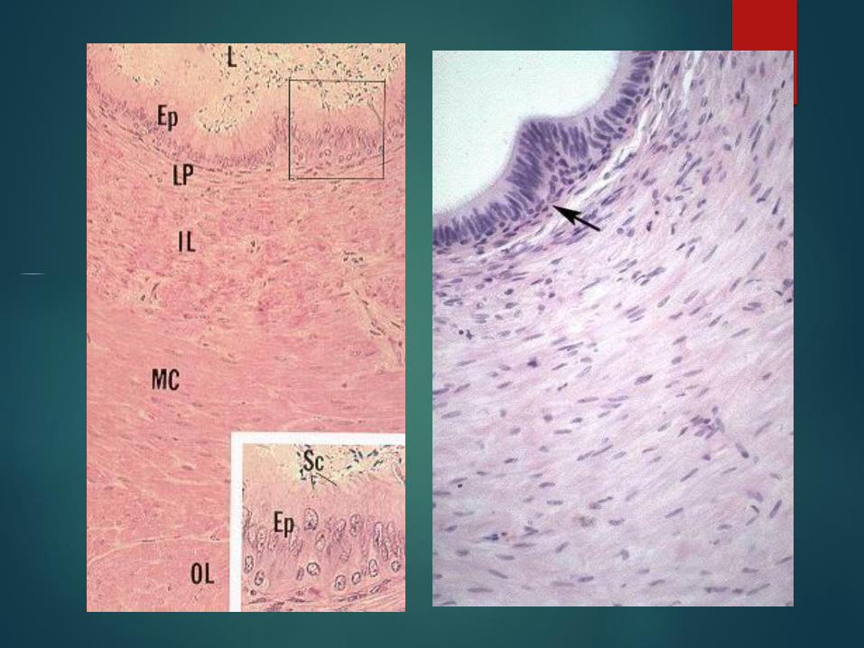
Ductus (Vas) Deferens
Straight tube from epididymis to ejaculatory duct
Narrow folded lumen with thick smooth muscle layer
Epithelium is pseudostratified columnar with stereocilia but principal cells are shorter
Lamina propria with loose fibroelastic connective tissue and vessels
Smooth muscle layer has inner longitudinal, middle circular and outer longitudinal layers
Smooth muscle layers surround by a thin layer of connective tissue
Dilated region, ampulla, has highly folded, thickened epithelium
Ejaculatory duct
A short straight tubule that enter prostate gland
The lumen lined by simple columnar
epithelium
Subepithelial connective tissue which is folded
support the epithelium
Ejaculatory duct has no smooth muscle cells in
its wall
Spermatic Cord
Testicular artery
Pampiniform (venous) plexus
Nerves
Ductus deferens
Surrounded by skeletal muscle, the cremaster muscle

Accessory Genital Glands
Paired seminal vesicle
Single prostate
Paired bulbourethral (Cowper’s) glands
Seminal Vesicle
Highly coiled tubular structures, 15 cm long
Duct joins with ductus deferens at prostate
Folded epithelium with pseudostratified columnar epithelium
Rounded basal cells and low columnar cells
Low columnar cells have short microvilli and a single flagellum, RER, golgi
apparatus, numerous mitochondria, lipid and lipochrome droplets, and many
secretory granules

Seminal Vesicle
Lamina propria is a fibroelastic CT
Thin smooth muscle layers, inner circular, outer longitudinal
Viscous yellowish fructose-rich secretion making up 70% of ejaculate volume
Pale yelloish color is due to lipochrome released by the seminal vesicle
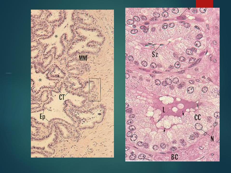
Prostate Capsule is composed of richly vascularized dense irregular
CT interspersed with smooth muscle
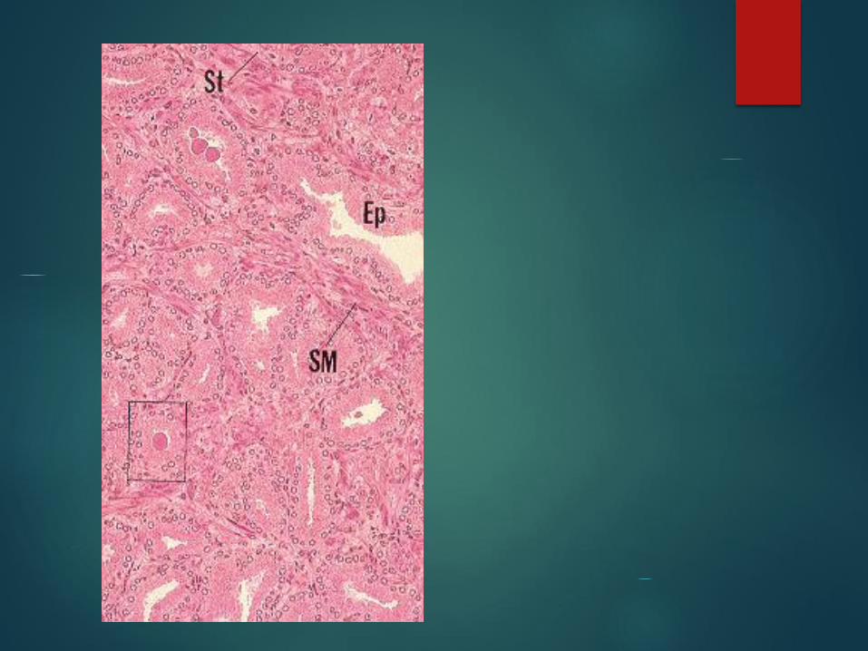
Stroma is derived from capsule and has smooth muscle cells in addition to normal CT cells
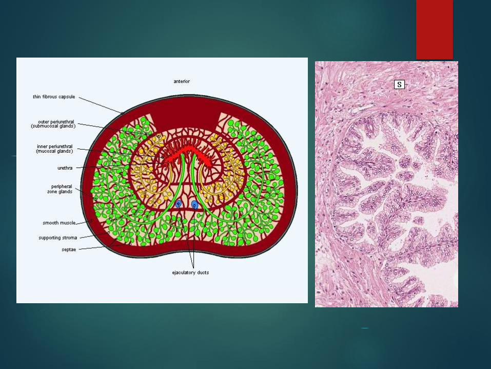
30-50 individual compound tubuloacinar glands with ducts leading to prostatic urethra
Gland arranged in three separate concentric layers *mucosal gland *submucosal gland *main gland
Epithelium is simple columnar or pseudostratified columnar cells with organelles endowed in protein synthesis

Prostate
Prostatic fluid stored for release during ejaculation
Secretions is a serous white fluid include amylase,
proteases, acid phosphatase and lipids
Prostatic concretions (Corpora Amylacea) found in
gland lumens increase with age, function unknown
They are composed of calcified glycoproteins
Past age 40 glands hypertrophy which can lead to
urethral blockage or cancer
Bulbourethral (Cowper’s) Glands
Its fibroelastic capsule contains smooth muscle cells as well as skeletal muscle fibers derived from urogenital diaphragm
Compound tubuloacinar glands with simple cuboidal to simple columnar
Gland acini surrounded by smooth muscle
Secretion is clear mucous released during sexual arousal for lubrication
Penis 3 cylindrical masses of highly vascularized erectile tissue
2 corpora cavernosa are dorsal
1 corpus spongiosum is ventral and surrounds urethra
Each of them enclosed by dense fibrous CT known as tunica albugina
corpus spongiosum end as a bulbous portion known as glans of penis
A common loose CT sheath surrounding all three corpora
Outer covering is skin
Skin form a retractable sheath over glans which is called prepuce
Prepuce lined by nonkeratinized stratified epithelium

Penis
Erectile tissue consists of venous spaces lined by
unfenestrated endothelium and separated by septa of
connective tissue and smooth muscle
Septa of corpus spongiosum has more elastic fiber and
less smooth muscle
Capillary plexus in septa supply some blood to
vascular spaces
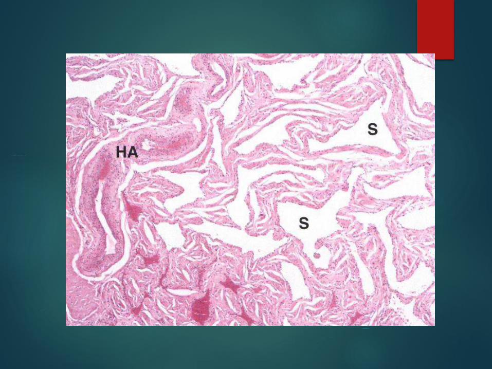
The main source of blood during erection that flow
into spaces are from helical arteries
Blood flows into venous spaces during erection and
smooth muscle contraction slows outflow of blood

Urethra
15 – 20 cm long in male
Prostatic urethra 3- 4 cm long located in prostate, and is lined by transitional epithelium
Membranous urethra 1-2 cm long, it is pass through urogenital diaphragm, is lined by stratified columnar with patches of Pseudostratified columnar epithelium
Spongy urethra(Penile) 15 cm long, is lined by stratified columnar with patches of pseudostratified columnar epithelium
In the glans (the navicular fossa) it is lined by stratified squamous
Mucous glands of Littre found along urethra in lamina propria and subepithelial loose connective tissue

HR Mahmoudzadeh Sagheb
Z Heidari
MH Noori Mugahi
Department of Histology