Embed Size (px)
Citation preview
MOL #88526
1
Pore exposed tyrosine residues of P-glycoprotein are important hydrogen bonding
partners for drugs
Yaprak Dönmez Cakil, Narakorn Khunweeraphong, Zahida Parveen, Diethart Schmid,
Matthias Artaker, Gerhard F. Ecker, Harald H. Sitte, Oliver Pusch, Thomas Stockner,
Peter Chiba
Institute of Medical Chemistry, Medical University of Vienna, Waehringerstrasse 10, 1090
Vienna, Austria (Y.D.C., N.K., Z.P., P.C.)
Department of Biochemistry, Abdul Wali Khan University Mardan, Pakistan (Z.P.)
Institute of Pharmacology, Medical University of Vienna, Waehringerstrasse 13A, 1090
Vienna, Austria (Y.D.C., T.S., H.H.S.)
Institute of Physiology, Medical University of Vienna, Schwarzspanierstrasse 17, 1090
Vienna, Austria (D.S.)
Department of Medical Biochemistry, Max F. Perutz Laboratories, Medical University of
Vienna, Dr. Bohrgasse 9, 1030 Vienna, Austria (M.A.)
Emerging Field Pharmacoinformatics, Dept. of Medicinal Chemistry, University of Vienna,
Althanstrasse 14, 1090 Vienna, Austria (G.F.E.)
Department of Cell and Developmental Biology, Medical University of Vienna,
Schwarzspanierstraße 17, 1090 Vienna, Austria (O.P.)
Molecular Pharmacology Fast Forward. Published on December 23, 2013 as doi:10.1124/mol.113.088526
Copyright 2013 by the American Society for Pharmacology and Experimental Therapeutics.
This article has not been copyedited and formatted. The final version may differ from this version.Molecular Pharmacology Fast Forward. Published on December 23, 2013 as DOI: 10.1124/mol.113.088526
at ASPE
T Journals on M
ay 1, 2018m
olpharm.aspetjournals.org
Dow
nloaded from
MOL #88526
2
Running title page Pore exposed ABCB1 tyrosines form hydrogen bonds with drugs
Address correspondence to:
Thomas Stockner, Institute of Pharmacology, Waehringerstrasse 13A, 1090 Vienna, Austria,
phone +43 1 40160 31361, Fax: +43 1 40160 931300, email:
Number of text pages: 27
Number of tables: 0
Number of figures: 5
Number of references: 33
Number of words in
Abstract: 142
Introduction: 441
Discussion: 1437
d) List of non-standard abbreviations
ABC, ATP-binding cassette; P-gp, P-glycoprotein; MDR, multidrug resistance; TMD,
transmembrane domain; NBD, nucleotide binding domain; rh123, rhodamine123;
This article has not been copyedited and formatted. The final version may differ from this version.Molecular Pharmacology Fast Forward. Published on December 23, 2013 as DOI: 10.1124/mol.113.088526
at ASPE
T Journals on M
ay 1, 2018m
olpharm.aspetjournals.org
Dow
nloaded from
MOL #88526
3
Abstract The multispecific efflux transporter P-glycoprotein plays an important role in drug disposition.
Substrate translocation occurs along the interface of its transmembrane domains. The
rotational C2 symmetry of ABC-transporters implies the existence of two symmetry related
sets of substrate-interacting amino acids. These sets are identical in homodimeric
transporters, and remain evolutionarily related in full transporters such as P-glycoprotein,
where substrates bind preferentially, but non-exclusively to one of two binding sites. We
explored the role of pore exposed tyrosines for hydrogen-bonding interactions with
propafenone type ligands in their preferred binding site 2. Tyrosine 953 is shown to form
hydrogen-bonds with propafenone analogs, but also with the preferred site 1 substrate
rhodamine123. Furthermore, an accessory role of tyrosine 950 for binding of selected
propafenone analogs is demonstrated. The present study demonstrates the importance of
domain interface tyrosine residues for interaction of small molecules with P-glycoprotein.
This article has not been copyedited and formatted. The final version may differ from this version.Molecular Pharmacology Fast Forward. Published on December 23, 2013 as DOI: 10.1124/mol.113.088526
at ASPE
T Journals on M
ay 1, 2018m
olpharm.aspetjournals.org
Dow
nloaded from
MOL #88526
4
Introduction
ATP-binding cassette (ABC) proteins form one of the largest families of transmembrane
proteins. The human genome contains 48 genes encoding for ABC proteins, the majority of
which are transporters. Mutations in at least 17 ABC transporters have been linked to
disease etiologies (Linton et al., 2011). The minimal functional unit of a transporter consists
of four domains: two transmembrane domains (TMDs), which form the solute conduits and
two nucleotide binding domains (NBDs), which provide the energy for solute translocation by
ATP binding and hydrolysis. Human P-glycoprotein (P-gp, ABCB1) is a multidrug resistance
transporter, which plays a central role in drug disposition. Therefore, early profiling of
developmental compounds includes routine screening for P-gp substrate properties
(Giacomini et al., 2010).
A mechanistic model for cargo transport of ABC efflux transporters remains elusive, despite
a large body of biochemical evidence. The present study characterizes the contribution of
hydrogen bonding interactions between propafenone type ligands and selected pore
exposed tyrosine OH-groups. Propafenones have been characterized extensively in previous
QSAR studies and demonstrated to be both substrates and inhibitors of P-glycoprotein
(Schmid et al., 1999). Tyrosine residues are known to play a pivotal role for molecular
recognition in biological systems, including domain interface and active site interactions.
Tyrosines are amphipathic residues, capable of forming hydrophobic, hydrogen-bonding, π-π
and π-cation interactions. They were shown to make a large contribution to protein stability
(Pace et al., 2001), but nevertheless contribute to structural plasticity of binding regions
(Mian et al., 1991). The rigidity of the aromatic ring is associated with a reduced loss of
conformational entropy upon immobilization in binding interfaces (Koide and Sidhu, 2009).
The amphipathic nature of tyrosines allows them to readily tolerate changes in the polarity of
their environment (MacCallum et al., 2007). Molecular dynamics simulations suggest that
rotamers adopt different positions in the drug binding pocket that allow them to contribute to
poly-specificity (Liu et al., 2013). The known requirement for hydrogen bonding interaction of
This article has not been copyedited and formatted. The final version may differ from this version.Molecular Pharmacology Fast Forward. Published on December 23, 2013 as DOI: 10.1124/mol.113.088526
at ASPE
T Journals on M
ay 1, 2018m
olpharm.aspetjournals.org
Dow
nloaded from
MOL #88526
5
P-gp substrates (Cramer et al., 2007; Gatlik-Landwojtowicz et al., 2006; Schmid et al., 1999)
prompted us to investigate the importance of tyrosine hydroxyl groups for interaction with
propafenone analogues.
A combination of photolabeling and mass spectrometry recently enabled us to demonstrate a
dual interaction mode of P-glycoprotein with substrates in two rotationally symmetric
positions. These involve tyrosine residues, which in the present study were mutated to
phenylalanine to assess the contribution of tyrosine hydroxyl groups to hydrogen bond
formation with propafenone type ligands. Data indicate an important role of these residues
for interaction with propafenones, but also with the paradigmatic P-gp substrate
rhodamine123 (rh123), thus providing additional experimental evidence for the dual
interaction mode of P-gp with solutes and drugs.
This article has not been copyedited and formatted. The final version may differ from this version.Molecular Pharmacology Fast Forward. Published on December 23, 2013 as DOI: 10.1124/mol.113.088526
at ASPE
T Journals on M
ay 1, 2018m
olpharm.aspetjournals.org
Dow
nloaded from
MOL #88526
6
Materials and Methods
Sequence Alignments and Homology Modeling. Generation of the ABCB1 model was
described previously (Stockner et al., 2009). Briefly, ClustalW was used to obtain multiple
sequence alignments of ABCB proteins. The models of human P-gp were based on the
crystal structures of the outward facing structure of Sav1866 from Staphylococcus aureus
and the inward facing structure of ABCB1 from C. elegans (PDB ID: 2HYD; 3.0 Å resolution
(Dawson and Locher, 2006) and 4F4C; 3.4 Å resolution (Jin et al., 2012)) using MODELLER
(version 9v12) (Marti-Renom et al., 2000; Sali and Blundell, 1993). The N-terminus before
the elbow helix as observed in the C. elegans ABCB1 structure was not included in the
model and the interrupted helix 10 was replaced by a de novo model of an ideal helix. This
replacement is supported by the observation of a contiguous helix 10 in all other structures
from the ABCB transporter family. Initial models were further optimized by relaxation
simulations of a membrane inserted transporter.
Knockdown of endogenous P-gp in HEK293 cells
Construction and prevalidation of shRNA Vectors
HEK293 cells endogenously express P-gp at a level corresponding to approximately 5% of
transiently expressed protein. In order to avoid interference from endogenous P-gp in
functional assays, the transporter was knocked down by transduction with pLKO.1 lentiviral
vectors (Moffat et al., 2006) containing P-gp shRNA constructs targeted towards the 3’UTR
of the endogenous sequence as described by Addgene (http://www.addgene.org/plko).
Briefly, five specific oligonucleotides (Sigma-Aldrich, St. Louis, MO, USA) targeting the 3’
UTR of the ABCB1 gene were introduced into the Age I – EcoR I sites of pLKO.1 (Addgene,
Cambridge MA, USA, plasmid # 10878).
ABCB1_1_fwd:
ccggAAGAGGTATCTGTTTAACATTctcgagAATGTTAAACAGATACCTCTTtttttg
ABCB1_2_fwd:
ccggGAATTATGAAGAGGTATCTGTctcgagACAGATACCTCTTCATAATTCtttttg
This article has not been copyedited and formatted. The final version may differ from this version.Molecular Pharmacology Fast Forward. Published on December 23, 2013 as DOI: 10.1124/mol.113.088526
at ASPE
T Journals on M
ay 1, 2018m
olpharm.aspetjournals.org
Dow
nloaded from
MOL #88526
7
ABCB1_3_fwd:
ccggGAACAGAGTGAGAGACATCATctcgagATGATGTCTCTCACTCTGTTCtttttg
ABCB1_4_fwd:
ccggGTGGAGAGAAATCATAGTTTActcgagTAAACTATGATTTCTCTCCACtttttg
ABCB1_5_fwd:
ccggGACTGTATGAGATGTTAAATActcgagTATTTAACATCTCATACAGTCtttttg
ABCB1_1_rev:
aattcaaaaaAAGAGGTATCTGTTTAACATTctcgagAATGTTAAACAGATACCTCTT
ABCB1_2_rev:
aattcaaaaaGAATTATGAAGAGGTATCTGTctcgagACAGATACCTCTTCATAATTC
ABCB1_3_rev:
aattcaaaaaGAACAGAGTGAGAGACATCATctcgagATGATGTCTCTCACTCTGTTC
ABCB1_4_rev:
aattcaaaaaGTGGAGAGAAATCATAGTTTActcgagTAAACTATGATTTCTCTCCAC
ABCB1_5_rev:
aattcaaaaaGACTGTATGAGATGTTAAATActcgagTATTTAACATCTCATACAGTC
A non-targeting shRNA vector (Addgene plasmid # 1864) was used as a negative control. All
shRNA expression cassettes were verified by sequencing. To test for efficiency and
specificity the five candidate shRNA constructs (#1-#5) were analyzed for target mRNA
degradation using the Dual-Luciferase Reporter Assay System (Promega, Mannheim,
Germany) according to the manufacturer's recommendations. The inhibitory effects
generated by shRNA constructs were expressed as normalized ratios between the activities
of the reporter luciferase gene (firefly) and the luciferase reporter target gene fusion (renilla)
relative to the negative control vector containing scrambled shRNA (Addgene, plasmid #
1864). The two most effective constructs (#1 and #4) targeting endogenous P-gp were used
to stably transduce HEK293 cells (see supplementary Fig. 1 for further details).
Viral Particle Production and Target Cell Infection
This article has not been copyedited and formatted. The final version may differ from this version.Molecular Pharmacology Fast Forward. Published on December 23, 2013 as DOI: 10.1124/mol.113.088526
at ASPE
T Journals on M
ay 1, 2018m
olpharm.aspetjournals.org
Dow
nloaded from
MOL #88526
8
Described shRNA-pLKO.1 constructs were co-transfected with the packaging plasmid pPax2
(Addgene, plasmid # 12260) and the envelop plasmid pMD2.G (Addgene, plasmid #12259)
into human embryonic kidney 293FT cells using Lipofectamine 2000 (Invitrogen, LifeTech
Austria, Vienna, Austria). Virus was harvested 72 h post transfection and concentrated using
a PEG virus precipitation kit (BioVision, Milpitas, CA, USA). Infections of HEK293 cells were
carried out in the presence of 10 µg/ml hexadimethrine bromide (synonym: polybrene,
Sigma-Aldrich). Following transduction, cells were selected with 2 μg/ml puromycin.
Construction of P-gp mutants
The following primers were used for generation of the Y307F, Y310F, Y307/Y310F, Y950F,
Y953F and Y950F/Y953F mutations of hexa-his tagged human P-gp in the entry vector
pENTR4:
Y307F-f 5’- CTTTCCTGCTGATCTTTGCATCTTATGCTCTGGCC-3’,
Y307F- r 5’- GGCCAGAGCATAAGATGCAAAGATCAGCAGGAAAG-3’,
Y310F-f 5’- CTTTCCTGCTGATCTATGCATCTTTTGCTCTGGCC-3’,
Y310F- r 5’- GGCCAGAGCAAAAGATGCATAGATCAGCAGGAAAG-3’,
Y307F/Y310F-f 5’- CTTTCCTGCTGATCTTTGCATCTTTTGCTCTGGCC-3’,
Y307F/Y310F-r 5’- GGCCAGAGCAAAAGATGCAAAGATCAGCAGGAAAG-3’,
Y950F-f 5’- TCACCCAGGCAATGATGTTTTTTTCCTATGCTGGATG-3’,
Y950F-r 5’- CATCCAGCATAGGAAAAAAACATCATTGCCTGGGTGA-3’,
Y953F-f 5’-ACCCAGGCAATGATGTATTTTTCCTTTGCTGGATGTTTC-3’,
Y953F-r 5’- GAAACATCCAGCAAAGGAAAAATACATCATTGCCTGGGT-3’,
Y950F/Y953F-f 5’-CCTTCACCCAGGCAATGATGTTTTTTTCCTTTGCTGGATGTTTCC -3’,
Y950F/Y953F-r 5’-GGAAACATCCAGCAAAGGAAAAAAACATCATTGCCTGGGTGAAGG-3’.
Further, Q132R and Q773R mutations were introduced to the constructs above by using
Q132R-f 5’- GCTGCTTACATTCGTGTTTCATTTTG-3’,
Q132R-r 5’- CAAAATGAAACACGAATGTAAGCAGC-3’,
Q773R-f 5’- CATTTTTCCTTCGAGGTTTCACATTTG-3’,
Q773R-r 5’- CATTTTTCCTTCGAGGTTTCACATTTG-3’.
This article has not been copyedited and formatted. The final version may differ from this version.Molecular Pharmacology Fast Forward. Published on December 23, 2013 as DOI: 10.1124/mol.113.088526
at ASPE
T Journals on M
ay 1, 2018m
olpharm.aspetjournals.org
Dow
nloaded from
MOL #88526
9
Gateway cloning technology (Hartley et al., 2000) was applied to shift the wild type and
mutant P-gp constructs to pCEP4 destination vector.
Surface expression of wild type P-gp and mutants
HEK293 cells were transiently transfected with wild type or mutant P-gp constructs using
TurboFect transfection reagent (Fisher Scientific, Vienna, Austria) according to the
manufacturer`s instructions. Expression was determined by using the mouse monoclonal - P-
gp specific - MRK16 antibody (5µg/mL) (Kamiya Biomedical Company, Seattle, WA, USA),
IgG2A (2.5 µg/mL) as the control antibody and a Becton Dickinson FACSCalibur flow
cytometer (BD Biosciences, Vienna, Austria) as described previously (Parveen et al., 2011).
Continuous monitoring of rhodamine123 zero trans efflux
Cells were trypsinized, centrifuged at 500g and washed with phosphate-buffered saline
(PBS). 106 cells per data point (0.5*106/ml) were resuspended in DMEM, pH 7.8 containing
rh123 (Sigma-Aldrich, St. Louis, MO) at a final concentration of 0.2 µg/ml (0.53 µmol/l) and
incubated at 37⁰C under gentle continuous agitation for 30 min. Loading was terminated by
chilling tubes on ice and cells were washed twice with ice cold DMEM, pH 7.4. Efflux was
initiated by resuspending the cell pellet in DMEM, pH 7.4 prewarmed to 37⁰C. Efflux was
monitored continuously over five minutes in a Becton Dickinson FACSCalibur flow cytometer
at 37°C using a temperature controlled water jacket. Viable cells were selected by setting
appropriate gates for forward and side scatter. The excitation and emission wavelengths
were 488 nm and 534 nm, respectively. Data points were exported to a graphic user
interface (GUI) programmed in LabView 2013 (National Instruments, Salzburg, Austria),
which allows the import of flow cytometry standard files. Average MFU values were
computed for selected time intervals (default 1 second intervals) and these MFU values were
displayed as a function of time. When selecting 1 second intervals, the time course consists
of 300 individual data points, each averaging about 3000 gated events. First order rate
constants (k-values) were calculated from an exponential fit according to the following
equation y=a*e-kt+c, where a is the difference between the zero and infinite time point of the
curve, e is the Euler number, k is the first order rate constant, t is the time in seconds and c
This article has not been copyedited and formatted. The final version may differ from this version.Molecular Pharmacology Fast Forward. Published on December 23, 2013 as DOI: 10.1124/mol.113.088526
at ASPE
T Journals on M
ay 1, 2018m
olpharm.aspetjournals.org
Dow
nloaded from
MOL #88526
10
is the background fluorescence of cells (refer to supplementary Fig.2 for further details).
Transport rates were calculated from k-values normalized to surface expression, which was
determined by MRK16 staining. Fractional transport rates were calculated for each individual
experiment.
Inhibition Assays
Cells were loaded with rh123 as described above and the cell pellet was resuspended in
medium pre-warmed to 37 °C that contained either no inhibitor or compounds (GPV005,
GPV031 or GPV366; refer to supplementary Fig. 3 for structures) at various concentrations
ranging from 4.6 nM to 90 µM, depending on solubility and expected potency. Eight
concentrations (serial 1:3 dilution) were tested for each propafenone analog. Inhibition of
rh123 efflux by these compounds was monitored continuously over five minutes at 37 °C.
First order rate constants (k) were plotted as a function of inhibitor concentration and IC50
values were calculated for each compound by non-linear regression analysis.
Statistical Analysis
All data are expressed as mean ± standard deviation (SD). Averages of IC50 values were
compared using one way ANOVA (GraphPad prism software, version 5). Post-hoc Tukey
analyses were carried out to find groups whose mean differences were significant.
This article has not been copyedited and formatted. The final version may differ from this version.Molecular Pharmacology Fast Forward. Published on December 23, 2013 as DOI: 10.1124/mol.113.088526
at ASPE
T Journals on M
ay 1, 2018m
olpharm.aspetjournals.org
Dow
nloaded from
MOL #88526
11
Results
Selection of residues and design of mutants
Our group previously demonstrated that photoactivated propafenone derivatives label
residues in pseudosymmetric position including helices 5 and 11 (Parveen et al., 2011) (Fig
1). These results imply the existence of two substrate-transporter interaction modes. Full
transporters have arisen from half transporters by gene duplication. In homodimeric half
transporters, each conformation of any set of amino acid residues is represented twice. On
theoretical grounds binding of ligands would therefore be possible in either of two modes. In
full transporters, these sets of amino acids were reshaped by evolution making them similar,
but nonidentical (Parveen et al., 2011). In order to designate sets of interacting amino acid
residues, the term “site” will be used subsequently. Please note that it is only meant to refer
to sets of ligand-interacting amino acid residues in pseudosymmetric positions of the
transporter that might either be separated in space or partially or fully overlapping.
Accordingly, rh123 was shown to prefer the first interaction site (site 1), whereas
propafenones, verapamil, and vinblastine have a preference for the second (site 2).
Introduction of arginine residues in positions 773 and 132 changes the binding probability for
protonatable compounds in site 1 or 2 by charge repulsion. Previous experiments identified
site 2 tyrosine residue Y953 in helix 11 as being photolabeled by propafenones (Parveen et
al., 2011; Pleban et al., 2005). The conserved residue Y953 is located in a consensus
950Y(F)xSYA954 motif that is also found in site 1. This amino acid residue lies at the apex of
site 2 (Fig. 1) and represents a potential interaction partner for propafenone type ligands.
Pore exposed tyrosine residue Y950, which lies one helical turn away from Y953 towards the
cell interior, was also mutated to phenylalanine as it was considered a potential additional
interaction partner for propafenones. This residue is a phenylalanine in some species, but a
tyrosine in humans. Interaction of propafenones with the transporter was quantified by
exploring the potential of the compounds to inhibit rh123 efflux. This proved necessary,
This article has not been copyedited and formatted. The final version may differ from this version.Molecular Pharmacology Fast Forward. Published on December 23, 2013 as DOI: 10.1124/mol.113.088526
at ASPE
T Journals on M
ay 1, 2018m
olpharm.aspetjournals.org
Dow
nloaded from
MOL #88526
12
because propafenone analogs used in this study rapidly diffuse through biomembranes and
thus a net transport is not measureable (Schmid et al., 1999).
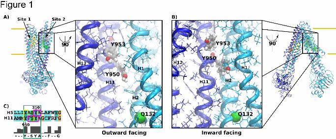
Position and local geometry of site 2 are illustrated in Fig. 1A and B and compared between
the outward (model based on Sav1866 from S. aureus (Dawson and Locher, 2006) and the
inward facing structures (model based on ABCB1 from C. elegans (Jin et al., 2012). The
local geometry at site 2 remained remarkably similar between the two structures. We
observed a reduction in the distance between transmembrane helices 1 and 12 by
approximately 2 Å during the transition from the inward to the outward facing structure, while
accessibility to residues Y950 and Y953 was comparable. The hydroxyl functional groups of
both tyrosine residues remained water exposed in both conformations and would therefore
be able to form hydrogen bonds to a ligand binding to site 2.
Effect of the removal of Y950 and Y953 OH-groups on rhodamine 123 efflux
P-gp mutants were first characterized for their ability to transport rh123 in order to assess
any potential effect of changes in its transport rate on evaluation of propafenones. Rh123 is a
preferred site 1 substrate, but also transported via site 2. First order rate constants k were
normalized by expression to account for differences in the amount of protein present at the
plasma membrane, which differs as a result of using a transient expression system (refer to
supplementary Fig. 4 for the linear relationship between first order rate constants and
expression). The (normalized) transport rates are therefore independent of the amount of
protein expressed. All mutants were detected at the plasma membrane, though the
expression levels of Q132R/Y950F, Q773R/Y950F and Y307F/Y310F were reduced
(supplementary Fig. 5). Correct formation of the contact interfaces between helices 2/11 and
5/8 is an important requirement for proper engagement of coupling helices 2 and 4 into the
sockets formed between the core and the alpha-helical domain of NBD1 and NBD2,
respectively. Less efficient folding of these mutants, which reside in the 2/11 and 5/8
interfaces, is likely the cause of lower plasma membrane expression.
This article has not been copyedited and formatted. The final version may differ from this version.Molecular Pharmacology Fast Forward. Published on December 23, 2013 as DOI: 10.1124/mol.113.088526
at ASPE
T Journals on M
ay 1, 2018m
olpharm.aspetjournals.org
Dow
nloaded from
MOL #88526
13
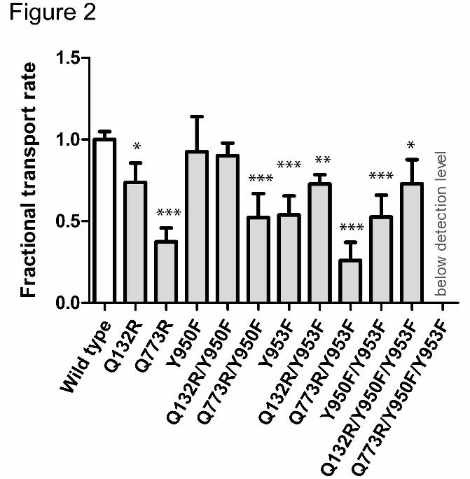
Mutant Y950F showed a similar transport rate as wild type protein, while mutant Y953F
showed a significantly decreased rate (54 ± 12% of wild type) (Fig. 2). The double mutant
Y950F/Y953F showed a decrease that was comparable to that observed for the Y953F
single mutant alone. Thus only the OH-group of residue Y953, but not that of Y950
contributes to rh123 transport.
Access of rh123 to binding site 1 and 2 can be controlled by the introduction of positive
charges (arginines) in the access path to the ligand binding sites. We have previously shown
that the Q132R mutation blocks access to site 2 for rh123, while the Q773R mutation
prevents rh123 access to site 1 (Parveen et al., 2011). The effect of the Y953F mutation on
rh123 efflux should be abolished by introducing selector residue R132 and be more
pronounced when deselecting site 1 by introducing selector residue R773. Introduction of
arginine residues in positions 132 or 773 led to a decrease in rh123 efflux to 75 ± 11% and
37 ± 8% of wild type, respectively (Fig. 2). These data are in agreement with our previous
results, in which the same rank order of transport activity was found. As expected, presence
of the selector residue R132 abrogated the effect of single and double tyrosine mutation on
transport activity. This is illustrated by comparable transport activity of the Q132R/Y950F,
Q132R/953F and Q132R/Y950F/Y953F mutants. These results also demonstrate that Y to F
mutations in positions 950 and 953 do not perturb protein mechanics. This is an important
finding, because impairment of function is a frequently encountered limitation of site directed
mutagenesis (Ito et al., 2001; Koike et al., 2002; Swartz et al., 2013).
In contrast, transport rates were found to be lower in the Q773R/Y953F mutant as compared
to the Q773R mutant alone, though this decrease did not reach statistical significance.
Definitely, no decrease in transport activity was observed for the Y950F mutant introduced in
the R773 background. In summary, these results demonstrate that the hydroxyl group of
residue 953, but not that of 950, plays a role for rh123 transport.
Interestingly, neither single nor double mutation of corresponding residues Y307 and Y310 in
binding site 1 changed rh123 efflux (supplementary Fig. 6). These findings indicate that
This article has not been copyedited and formatted. The final version may differ from this version.Molecular Pharmacology Fast Forward. Published on December 23, 2013 as DOI: 10.1124/mol.113.088526
at ASPE
T Journals on M
ay 1, 2018m
olpharm.aspetjournals.org
Dow
nloaded from
MOL #88526
14
hydrogen bonding interactions are of minor importance for interaction of rh123 with tyrosines
located on helix 5. This clearly reflects evolutionary divergence of the two sites. While
aromatic interactions likely are important, these were not subject of the present study.
Role of Y950 and Y953 for the formation of hydrogen bonds with propafenone
analogues
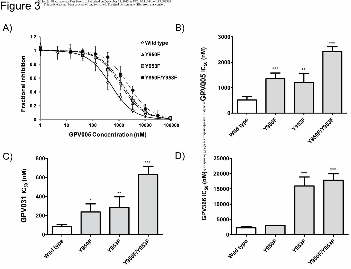
Protonatable propafenones GPV005 and GPV031 were chosen to be studied with wild type
protein and in the R132 background. The non-protonatable acid amide GPV366 served as a
control that is not influenced by the selector residue R132. Representative examples of
concentration response curves for compound GPV005 are shown for wild type, single and
double tyrosine mutants (Fig. 3A). Wild type showed an IC50 value of 518 ± 141 nM, while the
values for the single mutants Y950F and Y953F were 1348 ± 229 and 1207 ± 362 nM,
respectively. The double mutant had an IC50 value of 2418 ± 194 nM (Fig. 3B, refer to Table
1 of the supplemental material for numerical values and Tables 2A and 2B for statistical
significance of differences in IC50 values). An analogous pattern was seen for GPV031 (wild
type: 85 ± 22 nM, Y950F: 238 ± 84 nM, Y953F: 287 ± 109 nM, Y950F/Y953F: 630 ± 87 nM)
(Fig. 3C). The fold change in IC50 values was thus similar for both compounds. Data suggest
H-bonding interactions of compounds with both tyrosine OH-groups, because a comparable
increase in IC50 values was observed for each of them and an additive effect was seen when
both of them were mutated. Fig. 3D and supplementary Table 1 summarize data for the non-
protonatable acid amide GPV366. In contrast to protonatable propafenones, a higher IC50
value was observed for the Y953F, but not for the Y950F single mutant. The fold change for
the double mutant was comparable to that observed for the Y953F single mutant (wild type:
2239 ± 391 nM, Y950F: 2984 ± 79 nM (n.s.), Y953F: 15946 ± 2941 nM (7.1 fold change
relative to wild type), Y950F/Y953F: 17819 ± 2106 nM (8 fold change).
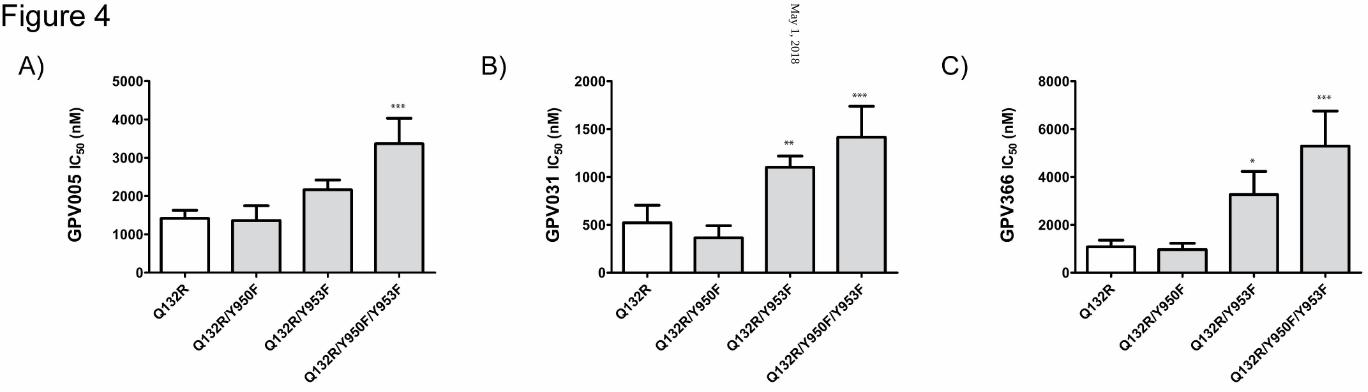
Effect of site 2 tyrosine mutants in the R132 background The at least 3-fold difference in IC50 values for wild type protein (GPV005: 518 ± 141 nM,
GPV031: 85 ± 22 nM) (Fig. 3B, C) and the Q132R mutant (GPV005: 1414 ± 215 nM,
This article has not been copyedited and formatted. The final version may differ from this version.Molecular Pharmacology Fast Forward. Published on December 23, 2013 as DOI: 10.1124/mol.113.088526
at ASPE
T Journals on M
ay 1, 2018m
olpharm.aspetjournals.org
Dow
nloaded from
MOL #88526
15
GPV031: 521 ± 183 nM) (Fig. 4A, B) demonstrates that an arginine residue in position 132
prevents access of protonatable compounds to site 2. Introducing tyrosine mutations in the
R132 background should in principle make IC50 values of protonatable compounds
independent of the presence of site 2 mutations. Indeed data in Fig. 4A, B and
supplementary Table 1 show that for the Q132R/Y950F mutant IC50 values were similar to
those observed in the Q132R background alone. However IC50 values were still higher in the
Q132R/Y953F mutation. This increase was not significant for GPV005, but reached
significance for GPV031. Thus, removal of the hydroxyl group of tyrosine 953 still affected
IC50 values of protonatable propafenones, indicating that protonatable compounds were still
able to access site 2, even though to a lesser extent. Incomplete prevention of access is a
result of ligand properties and not the transporter itself, because both, GPV005 and GPV031
have a pKa (Ecker et al., 1999), which results in charged and uncharged species to coexist
under physiological conditions (further details are given in the discussion section).
For the non-protonatable acid amide GPV366 a higher IC50 value was observed in the
Q132R/Y953F mutant (Q132R: 1074 ± 282 nM, Q132R/Y953F: 3261 ± 965 nM), while the
Y950F mutation in the same background did not affect potency (966 ± 259 nM) (Fig. 4C).
Although the pattern was similar to that observed in the wild type background, the fold
change was lower in the Q132R background and the IC50 value was somewhat lower in the
protein containing the Q132R mutation than for wild type. A possible explanation for this
effect is the lack of competition of GPV366 in the preferred binding site 2 with rh123,
because rh123 binding to site 2 is abolished by the Q132R mutation.
The higher IC50 values in the Q132R/Y950F/Y953F were not due to an additive effect of the
two tyrosine mutations on binding of propafenones. Therefore the observed effect cannot be
explained by a contribution from tyrosine mutations on small molecule binding and therefore
has to have other reasons. Certainly, the effect is not brought about by a global perturbance
of protein structure and function, because the Q132R mutant and the Q132R/Y950F/Y953F
triple mutant showed comparable rh123 transport rates (cf. Fig. 2). All three mutations are
This article has not been copyedited and formatted. The final version may differ from this version.Molecular Pharmacology Fast Forward. Published on December 23, 2013 as DOI: 10.1124/mol.113.088526
at ASPE
T Journals on M
ay 1, 2018m
olpharm.aspetjournals.org
Dow
nloaded from
MOL #88526
16
located at the contact interface of helices 2 and 11 and might thus locally influence the
geometry of binding site 2 in the triple mutant. Importantly, this effect does not have a
bearing on the interpretation of results in this study.
This article has not been copyedited and formatted. The final version may differ from this version.Molecular Pharmacology Fast Forward. Published on December 23, 2013 as DOI: 10.1124/mol.113.088526
at ASPE
T Journals on M
ay 1, 2018m
olpharm.aspetjournals.org
Dow
nloaded from
MOL #88526
17
Discussion
The presence of tyrosines in binding cavities is important both for specificity and affinity of
solutes towards proteins (Koide and Sidhu, 2009). This has been illustrated for a number of
bacterial, rodent and human transporters. The importance of tyrosine residues for the
interaction of the bacterial exporter QacA from Staphylococcus aureus with solutes was
studied by Wu et al (Wu et al., 2008). Mutation of residue Y410 in the drug binding pocket to
phenylalanine led to a decrease in the transport rate of QacA for several substrates,
including monovalent dyes. A study on the human ABCC1 transporter (multidrug resistance
protein, MRP1) found that mutation of two cytoplasmic loop tyrosines (Y1189 and Y1190) to
alanine or serine, respectively, resulted in a 50% reduced transport rate for different organic
anion substrates, particularly glutathione (Conseil et al., 2005). Mammalian ABC transporters
are rich in aromatic residues in the apical region of the central cavity as illustrated by the
crystal structure of mouse ABCB1, protein homology models of human ABCB1 and the
related human hepatic transporter ABCB4 (Gutmann et al., 2010), as well as the crystal
structure of ABCB10 (Shintre et al., 2013). Interactions formed at the apex of the central
cavity may be a prerequisite for substrate induced nucleotide occlusion in one of the
composite nucleotide binding site, which in turn affords the outward facing conformation and
substrate release (Sauna et al., 2007; Tombline et al., 2006; Tombline et al., 2005).
Our study of the importance of the hydrogen-bonding capability of Y950 and Y953 was
motivated by three lines of evidence: (i) the critical role that tyrosines play in molecular
recognition, (ii) the projected location of these residues in binding site 2 and (iii) the presence
of a conserved Y(F)xSYA motif, in which the second tyrosine is photolabeled by
propafenones (Parveen et al., 2011). Residue Y953 seems to be highly conserved in multiple
sequence alignments of annotated P-gp orthologs, while substitutions of Y950 by
phenylalanine are more frequent.
In order to address the role of H-bonding interactions of these two tyrosines for rh123 efflux,
the transport rate of this preferred site 1 substrate was determined either in wild type or the
This article has not been copyedited and formatted. The final version may differ from this version.Molecular Pharmacology Fast Forward. Published on December 23, 2013 as DOI: 10.1124/mol.113.088526
at ASPE
T Journals on M
ay 1, 2018m
olpharm.aspetjournals.org
Dow
nloaded from
MOL #88526
18
respective selector mutation background. Residues Q132 and Q773 are located below and
outside the binding sites, and replacement by arginine was previously shown to direct
protonatable compounds away from one and towards the respective alternate site by charge
repulsion. Experimental evidence indicates Y953 to represent an H-bonding partner for
rh123, because a significant decrease in transport rates in wild type and the Q773R
backgrounds was observed. Identical transport rates were found for the mutant Q132R and
the Y953F mutant in the Q132R background. This finding was expected, because (i) rh123
always carries one positive charge at physiological pH (Shinomiya et al., 1992) and (ii)
introducing selector residue R132 would abolish any influence that mutations in the site,
which is now inaccessible for ligands, would have.
For reasons detailed in the results section, hydrogen bonding interactions of tyrosines with
propafenone analogs GPV005, GPV031 and GPV366 were assessed by determining IC50
values for rh123 efflux inhibition. Both residues Y950 and Y953 were demonstrated to play a
role as hydrogen bonding partners for the protonatable propafenones GPV005 and GPV031,
whereby the effect was additive in the double mutant. GPV366 showed a hydrogen bonding
interaction with residue Y953, while residue Y950 did not contribute. As expected, the double
mutant showed an IC50 value that was comparable to that of the Y953F mutant.
In principle, access of charged propafenones to preferred site 2 should be prevented by
introduction of the selector mutation R132. IC50 values were therefore expected to be the
same in single and double tyrosine mutants generated in the Q132R background. Indeed no
difference was observed between the Q132R single and the Q132R/Y950F double mutant.
However, higher IC50 values were found for the Y953F mutant, though less pronounced than
in the wild type background. These results indicate that propafenones can still reach site 2.
Propafenones coexist in protonated and non-protonated forms at physiological pH. The
uncharged ligand species can still have access to site 2, which has been deselected for the
protonated ligand species by introducing the Q132R mutation. The permanently charged
rh123 is entirely prevented from reaching site 2 by introducing selector residue R132. The
This article has not been copyedited and formatted. The final version may differ from this version.Molecular Pharmacology Fast Forward. Published on December 23, 2013 as DOI: 10.1124/mol.113.088526
at ASPE
T Journals on M
ay 1, 2018m
olpharm.aspetjournals.org
Dow
nloaded from
MOL #88526
19
pKa value of GPV005 is 8.4 and that of GPV031 is 7.3 (Ecker et al., 1999). This allows
estimating the percentage of protonated species of GPV005 and GPV031 at intracellular pH.
Accordingly, the charged species amounts to >90% and 60-70%, respectively. GPV005
would therefore be prevented from accessing site 2 to a larger extent than GPV031. Indeed
results in Fig. 4A and B show that this is the case. For GPV005 mutant Q132R/Y953F
showed a 1.5 fold higher IC50 value than the Q132R mutation, while the fold change for
GPV031 was 2.1 fold and that for uncharged GPV366 was 3.0 fold. From experiments in the
wild type background (Fig. 3), an equal contribution of residues Y950 and Y953 was
expected. However, such an effect was not observed in the Q132R background (Figs. 4A
and B). Residue Y950 thus lost its importance as an interaction partner, possibly due to the
close proximity of residues Q132R and Y950, which are predicted in models to have a C-
alpha distance of approximately 7 Å. Though residue Q132 has been shown not to be a
direct interaction partner for propafenone type ligands (Parveen et al, 2011), a steric effect of
the arginine side chain is likely, but only seen for those propafenone analogs that interact
with Y950 (GPV005, GPV031, not GPV366).
The triple Q132R/Y950F/Y953F mutant showed a further increase in IC50 values for all
compounds, which is obviously not due to interaction with tyrosines. As discussed in the
results section, this finding is hypothesized to be due to local changes in site 2. The fact that
the extent of deselecting inversely correlates with the fold change (2.3 fold for GPV005, 2.7
fold for GPV031 and 4.9 fold for GPV366) is supporting this hypothesis.
Interaction of propafenones with the transporter was assessed by determining IC50 values for
rh123 efflux inhibition. In these experiments, both the interaction of P-gp with rh123 and with
propafenones contributes to the outcome of the experiments. We therefore addressed the
question if a change in rh123 efflux would affect propafenone IC50 values in the way we
observed. When the transporter is partially impaired, an inhibition should be easier to
accomplish. In contrast, our experiments showed the inverse, i.e. an increase in IC50 values,
rather than a decrease. Therefore the effects seen for the mutants are certainly not due to
This article has not been copyedited and formatted. The final version may differ from this version.Molecular Pharmacology Fast Forward. Published on December 23, 2013 as DOI: 10.1124/mol.113.088526
at ASPE
T Journals on M
ay 1, 2018m
olpharm.aspetjournals.org
Dow
nloaded from
MOL #88526
20
the observed decrease in rh123 flux, because in that case they should be the opposite of
what we observed.
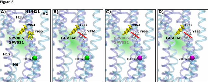
Fig. 5 summarizes results in a schematic way. Tyrosines are shown in yellow, when found
important for H-bond formation and in gray, when not. Tyrosines Y950 and Y953 are H-
bonding with protonatable compounds GPV005 and GPV031 (panel A), while only residue
Y953 is important for interaction with the non-protonatable acid amide GPV366 (panel B).
When the wild type glutamine in position 132 is replaced by arginine, the probability of
protonatable compounds to reach site 2 is decreased. This is indicated by gray font color of
compounds GPV005 and GPV031. Also residue Y950 loses its importance as a residue that
forms H-bonds with propafenones (panel C). Compound GPV366 only interacts with residue
Y953 (panel D).
Our results are in agreement with earlier work on tyrosine residues in mouse and human P-
gp. These previous studies evaluated toxicity (a composite of the influence of mutation on
trafficking and function) (Hanna et al., 1996) and ATPase activity of reconstituted P-gp (Loo
and Clarke, 2000), respectively. A direct evaluation of transport activity has not been
performed in earlier work. Philippe Gros’ group studied alanine mutants of tyrosine residues
Y946 and Y949 in mouse mdr3. These residues are found in analogous position to tyrosines
Y950 and Y953 in the human sequence. The ability of mutants to confer resistance to
actinomycin D, adriamycin, colchicine and vinblastine was determined. In these experiments
mutations to alanine probed for both H-bonding and aromatic interactions. The Y949A
mutation increased colchicine and adriamycin toxicity, the Y946A made cells more sensitive
to vinblastine, while both mutants increased sensitivity towards actinomycin D. These data
are in agreement with the finding that drugs, which we previously found to preferentially bind
to site 1 or site 2, respectively, are affected by mutation of tyrosines. Loo and Clarke
demonstrated altered ATPase stimulation of the human protein for the Y953A mutant by
verapamil, colchicine and vinblastine, but did not present evidence for a direct involvement in
drug binding.
This article has not been copyedited and formatted. The final version may differ from this version.Molecular Pharmacology Fast Forward. Published on December 23, 2013 as DOI: 10.1124/mol.113.088526
at ASPE
T Journals on M
ay 1, 2018m
olpharm.aspetjournals.org
Dow
nloaded from
MOL #88526
21
Conclusions
A dual substrate binding mode is rooted in the rotational C2 symmetry of human ABC
transporters. In this manuscript, the contribution of active site tyrosine residues for substrate
interaction with propafenones has been studied with respect to their preferred interaction site
2. Propafenones were used as model compounds because of the availability of photolabeling
data, which previously showed a dual pseudosymmetric mode of interaction with P-
glycoprotein. The photolabeled tyrosine residue Y953 is shown to form hydrogen bonds with
protonatable and non-protonatable propafenones, but also with preferred site 1 substrate
rh123. We provide proof of principle that the preferred propafenone binding site can be
studied individually and the influence of mutation can be resolved in a site specific manner.
We believe that dual symmetry related sites present in other human ABC exporters may
potentially be linked to disease etiologies. One candidate is the human bile salt export pump,
which specifically transports conjugated bile acids, but at the same time is inhibited by
numerous systemically administered drugs.
This article has not been copyedited and formatted. The final version may differ from this version.Molecular Pharmacology Fast Forward. Published on December 23, 2013 as DOI: 10.1124/mol.113.088526
at ASPE
T Journals on M
ay 1, 2018m
olpharm.aspetjournals.org
Dow
nloaded from
MOL #88526
22
Authorship contributions
Participated in research design: Dönmez Cakil, Stockner, Chiba
Conducted experiments: Dönmez Cakil, Khunweeraphong, Parveen, Artaker, Stockner
Contributed new reagents or analytic tools: Pusch, Schmid
Performed data analysis: Dönmez Cakil, Stockner, Chiba
Wrote or contributed to writing of the manuscript: Dönmez Cakil, Sitte, Ecker, Stockner,
Chiba
This article has not been copyedited and formatted. The final version may differ from this version.Molecular Pharmacology Fast Forward. Published on December 23, 2013 as DOI: 10.1124/mol.113.088526
at ASPE
T Journals on M
ay 1, 2018m
olpharm.aspetjournals.org
Dow
nloaded from
MOL #88526
23
References
Conseil G, Deeley RG and Cole SP (2005) Role of two adjacent cytoplasmic tyrosine residues in MRP1
(ABCC1) transport activity and sensitivity to sulfonylureas. Biochem Pharmacol69(3): 451-
461.
Cramer J, Kopp S, Bates SE, Chiba P and Ecker GF (2007) Multispecificity of drug transporters: probing
inhibitor selectivity for the human drug efflux transporters ABCB1 and ABCG2.
ChemMedChem2(12): 1783-1788.
Dawson RJ and Locher KP (2006) Structure of a bacterial multidrug ABC transporter.
Nature443(7108): 180-185.
Ecker G, Huber M, Schmid D and Chiba P (1999) The importance of a nitrogen atom in modulators of
multidrug resistance. Mol Pharmacol56(4): 791-796.
Gatlik-Landwojtowicz E, Aanismaa P and Seelig A (2006) Quantification and characterization of P-
glycoprotein-substrate interactions. Biochemistry45(9): 3020-3032.
Giacomini KM, Huang SM, Tweedie DJ, Benet LZ, Brouwer KL, Chu X, Dahlin A, Evers R, Fischer V,
Hillgren KM, Hoffmaster KA, Ishikawa T, Keppler D, Kim RB, Lee CA, Niemi M, Polli JW,
Sugiyama Y, Swaan PW, Ware JA, Wright SH, Yee SW, Zamek-Gliszczynski MJ and Zhang L
(2010) Membrane transporters in drug development. Nat Rev Drug Discov9(3): 215-236.
Gutmann DA, Ward A, Urbatsch IL, Chang G and van Veen HW (2010) Understanding polyspecificity
of multidrug ABC transporters: closing in on the gaps in ABCB1. Trends Biochem Sci35(1): 36-
42.
Hanna M, Brault M, Kwan T, Kast C and Gros P (1996) Mutagenesis of transmembrane domain 11 of
P-glycoprotein by alanine scanning. Biochemistry35(11): 3625-3635.
Hartley JL, Temple GF and Brasch MA (2000) DNA cloning using in vitro site-specific recombination.
Genome Res10(11): 1788-1795.
Ito K, Oleschuk CJ, Westlake C, Vasa MZ, Deeley RG and Cole SP (2001) Mutation of Trp1254 in the
multispecific organic anion transporter, multidrug resistance protein 2 (MRP2) (ABCC2),
alters substrate specificity and results in loss of methotrexate transport activity. J Biol
Chem276(41): 38108-38114.
Jin MS, Oldham ML, Zhang Q and Chen J (2012) Crystal structure of the multidrug transporter P-
glycoprotein from Caenorhabditis elegans. Nature490(7421): 566-569.
Koide S and Sidhu SS (2009) The importance of being tyrosine: lessons in molecular recognition from
minimalist synthetic binding proteins. ACS Chem Biol4(5): 325-334.
Koike K, Oleschuk CJ, Haimeur A, Olsen SL, Deeley RG and Cole SP (2002) Multiple membrane-
associated tryptophan residues contribute to the transport activity and substrate specificity
of the human multidrug resistance protein, MRP1. J Biol Chem277(51): 49495-49503.
Linton KJ, Zolnerciks JK and Schmitt L (2011) The ABC Transporters of Human Physiology and Disease:
Genetics and Biochemistry of ATP Binding Cassette Transporters, World Scientific Publishing,
London.
Liu M, Hou T, Feng Z and Li Y (2013) The flexibility of P-glycoprotein for its poly-specific drug binding
from molecular dynamics simulations. J Biomol Struct Dyn31(6): 612-629.
Loo TW and Clarke DM (2000) Identification of residues within the drug-binding domain of the
human multidrug resistance P-glycoprotein by cysteine-scanning mutagenesis and reaction
with dibromobimane. J Biol Chem275(50): 39272-39278.
MacCallum JL, Bennett WF and Tieleman DP (2007) Partitioning of amino acid side chains into lipid
bilayers: results from computer simulations and comparison to experiment. J Gen
Physiol129(5): 371-377.
Marti-Renom MA, Stuart AC, Fiser A, Sanchez R, Melo F and Sali A (2000) Comparative protein
structure modeling of genes and genomes. Annu Rev Biophys Biomol Struct29: 291-325.
Mian IS, Bradwell AR and Olson AJ (1991) Structure, function and properties of antibody binding
sites. J Mol Biol217(1): 133-151.
This article has not been copyedited and formatted. The final version may differ from this version.Molecular Pharmacology Fast Forward. Published on December 23, 2013 as DOI: 10.1124/mol.113.088526
at ASPE
T Journals on M
ay 1, 2018m
olpharm.aspetjournals.org
Dow
nloaded from
MOL #88526
24
Moffat J, Grueneberg DA, Yang X, Kim SY, Kloepfer AM, Hinkle G, Piqani B, Eisenhaure TM, Luo B,
Grenier JK, Carpenter AE, Foo SY, Stewart SA, Stockwell BR, Hacohen N, Hahn WC, Lander ES,
Sabatini DM and Root DE (2006) A lentiviral RNAi library for human and mouse genes applied
to an arrayed viral high-content screen. Cell124(6): 1283-1298.
Pace CN, Horn G, Hebert EJ, Bechert J, Shaw K, Urbanikova L, Scholtz JM and Sevcik J (2001) Tyrosine
hydrogen bonds make a large contribution to protein stability. J Mol Biol312(2): 393-404.
Parveen Z, Stockner T, Bentele C, Pferschy S, Kraupp M, Freissmuth M, Ecker GF and Chiba P (2011)
Molecular dissection of dual pseudosymmetric solute translocation pathways in human P-
glycoprotein. Mol Pharmacol79(3): 443-452.
Pleban K, Kopp S, Csaszar E, Peer M, Hrebicek T, Rizzi A, Ecker GF and Chiba P (2005) P-glycoprotein
substrate binding domains are located at the transmembrane domain/transmembrane
domain interfaces: a combined photoaffinity labeling-protein homology modeling approach.
Mol Pharmacol67(2): 365-374.
Sali A and Blundell TL (1993) Comparative protein modelling by satisfaction of spatial restraints. J Mol
Biol234(3): 779-815.
Sauna ZE, Kim IW, Nandigama K, Kopp S, Chiba P and Ambudkar SV (2007) Catalytic cycle of ATP
hydrolysis by P-glycoprotein: evidence for formation of the E.S reaction intermediate with
ATP-gamma-S, a nonhydrolyzable analogue of ATP. Biochemistry46(48): 13787-13799.
Schmid D, Ecker G, Kopp S, Hitzler M and Chiba P (1999) Structure-activity relationship studies of
propafenone analogs based on P-glycoprotein ATPase activity measurements. Biochem
Pharmacol58(9): 1447-1456.
Shinomiya N, Tsuru S, Katsura Y, Sekiguchi I, Suzuki M and Nomoto K (1992) Increased mitochondrial
uptake of rhodamine 123 by CDDP treatment. Exp Cell Res198(1): 159-163.
Shintre CA, Pike AC, Li Q, Kim JI, Barr AJ, Goubin S, Shrestha L, Yang J, Berridge G, Ross J, Stansfeld PJ,
Sansom MS, Edwards AM, Bountra C, Marsden BD, von Delft F, Bullock AN, Gileadi O,
Burgess-Brown NA and Carpenter EP (2013) Structures of ABCB10, a human ATP-binding
cassette transporter in apo- and nucleotide-bound states. Proc Natl Acad Sci U S A110(24):
9710-9715.
Stockner T, de Vries SJ, Bonvin AM, Ecker GF and Chiba P (2009) Data-driven homology modelling of
P-glycoprotein in the ATP-bound state indicates flexibility of the transmembrane domains.
FEBS J276(4): 964-972.
Swartz DJ, Weber J and Urbatsch IL (2013) P-glycoprotein is fully active after multiple tryptophan
substitutions. Biochim Biophys Acta1828(3): 1159-1168.
Tombline G, Donnelly DJ, Holt JJ, You Y, Ye M, Gannon MK, Nygren CL and Detty MR (2006)
Stimulation of P-glycoprotein ATPase by analogues of tetramethylrosamine: coupling of drug
binding at the "R" site to the ATP hydrolysis transition state. Biochemistry45(26): 8034-8047.
Tombline G, Muharemagic A, White LB and Senior AE (2005) Involvement of the "occluded
nucleotide conformation" of P-glycoprotein in the catalytic pathway. Biochemistry44(38):
12879-12886.
Wu J, Hassan KA, Skurray RA and Brown MH (2008) Functional analyses reveal an important role for
tyrosine residues in the staphylococcal multidrug efflux protein QacA. BMC Microbiol8: 147.
This article has not been copyedited and formatted. The final version may differ from this version.Molecular Pharmacology Fast Forward. Published on December 23, 2013 as DOI: 10.1124/mol.113.088526
at ASPE
T Journals on M
ay 1, 2018m
olpharm.aspetjournals.org
Dow
nloaded from
MOL #88526
25
Footnotes
This research was supported by grants from the Austrian Science Fund FWF [stand alone
project 23319, Special Research Fund 35-3509]; and the European Cooperation in Science
and Technology [e-COST Action CM0902]. NK acknowledges receipt of a scholarship from
the Austrian Federal Ministry of Science and Research (BMWF).
This article has not been copyedited and formatted. The final version may differ from this version.Molecular Pharmacology Fast Forward. Published on December 23, 2013 as DOI: 10.1124/mol.113.088526
at ASPE
T Journals on M
ay 1, 2018m
olpharm.aspetjournals.org
Dow
nloaded from
MOL #88526
26
Figure legends:
Figure 1: Location of site 1 (orange; preferred rh123 site) and site 2 (green; preferred
propafenone, verapamil and vinblastine site) are indicated in the homology models of P-gp.
Panel A shows their location in the outward facing P-glycoprotein structure and in a close up
of site 2 in a 90º rotated transporter. Site 2 is viewed from the pore. Residues Q773 (orange)
and Q132 (green) lie proximal in the path taken by transported compounds. When mutated to
arginine, these residues decrease the binding probability of protonatable compounds in
either of the two sites. Panel B shows the same representation of overall structure and a
close up of site 2 for the inward facing conformation. Panel C shows the sequence alignment
of helices 5 and 11 of P-gp. The cyan box shows the conserved Y(F)xSYA motif, yellow
boxes highlight mutated residues tyrosine Y950 and Y953. Residues which were previously
shown to be strongly labeled by photoactivated propafenones are indicated in magenta.
Figure 2: Bar graph showing changes in the fractional transport rate of mutants in
comparison with wild type. Rh123 efflux was monitored continuously for 5 min. First order
rate constants (k) were calculated from an exponential fit and normalized to surface
expression that was determined by MRK16 staining. Q773/Y950F/Y953F was expressed at
the surface, but no rh123 efflux was detectable. Each value represents the mean ± SD of at
least 3 independent experiments (*p< 0.05, ** p< 0.01 ***p< 0.001 compared with wild type
P-gp).
Figure 3: Inhibition of rh123 efflux was measured in the absence and presence of 8 serial
dilutions of GPV005. Wild type: open diamonds; Y950F/Y953F: filled circles; Y950F:
triangles; Y953F: squares (A). IC50 values for GPV005 (B), GPV031 (C) and GPV366 (D)
were determined by fitting hyperbolic concentration response curves to data points by non-
linear regression analysis and calculation of 50% occupancy values. Mean ± SD were
calculated from at least 3 independent experiments (*p< 0.05, **p< 0.01 ***p< 0.001
compared with wild type P-gp).
This article has not been copyedited and formatted. The final version may differ from this version.Molecular Pharmacology Fast Forward. Published on December 23, 2013 as DOI: 10.1124/mol.113.088526
at ASPE
T Journals on M
ay 1, 2018m
olpharm.aspetjournals.org
Dow
nloaded from
MOL #88526
27
Figure 4: Same as Fig. 3, but tyrosine mutants generated in the Q132R mutation
background. Mean ± SD were calculated from at least 3 independent experiments. GPV005
(A), GPV031 (B) and GPV366 (C) (*p< 0.05, **p< 0.01 ***p< 0.001 compared with Q132R
mutation).
Figure 5: Schematic presentation of the importance of tyrosine hydroxyl groups (tyrosines;
yellow) for interaction with propafenone analogs in the outward facing P-glycoprotein
structure (template Sav1866) panel A and B; Cα atom of residue Q132 shown as a green
sphere) and after introduction of the R132 selector mutation (panel C and D; Cα atom of
residue Q132 shown in magenta). The hydroxyl-groups of both Y950 and Y953 are able to
form hydrogen bonds with protonatable propafenones GPV005 and GPV031 (A), while
compound GPV366 only forms hydrogen bonds with residue Y953 (B). The gray color of
residue Y950 and red bar indicate that hydrogen bonds of residue Y950 are not important for
interaction. The presence of the selector residue R132 (panel C, D) eliminates the
importance of the hydroxyl group of Y950 for propafenone analog binding, while the
interaction of residue Y953 continues to contribute to the interaction. Use of grey font color
indicates incomplete prevention of access of compounds GPV005 and GPV031 in panel C.
This article has not been copyedited and formatted. The final version may differ from this version.Molecular Pharmacology Fast Forward. Published on December 23, 2013 as DOI: 10.1124/mol.113.088526
at ASPE
T Journals on M
ay 1, 2018m
olpharm.aspetjournals.org
Dow
nloaded from
This article has not been copyedited and formatted. The final version may differ from this version.Molecular Pharmacology Fast Forward. Published on December 23, 2013 as DOI: 10.1124/mol.113.088526
at ASPE
T Journals on M
ay 1, 2018m
olpharm.aspetjournals.org
Dow
nloaded from
This article has not been copyedited and formatted. The final version may differ from this version.Molecular Pharmacology Fast Forward. Published on December 23, 2013 as DOI: 10.1124/mol.113.088526
at ASPE
T Journals on M
ay 1, 2018m
olpharm.aspetjournals.org
Dow
nloaded from
This article has not been copyedited and formatted. The final version may differ from this version.Molecular Pharmacology Fast Forward. Published on December 23, 2013 as DOI: 10.1124/mol.113.088526
at ASPE
T Journals on M
ay 1, 2018m
olpharm.aspetjournals.org
Dow
nloaded from
This article has not been copyedited and formatted. The final version may differ from this version.Molecular Pharmacology Fast Forward. Published on December 23, 2013 as DOI: 10.1124/mol.113.088526
at ASPE
T Journals on M
ay 1, 2018m
olpharm.aspetjournals.org
Dow
nloaded from
This article has not been copyedited and formatted. The final version may differ from this version.Molecular Pharmacology Fast Forward. Published on December 23, 2013 as DOI: 10.1124/mol.113.088526
at ASPE
T Journals on M
ay 1, 2018m
olpharm.aspetjournals.org
Dow
nloaded from