Embed Size (px)
Citation preview
59
Acta Pharm. 72 (2022) 59–77 Original research paper https://doi.org/10.2478/acph-2022-0009
Molecular networking-assisted flavonoid profile of Gypsophila glomerata extract in relation to its protective effects on carbon
tetrachloride-induced hepatorenal damage in rats
The aim of the study was to provide an in-depth characteri-zation of the methanol-aqueous extract from the aerial parts of Gypsophila glomerata Pall. Ex Adams (Caryophylla-ceae) (EGG) and to assess its protective potential on carbon tetrachloride (CCl4)-induced liver and kidney damage in male Wistar rats. Twenty-two flavonoid C-, O- and C,O- -glycosides in EGG were annotated by mass spectrometry- -based molecular networking; nine of them are reported in this species for the first time. Fourteen-day oral admini-stration of EGG at a dose 200 mg kg–1 bm prevented signifi-cantly CCl4-induced liver injury, discerned by an ameliora-tion of the markers of oxidative stress (GSH and MDA) and transaminase activity. EGG decreased the serum level of urea and creatinine as well. The observed improvement of biochemical parameters was supported by histopatholo-gical observations. The protective hepatorenal effects of EGG, rich in 2“-О-pentosyl-6-С-hexosyl-apigenin/luteolin/methylluteolin and their acetyl- and methoxycinnamoyl-derivatives, were comparable with the effects of the posi-tive control silymarin.
Keywords: Gypsophila glomerata, CCl4, molecular network, hepatorenal toxicity, flavonoids
Nowadays, there is an increase in scientific interest in herbal medicines, since they could be a good natural alternative for synthetic drugs and a safe source of helpful sub-stances and antioxidants, e.g., flavonoids, xanthones, terpenoids, although some concerns about their safety are also reported (1). It is well known that oxidative damage of DNA, lipids or proteins is associated with some chronic diseases (2) like hypertension, coronary diseases (2, 3), diabetes (2, 4) and cancer (2, 5). In the light of the above, the role of herbal drugs in the prevention or control of the aforementioned diseases is attributed mainly to the antioxidative effects of their constituents (5, 6).
Proven antioxidative properties (7, 8), as well as nephroprotective (9, 10), hepatoprotec-tive and liver regenerative effects (11) along with anti-inflammatory, immunomodulatory
BOJANA DIMITROVA1,2
RUMYANA VITANSKA1,*
RENETA GEVRENOVA2
DIMITRINA ZHELEVA-DIMITROVA2
VESSELA BALABANOVA2
STOYCHO STOEV3
1 Department of Pharmacology Pharmacotherapy and Toxicology Faculty of Pharmacy, Medical University of Sofia, 1000 Sofia2 Department of Pharmacognosy and Pharmaceutical Botany, Faculty of Pharmacy, Medical University of Sofia 1000 Sofia3 Department of General and Clinical Pathology, Faculty of Veterinary Medicine, Trakia University, Students Campus, 6000 Stara Zagora, Bulgaria
Accepted February 18, 2021 Published online March 6, 2021
* Correspondence; e-mail address: [email protected]
60
B. Dimitorva et al.: Molecular networking-assisted flavonoid profile of Gypsophila glomerata extract in relation to its protective effects on carbon tetrachloride-induced hepatorenal damage in rats, Acta Pharm. 72 (2022) 59–77.
and membrane-stabilizing effects (12) were reported for different flavonoids, and espe-cially for the flavolignan complex silymarin from Silybum marianum fruits, which was used as a positive control in this work.
The Gypsophila species are highly valued and important medicinal herbs widely spread in Asia and Europe, with application in traditional Chinese medicine to treat vari-ous ailments such as fever, malnutrition syndrome, diabetes (13), immune disorders or liver diseases (14–16). There are reports on the antioxidant potential of Gypsophila species related to C-flavonoid glycosides (14, 15, 17, 18). Our recent studies on some Gypsophila spe-cies revealed the protective effects of apigenin-O/C-diglucoside (saponarin) isolated from G. trichotoma against carbon tetrachloride (CCl4) induced hepatotoxicity in vitro and in vivo in rats (19) or against paracetamol-induced liver damage (20) and cocaine-induced oxida-tive stress in rats (17); the protective effects were similar to that of silymarin. However, there is no information about the possible protective effect of the commonly found species, G. glomerata. Recently, a large number of triterpenoid saponins in G. glomerata roots was characterized (21). Eleven C-glycosyl flavones and 4 O-glycosyl flavonoids, including 2”-O- -pentosyl-6-C-hexosyl-apigenin/methylluteolin together with the common saponarin, homo-orientin, orientin, isovitexin and vitexin were already reported in the aerial parts of G. glomerata (18). In addition, the total phenolic and flavonoid contents were determined to be 20.59 mg gallic acid equivalent g–1 extract and 33.00 mg rutin equivalent g–1 extract, resp. C,O-glycosyl flavones were commonly found in Gypsophila species (18, 22–24). 2”-O-arabi-nosyl/rhamnosyl/glucosyl-6/8-C-hexosyl-derivatives of apigenin, luteolin and methyllute-olin were reported in G. paniculata, G. elegans and G. pacifica. Recently, a variety of C- and di-C-glycosyl flavones were evidenced in G. perfoliata, while acetylated C,O-glycosyl fla-vones were annotated only in G. glomerata (18). These investigations highlight the impor-tance of the aerial parts of the Gypsophila species as a new source of bioactive agents, e.g., antioxidants, acetylcholinesterase and α-glucosidase inhibitors. Furthermore, it has been reported that the extracts of G. glomerata possessed promising radical scavenging activity, reducing ability and metal chelating activity in the in vitro antioxidant assays (18).
Hence, the aim of the present study was to explore the possible protective effect of G. glomerata on CCl4-induced damages in the liver and kidneys in rats and to compare these beneficial effects with the well-known effect of silymarin. In addition, molecular network- -assisted flavonoid profiling of G. glomerata extract in relation to its beneficial properties was performed.
EXPERIMENTAL
Plant material and extract preparation
G. glomerata aerial parts were collected in September 2010, at Ognyanovo village (Pazardjik region, Bulgaria) (42°15‘ N – 24°42‘ E). The plants were identified by Dr. R. Gevrenova (Faculty of Pharmacy, Medical University-Sofia, Bulgaria). Voucher specimens were deposited at the Institute of Biodiversity and Ecosystems Research, Bulgarian Academy of Sciences, Sofia, Bulgaria (SOM 171499).
Air-dried powdered aerial parts (leaves, stems and flowers) were extracted with 80 % methanol (1:25, solid/solvent) (× 3) by sonication for 15 min (each time) at room temperature and lyophilized to yield the crude extract (EGG).
61
B. Dimitorva et al.: Molecular networking-assisted flavonoid profile of Gypsophila glomerata extract in relation to its protective effects on carbon tetrachloride-induced hepatorenal damage in rats, Acta Pharm. 72 (2022) 59–77.
Chemicals
Acetonitrile (hypergrade for LC-MS), formic acid (HPLC grade) and methanol (ana-lytical grade) were purchased from Merck (Germany). The standards of orientin, homoorientin, vitexin, isovitexin, kaempferol 3-glucoside (astragalin) (≥ 99 % HPLC purity), saponarin and kaempferol-3-rutinoside (≥ 98 % HPLC purity were provided from Extrasynthese (France).
Animals
The rats were housed, maintained and euthanized in accordance with the relevant international rules and recommendations as stated in the European Convention for the Protection of Vertebrate Animals used for Experimental and other Scientific Purposes (ETS 123) (25). The Animal Care Ethics Committee approved the study protocol and Ethics clearance (No 273 from 02/07/2020) was issued for the study by the Bulgarian Agency for Food Safety.
Thirty-six specific pathogen-free male Wistar rats, three months old (150–200 g) were used. The rats were housed in plexiglass cages (3 per cage) in a 12/12 light/ dark cycle, under standard laboratory conditions (ambient temperature 20 ± 2 °C and humidity 72 ± 4 %). Animals were purchased from the National Breeding Center, Sofia, Bulgaria. A minimum of 7 days of acclimatization was allowed before the commencement of the study. The stan-dard complete commercial pelleted rat feed suitable for their age and fresh drinking water were available ad libitum during the entire experimental period of 14 days.
Experimental design and outcome
Male Wistar rats were randomized into six experimental groups with six animals in each. Carbon tetrachloride, G. glomerata extract and silymarin were administered per os via oral gavage using a feeding needle as follows: group 1 (control animals) were treated with physiological saline (0.5 mL g–1 bm), group 2 rats were treated with silymarin alone solu-bilized with Tween 80 (0.1 %) in a physiological saline buffer (100 mg kg–1 per day, for 14 days) (26), group 3 was orally administered with EGG alone dissolved in a physiological saline buffer (200 mg kg–1 per day, for 14 days), group 4 received a single oral dose of 10 % CCl4 in olive oil (1.25 mL kg–1) (27) at day 7, group 5 animals were treated with silymarin (100 mg kg–1 ) for 7 days and on day 7 the animals were challenged with CCl4 (10 % solu-tion) and subsequently treated with silymarin (100 mg kg–1) for another 7 days; group 6 was pre-treated for 7 days with EGG (200 mg kg–1 ), intoxicated with 10 % CCl4 solution at 7th day, and additionally treated with EGG (mg kg–1) for another 7 days.
On the 15th day, the animals were euthanized using CO2, then sacrificed with a labora-tory guillotine in strict accordance with the rule of the Animal Ethics Committee and adopted Directive 2010/63/EU (28). Blood for serum biochemical investigations was collected in tubes containing a clot activator. After centrifugation at 3000 ×g for 10 min, the serum was separated. Afterward, the livers and kidneys were taken to assess the oxidative stress biomarkers – MDA, GSH. For all following experiments, the excised organs were perfused with cold saline solution (0.9 % NaCl), blotted dry, weighed and homogenized with appropriate buffers. Small pieces of livers and kidneys from all rats were preserved in 10 % neutral buffered formalin for histopathological assessment.
62
B. Dimitorva et al.: Molecular networking-assisted flavonoid profile of Gypsophila glomerata extract in relation to its protective effects on carbon tetrachloride-induced hepatorenal damage in rats, Acta Pharm. 72 (2022) 59–77.
UHPLC-ESI/HRMS
The LC-MS analyses were performed on a Q Exactive Plus heated electrospray ionization (HESI-II) – high-resolution mass spectrometer (HRMS) (Thermo Fisher Scientific, USA) equipped with an ultra high-performance liquid chromatography (UHPLC) system Dionex Ultimate 3000RSLC (Thermo Fisher Scientific). The UHPLC sepa-ration and mass spectrometry conditions were reported elsewhere (18). Briefly, the mass spectrometer was operated in negative ion mode with the HESI source of 2.5 kV and capil-lary temperature of 320 °C. The mass analyzer scanned over a mass range of 100–1500 m/z in Full МS-ddMS2/Top 5 scan type, 2 m/z quadrupole isolation window of precursor ions, higher energy collision-induced dissociation (hcd) was set at 25. The UHPLC separations were performed on a Kromasil EternityXT C18, 1.8 µm, 2.1×100 mm (AkzoNobel, Sweden). The binary mobile phase consisted of A: 0.1 % HCOOH and B: 0.1 % HCOOH in acetoni-trile. The following step gradient profile was used: 5 % B for 1.0 min, increased linearly to 25 % B in 14 min, held isocratic at 25 % B for 2.0 min, increased linearly to 50 % B in 1.0 min, held isocratic at 50 % B for 2.0 min, increased linearly to 95 % B in 2.0 min, held isocratic for 2.0 min, finally brought back down to 5 % B over 0.5 min. The flow was 0.3 mL min–1. Data were processed with Xcalibur software ver. 3.0 (Thermo Scientific). The calculation of the exact masses and mass measurement errors and prediction of molecular formulas were carried out with Xcalibur software.
Molecular networkingThe method of molecular networking (MN) was applied to improve the annotation
and dereplication of the chromatographic peaks (29). MN was created using the GNPS platform (Global Natural Product Social Molecular Networking) (30). Generally, MN dis-plays fragmentation similarities between metabolites in complex plant matrices via the establishment of a cosine score. The principle of MN is that the metabolites sharing similar fragmentation patterns are usually structurally correlated. Thus, precursor ions of differ-ent molecules but belonging to a common structural type are grouped in clusters. Within the cluster, each node corresponds to a consensus MS/MS spectrum; nodes with related fragmentation patterns are connected with edges reflecting the mass differences between the associated ions. MN allows for a visual molecular map by linking the nodes regarding the similarity score (cosine score) of their associated MS/MS data.
The first step in G. glomerata MN assessment consisted in the cluster discovery with C- and C,O-flavonoid glycosides. MN generated 3 clusters (A, B, C) allowing visual exami-nation of the consequent flavonoid structural types and facilitated their annotation. In the second step, the dereplication of some compounds was achieved by the GNPS library. Finally, the extracted ion chromatograms were used to verify and annotate isobaric structures.
Raw file in the negative ion mode was uploaded to the GNPS online platform where the molecular networking was generated with online workflow (30).
Animal physical activity and gross pathology
During the experimental period the housing-related activity (walking, running, climbing, rearing, passivity, fighting, eating and social behaviors), food and water quan-
63
B. Dimitorva et al.: Molecular networking-assisted flavonoid profile of Gypsophila glomerata extract in relation to its protective effects on carbon tetrachloride-induced hepatorenal damage in rats, Acta Pharm. 72 (2022) 59–77.
tity intake were surveilled daily by veterinary. Reactions of animals to “external stimuli” (response to handling, righting reflex, response to hand clapping, response to noise, response to light fluctuations, toe or tail pinch reflex) were assessed.
Blood biochemical assays and oxidative stress markers
AST, ALT, urea and creatinine in the serum were assessed using commercially avail-able standard diagnostic kits (Mindray, China) with an automatic biochemistry analyzer (BS-120, Mindray).
Oxidative damage was determined by measuring the quantity of thiobarbituric acid reactive substances (TBARS), expressed as malondialdehyde (MDA) equivalents as described by Polizio and Peña (31). Reduced glutathione (GSH) was assessed by measuring the non-protein sulfhydryls after precipitation of proteins with trichloroacetic acid (TCA), using the method described by Bump (32).
Pathomorphological examination
On the day of euthanasia (day 15), materials were taken from the kidneys and liver of all experimental and control animals for histological examination. These were fixed in 10 % neutral buffered formalin. The fixed tissues were processed for paraffin embedding, sec-tioned at 6 µm and stained with haematoxylin-eosin. Histological changes were observed and photo-documented with a light microscope with a built-in camera Leica (Germany).
Statistical methods
Statistical analysis was performed using the MEDCALC program. Results are expressed as mean ± SD for six rats in each group. Experimental groups were compared by the Kruskal-Wallis variance analysis test, and a post-hoc analysis using the Mann-Whitney U test was performed. Statistical significance was considered at p ≤ 0.05.
RESULTS AND DISCUSSION
Molecular networking-based annotation of G. glomerata flavonoids
Herein, non-targeted metabolic profiling of G. glomerata extract was performed by UHPLC-HRMS (Fig. SI). Putative flavonoid annotation was based on the high accuracy MS and dd-MS2 data, molecular formula and fragmentation pattern in comparison with the previously reported data (18), assisted by the MN investigation and GNPS spectral library search. Our approach for flavonoid structural elucidation was based on the diagnostic fragment ions for O-, C- and C,O-glycosides in Gypsophila species and reference standards (18). The main criteria in the peak annotation of flavonoid glycosides were the neutral losses of 132.042, 162.053 and 176.033 Da consistent with pentose, hexose and hexuronic acid units. The characteristic retro-Diels-Alder (RDA) cleavages of the flavonoid backbone (1,3B–, 1,3A–) supported by a series of fragment ions arising from the neutral losses of Н2О (–18), СО (–28), СН2О (–30), СО2 (–44), СО + C2HО2 (–85), CH2О2 (–46) and radical losses •СН3 (–15) and •НСO (–29), were used for the aglycone annotation (18, 33). The fragmenta-
64
B. Dimitorva et al.: Molecular networking-assisted flavonoid profile of Gypsophila glomerata extract in relation to its protective effects on carbon tetrachloride-induced hepatorenal damage in rats, Acta Pharm. 72 (2022) 59–77.
tion of the C-hexosides affords diagnostic cross ring cleavages of the sugar moieties as follows: 0,1X0 (–150), 0,2X0 (–120), 0,3X0 (–90), 0,4X0 (–60). The rules for the recognition of 6-C and 8-C glycosyl flavone isomers were described elsewhere (18). In addition, the acetyl (Ac) and methoxycinnamoyl (MeCin) groups are evidenced by the losses of Ac (–42), AcOH (–60), MeCin (–178). Further, the created MN allowed for the visualisation of the related deriva-tives and facilitated the annotation of the C-flavonoids. The mass difference (Δm/z) be-tween the compounds in each pair underlies a structure modification. For instance, the difference of 30.01 Da usually resulted from the aglycone structure – apigenin/methyl-luteolin, while 84.023 Da was diagnostic for diacetylated descendants. Diacetylated and methoxycinnamoyl-acetyl-derivatives were connected together with Δm/z 136.052 Da; Δm/z 220.075 Da indicated additional Ac and MeCin groups.
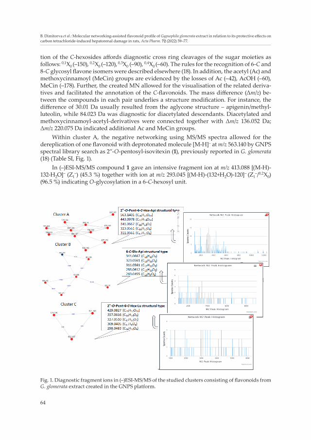
Within cluster A, the negative networking using MS/MS spectra allowed for the dereplication of one flavonoid with deprotonated molecule [M-H]– at m/z 563.140 by GNPS spectral library search as 2”-O-pentosyl-isovitexin (1), previously reported in G. glomerata (18) (Table SI, Fig. 1).
In (–)ESI-MS/MS compound 1 gave an intensive fragment ion at m/z 413.088 [(M-H)-132-Н2О]– (Z1
–) (45.3 %) together with ion at m/z 293.045 [(M-H)-(132+Н2О)-120]– (Z1–/0,2X0)
(96.5 %) indicating O-glycosylation in a 6-C-hexosyl unit.
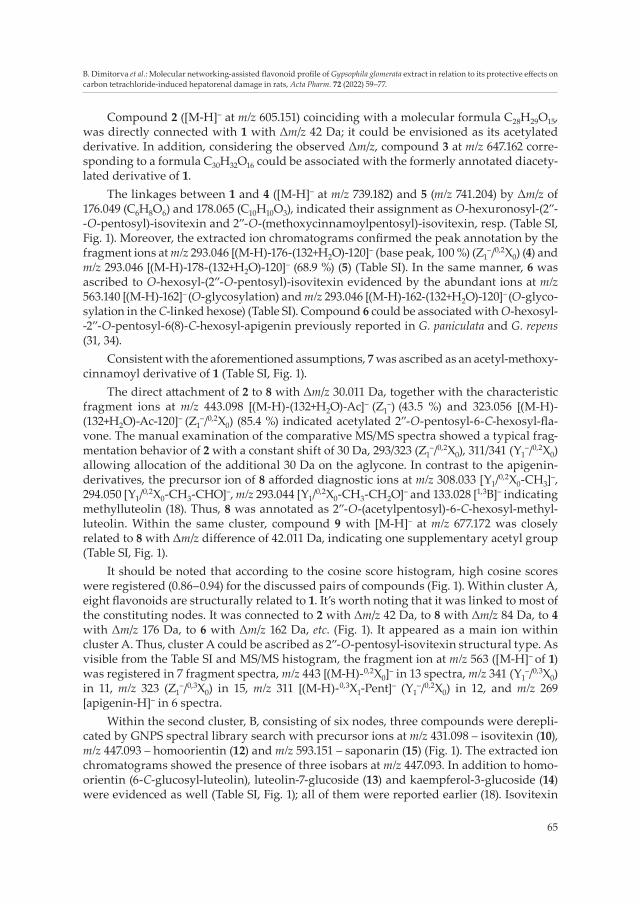
Fig. 1. Diagnostic fragment ions in (–)ESI-MS/MS of the studied clusters consisting of flavonoids from G. glomerata extract created in the GNPS platform.
65
B. Dimitorva et al.: Molecular networking-assisted flavonoid profile of Gypsophila glomerata extract in relation to its protective effects on carbon tetrachloride-induced hepatorenal damage in rats, Acta Pharm. 72 (2022) 59–77.
Compound 2 ([M-H]– at m/z 605.151) coinciding with a molecular formula C28H29O15, was directly connected with 1 with Δm/z 42 Da; it could be envisioned as its acetylated derivative. In addition, considering the observed Δm/z, compound 3 at m/z 647.162 corre-sponding to a formula C30H32O16 could be associated with the formerly annotated diacety-lated derivative of 1.
The linkages between 1 and 4 ([M-H]– at m/z 739.182) and 5 (m/z 741.204) by Δm/z of 176.049 (C6H8O6) and 178.065 (C10H10O3), indicated their assignment as O-hexuronosyl-(2”- -O-pentosyl)-isovitexin and 2”-O-(methoxycinnamoylpentosyl)-isovitexin, resp. (Table SI, Fig. 1). Moreover, the extracted ion chromatograms confirmed the peak annotation by the fragment ions at m/z 293.046 [(M-H)-176-(132+Н2О)-120]– (base peak, 100 %) (Z1
–/0,2X0) (4) and m/z 293.046 [(M-H)-178-(132+Н2О)-120]– (68.9 %) (5) (Table SI). In the same manner, 6 was ascribed to O-hexosyl-(2”-O-pentosyl)-isovitexin evidenced by the abundant ions at m/z 563.140 [(M-H)-162]– (O-glycosylation) and m/z 293.046 [(M-H)-162-(132+Н2О)-120]– (O-glyco-sylation in the C-linked hexose) (Table SI). Compound 6 could be associated with O-hexosyl- -2”-O-pentosyl-6(8)-C-hexosyl-apigenin previously reported in G. paniculata and G. repens (31, 34).
Consistent with the aforementioned assumptions, 7 was ascribed as an acetyl-methoxy-cinnamoyl derivative of 1 (Table SI, Fig. 1).
The direct attachment of 2 to 8 with Δm/z 30.011 Da, together with the characteristic fragment ions at m/z 443.098 [(M-H)-(132+Н2О)-Ас]– (Z1
–) (43.5 %) and 323.056 [(M-H)-(132+Н2О)-Ас-120]– (Z1
–/0,2X0) (85.4 %) indicated acetylated 2”-O-pentosyl-6-C-hexosyl-fla-vone. The manual examination of the comparative MS/MS spectra showed a typical frag-mentation behavior of 2 with a constant shift of 30 Da, 293/323 (Z1
–/0,2X0), 311/341 (Y1–/0,2X0)
allowing allocation of the additional 30 Da on the aglycone. In contrast to the apigenin-derivatives, the precursor ion of 8 afforded diagnostic ions at m/z 308.033 [Y1/0,2X0-СН3]–, 294.050 [Y1/0,2X0-СН3-СНО]–, m/z 293.044 [Y1/0,2X0-СН3-СН2О]– and 133.028 [1,3В]– indicating methylluteolin (18). Thus, 8 was annotated as 2”-O-(acetylpentosyl)-6-C-hexosyl-methyl-luteolin. Within the same cluster, compound 9 with [M-H]– at m/z 677.172 was closely related to 8 with Δm/z difference of 42.011 Da, indicating one supplementary acetyl group (Table SI, Fig. 1).
It should be noted that according to the cosine score histogram, high cosine scores were registered (0.86–0.94) for the discussed pairs of compounds (Fig. 1). Within cluster A, eight flavonoids are structurally related to 1. It’s worth noting that it was linked to most of the constituting nodes. It was connected to 2 with Δm/z 42 Da, to 8 with Δm/z 84 Da, to 4 with Δm/z 176 Da, to 6 with Δm/z 162 Da, etc. (Fig. 1). It appeared as a main ion within cluster A. Thus, cluster A could be ascribed as 2”-O-pentosyl-isovitexin structural type. As visible from the Table SI and MS/MS histogram, the fragment ion at m/z 563 ([M-H]– of 1) was registered in 7 fragment spectra, m/z 443 [(М-Н)-0,2X0]– in 13 spectra, m/z 341 (Y1
–/0,3X0) in 11, m/z 323 (Z1
–/0,3X0) in 15, m/z 311 [(М-Н)-0,3X1-Pent]– (Y1–/0,2X0) in 12, and m/z 269
[apigenin-Н]– in 6 spectra.Within the second cluster, B, consisting of six nodes, three compounds were derepli-
cated by GNPS spectral library search with precursor ions at m/z 431.098 – isovitexin (10), m/z 447.093 – homoorientin (12) and m/z 593.151 – saponarin (15) (Fig. 1). The extracted ion chromatograms showed the presence of three isobars at m/z 447.093. In addition to homo-orientin (6-C-glucosyl-luteolin), luteolin-7-glucoside (13) and kaempferol-3-glucoside (14) were evidenced as well (Table SI, Fig. 1); all of them were reported earlier (18). Isovitexin
66
B. Dimitorva et al.: Molecular networking-assisted flavonoid profile of Gypsophila glomerata extract in relation to its protective effects on carbon tetrachloride-induced hepatorenal damage in rats, Acta Pharm. 72 (2022) 59–77.
(6-C-glucosyl-apigenin) (10) at m/z 431.098 was connected to 12 with Δm/z of 15.995 Da (O). Furthermore, extracted ion chromatogram at m/z 431 showed the presence of isovitexin and its isomer vitexin (8-C-glucosyl-apigenin) (11), formerly identified in G. glomerata (18). Key points of recognition of 6-C- and 8-C-glucosyl-apigenin/luteolin are relative abundances of the diagnostic ions [M-H]–, 0,3X–, 0,2X–, 0,2X–/CO.
Manual examination of the MS/MS spectra of a precursor ion at m/z 593.151 revealed that along with saponarin (15), two previously annotated isobars 2”-O-pentosyl-6-C-hexo-syl-methylluteolin (16) and kaempferol 3-rutinoside (17) were evidenced (Table SI, Fig. 1). In (–)ESI-MS/MS 16 afforded diagnostic fragment ions at m/z 323.056 [(M-H)-(132+H2O)-120]− ([Z1/0,2X0]−) indicating 2”-O-pentosyl and C-hexosyl moieties as was seen in 1. The aglycone was discerned by the ions at m/z 308.033 (Z1
–/0,2X0/CH3), m/z 299.055 [MeLu-H]–, m/z 280.0377 (Z1
–/0,2X0/CH3/CO) and m/z 269.045 [MeLu-H-CH2O]– relevant for the methoxy group and methylluteolin. Regarding 17, the fragment ion at m/z 285.040 resulting from the concomi-tant loss of hexose and deoxyhexose units (308 Da) and relative abundance of 96 % led to its assignment as O-glycoside; kaempferol-3-rutinoside was additionally confirmed by comparison with an authentic standard.
Precursor ion at m/z 461.109 (18) was attached to 12 and 11 with Δm/z of 14.016 Da (CH2) and 30.011 (OCH2), resp. According to the previously established relative abundances of diagnostic ions for 6-/8-C-glucosyl-flavones, fragment ions at m/z 371.078 0,3X– (26 %), m/z 341.067 0,2X– (84 %) and m/z 298.048 0,2X–/СН3/СО (54 %) indicated 6-С-hexosyl flavone. A series of ions resulting from the neutral and methyl radical losses from 0,2X at m/z 298.048, 269.045 (0,2X–/СН3/С2НО2), 241.051 (0,2X–/СН3/С2НО2/СО), 225.055 (0,2X–/СН3/С2НО2/СО/СО2) are indicative for 6-C-hexosyl-methylluteolin. Precursor ion at m/z 609.161 (19) was linked to 10 by Δm/z of 178.064 Da suggesting methoxycinnamoyl ester of 6-C-hexosyl-apigenin. Within cluster B, flavonoids were depicted as structurally related congeners of 10, and the cluster could be ascribed to isovitexin structural type.
Cluster C has three closely related nodes in the spectral networking assuming their peculiar fragmentation (Fig. 1). They are tentatively ascribed to 2”-O-pentosyl-6-C-hexo-syl-luteolin (20) and its mono- (21) and diacetylated (22) derivatives. The MS/MS histogram of the cluster C showed a series of diagnostic ions at m/z 298.048 (Y1
–/0,2X0/CHO, C16H10O6), 309.041 (Z1
–/0,2X0, C17H9O6), 327.051 (Y1–/0,2X0, C17H11O7), 339.051 (Z1
–/0,3X0, C18H11O7), 357.062 (Y1
–/0,3X0, C18H13O8), 411.072 (Z1–/H2O, C21H15O9), 429.083 (Z1
–, C21H17O10) (Table SI). Conse-quently, the cluster was assessed as 2”-O-pentosyl-6-C-hexosyl-luteolin structural type.
In total, twenty-two flavonoids belonging to C, C,O-, and O-glycosides were derepli-cated or tentatively annotated in the G. glomerata extract by the means of molecular net-working and in-deep ESI-MS/MS characterization of the compounds.
The main finding of this study is that nine flavonoids, namely, 4, 5, 7, 9, 18–22 were reported for the first time in the genus Gypsophila. This is the first report on the annotation of compounds 4–7, 18, 19, 21 and 22 in G. glomerata. All of them are structurally related flavonoids, especially C,O-glycosides with supplementary acetyl and methoxycinnamoyl moieties. It’s worth noting that based on the detailed analysis of MS/MS data and MS/MS histograms, a series of relevant ions for each of the defined clusters is outlined (Fig. 1). The chromatographic profile of the EGG was dominated by 2”-O-pentosyl-6-C-hexosyl-apigenin/ luteolin/methylluteolin (1, 16, 20) and diacetylated derivative 3 (Fig. SI).
67
B. Dimitorva et al.: Molecular networking-assisted flavonoid profile of Gypsophila glomerata extract in relation to its protective effects on carbon tetrachloride-induced hepatorenal damage in rats, Acta Pharm. 72 (2022) 59–77.
Animal physical activity and gross pathology
During the 14 days of the investigational period, no mortality was observed in the experimental and control rats. The physical activity level in CCl4-treated rats from group 4 had decreased, as assessed by their reduced response to external stimuli. In this group, food and water intake also decreased. The abdomens of these animals were slightly swollen. All other groups were recorded as having normal activity levels and with normal intake of food and water during the whole experimental period.
Biochemical findings
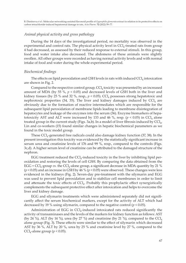
The effects on lipid peroxidation and GSH levels in rats with induced CCl4 intoxication are shown in Fig. 2.
Compared to the respective control group, CCl4 toxicity was presented by an increased amount of MDA (by 55 %, p < 0.05) and decreased levels of GSH both in the liver and kidney tissues (by 25 % and 32 %, resp., p < 0.05). CCl4 possesses strong hepatotoxic and nephrotoxic properties (34, 35). The liver and kidney damages induced by CCl4 are obviously due to the formation of reactive intermediates which are responsible for the subsequent lipid peroxidation of membrane lipids leading to membrane disintegration of hepatocytes and leakage of the enzymes into the serum (36). Enzyme biomarkers of hepa-totoxicity AST and ALT were increased by 133 and 46 %, resp. (p < 0.05) in CCl4 alone treated group in the current study (Figs. 3a,b). In a model of liver fibrosis induced by CCl4, Lin and co-workers (15) found similar changes in hepatic biochemical parameters as we found in the toxic model group.
These CCl4-generated free radicals could also damage kidney function (37, 38). In the present investigation this toxicity was evidenced by the statistically significant increase in serum urea and creatinine levels of 176 and 99 %, resp., compared to the controls (Figs. 3c,d). A higher serum level of creatinine can be attributed to the damaged structure of the nephron.
EGG treatment reduced the CCl4-induced toxicity in the liver by inhibiting lipid per-oxidation and restoring the levels of cell GSH. By comparing the data obtained from the EGG + CCl4-group vs. the CCl4-alone group, a significant decrease in MDA quantity by 21 % (p < 0.05) and an increase in GSH by 46 % (p < 0.05) were observed. These changes were less evidenced in the kidneys (Fig. 2). Seven-day pre-treatment with the silymarin and EGG was used to prevent lipid peroxidation and to stabilize cell membranes in order to limit and attenuate the toxic effects of CCl4. Probably this prophylactic effect synergistically complements the subsequent protective effect after intoxication and helps to overcome the liver and kidney damage.
EGG and silymarin treatments which were administered separately did not signifi-cantly affect the serum biochemical markers, except for the activity of ALT which had decreased by 19 % using silymarin, compared to the negative control (p < 0.05).
Administration of EGG in CCl4-induced intoxicated rats reduced significantly the activity of transaminases and the levels of the markers for kidney function as follows: AST (by 24 %), ALT (by 14 %), urea (by 27 %) and creatinine (by 21 %), compared to the CCl4 alone group (Fig. 3). These effects were similar to the effect of silymarin which decreased AST by 36 %, ALT by 20 %, urea by 25 % and creatinine level by 27 %, compared to the CCl4-alone group (p < 0.05).
68
B. Dimitorva et al.: Molecular networking-assisted flavonoid profile of Gypsophila glomerata extract in relation to its protective effects on carbon tetrachloride-induced hepatorenal damage in rats, Acta Pharm. 72 (2022) 59–77.
Herein, the presence of a variety of 2”-О-pentosyl-6-С-hexosyl-apigenin/luteolin/methylluteolin and their acetyl- and methoxycinnamoyl-derivatives together with C-gly-cosyl-flavones could be associated with the protective effect of EGG. Huang et al. (14) re-ported a significant protective effect and improvement of the liver function by the admini-stration of isoorientin-2”-O-α-L-arabinopyranosyl (IOA) isolated from G. elegans. The authors found decreased levels of ALT, AST, ALP, c-glutamyltransferase, interleukin-6 and tumor necrosis factor in alcohol-induced liver injury in rats. They showed that IOA effec-
Fig. 2. Effects of EGG and silymarin administered alone and after CCl4 intoxication, on hepatic and renal: a) MDA and b) GSH levels. Results are expressed as mean ± SD (n = 6).
Fig. 3. Effects of EGG and silymarin, administered alone and after CCl4 intoxication, on serum bio-chemical markers: a) AST, b) ALT, c) urea, and d) creatinine. Results are expressed as mean ± SD (n = 6).
a) b)
a) b)
c) d)
69
B. Dimitorva et al.: Molecular networking-assisted flavonoid profile of Gypsophila glomerata extract in relation to its protective effects on carbon tetrachloride-induced hepatorenal damage in rats, Acta Pharm. 72 (2022) 59–77.
tively reduced pathological tissue damage, as was found in the present study. Research on the protective mechanisms showed that IOA can inhibit CCl4-induced hepatic damages, due to its radical scavenging action, antioxidant activity, and modulation of NF-κB and TGF-β1/Smad signaling pathways (15, 16).
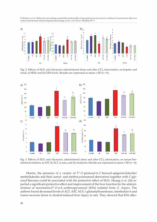
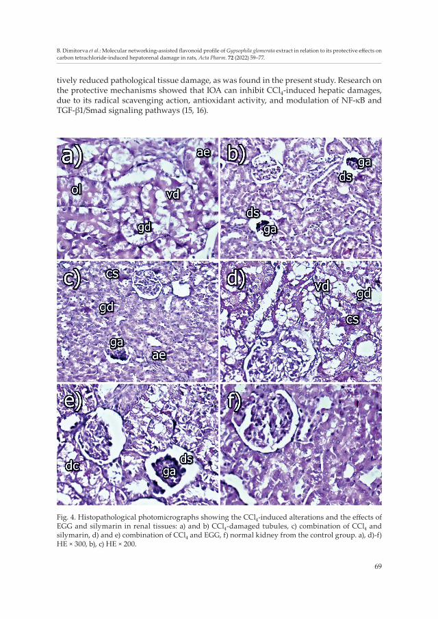
Fig. 4. Histopathological photomicrographs showing the CCl4-induced alterations and the effects of EGG and silymarin in renal tissues: a) and b) CCl4-damaged tubules, c) combination of CCl4 and silymarin, d) and e) combination of CCl4 and EGG, f) normal kidney from the control group. a), d)-f) HE × 300, b), c) HE × 200.
70
B. Dimitorva et al.: Molecular networking-assisted flavonoid profile of Gypsophila glomerata extract in relation to its protective effects on carbon tetrachloride-induced hepatorenal damage in rats, Acta Pharm. 72 (2022) 59–77.
The more pronounced antioxidant ability of the C-glycosyl flavonoids in comparison with the O-glycosyl flavonoids was reported by Barreca et al. (39). Moreover, homoorientin, orientin, isovitexin and vitexin also possess some beneficial activities such as anti-inflam-matory, antibacterial, antiproliferative, antimutagenic and anticarcinogenic (40), which could add significantly to the above-mentioned protective effects of the flavonoids.
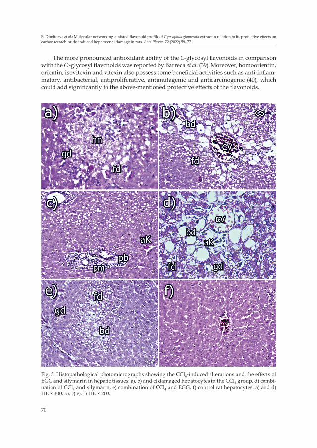
Fig. 5. Histopathological photomicrographs showing the CCl4-induced alterations and the effects of EGG and silymarin in hepatic tissues: a), b) and c) damaged hepatocytes in the CCl4 group, d) combi-nation of CCl4 and silymarin, e) combination of CCl4 and EGG, f) control rat hepatocytes. a) and d) HE × 300, b), c) e), f) HE × 200.
71
B. Dimitorva et al.: Molecular networking-assisted flavonoid profile of Gypsophila glomerata extract in relation to its protective effects on carbon tetrachloride-induced hepatorenal damage in rats, Acta Pharm. 72 (2022) 59–77.
Macroscopical and histopathological assessment
Macroscopical examination at slaughter time revealed that liver and kidney volume was significantly larger, with yellowish colour in the CCl4-exposed group compared with the control group. In other experimental groups, the color and the size of the examined organs were relatively preserved and similar in appearance/morphology to the control.
Histological photomicrographs provided a direct assessment of the effects of the in-vestigated compounds on the kidneys and livers (Figs. 4 and 5). The kidney and liver tis-sues in the control rats showed normal architecture. No pathological changes were ob-served in the glomeruli or in the tubular kidney epithelium (Fig. 4f), nor in the hepatocytes (Fig. 5f) in the control animals.
Renal histopathology from CCl4-treated rats showed moderate degenerative changes in the tubular epithelium of the renal cortex, e.g., cloudy swelling, granular (gd) and/or vacuolar degeneration (vd). The tubular epithelial cells were often swollen and tubular lumen was diminished and sometimes occluded (ol) by the enlarged epithelium (ae) (Fig. 4a). Some of the vacuolated epithelial cells were with foamy appearance, whereas other epithelial cells were completely necrotized. Some glomeruli showed mild dilatation of Bowman’s space (ds) and partial glomerular atrophy, whereas others were with total atrophy (ga) or sclerosis (Fig. 4b). Mild congestion of capillary loops or peritubular capillaries was also observed in some places and activation of capillary endothelium (ae) was present. Small foci of mononuclear proliferation in the renal interstitium were also visible in some areas. These alterations were also found in the study of Okolo et al. (41) who showed that the administration of CCl4 markedly disrupted the normal architecture of the kidney cell by degenerating the tubules and collapsing the glomerulus.
The same scientific group (41) found that the hepatocytes of CCl4-treated rats showed proliferation of bile duct epithelium with dilated and congested sinusoids. Similar find-ings have also been found in this study. In the liver, there was a strong fatty (fd) and granular degeneration (gd), which was more pronounced in the central part of the lobules, where disorderly hepatocyte cords were found (Fig. 5a). Cloudy swelling (cs) (Fig. 5b) of some hepatocytes and small hepatocellular necroses (hn) (Fig. 5a) were found mainly in the central parts of the lobules. A strong ballooning degeneration (bd) of hepatocytes was observed in the central parts of the lobules (Fig. 5b). A mild proliferation of mononuclear cells and/or connective tissue (pm) and/or proliferation of bile ducts (pb) was observed in Glisson’s triangles. A small cluster of macrophages or activated Kupffer cells (aK) were often found in these proliferates (Fig. 5c). Part of the necroses had infiltrated with inflam-matory mononuclear cells or single macrophages. Mild congestion of vessels (cv) was also seen, e.g., V. centralis (Fig. 5b).
In the kidneys of rats treated with CCl4 and EGG, there were slight to moderate dege-nerative changes (dc) in the tubular epithelium of the renal cortex, e.g., cloudy swelling (cs), granular (gd) and/or vacuolar degeneration (vd) (Fig. 4d). The tubular epithelial cells often appeared swollen and thereby obscured the tubular lumen. A few glomeruli showed mild dilatation of Bowman’s space (ds), and partial glomerular atrophy (ga) (Fig. 4e), whereas total atrophy or sclerosis of glomeruli was rarely seen. Mild congestion of capillary loops or peritubular capillaries was rarely noticed in some places. The capillary endothelium of peritubular capillaries was activated. Small foci of mononuclear proliferation in the renal interstitium were also seen in some areas.
72
B. Dimitorva et al.: Molecular networking-assisted flavonoid profile of Gypsophila glomerata extract in relation to its protective effects on carbon tetrachloride-induced hepatorenal damage in rats, Acta Pharm. 72 (2022) 59–77.
There was a moderate fatty (fd) and/or granular degeneration (gd) in the livers of these animals, which was well visible in the central part of the lobules. In some places, cloudy swelling of some hepatocytes was observed. A ballooning degeneration (bd) of hepato-cytes was seen in part of the hepatocytes in the central parts of some lobules (Fig. 5e), while small hepatocellular necroses infiltrated with macrophages or leucocytes were rarely found. A mild proliferation of mononuclear cells, and/or connective tissue and/or prolife-ration of bile ducts were observed in the Glisson’s triangles. A small cluster of macro-phages or activated Kupffer (aK) cells were often found in these proliferates. A mild to moderate congestion of vessels was also seen, e.g., V. centralis.
Gypsophila elegans isoorientin-2″-O-α-l-arabinopyranosyl (GEI) ameliorates porcine serum-induced immune liver injury as evidenced by decreased pathological damages (16). These researchers found that GEI significantly alleviated liver injury and collagen accu-mulation by reducing hepatocyte loosening, fatty degeneration, portal inflammation and necrosis, which is in accordance with the results of this study.
In the rats treated with CCl4 and silymarin, there were slight degenerative changes in the tubular epithelium of the renal cortex, e.g., cloudy swelling (cs), granular and/or vacu-olar degeneration (gd). The tubular epithelial cells often appeared swollen and the tubular lumen was diminished by the enlarged epithelium. A few glomeruli showed mild dilata-tion of Bowman’s space and partial glomerular atrophy (ga) (Fig. 4c), whereas total atrophy or sclerosis of glomeruli was rarely seen. Mild congestion of capillary loops or peritubular capillaries was rarely seen in some places as well as activation of capillary endothelium (ae) was presented. In some areas, small foci of mononuclear proliferation in the renal interstitium were also seen.
The well-known protective effect of silymarin has been reported previously in rat cisplatin-induced renal toxicity (42), as well as in dog gentamicin-induced renal damages (43) and in alloxan-induced diabetic nephropathy too (6). In our experimental study, the protective effect of silymarin was confirmed with the decreased levels of creatinine and urea (Figs. 3c,d) along with the less pronounced degenerative damages in epithelial cells of renal tubules (Fig. 4c).
In the group where rat livers were treated with CCl4 and silymarin, there was moderate fatty (fd) and granular degeneration (gd), which was more pronounced in the central area of the lobules. Cloudy swelling of some hepatocytes was observed in some places. A ballooning degeneration (bd) of hepatocytes was rarely seen in the central parts of some lobules, and small hepatocellular necroses infiltrated with macrophages were rarely found. A mild proliferation of mononuclear cells and/or connective tissue and/or proli-feration of bile ducts were seen in Glisson’s triangles. A small cluster of macrophages or activated Kupffer cells (aK) were often found in these proliferates. Mild congestion of vessels (cv) was also noticed, e.g., V. centralis (Fig. 5d).
The protective effect of silymarin is reported due to the observed decrease in lipid peroxidation and the increase in endogenous antioxidants, which are responsible for the integrity of the plasma membrane and, therefore, counteract the leakage of some target enzymes (44, 45). In this study, the 7-day pretreatment with silymarin and EGG before CCl4 intoxication aimed to stabilize the cell membranes and thus prevent enzyme leakage. Freitag et al. (46) reported that silymarin treatment has restored the hepatocyte functional and histopathological alterations induced by paracetamol in normotensive and hypertensive animals. In another study of experımental obstructıve jaundice model, the enlargement of
73
B. Dimitorva et al.: Molecular networking-assisted flavonoid profile of Gypsophila glomerata extract in relation to its protective effects on carbon tetrachloride-induced hepatorenal damage in rats, Acta Pharm. 72 (2022) 59–77.
hepatocytes, dilatation of canaliculi and the edema regressed in livers in the silymarin– -treated group was reported (47).
No pathological changes were seen in the glomeruli or in the tubular kidney epithe-lium of rats treated only with silymarin, nor were pathological changes in hepatocytes of their livers observed. A mild fatty degeneration and a mild mononuclear proliferation were found in the liver only in one of the rats treated with silymarin.
There was a slight mononuclear cell proliferation in the renal interstitium of two rats treated with EGG alone. Degenerative changes, which were shown to be not significant, were found in some hepatocytes in only two rats. A minor proliferation of mononuclear cells, activation of Kupffer cells and slight hyperaemia of sinusoidal capillaries were ob-served in the same two rats.
The analysis of the results showed that histopathological and biochemical changes were strongest in rats exposed to CCl4 without any supplemental treatment, followed by rats treated additionally with EGG and those treated with silymarin, whereas scarce changes or no changes were seen in control rats and in rats treated with EGG or silymarin only. It seems that the G. glomerata extract (EGG) has a good protective effect against the toxic effects of CCl4 on the liver and kidneys, which is similar to that of silymarin.
CONCLUSIONS
In summary, twenty-two flavonoid C-, O- and C,O-glycosides were dereplicated or tentatively annotated in the G. glomerata aerial parts extract including nine flavonoids pre-viously undescribed in the literature. In this study, molecular networking provides effi-cient tool for the annotation of new flavonoids in the genus Gypsophila: O-hexuronosyl-(2”-pentosyl-6-C-hexosyl)-apigenin (4), 2”-(methoxycinnamoylpentosyl)-6-C-hexosyl-apigenin (5), 2”- (methoxycinnamoyl-acetylpentosyl)-6-C-hexosyl)-apigenin (7), 2”-O-diacetylpento-syl-6-C-hexosyl-methylluteolin (9), 6-C-hexosyl-methylluteolin (18), 2”-methoxycinnamo-yl-6-C-hexosyl-apigenin (19), 2”-О-pentosyl-6-С-hexosyl-luteolin (20), 2”-О-(acetylpentosyl)-6-С-hexosyl-luteolin (21) and 2”-О-(diacetylpentosyl)-6-С-hexosyl-luteolin (22).
The observed hepatorenal protective effects of G. glomerata could be attributed to fla-vonoids, notably to the major flavonoids in the chromatographic profile: 2”-O-pentosyl-6-C-hexosyl-apigenin/luteolin/methylluteolin (1, 16, 20) and diacetylated derivative 3. How-ever, further investigations on the detailed mechanisms of action are needed.
Acknowledgements. – The authors thank Dr. Nisha Singh for English editing. Supplementary material available on request.
REFERENCES
1. M. Ekor, The growing use of herbal medicines: issues relating to adverse reactions and challenges in monitoring safety, Front. Pharmacol. 4 (2014) Article ID 177 (10 pages); https://doi.org/10.3389/fphar.2013.00177
2. R. F. F. de Araújo, D. B. G. Martins and M. A. C. S. M. Borba, Oxidative Stress and Disease, in A Master Regulator of Oxidative Stress – The Transcription Factor Nrf2 (Eds. J. A. Morales-Gonzalez, A. Morales-González and E. O. Madrigal-Santillan), IntechOpen, Rijeka 2016.
74
B. Dimitorva et al.: Molecular networking-assisted flavonoid profile of Gypsophila glomerata extract in relation to its protective effects on carbon tetrachloride-induced hepatorenal damage in rats, Acta Pharm. 72 (2022) 59–77.
3. T. Senoner and W. Dichtl, Oxidative stress in cardiovascular diseases: Still a therapeutic target?, Nutrients 11 (2019) Article ID 2090 (25 pages); https://doi.org/10.3390%2Fnu11092090
4. Y. Kayama, U. Raaz, A. Jagger, M. Adam, I. N. Schellinger, M. Sakamoto, H. Suzuki, K. Toyama, J. M. Spin and P. S. Tsao, Diabetic cardiovascular disease induced by oxidative stress, Int. J. Mol. Sci. 16 (2015) 25234–25263; https://doi.org/10.3390/ijms161025234
5. C. Forni, F. Facchiano, M. Bartoli, S. Pieretti, A. Facchiano, D. D’Arcangelo, S. Norelli, G. Valle, R. Nisini, S. Beninati, C. Tabolacci and R. N. Jadeja, Beneficial role of phytochemicals on oxidative stress and age-related diseases, Biomed. Res. Int. 2019 (2019) Article ID 8748253 (17 pages); https://doi.org/10.1155/2019/8748253
6. J. Teixeira, D. Chavarria, F. Borges, L. Wojtczak, M. R. Wieckowski, A. Karkucinska-Wieckowska and P. J. Oliveira, Dietary polyphenols and mitochondrial function: role in health and disease, Curr. Med. Chem. 26 (2019) 3376–3406; https://doi.org/10.2174/0929867324666170529101810
7. R. Saller, J. Melzer, J. Reichling, R. Brignoli and R. Meier, An updated systematic review of the phar-macology of silymarin, Forsch. Komp. Klas. Nat. 14 (2007) 70–80; https://doi.org/10.1159/000100581
8. E. Shaker, H. Mahmoud and S. Mnaa, Silymarin, the antioxidant component and Silybum marianum extracts prevent liver damage, Food Chem. Toxicol. 48 (2010) 803–806.
9. C. Soto, J. Pérez, V. García, E. Uría, M. Vadillo and L. Raya, Effect of silymarin on kidneys of rats suf-fering from alloxan-induced diabetes mellitus, Phytomedicine 17 (2010) 1090–1094; https://doi.org/10.1016/j.phymed.2010.04.011
10. S. D. Stoev, P. Njobeh, I. Zarkov, T. Mircheva, D. Zapryanova, S. Denev and B. Dimitrova, Selected herbal feed additives showing protective effects against ochratoxin A toxicosis in broiler chicks, World Mycotoxin J. 12 (2019) 257–268; https://doi.org/10.3920/WMJ2019.2432
11. M. Bahmani, H. Shirzad, S. Rafieian and M. Rafieian-Kopaei, Silybum marianum: Beyond hepatopro-tection, Evid-Based Complement. Alternat. Med. 20 (2015) 292–301; https://doi.org/10.1177/2156587215571116
12. S. K. Katiyar, Silymarin and skin cancer prevention: anti-inflammatory, antioxidant and immuno-modulatory effects, Int. J. Oncol. 26 (2005) 169–176.
13. Q. Chen, J.-G. Luo and L.-Y. Kong, Triterpenoid saponins from Gypsophila altissima L., Chem. Pharm. Bull. 58 (2010) 412–414; https://doi.org/10.1248/cpb.58.412
14. Q. F. Huang, S. J. Zhang, L. Zheng, M. Liao, M. He, R. Huang, L. Zhuo and X. Lin, Protective effect of isoorientin-2”-O-α-l-arabinopyranosyl isolated from Gypsophila elegans on alcohol induced hepatic fibrosis in rats, Food Chem. Toxicol. 50 (2012) 1992–2001; https://doi.org/10.1016/j.fct.2012.03.044
15. X. Lin, Y. Chen, S. Lv, S. Tan, S. Zhang, R. Huang, L. Zhuo, S. Liang, Z. Lu and Q. Huang, Gypsophila elegans isoorientin attenuates CCl4-induced hepaticfibrosis in rats via modulation of NF-κB and TGF-β1/Smad signaling pathways, Int. Immunopharmacol. 28 (2015) 305–312; https://doi.org/10.1016/j.intimp.2015.06.021
16. F. Bai, Q. Huang, J. Wei, S. Lv, Y. Chen, C. Liang, L. Wei, Z. Lu and X. Lin, Gypsophila elegans isoorientin- -2″-O-α-l-arabinopyranosyl ameliorates porcine serum-induced immune liver fibrosis by inhibiting NF-κB signaling pathway and suppressing HSC activation, Int. Immunopharmacol. 54 (2018) 60–67; https://doi.org/10.1016/j.intimp.2017.10.028
17. V. Vitcheva, R. Simeonova, I. Krasteva, M. Yotova, S. Nikolov and M. Mitcheva, Hepatoprotective effects of saponarin, isolated from Gypsophila trichotoma Wend. on cocaine-induced oxidative stress in rats, Redox Rep. 16 (2011) 56–61; https://doi.org/10.1179/174329211x12989133691530
18. D. Zheleva-Dimitrova, G. Zengin, V. Balabanova, Y. Voynikov, V. Lozanov, I. Lazarova and R. Gevrenova, Chemical characterization with in vitro biological activities of Gypsophila species, J. Pharm. Biomed. Anal. 155 (2018) 56–69; https://doi.org/10.1016/j.jpba.2018.03.040
19. R. Simeonova, M. Kondeva-Burdina, V. Vitcheva, I. Krasteva, V. Manov and M. Mitcheva, Protective effects of the apigenin-O/C-diglucoside saponarin from Gypsophila trichotoma on carbone tetrachlo-
75
B. Dimitorva et al.: Molecular networking-assisted flavonoid profile of Gypsophila glomerata extract in relation to its protective effects on carbon tetrachloride-induced hepatorenal damage in rats, Acta Pharm. 72 (2022) 59–77.
ride-induced hepatotoxicity in vitro/in vivo in rats, Phytomedicine 21 (2014) 148–154; https://doi.org/10.1016/j.phymed.2013.07.014
20. R. Simeonova, V. Vitcheva, M. Kondeva-Burdina, I. Krasteva, V. Manov and M. Mitcheva, Hepato-protective and antioxidant effects of saponarin, isolated from Gypsophila trichotoma Wend. on paracetamol-induced liver damage in rats, Biomed. Res. Int. 2013 (2013) Article ID 757126; https://doi.org/10.1155/2013/757126
21. R. Gevrenova, K. Bardarov, S. Bouguet-Bonnet, Y. Voynikov, V. Balabanova, D. Zheleva-Dimitrova and M. Henry, A new liquid chromatography-high resolution Orbitrap mass spectrometry-based strategy to characterize glucuronide oleanane-type triterpenoid carboxylic acid 3,28-O-bidesmo-sides (GOTCAB) saponins. A case study of Gypsophila glomerata Pall ex M. B. (Caryophyllaceae), J. Pharm. Biomed. Anal. 159 (2018) 567–581; https://doi.org/10.1016/j.jpba.2018.07.041
22. F. Ferreres, A. Gil-Izquierdo, P. B. Andrade, P. Valentão and F. Tomás-Barberán, Characterization of C-glycosyl flavones O-glycosylated by liquid chromatography–tandem mass spectrometry, J. Chro-matogr. A. 1161 (2007) 214–223; https://doi.org/10.1016/j.chroma.2007.05.103
23. W. Nie, J.-G. Luo and L.-Y. Kong, C-glycosyl flavonoids from the aerial part of Gypsophila pacifica, Chin. J. Nat. Med. 8 (2010) 250–252; https://doi.org/10.3724/SP.J.1009.2010.00250
24. F. M. Zhang, Z. G. Tai, L. Cai, Y. B. Yang, F. Li and Z. T. Ding, Flavonoids from Gypsophila elegans and their antioxidant activities, J. Yunnan Univ. Nat. Sci. 33 (2011) 93–95.
25. Council of Europe. European Convention for the Protection of Vertebrate, Animals Used for Experi-mental and other Scientific Purposes, CETS No. 123, 1991 [displayed 30 May 2007]; https://ec.europa.eu/world/agreements/downloadFile.do?fullText=yes&treaty TransId=1346; last access date Nov 4, 2020
26. P. V. Habbu, R. A. Shastry, K. M. Mahadevan, H. Joshi and S. K. Das, Hepatoprotective and antioxi-dant effects of Argyreia speciosa in rats, Afr. J. Tradit. Complement. Altern. Med. 5 (2008) 158–164; https://doi.org/10.4314%2Fajtcam.v5i2.31268
27. T. H. Ahn, Y. S. Yang, J. C. Lee, C. J. Moon, S. H. Kim, W. Jun, S. C. Park and J. C. Kim, Ameliorative effects of pycnogenol on carbon tetrachloride-induced hepaticoxidative damage in rats, Phytother. Res. 21 (2007) 1015–1019; https://doi.org/10.1002/ptr.2146
28. European Parliament and of the Council, Directive 2010/63/EU of the European Parliament and of the Council of 22 September 2010 on the protection of animals used for scientific purposes, Off. J. Eur. Union (2010) L 276/33–79; https://eur-lex.europa.eu/LexUriServ/LexUriServ.do?uri=OJ:L:2010:276:0033:0079:en:PDF; last access date Nov 4, 2020
29. J. L. Wolfender, P. M. Allard, M. Kubo and E. F. Queiroz, Меtabolomics Strategies for the Dereplication of Polyphenols and Other Metabolites, in Recent Advances in Polyphenol Research (Eds. H. Halbwirth, K. Stich, V. Cheynier and S. Quideau), 1st ed, Wiley, Hoboken (NJ) 2019, pp. 183–205.
30. M. Wang, J. J. Carver, W. Phelan, L. M. Sanchez, N. Garg, Y. Peng, D. D. Nguyen, J. Watrous, C. A. Capono, T. Luzzatto-Knaan, C. Porto, A. Bouslimani, A. V. Melnik, M. J. Meehan, W.-T. Liu, M. Crüse-mann, P. D. Boudreau, E. Esquenazi, M. Sandoval-Calderón, R. D. Kersten, L. A. Pace, R. A. Quinn, K. R. Duncan, C.-C. Hsu, D. J. Floros, R. G. Gavilan, K. Kleigrewe, T. Northen, R. J. Dutton, D. Parrot, E. E. Carlson, B. Aigle, C. F. Michelsen, L. Jelsbak, C. Sohlenkamp, P. Pevzner, A. Edlund, J. McLean, J. Piel, B. T. Murphy, L. Gerwick, C.-C. Liaw, Y.-L. Yang, H.-U. Humpf, M. Maansson, R. A. Keyzers, A. C. Sims, A. R. Johnson, A. M. Sidebottom, B. E. Sedio, A. Klitgaard, C. B. Larson, C. A. Boya, D. Torres-Mendoza, D. J. Gonzalez, D. B. Silva, L. M. Marques, D. P. Demarque, E. Pociute, E. C. O’Neill, E. Briand, E. J. N. Helfrich, E. A. Granatosky, E. Glukhov, F. Ryffel, H. Houson, H. Mohimani, J. J. Kharbush, Y. Zeng, J. A. Vorholt, K. L. Kurita, P. Charusanti, K. L. McPhail, K. F. Nielsen, L. Vuong, M. Elfeki, M. F. Traxler, N. Engene, N. Koyama, O. B. Vining, R. Baric, R. R. Silva, S. J. Mascuch, S. Tomasi, S. Jenkins, V. Macherla, T. Hoffman, V. Agarwal, P. G. Williams, J. Dai, R. Neupane, J. Gurr, A. M. C. Rodríguez, A. Lamsa, C. Zhang, K. Dorrestein, B. M. Duggan, J. Almaliti, P.-M. Allard, P. Phapale, L.-F. Nothias, T. Alexandrov, M. Litaudon, J.-L. Wolfender, J. E. Kyle, T. O. Metz, T. Peryea, D.-T. Nguyen, D. VanLeer, P. Shinn, A. Jadhav, R. Müller, K. M. Waters, W. Shi, X. Liu, L. Zhang, R.
76
B. Dimitorva et al.: Molecular networking-assisted flavonoid profile of Gypsophila glomerata extract in relation to its protective effects on carbon tetrachloride-induced hepatorenal damage in rats, Acta Pharm. 72 (2022) 59–77.
Knight, P. R. Jensen, B. O. Palsson, K. Pogliano, R. G. Linington, M. Gutiérrez, N. P. Lopes, W. H. Gerwick, B. S. Moore, P. C. Dorrestein and N. Bandeira, Sharing and community curation of mass spectrometry data with global natural products social molecular networking, Nat. Biotechnol. 34 (2016) 828–837; https://doi.org/10.1038/nbt.3597
31. A. H. Polizio and C. Peña, Effects of angiotensin II type 1 receptor blockade on the oxidative stress in spontaneously hypertensive rat tissues, Regul. Pept. 128 (2005) 1–5; https://doi.org/10.1016/j.reg-pep.2004.12.004
32. E. A. Bump, Y. C. Taylor and M. J. Brown, Role of glutathione in the hypoxic cell cytotoxicity of miso-nidazole, Cancer Res. 43 (1983) 997–1002.
33. R. Gevrenova, D. Zheleva-Dimitrova, V. Balabanova, Y. Voynikov, K. I. Sinan, M. F. Mahomoodally and G. Zengin, Integrated phytochemistry, bio-functional potential and multivariate analysis of Tanacetum macrophyllum (Waldst. & Kit.) Sch. Bip. and Telekia speciosa (Schreb.) Baumg. (Asteraceae), Ind. Crops Prod. 155 (2020) Article ID 112817; https://doi.org/10.1016/j.indcrop.2020.112817
34. A. A. Rahmat, F. A. Dar and I. M. Choudhary, Protection of CCl4-Induced liver and kidney damage by phenolic compounds in leaf extracts of Cnestis ferruginea (de Candolle), Pharmacog. Res. 6 (2014) 19–28; https://doi.org/10.4103%2F0974-8490.122913
35. A. S. M. Al Amin and R. G. Menezes, Carbon tetrachloride toxicity, StatPearls [Internet], StatPearls Publishing, Treasure Island (FL) 2020; https://www.ncbi.nlm.nih.gov/books/NBK562180/
36. S. Sahreen, M. R. Khan and R. A. Khan, Hepatoprotective effects of methanol extract of Carissa opaca leaves on CCl4-induced damage in rat, BMC Complement. Altern. Med. 11 (2011) Article ID 48 (9 pages); https://doi.org/10.1186/1472-6882-11-48
37. M. Ogeturk, I. Kus, N. Colakoglu, I. Zararsiz, N. Ilhan and M. Sarsilmaz, Caffeic acid phenyl ester protects kidneys against carbon tetrachloride toxicity in rats, J. Ethnopharmacol. 97 (2005) 273–280.
38. F. Ozturk, M. Ucar, I. C. Ozturk, N. Vardi and K. Batcioglu, Carbon tetrachloride induced nephro-toxicity and protective effect of betaine in Sprague-Dawley rats, Urology 62 (2003) 353–356.
39. D. Barreca, E. Bellocco, C. Caristi, U. Leuzzi and G. Gattuso, Distribution of C-and O-glycosyl flavo-noids, (3-hydroxy-3-methylglutaryl)glycosyl flavanones and furocoumarins in Citrus aurantium L. juice, Food Chem. 124 (2011) 576–582; https://doi.org/10.1016/j.foodchem.2010.06.076
40. H. F. dos Santos, J. F. Campos, C. M. dos Santos, J. B. Perrella Balestieri, D. B. Silva, C. A. Carollo, K. de Picoli Souza, L. M. Estevinho and E. L. dos Santos, Chemical profile and antioxidant, anti-inflam-matory, antimutagenic and antimicrobial activities of geopropolis from the stingless bee Melipona orbigny, Int. J. Mol. Sci. 18 (2017) Article ID 953 (18 pages); https://doi.org/10.3390%2Fijms18050953
41. K. O. Okolo, I. M. Siminialayi and O. E. Orisakwe, Carbon tetrachloride induced hepatorenal toxicity in rats: possible protective effects of wild Pleurotus tuber-regium, Clin. Phytosci. 3 (2017) Article ID 2 (7 pages); https://doi.org/10.1186/s40816-016-0040-5
42. G. Karimi, M. Ramezani and Z. Tahoonian, Cisplatin nephrotoxicity and protection by milk thistle extract in rats, Evid-Based Complement. Alternat. Med. 2 (2005) 383–386; https://doi.org/10.1093/ecam/neh103
43. H. N. Varzi, S. Esmailzadeh, H. Morovvati, R. Avizeh, A. Shahriari and M. E. Givi, Effect of silymarin and vitamin E on gentamicin-induced nephrotoxicity in dogs, J. Vet. Pharmacol. Ther. 30 (2007) 477–481; https://doi.org/10.1111/j.1365-2885.2007.00901.x
44. G. Upadhyay, M. N. Tiwari, O. Prakash, A. Jyoti, R. Shanker and M. P. Singh, Involvement of multiple molecular events in pyrogallol-induced hepatotoxicity and silymarin-mediated protection: Evidence from gene expression profiles, Food Chem. Toxicol. 48 (2010) 1660–1670; https://doi.org/10.1016/j.fct.2010.03.041
45. G. Karimi, M. Vahabzadeh, P. Lari, M. Rashedinia and M. Moshiri, Silymarin, a promising pharma-cological agent for treatment of diseases, Iranian J. Basic Med. Sci. 14 (2011) 308–317.
77
B. Dimitorva et al.: Molecular networking-assisted flavonoid profile of Gypsophila glomerata extract in relation to its protective effects on carbon tetrachloride-induced hepatorenal damage in rats, Acta Pharm. 72 (2022) 59–77.
46. A. F. Freitag, G. F. Cardia, B. A. da Rocha, R. P. Aguiar, F. M. de Souza Silva-Comar, R. A. Spironello, R. Grespan, S. M. Caparroz-Assef, C. A. Bersani-Amado and R. K. Nakamura Cuman, Hepatoprotec-tive effect of silymarin (Silybum marianum) on hepatotoxicity induced by acetaminophen in spontane-ously hypertensive rats, Evid-Based Complement. Alternat. Med. 2015 (2015) Article ID 538317 (8 pages); https://doi.org/10.1155/2015/538317
47. A. K. Onalan, S. Tuncal, S. Kilicoglu, S. Celepli, E. Durak, B. Kilicoglu, E. Devrim, A. Mutlu Barlas and K. Kismet, Effect of silymarin on oxidative stress and liver histopathology in experimental obstruc-tive jaundice model, Acta Cir. Bras. 31 (2016) 801–806; https://doi.org/10.1590/s0102-865020160120000004