Embed Size (px)
Citation preview
Myat Htut Nyunt, Thinzar Shein, Ni Ni Zaw, Soe Soe Han, Fauzi Muh, Seong-Kyun Lee,
Jin-Hee Han, Kyaw Zin Thant, Eun-Taek Han,1 Myat Phone Kyaw1
Artemisinin resistance containment in Myanmar was initi-ated in 2011 after artemisinin-resistant Plasmodium falci-parum malaria was reported. Molecular evidence suggests that asymptomatic malaria infections harboring drug resis-tance genes are present among residents of the Myanmar artemisinin resistance containment zone. This evidence supports efforts to eliminate these hidden infections.
The global burden of malaria has been decreasing in recent years as a result of high levels of control of
the spread of infection, and the ultimate goal of malaria elimination by 2030 in all Greater Mekong Subregion countries in Southeast Asia seems attainable (1). However, artemisinin-resistant Plasmodium falciparum malaria has been reported in Cambodia, Thailand, Myanmar, Laos, and Vietnam (2). Chloroquine-resistant P. vivax malaria has also been confirmed in 10 countries, including Myanmar (3), and mutations in the mefloquine-resistance molecu-lar marker (pvmdr1 mutation) and sulfadoxine/pyrimeth-amine-resistance markers (pvdhps, pvdhfr mutations) have been reported in Myanmar (4).
A containment program for artemisinin-resistant ma-laria was initiated in 2011 according to the Global Plan for Artemisinin Resistance Containment. Areas where artemis-inin resistance was documented were ranked as Tier I under the protocol, whereas areas where resistance was suspected were ranked as Tier II. After Myanmar artemisinin resis-tance containment (MARC) was initiated, malaria morbid-ity and mortality rates decreased dramatically, especially in MARC Tier I areas (5). However, there are no reports on the prevalence of asymptomatic infections, which may represent a reservoir of local malaria transmission. In this study, we aimed to determine the prevalence of asymptom-atic malaria infection and to analyze drug-resistance mark-ers in asymptomatic P. falciparum and P. vivax infections.
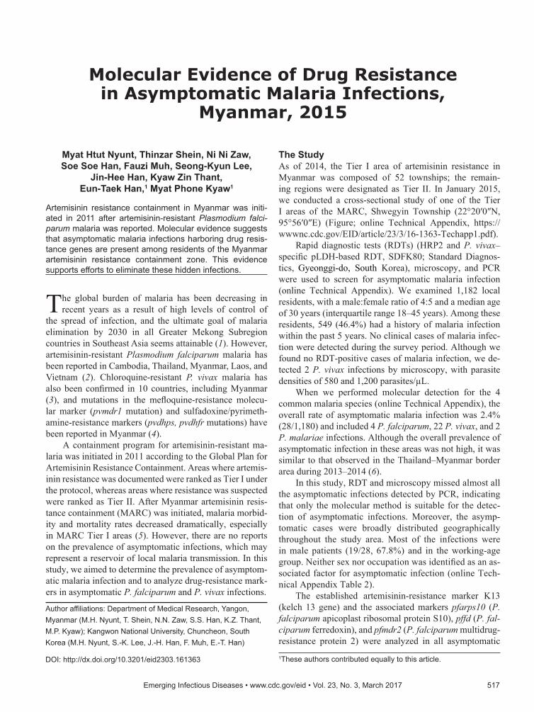
The StudyAs of 2014, the Tier I area of artemisinin resistance in Myanmar was composed of 52 townships; the remain-ing regions were designated as Tier II. In January 2015, we conducted a cross-sectional study of one of the Tier I areas of the MARC, Shwegyin Township (22°20′0″N, 95°56′0″E) (Figure; online Technical Appendix, https://wwwnc.cdc.gov/EID/article/23/3/16-1363-Techapp1.pdf).
Rapid diagnostic tests (RDTs) (HRP2 and P. vivax–specific pLDH-based RDT, SDFK80; Standard Diagnos-tics, Gyeonggi-do, South Korea), microscopy, and PCR were used to screen for asymptomatic malaria infection (online Technical Appendix). We examined 1,182 local residents, with a male:female ratio of 4:5 and a median age of 30 years (interquartile range 18–45 years). Among these residents, 549 (46.4%) had a history of malaria infection within the past 5 years. No clinical cases of malaria infec-tion were detected during the survey period. Although we found no RDT-positive cases of malaria infection, we de-tected 2 P. vivax infections by microscopy, with parasite densities of 580 and 1,200 parasites/µL.
When we performed molecular detection for the 4 common malaria species (online Technical Appendix), the overall rate of asymptomatic malaria infection was 2.4% (28/1,180) and included 4 P. falciparum, 22 P. vivax, and 2 P. malariae infections. Although the overall prevalence of asymptomatic infection in these areas was not high, it was similar to that observed in the Thailand–Myanmar border area during 2013–2014 (6).
In this study, RDT and microscopy missed almost all the asymptomatic infections detected by PCR, indicating that only the molecular method is suitable for the detec-tion of asymptomatic infections. Moreover, the asymp-tomatic cases were broadly distributed geographically throughout the study area. Most of the infections were in male patients (19/28, 67.8%) and in the working-age group. Neither sex nor occupation was identified as an as-sociated factor for asymptomatic infection (online Tech-nical Appendix Table 2).
The established artemisinin-resistance marker K13 (kelch 13 gene) and the associated markers pfarps10 (P. falciparum apicoplast ribosomal protein S10), pffd (P. fal-ciparum ferredoxin), and pfmdr2 (P. falciparum multidrug-resistance protein 2) were analyzed in all asymptomatic
Molecular Evidence of Drug Resistance in Asymptomatic Malaria Infections,
Myanmar, 2015
Emerging Infectious Diseases • www.cdc.gov/eid • Vol. 23, No. 3, March 2017 517
Author affiliations: Department of Medical Research, Yangon, Myanmar (M.H. Nyunt, T. Shein, N.N. Zaw, S.S. Han, K.Z. Thant, M.P. Kyaw); Kangwon National University, Chuncheon, South Korea (M.H. Nyunt, S.-K. Lee, J.-H. Han, F. Muh, E.-T. Han)
DOI: http://dx.doi.org/10.3201/eid2303.161363 1These authors contributed equally to this article.
DISPATCHES
P. falciparum cases. Nonsynonymous mutations in the propeller region of K13 were found to be associated with artemisinin resistance and associated delayed clearance of the parasite beyond 72 hours after treatment with artemis-inin-based combination therapy (7). A previous study in the same region of patients with uncomplicated P. falciparum malaria indicated that 25.3% carried mutant K13 alleles (8). Markers that showed the underlying genetic back-ground predisposing to the K13 mutant were also reported, including pfarps10, pffd, pfmdr2, and pfcrt. Specific single nucleotide polymorphisms of these genes, such as V127M of pfarps10, D193Y of pffd, and T484I of pfmdr2, were found at a similar prevalence as K13 mutations (9).
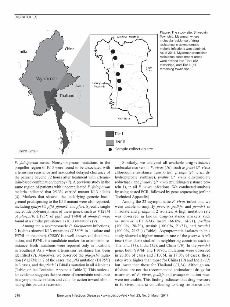
Among the 4 asymptomatic P. falciparum infections, 2 isolates showed K13 mutations (C580Y in 1 isolate and P574L in the other). C580Y is a well-known validated mu-tation, and P574L is a candidate marker for artemisinin re-sistance. Both mutations were reported only in locations in Southeast Asia where artemisinin resistance has been identified (2). Moreover, we observed the pfarps10 muta-tion (V127M) in 2 of the cases, the pffd mutation (D195Y) in 3 cases, and the pfmdr2 (T484I) mutation in all 4 isolates (Table; online Technical Appendix Table 3). This molecu-lar evidence suggests the presence of artemisinin resistance in asymptomatic isolates and calls for action toward elimi-nating this parasite reservoir.
Similarly, we analyzed all available drug-resistance molecular markers in P. vivax (10), such as pvcrt (P. vivax chloroquine-resistance transporter), pvdhps (P. vivax di-hydropteroate synthase), pvdhfr (P. vivax dihydrofolate reductase), and pvmdr1 (P. vivax multidrug-resistance pro-tein 1), in all P. vivax infections. We conducted analysis by using nested PCR, followed by gene sequencing (online Technical Appendix).
Among the 22 asymptomatic P. vivax infections, we were unable to amplify pvcrt-o, pvdhfr, and pvmdr1 in 1 isolate and pvdhps in 2 isolates. A high mutation rate was observed in known drug-resistance markers such as pvcrt-o K10 AAG insert (66.6%, 14/21), pvdhps (100.0%, 20/20), pvdhfr (100.0%, 21/21), and pvmdr1 (100.0%, 21/21) (Table). Asymptomatic isolates in this study showed a higher mutation rate of the pvcrt-o AAG insert than those studied in neighboring countries such as Thailand (11), India (12), and China (10). In the pvmdr1 gene, both Y976F and F1076L mutations were observed in 23.8% of cases and F1076L in 19.0% of cases; these rates were higher than those for China (10) and India (12) but lower than those for Thailand (13,14). Although an-tifolates are not the recommended antimalarial drugs for treatment of P. vivax, pvdhfr and pvdhps mutation rates were noticeable. This finding indicates that drug pressure in P. vivax malaria contributing to drug resistance also
518 Emerging Infectious Diseases • www.cdc.gov/eid • Vol. 23, No. 3, March 2017
Figure. The study site, Shwegyin Township, Myanmar, where molecular evidence of drug resistance in asymptomatic malaria infections was obtained. As of 2014, Myanmar artemisinin resistance containment areas were divided into Tier I (52 townships) and Tier II (all remaining townships).
Drug Resistance in Asymptomatic Malaria, Myanmar
needs to be considered in addition to emphasizing the artemisinin-resistant P. falciparum malaria.
One limitation of this study is the exclusive focus on the local residents in the MARC area, where all available control and prevention measures had already been imple-mented. Unlike the mobile and migrant population, local residents have not been a top priority for the artemisinin re-sistance containment program, leading to a niche of hidden infection. Moreover, blood pooling before DNA extraction was used in this study for molecular detection of malaria infection. Although this method is not ultrasensitive, it has a higher sensitivity than RDT and microscopy. The hidden asymptomatic infections and associated molecular markers for drug resistance among the asymptomatic cases detected in this study represent a threat to containment and elimina-tion efforts with regard to drug-resistant parasites.
ConclusionsAll countries in the Greater Mekong Subregion have set an ultimate goal of eliminating malaria by 2030. One of the main challenges to achieving this goal is hidden asymp-tomatic infection, which maintains a reservoir for local transmission of malaria (15). Critically, these asymptom-atic infections may carry drug-resistance genes, including genes for artemisinin resistance. Our results indicated that drug-resistant malaria parasites may be spreading, even in the containment areas or (pre-)elimination areas; this issue should, therefore, be addressed at a policy level. Detection and elimination of asymptomatic infections are of vital im-portance. Our evidence highlights the need for a strategy
for eliminating drug-resistant malaria in asymptomatic in-fections in the containment areas.
AcknowledgmentsWe thank all participants in this study and local health authority personnel and staff from the Parasitology Research Division and Advanced Molecular Research Center, Department of Medical Research and Kangwon Malaria Research Lab, Kangwon National University, South Korea.
This study was supported by the Korea Association of Health Promotion (2016_02), the National Research Foundation of Korea (NRF) grant funded by the South Korea government (MSIP) (NRF-2014R1A2A1A11052079), and by the Basic Science Research Program through the NRF funded by the Ministry of Science, ICT and Future Planning (2015R1A4A1038666).
Dr. Nyunt is a research scientist in the Department of Medical Research, Republic of the Union of Myanmar, and is currently studying in Kangwon National University, Chuncheon, South Korea. His research interests include drug-resistant malaria and neglected tropical diseases.
References 1. World Health Organization. Strategy for Malaria Elimination
in the Greater Mekong Subregion (2015–2030). WHO Regional Office for the Western Pacific; 2015 [cited 2016 Aug 16]. http://www.wpro.who.int/mvp/documents/strat_mal_elim_gms/en/
2. World Health Organization. Status report: Artemisinin and artemisinin-based combination therapy resistance. Geneva: The Organization; 2016 [cited 2016 Aug 16]. http://apps.who.int/iris/handle/10665/208820
Emerging Infectious Diseases • www.cdc.gov/eid • Vol. 23, No. 3, March 2017 519
Table. Plasmodium falciparum and P. vivax drug-resistance molecular markers in asymptomatic infections, Myanmar, 2015 Target Description* No. isolates/total (%) kelch 13 (K13) Wild 2/4 (50.0)
C580Y 1/4 (25.0) P574L 1/4 (25.0)
P. falciparum apicoplast ribosomal protein S10 (pfarps10)
Wild 2/4 (50.0) V127M 2/4 (50.0)
P. falciparum ferredoxin (pffd) Wild 1/4 (25.0) D193Y 3/4 (75.0)
P. falciparum multidrug-resistance protein 2 (pfmdr2) Wild 0/4 (0.0) T484I 4/4 (100.0)
P. vivax chloroquine-resistance transporter (pvcrt-o) Wild 7/21 (33.3) Mutant (AAG insert) 14/21 (66.7)
P. vivax multidrug-resistance protein 1 (pvmdr1) Wild (T, Y, F) (958, 976, 1076) 0/21 (0.0) Double mutant (M,Y, L) 4/21 (19.0) Single mutant (M, Y, F) 12/21 (57.1) Triple mutant (M, F, L) 5/21 (23.8)
P. vivax dihydropteroate synthase (pvdhps) Wild (S, A, K, A) (382, 383, 512, 553) 0/20 (0.0) Single mutant (S, G, K, A) 4/20 (20.0) Double mutant (S, G, K, G) 9/20 (45.0) Triple mutant (A, G, K, G) 5/20 (25.0)
Quadruple mutant (A, G, M, G) 2/20 (10.0) P. vivax dihydrofolate reductase (pvdhfr) Wild (F, S, T, S) (57, 58, 61, 117) 0/21 (0.0)
Single mutant (L, S, T, S) 1/21 (4.8) Double mutant (F, R, T, N) 2/21 (9.5)
Quadruple mutant (L/I, R, M, T) 18/21 (85.7) *Numbers in parentheses indicate the amino acid position. Mutant amino acids are shown in bold. All sequences were aligned with 3D7 (P. falciparum) and Sal-1 (P. vivax) reference sequences from http://www.plasmodb.org.

DISPATCHES
3. World Health Organization. World Malaria Report 2015. Geneva: The Organization; 2015 [cited 2016 Aug 16]. http://www.who.int/malaria/publications/world-malaria-report-2015/report/en/
4. World Health Organization. Global report on antimalarial drug efficacy and drug resistance: 2000–2010. Geneva: The Organization; 2010 [cited 2016 Aug 16]. http://www.who.int/malaria/publications/ atoz/9789241500470/en/
5. Township Health Department. Township Health Profile 2012: Shwegyin Township. Department of Health, Myanmar; 2013.
6. Baum E, Sattabongkot J, Sirichaisinthop J, Kiattibutr K, Jain A, Taghavian O, et al. Common asymptomatic and submicroscopic malaria infections in western Thailand revealed in longitudinal molecular and serological studies: a challenge to malaria elimination. Malar J. 2016;15:333. http://dx.doi.org/10.1186/s12936-016-1393-4
7. Ariey F, Witkowski B, Amaratunga C, Beghain J, Langlois AC, Khim N, et al. A molecular marker of artemisinin-resistant Plasmodium falciparum malaria. Nature. 2014;505:50–5. http://dx.doi.org/10.1038/nature12876
8. Ashley EA, Dhorda M, Fairhurst RM, Amaratunga C, Lim P, Suon S, et al.; Tracking Resistance to Artemisinin Collaboration (TRAC). Spread of artemisinin resistance in Plasmodium falciparum malaria. N Engl J Med. 2014;371:411–23. http://dx.doi.org/10.1056/NEJMoa1314981
9. Miotto O, Amato R, Ashley EA, MacInnis B, Almagro-Garcia J, Amaratunga C, et al. Genetic architecture of artemisinin- resistant Plasmodium falciparum. Nat Genet. 2015;47:226–34. http://dx.doi.org/10.1038/ng.3189
10. Lu F, Wang B, Cao J, Sattabongkot J, Zhou H, Zhu G, et al. Prevalence of drug resistance-associated gene mutations in Plasmodium vivax in Central China. Korean J Parasitol. 2012;50:379–84. http://dx.doi.org/10.3347/kjp.2012.50.4.379
11. Lu F, Lim CS, Nam D-H, Kim K, Lin K, Kim T-S, et al. Genetic polymorphism in pvmdr1 and pvcrt-o genes in relation to in vitro drug susceptibility of Plasmodium vivax isolates from malaria-endemic countries. Acta Trop. 2011;117:69–75. http://dx.doi.org/10.1016/j.actatropica.2010.08.011
12. Ganguly S, Saha P, Guha SK, Das S, Bera DK, Biswas A, et al. In vivo therapeutic efficacy of chloroquine alone or in combination with primaquine against vivax malaria in Kolkata, West Bengal, India, and polymorphism in pvmdr1 and pvcrt-o genes. Antimicrob Agents Chemother. 2013;57:1246–51. http://dx.doi.org/10.1128/AAC.02050-12
13. Suwanarusk R, Chavchich M, Russell B, Jaidee A, Chalfein F, Barends M, et al. Amplification of pvmdr1 associated with multidrug-resistant Plasmodium vivax. J Infect Dis. 2008;198:1558–64. http://dx.doi.org/10.1086/592451
14. Rungsihirunrat K, Muhamad P, Chaijaroenkul W, Kuesap J, Na-Bangchang K. Plasmodium vivax drug resistance genes; Pvmdr1 and Pvcrt-o polymorphisms in relation to chloroquine sensitivity from a malaria endemic area of Thailand. Korean J Parasitol. 2015;53:43–9. http://dx.doi.org/10.3347/kjp.2015.53.1.43
15. Okell LC, Ghani AC, Lyons E, Drakeley CJ. Submicroscopic infection in Plasmodium falciparum-endemic populations: a sys-tematic review and meta-analysis. J Infect Dis. 2009;200:1509–17. http://dx.doi.org/10.1086/644781
Address for correspondence: Eun-Taek Han, Department of Medical Environmental Biology and Tropical Medicine, Kangwon National University, Chuncehon-si, Gangwon-do, 200-701, South Korea; email: [email protected]; [email protected]
520 Emerging Infectious Diseases • www.cdc.gov/eid • Vol. 23, No. 3, March 2017
Page 1 of 9
Article DOI: http://dx.doi.org/10.3201/eid2303.161363
Molecular Evidence of Drug Resistance in Asymptomatic Malaria Infections, Myanmar,
2015
Technical Appendix
Study Population
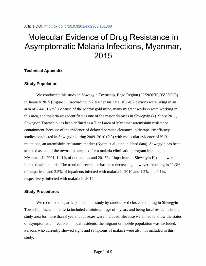
We conducted this study in Shwegyin Township, Bago Region (22°200″N, 95°560″E)
in January 2015 (Figure 1). According to 2014 census data, 107,462 persons were living in an
area of 2,440.1 km2. Because of the nearby gold mine, many migrant workers were working in
this area, and malaria was identified as one of the major diseases in Shwegyin (1). Since 2011,
Shwegyin Township has been defined as a Tier I area of Myanmar artemisinin resistance
containment because of the evidence of delayed parasite clearance in therapeutic efficacy
studies conducted in Shwegyin during 2009–2010 (2,3) with molecular evidence of K13
mutations, an artemisinin-resistance marker (Nyunt et al., unpublished data). Shwegyin has been
selected as one of the townships targeted for a malaria elimination program initiated in
Myanmar. In 2001, 14.1% of outpatients and 20.1% of inpatients in Shwegyin Hospital were
infected with malaria. The trend of prevalence has been decreasing, however, resulting in 11.3%
of outpatients and 3.5% of inpatients infected with malaria in 2010 and 1.2% and 0.1%,
respectively, infected with malaria in 2014.
Study Procedures
We recruited the participants in this study by randomized cluster sampling in Shwegyin
Township. Inclusion criteria included a minimum age of 6 years and being local residents in the
study area for more than 3 years; both sexes were included. Because we aimed to know the status
of asymptomatic infections in local residents, the migrant or mobile population was excluded.
Persons who currently showed signs and symptoms of malaria were also not included in this
study.
Page 2 of 9
We obtained written informed consent from all the participants. All the patients with
parasite infections detected by any method (rapid diagnostic test [RDT], microscopy, or
molecular method) were treated according to the National Malaria Treatment Guideline. This
study was approved by the Ethical Committee of the Department of Medical Research, Myanmar
(approval no. 49/Ethics-2014). The study was also registered with ClinicalTrial.gov (identifier
NCT02708199).
Sample Size Determination
The prevalence of asymptomatic infection in the study population was unknown. Based
on the previous studies (4,5) conducted in Southeast Asia, we assumed the maximum possibility
of infection was 25% in the study site. A required sample size was calculated by anticipated
population proportion of asymptomatic infection (25%) among the population of 107,462 (as of
the 2014 census), with marginal error (2.5%), and 95% CI. The minimum sample size required
was 1,141; in this study, 1,182 participants were involved.
Sampling Procedure
Randomized cluster sampling method was used in this study. Shwegyin Township was
selected according to the rationale described previously. Two of the 4 local health centers were
randomly selected. All villages belonging to the local health centers were listed and 6 villages
were randomly selected. Among these villages, a sampling interval was calculated to get the
required blood samples in the villages, which we determined to be at least 191 per village.
Household visits or meeting places were used to collect the samples, depending on the
convenience of the participants.
Laboratory Procedures
We collected 1 mL of venous blood from the participant’s forearm under aseptic
conditions, using a disposable syringe. The blood was used for detection of asymptomatic
infections.
Page 3 of 9
Rapid Diagnostic Test
We detected malarial infection in all participants by PfHRP2- and Pv-specific pLDH-
based RDT (SDFK80; Standard Diagnostics, Gyeonggi-do, South Korea) and peripheral blood
film examination to exclude malaria infection in the field. We used venous blood for malaria
detection by RDT according to the manufacturer’s instructions.
Malaria Microscopy
We followed the World Health Organization standardized protocol (6) for malaria
microscopy. Briefly, 10% Giemsa was used to stain thick and thin blood films in the field for
initial screening. In the main laboratory, another set of thick and thin blood films was stained
with 3% Giemsa stain for confirmation and validation of the result. We prepared a fresh Giemsa
stain dilution at least once a day and possibly more often, depending on the number of slides
processed. We examined the Giemsa-stained thick and thin blood films at a magnification of
1,000 to identify the parasite species and to determine the parasite density. We calculated the
parasite density, expressed as the number of asexual parasites per microliter of blood, by
dividing the number of asexual parasites by the number of leukocytes counted and then
multiplying by an assumed leukocyte density (6,000 leukocytes/µL). All slides were stained and
checked by World Health organization–certified microscopists and validated by an expert
microscopist. A blood slide was considered negative when examination of 1,000 leukocytes
revealed no asexual parasites. Two qualified microscopists read all the slides independently, and
parasite densities was calculated by averaging the 2 counts. Blood smears with discordant results
(differences between the 2 microscopists’ results in species diagnosis, in parasite density of
>50%, or in the presence of parasites) were reexamined by a third, independent microscopist,
and parasite density was calculated by averaging the 2 counts closest to each other.
Molecular Detection
We adapted the pooling strategy described previously (7). In brief, 20 µL from each of 10
samples was combined to make 1 pool of 200 µL whole blood. These pooled blood samples were
designated for DNA extraction with QIAamp DNA Blood Mini Kit (QIAGEN, Hilden,
Germany) and eluted into a final volume of 100 µL. Ten microliters of the DNA eluent was used
for Plasmodium genus-specific amplification of the 18S rRNA gene, as described elsewhere (8).
Only Plasmodium-positive pools were included to do individual DNA extraction again; 1 µL of
Page 4 of 9
the DNA eluent was used for individual genus and species identification for asymptomatic
malaria infection by using genus and specific primer pairs (8).
Artemisinin Resistance Molecular Markers for P. falciparum Infection
We analyzed all the samples that tested positive for P. falciparum malaria infection for
artemisinin-resistance molecular markers (9,10) such as K13 kelch genes (PF3D7_1343700),
pfarps10 (PF3D7_1460900.1), pffd (PF3D7_1318100), and pfmdr2 (PF3D7_1447900) by using
the pairs of primers (Table 1. The details for amplification of these targets are available at our
institutional web page (http://kmrl.kangwon.ac.kr/).
We performed amplification with an Accupower Premix (Bioneer, Daejon, South Korea)
in a final volume of 20 µL. The final volume included 250 nmol/L of each primer, 0.25 mmol/L
of each dNTP, 10 mmol/L Tris-HCl (pH 9.0), 30 mmol/L MgCl2, 1.0 units of Taq polymerase,
and 2 µL of genomic DNA template.
For nested-1 PCR amplification of the K13 kelch propeller gene, initial denaturation at
95°C for 5 min was followed by 35 cycles at 95°C for 30 s, 58°C for 1 min, 72°C for 1.5 min,
and a final extension at 72°C for 10 min. Using 1 μL of the nested-1 PCR product as a template,
we applied the same conditions for the nested-2 PCR except for an annealing temperature of
60°C for 1 min and 72°C for 1 min with 30 cycles.
For nested-1 PCR amplification of the pfarps10 gene, initial denaturation at 95°C for 5
min was followed by 35 cycles at 95°C for 30 s, 58°C for 1 min, 72°C for 1.5 min, and a final
extension of 72°C for 10 min. Using 1 μL of the nested-1 PCR product as a template, we applied
the same conditions for the nested-2 PCR except for an annealing temperature of 62°C for 1 min
and 72°C for 1 min with 30 cycles.
For nested-1 PCR amplification of the pffd gene, initial denaturation at 95°C for 5 min
was followed by 35 cycles at 95°C for 30 s, 62°C for 1 min, 72°C for 1 min, and a final
extension at 72°C for 10 min. Using 1 μL of the nested-1 PCR product as a template, we applied
the same conditions for the nested-2 PCR except for an annealing temperature of 60°C for 1 min
and 72°C for 1 min with 30 cycles.
Page 5 of 9
For nested-1 PCR amplification of the pfmdr2 gene amplification, initial denaturation at
95°C for 5 min was followed by 35 cycles at 95°C for 30 s, 58°C for 1 min, 72°C for 1.5 min,
and a final extension at 72°C for 10 min. Using 1 μL of the nested-1 PCR product as a template,
we applied the same conditions for the nested-2 PCR except for an annealing temperature of
60°C for 1 min and 72°C for 1 min with 35 cycles.
We visualized the PCR products by 1% agarose gel electrophoresis stained with 0.05%
Redsafe dye (iNtRON Biotechnology, Daejeon, South Korea). We purified the PCR products by
using a MEGA quick-spin Total Fragment DNA Purification Kit (iNtRON Biotechnology) and
sequenced them with primers from a commercial sequencing company (Genotech, Daejeon,
South Korea). We compared all the nucleotide and amino acid sequences with the reference
sequence of 3D7 version 3 (www.plasmodb.org) and aligned them by using software in the
Lasergene Genomic Suite (MegAlign, version 7.1; DNAStar, Madison, WI, USA).
Drug Resistance Molecular Marker Analysis for P. vivax Isolates
We further analyzed all the P. vivax isolates for pvcrt-o (P. vivax chloroquine resistance
transporter gene, PVX_087980), pvmdr1 (P. vivax multidrug resistance protein 1, PVX_080100),
pvdhps (P. vivax hydroxymethyl pterinpyrophosphokinase dihydropteroate synthetase,
PVX_123230), and pvdhfr (P. vivax dihydrofolate reductase thymidylate synthase,
PVX_089950) by using the modified procedures described previously (11). We performed
amplification by using an Accupower premix (Bioneer, Daejon, South Korea) in a final volume
of 20 µL, which included 250 nmol/L of each primers, 0.25 mmol/L of each dNTP, 10 mmol/L
Tris-HCl (pH 9.0), 30 mmol/L MgCl2, 1.0 units of Taq polymerase, and 2 µL of genomic DNA
template and pairs of primers (Table 1).
For the pvcrt-o PCR amplification, initial denaturation at 94°C for 10 min was followed
by 35 cycles at 94°C for 30 s, 60°C for 45 s, 72°C for 1 min, and a final extension at 72°C for 10
min for nested-1 PCR. Using 1 µL of the nested-1 PCR product as a template, we applied the
same conditions for the nested-2 PCR except for an annealing temperature of 60°C for 45 s with
30 cycles of amplification.
For the pvmdr1 PCR amplification, initial denaturation at 94°C for 10 min was followed
by 35 cycles at 94°C for 30 s, 58°C for 45 s, 72°C for 2 min, and a final extension at 72°C for 10
min for nested-1 PCR. Using 1 µL of the nested-1 PCR product as a template, we applied the
Page 6 of 9
same conditions for the nested-2 PCR except for an annealing temperature of 62°C for 45 s and
extension at 72°C for 45 s with 33 cycles of amplification.
For the pvdhps PCR amplification, initial denaturation at 94°C for 10 min was followed
by 40 cycles at 94°C for 30 s, 58°C for 45 s, 72°C for 1.5 min, and a final extension at 72°C for
10 min for nested-1 PCR. Using 1 µL of the nested-1 PCR product as a template, the same
conditions were applied for the nested-2 PCR except for 35 cycles of amplification.
For the pvdhfr PCR amplification, initial denaturation at 94°C for 10 min was followed
by 35 cycles at 94°C for 30 s, 58°C for 45 s, 72°C for 1 min, and a final extension at 72°C for 10
min for nested-1 PCR. Using 1 µL of the nested-1 PCR product as a template, we applied the
same conditions for the nested-2 PCR except for an annealing temperature of 62°C for 30 s and
extension at 72°C for 1 min with 30 cycles of amplification.
We visualized the PCR products by 1% agarose gel electrophoresis and staining with
0.05% Redsafe dye (iNtRON Biotechnology). We purified the PCR products by using a MEGA
quick-spin Total Fragment DNA Purification Kit (iNtRON Biotechnology) and sequenced them
with primers from a commercial sequencing company (Genotech).
Sequence Analysis and Statistics
We compared all the nucleotide and amino acid sequences with the reference sequence of
K13 kelch protein (gene ID: PF3D7_1343700), pfarps10 (PF3D7_1460900.1), pffd
(PF3D7_1318100), pfmdr2 (PF3D7_1447900), pvcrt (PVX_087980), pvdhps (PVX_123230),
pvdhfr (PVX_089950), and pvmdr1 (PVX_080100) from www.plasmodb.org and aligned them
by using software in the Lasergene Genomic Suite (MegAlign, version 7.1). We used SPSS
software (version 22.0, IBM SPSS Statistics, Armonk, NY, USA) for all statistical analysis. We
compared the frequency of mutations and haplotypes of the target genes among the groups by
using 2 and Fisher exact tests with a two-sided confidence interval at the 95% confidence level.
The sequences were deposited at GenBank (accession nos. KX000945–KX000959 and
KX384672–KX384687).
Page 7 of 9
References
1. Township-Health-Department. Shwegyin Township Health Profile 2012: Department of Health,
Myanmar; 2013.
2. World Health Organization. Global report on antimalarial drug efficacy and drug resistance: 2000–
2010. Geneva, Switzerland; 2010 [cited 2016 Aug 16].
http://www.who.int/malaria/publications/atoz/9789241500470/en/
3. World Health Organization Country Office Myanmar. Strategic Framework for Artemisinin Resistance
Containment in Myanmar (MARC) 2011–2015; 2011 [cited 2016 Aug 16].
http://www.searo.who.int/myanmar/documents/MARCframeworkApril2011.pdf
4. Imwong M, Nguyen TN, Tripura R, Peto TJ, Lee SJ, Lwin KM, et al. The epidemiology of subclinical
malaria infections in South-East Asia: findings from cross-sectional surveys in Thailand–
Myanmar border areas, Cambodia, and Vietnam. Malar J. 2015;14:381.
http://dx.doi.org/10.1186/s12936-015-0906-x
5. Starzengruber P, Fuehrer HP, Ley B, Thriemer K, Swoboda P, Habler VE, et al. High prevalence of
asymptomatic malaria in south-eastern Bangladesh. Malar J. 2014;13:16.
http://dx.doi.org/10.1186/1475-2875-13-16
6. World Health Organization. Methods for surveillance of antimalarial drug efficacy. Geneva: WHO;
2009 [cited 2016 Aug 16]. http://www.who.int/malaria/publications/atoz/9789241597531/en/
7. Nyunt MH, Kyaw MP, Thant KZ, Shein T, Han SS, Zaw NN, et al. Effective high-throughput blood
pooling strategy before DNA extraction for detection of malaria in low-transmission settings.
Korean J Parasitol. 2016;54:253–9. http://dx.doi.org/10.3347/kjp.2016.54.3.253
8. Snounou G, Singh B. Nested PCR analysis of Plasmodium parasites. Methods Mol Med. 2002;72:189–
203. http://dx.doi.org/10.1385/1-59259-271-6:189
9. Ariey F, Witkowski B, Amaratunga C, Beghain J, Langlois AC, Khim N, et al. A molecular marker of
artemisinin-resistant Plasmodium falciparum malaria. Nature. 2014;505:50–5.
http://dx.doi.org/10.1038/nature12876
10. Miotto O, Amato R, Ashley EA, MacInnis B, Almagro-Garcia J, Amaratunga C, et al. Genetic
architecture of artemisinin-resistant Plasmodium falciparum. Nat Genet. 2015;47:226–34.
http://dx.doi.org/10.1038/ng.3189
Page 8 of 9
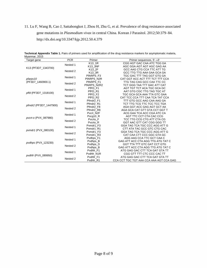
11. Lu F, Wang B, Cao J, Sattabongkot J, Zhou H, Zhu G, et al. Prevalence of drug resistance-associated
gene mutations in Plasmodium vivax in central China. Korean J Parasitol. 2012;50:379–84.
http://dx.doi.org/10.3347/kjp.2012.50.4.379
Technical Appendix Table 1. Pairs of primers used for amplification of the drug resistance markers for asymptomatic malaria, Myanmar, 2015
Target gene PCR Primer Primer sequences, 53
K13 (PF3D7_1343700) Nested 1
K13_1R CGG AGT GAC CAA ATC TGG GA K13_3NF AGC GGA AGT AGT AGC GAG AA
Nested 2 K13_2F GCC AAG CTG CCA TTC ATT TG K13_3R GCC TTG TTG AAA GAA GCA GA
pfarps10 (PF3D7_1460900.1)
Nested 1 PfARPS_F3 TGC GAC TTT TAG GGT GTG GA
PfARPS_N2R CAT GGT ACC ACT TTT TCT TTT CCA
Nested 2 PfARPS_F1 TTG TAG CAG GCC CAA TTC CC
PfARPS_N2R2 TCT GGG TAA TTT GAC ATT CAT
pffd (PF3D7_1318100) Nested 1
PfFD_F1 AGT TGT TCT ACA TGC GCA GC PfFD_R1 AAT GTG CGC TTG TAG TGC AT
Nested 2 PfFD_F2 TGC GCA GCA AAA TTA GTC GAA PfFD_R2 CAT TCC CCA TTT CAA TCA TAT CCA
pfmdr2 (PF3D7_1447900) Nested 1
Pfmdr2_F1 TTT GTG GCC AAG CAA AAG GA Pfmdr2_R1 TCT TTG TCG TTC TCC TCC TGA
Nested 2 Pfmdr2_F8 AGA GGT ACC GAG AGT GCT AA Pfmdr2_R8 AGA GCA CAT GTT GTA CCT GGT T
pvcrt-o (PVX_087980) Nested 1
Pvcrt_N2F ACG GAA TCA ACC CGA ATC CA Pvcg10_R AGT TTC CCT CTA CAC CCG
Nested 2 Pvcrto_F TCC TTG CCG CTG ATT CTA CG Pvcrto_R GGT AAC GTT CAT CGG GGG TT
pvmdr1 (PVX_080100) Nested 1
Pvmdr1_F3 GGA TAG TCA TGC CCC AGG ATT G Pvmdr1_R1 CTT ATA TAC GCC GTC CTG CAC
Nested 2 Pvmdr1_F3 GGA TAG TCA TGC CCC AGG ATT G Pvmdr1_R3 CAT CAA CTT CCC GGC GTA GC
pvdhps (PVX_123230) Nested 1
Pvdhps_F1 AGG AAG CCA TTC GCT CAA C Pvdhps_B GAG ATT ACC CTA AGG TTG ATG TAT C
Nested 2 Pvdhps_D GGT TTA TTT GTC GAT CCT GTG Pvdhps_B GAG ATT ACC CTA AGG TTG ATG TAT C
pvdhfr (PVX_089950) Nested 1
Pvdhfr_F1 ATG GAG GAC CTT TCA GAT GTA TT
Pvdhfr_N1R CGG GTT TTT CTC CCC CAC TT
Nested 2 Pvdhfr_F1 ATG GAG GAC CTT TCA GAT GTA TT
Pvdhfr_R1 CCA CCT TGC TGT AAA CCA AAA AGT CCA GAG
Page 9 of 9
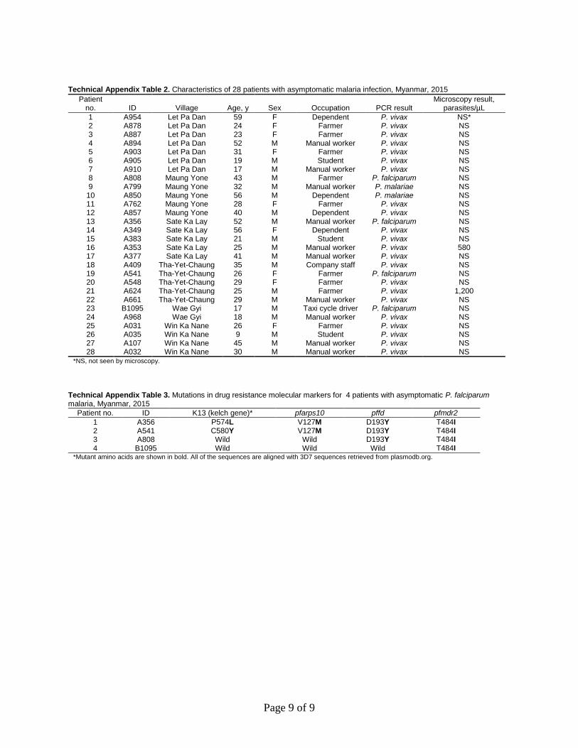
Technical Appendix Table 2. Characteristics of 28 patients with asymptomatic malaria infection, Myanmar, 2015
Patient no. ID Village Age, y Sex Occupation PCR result
Microscopy result, parasites/µL
1 A954 Let Pa Dan 59 F Dependent P. vivax NS* 2 A878 Let Pa Dan 24 F Farmer P. vivax NS 3 A887 Let Pa Dan 23 F Farmer P. vivax NS 4 A894 Let Pa Dan 52 M Manual worker P. vivax NS 5 A903 Let Pa Dan 31 F Farmer P. vivax NS 6 A905 Let Pa Dan 19 M Student P. vivax NS 7 A910 Let Pa Dan 17 M Manual worker P. vivax NS 8 A808 Maung Yone 43 M Farmer P. falciparum NS 9 A799 Maung Yone 32 M Manual worker P. malariae NS
10 A850 Maung Yone 56 M Dependent P. malariae NS 11 A762 Maung Yone 28 F Farmer P. vivax NS 12 A857 Maung Yone 40 M Dependent P. vivax NS 13 A356 Sate Ka Lay 52 M Manual worker P. falciparum NS 14 A349 Sate Ka Lay 56 F Dependent P. vivax NS 15 A383 Sate Ka Lay 21 M Student P. vivax NS 16 A353 Sate Ka Lay 25 M Manual worker P. vivax 580 17 A377 Sate Ka Lay 41 M Manual worker P. vivax NS 18 A409 Tha-Yet-Chaung 35 M Company staff P. vivax NS 19 A541 Tha-Yet-Chaung 26 F Farmer P. falciparum NS 20 A548 Tha-Yet-Chaung 29 F Farmer P. vivax NS 21 A624 Tha-Yet-Chaung 25 M Farmer P. vivax 1,200 22 A661 Tha-Yet-Chaung 29 M Manual worker P. vivax NS 23 B1095 Wae Gyi 17 M Taxi cycle driver P. falciparum NS 24 A968 Wae Gyi 18 M Manual worker P. vivax NS 25 A031 Win Ka Nane 26 F Farmer P. vivax NS 26 A035 Win Ka Nane 9 M Student P. vivax NS 27 A107 Win Ka Nane 45 M Manual worker P. vivax NS 28 A032 Win Ka Nane 30 M Manual worker P. vivax NS
*NS, not seen by microscopy.
Technical Appendix Table 3. Mutations in drug resistance molecular markers for 4 patients with asymptomatic P. falciparum malaria, Myanmar, 2015
Patient no. ID K13 (kelch gene)* pfarps10 pffd pfmdr2
1 A356 P574L V127M D193Y T484I 2 A541 C580Y V127M D193Y T484I 3 A808 Wild Wild D193Y T484I 4 B1095 Wild Wild Wild T484I
*Mutant amino acids are shown in bold. All of the sequences are aligned with 3D7 sequences retrieved from plasmodb.org.